Embed Size (px)
Citation preview

The Panel Approach to Diagnostics
Lauren Hopson International Product Specialist
Cell Marque Corporation

Cell Marque Rocklin, California

About Cell Marque:
• IVD primary antibody manufacturer
• Distributors in 50+ countries
• 60+ Employees
• 2 pathologists on Staff
• Staff of primarily Biologists
• All employees trained on the science of IHC
• Focus on education, and increasing the diagnostic power of the pathologist

What is immunohistochemistry?
• Immunology: branch of science dealing with the immune system Immuno
• Histology: branch of biology dealing with the study of organic tissues Histo
• The science of the composition, structure, properties and reactions of matter
Chemistry

Definition of Immunohistochemistry:
Using a microscope to localize specific antigens in tissues by
staining them with antibodies labeled with pigmented material.

Importance of IHC
• Allows for visualization of proteins under a microscope
• Provides a diagnostic and prognostic tool for pathologists
• Detects infectious agents

What is an antibody?
Antibodies are proteins used by the immune
system to identify and neutralize foreign
objects.
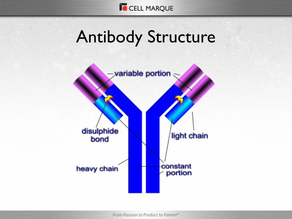
Antibody Structure

• Antigen: a molecule
that stimulates an
immune response
• Epitope: three
dimensional surface
features found on an
antigen molecule
Cell Membrane/Nucleus/Cytoplasm
Antigen
Antigens

What is a Panel?
Panels are diagnostic algorithms used
to immunophenotype specific types of
tumors and neoplasms.

Panel Types
Flo
w C
har
t
GCDFP-15+/ Mammaglobin+
E-Cadherin and p120 catenin
E-cadherin-/ p120 catenin+
E-cadherin+/ p120 catenin-
E-cadherin+/ p120 catenin+
Breast Lesion
GCDFP-15 Mammaglobin B-catenin E-Cadherin CK, 34betaE12 p120
Lobular + + - - + + (cytoplasmic)
Ductal + + + (membranous) + - + (membranous)
Imm
mu
no
ph
en
oty
pe
G
rid
s
GCDFP15+ Mammaglobin+ E-Cadherin - P120 Catenin -

? ? ? ? ? ?
Undifferentiated Tumor Panel

Novel Markers:
Completing the Panel

PAX-8 • Clone: MRQ-50
• Visualization: Nuclear
• Ovarian Carcinoma (high
sensitivity for serous ca)
• Thyroid transcription factor
• Kidney metastasis
sensitivity over 85%
• Multiple applications
• USCAP 2010, IAP 2010
Pax 8 on ovarian ca
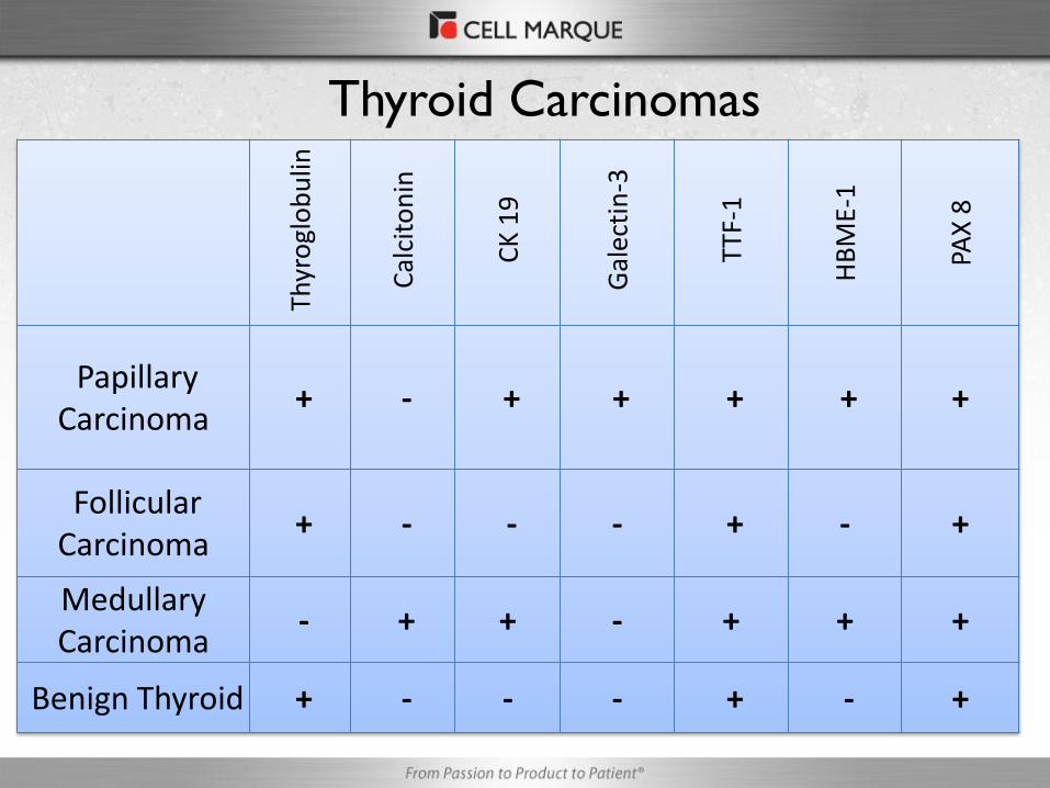
Thyroid Carcinomas
Th
yro
glo
bu
lin
Cal
cito
nin
CK
19
Gal
ecti
n-3
TTF
-1
HB
ME-
1
PAX
8
Papillary Carcinoma
+ - + + + + +
Follicular Carcinoma
+ - - - + - +
Medullary Carcinoma
- + + - + + +
Benign Thyroid + - - - + - +

Ovarian Carcinoma
PAX8 WT1 CA-125 CEA
Ovarian CA, Serous + + + +
Ovarian CA, Mucinous - - - -
Ovarian endometrioid CA + - + -
Ovarian Clear Cell Carcinoma + - + -

Kidney
RC
C
CD
10
PAX
-2
PAX
-8
Ksp
-C
adh
erin
Ep-C
AM
Cav
eolin
-1
Clear cell + + + + - - -
Chromophobe - - - + + + +
Oncocytoma - - - + + - -
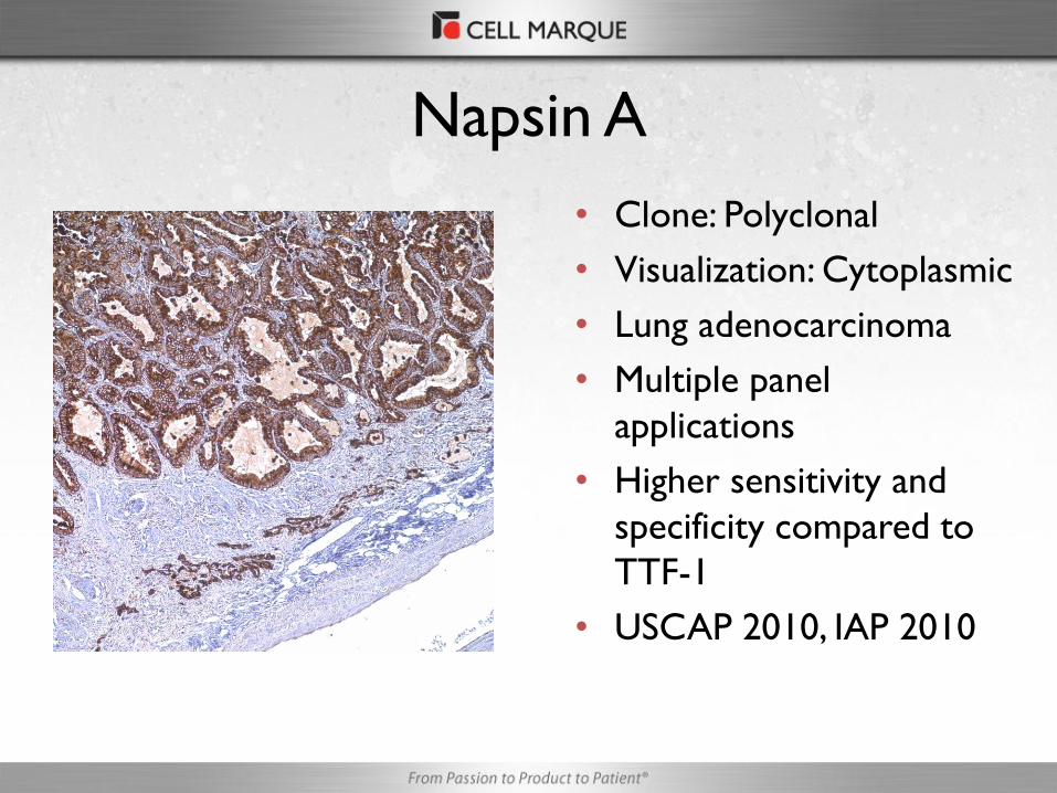
Napsin A
• Clone: Polyclonal
• Visualization: Cytoplasmic
• Lung adenocarcinoma
• Multiple panel
applications
• Higher sensitivity and
specificity compared to
TTF-1
• USCAP 2010, IAP 2010

• Clone: SP76
• Visualization: Nuclear
• Rabbit Monoclonal
• Differentiates lung squamous cell
carcinoma from lung
adenocarcinoma
• Distinguishes embryonal carcinoma
from other germ cell tumors
• Useful in the identification of
astrocytomas
• Important for general pathologists
and GU pathologists
SOX-2
SOX-2 on Lung Squamous Cell Carcinoma

Lung Adenocarcinoma vs.
Squamous Cell Carcinoma
Napsin A +
TTF-1 +
CK 5/14 -
Sox-2 -
Well Differentiated Lung Adenocarcinoma
Napsin A +
TTF-1 -
CK 5/14 -
Sox-2 -
Poorly Differentiated Lung Adenocarcinoma
Napsin A -
TTF-1 +
CK 5/14 -
Sox-2 -/+
Neuroendocrine Tumor (verify w/NE marker)
Napsin A -
TTF-1 -
CK 5/14 +
Sox-2 +
Squamous Cell Carcinoma

Arginase-1
• Clone: SP156
• Visualization: cytoplasmic and nuclear
• High sensitivity for HCC
• Useful for HCC but also stains hepatic adenoma and cirrhotic liver
• Used in combination with Glypican 3 and HepPar-1 to distinguish benign from malignant
Arginase 1 on cirrhotic liver

Glypican-3
• Clone: 1G12
• Visualization: Cytoplasmic
• Differentiates benign liver from
hepatocellular ca
• Also useful in identifying
choriocarcinoma and yolk sac
tumor
• Benign vs. malignant marker
• Unique to Cell Marque Glypican-3 on HCC

Liver Neoplasms
Arginase-1 Hep Par-1 Glypican-3 CD10 pCEA
Hepatic Adenoma + + - + +
Hepatocellular Carcinoma
+ + + + +
Metastatic Adenocarcinoma
- - - -/+ -/+
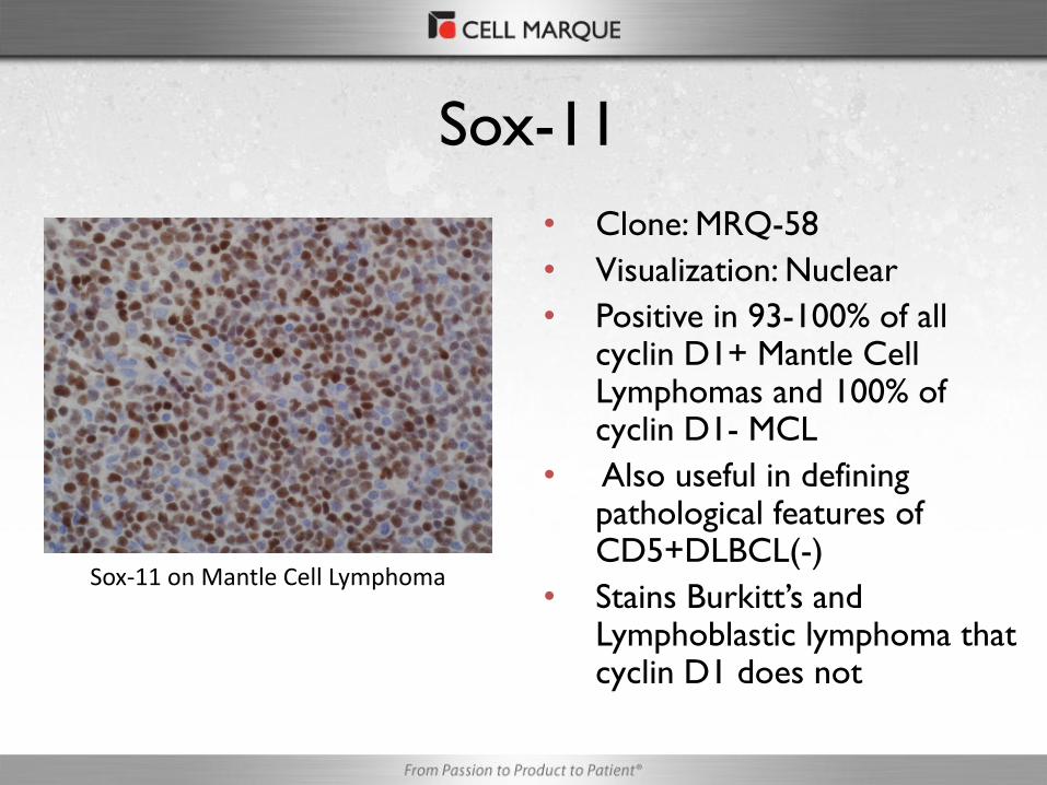
Sox-11
• Clone: MRQ-58
• Visualization: Nuclear
• Positive in 93-100% of all cyclin D1+ Mantle Cell Lymphomas and 100% of cyclin D1- MCL
• Also useful in defining pathological features of CD5+DLBCL(-)
• Stains Burkitt’s and Lymphoblastic lymphoma that cyclin D1 does not
Sox-11 on Mantle Cell Lymphoma

Hematolymphoid Neoplasm
Sox-11 CD20 CD5 CD10 CD23 Cyclin D1
MCL + + + - - +
FL - + - + - -
SLL/CLL - + + - + -
MZL - + - - - -
LBL + + - +/- - -
BL -/+ + - - - -
CD5+ DLBCL - + + + - -
Blastoid Variant MCL
+ + + - - +

E-Cadherin/p120 Catenin Dual Stain
E-cadherin-DAB, p120 catenin-Red

Breast Carcinoma
GCDFP-15+/ Mammaglobin+
E-Cadherin and p120 catenin
E-Cadherin-/ p120 catenin+
E-Cadherin+/ p120 catenin-
E-Cadherin+/ p120 catenin+
Ductal breast carcinoma
Lobular breast carcinoma
Tubulolobular breast
carcinoma

• Clone: Polyclonal
• Visualization: Nuclear
• Sensitive marker for melanoma (including conventional, spindle cell and
desmoplastic subtypes)
• Superior to other immunostains
in detecting residual invasive and
in situ melanoma
• Useful in detecting in situ and
invasive components of
desmoplastic melanoma
• Antibody unique to CM
SOX-10
SOX-10 on Melanoma

Cutaneous Lesion
SOX-10 CK Cocktail
HMB-45
S-100
MART-1 (Melan A)
Conventional Melanoma
+ -/+ + + +
Desmoplastic Melanoma
+ - - +/- -
Squamous Cell Carcinoma
- + - -/+ -
Basal Cell Carcinoma
- + - - -
Merkel Cell Carcinoma
- -/+ - -/+ -

Why are panels important?
• Increase the number of diagnostic tools for the
pathologists
• See a macroview of the disease state
• Faster turn around time
• No single antibody is 100% sensitive and specific
• More specific diagnosis leads to more specific
treatment

Questions?