Embed Size (px)
Citation preview
JOURNAL OF BACTERIOLOGY, Sept. 2004, p. 6286–6297 Vol. 186, No. 180021-9193/04/$08.00�0 DOI: 10.1128/JB.186.18.6286–6297.2004Copyright © 2004, American Society for Microbiology. All Rights Reserved.
The Paralogous Pairs of Genes Involved in Clavulanic Acid andClavam Metabolite Biosynthesis Are Differently Regulated in
Streptomyces clavuligerusKapil Tahlan, Cecilia Anders, and Susan E. Jensen*
Department of Biological Sciences, University of Alberta, Edmonton, Alberta, Canada
Received 7 April 2004/Accepted 17 June 2004
Carboxyethylarginine synthase, encoded by the paralogous ceaS1 and ceaS2 genes, catalyzes the first reactionin the shared biosynthetic pathway leading to clavulanic acid and the other clavam metabolites in Streptomycesclavuligerus. The nutritional regulation of ceaS1 and ceaS2 expression was analyzed by reverse transcriptasePCR and by the use of the enhanced green fluorescent protein-encoding gene (egfp) as a reporter. ceaS1 wastranscribed in complex soy medium only, whereas ceaS2 was transcribed in both soy and defined starch-asparagine (SA) media. The transcriptional start points of the two genes were also mapped to a C residue 98bp upstream of ceaS1 and a G residue 51 bp upstream of the ceaS2 start codon by S1 nuclease protection andprimer extension analyses. Furthermore, transcriptional mapping of the genes encoding the beta-lactamsynthetase (bls1) and proclavaminate amidinohydrolase (pah1) isoenzymes from the paralogue gene clusterindicated that a single polycistronic transcript of �4.9 kb includes ceaS1, bls1, and pah1. The expression ofceaS1 and ceaS2 in a mutant strain defective in the regulatory protein CcaR was also examined. ceaS1transcription was not affected in the ccaR mutant, whereas that of ceaS2 was greatly reduced compared to thewild-type strain. Overall, our results suggest that different mechanisms are involved in regulating the expres-sion of ceaS1 and ceaS2, and presumably also of other paralogous genes that encode proteins involved in theearly stages of clavulanic acid and clavam metabolite biosynthesis.
Members of the genus Streptomyces exhibit a complex lifecycle that involves a hierarchy of regulatory genes controllingand coordinating antibiotic production and sporulation (14,20). Streptomyces clavuligerus produces a number of �-lactamcompounds, including cephamycin C, clavulanic acid, and atleast four other known clavam metabolites (8, 12). Clavulanicacid and the other clavams differ from cephamycin C in thattheir bicyclic nucleus contains an oxygen atom instead of thesulfur atom found in the more conventional cephamycin-typeantibiotics (6). Clavulanic acid is a clinically important inhibi-tor of �-lactamases, whereas the other clavam metabolitesproduced by S. clavuligerus show weak antibacterial and anti-fungal activities (35). The four clavam metabolites produced byS. clavuligerus are commonly referred to as the 5S clavams dueto their stereochemistry, which differs from the 5R stereochem-istry of clavulanic acid. The �-lactamase-inhibitory activity ofclavulanic acid has been associated with this 5R stereochemis-try (6). Since clavulanic acid is produced industrially by fer-mentation using S. clavuligerus, the regulation of clavulanicacid and 5S clavam biosynthesis is a point of great interest.
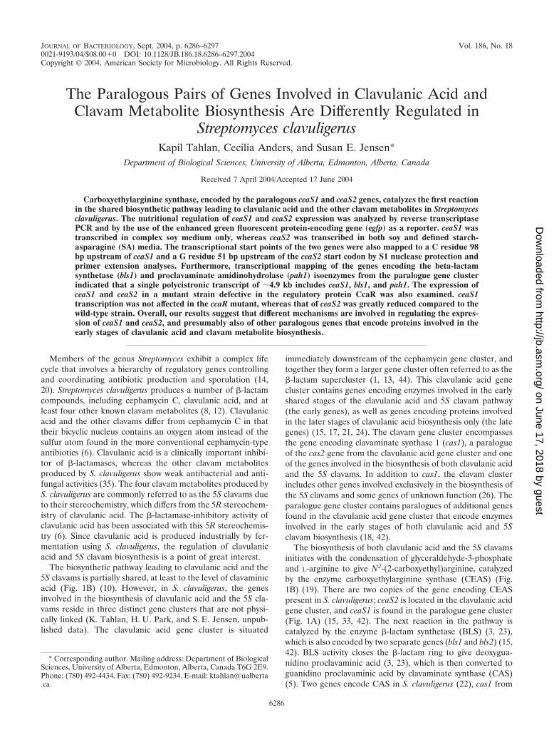
The biosynthetic pathway leading to clavulanic acid and the5S clavams is partially shared, at least to the level of clavaminicacid (Fig. 1B) (10). However, in S. clavuligerus, the genesinvolved in the biosynthesis of clavulanic acid and the 5S cla-vams reside in three distinct gene clusters that are not physi-cally linked (K. Tahlan, H. U. Park, and S. E. Jensen, unpub-lished data). The clavulanic acid gene cluster is situated
immediately downstream of the cephamycin gene cluster, andtogether they form a larger gene cluster often referred to as the�-lactam supercluster (1, 13, 44). This clavulanic acid genecluster contains genes encoding enzymes involved in the earlyshared stages of the clavulanic acid and 5S clavam pathway(the early genes), as well as genes encoding proteins involvedin the later stages of clavulanic acid biosynthesis only (the lategenes) (15, 17, 21, 24). The clavam gene cluster encompassesthe gene encoding clavaminate synthase 1 (cas1), a paralogueof the cas2 gene from the clavulanic acid gene cluster and oneof the genes involved in the biosynthesis of both clavulanic acidand the 5S clavams. In addition to cas1, the clavam clusterincludes other genes involved exclusively in the biosynthesis ofthe 5S clavams and some genes of unknown function (26). Theparalogue gene cluster contains paralogues of additional genesfound in the clavulanic acid gene cluster that encode enzymesinvolved in the early stages of both clavulanic acid and 5Sclavam biosynthesis (18, 42).
The biosynthesis of both clavulanic acid and the 5S clavamsinitiates with the condensation of glyceraldehyde-3-phosphateand L-arginine to give N2-(2-carboxyethyl)arginine, catalyzedby the enzyme carboxyethylarginine synthase (CEAS) (Fig.1B) (19). There are two copies of the gene encoding CEASpresent in S. clavuligerus; ceaS2 is located in the clavulanic acidgene cluster, and ceaS1 is found in the paralogue gene cluster(Fig. 1A) (15, 33, 42). The next reaction in the pathway iscatalyzed by the enzyme �-lactam synthetase (BLS) (3, 23),which is also encoded by two separate genes (bls1 and bls2) (15,42). BLS activity closes the �-lactam ring to give deoxygua-nidino proclavaminic acid (3, 23), which is then converted toguanidino proclavaminic acid by clavaminate synthase (CAS)(5). Two genes encode CAS in S. clavuligerus (22), cas1 from
* Corresponding author. Mailing address: Department of BiologicalSciences, University of Alberta, Edmonton, Alberta, Canada T6G 2E9.Phone: (780) 492-4434. Fax: (780) 492-9234. E-mail: [email protected].
6286
on June 17, 2018 by guesthttp://jb.asm
.org/D
ownloaded from
the clavam gene cluster and cas2 from the clavulanic genecluster (22, 26). Guanidino proclavaminic acid is hydrolyzed togive proclavaminic acid by the action of proclavaminate amidi-nohydrolase (PAH) (46), which is encoded by pah1 and pah2(1, 15, 18, 46). Next, in a series of two sequential reactions,CAS converts proclavaminic acid to clavaminic acid (4, 36).Clavaminic acid is thought to be the branch point of the path-way leading to clavulanic acid and the 5S clavams (10). The
only other step known in the pathway beyond clavaminic acidis the reduction of clavaldehyde to clavulanic acid by the actionof the enzyme clavulanic acid dehydrogenase (27), which isencoded by cad, a late gene from the clavulanic acid genecluster (15). It is still not known how clavaminic acid is con-verted to clavaldehyde, nor have any of the steps specificallyleading from clavaminic acid to the 5S clavams been elucidated(16).
FIG. 1. Early steps of clavulanic acid and 5S clavam metabolite biosynthesis in S. clavuligerus. (A) Genes involved in the early steps of clavulanicacid and 5S clavam biosynthesis from the clavam, clavulanic acid, and paralogue gene clusters. Genes flanking cas1 in the clavam cluster are shownonly to provide context; they do not encode early enzymes. (B) Diagrammatic representation of the early steps of clavulanic acid and 5S clavammetabolite biosynthesis showing the enzymes and genes involved. The 5S clavams are shown as a family of metabolites; R represents the site ofside chain modification giving rise to the different members.
VOL. 186, 2004 REGULATION OF PARALOGOUS GENES 6287
on June 17, 2018 by guesthttp://jb.asm
.org/D
ownloaded from
The transcriptional activators CcaR and ClaR are known toregulate the expression of the clavulanic acid biosyntheticgenes (16). The ccaR gene lies within the cephamycin biosyn-thetic gene cluster, and CcaR is the pathway-specific transcrip-tional regulator for cephamycin biosynthesis, as well as con-trolling expression of the claR gene from the clavulanic acidgene cluster (2, 31, 32). ClaR is the pathway-specific transcrip-tional regulator for clavulanic acid biosynthetic genes, but itaffects only expression of the late genes for clavulanic acidbiosynthesis (29). The early genes responsible for the stepsshared with the 5S clavams are not regulated by ClaR (29). Inthis manner, CcaR coordinates the biosynthesis of cephamycinC, a �-lactam antibiotic, with clavulanic acid, a �-lactamaseinhibitor (16).
The cas1 and cas2 paralogues can functionally replace eachother and are known to be nutritionally regulated. The cas1paralogue is expressed only when S. clavuligerus is grown oncomplex soy medium, whereas the cas2 paralogue is expressedduring growth on both complex soy and defined SA media (30).Thus, a cas1 mutant can still produce clavulanic acid in bothsoy and SA media, because cas2 is expressed in both media. Incontrast, a cas2 mutant produces clavulanic acid only whengrown on soy medium, because cas1 is not expressed in SAmedium (30). Similar phenotypes were also observed when
mutants defective in each of the ceaS, bls, and pah paralogueswere prepared and tested (15, 18, 42).
Since ceaS1 and ceaS2 encode proteins catalyzing the firstreaction in the shared clavulanic acid and 5S clavam biosyn-thetic pathways, and since they are both located at the bound-aries of their respective gene clusters (Fig. 1A), they seemed tobe likely candidates for points of regulation. With this in mind,the nutritional regulation of ceaS1 and ceaS2 was examined atthe transcriptional level to determine if they are regulated in amanner similar to cas1 and cas2. Previous studies have sug-gested that ClaR does not regulate ceaS2 transcription (29),but the effect of CcaR on ceaS1 and ceaS2 transcription is stillnot known. These effects were also examined and are discussedin the present study. Lastly, the transcriptional start points(TSPs) of ceaS1, bls1, pah1, and ceaS2 were also mapped.
MATERIALS AND METHODS
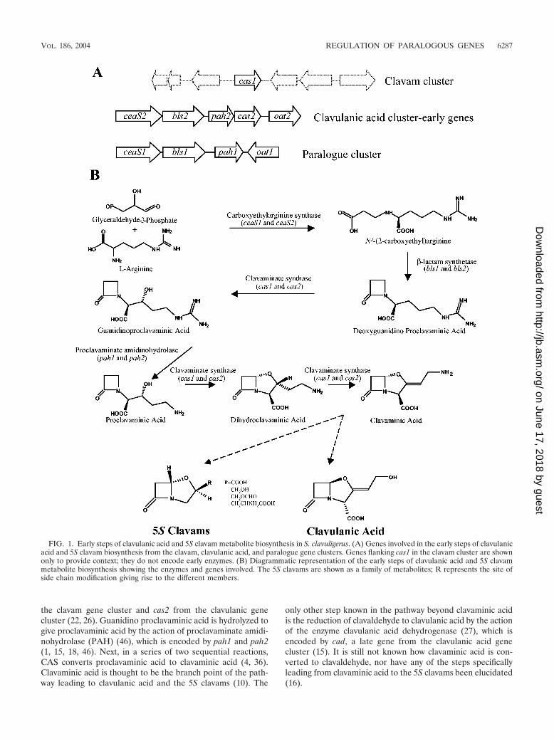
Bacterial strains, plasmids, media, and culture conditions. The bacterialstrains and plasmids used in this study are described in Table 1. Escherichia colicultures were grown as described earlier (38), and cultures containing plasmidswere supplemented with ampicillin (100 �g/ml), apramycin (50 �g/ml), chlor-amphenicol (25 �g/ml), kanamycin (50 �g/ml), or spectinomycin (100 �g/ml), asappropriate. S. clavuligerus cultures were maintained on either MYM (40) or ISP4 medium, as described previously (42), and cultures containing plasmids weresupplemented with apramycin (25 �g/ml) or thiostrepton (5 �g/ml). S. clavulig-
TABLE 1. Bacterial strains and plasmids used in this study
Strain, plasmid, or cosmid Descriptiona Reference or source
Bacillus sp. strain ATCC 27860 Indicator strain for alanyl clavam bioassay 34K. pneumoniae ATCC 15380 Indicator strain for clavulanic acid bioassay 35S. clavuligerus NRRL3585 Wild type National Regional Research
Laboratory (NRRL)S. clavuligerus �ccaR::tsrA ccaR deletion mutant; ccaR replaced by tsr; Tsrr 2S. clavuligerus C1G Contains the egfp reporter gene fused to the ceaS1 promoter; integrated
at the �C31 attB site in the chromosomeThis study
S. clavuligerus C2G Contains the egfp reporter gene fused to the ceaS2 promoter; integratedat the �C31 attB site in the chromosome
This study
Plasmids and cosmidsK6L2 pLAFR3-based cosmid clone spanning the clavulanic acid gene cluster 1p2.8-18 pUC18 containing the 5� end of ceaS1 and upstream region 42p5.7 pUC118 containing blsI, pahI, and flanking sequences 42pBB5.3A pUC119 containing the 5.3-kb BamHI/BglII fragment encompassing
pcbR and ceaS2 from pTZ40; AmprJensen et al., unpublished
pCR2.1TOPO E. coli phagemid cloning vector for rapid cloning of PCR fragments;Ampr Kanr
Invitrogen
pIJ8660 Promoterless EGFP reporter plasmid; free living in E. coli and integratesinto the Streptomyces chromosome; Aprr
41
pIJ8660-ceaS2 EGFP reporter construct; pIJ8660 containing the 707-bp BamHI/KpnIfragment from pTOPO-ceaS2-8 encompassing the ceaS2 promoterregion fused to the egfp gene
This study
pSET152 E. coli cloning vector; integrates into the Streptomyces chromosome; Aprr NRRLpSET-ceaS1 EGFP reporter construct; pSET152 containing the 2.37-kb
BamHI/EcoRI fragment from pTO6-ceaS1 encompassing the ceaS1promoter fused to the egfp gene
This study
pTO6 pIJ8660 with aac(3)IV replaced by aadA; Spcr 28pTO6-ceaS1 EGFP reporter construct; pTO6 containing the 777-bp BamHI/KpnI
fragment from pTOPO-ceaS1-4This study
pTOPO-cea52-8 pCR2.1TOPO containing a 721-bp PCR fragment encompassing theceaS2 promoter region
This study
pTOPO-ceaS1-4 pCR2.1TOPO containing a 784-bp PCR fragment encompassing theceaS1 promoter region
This study
pTZ40 pTZ18R containing an �26-kb BamHI/PstI fragment from cosmid K6L2spanning the clavulanic acid gene cluster; Ampr
Jensen et al., unpublished
a Ampr, ampicillin resistance; Aprr, apramycin resistance; Kanr, kanamycin resistance, Spcr, spectinomycin resistance; Tsrr, thiostrepton resistance.
6288 TAHLAN ET AL. J. BACTERIOL.
on June 17, 2018 by guesthttp://jb.asm
.org/D
ownloaded from
erus cultures for the isolation of chromosomal DNA were grown in trypticase soybroth supplemented with 1% starch (TSBS), and cultures for the isolation ofexconjugants were grown in AS-1 medium supplemented with 10 mM MgCl2, asdescribed earlier (42). To prepare RNA from S. clavuligerus, spores were pre-germinated for 4 h at 28°C in 2YT medium (38) and then used to inoculate SAor soy culture medium (30). RNA was isolated after 96 and 120 h of growth insoy medium and after 72 and 96 h of growth in SA medium. All Streptomycesliquid cultures were grown at 28°C on a rotary shaker at 250 rpm.
DNA isolation, manipulation, and Southern analysis. Routine manipulationof plasmid DNA isolated from E. coli cultures, including labeling of double-stranded DNA probes with [�-32P]dCTP by nick translation and end labelingusing [-32P]dATP, was performed using standard procedures (38). DNA se-quencing was carried out using the DYEnamic ET Terminator Cycle Sequencingkit (Amersham Pharmacia, Baie d’Urfe, Quebec, Canada) by the MolecularBiology Service Unit, University of Alberta. Manual sequencing of DNA wasperformed using the Thermo Sequenase Radiolabeled Terminator Cycle Se-quencing kit (U.S. Biochemical) according to the manufacturer’s directions.DNA fragments fractionated by agarose and polyacrylamide gel electrophoresis(PAGE) were isolated using the QIAquick Gel Extraction kit (Qiagen Inc.) andthe crush-and-soak method (38), respectively. PCRs were carried out using theExpand high-fidelity PCR system (Roche) according to the manufacturer’s in-structions. Plasmid DNA was introduced into S. clavuligerus by intergenericconjugation as described previously (42). Southern analysis of S. clavuligerusDNA was also carried out as described elsewhere (38), using the following washconditions. After overnight incubation with labeled probes, the nylon membraneswere washed twice for 15 min each time with 2 SSC (1 SSC is 0.15 M NaClplus 0.015 M sodium citrate)–0.1% sodium dodecyl sulfate (SDS) (38) at roomtemperature, once for 20 min with 1.0 SSC–0.1% SDS, and once for 20 minwith 0.1 SSC–0.1% SDS. All incubations and washes were carried out at 65°Cunless otherwise indicated.
RNA isolation, RT-PCR, S1 nuclease mapping, primer extension, and North-ern blot analysis. RNAs from wild-type S. clavuligerus and S. clavuligerus�ccaR::tsrA were isolated using the modified Kirby procedure (20). Reversetranscriptase (RT)-PCR analysis of RNA was carried out using C. therm poly-merase (Roche) for reverse transcription in two-step RT-PCR. All RT reactionswere carried out at 62°C for 30 min according to the manufacturer’s instructionswith the following changes. The reactions were set up in a final volume of 10 �lusing 0.5 �g of total RNA per reaction and 15.8 U of RNAguard RNaseInhibitor (Amersham). The reverse primers ceaS1-RT-Rev, ceaS2-RT-Rev, andCAN 122 (Table 2) were used to synthesize cDNA corresponding to the ceaS1,ceaS2, and hrdB transcripts, respectively. The PCRs were performed using 5 �lof the RT product from the reaction described above in a final volume of 50 �l,employing the Expand high-fidelity PCR system with buffer 2. The primer pairsceaS1-RT-For plus ceaS1-RT-Rev and ceaS2-RT-For plus ceaS2-RT-Rev wereused for PCR amplification of the ceaS1 and the ceaS2 RT products, respectively.
Dimethyl sulfoxide (DMSO; 5% [vol/vol] final concentration) was used in thesereactions with the following program: 94°C for 2 min, followed by 25 cycles of94°C for 1 min, 62°C for 1 min, and 72°C for 1 min. The primers CAN 123 andCAN 122 were used to amplify the hrdB RT product by PCR using 10% (vol/vol)DMSO and the following program: 94°C for 2 min, followed by 25 cycles of 94°Cfor 1 min, 66°C for 1 min, and 72°C for 1 min. The RT-PCR products wereanalyzed by fractionation on 1.5% agarose Tris-borate-EDTA gels. The identi-ties of the RT-PCR products were also verified by restriction analysis, followedby PAGE and by sequencing of both DNA strands (data not shown).
High-resolution S1 nuclease mapping was carried out using the sodium tri-chloroacetate method (20). All double-stranded DNA probes were prepared byPCR using custom primers. DMSO (5% [vol/vol]) was included in the reactions,with the following program: 94°C for 2 min, followed by 10 cycles of 94°C for 45 s,60°C for 45 s, and 72°C for 45 s, and finally 15 cycles of 94°C for 45 s, 65°C for45 s, and 72°C for 45 s. The primers ceaS1-S1-For and ceaS1-PR-EX, along withp2.8-18 as a template, were used to prepare the probe to map the ceaS1 TSP. Theprimers ceaS2-S1-For and ceaS2-PR-EX, along with the template plasmidpBB5.3A, were used to prepare the probe for mapping the TSP of ceaS2. Theprimers bls1-S1-For and bls1-S1-Rev, and pah1-S1-For and pah1-UP-rev, alongwith p5.7 as a template, were used to prepare probes for S1 protection analysisof bls1 and pah1, respectively.
Primer extension analysis was performed using C. therm polymerase for re-verse transcription in two-step RT-PCR (Roche) according to the manufactur-er’s instructions with the following changes. Twenty-microliter reaction mixtureswere set up using 5 pmol of the end-labeled reverse primers ceaS1-PR-EX andceaS2-PR-EX (Table 2) and 40 U of RNaseOUT Recombinant RNase Inhibitor(Invitrogen), with the following program: extension at 60°C for 60 min andtermination at 80°C for 10 min.
DNA sequencing ladders were prepared for size estimation using the reverseprimers and the template plasmids used in the preparation of the S1 probes orin the primer extension reactions. Samples were separated on 6% denaturingpolyacrylamide sequencing gels for analysis as described earlier (38).
Northern blot analysis was carried out using established techniques (30) with40 �g of RNA isolated from wild-type S. clavuligerus grown on soy medium for96 and 120 h. Molecular Weight Marker III (Roche) was run along with the RNAsamples for size estimation. The primers pah1-S1-For and pah1-S1-Rev wereused to generate a 288-bp probe by PCR using p5.7 as a template. This probe wasused for both S1 nuclease protection analysis (data not shown) and Northernhybridization. Probe annealing and subsequent washes were carried out underthe same high-stringency conditions used in Southern blot analysis.
Preparation of enhanced green fluorescent protein (EGFP) reporter con-structs. A 781-bp DNA fragment spanning the ceaS1 promoter region wasamplified by PCR using p2.8-18 as a template and the primer pair KTA-ceaS1-For and KTA-ceaS1-Rev. Similarly, a 721-bp DNA fragment encompassing theceaS2 promoter region was amplified by PCR using pBB5.3A as the template and
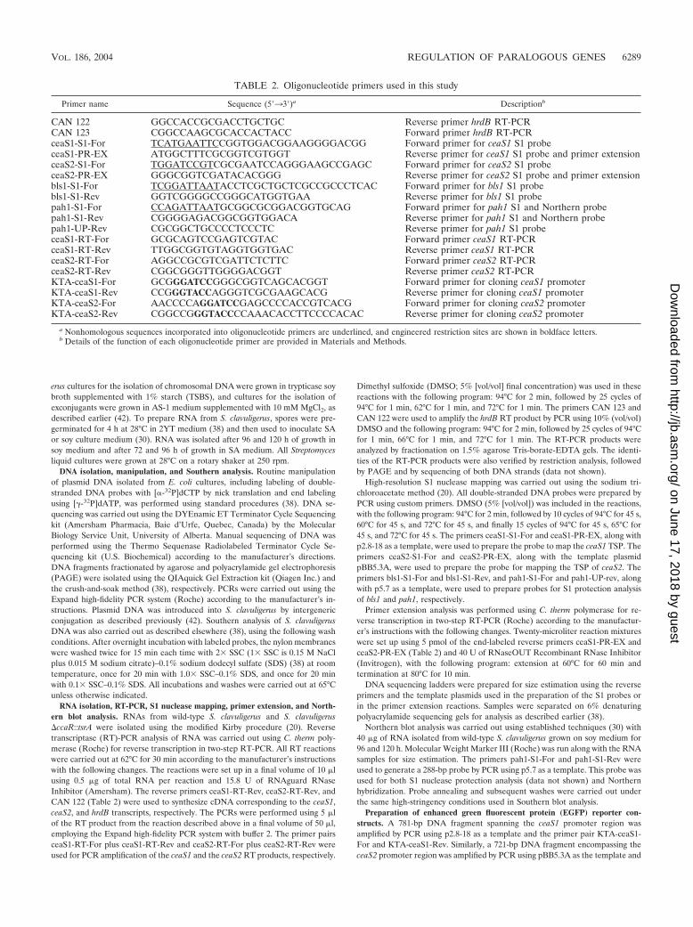
TABLE 2. Oligonucleotide primers used in this study
Primer name Sequence (5�33�)a Descriptionb
CAN 122 GGCCACCGCGACCTGCTGC Reverse primer hrdB RT-PCRCAN 123 CGGCCAAGCGCACCACTACC Forward primer hrdB RT-PCRceaS1-S1-For TCATGAATTCCGGTGGACGGAAGGGGACGG Forward primer for ceaS1 S1 probeceaS1-PR-EX ATGGCTTTCGCGGTCGTGGT Reverse primer for ceaS1 S1 probe and primer extensionceaS2-S1-For TGGATCCGTCGCGAATCCAGGGAAGCCGAGC Forward primer for ceaS2 S1 probeceaS2-PR-EX GGGCGGTCGATACACGGG Reverse primer for ceaS2 S1 probe and primer extensionbls1-S1-For TCGGATTAATACCTCGCTGCTCGCCGCCCTCAC Forward primer for bls1 S1 probebls1-S1-Rev GGTCGGGGCCGGGCATGGTGAA Reverse primer for bls1 S1 probepah1-S1-For CCAGATTAATGCGGCGCGGACGGTGCAG Forward primer for pah1 S1 and Northern probepah1-S1-Rev CGGGGAGACGGCGGTGGACA Reverse primer for pah1 S1 and Northern probepah1-UP-Rev CGCGGCTGCCCCTCCCTC Reverse primer for pah1 S1 probeceaS1-RT-For GCGCAGTCCGAGTCGTAC Forward primer ceaS1 RT-PCRceaS1-RT-Rev TTGGCGGTGTAGGTGGTGAC Reverse primer ceaS1 RT-PCRceaS2-RT-For AGGCCGCGTCGATTCTCTTC Forward primer ceaS2 RT-PCRceaS2-RT-Rev CGGCGGGTTGGGGACGGT Reverse primer ceaS2 RT-PCRKTA-ceaS1-For GCGGGATCCGGGCGGTCAGCACGGT Forward primer for cloning ceaS1 promoterKTA-ceaS1-Rev CCGGGTACCAGGGTCGCGAAGCACG Reverse primer for cloning ceaS1 promoterKTA-ceaS2-For AACCCCAGGATCCGAGCCCCACCGTCACG Forward primer for cloning ceaS2 promoterKTA-ceaS2-Rev CGGCCGGGTACCCCAAACACCTTCCCCACAC Reverse primer for cloning ceaS2 promoter
a Nonhomologous sequences incorporated into oligonucleotide primers are underlined, and engineered restriction sites are shown in boldface letters.b Details of the function of each oligonucleotide primer are provided in Materials and Methods.
VOL. 186, 2004 REGULATION OF PARALOGOUS GENES 6289
on June 17, 2018 by guesthttp://jb.asm
.org/D
ownloaded from
the primers KTA-ceaS2-For and KTA-ceaS2-Rev. The PCR products weretreated with Taq DNA polymerase before ligation to pCR2.1TOPO (Invitrogen)according to the manufacturer’s instructions. This gave pTOPO-ceaS1-4 andpTOPO-ceaS2-8, which contain the ceaS1 and ceaS2 promoter regions inpCR2.1TOPO, respectively. The double-stranded DNA sequence of the insertswas obtained using universal primers to ensure that no mutations were intro-duced.
The ceaS2 promoter region from pTOPO-ceaS2-8 was isolated as a BamHI/KpnI fragment and ligated into the corresponding sites of pIJ8660 to givepIJ8660-ceaS2. pIJ8660-ceaS2 contains the ceaS2 promoter region fused to apromoterless egfp gene for use as a reporter of expression driven by the ceaS2promoter.
For unexplained reasons, we were unable to subclone the ceaS1 promoterregion into pIJ8660 and therefore used an alternative approach. The ceaS1promoter region from pTOPO-ceaS1-4 was isolated as a BamHI/KpnI fragmentand ligated into the corresponding sites of pTO6 to give pTO6-ceaS1. The entirecassette encompassing the ceaS1 promoter region fused to the egfp gene frompTO6-ceaS1 was isolated as a 2.37-kb BamHI/EcoRI fragment and introducedinto the corresponding sites of pSET152 to give pSET-ceaS1, which served as theceaS1 reporter construct.
The plasmids pSET-ceaS1 and pIJ8660-ceaS2 were introduced into wild-typeS. clavuligerus by conjugation. Strains that had the plasmids integrated at the�C31 attB site in the chromosome were isolated based on apramycin resistanceand were verified by Southern hybridization (data not shown).
Confocal microscopy. S. clavuligerus cultures harboring egfp reporter con-structs were grown in TSBS for 36 h. Five-hundred-microliter amounts of theTSBS cultures were used to inoculate 25 ml of either SA or soy medium. After72 h of growth, 1-ml amounts of the cultures were harvested and washed once inacetonitrile and then twice in sterile distilled water. The washed mycelia weremounted in 40% (vol/vol) glycerol before observations were made under themicroscope.
Confocal microscopy was carried out using a Leica DM IRB inverted micro-scope. An argon laser (50 to 52% attenuation) provided excitation at 488 nm.Fluorescence due to EGFP excitation was detected between 505 and 520 nm, andcorresponding differential interference contrast images were also obtained.
Western analysis. Five-milliliter amounts of S. clavuligerus cultures grown insoy and SA media were harvested and resuspended in 1 ml of lysing buffer (100mM HEPES [pH 7.2], 0.5 mg of lysozyme/ml, 2 Complete EDTA free proteaseinhibitor cocktail [Roche]). The suspensions were incubated at 37°C for 30 min,the cell membranes were broken by ultrasonic disruption, and then the cell debriswas removed by centrifugation. Samples of cell extracts (CFEs) containing 50 �gof total protein were separated by SDS-PAGE (12% gels) as described earlier(38). The proteins were transferred onto polyvinylidene difluoride membranes(Immobilon-P; Millipore) using a Bio-Rad Transblot apparatus. The BM Chemi-luminescence Western Blotting Kit (Mouse/Rabbit) (Roche) was used to detectproteins in accordance with the manufacturer’s instructions. The primary anti-body used to detect EGFP was the commercially available BD Living Colors A.v.Peptide Antibody (BD Biosciences) and was used at 1/400 dilution.
HPLC analyses of culture filtrates, bioassays, and growth determination.High-performance liquid chromatographic (HPLC) analysis of culture superna-tants after imidazole derivatization was performed under previously describedconditions (30). Bioassays were used to detect clavulanic acid and alanyl clavamproduction using Klebsiella pneumoniae ATCC 15380 and Bacillus sp. strainATCC 27860, respectively, as the indicator organisms (18, 30). Growth of S.clavuligerus in fermentation medium was estimated by measuring the opticaldensity of broken mycelia at 495 nm as described earlier (7).
RESULTS
Nutritional regulation of ceaS1 and ceaS2. In previous stud-ies, when ceaS1 and ceaS2 mutant strains were prepared indi-vidually, both mutant strains still retained some ability to pro-duce both clavulanic acid and the 5S clavams, depending onthe fermentation medium used (15, 33, 42). The ceaS1 mutantproduced both clavulanic acid and the 5S clavams in soy me-dium but only clavulanic acid in SA medium. In contrast, theceaS2 mutant produced small amounts of clavulanic acid andthe 5S clavam metabolites in soy medium only, while no cla-vulanic acid or 5S clavam production was observed in culturesgrown in SA medium (15, 42). Based on the observed pheno-
types, ceaS1 was postulated to be expressed in soy mediumonly, whereas ceaS2 was expressed in both soy and SA media.To verify this hypothesis, we examined the effects of growth insoy and SA media on both ceaS1 and ceaS2 expression at thetranscriptional level.
RT-PCR was used to detect ceaS1 and ceaS2 transcripts,using RNA isolated from wild-type S. clavuligerus grown on SAmedium for 72 and 96 h and on soy medium for 96 and 120 h.When RNA isolated from SA cultures was subjected to anal-ysis, ceaS1 transcripts were not detected, whereas the samesamples showed the presence of ceaS2 transcripts (Fig. 2A). Insimilar analyses of RNA isolated from soy-grown cultures,both ceaS1 and ceaS2 transcripts were detected by RT-PCR(Fig. 2A). The expression of hrdB, which encodes a constitu-tively expressed sigma factor in Streptomyces, was monitored asa control, and hrdB transcripts were detected at similar levelsin all samples tested (Fig. 2). HPLC analysis of culture super-natants showed the expected levels of clavulanic acid and 5Sclavams in cultures grown in both media used for RNA isola-tion and analysis (data not shown).
The promoter regions of ceaS1 and ceaS2 (�501 to �215and �551 to �95 bp relative to the putative start codons ofceaS1 and ceaS2, respectively) were subcloned in front of apromoterless egfp gene to give plasmids pSET-ceaS1 andpIJ8660-ceaS2, respectively. Fluorescence arising due toEGFP expression was used as a reporter to monitor transcrip-tion driven from the promoters. These promoter constructswere introduced into wild-type S. clavuligerus, where they in-tegrated into the chromosome via the �C31 attB site. Theintegration of the reporter plasmids into the S. clavuligeruschromosome was confirmed by Southern hybridization usingDNA probes specific for both egfp and the respective promot-ers (data not shown).
The S. clavuligerus reporter strains C1G and C2G (Table 1)were grown on soy and SA media for 72 h, after which myceliawere harvested and analyzed by confocal microscopy to exam-
FIG. 2. Assessment of ceaS1 and ceaS2 transcript levels by RT-PCR. RNA samples isolated from various strains of S. clavuligeruswere analyzed by RT-PCR using primers specific for ceaS1, ceaS2, andhrdB. (A) Analysis of RNAs from S. clavuligerus wild-type (WT) cul-tures grown on SA medium for 72 and 96 h and from cultures grownon soy medium for 96 and 120 h. (B) Analysis of RNAs from S.clavuligerus wild-type cultures grown on soy medium for 96 and 120 hand S. clavuligerus �ccaR::tsrA mutant cultures grown on soy mediumfor 96 and 120 h.
6290 TAHLAN ET AL. J. BACTERIOL.
on June 17, 2018 by guesthttp://jb.asm
.org/D
ownloaded from
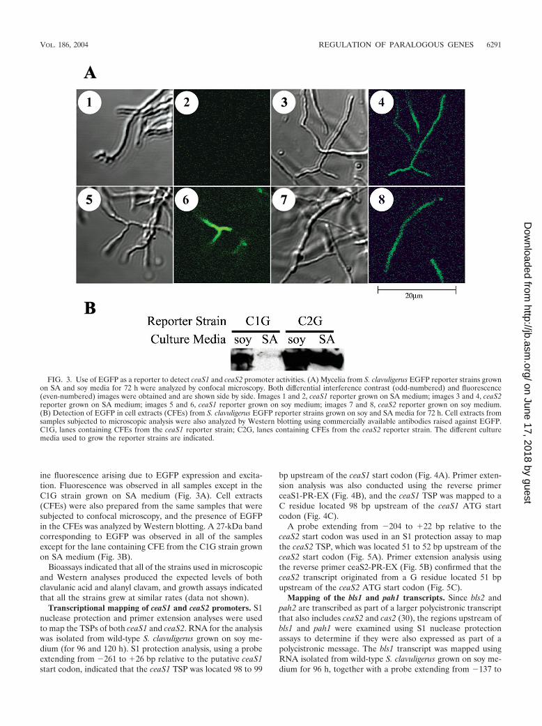
ine fluorescence arising due to EGFP expression and excita-tion. Fluorescence was observed in all samples except in theC1G strain grown on SA medium (Fig. 3A). Cell extracts(CFEs) were also prepared from the same samples that weresubjected to confocal microscopy, and the presence of EGFPin the CFEs was analyzed by Western blotting. A 27-kDa bandcorresponding to EGFP was observed in all of the samplesexcept for the lane containing CFE from the C1G strain grownon SA medium (Fig. 3B).
Bioassays indicated that all of the strains used in microscopicand Western analyses produced the expected levels of bothclavulanic acid and alanyl clavam, and growth assays indicatedthat all the strains grew at similar rates (data not shown).
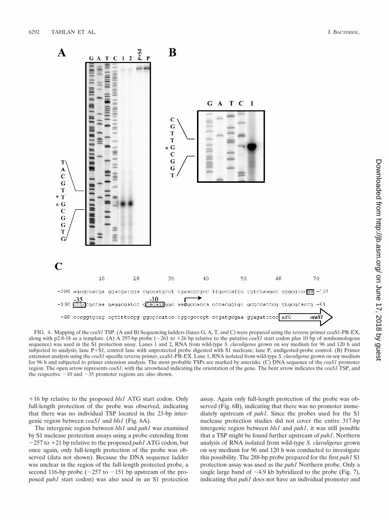
Transcriptional mapping of ceaS1 and ceaS2 promoters. S1nuclease protection and primer extension analyses were usedto map the TSPs of both ceaS1 and ceaS2. RNA for the analysiswas isolated from wild-type S. clavuligerus grown on soy me-dium (for 96 and 120 h). S1 protection analysis, using a probeextending from �261 to �26 bp relative to the putative ceaS1start codon, indicated that the ceaS1 TSP was located 98 to 99
bp upstream of the ceaS1 start codon (Fig. 4A). Primer exten-sion analysis was also conducted using the reverse primerceaS1-PR-EX (Fig. 4B), and the ceaS1 TSP was mapped to aC residue located 98 bp upstream of the ceaS1 ATG startcodon (Fig. 4C).
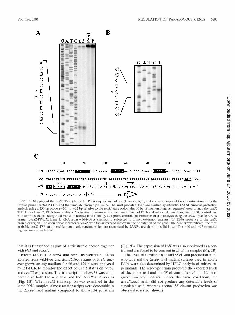
A probe extending from �204 to �22 bp relative to theceaS2 start codon was used in an S1 protection assay to mapthe ceaS2 TSP, which was located 51 to 52 bp upstream of theceaS2 start codon (Fig. 5A). Primer extension analysis usingthe reverse primer ceaS2-PR-EX (Fig. 5B) confirmed that theceaS2 transcript originated from a G residue located 51 bpupstream of the ceaS2 ATG start codon (Fig. 5C).
Mapping of the bls1 and pah1 transcripts. Since bls2 andpah2 are transcribed as part of a larger polycistronic transcriptthat also includes ceaS2 and cas2 (30), the regions upstream ofbls1 and pah1 were examined using S1 nuclease protectionassays to determine if they were also expressed as part of apolycistronic message. The bls1 transcript was mapped usingRNA isolated from wild-type S. clavuligerus grown on soy me-dium for 96 h, together with a probe extending from �137 to
FIG. 3. Use of EGFP as a reporter to detect ceaS1 and ceaS2 promoter activities. (A) Mycelia from S. clavuligerus EGFP reporter strains grownon SA and soy media for 72 h were analyzed by confocal microscopy. Both differential interference contrast (odd-numbered) and fluorescence(even-numbered) images were obtained and are shown side by side. Images 1 and 2, ceaS1 reporter grown on SA medium; images 3 and 4, ceaS2reporter grown on SA medium; images 5 and 6, ceaS1 reporter grown on soy medium; images 7 and 8, ceaS2 reporter grown on soy medium.(B) Detection of EGFP in cell extracts (CFEs) from S. clavuligerus EGFP reporter strains grown on soy and SA media for 72 h. Cell extracts fromsamples subjected to microscopic analysis were also analyzed by Western blotting using commercially available antibodies raised against EGFP.C1G, lanes containing CFEs from the ceaS1 reporter strain; C2G, lanes containing CFEs from the ceaS2 reporter strain. The different culturemedia used to grow the reporter strains are indicated.
VOL. 186, 2004 REGULATION OF PARALOGOUS GENES 6291
on June 17, 2018 by guesthttp://jb.asm
.org/D
ownloaded from
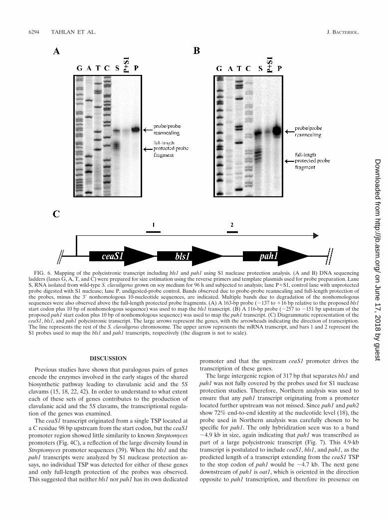
�16 bp relative to the proposed bls1 ATG start codon. Onlyfull-length protection of the probe was observed, indicatingthat there was no individual TSP located in the 23-bp inter-genic region between ceaS1 and bls1 (Fig. 6A).
The intergenic region between bls1 and pah1 was examinedby S1 nuclease protection assays using a probe extending from�257 to �21 bp relative to the proposed pah1 ATG codon, butonce again, only full-length protection of the probe was ob-served (data not shown). Because the DNA sequence ladderwas unclear in the region of the full-length protected probe, asecond 116-bp probe (�257 to �151 bp upstream of the pro-posed pah1 start codon) was also used in an S1 protection
assay. Again only full-length protection of the probe was ob-served (Fig. 6B), indicating that there was no promoter imme-diately upstream of pah1. Since the probes used for the S1nuclease protection studies did not cover the entire 317-bpintergenic region between bls1 and pah1, it was still possiblethat a TSP might be found further upstream of pah1. Northernanalysis of RNA isolated from wild-type S. clavuligerus grownon soy medium for 96 and 120 h was conducted to investigatethis possibility. The 288-bp probe prepared for the first pah1 S1protection assay was used as the pah1 Northern probe. Only asingle large band of �4.9 kb hybridized to the probe (Fig. 7),indicating that pah1 does not have an individual promoter and
FIG. 4. Mapping of the ceaS1 TSP. (A and B) Sequencing ladders (lanes G, A, T, and C) were prepared using the reverse primer ceaS1-PR-EX,along with p2.8-18 as a template. (A) A 297-bp probe (�261 to �26 bp relative to the putative ceaS1 start codon plus 10 bp of nonhomologoussequence) was used in the S1 protection assay. Lanes 1 and 2, RNA from wild-type S. clavuligerus grown on soy medium for 96 and 120 h andsubjected to analysis; lane P�S1, control lane with unprotected probe digested with S1 nuclease; lane P, undigested-probe control. (B) Primerextension analysis using the ceaS1-specific reverse primer, ceaS1-PR-EX. Lane 1, RNA isolated from wild-type S. clavuligerus grown on soy mediumfor 96 h and subjected to primer extension analysis. The most probable TSPs are marked by asterisks. (C) DNA sequence of the ceaS1 promoterregion. The open arrow represents ceaS1, with the arrowhead indicating the orientation of the gene. The bent arrow indicates the ceaS1 TSP, andthe respective �10 and �35 promoter regions are also shown.
6292 TAHLAN ET AL. J. BACTERIOL.
on June 17, 2018 by guesthttp://jb.asm
.org/D
ownloaded from
that it is transcribed as part of a tricistronic operon togetherwith bls1 and ceaS1.
Effects of CcaR on ceaS1 and ceaS2 transcription. RNAsisolated from wild-type and �ccaR::tsrA strains of S. clavulig-erus grown on soy medium for 96 and 120 h were analyzedby RT-PCR to monitor the effect of CcaR status on ceaS1and ceaS2 expression. The transcription of ceaS1 was com-parable in both the wild-type and the �ccaR::tsrA strains(Fig. 2B). When ceaS2 transcription was examined in thesame RNA samples, almost no transcripts were detectable inthe �ccaR::tsrA mutant compared to the wild-type strain
(Fig. 2B). The expression of hrdB was also monitored as a con-trol and was found to be constant in all of the samples (Fig. 2B).
The levels of clavulanic acid and 5S clavam production in thewild-type and the �ccaR::tsrA mutant cultures used to isolateRNA were also determined by HPLC analysis of culture su-pernatants. The wild-type strain produced the expected levelsof clavulanic acid and the 5S clavams after 96 and 120 h ofgrowth on soy medium. Under the same conditions, the�ccaR::tsrA strain did not produce any detectable levels ofclavulanic acid, whereas normal 5S clavam production wasobserved (data not shown).
FIG. 5. Mapping of the ceaS2 TSP. (A and B) DNA sequencing ladders (lanes G, A, T, and C) were prepared for size estimation using thereverse primer ceaS2-PR-EX and the template plasmid pBB5.3A. The most probable TSPs are marked by asterisks. (A) S1 nuclease protectionanalysis using a 236-bp probe (�204 to �22 bp relative to the ceaS2 start codon plus 10 bp of nonhomologous sequence) used to map the ceaS2TSP. Lanes 1 and 2, RNA from wild-type S. clavuligerus grown on soy medium for 96 and 120 h and subjected to analysis; lane P�S1, control lanewith unprotected probe digested with S1 nuclease; lane P, undigested-probe control. (B) Primer extension analysis using the ceaS2-specific reverseprimer, ceaS2-PR-EX. Lane 1, RNA from wild-type S. clavuligerus subjected to primer extension analysis. (C) DNA sequence of the ceaS2promoter region. The open arrow represents ceaS2, with the arrowhead indicating the orientation of the gene. The bent arrow indicates the mostprobable ceaS2 TSP, and possible heptameric repeats, which are recognized by SARPs, are shown in solid boxes. The �10 and �35 promoterregions are also indicated.
VOL. 186, 2004 REGULATION OF PARALOGOUS GENES 6293
on June 17, 2018 by guesthttp://jb.asm
.org/D
ownloaded from
DISCUSSION
Previous studies have shown that paralogous pairs of genesencode the enzymes involved in the early stages of the sharedbiosynthetic pathway leading to clavulanic acid and the 5Sclavams (15, 18, 22, 42). In order to understand to what extenteach of these sets of genes contributes to the production ofclavulanic acid and the 5S clavams, the transcriptional regula-tion of the genes was examined.
The ceaS1 transcript originated from a single TSP located ata C residue 98 bp upstream from the start codon, but the ceaS1promoter region showed little similarity to known Streptomycespromoters (Fig. 4C), a reflection of the large diversity found inStreptomyces promoter sequences (39). When the bls1 and thepah1 transcripts were analyzed by S1 nuclease protection as-says, no individual TSP was detected for either of these genesand only full-length protection of the probes was observed.This suggested that neither bls1 nor pah1 has its own dedicated
promoter and that the upstream ceaS1 promoter drives thetranscription of these genes.
The large intergenic region of 317 bp that separates bls1 andpah1 was not fully covered by the probes used for S1 nucleaseprotection studies. Therefore, Northern analysis was used toensure that any pah1 transcript originating from a promoterlocated further upstream was not missed. Since pah1 and pah2show 72% end-to-end identity at the nucleotide level (18), theprobe used in Northern analysis was carefully chosen to bespecific for pah1. The only hybridization seen was to a band�4.9 kb in size, again indicating that pah1 was transcribed aspart of a large polycistronic transcript (Fig. 7). This 4.9-kbtranscript is postulated to include ceaS1, bls1, and pah1, as thepredicted length of a transcript extending from the ceaS1 TSPto the stop codon of pah1 would be �4.7 kb. The next genedownstream of pah1 is oat1, which is oriented in the directionopposite to pah1 transcription, and therefore its presence on
FIG. 6. Mapping of the polycistronic transcript including bls1 and pah1 using S1 nuclease protection analysis. (A and B) DNA sequencingladders (lanes G, A, T, and C) were prepared for size estimation using the reverse primers and template plasmids used for probe preparation. LaneS, RNA isolated from wild-type S. clavuligerus grown on soy medium for 96 h and subjected to analysis; lane P�S1, control lane with unprotectedprobe digested with S1 nuclease; lane P, undigested-probe control. Bands observed due to probe-probe reannealing and full-length protection ofthe probes, minus the 3� nonhomologous 10-nucleotide sequences, are indicated. Multiple bands due to degradation of the nonhomologoussequences were also observed above the full-length protected probe fragments. (A) A 163-bp probe (�137 to �16 bp relative to the proposed bls1start codon plus 10 bp of nonhomologous sequence) was used to map the bls1 transcript. (B) A 116-bp probe (�257 to �151 bp upstream of theproposed pah1 start codon plus 10 bp of nonhomologous sequence) was used to map the pah1 transcript. (C) Diagrammatic representation of theceaS1, bls1, and pah1 polycistronic transcript. The large arrows represent the genes, with the arrowheads indicating the direction of transcription.The line represents the rest of the S. clavuligerus chromosome. The upper arrow represents the mRNA transcript, and bars 1 and 2 represent theS1 probes used to map the bls1 and pah1 transcripts, respectively (the diagram is not to scale).
6294 TAHLAN ET AL. J. BACTERIOL.
on June 17, 2018 by guesthttp://jb.asm
.org/D
ownloaded from

the 4.9-kb polycistronic transcript can be ruled out. In addition,computational analysis of the 126-bp intergenic region be-tween pah1 and oat1 predicted the presence of considerablesecondary structure, consisting of multiple stem-loops with acumulative G of �101.4, which could function as a transcrip-tional terminator (data not shown).
The transcriptional arrangement of ceaS1, bls1, and pah1 issimilar to that of their paralogous counterparts from the cla-vulanic acid gene cluster (Fig. 1A). The ceaS2, bls2, pah2, andcas2 genes from the clavulanic acid gene cluster are transcribedas a 5.3-kb polycistronic transcript. In addition, cas2 is alsotranscribed as a 1.2-kb monocistronic transcript (30). The mostsignificant difference between the 5.3-kb transcript arisingfrom the clavulanic acid gene cluster and the 4.9-kb transcriptarising from the paralogue gene cluster is the absence of thecas1 coding sequence in the paralogue gene cluster. cas1 islocated elsewhere on the S. clavuligerus chromosome and isexpressed as a 1.4-kb monocistronic transcript (30).
The transcript comprising ceaS2 was also mapped and wasalso found to arise from a single TSP located 51 bp upstreamof the ceaS2 start codon. As was the case for ceaS1, the pro-posed ceaS2 promoter region did not show any significant
similarity to any known Streptomyces promoters. Since S1 nu-clease and primer extension analyses were used to identify allof the TSPs described in this study, it should be noted that bothof the analysis methods employed are predictive of the TSP,provided the mRNA is not processed.
The nutritional regulation of ceaS1 and ceaS2 transcriptionwas examined using RT-PCR, which demonstrated that ceaS1was transcribed in soy medium only and not in SA medium.Under the same conditions and using the same RNA prepa-rations, ceaS2 was transcribed in both soy and SA media atcomparable levels. The ceaS1 and ceaS2 promoter regionswere also subcloned in front of a promoterless egfp gene, andEGFP expression was used as a reporter to monitor transcrip-tion driven by the respective promoters. Confocal microscopywas used to detect fluorescence due to EGFP expression andexcitation, and the results confirmed that ceaS1 was expressedin soy medium only and not in SA medium, whereas ceaS2 wasexpressed in both media tested. The results obtained fromconfocal microscopy were confirmed by Western analysis,which indicated that the fluorescence observed in the sampleswas due to true EGFP expression. Since ceaS1, bls1, and pah1are expressed only as a 4.9-kb polycistronic message, it can beinferred that bls1 and pah1 will show the same general trend ofnutritional regulation as ceaS1. In combination, our resultsindicate that ceaS1, bls1, and pah1 are expressed in soy me-dium but not in SA medium, whereas ceaS2, bls2, pah2, andcas2 are expressed in both soy and SA media. This explains theclavulanic acid- and 5S clavam-producing phenotypes observedwhen mutants defective in these genes were prepared andtested in previous studies (15, 18, 42).
The ccaR gene from the cephamycin gene cluster encodes apathway-specific transcriptional regulator that coordinates theproduction of both cephamycin C and clavulanic acid (2, 31).Its effect on clavulanic acid biosynthesis is exerted, at least inpart, through activation of the expression of a second pathway-specific transcriptional regulator, ClaR, from the clavulanicacid gene cluster. Mutants defective in CcaR do not produceany cephamycin C, and clavulanic acid production is alsoknocked out (2, 31), as the transcription of claR is reduced tonear zero in these mutants (32). ClaR positively regulatesgenes involved exclusively in the biosynthesis of clavulanic acid(29). Previous studies have shown that ceaS2 expression is notunder the control of claR (29), but the detailed effects of CcaRon ceaS1 and ceaS2 expression are not known.
Our results indicate that ceaS1 transcription is unaffected inthe ccaR mutant compared to the wild-type strain, whereas thetranscription of ceaS2 is almost eliminated in the ccaR mutant.Therefore, CcaR controls the production of clavulanic acidthrough at least two routes, an indirect route mediated byClaR and a second route that may involve CcaR directly reg-ulating ceaS2 promoter activity.
CcaR belongs to a family of transcriptional regulators calledthe Streptomyces antibiotic regulatory proteins (SARPs), whichbind to specific heptameric repeats and promote transcription(45). Imperfect heptameric repeats can be identified in theregion upstream of ceaS2 (Fig. 5C), consistent with the ideathat CcaR may bind directly to the ceaS2 promoter region toregulate transcription. Such a notion, however, has not yetbeen demonstrated experimentally, and it is also possible thatCcaR exerts its effect indirectly through additional proteins
FIG. 7. Northern analysis of wild-type S. clavuligerus RNA using apah1-specific probe. Lane 1, molecular size marker; lanes 2 and 3,RNA from wild-type S. clavuligerus grown on soy medium for 96 and120 h, respectively.
VOL. 186, 2004 REGULATION OF PARALOGOUS GENES 6295
on June 17, 2018 by guesthttp://jb.asm
.org/D
ownloaded from
(20a, 38a). Since claR is not expressed in the ccaR mutant, itcan also be inferred that claR has no effect on ceaS1 transcrip-tion, which was unaffected in the ccaR mutant. Similarly, dueto the polycistronic nature of the transcript including ceaS1,bls1, and pah1, it follows that neither bls1 nor pah1 is affectedby CcaR or ClaR.
The reason why S. clavuligerus possesses two sets of genesencoding enzymes involved in the early stages of clavulanicacid and 5S clavam biosynthesis is still unclear. One suggestionis that this could be a strategy to increase precursor and me-tabolite flux through the shared part of the pathway by increas-ing the gene dosage. This is consistent with the observationthat both sets of the paralogous genes are expressed in com-plex soy medium, where precursor availability and growthwould support greater metabolite production levels than arepossible on defined SA medium, where only the ceaS2-oat2 setof paralogues is expressed. In addition, the increased produc-tion of these secondary metabolites may be of greater advan-tage in complex medium to ward off competition, especiallythat posed by faster-growing organisms. In defined media, suchas SA, the expression of only one set of paralogous genes maysuffice to provide an adequate supply of precursors, given thelower levels of clavulanic acid and 5S clavam production.
Another explanation put forth is that the two sets of paralo-gous genes may belong to two separate pathways, with oneleading to clavulanic acid and the other to the 5S clavams.Since the two pathways proceed through common early steps,a sharing of biosynthetic intermediates results, but the twopathways may be regulated differently. This is consistent withour observation that, although clavulanic acid production wasknocked out in the ccaR mutant, the 5S clavams were stillproduced during growth on soy medium. The ccaR mutantshows ceaS1 transcription equivalent to that seen in the wild-type strain and still produces wild-type levels of 5S clavams,whereas ceaS2 transcription is almost absent and clavulanicacid production is lost. This suggests that ceaS1, bls1, and pah1may be more closely associated with the production of 5Sclavams via a CcaR-independent pathway, whereas ceaS2 andits related paralogues are associated with clavulanic acid pro-duction and are regulated by CcaR. This is an attractive hy-pothesis from the point of view that the producer organismswould be best served by coordinating production of a �-lactamantibiotic (cephamycin C) with production of a �-lactamaseinhibitor (clavulanic acid) through the action of a commonregulator (CcaR). In contrast, no apparent advantage would begained by coordinating production of the 5S clavams with pro-duction of cephamycin C. Further investigation is requiredbefore any firm conclusions can be drawn. In previous studies(2), it was reported that production of both 5S clavam andclavulanic acid was lost in a ccaR mutant, whereas in ourhands, mutation of ccaR had no effect on 5S clavam produc-tion. This inconsistency may be attributed to differences in themethodologies and growth media used for culture propagationin the two studies or to the extensive variability that has beenobserved in 5S clavam production profiles within this species(43).
The functional holoenzyme forms of CEAS2 (9), BLS2 (25),and PAH2 (11) have all been characterized structurally andshown to be oligomers. Since these proteins were overex-pressed and purified from E. coli, only homo-oligomers were
observed. It is reasonable to expect that when the correspond-ing homo-oligomeric forms of CEAS1, BLS1, and PAH1 areexpressed and purified, they may have somewhat different ac-tivities or kinetic properties, just as was seen for the CAS1 andCAS2 monomers (37). These differences in activities may beimportant under the specific nutritional conditions in whicheach of these isoenzymes is expressed. It is also possible thatwithin S. clavuligerus the two isozymic forms of each proteincan form hetero-oligomers, which could provide anothermechanism to modulate enzyme activity based on nutritionaland precursor availability.
Studies are under way to investigate the interactions amongthe different isoenzymes to gain greater understanding of theroles of the paralogous genes involved in clavulanic acid and 5Sclavam biosynthesis.
ACKNOWLEDGMENTS
This work was supported by a grant from the Canadian Institutes ofHealth Research. K.T. was supported by a studentship from the Al-berta Heritage Foundation for Medical Research.
We thank B. K. Leskiw and her laboratory members for help withRNA analysis. We also thank R. Bhatnagar and J. Scott from theMicroscopy Unit at the Department of Biological Sciences, Universityof Alberta, for contributing their expertise on confocal microscopy.
REFERENCES
1. Aidoo, K. A., A. Wong, D. C. Alexander, R. A. Rittammer, and S. E. Jensen.1994. Cloning, sequencing and disruption of a gene from Streptomyces cla-vuligerus involved in clavulanic acid biosynthesis. Gene 147:41–46.
2. Alexander, D. C., and S. E. Jensen. 1998. Investigation of the Streptomycesclavuligerus cephamycin C gene cluster and its regulation by the CcaR pro-tein. J. Bacteriol. 180:4068–4079.
3. Bachmann, B. O., R. Li, and C. A. Townsend. 1998. �-Lactam synthetase: anew biosynthetic enzyme. Proc. Natl. Acad. Sci. USA 95:9082–9086.
4. Baldwin, J. E., R. M. Adlington, J. S. Bryans, A. O. Bringhen, J. B. Coates,N. P. Crouch, M. D. Lloyd, C. J. Schofield, S. W. Elson, K. H. Baggaley, R.Cassels, and N. H. Nicholson. 1991. Isolation of dihydroclavaminic acid, anintermediate in the biosynthesis of clavulanic acid. Tetrahedron 47:4089–4100.
5. Baldwin, J. E., M. D. Lloyd, B. Wha-Son, C. J. Schofield, S. W. Elson, K. H.Baggaley, and N. H. Nicholson. 1993. A substrate analog study on clavaminicacid synthase: possible clues to the biosynthetic origin of proclavaminic acid.J. Chem. Soc. Chem. Commun. 1993:500–502.
6. Baldwin, J. E., and C. J. Schofield. 1992. The biosynthesis of �-lactams, p.1–78. In M. I. Page (ed.), The chemistry of �-lactams. Blackie AcademicProfessional, London, United Kingdom.
7. Brana, A. F., N. Paiva, and A. L. Demain. 1986. Pathways and regulation ofammonium assimilation in Streptomyces clavuligerus. J. Gen. Microbiol. 132:1305–1317.
8. Brown, A. G., D. Butterworth, M. Cole, G. Hanscombe, J. D. Hood, C.Reading, and G. N. Robinson. 1976. Naturally occurring �-lactamase inhib-itors with antibacterial activity. J. Antibiot. 29:668–669.
9. Caines, M. E. C., J. M. Elkins, K. S. Hewitson, and C. J. Schofield. 2004.Crystal structure and mechanistic implications of N2-(2-carboxyethyl)argin-ine synthase, the first enzyme in the clavulanic acid biosynthesis pathway.J. Biol. Chem. 279:5685–5692.
10. Egan, L. A., R. W. Busby, D. IwataReuyl, and C. A. Townsend. 1997. Prob-able role of clavaminic acid as the terminal intermediate in the commonpathway to clavulanic acid and the antipodal clavam metabolites. J. Am.Chem. Soc. 119:2348–2355.
11. Elkins, J. M., I. J. Clifton, H. Hernandez, L. X. Doan, C. V. Robinson, C. J.Schofield, and K. S. Hewitson. 2002. Oligomeric structure of proclavaminicacid amidino hydrolase: evolution of a hydrolytic enzyme in clavulanic acidbiosynthesis. Biochem. J. 366:423–434.
12. Higgens, C. E., and R. E. Kastner. 1971. Streptomyces clavuligerus sp. nov., a�-lactam antibiotic producer. Int. J. Syst. Bacteriol. 21:326–331.
13. Hodgson, J. E., A. P. Fosberry, N. S. Rawlinson, H. N. M. Ross, R. J. Neal,J. C. Arnell, A. J. Earl, and E. J. Lawlor. 1995. Clavulanic acid biosynthesisin Streptomyces clavuligerus: gene cloning and characterization. Gene 166:49–55.
14. Horinouchi, S., and T. Beppu. 1992. Autoregulatory factors and communi-cation in Actinomycetes. Annu. Rev. Microbiol. 96:377–392.
15. Jensen, S. E., K. J. Elder, K. A. Aidoo, and A. S. Paradkar. 2000. Enzymescatalyzing the early steps of clavulanic acid biosynthesis are encoded by two
6296 TAHLAN ET AL. J. BACTERIOL.
on June 17, 2018 by guesthttp://jb.asm
.org/D
ownloaded from
sets of paralogous genes in Streptomyces clavuligerus. Antimicrob. AgentsChemother. 44:720–726.
16. Jensen, S. E., and A. S. Paradkar. 1999. Biosynthesis and molecular geneticsof clavulanic acid. Antonie Leeuwenhoek 75:125–133.
17. Jensen, S. E., A. S. Paradkar, R. H. Mosher, C. Anders, P. H. Beatty, M. J.Brumlik, A. Griffin, and B. Barton. 2004. Five additional genes are involvedin clavulanic acid biosynthesis in Streptomyces clavuligerus. Antimicrob.Agents Chemother. 48:192–202.
18. Jensen, S. E., A. Wong, A. Griffin, and B. Barton. 2004. Streptomyces clavu-ligerus has a second copy of the proclavaminate amidinohydrolase gene.Antimicrob. Agents Chemother. 48:514–520.
19. Khaleeli, N., R. F. Li, and C. A. Townsend. 1999. Origin of the �-lactamcarbons in clavulanic acid from an unusual thiamine pyrophosphate-medi-ated reaction. J. Am. Chem. Soc. 121:9223–9224.
20. Kieser, T., M. J. Bibb, M. J. Buttner, K. F. Chater, and D. A. Hopwood. 2000.Practical Streptomyces genetics. John Innes Foundation, Norwich, England.
20a.Kyung, Y. S., W. S. Hu, and D. H. Sherman. 2001. Analysis of temporal andspatial expression of the CcaR regulatory element in the cephamycin Cbiosynthetic pathway using green fluorescent protein. Mol. Microbiol. 40:530–541.
21. Li, R., N. Khaleeli, and C. A. Townsend. 2000. Expansion of the clavulanicacid gene cluster: identification and in vivo functional analysis of three newgenes required for biosynthesis of clavulanic acid by Streptomyces clavulig-erus. J. Bacteriol. 182:4087–4095.
22. Marsh, E. N., M. D. Chang, and C. A. Townsend. 1992. Two isozymes ofclavaminate synthase central to clavulanic acid formation: cloning and se-quencing of both genes from Streptomyces clavuligerus. Biochemistry 31:12648–12657.
23. McNaughton, H. J., J. E. Thirkettle, Z. H. Zhang, C. J. Schofield, S. E.Jensen, B. Barton, and P. Greaves. 1998. �-Lactam synthetase: implicationsfor �-lactamase evolution. Chem. Commun. 1998:2325–2326.
24. Mellado, E., L. M. Lorenzana, M. Rodríguez-Saiz, B. Díez, P. Liras, and J. L.Barredo. 2002. The clavulanic acid biosynthetic cluster of Streptomyces cla-vuligerus: genetic organization of the region upstream of the car gene. Mi-crobiology 149:1427–1438.
25. Miller, M. T., B. O. Bachmann, C. A. Townsend, and A. C. Rosenzweig. 2001.Structure of �-lactam synthetase reveals how to synthesize antibiotics insteadof asparagine. Nat. Struct. Biol. 8:684–689.
26. Mosher, R. H., A. S. Paradkar, C. Anders, B. Barton, and S. E. Jensen. 1999.Genes specific for the biosynthesis of clavam metabolites antipodal to cla-vulanic acid are clustered with the gene for clavaminate synthase 1 in Strep-tomyces clavuligerus. Antimicrob. Agents Chemother. 43:1215–1224.
27. Nicholson, N. H., K. H. Baggaley, R. Cassels, M. Davison, S. W. Elson, M.Fulston, J. W. Tyler, and S. R. Woroniecki. 1994. Evidence that the imme-diate biosynthetic precursor of clavulanic acid is its N-aldehyde analog.J. Chem. Soc. Chem. Commun. 1994:1281–1282.
28. O’Connor, T. J, P. Kanellis, and J. R. Nodwell. 2002. The ramC gene isrequired for morphogenesis in Streptomyces coelicolor and expressed in a celltype-specific manner under the direct control of RamR. Mol. Microbiol.45:45–57.
29. Paradkar, A. S., K. A. Aidoo, and S. E. Jensen. 1998. A pathway-specifictranscriptional activator regulates late steps of clavulanic acid biosynthesis inStreptomyces clavuligerus. Mol. Microbiol. 27:831–843.
30. Paradkar, A. S., and S. E. Jensen. 1995. Functional analysis of the geneencoding the clavaminate synthase 2 isoenzyme involved in clavulanic acidbiosynthesis in Streptomyces clavuligerus. J. Bacteriol. 177:1307–1314.
31. Perez-Llarena, F. J., P. Liras, A. Rodríguez-García, and J. F. Martín. 1997.
A regulatory gene (ccaR) required for cephamycin and clavulanic acid pro-duction in Streptomyces clavuligerus: amplification results in overproductionof both �-lactam compounds. J. Bacteriol. 179:2053–2059.
32. Perez-Redondo, R., A. Rodríguez-García, J. F. Martín, and P. Liras. 1998.The claR gene of Streptomyces clavuligerus, encoding a LysR-type regulatoryprotein controlling clavulanic acid biosynthesis, is linked to the clavulanate-9-aldehyde reductase (car) gene. Gene 211:311–321.
33. Perez-Redondo, R., A. Rodríguez-García, J. F. Martín, and P. Liras. 1999.Deletion of the pyc gene blocks clavulanic acid biosynthesis except in glyc-erol-containing medium: evidence for two different genes in formation of theC3 unit. J. Bacteriol. 181:6922–6928.
34. Pruess, D. L., and M. Kellett. 1983. Ro-22–5417, a new clavam antibioticfrom Streptomyces clavuligerus. I. Discovery and biological activity. J. Anti-biot. 36:208–212.
35. Reading, C., and M. Cole. 1977. Clavulanic acid: a beta-lactamase-inhibitingbeta-lactam from Streptomyces clavuligerus. Antimicrob. Agents Chemother.11:852–857.
36. Salowe, S. P., W. J. Krol, D. Iwata-Reuyl, and C. A. Townsend. 1991. Elu-cidation of the order of oxidations and identification of an intermediate inthe multistep clavaminate synthase reaction. Biochemistry 30:2281–2292.
37. Salowe, S. P., E. N. Marsh, and C. A. Townsend. 1990. Purification andcharacterization of clavaminate synthase from Streptomyces clavuligerus: anunusual oxidative enzyme in natural product biosynthesis. Biochemistry 29:6499–6508.
38. Sambrook, J., E. F. Fritsch, and T. Maniatis. 1989. Molecular cloning: alaboratory manual, 2nd ed. Cold Spring Harbor Laboratory Press, ColdSpring Harbor, N.Y.
38a.Santamarta, I., A. Rodríguez-García, R. Perez-Redondo, J. F. Martín, and P.Liras. 2002. CcaR is an autoregulatory protein that binds to the ccaR andcefD-cmcI promoters of the cephamycin C-clavulanic acid cluster in Strep-tomyces clavuligerus. J. Bacteriol. 184:106–113.
39. Strohl, W. R. 1992. Compilation and analysis of DNA sequences associatedwith apparent streptomycete promoters. Nucleic Acids Res. 20:961–974.
40. Stuttard, C. 1982. Temperate phages of Streptomyces venezuelae: lysogenyand host specificity shown by phages SV1 and SV2. J. Gen. Microbiol.128:115–121.
41. Sun, J., G. H. Kelemen, J. M. Fernandez-Abalos, and M. J. Bibb. 1999.Green fluorescent protein as a reporter for spatial and temporal gene ex-pression in Streptomyces coelicolor A3(2). Microbiology 145:2221–2227.
42. Tahlan, K., H. U. Park, A. Wong, P. H. Beatty, and S. E. Jensen. 2004. Twosets of paralogous genes encode the enzymes involved in the early stages ofclavulanic acid and clavam metabolite biosynthesis in Streptomyces clavulig-erus. Antimicrob. Agents Chemother. 48:930–939.
43. Thai, W., A. S. Paradkar, and S. E. Jensen. 2001. Construction and analysisof �-lactamase-inhibitory protein (BLIP) non-producer mutants of Strepto-myces clavuligerus. Microbiology 147:325–335.
44. Ward, J. M., and J. E. Hodgson. 1993. The biosynthetic genes for clavulanicacid and cephamycin production occur as a �super-cluster’ in three Strepto-myces. FEMS Microbiol. Lett. 110:239–242.
45. Wietzorrek, A., and M. Bibb. 1997. A novel family of proteins that regulatesantibiotic production in streptomycetes appears to contain an OmpR-likeDNA-binding fold. Mol. Microbiol. 25:1181–1184.
46. Wu, T. K., R. W. Busby, T. A. Houston, D. B. McIlwaine, L. A. Egan, andC. A. Townsend. 1995. Identification, cloning, sequencing, and overexpres-sion of the gene encoding proclavaminate amidino hydrolase and character-ization of protein function in clavulanic acid biosynthesis. J. Bacteriol. 177:3714–3720.
VOL. 186, 2004 REGULATION OF PARALOGOUS GENES 6297
on June 17, 2018 by guesthttp://jb.asm
.org/D
ownloaded from