Embed Size (px)
Citation preview
1
The Pathophysiology of VEGF in Renal Disease Surya V. Seshan, M.D. Department of Pathology and Laboratory Medicine Weill Cornell Medical College New York, NY The endothelial cells (EC) cover the luminal side of blood vessels of all calibers including the microscopic capillaries (composed of basement membranes lined by EC) in the body. Due to their constant association with the circulating blood, they are uniquely positioned to be involved to modulate a number biologic functions of the elements of the blood (1). The EC together with the basement membrane of the vasculature forms a secondary barrier to the passage of fluid and formed elements into the extravascular compartment as well as extracellular matrix. They also contribute actively to the composition of the extracellular matrix, some of which are also incorporated in the basement membranes (various types of collagen, fibronectin, thrombospondin, mucopolysaccharides, laminin, elastin and some collagenase enzymes in activated EC) and its remodeling (1). The properties/functions of the endothelial cells are varied including procoagulation, anticoagulation, fibrinolysis, metabolic activities, immunologic interaction and grown regulation, e.g. platelet‐derived growth factor (PDGF)‐like protein.
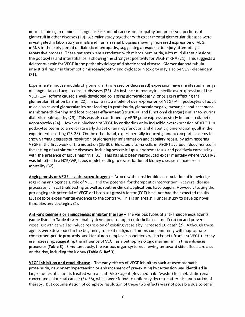
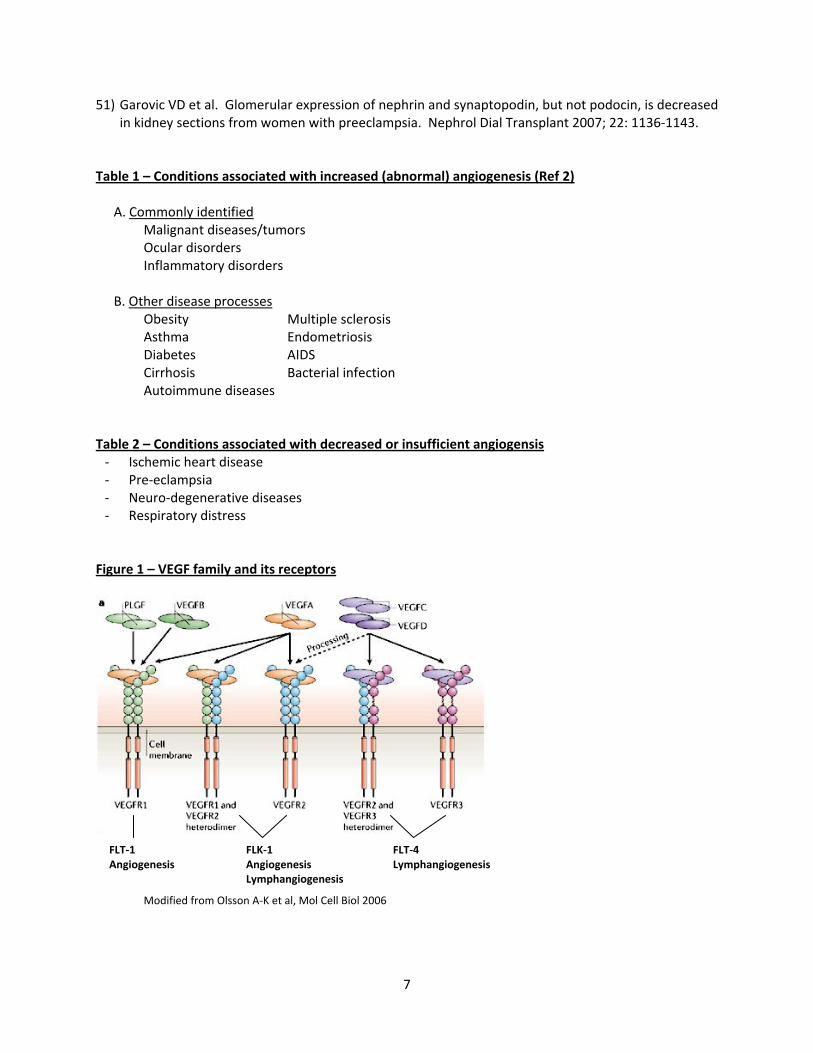
The growth or the formation of new blood vessels, known as “angiogenesis,” in the normal physiologic setting is required for organ growth in the embryonic phase and also for repair of wounded tissue in the adult. However, an imbalance in the growth of blood vessels has contributed to the pathogenesis of many diseases (Table 1, Table 2). The key mechanisms and molecular players are only now being postulated, investigated and studied during the last couple of decades (2). Thus both pro‐angiogenic and anti‐angiogenic factors are important in maintaining this vascular homeostasis (3). The presence of “diffusible” growth factors stimulating neovascularization in tumors and as a means of communication between the tumor cells and vascular endothelial cells was first proposed by J. Folkman in 1971 (4). Later, it was also shown that normal tissues are also a source of angiogenic activity, particularly the Vascular Endothelial Growth Factor (VEGF) family of proteins. Other molecules which have participated as positive regulators of angiogenesis include fibroblast growth factor (FGF), transforming growth factor (TGF – alpha and beta), hepatocyte growth factor (HGF), tumor necrosis factor – alpha (TNF), angiogenin, interleukin (IL)‐8 and the angiopoietius (5). VEGF – VEGF and related proteins in this gene family are dimeric (usually homodimers) gylcoproteins of about 40kDa and are similar to the PDGF family of growth factors (Fig 1). They are VEGFA, B, C, D and placenta growth factor (PlGF) (5, 6). However, the prototype member of this family is VEGFA, well known as a key regulator in both physiological and pathological angiogenesis. VEGFC and VEGFD are involved in the regulation of another anatomic component of the vascular system, lymphatic angiogenesis. From the human VEGFA gene, four different isoforms have been generated following alternative exon splicing depending on the number of amino acids after signal sequence cleavage (VEGF121, VEGF165, VEGF189, VEGF206). While VEGF121 resembles the native VEGF, the others differ in their physicochemical properties and the responses elicited following receptor binding (5). All the VEGF ligands (A, B, C, D, PlGF) bind in an overlapping pattern to 3 VEGF receptors, which are receptor tyrosine kinases (VEGFR‐1 or Flt‐1, VEGFR‐2 or Flk‐1 and VEGFR‐3 [fms‐like‐tyrosine kinase or Flt‐4)]). The last one is not a receptor for VEGF, but bind to VEGFC and VEGFD. VEGF also interacts with a family of co‐
2
receptors, the neuropilins on the EC membrane. These VEGF receptors are primarily expressed on EC or the vascular system suggesting that VEGF/VEGFRs major site of function is in the vascular system. The VEGFR (VEGFR‐1 and VEGFR‐2) activity is regulated by the availability of the ligands as well as hypoxia, directly or indirectly by stabilization of the hypoxia inducible factors (HIFs) which bind to specific promoter elements of VEGFA (6). Activities of VEGF – VEGF can promote growth of EC from various vascular sources in vitro as well as induce a potent angiogenic response in vivo (5). It also serves as a survival factor for EC and induces anti‐apoptotic proteins Bcl‐2 and A1 in endothelial cells. Apart from its varied effect on bone marrow‐derived cells, by virtue of its ability to induce vascular leakage, VEGF is also known as a vascular permeability factor, thus participating in inflammation and other pathological conditions (7). Endothelial fenestration in some organ vascular beds and vasodilatation via EC derived nitric oxide are also induced by VEGF (8, 9). A more complete list of the role of VEGF in physiological angiogenesis and pathologic conditions in several organ systems is found in Refs 2 and 5. Both VEGF and its receptors are continuously secreted and expressed respectively from/in ECs of many vascular adult tissues as well as several glands, cardiac and skeletal myocytes, liver and spleen (10). VEGF and the Kidney – Due to the diverse functions of the EC and their dependency on a variety of activation mechanisms, the vascular endothelium in the kidney plays a vital role in modulation of renal function and not merely perceived as a passive cellular barrier in the filtration apparatus and coagulation. It has been shown that VEGF dependant metanephric endothelial differentiation may occur from extrarenal mesenchymal vessels by angiogenesis or partly from angioblasts by vasculogenesis, particularly in the presence of threshold levels of VEGF during development (11). This has been borne out by numerous experimental models including VEGF knockout mice. In addition, A. Tufro has shown that VEGF produced by the differentiating nephrons acts as a chemoattractant, providing spatial direction to developing capillaries toward forming nephrons during metanephric development in vitro (11). VEGF is generally expressed in the glomerular podocytes and tubular epithelial cells (proximal, distal and collecting ducts) and VEGF receptors are localized in the mesangium, glomerular and peritubular capillaries (10). By double‐label immunohistochemistry, VEGF receptor proteins were identified in the endothelial cells of preglomerular, glomerular and post glomerular vessels in the adult, while the VEGF receptor protein appeared as soon as endothelial cells expressed von Willebrand factor in the embryonic stage. The affinity factor in adults and fetal kidneys for radiolabeled I125‐VEGF was similar within the vasculature, glomeruli and cortical tubulo‐interstitium. A high density of VEGFR‐1 or Flt‐1 receptor binding were demonstrated in the normal kidney emphasizing the need for VEGF in the adult kidney for other than angiogenesis (12). In addition to its role in the maintenance of glomerular endothelial cells, glomerular VEGF is increased in response to hypertension and activation of the renin‐angiogensin system in the experimental setting, thus showing a protective role for VEGF signaling in stressful vascular conditions (13). Table 3 shows participation of VEGF and its receptors in normal physiological functions of the kidney. Abnormal VEGF expression in Renal disease – The expression of VEGF and VEGF mRNA was studied by in situ hybridization and immunohistochemistry in renal biopsies with a variety of glomerular diseases in 47 cases (20). In most of the chronic glomerular lesions, there were decreased cells expressing VEGF such as focal or global glomerulosclerosis, amyloidosis, advanced diabetic nephropathy, crescentic glomerulonephritis and diffuse lupus nephritis. Some of these lesions also lead to alterations of glomerular endothelial function as it is dependent on adequate secretion of VEGF. However, there was
3
normal staining in minimal change disease, membranous nephropathy and preserved portions of glomeruli in other diseases (20). A similar study together with experimental glomerular diseases were investigated in laboratory animals and human renal biopsies showing increased expression of VEGF mRNA in the early period of diabetic nephropathy, suggesting a response to injury attempting a reparative process. These patients were associated with microalbuminuria, with mild diabetic lesions, the podocytes and interstitial cells showing the strongest positivity for VEGF mRNA (21). This suggests a deleterious role for VEGF in the pathophysiology of diabetic renal disease. Glomerular and tubulo‐interstitial repair in thrombotic microangiopathy and cyclosporin toxicity may also be VEGF‐dependant (21). Experimental mouse models of glomerular (increased or decreased) expression have manifested a range of congenital and acquired renal diseases (22). An instance of podocyte‐specific overexpression of the VEGF‐164 isoform caused a well‐developed collapsing glomerulopathy, once again affecting the glomerular filtration barrier (22). In contrast, a model of overexpression of VEGF‐A in podocytes of adult mice also caused glomerular lesions leading to proteinuria, glomerulomegaly, mesangial and basement membrane thickening and foot process effacement (structural and functional changes) similar to murine diabetic nephropathy (23). This was also confirmed by VEGF gene expression study in human diabetic nephropathy (24). However, blockade of VEGF by antibodies or by inducible overexpression of sFLT‐1 in podocytes seems to ameliorate early diabetic renal dysfunction and diabetic glomerulopathy, all in the experimental setting (25‐28). On the other hand, experimentally induced glomerulonephritis seems to show varying degrees of resolution of glomerular inflammation and capillary repair, by administering VEGF in the first week of the induction (29‐30). Elevated plasma cells of VEGF have been documented in the setting of autoimmune diseases, including systemic lupus erythematosus and positively correlating with the presence of lupus nephritis (31). This has also been reproduced experimentally where VEGFR‐2 was inhibited in a NZB/WF, lupus model leading to exacerbation of kidney disease in increase in mortality (32). Angiogenesis or VEGF as a therapeutic agent – Armed with considerable accumulation of knowledge regarding angiogenesis, role of VEGF and the potential for therapeutic intervention in several disease processes, clinical trials testing as well as routine clinical applications have begun. However, testing the pro‐angiogenic potential of VEGF or fibroblast growth factor (FGF) have not had the expected results (33) despite experimental evidence to the contrary. This is an area still under study to develop novel therapies and strategies (2). Anti‐angiogenesis or angiogenesis inhibitor therapy – The various types of anti‐angiogenesis agents (some listed in Table 4) were mainly developed to target endothelial cell proliferation and prevent vessel growth as well as induce regression of existing vessels by increased EC death (2). Although these agents were developed in the beginning to treat malignant tumors concomitantly with appropriate chemotherapeutic protocols, additional non‐neoplastic conditions which benefit from antiVEGF therapy are increasing, suggesting the influence of VEGF as a pathophysiologic mechanism in these disease processes (Table 5). Simultaneously, the various organ systems showing untoward side effects are also on the rise, including the kidney (Table 6, Ref 3). VEGF inhibition and renal disease – The early effects of VEGF inhibitors such as asymptomatic proteinuria, new onset hypertension or enhancement of pre‐existing hypertension was identified in large studies of patients treated with an anti‐VEGF agent (Bevacizumab, Avastin) for metastatic renal cancer and colorectal cancer (34‐36), which were found to uniformly decrease after discontinuation of therapy. But documentation of complete resolution of these two effects was not possible due to other
4
patient variables including death. Proteinuria is directly as a result of depletion of podocyte VEGF during antitumor therapy leading to down regulation of podocyte tight junction protein, while hypertension is thought to be nitric oxide dependent increase in risk of proteinuria and hypertension in these patients (37, 38). Induction of proteinuria was also shown earlier in mice by neutralization of circulating vascular endothelial growth factor by anti‐VEGF antibodies and soluble VEG receptor 1 (sFlt1) (39). Varying degrees of proteinuria up to nephrotic syndrome is also attributed to glomerular endothelial injury, thrombotic microangiopathy and rarely immune complex glomerulonephritis including IgA deposits by renal biopsies (40‐42). Acute kidney injury and isolated acute or chronic thrombotic microangiopathy associated with anti‐VEGF therapy in anecdotal cases has been reported following renal biopsies (43‐45). Acute kidney injury is usually reversible and these patients are rarely biopsied. Thus, anti‐VEGF therapy should be included as yet another therapeutic agent causing drug‐induced TMA in both neoplastic and non‐neoplastic settings (46‐47). Role of VEGF in preeclampsia – Preeclampsia is a serious complication of pregnancy associated with significant maternal and fetal morbidity and mortality. It is defined as a syndrome of new onset hypertension and proteinuria of varying severity during the last trimester of pregnancy, often accompanied by edema and hyperuricemia along with an abnormal placenta (48). The severe form may manifest systemic endothelial dysfunction and microangiopathy affecting target organs such as brain, liver or kidney. Proteinuria ranges from subnephrotic to nephrotic range with no significant urinary sediment and usually disappears following termination of pregnancy. Renal insufficiency is rare, unless there is severe hypertension and thrombotic microangiopathy. A striking renal pathological finding in the setting of preeclampsia is known as “glomerular capillary endotheliosis.” This is characterized by endothelial and mesangial swelling with minimal or no increase in cellularity. The overlying epithelial cells may also appear swollen. The glomeruli appear bloodless and mildly enlarged. While immunofluorescence microscopy may reveal only focal glomerular fibrin deposits in the acute or early cases, the electron microscopic findings confirm endotheliosis showing endothelial swelling, loss of fenestrae and no significant loss of epithelial foot processes. Focal subendothelial widening and accumulation of fibrin‐like protein with varying degrees of mesangial interposition in the healing stages and focal segmental sclerosis may be noted. Until about 8‐10 years ago, the pathogenesis was not known. Recent observations have demonstrated that placental soluble fms‐like tyrosine kinase 1 (sFlt‐1), an inhibitor of VEGF and placental growth factor, most probably elaborated by abnormal placenta may participate in the pathogenesis of pre‐eclampsia (49, 50). They found significantly lower levels of placental growth factor in circulation, thus leading to depletion of VEGF in the glomeruli and other organs. This could result in dysregulation of nephrin and synaptopodin in the podocyte foot processes and the filtration barrier causing proteinuria (51). References 1) Jaffe EA. Cell biology of endothelial cells. Hum Pathol 1987; 18(3): 234‐239. 2) Carmeliet P. Angiogenesis in life, disease and medicine. Nature 2005; 438(7070): 932‐936. 3) Gurevich F, Perazella MA. Renal effects of anti‐angiogenesis therapy: update for the internist. Am J
Med 2009; 122: 322‐328. 4) Folkman J. Tumor angiogenesis. Therapeutic implications. N Engl J Med 1971; 285: 1182‐1186. 5) Ferrara N, Gerber H‐P, LeCouter J. The biology of VEGF and its receptors. Nat Med 2003; 9(6): 669‐
676.
5
6) Olsson A‐K, Dimberg A, Kreuger J, Claesson‐Welsh L. VEGF receptor signalling – in control of vascular function. Nat Rev Mol Cell Biol 2006; 7(5): 359‐371.
7) Dvorak HF et al. Vascular permeability factor/vascular endothelial growth factor, microvascular hyperpermeability, and angiogenesis. Am J pathol 1995; 146: 1029‐1039.
8) Roberts WG, Palade GE. Increased microvascular permeability and endothelial fenestration induced by vascular endothelial growth factor. J Cell Sci 1995; 108: 2369‐2379.
9) Ku DD et al. Vascular endothelial growth factor induces EDRF‐dependent relaxation in coronary arteries. Am J Physiol 1993; 265: H586‐H592.
10) Maharaj ASR et al. Vascular endothelial growth factor localization in the adult. Am J Pathol 2006; 168(2): 639‐648.
11) Tufro A. VEGF spatially directs angiogenesis during metanephric development in vitro. Dev Biol 2000; 227: 558‐566.
12) Simon M et al. Receptors of vascular endothelial growth factor/vascular permeability factor (VEGF/VPF) in fetal and adult human kidney: localization and [125I] VEGF binding sites. J Am Soc Nephrol 1998; 9(6): 1032‐1044.
13) Advani A et al. Role of VEGF in maintaining renal structure and function under normotensive and hypertensive conditions. Proc Natl Acad Sci USA 2007; 104(36): 14448‐14453.
14) Wakelin SJ et al. The role of vascular endothelial growth factor in the kidney in health and disease. Nephron Physiol 2004; 98: 73‐79.
15) Eremina V, Baelde HJ, Quaggin WE. Role of the VEGF‐A signaling pathway in the glomerulus: evidence for crosstalk between components of the glomerular filtration barrier. Nephron Physiol 2007; 106: 32‐37.
16) Guan F et al. Autocrine VEGF‐A system in podocytes regulates podocin and its interaction with CD2AP. Am J Physiol Renal Physiol 2006; 291: F422‐F428.
17) Kanellis J et al. Vascular endothelial growth factor is a survival factor for renal tubular epithelial cells. Am J Phsiol Renal Physiol 2000; 278: F905‐F915.
18) Pepper MS et al. Vascular endothelial growth factor (VEGF) induces plasminogen activators and plasminogen activator inhibitor‐1 in microvascular endothelial cells. Biochem Biophys Res Commun 191; 181: 902‐906.
19) Foster RR et al. Functional evidence that vascular endothelial growth factor may act as an autocrine factor on human podocytes. Am J Physiol Renal Physiol 2003; 284(6): F1263‐73.
20) Shulman K et al. Expression of vascular permeability factor (VPF/VEGF) is altered in many glomerular diseases. J Am Soc Nephrol 1996; 7: 661‐666.
21) Schrijvers BF, Flyvbjerg A, De Vriese AS. The role of vascular endothelial growth factor (VEGF) in renal pathophysiology. Kidney Int 2004; 65: 2003‐2017.
22) Eremina V et al. Glomerular‐specific alterations of VEGF‐A expression lead to distinct congenital and acquired renal diseases. J Clin Invest 2003; 111: 707‐716.
23) Veron D et al. Overexpression of VEGF‐A in podocytes of adult mice causes glomerular disease. Kidney Int 2010; 77: 989‐999.
24) Kanesaki Y et al. Vascular endothelial growth factor expression is correlated with glomerular neovascularization in human diabetic nephropathy. Am J Kid Dis 2005; 45(2): 288‐294.
25) De Vriese AS et al. Antibodies against vascular endothelial growth factor improve early renal dysfunction in experimental diabetes. J Am Soc Nephrol 2001; 12: 993‐1000.
26) Flyvbjerg A et al. Amelioration of long‐term renal changes in obese type 2 diabetic mice by a neutralizing vascular endothelial growth factor antibody. Diabetes 2002; 51: 3090‐3094.
27) Sung SH et al. Blockade of vascular endothelial factor signaling ameliorates diabetic albuminuria in mice. J Am Soc Nephrol 2006; 17: 3093‐3104.
6
28) Ku C‐H et al. Inducible overexpression of sFlt‐1 in podocytes ameliorates glomerulopathy in diabetic mice. Diabetes 2008; 57: 2824‐2833.
29) Masuda Y. Vascular endothelial growth factor enhances glomerular capillary repair and accelerates resolution of experimentally induced glomerulonephritis. Am J Pathol 2001; 159: 599‐608.
30) Shimizu A et al. Vascular endothelial growth factor165 resolves glomerular inflammation and accelerates glomerular capillary repair in rat anti‐glomerular basement membrane glomerulonephritis. J Am Soc Nephrol 2004; 15: 2655‐2665.
31) Navarro C et al. Vascular endothelial growth factor plasma levels in patients with systemic lupus erythematosus and primary antiphospholipid syndrome. Lupus 2002; 11: 21‐24.
32) Watanabe et al. Anti‐vascular endothelial growth factor receptor‐2 antibody accelerates renal disease in the NZB/W F1 murine systemic lupus erythematosus model. Clin Cancer Res 2005; 11(1): 407‐409.
33) Simon M. Angiogenesis: where do we stand now? Circulation 2005; 111, 1556‐1566. 34) Yang JC et al. A randomized trial of bevacizumab, an anti‐vascular endothelial growth factor
antibody, for metastatic renal cancer. N Eng J Med 2003; 349: 427‐434. 35) Hurwitz H et al. Bevacizumab plus irinotecan, fluorouracil, and leucovorin for metastatic colorectal
cancer. N Eng J Med 2004; 350: 2335‐2342. 36) Ferrara N, Hillan KJ, Novotny W. Bevacizumab (avastin), a humanized anti‐VEGF monoclonal
antibody for cancer therapy. Biochem Biophys Res Commun 2005; 333(2): 328‐335. 37) Izzedine H et al. Angiogenesis inhibitor therapies: focus on kidney toxicity and hypertension. Am J
Kid Dis 2007; 50(2): 203‐218. 38) Zhu X et al. Risks of proteinuria and hypertension with bevacizumab, an antibody against vascular
endothelial growth factor: systematic review and meta‐analysis. 39) Sugimoto H et al. Neutralization of circulating vascular endothelial growth factor (VEGF)l l by anti‐
VEGF antibodies and soluble VEGF receptor 1 (sFlt‐1) induces proteinuria. J Biol Chem 2003; 278(15): 12605‐12608.
40) George BA, Zhou XJ, Toto R. Nephrotic syndrome after bevacizumab: case report and literature review. Am J Kid Dis 2007; 49(2): E23‐E29.
41) Roncone D et al. Proteinuria in a patient receiving anti‐VEGF therapy for metastatic renal cell carcinoma. Nat Clin Pract Nephrol 2007; 3(5): 287‐293.
42) Stokes MB, Erazo MC, D’Agati VD. Glomerular disease related to anti‐VEGF therapy. Kidney Int 2008; 74(11): 1487‐1491.
43) Izzedine H et al. Thrombotic microangiopathy and anti‐VEGF agents. Nephrol Dial Transplant 2007; 22(5): 1481‐1482.
44) Frangie C et al. Renal thrombotic microangiopathy caused by anti‐VEGF antibody treatment for metastatic renal‐cell carcinoma. Lancet Oncol 2007; 8(2): 177‐178.
45) Eremina V et al. VEGF inhibition and renal thrombotic microangiopathy. N Eng J Med 2008; 358(11): 1129‐1136.
46) Dlott JS et al. Drug‐induced thrombotic thrombocytopenic purpura/hemolytic uremic syndrome: a concise review. Ther Apher Dial 2004; 8(2): 102‐111.
47) Zakarija A, Bennett C. Drug‐induced thrombotic microangiopathy. Semin Thromb Hemost 2005; 31(6): 681‐690.
48) Karumanchi SA et al. Preeclampsia: a renal perspective. Kidney Int 2005; 67(6): 2101‐2113. 49) Maynard SE et al. Excess placental soluble fms‐like tyrosine kinase 1 (sFlt1) may contribute to
endothelial dysfunction, hypertension and proteinuria in preeclampsia. J Clin Invest 2003; 111: 649‐658.
50) Levine RJ et al. Circulating angiogenic factors and the risk of preeclampsia. N Engl J Med 2004; 350(7): 672‐683.
7
51) Garovic VD et al. Glomerular expression of nephrin and synaptopodin, but not podocin, is decreased in kidney sections from women with preeclampsia. Nephrol Dial Transplant 2007; 22: 1136‐1143.
Table 1 – Conditions associated with increased (abnormal) angiogenesis (Ref 2) A. Commonly identified Malignant diseases/tumors Ocular disorders Inflammatory disorders B. Other disease processes
Obesity Multiple sclerosis Asthma Endometriosis Diabetes AIDS Cirrhosis Bacterial infection Autoimmune diseases
Table 2 – Conditions associated with decreased or insufficient angiogensis ‐ Ischemic heart disease ‐ Pre‐eclampsia ‐ Neuro‐degenerative diseases ‐ Respiratory distress
Figure 1 – VEGF family and its receptors
Modified from Olsson A‐K et al, Mol Cell Biol 2006
FLT‐1 Angiogenesis
FLK‐1 Angiogenesis Lymphangiogenesis
FLT‐4 Lymphangiogenesis
8
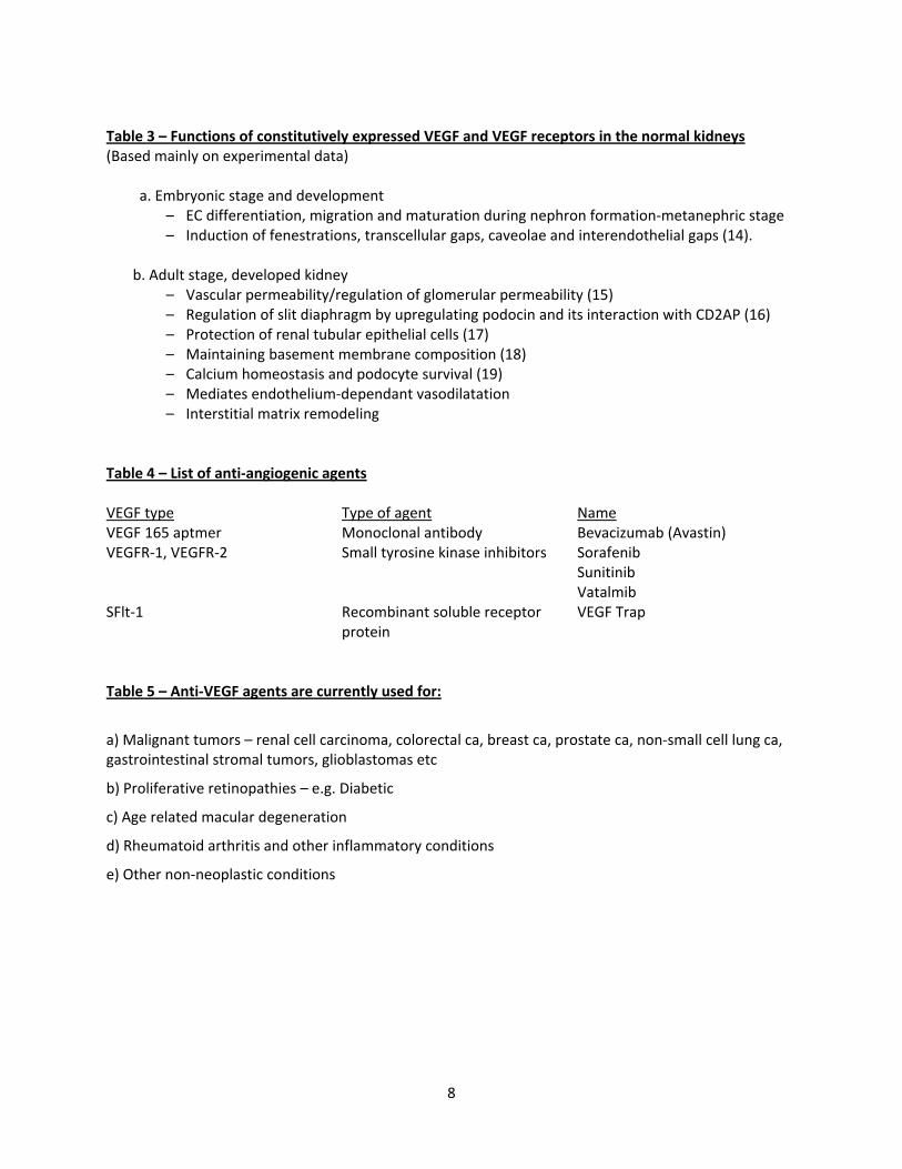
Table 3 – Functions of constitutively expressed VEGF and VEGF receptors in the normal kidneys (Based mainly on experimental data)
a. Embryonic stage and development – EC differentiation, migration and maturation during nephron formation‐metanephric stage – Induction of fenestrations, transcellular gaps, caveolae and interendothelial gaps (14).
b. Adult stage, developed kidney – Vascular permeability/regulation of glomerular permeability (15) – Regulation of slit diaphragm by upregulating podocin and its interaction with CD2AP (16) – Protection of renal tubular epithelial cells (17) – Maintaining basement membrane composition (18) – Calcium homeostasis and podocyte survival (19) – Mediates endothelium‐dependant vasodilatation – Interstitial matrix remodeling
Table 4 – List of anti‐angiogenic agents VEGF type Type of agent Name VEGF 165 aptmer Monoclonal antibody Bevacizumab (Avastin) VEGFR‐1, VEGFR‐2 Small tyrosine kinase inhibitors Sorafenib
Sunitinib Vatalmib
SFlt‐1 Recombinant soluble receptor protein
VEGF Trap
Table 5 – Anti‐VEGF agents are currently used for:
a) Malignant tumors – renal cell carcinoma, colorectal ca, breast ca, prostate ca, non‐small cell lung ca, gastrointestinal stromal tumors, glioblastomas etc
b) Proliferative retinopathies – e.g. Diabetic
c) Age related macular degeneration
d) Rheumatoid arthritis and other inflammatory conditions
e) Other non‐neoplastic conditions
9
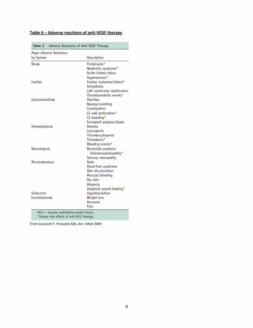
Table 6 – Adverse reactions of anti‐VEGF therapy
From Gurevich F, Perazella MA, Am J Med 2009
10
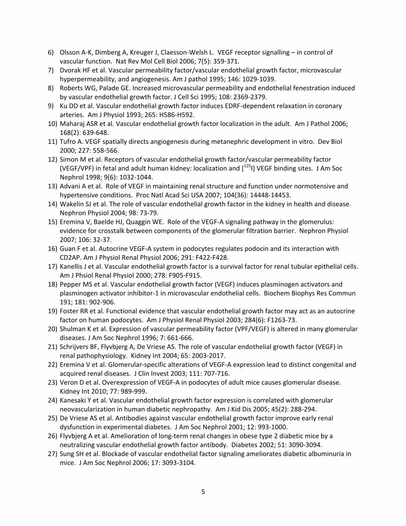
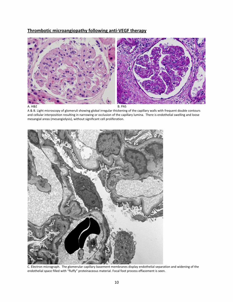
Thrombotic microangiopathy following anti‐VEGF therapy
A. H&E B. PAS A & B. Light microscopy of glomeruli showing global irregular thickening of the capillary walls with frequent double contours and cellular interposition resulting in narrowing or occlusion of the capillary lumina. There is endothelial swelling and loose mesangial areas (mesangiolysis), without significant cell proliferation.
C. Electron micrograph. The glomerular capillary basement membranes display endothelial separation and widening of the endothelial space filled with “fluffy” proteinaceous material. Focal foot process effacement is seen.
11
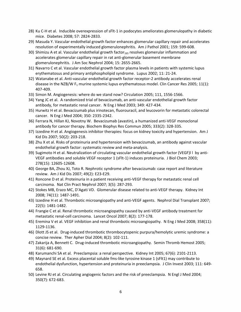
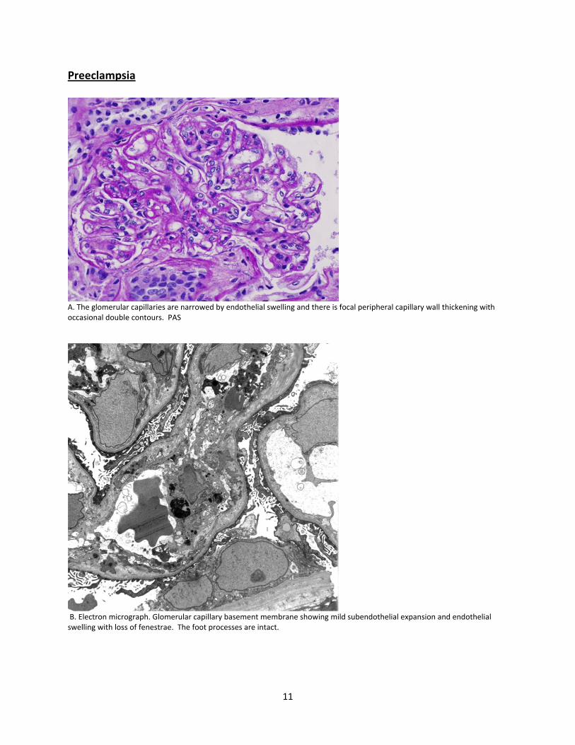
Preeclampsia
A. The glomerular capillaries are narrowed by endothelial swelling and there is focal peripheral capillary wall thickening with occasional double contours. PAS
B. Electron micrograph. Glomerular capillary basement membrane showing mild subendothelial expansion and endothelial swelling with loss of fenestrae. The foot processes are intact.