Embed Size (px)
Citation preview
The Pavlik Harness in the Treatment of Congenital Dislocating Hip: Report on a Multicenter Study of the European Paediatric Orthopaedic Society F. Grill, M.D., *H. Bensahel, M.D., †J. Cañadell, M.D., ‡P. Dungl, M.D., ¶T. Matasovic, M.D., and §T. Vizkelety, M.D. Department of Pediatric Orthopedics Speising Hospital Vienna, Austria; †University Clinic of Navarra, Pamplona, Spain; *Department of Pediatric Orthopedics, Hospital Bretonneau, Paris, France; ‡Bulovka Hospital, Prague, Czechoslovakia; ¶Orthopedic Clinic of Zagreb University, Yugoslavia; and §Orthopedic University Clinic, Budapest, Hungary SUMMARY The results of functional treatment using the Pavlik harness in congenital dislocation and congenital dysplasia of the hip in children aged <11 months were examined by an EPOS study group. This study was conducted on 3,611 hips in 2,636 patients for a period of 1-9 years after treatment. The reduction rate was 92% in grade Tonnis 2 and 3; the healing rate was 80%. In children with dysplastic hips, the healing rate was 95.35%. Avascular necrosis of the femoral head was observed in 2.38%. The Pavlik harness is designed for outpatient treatment if the parents are compliant. KEY WORDS Congenital hip dysplasia—Hip dislocation—Pavlik harness —Treatment guideline.
Address correspondence and reprint requests to Dr. F. Grill at Department of Pediatric Orthopedics, Speising-Hospital, Speisinger StraBe 109, A-1134, Vienna, Austria.
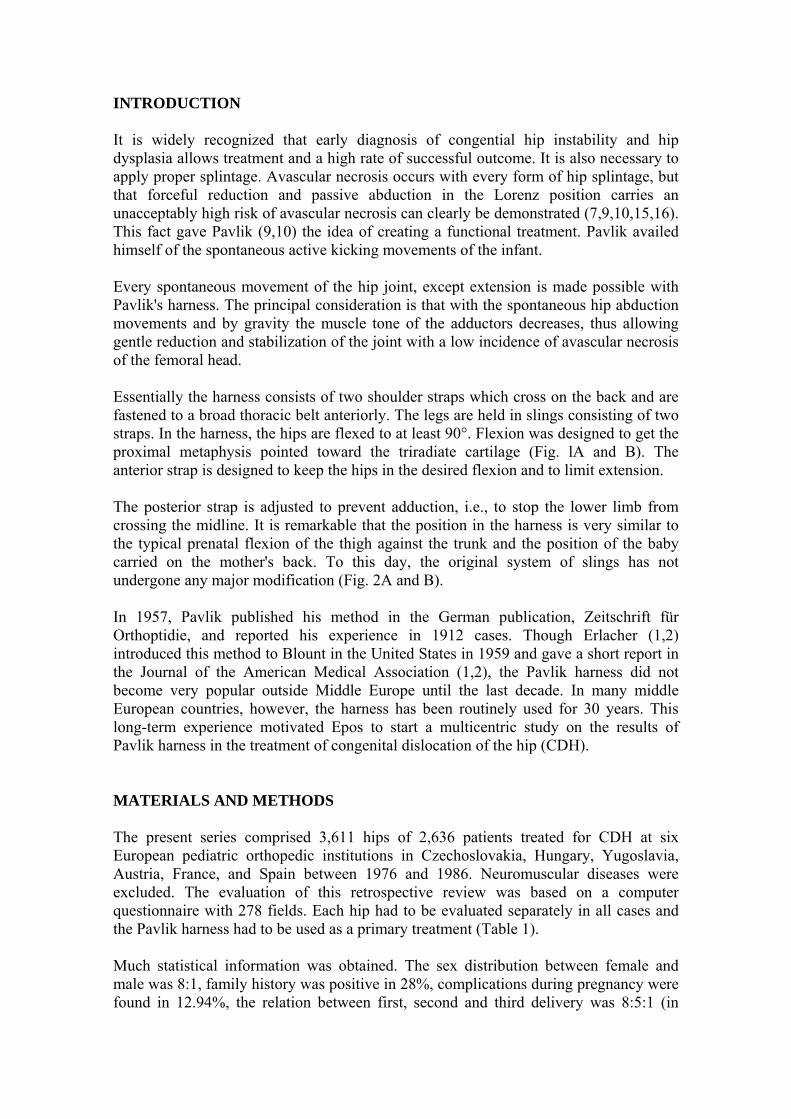
INTRODUCTION It is widely recognized that early diagnosis of congential hip instability and hip dysplasia allows treatment and a high rate of successful outcome. It is also necessary to apply proper splintage. Avascular necrosis occurs with every form of hip splintage, but that forceful reduction and passive abduction in the Lorenz position carries an unacceptably high risk of avascular necrosis can clearly be demonstrated (7,9,10,15,16). This fact gave Pavlik (9,10) the idea of creating a functional treatment. Pavlik availed himself of the spontaneous active kicking movements of the infant. Every spontaneous movement of the hip joint, except extension is made possible with Pavlik's harness. The principal consideration is that with the spontaneous hip abduction movements and by gravity the muscle tone of the adductors decreases, thus allowing gentle reduction and stabilization of the joint with a low incidence of avascular necrosis of the femoral head. Essentially the harness consists of two shoulder straps which cross on the back and are fastened to a broad thoracic belt anteriorly. The legs are held in slings consisting of two straps. In the harness, the hips are flexed to at least 90°. Flexion was designed to get the proximal metaphysis pointed toward the triradiate cartilage (Fig. lA and B). The anterior strap is designed to keep the hips in the desired flexion and to limit extension. The posterior strap is adjusted to prevent adduction, i.e., to stop the lower limb from crossing the midline. It is remarkable that the position in the harness is very similar to the typical prenatal flexion of the thigh against the trunk and the position of the baby carried on the mother's back. To this day, the original system of slings has not undergone any major modification (Fig. 2A and B). In 1957, Pavlik published his method in the German publication, Zeitschrift für Orthoptidie, and reported his experience in 1912 cases. Though Erlacher (1,2) introduced this method to Blount in the United States in 1959 and gave a short report in the Journal of the American Medical Association (1,2), the Pavlik harness did not become very popular outside Middle Europe until the last decade. In many middle European countries, however, the harness has been routinely used for 30 years. This long-term experience motivated Epos to start a multicentric study on the results of Pavlik harness in the treatment of congenital dislocation of the hip (CDH). MATERIALS AND METHODS The present series comprised 3,611 hips of 2,636 patients treated for CDH at six European pediatric orthopedic institutions in Czechoslovakia, Hungary, Yugoslavia, Austria, France, and Spain between 1976 and 1986. Neuromuscular diseases were excluded. The evaluation of this retrospective review was based on a computer questionnaire with 278 fields. Each hip had to be evaluated separately in all cases and the Pavlik harness had to be used as a primary treatment (Table 1). Much statistical information was obtained. The sex distribution between female and male was 8:1, family history was positive in 28%, complications during pregnancy were found in 12.94%, the relation between first, second and third delivery was 8:5:1 (in
CDH children first delivery was found in 57.12%), and siblings were found in 1.06%. Mean length at birth was 49.36 cm, and mean weight at birth was 3329.68 g. The distribution of CDH according to month of birth showed two not very significant peaks during winter and summer. 56.17% of all babies with CDH were born in January, February, March, July, August, and September. The classification of associated deformities in this multicenter study was not without problems and cannot be assumed to be objective; e.g., foot deformities were found in a range between 3.5 and 39%. For this retrospective review, the diagnosis of hip abnormality as well as consequent evaluations were based on clinical and roentenographic criteria. Clinical evidence was based only on hip instability, which was found in 32.6% of all hips. Limitation of abduction, asymmetric thigh fold, and leg length discrepancy were not registered because of their slight diagnostic value. Radiographs or sonography were taken before treatment in all cases. Radiographic observations included Acetabular Index angle and, for CDH classification, the degrees according to Tönnis (15,16) as follows: grade 0, normal hip; grade 1, ossification center of the capital epiphysis is medial to Perkin's line; grade 2, ossification center of the capital epiphysis is lateral to Perkin's line but below the superolateral margin of the acetabulum; grade 3, ossification center is at the level of the superolateral margin of the acetabulum; and grade 4, ossification center is aboye the level of the superolateral margin of the acetabulum. The average AC angle before treatment was 39.57° in the first month, 37.67° in the second, 35.98° in the third, and 34.13° in the fourth. In several cases, sonography was used and classified according to Graf (4). For evaluation reasons we found grade Tönnis 1 and Graf 2a - , 2b, and 2g to be similar as were grade Tönnis 2 and Graf 2d, 3a, and 3b. We graded Graf 4 degree equal to Tönnis 3. This correlation made it possible to evaluate our material in one block. Thus, separate evaluation of roentgenogram and sonography cases could be avoided. One thousand ninety-two hips were grade Tönnis 1, 1,698 hips were grade Tönnis 2,742 hips were grade Tönnis 3, and 79 hips were grade Tönnis 4. All four Tönnis groups were evaluated separately. The average age at the time of the initial treatment was 4.1 months (range 2 days to 11 months). In most patients the harness was used as described by Pavlik. It was, however, individually adapted to allow free movement of the limb within the safety zone of Ramsey et al. (11), avoiding adduction <35°, which may cause dislocation, or abduction >75°, which eventually leads to avascular necrosis. This latter complication is prevented if overstress of the posterior strap of the harness is avoided or if small pillows are placed under the knees of a supine baby. All physicians used a range of hip flexion of 80 and 110° in the harness. Some started routinely with an intermediate degree of flexion of ~60° and increased it slowly, tightening the anterior strap to 110° within the first 2 weeks. The argument for this procedure is that the baby and the hip should have time to accept the therapeutic position of the harness (Fig. 3).

In children aged >3 months with rather fatty morphology in which the hip was in adduction and lateral rotation, Bensahel found that the medial side of the thigh was compressed by the anteromedial strap, which produced edema and pain at the thigh and the inguinal region. To solve this problem, he recommends adding a third bandage under the patella on the anterior part of the knee (Fig. 4A). This bandage leads the anterior medial strap laterally, thus preventing it from being excessively straight (Fig. 4B). All physicians kept the harness on dislocated hips at all times during the first 4-6 weeks. During that time the harness was not even removed while the child was being washed. To give the mother time to adjust to handling the harness, during the first days of treatment Grill allowed the harness to be kept on only as long as mother and child tolerated it. All considered the mother's compliance very important; therefore, follow-ups were very frequent and regular. At first, follow-ups were weekly. After 6 weeks, the child was seen at intervals of 2 or 3 weeks. All but two physicians used the Pavlik harness on an outpatient basis. Hospitalization was exceptional and mostly used only for sociologic reasons. Bensahel prepared all his CDH patients systematically with vertical skin-zenith traction at first, then traction in slight abduction (~30°) to make their muscles supple, using onetenth of body weight. After traction, the harness was fitted and kept on until healing occurred. Open reduction of dislocated hips previously treated by overhead traction with stretched knees often shows soft tissue edema and hydrops of the hip joint. Today, this can also easily be demonstrated by sonography. By traction in stretched knee position, spontaneous kicking movements of the child are probably hindered and hamstring tension probably reduces hip reduction and increases the risk of avascular necrosis. Hip reduction by conservative treatment should not be a one-step action but a repeated processs stimulated by spontaneous kicking movements of the leg in hip abduction until the hip becomes stable, thus avoiding avascular necrosis. This can be achieved best by a method that allows traction of the upper thigh in flexed knee position as well as kicking movements. Therefore, in grade Tönnis 3 cases with limitation of abduction due to severe adductor tightness, Grill, inspired by Wientroub, used the Pavlik harness in combination with upper thigh skin traction (Fig. 5). After clinical reduction, all cases treated by this method were controlled by arthrography. If reduction had taken place and no soft tissue interposition could be found, treatment was continued with the Pavlik harness until healing occurred. For the entire study, once a stable concentric reduction of the dislocated hip was obtained (i.e., the hip did not slide out of the socket on gentle examination), the rest of the harness treatment program was essentially the same as for dysplastic hips. Clinical reduction was confirmed by roentgenogram. The roentgenograms were done every 3 months until the end of treatment. The last roentgenogram was taken at the time of follow-up. In all cases, the treatment program was considered complete when radiographs of the hips were normal. The average length of time during which the harness was worn permanently was 6.3 months and depended on severity of the disease and age at onset of therapy. Only those children who could be followed for at least 1 year after the beginning of treatment have been included in the present series.
RESULTS All patients were followed for >1 year with an average follow-up of 4.46 years (range 1-8.9 years). Roentgenographic analysis at follow-up included measurement of AC angles, Ullmann Sharp angles, center edge angles, migration percentage according to Reimers and degree of dislocation according to Tönnis. The ossification centers of the femoral heads were assessed with regard to the time of appearance and the degree of avascular necrosis seen at its maximal development according to Tönnis: 1. Mildest degree of alteration: The ossification center presents a somewhat
irregular contour and its structure is slightly granular and irregular; the alterations probably subside without consequences.
2. Distinct alterations of structure occur, but without fragmentation: a subtype included in this grade presents punched-out defects of the femoral head, often recognizable as small lateral notches in the surface of the head.
3. The capital center of ossification is broken into pieces and is recognizable only by small fragmente or as a fíat strip; very small centers may disintegrate completely. Marked deformation of the femoral head and neck remains throughout life. According to Salter (also grade 3): no nucleus is present and there is no growth of the nucleus for 1 year.
The course of treatment was recorded accurately, especially with regard to time of reposition, failure of treatment, change of therapy, complications, and additional surgical interventions. All four Tönnis groups were evaluated separately. Grade Tönnis 1 There were 1,092 hips. Age at beginning of walking was normal. The AC angle averaged 31° before and 17.4° (5-29°) at follow-up. The center edge (CE) angle averaged 23.5° (13-32°). The migration index was 6.1 (0-27°). The ossification center appeared within 5.4 months. Fifty-seven hips showed fragmentation of the femoral head, 42 showed grade 1, 12 showed grade 2, and 2 showed grade 3. If grade 1 is disregarded, the rate of avascular necroses in this group was 1.28%. There were no other complications. According to Tönnis the healing rate at follow-up was 95.35%. Slight dysplasia was apparent in 4.65%. They were graded Tönnis 1. Grade Tönnis 2 There were 1,698 hips. All children walked within a normal period ranging from 9 to 14 months. The acetabular index averaged 34.8 before and 18.1 (6-27) at follow-up. The CE angle was 24.9° (5-33°), and the migration index was 15.7%. The ossification center appeared with 5.8 months. There were 112 hips with fragmentation of the femoral epiphysis, 76 grade 1, 23 grade 2, and 13 grade 3. If grade 1 (which in all cases leads to a normal later development of the hip) is disregarded, the rate of avascular necroses in this group was 2.12%. It is remarkable that in 17% of the patients in this group, after reduction and stabilization of the hip joint, the harness was changed for Hilgenreiner rigid abduction splint. In 3.63%, after reduction by Pavlik harness, a plaster cast was applied. Primary reduction by the Pavlik harness was achieved in 94.55%. In 1.2%, open reduction was necessary. The healing rate was 92.31%; 5.77% were graded Tönnis

1, and 1.92% were graded Tönnis 2. For the latter, surgical interventions will become necessary. In a few cases, limb swelling and hip pain with pseudoparalysis of the quadriceps was recognized but did not influence the result (Figs. 6-8). Grade Tönnis 3 There were 742 hips. Age at start of walking was normal in all children. AC angle was 37.3° before treatment and 21.5° at follow-up. CE angle was 17.6°, and migration index was 15.1 at follow-up. The ossification center appeared within 6 months. There were 75 hips with fragmentation of the femoral nucleus; 52 were graded 1, 14 were graded 2, and 9 were graded 3 according to Tönnis. If grade 1 is disregarded, the rate of avascular necroses in this group was 3.1%. In this group, treatment at the different clinics was not uniform. This may have been because the classification between grades 2 and 3 was not done accurately; e.g., the average rate of open reduction was 6.2% and ranged from 0-100% among the participating clinics. The Pavlik harness in this group had a failure rate of 14%. In these cases, reduction could not be achieved with the bandage within 4 weeks. Therefore, a change to the conventional program of traction followed by closed or open reduction became necessary. Two clinics which in certain cases started treatment with traction or in combination with traction and bandage are included in the percentage. Later, 47 hips needed further surgical interventions, 28 a Salter or Pemberton osteotomy and 25 a derotational osteotomy. The healing rate was 52%, grade 1 was 41%, and grade 2 was 7%. Grade Tönnis 4 There were 79 hips. Only two clinics used the Pavlik harness as primary treatment for grade 4 hips. The other clinics used the conventional program of traction as primary treatment, later combined with the Pavlik harness. The rate of avascular necrosis in this group was excessively high (16.4%). In all such cases, therefore, arthrography should be performed routinely to derive full information about the soft tissues inside the hip joint to avoid ineffective therapeutic methods (Fig. 9A and B). The Pavlik harness treatment was successful in achieving normal hips in 95.35% of patients with dysplastic hips and in 80% of patients with dislocated hips; 92% of all dislocated hips could be reduced by use of the harness. The total rate of avascular necrosis (grade 2 and 3) was 2.38%. Reduction of hip dislocation could be achieved with the harness as early as the first day in 29%, by the second day in 35%, within the first week in 20%, and within 4 weeks in 16% in this series. DISCUSSION As compared with several other devices in use, the Pavlik harness is simple and allows movements of the lower extremity other than extension so that it is very comfortable for the child. To achieve successful healing without complications, however, control during application is quite important. The Pavlik harness is designed for outpatient treatment
but only if patient compliance is guaranteed. Close contact between the doctor and the child's family is a must. With these assumptions, this study shows that the Pavlik harness should be the preferred treatment in patients with congenital dislocation or dysplasia from the neonata) period up to the age of ~6 or 7 months before the child attempt to stand and before hip contractures and soft tissue obstacles have developed in the hip joint. Avascular necrosis occurs with every form of hip splintage. This study shows that the rate of avascular necrosis with the Pavlik harness is very low (2.38%) as compared with other methods, especially rigid splints and plaster (Lorenz (8) position 27%, Lange (7) position 16%). As compared with severa) other publications (3,5,6,10,12-14,16-18) describing avascular necrosis and the Pavlik harness, our rate of avascular necrosis study is nearly the same (Table 2) Our study shows that if treatment is started within the first 3 months of life, the rate of avascular necroses is only 50% of the rate that occurs if treatment begins between the third and sixth month of life. The earlier the treatment is started, the better the result achieved. Age at beginning of treatment influences not only the rate of avascular necrosis but also the healing rate. This study summarizes the experiences of severa] European experts and members of EPOS to create a treatment guideline for Pavlik harness usage. Routinely, only grades 1, 2 (and 3) patients should be treated as outpatients. Grade 4 patients should always be hospitalized. Therapy must at first be gently. Flexion should not be more than 70° at the beginning. The harness should be worn as long as mother and child tolerate, rather than for 24 h. During the first week, flexion of the hip joint in the harness should be increased slowly but constantly. Until abduction is free and the hip is reduced, the child should be kept in supine position. Hip flexion and abduction must be stimulated by the mother. After reduction, the harness must be worn permanently until the joint has stabilized (~4 weeks). No bathing is allowed during this period, and no “rigid” clothes should be worn. Close follow-up (weekly during the first 6 weeks) is essential. In all cases of Pavlik-harness-treatment, longer than 4 weeks should be allowed for reduction. Reduction must be confirmed by radiograph in the harness. Radiographic control or sonography must be done in uncomplicated cases after 3 months. The Pavlik harness is an excellent treatment device that is comfortable for the patient, simple, sufficiently effective, and cheap. Pavlik introduced his harness in clinical practice in 1945. In 1950, he wrote: “The harness does not shorten the time of treatment but it cardes it out with greater safety. The main aim of the treatment is to achieve concentric reduction and to prevent avascular necrosis, which cripples the child for the whole of his life”. These sentences sum up the value of the treatment.
ACKNOWLEDGMENT The present study consisting of such a large number of patients could not be done by the authors alone. Co-authors contributed much work. We wish to thank them for their help. They are: O. Badelon, Ch. Themar Noel, A. Vital (Paris, France); M. Haspl, J. Aljinovic, J. Barle, A. Marinkovic, S. Solaric (Zagreb, Split, Slavonski Brod-Yugoslavia); E Cornejo, J. Cañadell and J. L. Beguiristain (Pamplona, Spain); and J. L. Beriguistan, R. Grill, M. Vitek, J. Altenhuber, P. Altenhuber (Vienna, Austria). Further gratitude is extended to the Mayor of the City of Vienna who sponsored the computer program and evaluation by a grant. REFERENCES
1. Erlacher P. Early treatment of dysplasia of the hip. J Int Coll Surg 1962;38:248.
2. Erlacher P. Congenital dislocation of the hip. Foreign letters. J Am Med Assoc 1959;170:14:1707.
3. Filipe G, Carlioz H. Use of the Pavlik harness in treating congenital dislocation of the hip. J Pediatr Orthop 1982;2: 357-62.
4. Graf R. Sonographie der Säuglingshiifte Bücherei des Orthopüden. Stuttgart: Enke 1985, Bd. 43.
5. Iwasaki K. Treatment of congenital dislocation of the hip by the Pavlik harness. J Bone Joint Surg [Am] 1983;65:760-7.
6. Kalamchi A, McFarland R III. The Pavlik harness: results in patients over three months of age. J Pediatr Orthop 1982;2: 3-8.
7. Lange M. Zur Frage der Femurkopfverunstaltung nach unblutig eingerenkenten angeborenen Hüftluxationen. ROFO 1931;44:227-34.
8. Lorenz A. Der gegenwärtige Stand der Hüftluxations Therapie. Z Orthop 1935;63:93-128.
9. Pavlik A. Nový, směr v léčeni vrozených vykloubení kyčlí u děti do prvního roku aktivním pohybem s pomocí třmenu. Lek Listy 1950;5:81.
10. Pavlik A. Die funktionelle Behandlungsmethode mittels Riemenbügel als Prinzip der Konservativen Therapie bei angeborenen Hüftgelenksverrenkungen der Säuglinge. Z Orthop 1957;89:341.
11. Ramsey Pl, Lasser S, MacEwen GD. Congenital dislocation of the hip. Use of the Pavlik harness in the child during the first six months of life. J Bone Joint Surg [Am] 1976;58: 1000-4.
12. Reiter R. Erfahrungen mit dem Riemenzügel nach Pavlik. Z Orthop 1961;95:220-32.
13. Salter RB, Kostuik J, Dallas S. Avascular necrosis of the femoral head as complication of treatment for congenital dislocation of the hip in young children: a clinical and experimental investigation. Can J Surg 1969;12:44-61.
14. Takahasi J. Functional treatment of congenital dislocation of the hip using Pavlik harness. Nippon Seikeigeka Gakkai Zasshi 1985;59:973-84.
15. Tönnis D. Die angeborene Hüftdysplasie und Hüftluxation im Kindes - und Erwachsenenalter. Berlin: SpringerVerlag, 1984.
16. Tönnis D. Congenital hip dislocation. Avascular necrosis. Stuttgart: Thieme-Stratton, 1982.

17. Ueno R, Funauchi M, Kura K, Tamaj A, Nagatsuku Y. Results of treatment with Pavlik harness for congenital dislocation of the hip. Z Orthop 1975;113:1090-5.
18. Vizkelety T, Glauber A. Ergebnisse der Behandlung der angeborenen Hüftgelenksluxation mit dem Pavlikzügel nach Pavlik. Z Orthop 1972;110:108-15.
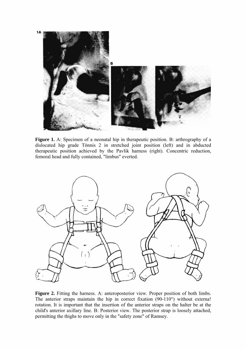
Figure 1. A: Specimen of a neonatal hip in therapeutic position. B: arthrography of a dislocated hip grade Tönnis 2 in stretched joint position (left) and in abducted therapeutic position achieved by the Pavlik harness (right). Concentric reduction, femoral head and fully contained, "limbus" everted.
Figure 2. Fitting the harness. A: anteroposterior view. Proper position of both limbs. The anterior straps maintain the hip in correct fixation (90-110°) without externa! rotation. It is important that the insertion of the anterior straps on the halter be at the child's anterior axillary line. B: Posterior view. The posterior strap is loosely attached, permitting the thighs to move only in the "safety zone" of Ramsey.
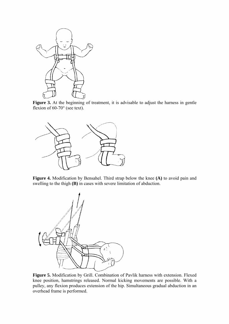
Figure 3. At the beginning of treatment, it is advisable to adjust the harness in gentle flexion of 60-70° (see text).
Figure 4. Modification by Bensahel. Third strap below the knee (A) to avoid pain and swelling to the thigh (B) in cases with severe limitation of abduction.
Figure 5. Modification by Grill. Combination of Pavlik harness with extension. Flexed knee position, hamstrings released. Normal kicking movements are possible. With a pulley, any flexion produces extension of the hip. Simultaneous gradual abduction in an overhead frame is performed.
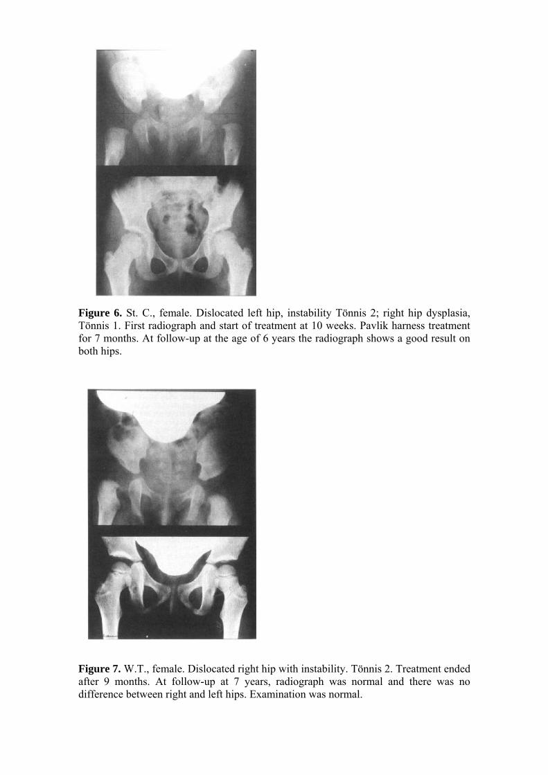
Figure 6. St. C., female. Dislocated left hip, instability Tönnis 2; right hip dysplasia, Tönnis 1. First radiograph and start of treatment at 10 weeks. Pavlik harness treatment for 7 months. At follow-up at the age of 6 years the radiograph shows a good result on both hips.
Figure 7. W.T., female. Dislocated right hip with instability. Tönnis 2. Treatment ended after 9 months. At follow-up at 7 years, radiograph was normal and there was no difference between right and left hips. Examination was normal.
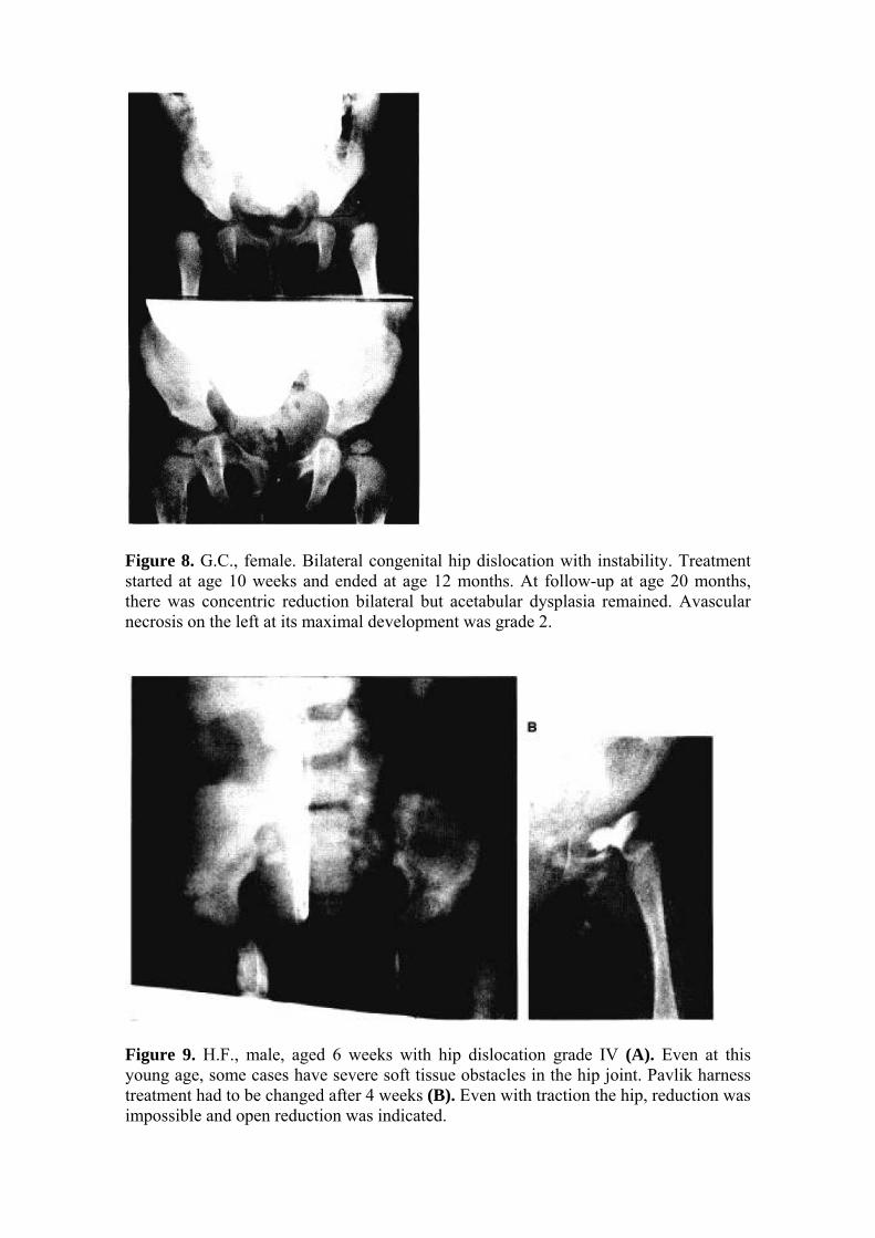
Figure 8. G.C., female. Bilateral congenital hip dislocation with instability. Treatment started at age 10 weeks and ended at age 12 months. At follow-up at age 20 months, there was concentric reduction bilateral but acetabular dysplasia remained. Avascular necrosis on the left at its maximal development was grade 2.
Figure 9. H.F., male, aged 6 weeks with hip dislocation grade IV (A). Even at this young age, some cases have severe soft tissue obstacles in the hip joint. Pavlik harness treatment had to be changed after 4 weeks (B). Even with traction the hip, reduction was impossible and open reduction was indicated.
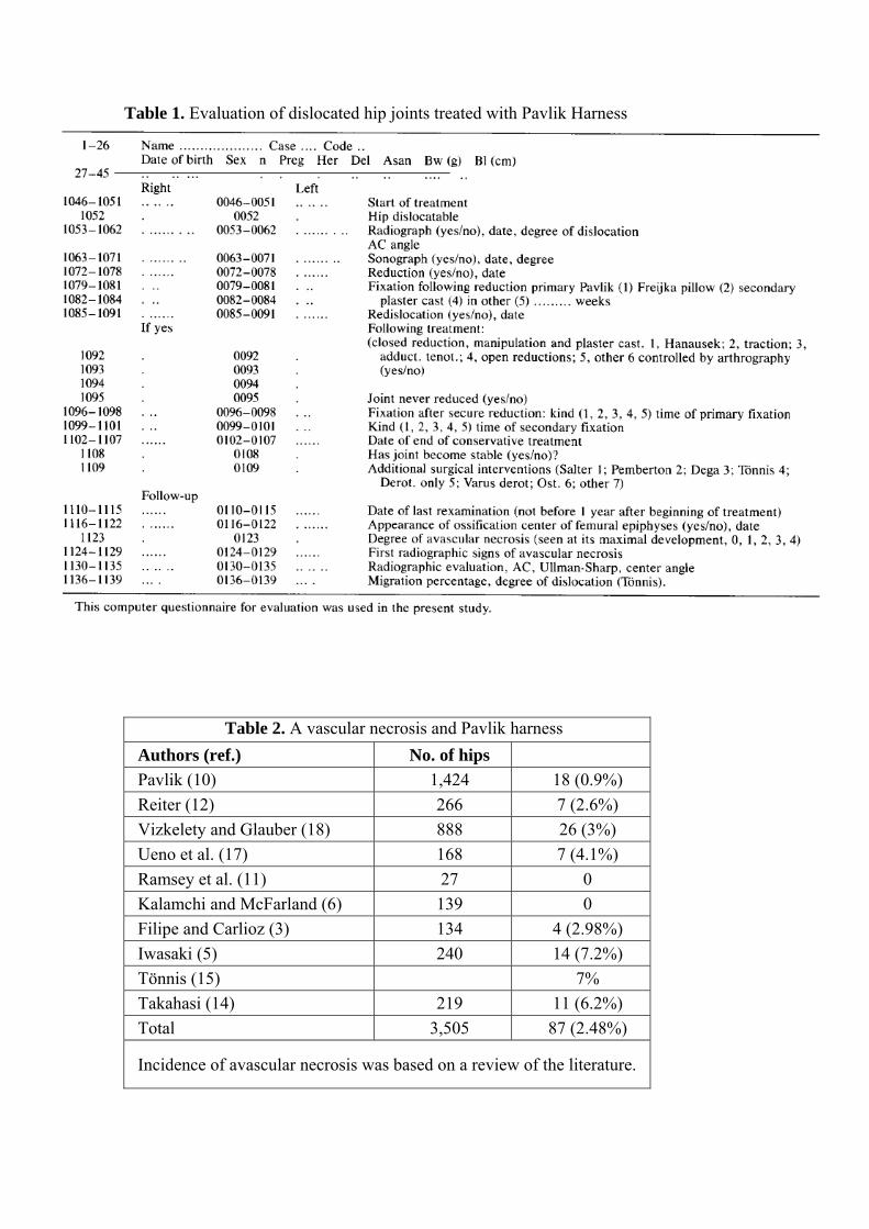
Table 1. Evaluation of dislocated hip joints treated with Pavlik Harness
Table 2. A vascular necrosis and Pavlik harness Authors (ref.) No. of hips Pavlik (10) 1,424 18 (0.9%) Reiter (12) 266 7 (2.6%) Vizkelety and Glauber (18) 888 26 (3%) Ueno et al. (17) 168 7 (4.1%) Ramsey et al. (11) 27 0 Kalamchi and McFarland (6) 139 0 Filipe and Carlioz (3) 134 4 (2.98%) Iwasaki (5) 240 14 (7.2%) Tönnis (15) 7% Takahasi (14) 219 11 (6.2%) Total 3,505 87 (2.48%)
Incidence of avascular necrosis was based on a review of the literature.