Embed Size (px)
Citation preview

Phorocheviisfry orid Pliofobiology. 19G4 Vol. 3, pp. 415427 . Pergamon Press Ltd. Printed in Great Britain
THE PHOTOCHEMICAL INTERACTION OF DEOXYRIBONUCLEIC ACID AND PROTEIN IN VWO
AND ITS BIOLOGICAL 1MPORTANCE"t
KENDRIC C. SMITH Department of Radiology, Stanford University School of Medicine
Palo Alto, California
(Received 21 July 1964)
GREAT advances have been made in the last few years in our knowledge of the photo- chemistry of the nucleic acids and of the proteins,(11J7) but not until recently has the oppor- tunity to study the photochemical interaction of the nucleic acids and proteins presented itself. The deoxyribonucleic acids are in intimate contact with proteins within the cell and if there were a photochemical reaction that caused the crosslinking of DNA and protein then it might well have a greater photobiological significance than an isolated photo- chemical event within either the DNA or the protein. The present report will deal with such a phenomenon.
1. The decrease in extractability of DNA from U.V. irradiated E. coli When bacteria are irradiated with increasing doses of u.v. light there is a dose dependent
decrease in the amount of DNA that can be extracted from these cells with detergent.('J6) This appears to be the consequence of a crosslinking between DNA and protein and the subsequent precipitation of the linked DNA when the denatured proteins are precipitated in the procedure used for the isolation of the DNA.(la)
If aliquots of a culture of E. coli are irradiated with increasing doses of U.V. light at 2537A and then treated with detergent for the isolation of their DNA a curve can be constructed for the percent recovery of DNA V.S. the dose of U.V. delivered to the aliquot. This is shown in Fig. 1 for three strains of E. coli. Two features are immediately apparent. First there seems to be two populations of DNA that are sensitive to this photochemical response. One population (comprising about 30 per cent of the total DNA) is about seven times as sensitive as the other. Second, the response is analytically very sensitive. At the dose of u.v. to kill 99 per cent of a culture of E. coli B/r about 10 per cent of the DNA has been rendered non-extractable. Although the biological importance of thymine dimer formation by U.V. light seems well documented,Q') it is rather an insensitive parameter analytically. The top line in Fig. 1 indicates the rate of formation of thymine dimers in E. coli BJr plotted here as the loss in recovery of thymine. At the dose of U.V. where 10 per cent of the DNA has been rendered non-extractable only one-tenth of one percent of the thymine has been dimerized.
* Presented at the Symposium on Molecular Mechanisms in Photobiology, Wakulla Springs, Florida, February 16-21, 1964, sponsored by the Committee on Photobiology, National Academy of Sciences- National Research Council. t This investigation was supported by Grant CA-02896 f ro3 the Nalional Cancer Institute of the US. Public Health Service.
41 5

41 6 KENDRIC C. SMITII
10 0
90
80
70
60 f 0) >
50 tr + c $ 4 0 L z 30
20
10
k THYMINE (B/r )
r 99 % Killing
B/r
o T - o , * B
B/r)
I- I I I
1.0 2.0 3.0 Ergs/mm*(x
FIG. 1. The extractability of DNA from E. coli following irradiation with increasing doses of U.V. light.
Thymine-2-C-14 labeled E. coli were grown to saturation (24 hr) and then irradiated with increasing doses of U.V. and extracted with sodium lauryl sulfate to isolate the DNA. The graph represents the recovery of free DNA. The DNA not isolated remained associated with the denatured proteins. For comparison, data of Smith(16) are included on the rate of forma- tion of thymine dimer in E. coli B/r with increasing dose of u.v., plotted here as the loss of
recovery of thymine(").
2. Evidence for the biological importance of' the crosslinking of DNA to protein by U . V .
The remarkable analytical sensitivity of the crosslinking of DNA and protein by U.V.
light and the fact that there are two populations of DNA with differing sensitivities en- couraged us in the belief that this phenomenon must be a biologically important photo- chemical reaction. The following experiments were therefore designed to determine if the same treatments that can drastically alter the intrinsic sensitivity of cells to killing by U.V.
can also cause a similar alteration in the ability of the DNA of these cells to become cross- linked with protein.
The response to U.V. crosslinking of DNA and protein can be significantly altered in E. coli 15 TAU by varying its growth conditions. Since this mutant requires thymine, arginine and uracil one can selectively inhibit DNA, or RNA and protein synthesis by with- holding the proper nutrient. ( B l l o )

Photochemical interaction of DNA and protein 41 7
When log phase cells are suddenly switched to a medium devoid of thymine the total cell number stays essentially constant for the first 30 min and then the cells begin to die rapidly of what is called thymineless death.c2) We have sampled cultures at various times after transferring to a thymineless medium, irradiated these aliquots with a single dose of U.V. light and then assayed for cell viability and for the extractability of the DNA relative to unirradiated aliquots taken at the same time. The results are shown in Fig. 2. The upper
f 10' 7 FIG. 2. The susceptibility of E. coli 15 TAU to killing and to the crosslinking of their DNA
and Protein by U.V. at various times of growth under conditions of (-T+AU). A logarithmic culture of E. coli 15 TAU was suddenly switched to a medium devoid of thymine by filtration and resuspension.('O) At various times thereafter, two aliquots were withdrawn from the culture. One of these aliquots was irradiated with 480 ergs/mm2 (2537A) and then both aliquots were assayed for viable cells and for the amount of DNA that could be extracted
free of protein.
curve on the left shows the loss in viable titer due to thymineless death vs. time of growth in the absence of thymine. The lower curve indicates the viable titer following a dose of 480 ergs/mm2 vs. time of growth in the absence of thymine. The middle curve is the result of dividing the lower curve by the upper one to give the fraction of the total population sur- viving after the irradiation. Deviation of these data from a horizontal line indicates an alteration in the intrinsic sensitivity of the cells to killing by U.V.
Shortly after removing thymine from the medium the cells become fifteen times more sensitive to killing by U.V. light. This sensitization reaches a maximum after 45-50 min and this is at a time when only about 50 per cent of the culture has died of thymineless death. After this time there is an abrupt change in the sensitivity to killing by U.V. and the remaining viable cells become much more resistant than the controls.
If we now look at the curve for the U.V. crosslinking of DNA and protein (right half of Fig. 2) we see that the thymineless state also produces a marked increase in crosslinking and therefore a greater loss in DNA by the same dose of U.V. and the time course of events is very similar to that for changes in sensitivity to killing. The viability experiments of course only measure those cells in the culture that are still viable but the chemistry part looks a t all the cells in the culture regardless of viability. There is a marked increase in crosslinking for
41 8 KENDRIC C. SMITH
the first 40 min and then the DNA of the cells becomes much less sensitive and finally more resistant than the log phase cells.
It has been pointed out that messenger RNA synthesis appears to be required before the withholding of thymine causes death.(') The enhanced association of protein with DNA during the first 40 min of thymine starvation may reflect an enhanced production of messenger RNA and the major protein crosslinked to the DNA by U.V. under these conditions may be the DNA-directed RNA polymerase.
We can conclude that the crosslinking of DNA and protein by U.V. light is not a random, non-specific process but changes under the same conditions that lead to the production of cells that are more sensitive to U.V. killing. The near equivalence in the timing of changes in viability and crosslinking are suggestive that the crosslinking may play a role in the loss of viability.
Figure 3 shows the complete dose response curves for cultures of E. coli 15 TAU grown for various times under several conditions. The results for E. coli B, B/r and T- given in Fig. 1 show that only about 30 per cent of the DNA of these cultures was exquisitely
I I I
0 I .o 2.0 SO erps/mm2 ( x 10-4)
FIG. 3. The extractability of DNA from cultures of E. coli 15 TAU following irradiation with increasing doses of U.V. light.
Log phase cultures were switched to a particular nutritionally restricted medium and main- tained at 37" for the times indicated. The cultures were then harvested by filtration and re- suspended in phosphate buffer. Aliquots were irradiated with different doses of U.V. (2537A) and the amount of free DNA that could be extracted from these aliquots is plotted vs. the dose
of U.V.
sensitive to crosslinking but the results for E. coli 15 TAU (Fig. 3) show that for log phase cells about 75 per cent of the DNA is exquisitely sensitive. This is also true for another mutant of 15 TAU that further requires methionine, tryptophan and proline and is called 15 TAU-Bar.(') We are unable at this time to explain this gross difference in response of the DNA of these five strains of E. coli.
The results in Fig. 3 show again quite clearly (compare Fig. 2) that after 45 min of thymineless-growth that both the absolute sensitivity and the total amount of the DNA

Photochemical lnteraction of DNA and protein 419
that is sensitive to crosslinking is increased above that for the log culture. After 120 min of thymineless-growth the culture becomes more resistant. A culture deprived of all three nutritional requirements (-T-AU) also becomes more sensitive to crosslinking after about 40 min but after 120 min becomes the most resistant to crosslinking of all the culture conditions thus far studied.
We have also studied the variation in sensitivity to U.V. crosslinking of cultures grown in the presence of thymine but in the absence of arginine and uracil (fT-AU). Under these conditions a small percentage of the total population (about 27 per cent) is capable of dividing even in the absence of arginine and uracil. The rest of the cells complete the doubling of their DNA but cannot divide because they cannot make the protein and/or RNA necessary for the final stages of division.(lO) The continued replication of DNA would not be expected to greatly alter the sensitivity of the DNA of the cells to crosslinking over that for log phase cells. However, the time course of the division of those cells that are capable of dividing in the absence of arginine and uracil is the same as that for a transitory increase in the crosslinking of DNA and protein by a constant dose of U.V. delivered to the culture at various times after withdrawing the arginine and uracil. It therefore seems reasonable to conclude that on the average the DNA of E. coli 15 TAU is somewhat more sensitive to crosslinking to protein by U.V. at the time of cell division than it is during replication. Helmstetter and Uretz@) have shown that E. coli are most sensitive to killing by U.V. just after division.
These studies on the photobiology and photochemistry of nutritionally restricted cultures lead to some interesting conclusions concerning changes in the state of the DNA and shed further light upon the mechanisms involved in the phenomenon of thymineless death but this lies outside the interest of this symposium and will be reported more fully elsewhere.
3. The increased sensitivity of the replicatingportion of the DNA of E coli to U . V . crosslinking with protein
Another way to show that the sensitivity of DNA to U.V. crosslinking with protein is directly dependent upon the state of the DNA is to pulse label the DNA of log phase cultures with tritiated thymine and then measure the sensitivity of the part of the genome that is labelled with radioactivity at various times after the pulse labeling. Fig. 4 indicates schematically that the DNA of eachcell in a logarithmic culture is on the average going to be labelled to the same extent by a pulse of thymine -H-3 but the position along the DNA that becomes labelled will depend upon the time in the division cycle for that particular cell. It has been shown that the DNA in E. coli is in a closed circle(5) and that replication of the DNA continues throughout most of the generation time of the bacterium.(14) During the pulse a short segment of the DNA becomes labelled but when the radioactive thymine is removed from the culture and replaced by non-radioactive thymine the remaining DNA will be unlabelled. At a period half a generation time away from the pulse, which in this case is 20 min, the labelled portion of the DNA should be at its furthest linear distance from that portion of the genome then being actively duplicated.
The circular chromosomes of the bacteria are broken up into as many as 50-200 pieces of DNA during the usual procedures used for the isolation of DNA. Therefore when the DNA is isolated from a pulse labeled culture the majority of the DNA pieces isolated will be unlabeled and there will be a distribution of pieces with various amounts of labeling in them. It is only the labeled molecules of DNA that we are assaying.

420 KENDRIC C. SMITH
We know that for a given culture a given dose of ultraviolet light will crosslink a given amount of DNA (see Figs. 1 and 3). If the phenomenon of crosslinking were a random process involving photochemically altered DNA and protein in a non-selective manner then samples taken from a pulse labeled logarithmic culture at various times after the pulse should show a constant percentage of crosslinked DNA for a constant dose of U.V. How- ever, if the crosslinking depends upon the state of the DNA then the short segment of the
FIG. 4. A schematic representation of the pulse labeling of the DNA of a culture of E. coli with thymine-H-3.
E. coli 15 TAU (generation time: 40 inin) were pulse labeled for 5 min with thymine-H-3 (shaded area in dark lines representing DNA molecules). Since the DNA of bacteria is synthe- sized throughout most of the generation time almost all of the cells will be labeled and to about the same extent, but the area along the chromosome will be different for each cell. Since the chromosome is circular(6) one-half of a generation time after the pulse (20 min) the portion of the genome being replicated should be the furthest hear distance from the radioactive section of the DNA. Again one generation time after the pulse all of the cells should again be
copying the radioactive section of their chromosome.
DNA that is labeled should show some variation in its sensitivity to labeling versus time after the pulse and this variation should be keyed to the generation time.
Figure 5 indicates the type of results that we have obtained from several experiments in which a log culture of E. coli 15 TAU was pulsed for 5 min with tritiated thymine and then the radioactive thymine was removed and replaced with non-radioactive thymine. This switch in medium takes less than 1 min and the culture continues in log growth. The percent recovery of radioactive DNA after a given dose of U.V. has been plotted for various times after the pulse. Since the response differs significantly from a straight line, we can conclude that the crosslinking is not a random, nonselective process. The response is in fact keyed to the generation time of these bacteria. Right after the pulse and again one generation time later the radioactive portion of the genome is most sensitive to cross- linking. At 20 min after the pulse the radioactive area on the genome should be the furthest linear distance from that portion of the genome that is then being actively copied. The peaks and valleys in Fig. 5 can therefore be explained by the conclusion that that portion of the genome that is actively being copied is themost sensitiveto U.V. crosslinking with protein.
4. Observations on the chemical mechanisms involved in fhe crosslinking of DNA and protein by U . V .
The DNA that is not isolated free of protein after U.V. irradiation is noi lost but can be quantitatively accounted for in the precipitate containing the denatured proteins. When this

Photochemical interaction of DNA and protein 42 I
100
90
8b
a 70
u .- c
.- 60
a
t
a" 40
Q
c 50
> 0
L E
U
0" 30
20
10
, 1 1 , , , , 1 1 1 , 1 1 , 1 , -5 0 10 20 30 40 50 60 70 8 0
Minutes After 5 min. Pulse of Thymine - H3
FJG. 5. The sensitivity of a pulse labeled section of the bacterial DNA to be crosslinked to protein by a constant dose of U.V. as a function of the time after the pulse.
A log phase culture of E. coli 15 TAU was pulsed with thymine-H-3 for 5 min, the radioactive thymine was removed by filtration, the cells returned to a medium containing non-radio- active thymine and then allowed to continue logarithmic growth. At various times two aliquots were removed from the culture. One of these was irradiated with 133 ergs/mma and then both were treated with detergent for the isolation of their DNA. The percent recovery of DNA (vs. the unirradiated control aliquots) is plotted against the time following the pulse of thymine-H-3.
Only the radioactive DNA is being assayed in this experiment.
precipitate is homogenized in 55 per cent CsCl and spun in an ultracentrifuge (Spinco rotor SW 39L; 29,000 rev/min for 65 hr) less than 20 per cent of this DNA bands free of protein and the remainder floats on top of the gradient with the protein. Trypsin digestion of this floating layer will free about 50 per cent of the DNA, as assayed by rebanding in cesium chloride. These results suggest that the DNA was crosslinked to protein.
Conditions which photoreactivate colony formation do not alter the amount of DNA that can be extracted from irradiated cells. Current information on the specificity of the photoreactivating en~yme('~*l') would seem to eliminate thymine dimerization as being important in this phenomenon. This is further substantiated by the fact that the amount of thymine dimer in the extractable DNA is not appreciably different from that in the DNA
422 KENDRIC C. SMITH
that is precipitated with the proteins. However, at least four other photoproducts of thymine as yet unidentified, are found in significantly greater yield in the protein bound DNA.(l*j Bromouracil substitution of the DNA makes the cells (E. coli B/r) about five times more sensitive to crosslinking but doesn't increase the total amount of the DNA that is sensitive to crosslinking.(17j
Since the crosslinking phenomenon is not altered under conditions allowing photo- reactivation to occur we may conclude that the crosslinking phenomenon must be the consequence of the primary absorption of U.V. rather than a secondary attachment of photoreactivating enzymes subsequent to irradiation.
This argument can now be extended to include the recently described dark repair or excision enzymes (Setlow and Carrier, Proc. Nat. Acad. Sci., U.S. 51, 293, 1964; Boyce and Howard-Flanders, Ibid., 51, 293, 1964). We find that E. coli B, (which contains no dark repair enzymes; Caslellani, Jagger and Setlow, Science 143, 1170, 1964) gives essen- tially the same dose response curve for the production of DNA-protein crosslinks as does E. coli B/r, which possesses a very efficient system for the dark repair of thymine dimers.
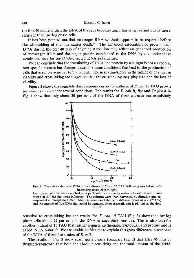
5. The crosslinking of DNA and protein in vitro Our best evidence that this phenomenon really involves the crosslinking of DNA and
protein comes from our in vitro studies. When purified E. coli DNA is irradiated with
100
80
60
40
20
10 ! I I f I I I I I
0 I 2 3 4 5 6 7 8 9
ergs /mm2 ( x ~ 0 - 4 )
FIG. 6. The in vitro crosslinking of E. coli DNA and bovine serum albumin by U.V. light (2537A).
The DNA (labeled with thymine-2-C-14) was isolated from E. coli with detergent,(l6) treated with ribonuclease and resubmitted to the isolation technique. The samples labeled NON- AGED were assayed shortly after isolation and again 5 months later (AGED) after storage at -20°C. Another sample was assayed NON-AGED and after treatment with 90 per cent phenol (PhOH) (shake 60 min at 23"C, centrifuge, recover DNA by alcohol precipitation) before and after ribonuclease (RNAase) treatment. The sample to be irradiated (in a 5-cm petri dish) contained 20 mg bovine serum albumin and 0.03 mg of DNA-C-14 (2 x lo6 cpm/mg) in 4.2 ml of 0.007 M NaCI. After various times of irradiation 0.5 ml was removed and mixed with an equal volume of 4 per cent sodium lauryl sulfate. These solutions were mixed occasionally over 60 min (at room temp.) and then treated with 1 M KCI and quantitated for the recovery
of DNA.(16)

Photochemical interaction of DNA and protein 423
heavy doses of U.V. (8 x lo4 ergs/mm2) and then resubmitted to our procedure for the isolat- ion of DNA all of the DNA is recovered. This is shown by the top line in Fig. 6 . However, if the DNA is irradiated in the presence of bovine serum albumin and then submitted to the isolation procedure for DNA there is a dose dependent decrease in the amount of DNA that can be isolated (bottom line, Fig. 6).
Storage of the DNA in the freezer for several months (AGED sample) or treatment of the DNA with phenol (PhOH) either before or after ribonudease (RNAase) treatment de- creases the efficiency of the DNA for crosslinking with bovine serum albumin. These results may be the consequence of shearing of the DNA.
No crosslinking was observed when the DNA was assayed prior to the removal of the RNA from the sample, yet when a 10-fold excess of yeast RNA was added to a sample of purified DNA it altered the response of the DNA only slightly. It is interesting to speculate that perhaps isologous messenger RNA combines with the DNA and thereby protects the DNA from this reaction, whereas the heterologous RNA does not. This experiment should be repeated with isologous RNA.
We have performed mixing experiments in which the DNA and the protein are irradiated separately and then mixed either with an irradiated or non-irradiated counter part. These solutions were then allowed to stand for 30 min at room temperature and then the DNA was extracted. The underlines in Fig. 7 indicate which of the materials were irradiated. Un- irradiated DNA mixed with unirradiated protein, and irradiated DNA mixed with water
4 2 2.9 x i 0 ergdmm
1 DNA +BAA I
I I I I 1 I 0 20 40 60 80 too
Percent Recovery of DNA
FIG. 7. The relative contribution of DNA and protein to U.V. crosslinking. Crystalline bovine serum albumin (BSA) at 5 m g / d in water, or thymine-2-C-14 labeled E. coli DNA (2.1 x lo* cprnimg) at 0.0075 mg/ml in 0.1 MNaCl were irradiated speparately and then mixed (equal aliquots) with an irradiated (heavy underline, ) or unirradiated coun- terpart. The mixtures were allowed to stand for 30 min at room temperature and then were treated with 2 per cent sodium lauryl sulfate for 60 min followed by 1 MKCl for the reisolation of the DNA.(ls) The crosslinking of DNA and protein is indicated when the recovery of DNA
is less than 100 per cent.
gave 100 per cent recovery. When the DNA was irradiated and mixed with unirradiated protein only 85 per cent of the DNA was recoverable; when the protein was irradiated and mixed with unirradiated DNA only 60 per cent of the DNA was recoverable. When both the DNA and the protein were irradiated about 45 per cent of the DNA was recovered. These data indicste that irradiated DNA can combine with unirradiated protein and irradiated protein can react with unirradiated DNA to form a linkage which is not separated by the action of detergent or high salt concentration.

424 KENDRIC c. SMITH
We don’t know if the crosslinking that results from these mixing experiments is by covalent bonds or not. If the bonding is truly covalent then it must have resulted from the interaction of exceedingly long lived excited foci produced in the DNA and protein by the U.V. light. Another alternative is that the crosslinking is due to the formation of extra- ordinarily strong salt linkages due to configurational changes brought about in the DNA and protein by photochemical events in these molecules. If this is so, then it can’t be by simple denaturation mechanisms since various stages of heat denaturation of bovine serum albumin have caused DO crosslinking with DNA.
One mechanism by which the crosslinking of DNA and protein could be accomplished is by the addition of the OH group of serine, tyrosine, etc., or the SH group of cysteine to the 6 position of cyosine or uracil, analogous to the photochemical addition of the OH group of water to uracil. Consistent with this postulate is the fact that if uracil is irradiated in anhydrous alcohol, the alcohol adds to the 5-6 double bond of uracil. The water addition product of uracil is labile to heat and changes in pH but it would be expected that if a protein were joined to a DNA molecule through several of these hydration bridges, a rather firm binding might result. The bonds formed between DNA and protein in vitro are sensitive to heat and so are the bonds formed in vivo at very low doses of U.V. but not at higher doses. We hope to test this hypothesis with the use of model compounds (syn- thetic polynucleotides and polyamino acids).
We have recently produced a hetero dimer of uracil and cysteine by the irradiation (2537A) of these two compounds in solution. This photoproduct has been crystallized and its structure is currently under investigation.
6 . The eficiency of X-rays and of acridine orange plus visible light in producing crosslinks between DNA and protein
X-rays at doses up to 40 Kr do not produce crosslinks between DNA and protein in vivo that are stable to the action of detergent and high concentrations of salt. Acridine orange plus visible light does produce this phenomenon and furthermore it appears to be more efficient than ultraviolet light. For cultures ( E . coli T-) where only 30 per cent of the DNA was sensitive to crosslinking by the action of U.V. (Fig. 1) essentially all of the DNA was sensitive to crosslinking by the action of acridine orange and visible light.(*6~17) The crosslinking of DNA and protein by the action of visible light and acridine orange can also be demonstrated in vitro. Contrary to the results with ultraviolet light, however, protein and DNA that have been separately irradiated with visible light in the presence of acridine orange and then mixed show no crosslinking. The DNA, protein and acridine orange all have to be present at the time of irradiation in order to produce this reaction. A covalent bond is therefore much easier to envisage here than in the case for u.v.. We are currently entertaining the hypothesis that the crosslinking of DNA and protein by acridine orange and visible light may well be its most biologically important reaction rather than the pro- duction of isolated photochemical events in either the DNA or the protein.
7. The use of sucrose gradients to demonstrate the crosslinking of DNA and protein in vitro and in vivo
The crosslinking of DNA and protein by ultraviolet light in vitro can also be demon- strated in sucrose gradients. Fig. 8 shows the sedimentation of heavily irradiated DNA (peak #3), of unirradiated DNA mixed with unirradiated protein (peak #2) and of DNA irradiated in the presence of bovine serum albumin and simply placed on top of the gradient

Photochemical interaction of DNA and protein
800 3 .-
I ! , , I I , I
roo
600
W I- = 5 0 0 5 I K W
400 a
$ 3 0
300
200
100
5 0
0 0 10 20 30 40 50 60 70 80
DROP NUMBER
FIG. 8. The use of sucrose gradients to detect the in vifro crosslinking of E. coli DNA and bovine serum albumin by U.V. light.
Condtions are similar to those described in Fig. 7. Sample # 1 : Eight pgin of DNA (thymine- c-14) and 15 mg bovine serum albumin in 1.65 cc of 0.014 M NaCl were irradiated with 3 a 6 x 10⁴ ergs/mm* (2537A). Sample #2.: Same as Sample # I but no irradiation. Sample #3: Same as sample # 1 but without the bovine serum albumin. 0.4 ml of each sample was placed On top of a linear sucrose gradient (5-20 %) and was spun in a Spinco rotor SW 39L for 25 min at 37,000 rev/min at room temperature. Drops were collected from the bottom of the tubes and
the distribution of the radioactivity (DNA) was determined.
425
(peak # 1). The sedimentation of heavily irradiated DNA (3.6 x lo4 ergs/mm2) does not significantly differ from that for unirradiated DNA. However, when the DNA is irradiated in the presence of bovine serum albumin the apparent molecular weight of the DNA is greatly increased due to crosslinking with the protein. Unfortunately no unique species of crosslinked DNA is formed but rather a wide distribution of molecular sizes is produced.
Our attempts to show the crosslinking of DNA (thymine-H-3 labeled) and protein (labeled with (2-14 amino acids) in vivo by direct isolation of this combination in sucrose gradients have thus far been somewhat less than satisfactory. We have observed peaks of DNA associated with protein in irradiated samples that were not present in the un- irradited controls, but the molecular weight of these DNA-protein complexes kept chang- ing, probably as a consequence of shearing forces. The main difference that we find in the sedimentation of the DNA from the control and irradiated samples is quantitative rather than qualitative. Even in the unirradiated control sample there is a small amount of DNA that sediments at low centrifugal speeds to the bottom of the tube in association with protein but with irradiation there is a dose dependent increase in the amount of DNA sedimenting to the bottom of the tube. We have thus far been unable to solubilize and purify this DNA-protein pellet sufficiently to allow us to obtain satisfactory information on the type of linkage or the type of protein involved.

426 KENDRIC c. SMJTIi
DISCUSSION DNA polymerase has been found associated with bacterial DNA when the cells are
lysed and banded in sucrose gradients.(4) The polymerase would presumably be associated with the replicating segment of the chromosome. It has been shown that newly synthesized DNA is the most difficult to isolate free of protein.(sB12) Newly synthesized DNA is also more resitant to sonic disruption.@) This may be due to protection by associated protein. We find that the newly synthesized DNA is much more sensitive to crosslinking to protein by U.V. than is the remainder of the DNA (Section #3). Presumably therefore, the enzymes involved in replicating the DNA are the ones most easily crosslinked to the DNA by the action of U.V.
Although the replicating portion of the genome is the most sensitive to crosslinking with protein the major portion of the remainder of the DNA of E. coli 15 TAU is also capable of becoming crosslinked to protein (Fig. 3). This should be contrasted with the situation for three other strains of E. coli (Fig. 1) where only about 30 per cent of the total DNA is markedly sensitive to crosslinking. The biological significance of this striking difference between these strains of E. coli is not apparent.
Other data (Section #3) suggest that on the average, the DNA of cells undergoing cell fission is more sensitive to U.V. induced crosslinking with protein than is the DNA of cells that are replicating their DNA. It is therefore of particular interest that E. coli have been shown to be most sensitive to killing by u.v. just after division.(8)
Under conditions of thymine starvation where the intrinsic sensitivity of the cells to killing by U.V. i s greatly increased, the intrinsic sensitivity of the DNA of these cells to be crosslinked to protein by U.V. is also significantly increased and follows the same time course as the change in susceptibility to killing. This near equivalence in the timing of changes in the sensitivity to killing and to crosslinking are suggestive that the crosslinking may play a role in the loss of viability. This greatly increased association of protein with DNA during the thymineless state may also play a significant role in the mechanisms involved in thymineless death.
There thus seems to be sufficient data to suggest that the crosslinking of DNA and pro- tein by U.V. in vivo is not a non-specific random process, on the contrary, it appears to be quite specific and of significant biological importance.
Our evidence to support the conclusion that we are studying the photochemical cross- linking of DNA and protein in vivo is all indirect. The major support comes from the obser- vation that DNA and protein can be crosslinked in vitro by U.V. (Section # 5 and 7). The precise chemical nature of the linkage is unknown except that the photochemical attachment of the DNA to the protein is resistant to the sequential action of 2 per cent sodium lauryl sulfate and of O.5M KCI or 55 per cent CsCl. Several new photoproducts of thymine have been found in significantly greater yield in the DNA that is crosslinked to protein but their role in the crosslinking phenomenon is not known. Cells that have had a large share of the thymine in their DNA replaced by the analog, 5-bromouracil, are about five-times more sensitive to DNA-protein crosslinking than are normal cells.
It can be reasoned that DNA is the most important target for radiation within a cell, since it is present with the least multiplicity of all molecules within the cell and has the largest molecular weight and therefore presents the largest physical target. By the same argument it can be concluded that the destruction of proteins by radiation is probably not too significant because of the presence of extra copies of these proteins and the ease with
Photochemical internction of DNA and protein 42 7
which extra copies can be produced. However, the DNA is not present as a pure molecule within the cell but exists in close association with proteins such as the enzymes involved in its replication and in the transcription of the genetic code. Although the destruction of an isolated enzyme molecule within a cell may be biologically insignificant, if the enzyme were crosslinked to DNA as a consequence of the action of U.V. light then the destruction of that particular enzyme molecule would be of singular importance since it would un- doubtedly block the continuity of metabolic events surrounding the DNA. What may be of equal importance is that the crosslinked protein may block the access of repair enzymes to regions in the DNA that contain thymine dimers, lesions that otherwise could be repaired and would therefore not be detrimental to the cell.
REFERENCES 1 . P. ALEXANDER and H. MOROSON, Nuture, Lond. 194, 882 (1962). 2. H. D. BARNER and S. S. COHEN, J. Bacteriol. 68, 80 (1954). 3. T. BEN-PORAT, A, STERE and A. S. KAPLAN, Biochini. Biophys. Acta, 61, 150 (1962). 4. D. BILLEN, Biochini. Biophys. Acta, 68, 342 (1963). 5. J. CAIRNS, J. Mol. Biol. 6, 208 (1963). 6. A. GOLDSTEIN and B. J. BROWN, Biochini. Biophys. Acta, 53, 19 (1961). 7. P. C. HANAWALT, Nature, Lond. 198, 286 (1963). 8. C. E. HELMSTBTTER and R. B. URETZ, Biophys. J . 3, 35 (1963). 9. D. KANAZIR, H. D. BARNER, J. G. FLAKS and S. S. COHEN, Biochin?. Biophys. Acta, 34, 341 (1959).
10. 0. MAAWE and P. C. HANAWALT, J. Mol. Biol. 3, 144 (1961). 1 1 . A. D. MCLAREN and D. SHUGAR, Photochemistry of proteins and nucleic acids, Pergamon Press, Oxford
12. R. ROLFE, Proc. Nat. Acad. Sci. US. 49, 386 (1963). 13. C. S. RUPERT, in Photophysiology, (A. C. GIESE, ED.) Volunie 11, Academic Press, New York (1964). 14. M. SCHAECHTER, M. W. BENTZON and 0. MAAWE, Nature, 183, 1207 (1959). 15. K. C. SMITH, Biochem. Biophys. Res. Cominuns. 6, 458 (1962). 16. K. C. SMITH, Biochem. Biophys. Res. Conzmrms. 8, 157 (1962). 17. K. C. SMITH, Photochemistry of the nucleic acids. In Photophysiology, (A. C. GIESE, ED.) Volume 11,
18. K. C. SMITH, Photocheni. Pliorobiol. 3, 1 (1964).
(1 964).
Academic Press, New York (1964).
Summary-When bacteria are irradiated with U.V. light there is a dose dependent decrease in the amount of DNA that can subsequently be extracted free of protein with detergent. This appears to be due to the crosslinking of the DNA with protein and the precipitation of the linked DNA when the denatured proteins are precipitated in the procedure used for the iso- lation of the DNA, The type of linkage between the DNA and the protein is unknown except that it resists the sequential attack of 2% sodium lauryl sulfate and 0.5 M KCI or 55 % CsCI. The main evidence that the loss of DNA in vivo is due to the crosslinking of DNA and protein is that the crosslinking of DNA and protein can be demonstrated in vitro. X-rays do not crosslink DNA and protein in vivo, but acridine orange and visible light cause the crosslinking of DNA and protein both in vivo and in vitro.
By pulse labeling the DNA of bacteria with tritated thymine it can be shown that newly synthesized DNA is most sensitive to crosslinking and that this sensitivity shows a cyclic response keyed to the generation time of the bacteria. Under conditions of thymine star- vation where the intrinsic sensitivity of the cells to killing by U.V. is markedly increased, there is a parallel increase in the sensitivity of the DNA of these cells to be crosslinked to protein. The similarity in the time sequence of these two events strongly suggests that the crosslinking may play an important role in the loss of viability following U.V. irradiation.