Embed Size (px)
Citation preview

Portland State University Portland State University
PDXScholar PDXScholar
Dissertations and Theses Dissertations and Theses
8-2-1991
The Physiology and Molecular Biology of Iron The Physiology and Molecular Biology of Iron
Nutrition for Cyanobacteria Nutrition for Cyanobacteria
Nancy Walters Unsworth Portland State University
Follow this and additional works at: https://pdxscholar.library.pdx.edu/open_access_etds
Part of the Biology Commons
Let us know how access to this document benefits you.
Recommended Citation Recommended Citation Unsworth, Nancy Walters, "The Physiology and Molecular Biology of Iron Nutrition for Cyanobacteria" (1991). Dissertations and Theses. Paper 4529. https://doi.org/10.15760/etd.6413
This Thesis is brought to you for free and open access. It has been accepted for inclusion in Dissertations and Theses by an authorized administrator of PDXScholar. Please contact us if we can make this document more accessible: [email protected].

AN ABSTRACT OF THE THESIS OF Nancy Walters Unsworth for the Master
of Science in Biology presented August 2, 1991.
Title: The Physiology and Molecular Biology of Iron Nutrition for Cyanobacteria.
APPROVED BY THE MEMBERS OF THE THESIS COMMITTEE:
oh
W. Herman Taylor
Richard R. Petersen
Dennis Barnum
In addition to nitrogen and phosphorus, iron is an essential nutrient
for oceanic primary productivity. Unlike nitrogen and phosphorus however,
negligible amounts of iron are supplied to surface waters through recycling or
mixing but instead from the limited and sporadic input of aeolian particulate.
The low concentration of iron that becomes biologically available from the·
dust places a serious constraint on the heavily iron-dependent processes of
photosynthesis and nitrate reduction which affect primary productivity. As

much as 47% of the total oceanic primary productivity can be attributed to
cyanobacteria making them critical organisms in the biogeochemical cycles.
This thesis addresses the effect of iron on primary productivity using a
combined approach of physiological and molecular biology.
2
The physiological response of three marine strains of Synechococcus to
growth on different concentrations of FeEDTA was investigated. Cells grown
with higher concentrations of iron had greater cell density, more Chl-~ and
phycobiliproteins and higher carbon fixation rates than cells grown at
limiting iron concentrations (l0-8 M Fe). Iron enrichment of iron limited
cultures stimulated carbon fixation, growth rate, and pigment and protein
synthesis. Iron limited cells spiked with SJ.l.M Nlf4Cl prior to short term
incubations had higher dark carbon fixation than cells gro·wn at higher iron
and also spiked to 5J.1M Nlf4Cl. The addition of ammonium relieves a
restricted nitrogen assimilatory pathway in the low iron cells that is
evidenced by increased dark carbon fixation. We propose that this
measurement of enhanced dark carbon fixation could be a useful assay in
supporting the contention that populations of Synechococcus in nitrate rich
waters are iron limited.
Molecular genetic techniques were used to look for the presence of an
iron uptake gene in cyanobacteria. Preliminary results indicate that there is a
gene that is homologous to the ferric uptake regulation (fur) gene in E. coli.
This hybridization occurred in siderophore-producing cyanobacteria, but not
in marine cyanobacteria that do not produce siderophores. The fact that
marine Synechococcus do not produce siderophores and did not hybridize to
the fur gene suggest that fundamentally different mechanisms for iron

uptake operate in high biomass freshwater cyanobacteria and cyanobacteria
from dilute oligotrophic waters.
3

THE PHYSIOLOGY AND MOLECULAR BIOLOGY OF IRON NUTRITION FOR CYANOBACTERIA
by
NANCY WALTERS UNSWORTH
A thesis submitted in partial fulfillment of the requirements for the degree of
MASTER OF SCIENCE in
BIOLOGY
Portland State University 1992

TO THE OFFICE OF GRADUATE STUDIES:
The members of the Committee approve the thesis of Nancy Walters
Unsworth presented August 2, 1991.
W. Herman Taylor
Richard R. Petersen
Dennis Barnum
Robert 0. Tinnin, Chair, Department of Biology
C. William Savery, Interim Vice Provost for.kraduate Studies and Research

ACKNOWLEDGEMENTS
I would like to thank first and foremost, John Rueter whose unending
support and faith in my ability to succeed generated a new sense of excitement
and self confidence that convinced me I was capable of this task. I would also
like to thank Karen Elardo for many months of moral support, trips to the
coffee shop and companionship in the lab Finally, I thank my husband Dave
for his valuable computer skills and for the endless amount patience he
continually shows with me.

TABLE OF CONTENTS
PAGE
ACKNOWLEDGEMENTS ......................................................................................... iii
LIST OF TABLES .......................................................................................................... vi
LIST OF FIGURES ........................................................................................................ vii
CHAPTER
I INTRODUCTION ................................................................................ 1
II INCREASED PIGMENT SYNTHESIS IN CULTURES AND NATURAL SAMPLES OF CYANOBACTERIA DUE TO IRON ADDITIONS ....................................................................... 7
Introduction .............................................................................. 7
Materials and Methods ........................................................... 9
Results ........................................................................................ 10
Discussion .................................................................................. 14
III ENHANCEMENT OF DARK CARBON FIXATION IN IRON-LIMITED CULTURES OF SYNECHOCOCCUS WH6501 .................................................................................................. 17
lntroduction .............................................................................. 17
Materials and Methods ........................................................... 18
Results ........................................................................................ 19
Discussion .................................................................................. 23
IV IRON REGULATION AT THE GENETIC LEVEL: DO CYANOBACTERIA POSSESS A SEQUENCE HOMOLOGOUS TO THE E. COU FERRIC UPTAKE REGULATION (FUR) GENE? ........................................................... 26
Introduction .............................................................................. 26

v
Materials and Methods ........................................................... 27 Results ........................................................................................ 31
Discussion .................................................................................. 33
V SUMMARY AND CONCLUSIONS ................................................. 36
REFERENCES ............................................................................................................... 38
APPENDICES
A ALGAL CULTURE TECHNIQUES ................................................... 43
B BIOCHEMICAL AND PHYSIOLOGICAL TECHNIQUES ............ 48
C MOLECULAR BIOLOGY TECHNIQUES ........................................ 55

TABLE
I
LIST OF TABLES
The Effect of Iron on Phycobilin Content of Synechococcus WH7803 After Six Days (the Data are the Mean of
PAGE
Two Experiments ± Range) ................................................. 13
II Increase in ALA synthesis rate in Synechococcus WH6501 cultures preincubated with 5x1Q-7 M Fe for 16
hours ........................................................................................ 14
Ill Short term reponse (13 hours) of Chi-~ and phycoerythrin synthesis in Trichodesmium to added iron ................... 14
IV Cellular parameters and dark carbon fixation rate for four
experiments. "Experiment a" was measured after four days and b,c and d were measured after 5 days ...... 20
V Carbon fixation rate for cells grown in the light with no
ammonium added. Relative carbon fixation given
in counts per minute per microgram Chl-~ .................... 21

FIGURE
1.
2.
3.
4.
5.
LIST OF FIGURES
Growth response of Synechococcus strains WH6501, WH7803 and WH8018 to a range of iron
PAGE
concentrations (1Q-9M Fe-10-6M Fe) .................................. II
Measurement of total cellular protein per milliliter of culture for the three Synechococcus strains studied ..... ll
Chl-g_ concentration for the three Synechococcus strains sampled ................................................................................... 12
Growth response of Synechococcus WH6501 to iron grown withl0-8 M Fe (A) lQ-7 M Fe (b) ............................. 21
Enhancement of dark carbon fixation rates with addition of ammonium for "Experiment c" ................................... 22
6. Standard curve generated from a 2 hour restriction digest of Lambda DNA with Hindiii. ........................................... 29
7. Standard curve generated by a 2 hour restriction digest of plasmid pBR332 with Alu1 ................................................. 30
8. A 1% agarose gel containing DNA from Synechococcus spp. cut with restriction enzymes EcoRI, Hindiii and Pstl .................................................................................... 32

CHAPTER!
INTRODUCTION
The recent hypothesis that iron flux to the open ocean from transport
of aeolian-borne particulate dust controls primary productivity in these
regions (Moore and others 1984) has generated a keen interest in the role of
iron in controlling productivity in the world's oceans. Duce (1986) estimates
that 95% of the iron available for phytoplankton growth comes from this
aeolian transport of dust. Continental dust contains approximately 5.6% Fe by
weight (Moore and others 1984) and estimates for the amount of biologically
available Fe from dust range from 1 to 50% (1 %, (Hodge and others 1978);
10%, (Moore and others 1984) and 50%, (Zhuang and others 1990). The
biological availability of iron in dust and the biogeochemical cycle of iron in
the open ocean are of crucial importance to metabolic processes in
cyanobacteria that are heavily dependent on iron nutrition including
photosynthesis, nitrogen fixation and nitrate use. Trichodesmium, a main
contributor of fixed nitrogen to tropical oceans, and Synechococcus, which
accounts for as much as 47% of oceanic primary productivity (Glover and
others 1988) are two marine cyanobacteria that have elevated iron
requirements yet are major contributors to primary productivity. Although
both of these organisms remain viable during periods of iron deprivation, a
flux of iron to the system enhances new productivity by stimulating nitrogen
fixation by Trichodesmium and enabling Synechococcus to more efficiently
use nitrate at lower light intensities (Rueter and others man.) at which they

2
often predominate. Waters of the North Pacific, the Equatorial Pacific and the
Antarctic Oceans contain excess surface nitrate concentrations and only a low
biomass. Based on low iron concentrations of< 0.1nM and increased growth
following iron enrichments, Martin and Fitzwater (1988) and Martin and
Gordon (1988) hypothesized that the high nitrate, low biomass conditions
result from a lack of dust transport. Martin (1990) also generated vigorous
debate by suggesting that iron additions to these regions could possibly
ameliorate the consequences of increased atmospheric C{h by increasing
primary productivity and creating oceanic carbon sinks.
Iron is an essential nutrient for cyanobacterial growth and is potentially
growth limiting in natural environments (Carr and Wyman 1987). Iron
availability is limited due to its low solubility in oxic oceanic surface waters
where ferrous iron is rapidly oxidized to ferric iron and results in insoluble
iron hydroxides. This precipitate is rapidly transported to deeper water and
sediments producing iron concentrations in the euphotic zone that often are
nearly analytically undetectable. Because the source of iron differs from other
major nutrients such as nitrate and phosphorus that are resupplied to the
surface waters through advection, autotrophic organisms in the euphotic
zone are posed with the perpetual problem of meeting cellular demands for
iron (Boyer and others 1987). Additions of iron as FeEDTA and Saharan dust
to iron-limited cultures of Synechococcus and natural samples of
Trichodesmium resulted in increased growth rates and changes in several
physiological parameters (Rueter and Unsworth 1991 ).
Phytoplankton have a crucial cellular iron requirement to synthesize
the many iron-containing compounds that serve major physiological

3
functions including photosynthesis, respiration, nitrogen assimilation and
pigment synthesis (Sandmann and Malkin 1983). In photosynthesis, iron is
vital to iron-containing proteins involved in electron transport including
cytochrome b6-f, ferredoxin and non-heme Fe proteins. Iron is vital to nitrate
metabolism since the iron-containing protein ferredoxin serves as the
electron donor for nitrate and nitrite reductases in the cyanobacteria (Hardie
and others 1983). Although iron is not contained in chlorophyll and
phycobilin pigments, it is required for their synthesis. These requirements for
cyanobacterial physiology combined with a limited bioavailability of iron in
the natural environment raises several questions concerning the effect of
iron limitation on cyanobacterial physiology. What are the physiological
consequences of iron limitation in cyanobacteria? How do cyanobacteria take
up iron? How does iron interact with other nutrients such as nitrogen? This
thesis builds on previous work focusing on marine cyanobacteria to address
these questions.
What are the physiological consequences of Fe limitation in cyanobacteria?
Iron limitation results in a myriad of deleterious effects on the two
major energy demanding systems: carbon and nitrogen metabolism. Its
importance to biochemical pathways is displayed by cellular response to
periods of iron starvation during which ferredoxin, the cytochrome b6-f
complex and all PSI and PSII iron-containing redox protein concentrations are
reduced and photosynthetic electron flow is less efficient (Sandmann and
Malkin 1983; Laudenbach and others 1988). Iron stress interferes with the
4
transfer efficiency of the thylakoid ultrastructure, causing a less than optimal
function of the photosynthetic apparatus (Guikema and Sherman 1984). In
green algae this results in a bioenergetic limitation which reduces the rate of
photosynthesis and nitrogen metabolism (Rueter and Ades 1987).
Cyanobacterial cells under iron stress reduce their iron requirement by
replacing ferredoxin as the physiological electron donor with Flavodoxin,
which contains no iron(Flores and others 1983; Jones thesis). Under periods
of iron stress cyanobacteria can also decrease iron-requiring components such
as enzymes involved in nitrogen metabolism and photosynthetic membrane
proteins. Biochemical and physiological components may be optimized by
various light levels or growth on N2 or N03 vs Nlf4.
Iron uptake by marine cyanobacteria: How do they get it?
Duce (1986) estimates that 95% of the iron that is available for
phytoplankton growth in the open ocean is derived from aeolian borne
particulate dust. Since only a small percentage of the iron in this dust
becomes biologically available to phytoplankton, dust input has been
hypothesized to control primary productivity in some regions (Moore and
others 1984). In both cultures of Synechococcus and natural samples of
Trichodesmium, addition of iron as FeEDTA and as Saharan dust resulted in
increased growth rate and pigment synthesis. Some cyanobacteria excrete
iron binding compounds called siderophores during iron limiting conditions
that are involved in the solubilization and transport of ferric iron into the
cell (Boyer and others 1987). Neither of these marine species tested seems to
produce extracellular siderophores, therefore other mechanisms must be

important. Microscopic examination of Trichodesmium to which dust
particles had been added revealed that the colonies readily adsorbed dust
particles. With Synechococcus however, no attachment of dust to the cell
surface was observed by microscopic examination. Recent success in
cyanobacterial molecular techniques such as the isolation of structural genes
for ferredoxin and another iron regulated protein (irpA) (Reddy and others
1988) are evidence that molecular genetics offers possibilities for obtaining
important information on the little-understood mechanisms of iron uptake
and regulation in these organisms.
How does iron interact with nitrogen?
5
Nitrogen is one of the most important macro-nutrients constituting 4-
9% (proteins, pigments and nucleic acids) of the dry weight of a cyanobacterial
cell (Carr and Wyman 1987). Because iron-containing proteins play a crucial
role in energy transfer and nitrogen metabolism, iron-limitation results in
impaired energetics and nitrogen metabolism. The interaction between iron
and nitrogen is important for nitrogen transformations in the water column.
For example the nitrogenase enzyme, which catalyses N2 fixation has a high
iron requirement (28-36 atoms per complex). Organisms relying on nitrate for
photoautotrophic growth require more iron than those growing on
ammonium (Rueter and Ades 1987). Dependence on nitrate or N2 verses
ammonium for growth determines the ratio of "new" to "recycled"
production. Iron-nitrogen interaction identifies the water column dynamics
of oceanic regions and influences the phytoplankton community structure.
In low iron or iron-limited environments Synechococcus and

6
Trichodesmium are able to optimize biochemical and physiological
components; this optimization may relieve them of their dependence on
recycled nitrogen (ammonium) creating an increase in "new" productivity
(nitrate). Measured changes in the ratio of new to recycled productivity
indicates changes in water column properties (Platt and Sathyendranath
1988). Increased use of N2 or NOJlinked to of iron input to the system would
offer strong evidence supporting the key role of cyanobacteria in carbon and
nitrogen metabolism and energy transfer in the water column.
The goal of this thesis is to determine the effect of iron nutrition on
cyanobacterial physiology and to define or hypothesize what regulates these
changes. This includes the effects of iron on photosynthetic and nitrogen
assimilation pathways. Chapter II presents data on the physiological and
biochemical changes observed for several strains of marine Synechococcus
following iron addition. Chapter III follows with physiological and
biochemical analyses of the role of iron nutrition in regulating nitrate
assimilation. A suggested method to be used as an indicator of iron
limitation in a natural population is also presented. Chapter IV concentrates
on iron regulation at the genetic level focusing primarily on freshwater
species due to the ease in manipulation of such species as Synechococcus
PCC7942. Chapter V offers a summary and conclusion of the experimental
work. The desired outcome is a clarification of the interaction between light,
iron and nitrogen assimilation through an integration of the analysis and
understanding of the biochemical, physiological and genetic response of these
organisms.

CHAPTER II
INCREASED PIGMENT SYNTHESIS IN CULTURES AND NATURAL SAMPLES OF CYANOBACTERIA DUE TO IRON ADDITIONS
INTRODUCTION
Iron is important to the catalysis of biochemical reactions involving
photosynthesis, respiration and nitrogen assimilation in cyanobacteria. Iron
acts as an ideal electron carrier in the reactions because there is a large
variation in redox potential between ferrous and ferric compounds of iron.
The steady state effects of iron limitation on fundamental metabolic pathways
are indicated by decreased pigment concentrations and changes in the
photosynthetic ultrastructure (disruption of the thylakoid membrane) which
result in an overall decrease in photosynthetic efficiency. These effects were
discussed in the preceding chapter. Iron enrichment of iron limited cultures
and natural populations of cyanobacteria has been shown to stimulate carbon
fixation, pigment and protein synthesis and growth rate (Rueter and
Unsworth 1991; Rueter and Ades 1987). This stimulation by iron or recovery
from iron limitation requires a coordinated uptake of iron and incorporation
into proteins, which can happen only as fast as protein synthesis is taking
place. Thus physiological responses to iron addition such as increased carbon
fixation are a function of initial growth rate. The results of this study show
that chlorophyll and accessory pigments (phycoerythrin) are synthesized
more rapidly than the overall physiological response of the cell to iron. This

8
rapid response of pigment synthesis to iron addition is important as an
indicator of iron limitation in cultures and natural populations. Increased
pigment synthesis as a response to iron addition is especially important as an
estimation of primary productivity measured from remote sensing of ocean
color (Platt and Sathyendranath 1988). Algorithims for estimating oceanic
primary productivity are being developed based on satellite measurement of
pigment distribution in oceanic surface waters. Understanding the effects of
iron on the rapid response of chlorophyll and phycobilin synthesis is perhaps
the first step in elucidating how cyanobacteria efficiently optimize iron use to
support iron-requiring biochemical reactions. The activity of the Fe-requiring
enzyme that catalyses the synthesis of protochlorophyllide from Mg
protophorphyrin IX monomethyl ester (Mg-Proto Me) is inhibited by iron
limitation causing an accumulation of the intermediate Mg-Proto(Me) in the
tetrapyrrole biosynthetic pathway (Spiller and others 1982). This
accumulation of Fe- and Mg-containing tetrapyrroles inhibits the synthesis of
ALA (a-aminolevulinic acid). Iron deficient cells also exhibit a decrease in
heme synthesis which possibly explains a decrease in phycobilin synthesis
observed in iron-limited cyanobacterial cells. In barley, heme and chlorophyll
synthesis share a common ALA pool and a decrease in iron availability
would therefore interfere with the synthesis of Chl-~ and phycobilin
pigments (Chereskin and Castelfranco 1982). This study measured the
increase in Chl-~ phycobiliproteins and ALA in marine Synechococcus spp.
and Trichodesmium two strains of cyanobacteria that are important in open
ocean productivity.

9
MATERIALS AND METHODS
Synechococcus WH7803, WH6501, and WH8018 were obtained from
the Center for the Culture of Marine Phytoplankton, Bigelow Laboratory for
Ocean Sciences. Cultures were grown in 1-liter or 2-L polycarbonate bottles at
25°C with cool-white fluorescent bulbs providing continuous illumination at
50-70 J.Lmol photons m-2 s-1. Cultures were grown in AQUIL medium (Morel
and others 1979). Iron was added to the medium in total concentrations from
10-9 to 10-6M. Cell density was monitored ever other day after inoculation
using a Coulter Counter (Model2907 ZBI). Cultures were transferred into
new medium with the corresponding iron concentration at least three times;
a one-fifth inoculum each time allowed the cells to adapt and go through at
least six divisions.
Pigment and protein concentrations were measured as before (Rueter
and Unsworth 1991). Phycobilin concentrations were calculated using an
equation correcting for spectral overlaps of the pigments (Siegelman and
Kycia 1978). We calculated the phycourobilin from measured absorption
maxima by assuming the same tabulated absorption coefficient for PE. Chl-!
was measured fluorometrically on homogenized cell material extracted
overnight in 90% acetone (Parsons and others 1984). ALA on Synechococcus
cultures was measured by the modified method of Schneegurt and Beale
(1988). 400mL of culture was filtered onto Whatman GF /F filters and the
filter was ground in l.SmL of homogenization buffer. Microcentrifuge tubes
containing the sample were centrifuged at high speed for 10 minutes in an
Eppendorf centrifuge. SOJ.!L of supernatant from the homogenized sample
was added to 200J.1L of reaction buffer and tubes were then incubated in two
10
seperate batches at 30°C and room temperature for 30 minutes. After the 30
minute incubation 12.5 JlL of 100% w/v TCA was added to terminate the
reaction. 200J.1L of sample was then added to glass test tubes and the tubes
cooled on ice for 10 minutes. An equal volume of freshly prepared Ehrlich
Hg reagent was added, mixed gently and absorbance was measured on a
spectrophotometer at 552nm. Time zero and 30 minute incubations of an
ALA standard curve comprised of 0, 5, 10 and 30 JlM concentrations were also
measured. ALA formation rate was calculated by plotting the sample
absorbance readings at time zero and 30 minute against the standard curve at
time zero and 30 minute incubations. A more complete description of all
culture and analytical methods are provided in Appendix A and B.
RESULTS
From previous experiments carried out on cultures grown at different
iron concentrations we determined that 1Q-6 Miron cultures grew faster and
reached stationary phase after about 6 days and the slower growing iron
limited cultures continued active growth for up to 11 days. Based on this cells
were harvested at 6 days so that all cultures would be in active growth phase.
Cell density was a function of the initial iron concentration of the medium
for all three strains of Synechococcus (Figure 1). Both total cellular protein
and cell iron concentration varied with total iron concentration. (Figure 2).
The effect of iron on cell number and protein was similar for the three clones,
indicating that protein is a conservative parameter of cellular response. Chi-~
and phycobilin concentrations were much more flexible characteristics of the
cultures than total cell number and protein. A cell can remain viable and
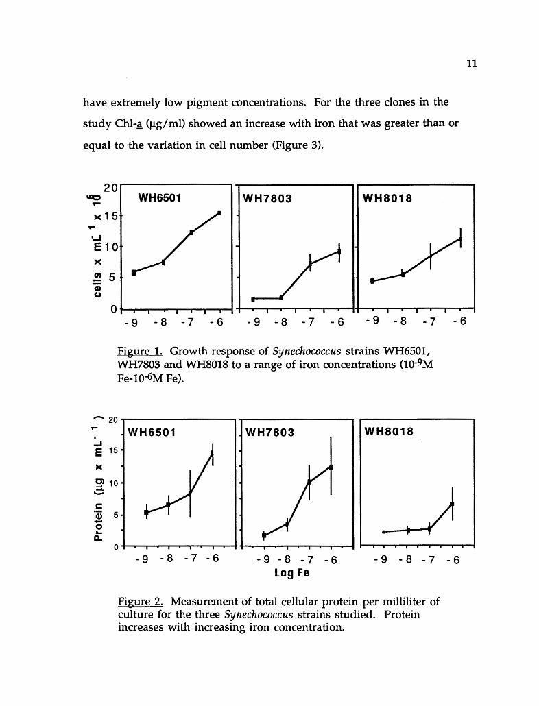
have extremely low pigment concentrations. For the three clones in the
study Chl-g_ (Jlg/ml) showed an increase with iron that was greater than or
equal to the variation in cell number (Figure 3).
20 u::o I ..... WH6501 II WH7803 II WH8018
><15 .... w E10 >< !!J. 5 -CD Cl
0 -9 -8 -7 -6 -9 -8 -7 -6 -9 -8 -7
Figure 1. Growth response of Synechococcus strains WH6501, WH7803 and WH8018 to a range of iron concentrations (1Q-9M Fe-10-6M Fe).
- 20 .....----------, .... I
..J E 15
>< C) 10 ::i .._.
c: "i 5 .... 0 ... Q.
WH6501 WH7803 WH8018
-6
11
0 I I I I I I I I I I I I I I I I I I I I I I I I I I I I I I I I I
-9 -8 -7 -6 -9 -8 -7 -6 Log Fe
-9 -8 -7 -6
Figure 2. Measurement of total cellular protein per milliliter of culture for the three Synechococcus strains studied. Protein increases with increasing iron concentration.

12
0.3,------------------- ~------------------ ,-------------------- WH6501 WH7803 WH8018
..J E 0.2
>< C) :i. -0.1 as .c 0
0.0 I .-- I I I I I I I I I I I I I I I I
-9 -8 -7 -6 -9 -8 -7 -6
Log Fe -9 -8 -7 -6
Figure 3. Chi-~ concentration for the three Synechococcus strains sampled. Chi-~ concentration increased with an increase in iron.
Phycobiliprotein pigment (PBP) content can make up a substantial portion of
the cell protein (15-25%) and varies among different species in the relative
composition of phycoerythrin (PE), phycocyanin (PC), and allophycocyanin
(APC). For marine Synechococcus, phycoerythrin occurs in the highest ratio.
Total PBP concentration increased with iron for all three clones, but each
showed a distinct response to Fe availability. For WH6501 PBP increased
proportional to protein, maintaining an average PBP: protein ratio of 23%. A
relatively constant fraction of that PBP was PE (43% ± 9%). WH8018 also had
a relatively constant PBP:protein ratio (24.4% ± 9.6%), and the ratio of PE:PBP
was also similar (46% ± 1.6%) for all but the lowest iron concentration in
which PE was 59% of the PBP. In contrast, PBP: protein in WH7803 had a
sixfold increase with iron and the relative PE content decreased with
increasing iron concentration (Table I). Previous studies using Synechococcus
and Trichodesmium to which iron was added as aeolian dust and as FeEDTA

resulted in increased concentrations of phycoerythrin and Chl-_g_ for both
organisms (Rueter and others 1991).
TABLE I
13
THE EFFECT OF IRON ON PHYCOBILIN CONTENT OF SYNECHOCOCCUS WH7803 AFTER SIX DAYS (THE DATA ARE THE MEAN OF TWO
EXPERIMENTS ± RANGE)
Iron Concentration PBP Content (J.tg/ mL) % Phycoerythrin
10-9M Fe 0.047 ± 0.003 39 10-8M Fe 0.160 ± 0.017 47 10-7M Fe 0.895 ± 0.075 30 10-6M Fe 2.245 ± 0.205 24
Cultures of marine Synechococcus WH6501 were grown at several
different iron concentrations to examine the effect of iron on ALA synthesis
rate. Chl-~ protein, PBP and cell counts were measured daily on these
cultures to ensure that cells harvested were still in exponential growth.
Cultures were spiked with 10 times more iron relative to the initial iron
concentration and incubated from 12 to 20 hours. ALA synthesis was
measured at 4 hour intervals commencing after 12 hours. Cultures grown at
5 X 10⁻⁸M Fe and spiked to 5 X 10⁻⁷ Fe prior to the 12 hour incubation had a
higher rate of ALA synthesis 17 hours after the addition of iron (Table II). In
two similar shipboard experiments 10-7M FeEDTA added to natural samples
ofTrichodesmium produced an increase in ALA synthesis rate by 17 and 42%
of the control after a 3 hour incubation. Natural samples of Trichodesmium

to which FeEDTA and dust were added had higher concentrations of Chl-~
and PE with increased iron concentration (Table III).
TABLE II
INCREASE IN ALA SYNTHESIS RATE IN SYNECHOCOCCUS WH6501 CULTURES PREINCUBATED WITH 5X10⁻⁷ M Fe FOR 16 HOURS
ALA svnthesis rate
No iron added I 5x10⁻⁷M Fe
3.0 Jlmol/h I 3.9 Jlmol/h
TABLE III
14
SHORT TERM REPONSE (13 HOURS) OF Chl-A AND PHYCOERYTHRIN SYNTHESIS IN TRICHODESMIUM TO ADDED IRON
PE Chl-~ (Relative
Treatment (Jl~/ml) fluoresence units)
+ 20nMFe .065 1.4
+ 100 nM Fe .073 1.4
+ 500 nM Fe .080 2.0
+ 1 mg dust ---- 2.2
+ 5 mg dust .058 3.0
+ 25 mg dust .067 4.4
DISCUSSION
The range of responses of the Synechococcus cultures to iron was much
greater for Chl-~ and phycobilin pigment proteins than it was for cell number

15
or protein. Cell number was 2.5-7.7 times higher from lowest to highest iron
concentration for these strains. Protein increased from 3-7.8 times, and Chl-g_
increased from 2.8-28 times. The PBP content was similar to protein. These
relative changes in cell composition are much lower than the changes in
iron availability which ranged from l0-9 to 10-6M Fe (a 1000-fold range). This
range drops to 500 if iron contamination estimates in AQUIL (Anderson and
Morel 1982) are taken into account. Cellular response to iron for the
measured parameters was less than linear to available iron and cellular iron.
A regression of cell parameters against total iron concentration results in
functions that are roughly the cube root of iron cocentration for cell density
and total protein and roughly the square root of iron concentration for
pigments and productivity rates. This suggests that iron-limited cells
maintain a higher iron use efficiency resulting in only slightly diminished
growth capacities by conserving crucial iron containing components whereas
increased iron availability has diminishing marginal returns in terms of
biomass, pigments and productivity. Although the wide range and rapid
response of Chl-g_ to iron addition is a good indicator of iron limitation it is
not a good choice for use as a normalization parameter since iron availability
is not consistent either in culture or in the natural environment.
Although the results are preliminary, a definite stimulation of ALA
synthesis following incubation with increased iron concentrations was
measured for Synechococcus cultures and natural samples of
Trichodesmium. This increase in ALA synthesis, an increase in Chl-g_ per cell
in the first 12-24 hours after iron addition and an increase in PBP
concentration with added iron shows that pigment synthesis is the first

16
parameter to respond to iron addition. This emphasizes the importance of
considering iron in algorithims for oceanic primary productivity via satellite
sensing based on pigment absorbance.

CHAPTER III
ENHANCEMENT OF DARK CARBON FIXATION IN IRON-LIMITED CULTURES OF SYNECHOCOCCUS WH6501
INTRODUCTION
The inorganic nitrogen assimilatory pathway involving the reduction
of nitrate to nitrite is energetically expensive requiring almost 50% as much
of the cells metabolic energy as carbon fixation (Syrett 1981; Turpin 1991). The
formation of glutamate from this assimilatory process requires ATP and
reductant generated from photosynthesis thus tightly linking light to
nitrogen metabolism. Iron too has a vital role in nitrate metabolism since the
iron-containing protein ferredoxin serves as the electron donor for nitrate
and nitrite reductase in the cyanobacteria and both of these reductases require
iron as a structural component (Peschek 1979). Nitrate and nitrite reductase,
in addition to their dependence on reduced ferredoxin, are intimately
associated with photosynthetic membranes emphasizing the link between
nitrate reduction and reductive energy generated by photosynthesis (Flores
and others 1983). Even though these processes are all inter-related, it is
important to assess whether an algal population is limited by a single factor or
by a combination of light, nitrogen and/or iron. Vincent (1981) and Pick
(1987) have successfully identified nitrogen and phosphorus limitation of
algal populations by monitoring increases in 14C fixation rates with added
ammonium of phosphorus. The rapid uptake and assimilation of nitrate,

18
phosphate or ammonium into amino acids or sugar-phosphate metabolites
and the resulting availability of these metabolites creates optimal biosynthetic
machinery which produces accelerated rates of photosynthetic carbon fixation.
Stimulation by iron or recovery from iron limitation differs from this due to
its coupling with protein synthesis. There are no pools of apo-proteins which
iron insertion would make metabolically active. This chapter presents a
method for assessing iron limitation based on an enhanced rate of dark
carbon fixation in nitrate grown, iron limited Synechococcus cultures with
ammonium chloride added to a SJ!M concentration.
MATERIALS AND METHODS
Cultures of Synechococcus WH6501 were grown in AQUIL medium as
previously described in this thesis. Iron from a 10-2M FeEDTA stock was
added to give final concentrations of 1Q-8 M Fe and 1Q-7 M Fe. Cultures were
transferred into fresh media at 1Q-8 and 1Q-7 M Fe concentrations every six
days to insure that the cells were not nitrogen limited and remained in active
growth phase (Rueter and Unsworth 1991). At least three of these transfers
with a one-fifth volume inoculum (100ml) were carried out to allow the cells
to go through a minimum of six divisions and adapt to the iron content of
the medium.
Following the initial inoculation, cell number was measured daily by
microscopic counts. Total protein, Chi-~ concentration and N03 were
measured as previously described (Rueter and Unsworth 1991; Rueter and
Ades 1987). Carbon fixation rate was estimated by the uptake of 14C. Cultures
were spiked with NaH14C03 that had been passed through a Chelex-100

19
column to remove contaminating trace metals. 15-mL aliquots in Oakridge
polycarbonate tubes were spiked with 5 mM ammonium chloride to a final
concentration of 5 J.!M and replicate samples for each iron concentration were
incubated for three hours both in 70J.!mol photons m-2s-1 and in foil wrapped
tubes for dark incubation. For use as controls, untreated aliquots from both
iron concentrations were incubated under the same conditions. Following a
three-hour incubation, samples were filtered onto 0.2Jlffi Nuclepore filters,
the filters dried and uptake rates measured with a liquid scintillation counter.
Culture and analytical methods are more completely described in Appendix A
and B.
RESULTS
Cultures were sampled daily over a 4-5 day period for cell number, total
protein and Chl-~ concentration. The 1Q-7 M Fe cultures had higher
concenrations of total protein and Chi-~ (Table IV) and a faster growth rate
than the iron limited (1Q-8 M Fe) cultures (Figure 4). Amounts of the
measured parameters for each iron concentration varied between
experiments a, band c since cultures for each experiment were at slightly
different points in their growth curve when sampled. All cultures, however,
were sampled during active growth phase. Cellular responses to these
parameters correspond to previous results obtained with Synechococcus
WH6501 grown at the same iron concentrations (Rueter and Unsworth 1991).
During the sampling period, growth rates at each of the iron concentrations
never led to the depletion of nitrate from the medium. Light-incubated 1Q-7
M Fe cells had higher 14C fixation rates than 10-8 M Fe light-incubated cells,

20
but normalization of these rates to Chl-g_ content showed uptake rates for 10-7
M Fe cells to be only 62-83% of the iron-limited (10-8 ) culture (Table V).
TABLE IV
CELLULAR PARAMETERS AND DARK CARBON FIXATION RATE FOR FOUR EXPERIMENTS. "EXPERIMENT A" WAS MEASURED AFTER FOUR
DAYS AND B,C AND D WERE MEASURED AFTER 5 DAYS
Protein Chl-~ dark 14C fix Experiment Fe cone. (Jlg/mL) (Jlg/mL) +NH4 -NH4
a 10-8 6.9 .088 1074 720 10-7 14.4 .312 453 481
b 10-8 4.8 .088 198 162 10-7 8.7 .163 122 185
c 10-8 12.6 .195 908 605 10-1 17.4 .346 588 651
d 10-7 12.0 .130 150 175 5x10⁻⁷ 15.0 .150 170 166
This suggests that the iron limited cells maintained an efficient energy
metabolism in the light and that reduced photosynthetic capacity is not solely
responsible for lowered nitrate reduction. Ammonium addition to both low
and high Fe cultures prior to light incubation produced only a slight effect on
the carbon fixation rate (a mean increase of 1.3% ± 3.5%). Dark carbon fixation
rate in these cultures showed two interesting features. First, there was a
significant dark carbon fixation rate which ranged from 1.3-11% with a mean
of 6.8% of the light carbon fixation rate. Second, the addition of ammonium
prior to incubation resulted in an enhanced dark carbon fixation rate. This

21
enhancement, calculated as [(+NH4/NH4)-1]x100%, ranged from 22-50% for
10-8M Fe cultures spiked with ammonium compared to untreated cells. No
enhancement was observed for 10-7 M Fe samples (Figure 5).
78+7~----------------------------~ 7.008+7 -,----------------.
10..8M Fe 10..7 M Fe Iii
68+7- 6.00e+7-
58+7-t I 5.008+7-
~4.00e+7 ~ •
..J • E 4e+7 .. II & a & . fl)
fl)
~ 38+7 El ~ 3.00e+7-
. • • 28+7 • 2.00e+71 ' • • 1e+7~ ' 1.008+7
* I • II
II Oe+O 0.00e+0 1 I I I I I I I I I I I
0 1 2 3 4 5 6 0 1 2 3 4 5 6
Days Days
Figure 4. Growth response of Synechoccuccs WH6501 to iron grown with 10-8M Fe (A) l0-7 M Fe (b). The mean value for three separate experiments is shown; a =a,c =b,• =c.
TABLE V
CARBON FIXATION RATE FOR CELLS GROWN IN THE LIGHT WITH NO AMMONIUM ADDED. RELATIVE CARBON FIXATION GIVEN IN
COUNTS PER MINUTE PER MICROGRAM Chl-A
CPM/ J.LgChl-ft Ratio Ex~:eriment 10-7 10-8 l0-7 /l0-8
a 1.2 X 10⁵ 1.4 X 10⁵ 0.83 b 1.6 X 10⁴ 2.0 X 10⁴ 0.80 c 1.6 X 10⁴ 2.6 X 10⁴ 0.62

1000-------------------------------------------------,
:E a. 0
50%
• Dark II Dark+NH4
800
<0% 600
400
200
0 ...,__ _ _..
-8Fe -7Fe
Figure 5. Enhancement of dark carbon fixation rates with addition of ammonium for "Experiment c".
22

23
In a fourth experiment, one large culture was grown through the same
transfer regime at 1Q-7 M Fe and monitored in the same manner as above.
Twenty-four hours prior to assaying for the ammonium effect on dark carbon
fixation rate, the culture was split and 5 X 1Q-7 M Fe was added to one of the
cultures. Protein concentration, Chl-g_ concentration and CPM (counts per
minute)/f..Lg Chl-~ were 25%, 15% and 85% higher respectively for the 5 X 1Q-7
Fe culture. There was no light or dark enhancement effect on samples with
added ammonium (Table IV). This suggests that iron controls growth rate;
even though 1Q-7 M Fe cells are "iron-limited" in that they grow better with
added iron, there is a physiological difference between these cells and 10-8M
Fe grown cells.
DISCUSSION
In this study and previous work (Rueter and Unsworth 1991) there
seems to be a physiological difference between cells grown at 1Q-8 M Fe and 10-
7M Fe. The response of WH6501 cells to restricted iron nutrition (lQ-8 Fe) is
characterized by slower growth rates, reduced concentrations of Chl-~ and
protein and lower carbon fixation rates. These results emphasize the crucial
roles that iron plays in metabolism and this integrated control makes it
impossible to predict whether a cell is iron-limited simply by looking at ratios
of these parameters as might be done for nitrogen or phosphorus limitation.
We were able to demonstrate that ammonium-dependent dark carbon
fixation enhancement is a feature of iron-limited cultures and is not seen in
iron-replete cultures.

24
We hypothesize that ammonium enhanced dark carbon fixation rates
for iron-limited cultures is an effect of the decrease in the cells ability to take
up nitrate from the media and to undergo subsequent intracellular reduction
of this nitrate to ammonia. Because metabolism is slowed due to iron
deficiency, the potential for carbon flux is not realized. Thus, ammonium
addition to iron-limited cultures causes an acceleration of amino acid
synthesis due to a metabolic energy requirement that is much less than that
required for the reduction of nitrate. This establishes that iron nutrition
plays a biochemical role in nitrogen assimilation, but it can not be determined
from our results what specifically is restricting the reduction of nitrate to
ammonia. We offer two possible explanations. First, that iron-limited cells
have a low efficiency of energy transfer due to ferredoxin, which requires
iron, operating as electron donor. Although nitrogen is available in the
medium, the cells are unable to use it effectively. Second, the cellular
efficiency of the iron-containing enzyme nitrite reductase, which relies on
ferredoxin as its immediate electron donor may be a site for the limitation of
nitrate assimilation. Regardless of an individual or collective effect causing
this restriction there is insufficient reduction of N03 and N02 to match the
potential carbon flow to amino acid synthesis.
In addition to the primary uptake of C02 via the carboxylation of RuBP
in the Calvin cycle by cyanobacteria, significant quantities of 14C are
incorporated into glutamate, aspartate, alanine and phosphoenyl pyruvate
(PEP) (Tabita 1988). The carboxylation of PEP by PEP carboxylase which
ultimately results in the flow of carbon to amino acids such as glutamate and
aspartate is evidence of an alternative C02 fixation pathway that is important

25
and which significantly complements the Calvin cycle in cyanobacteria
(Tabita 1988). The fixation of 14C via this alternative pathway occurs
predominately at low light and may be advantageous to cyanobacteria during
limiting light conditions by enabling them to preferentially channel carbon to
amino acids and bypass the more energy expensive Calvin Cycle.
The ammonium enhancement technique that we are proposing could
be a useful assay in supporting the contention that populations of
Synechococcus in nitrate rich waters are Fe limited. Knowledge of the iron
nutritional status of these populations in turn would indicate whether the
cells are contributing to new production or are relying on regenerated
ammonium (Wheeler and Kokkinakis 1990) in which case there would be no
evidence of ammonium enhancement.

CHAPTER IV
IRON REGULATION AT THE GENETIC LEVEL: DO CYANOBACTERIA POSSESS A SEQUENCE HOMOLOGOUS TO THE E. COU FERRIC UPTAKE
REGULATION (FUR) GENE?
INTRODUCTION
Many bacteria, including cyanobacteria have adaptations that allow
them to maintain efficient energy harvesting and transduction during
periods of iron limitation. The mechanism of regulation of iron uptake and
metabolism is well understood in E. coli. During periods of iron starvation
they excrete siderophores which are specialized high affinity iron transport
systems (Simpson and Neilands 1976) and synthesize membrane proteins that
transport iron into the cell. The characterization of five iron transport
systems associated with siderophore production in E. coli has been the basis
for a focus on molecular techniques as a tool for identifying a similar iron
regulation mechanism operating in cyanobacteria that might explain their
widespread success. The response in E. coli involves 26 genes, all of which
are derepressed by iron limitation (Schaffer and others 1985). Many
freshwater cyanobacteria are known to produce siderophores during periods
of iron stress in their natural environment. Likewise then, it would seem
that marine cyanobacteria such as Synechococcus spp., that have optimized
their cellular iron metabolism and uptake to maintain successful growth rates
in a system often limited by iron, light and nutrient availability would posses
the same or a similar mechanism.

27
This chapter presents work that looked for genes in cyanobacteria that
are homologous to the ferric uptake regulation gene (fur) in E. coli.
Zetaprobe membrane blot hybridizations containing chromosomal DNA
from four different species of cyanobacteria were probed with a 1.1kb fragment
of the iron-regulated E. coli fur gene to determine if a similar gene sequence
could be found in cyanobacteria. Three freshwater and one marine strain
were selected for their physiological and biochemical characteristics.
Anabaena 7120 fixes nitrogen and produces siderophores. DNA from two
other freshwater strains (1 PCC7942 which contains an iron regulated gene
(irpA, (Reddy et al. 1988) and produces a hydroxamate siderophore (Scanlan
et al. 1989) and 2) PCC6301 which has a close genetic relationship to PCC7942
were compared. A fourth marine form, Synechococcus WH6501, does not fix
nitrogen and has no known production of extracellular siderophores but is
able to grow efficiently at low light and under conditions of nutrient and iron
limitation. DNA from each organism underwent complete digestion with
several restriction enzymes and analysis of the Southern hybridizations
revealed weak hybridization in Anabaena 7120, and Synechococcus PCC6301
and PCC7942. No band was observed for the DNA from the marine
Synechococcus.
MATERIALS AND METHODS
Cultures of Anacystis nidulans R2 (PCC7942) were obtained from the
laboratories of Susan Golden and David Laudenbach. Anacystis nidulans
was grown in BG-11 medium (Thiel and others 1989) at 25°C under cool
white fluorescent bulbs. Cultures were inoculated in sterile media that had

28
been autoclaved for 20 minutes and bubbled with 1% C02 to resolubilize
precipitates and then grown in sterile glass Ehrlenmeyer flasks. Cultures of
Synechococcus PCC6301 and Anabaena 7120 obtained from John Goldbeck,
PSU and William Fish, OGI respectively, were grown in Kratz and Meyers
medium also using sterile technique. All three strains received the same
irradiance levels and culture vessels were manually agitated daily and
periodically checked for bacterial contamination. Synechococcus WH6501
was obtained from the Center for the Culture of Marine Phytoplankton.
Cultures were grown as previously described. Chromosomal DNA was
prepared from 200-SOOmL to obtain a minimum of Sflg of DNA. DNA from
the freshwater species was isolated by the method of Mazur et al. (1980) and
DNA was isolated from Synechococcus WH6501 by the methods of Wood and
Townsend (1990).
A bacterial stab consisting of the P ACYC184 vector with plasmid
pMH15 which carries a 2.2kb insert in containing the E. coli fur gene was
provided by Klaus Hantke (University of Tubingen). To identify sequence
homology between the fur gene and cyanobacterial DNA, a 1.1kb Acci/Bgli
fragment containing the fur gene was used as a hybridization probe. This
probe was prepared by a double restriction digest of pMH15 obtained from
mini plasmid preps (Maniatis and others 1982) of SmL overnight bacterial
cultures. The correct fragment was determined by electrophoresis of the
restriction digest on a 1% LMT (low melting temperature) agarose gel
containing ethidium bromide and comparison to Lambda/Hindiii and
PBR322/ Alu size markers (Figures 6 and 7) The correct band was excised from
the gel and purified from the agarose using a Prep-a-Gene kit (BioRad).

c.. .a ~
c ~ .§
Lambda DNA!Hindlll marker
102
lq
\ 1% agarose
101 t'-
' a. "'Ill
"a.. ~
1'-a ~ -........ ~
' ..... 100 ~
~
--------- ----------- --------------------------- - l·~
1 o-1
0 2 3 4 5
distance migrated (em)
Figure 6. Standard curve generated from a 2 hour restriction digest of Lambda DNA with Hindiii. By plotting the distance traveled on the gel for the Acc/Bgl restriction of plasmid pMH15, the 1.1 kb. insert could be identified.
29
6

Q.
~ c ~ 0 c .¥
pBR322/Aiul marker
100 - ~
._ 1:1
" 1....
-
' """'1
I
L-- ' ..., _,
' " ~ ~ ___,
'a ..._,
I J J J J i
1 o-1
4 5 6 7
distance migrated (em)
Figure 7. Standard curve generated by a two hour restriction digest of plasmid pBR332 with Alul. This marker is run with a Lambda/Hindlll marker to overlap the smaller kbp regions where the 1.1 kbp insert is located.
30

31
Chromosomal DNA from each species underwent complete digestion
with four different restriction enzymes (EcoRI, Hindll, Hindlll and Pstl) in a
SOJ.il volume and 20J.il of each sample was added to a 1% agarose gel (Figure
8). The gel was run at 20V for 16-20 hours and the DNA subsequently
transferred onto Zetaprobe membrane (BioRad) using the technique of
Southern. The probe was radiolabeled with 32p dCTP using a random
priming kit (Boehringer Mannheim). Hybridization of the fur probe to
chromosomal DNA digestions was determined by autoradiography. Cassettes
containing labeled membrane filters and X-ray film (Kodak) were foil
wrapped and placed in a -70°C freezer for 2-3 days. (For a more detailed
description of protocols such as prehybridization, hybridization and washing
conditions; see Appendices). The size of the band (in kb) was measured by
comparison to a pBR322 marker standard run on each side of the sample
wells.
RESULTS
The weak hybridization of the E. coli fur probe to DNA from PCC7942,
6301 and Anabaena 7120 is particularly significant due to the fact that all of
these strains produce siderophores at low iron concentrations. In E. coli, the
repression of the enzymes that synthesize siderophores are effected by the Fur
protein which is in turn coded for by the fur gene. When iron is readily
available to the cell the Fur potein binds to iron forming a repressor complex
that binds to an "iron box." The binding of this complex to the "iron box"
blocks transcription of downstream genes. When intracellular iron
concentration is lowered the enzymes are derepressed and siderophores are
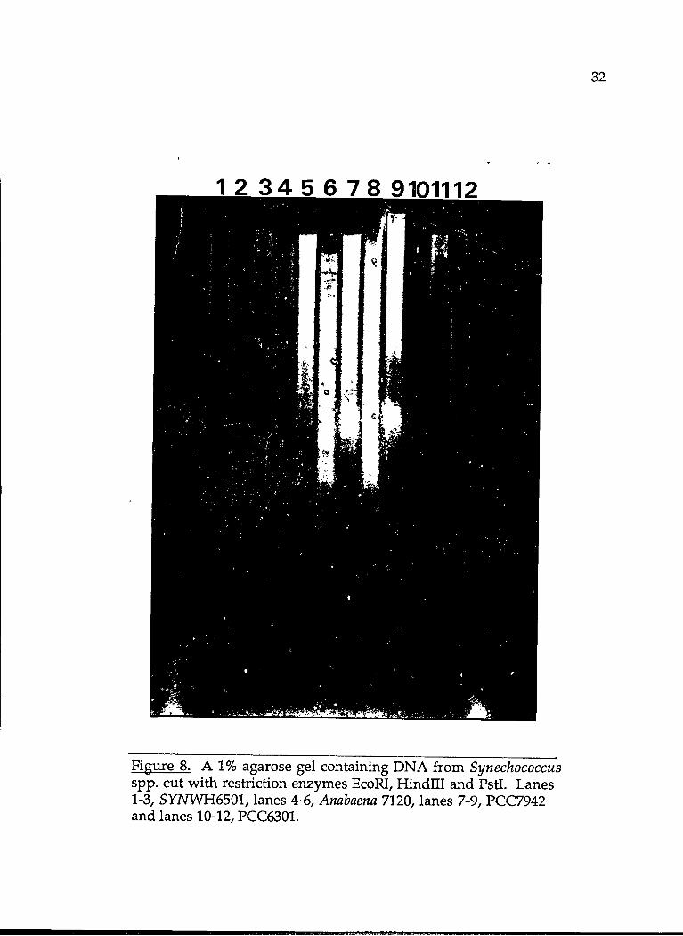
Figure 8. A 1% agarose gel containing DNA from Synechococcus spp. cut with restriction enzymes EcoRI, Hindiii and Pstl. Lanes 1-3, SYNWH6501, lanes 4-6, Anabaena 7120, lanes 7-9, PCC7942 and lanes 10-12, PCC6301.
32

33
synthesized. Hybridization of the E. coli fur gene to cyanobacterial DNA
offers evidence that the two gene sequences are homologous and suggests
then that a similar mechanism for iron uptake is operating within these
organisms. In contrast, the absence of hybridization of the fur probe to DNA
from marine Synechococcus using the same methodology indicates that
possibly a different mechanism is regulating iron aquisition. This
information on its own is not be significant, but combined with the absence of
measurable siderophores from marine strains may indicate a fundamental
difference in the iron uptake mechanisms.
DISCUSSION
Since iron supply and iron nutrition in these two systems is quite
different, (Hutchins and others 1991) the presence of distinctly different
mechanisms for optimizing cell growth in iron-limited environments is not
surprising. The results do however, direct a focus on acquiring knowledge of
what regulates the seemingly competitively advantageous strategies that
enable Synechococcus spp. to achieve its numerical abundance. Such a
regulatory mechanism, like siderophores for the freshwater species, will
possibly be linked to the water column dynamics of iron input and
availability in the marine system and would be of great significance in
understanding the separate strategies for competition and success of
cyanobacteria in these two natural environments. Probing DNA from
Synechococcus 7002, a marine cyanobacteria that produces siderophores, with
the E. coli fur gene is one approach that may offer more direct evidence for
the occurrance of different uptake mechanisms.

34
One complication of cyanobacterial molecular genetics is that the
morphological and physiological diversity of the group creates the necessity of
independent interpretation of data since findings for one species cannot
necessarily be applied to another (Golden and others 1989). This preliminary
data then, while providing evidence for different iron uptake mechanisms
for freshwater and marine cyanobacteria, demands a more detailed analysis of
gene expression in these organisms enlisting the use of reporter genes and
regulatory sequences. The combination of these genetic approaches and the
wealth of information on physiology and biochemical aspects of the cell
provide a powerful tool for understanding the complex systems operating in
these organisms. An example of this is the absence of siderophore production
determined physiologically and the absence of homology to the fur probe
determined with genetic techniques.
To date, the naturally transformable Synechococcus PCC7942 has been
chosen as a model system for studies on photosynthesis and iron regulation
due to its ease in genetic manipulation. Cloning in PCC7942 and other
transformable cyanobacteria can be carried out using shuttle vectors which
replicate in both E. coli and cyanobacterial cells or by direct incorporation of
the DNA into the genome (Friedberg 1988). It is through these
transformations and the use of reporter genes that we hope to further
elucidate the iron uptake mechanisms in marine and freshwater
Synechococcus spp. Although marine Synechococcus spp. have almost the
same morphological characteristics as PCC7942, only Matsunaga et al. (1990)
have successfully transformed a marine strain. While techniques are being
developed to make marine Synechococcus amenable to genetic manipulation,

35
more sophisticated techniques for the already amenable PCC7942 could
potentially unfold significant findings on iron regulation in the
cyanobacteria. Our lab is in the process of transforming PCC7942 with the
shuttle plasmid pSGlll which contains Ampr, lac Z as a reporter gene and the
E. coli "iron box" sequence to provide an iron controlled reporter plasmid.
This strategy has been used by Calderwood (Calderwood and Mekalanos 1988)
to confirm "Fe box" control in E. coli. Iron regulation of this gene could be
determined by measuring the activity of the reporter gene transcriptional
product, in this case iS-galactosidase in transformed cells of 7942. Cells grown
at low iron concentrations should have high levels of activity and the
addition of iron should cause a decrease the measured activity.

CHAPTER V
SUMMARY AND CONCLUSIONS
An optimal photosynthetic apparatus relys on the efficient transfer of
light energy from Chi-~ and the phycobilin pigments which are important
accessory pigments in cyanobacteria. Iron limitation results in decreased
synthesis of these pigments impairing energy transfer for carbon and nitrogen
metabolism which depend on reduced ferredoxin generated from
photosynthesis. One aspect of the effect of low iron on nitrogen metabolism
was explored further in marine strains of Synechococcus. Cells grown on low
iron and high nitrate and incubated with added ammonium in the dark had
high rates of dark carbon fixation. This response was not seen in cells grown
with high nitrate and high iron concentrations. We propose that this
increase reflects a shift to an alternate C02 fixation pathway that enables the
cell to bypass the more energetically expensive photosynthetic and nitrate
reduction pathway and preferentially channel carbon to amino acids and
other important cell components. We suggest that this finding could be
important for developing an assay that would identify iron-limited
populations in nitrate-rich waters.
The physiological and biochemical aspects of photosynthesis and
primary productivity in cyanobacteria has been the focus of extensive research
and as a result, much is known about these autotrophic organisms. Through
the recent successes of genetic manipulation using E. coli techniques on

37
cyanobacteria many of the processes controlling primary productivity can
now be analyzed at the molecular level. For example, the recent cloning and
sequencing of the nitrogen fixation (nif) gene has been useful in determining
factors regulating expression of the nitrogenase genes in natural systems. The
significance of homology of the E. coli fur gene to cyanobacterial DNA
discussed in this work is the first step in determining a sequence in
cyanobacteria that regulates the assimilation of iron which is central to an
efficient photosynthetic apparatus and energy transfer.
The degree of iron stress linked to co-limitation by light or nitrogen
availability quite possibly determines the mode by which cyanobacteria select
competitive mechanisms for success in dilute open ocean systems. This work
applies physiological and molecular biology techniques to underline the
geochemical importance of cyanobacteria and their role in controlling
primary productivity in the open ocean. Continuation of molecular
techniques to construct reporter plasmids for looking at gene expression will
be instrumental in identifying iron controlled genes in the cyanobacteria that
can further elucidate how these organisms obtain and transfer iron.

REFERENCES
Alberte, R.S., A.M. Wood, T. A. Kursar, and R. R. L. Guillard. "Novel phycoerythrins in Marine Synechococcus spp.: characterization and evolutionary and ecological implications." Plant Physiol. 75 (1984): 732 -739.
Anderson, M. A. and F. M. M. Morel. "The influence of aqueous iron chemistry on the uptake of iron by the coastal diatom Thalassiosira weissflogii." Limnol. Oceanogr. 27 (1982): 789-813.
Boyer, G. L., A. H. Gillam, and C. Trick. ''Iron Chelation and uptake." In The Cyanobacteria, ed. P. Fay and C. Van Baalen. 415-436. Amsterdam: Elsevier Science, 1987.
Calderwood, S.B. and J.J. Mekalanos. "Confirmation of the fur operator site by insertion of a synthetic oligonucliotide into an operon fusion plasmid." J. Bacteriol. 170 (1988): 1015-1017.
Carr, N.G. and M. Wyman. "Cyanobacteria: Their biology in relation to the oceanic picoplankton." In Photosynthetic Picoplankton, ed. T. and Li Platt W.K.W. 159-204. 214. Ottawa: Canadian Bulletin of Fisheries and Aquatic Sciences, 1987.
Chereskin, Barbara M. and Paul A. Castelfranco. ''Effects of Iron and Oxygen on Chlorophyll Biosynthesis." Plant Physiol 69 (1982): 112-116.
Duce, R. A. ''The impact of atmospheric nitrogen, phosphorus, and iron species on marine biological productivity." In The Role of Air-Sea Exchange in Geochemical Cycling., ed. P. Buat-Menard. 497-529. Dordrecht, Holland: Reidel Publ. Co., 1986.
Flores, D., J.L. Ramos, A Herrero, and M.G. Gurerrero. ''Nitrate assimilation by cyanobacteria." In Photosynthetic Prokaryotes: Cell Differentiation and Function., ed. G.C. Papageorgiou and L. Packer. 363-87. Elsevier Science Publishing Co., Inc., 1983.
Friedberg, D. ''Use of Reporter genes in cyanobacteria." Meth. Enzymol. 167 (1988): 736-747.

Glover, H. E., B. B. Prezelin, L. Cambell, M. Wyman, and C. Garside. "A nitrate-dependent Synechococcus bloom in surface Sargasso Sea water." Nature 331 (1988): 161-163.
Golden, S.S., M.S. Nalty, and D.C. Cho. "Genetic relationship of two highly studied Synechococcus strains designated Anacystis nidulans." 1 Bacteriol. 171 (1989): 24-29.
Golden, S.S. and L.A. Sherman. "Optimal conditions for genetic transformation of the cyanobacterium Anacystis nidulans R2." 1 Bacteriol. 158 (1984): 36-42.
Guikema, J. A. and L. A. Sherman. "Influence of iron deprivation on the membrane composition of Anacystis nidulans." Plant Physiol. 74 (1984): 90-95.
39
Hardie, L.P., D.L. Balkwill, and Jr. S.E. Stevens. "Effects of iron starvation on the physiology of the cyanobacterium Agmenellum quadruplicatum." Applied and Env. Microbiol. 45 (1983): 999-1006.
Hodge, V., S.R. Johnson, and E.D. Goldberg. ''Influence of atmospherically transported aerosols on surface water composition." Geochem. I. 12 (1978): 7-20.
Hutchins, D. A., J. G. Rueter, and W. Fish. "Siderophore production and nitrogen fixation are mutually exclusive strategies in Anabaena 7120." Limnol. Oceanogr. 36 (1991): 1-12.
Jones, K. L. "Analysis of ferredoxin and flavodoxin in Anabaena and Trichodesmium using fast protein liquid chromatography." Master of Science, Portland State University, Portland, Oregon, 1988.
Keller, M.D., W.K. Bellows, and R.R.L Guillard. "Microwave treatment for sterilization of phytoplankton culture media." I. exp. Mar. Bioi. Ecol. 117 (1988): 279-283.
Laudenbach, D.E., M.E. Reith, and N.A. Straus. ''Isolation, sequence analysis, and transcriptional studies of the flavodoxin gene from Anacystis nidulans R2." I. Bacterial. 170 (1988): 258-265.
Maniatis, T., E.F. Fritsch, and J. Sambrook. Molecular Cloning, a laboratory Manual. Cold Spring Harbor Laboratory, 1982.

40
Martin, J.H. "Glacial-interglacial C02 change: the iron hypothesis." 5 (1990): 1-13.
Martin, J. H. and S.E. Fitzwater. ''Iron deficiency limits phytoplankton growth in the north-east Pacific subarctic." Nature 331 (1988a): 341-343.
Martin, J. H. and S.E. Fitzwater. ''Iron deficiency limits phytoplankton growth in the north-east Pacific subarctic." Nature 331 (1988b): 341-343.
Martin, J. H. and R.M. Gordon. "Northeast Pacific iron distributions in relation to phytoplankton productivity." Deep-Sea Res. 35 (1988): 177-196. '
Matsunaga, T., H. Takeyama, and N. Nakamura. "Characterization of cryptic plasmids from marine cyanobacteria and construction of a hybrid plasmid potentially capable of transformation of marine cyanobacterium, Synechococcus sp., and its transformation." 24/25 (1990): 151-160.
Mazur, B. J., D. Rice, and R. Haselkom. ''Identification of blue-green algal nitrogen fixation genes by using heterologous DNA hybridization probes." Proc. Natl. Acad. Science (USA) 77 (1980): 186-190.
Moore, R. M., J. E. Milley, and A. Chatt. "The potential for biological mobilization of trace elements from aeolian dust in the ocean and its importance in the case of iron." Oceanologica Acta 7 (1984): 221-8.
Morel, F.M.M., J.G. Rueter, and D.M. Anderson. "Aquil: A chemically defined phytoplankton culture medium for trace metal studies." 1 Phycol. 15 (1979): 135-141.
Parsons, T.R., Y. Maita, and C.M. Lalli. "A manual of chemical and biological methods for seawater analysis." Pergamon Press, 1984.
Peschek, G.A. "Nitrate and nitrite reductase and hydrogenase in Anacystis nidulans grown in Fe- and Mo-deficient media." FEMS Microbiol. Lett. 6 (1979): 371-374.
Pick, F. R. and D. R. S. Lean. "The role of macronutrients (C, N, P) in controlling cyanobacterial dominance in termperate lakes." N. Z. I. Mar. & Freshwater. Res. 21 (1987a): 425-434.

Pick, F. R.] and D. R. S. Lean. "The role of macronutrients (C, N, P) in controlling cyanobacterial dominance in term per ate lakes." N. Z. I. Mar. & Freshwater. Res. 21 (1987b): 425-434.
Platt, T. and W. G. Harrison. ''Biogenic fluxes of carbon and oxygen in the ocean." 318 (1985): 55-58.
41
Platt, T. and S. Sathyendranath. "Oceanic primary production: Estimation by remote sensing at local and regional scales." 241 (1988): 1613-1619.
Reddy, K.J., G.S. Bullerjahn, D.M. Sherman, and L.A. Sherman. "Cloning, nucleotide sequence, and mutagenesis of a gene (irpA) involved in iron-deficient growth of the cyanobacterium Synechococcus sp. strain PCC7942." J. Bacteriol. 170 (1988): 4466-4476.
Rueter, J. G. and D. R. Ades. "The role of iron nutrition in photosynthesis and nitrogen assimilation in Scenedesmus quadricauda (Chlorophyceae)." J. Phycol. 23 (1987): 452-7.
Rueter, J. G. and N. L. Unsworth. "Response of marine Synechococcus (Cyanophyceae) cultures to iron nutrition." J. Phycol. 27 (1991): in press.
Rueter, J. G., N. L. Unsworth, R. W. Collier, and C. Meredith. "Uptake of iron by the marine cyanobacterium, Synechococcus ." Bioi. Ocean. Manuscript in prep. (man.):
Sandmann, G. and R. Malkin. "Iron-sulfur centers and activities of the photosynthetic electron transport chain in iron-deficient cultures of the blue-green alga Aphanocapsa." Plant Physiol. 73 (1983): 724-728.
Scanlan, D.J., N.H. Mann, and N.G. Carr. "Effect of iron and other nutrient limitations on the pattern of outer membrane proteins in the cyanobacterium Synechococcus PCC7942." Arch. Microbiol. 152 (1989): 224-228.
Schaffer, S., K. Hantke, and V. Braun. "Nucleotide sequence of the regulator gene fur." Mol Gen. Genet. 200 (1985): 110-113.
Schneegurt, M.A. and S.l. Beale. "Characterization of the RNA required for biosynthesis of a-aminolevulinic acid from glutamate." Plant Physiol. 86 (1988): 497-504.

42
Siegelman, H.W. and J.H. Kycia. "Algal biliproteins." In Handbook of Phycological Methods: Physiological and Biochemical Methods., ed. J.A. Hellebust and J.S. Craigie. 71-79. Cambridge: Cambridge University Press., 1978.
Simpson, F.B. and J.G. Neilands. "Siderochromes in cyanophyceae: isolation and characterization of schizokinen from Anabaena sp." I. Phycol. 12 (1976): 44-48.
Spiller, Susan C., Ann M. Castelfranco, and Paul A. Castelfranco. "Effects of Iron and Oxygen on Chlorophyll Biosynthesis." Plant Physiol 69 (1982): 107-111.
Syrett, P.J. "Nitrogen metabolism in microalgae." In Physiological bases of phytoplankton ecology. ed. T. Platt. 182-210. Hull, Quebec: Government Publishing Cen~re, 1981.
Tabita, F. Robert. ''Molecular and Cellular Regulation of Autotrophic Carbon Dioxide Fixation In Microorganisms." 52 number No.2 (1988): 155-189.
Theil, T., J. Bramble, and S. Rogers. "Optimum conditions for growth of cyanobacteria on solid media." FEMS Microbiol. Lett. 61 (1989): 27-32.
Turpin, D.H. ''Effects of inorganic N availability on algal photosynthesis and carbon metabolism." I. Phycol. 27 (1991): 14-20.
Vincent, W. F. ''Rapid physiological assays for nutrient demand by the plankton. I. Nitrogen." I. Plankton Res. 3 (1981): 685-697.
Waterbury, J. B., S. W. Watson, R. R. L. Guillard, and L. E. Brand. ''Widespread occurrence of a unicellular, marine, planktonic, cyanobacterium." Nature 277 (1979): 293-294.
Wheeler, P. A. and S. A. Kokkinakis. "Ammonium recycling limits nitrate use in the oceanic subarctic Pacific." Limnol. Oceanogr. 35 (1990): 1267-1278.
Wood, A. M. and D. Townsend. "DNA polymorphism within the WH7803 serogroup of marine Synechococcus spp. (Cyanobacteria)." I. Phycol. 26 (1990): 576-585.
Zhuang, G., R.A. Duce, D.R. Kester. ''The Dissolution of Atmospheric Iron in Surface Seawater of the Open Ocean." I. Geophys. Research. 95 (1990): 16,207-16,216.

S3illJINH::)3.L trnil.L1il::) 1V~1V
VXIGNHddV

44
Cultures of Synechococcus WH7803, WH6501 and WH8018 were
obtained from the Center for the Culture of Marine Phytoplankton, Bigelow
Laboratory for Ocean Sciences. Agar slants of Anacystis nidulans R2
(PCC7942) were kindly provided from the laboratories of Dr. Susan Golden
and Dr. David Laudenbach. Synechococcus 6301 was obtained from Dr. John
Goldbeck, Dept. of Chemistry, PSU. Anabaena 7120 was obtained from Dr.
William Fish, Oregon Graduate Institute.
Marine strains of Synechococcus spp. were grown in AQUIL medium
(Morel and others 1979) used previously in iron nutritional studies in our
laboratory because it has characterized speciation and low background iron
levels ( 2 X lQ-9 M or less). The synthetic ocean water (SOW), nutrients (N03-
and P04) and vitamins were made up in 1 liter batches and passed through a
column containing Chelex-100 ion exchange resin (Bio Rad) to remove any
contaminating trace metals. The trace metals (containing 5 X 10⁻⁶M EDTA
with no iron) were then added and the medium was filter sterilized by
passage through a 0.2Jlm pore size Nuclepore filter in an acid washed
polycarbonate (Millipore) filter apparatus. The medium then underwent a
final microwave sterilization procedure to reduce the risk of algal or bacterial
contamination (Keller and others 1988)
Cultures were grown in 1 or 2 L polycarbonate bottles at 25°C with cool white
fluorescent bulbs providing continuous illumination at 50-70 J..Lmol photons
m-2 s-1. These irradiance levels were chosen based on reports that these
clones (WH7803 and WH8018, for example) saturate in the region of 75Jlmol
photons m-2 s-1 and are very sensitive to photoinhibition (Alberte and others
1984). All culture work was carried out in a plastic enclosure containing a

45
REP A-filtered laminar flow hood drawing air from outside the enclosure to
create positive pressure in an effort to keep dust out. This "tent" is necessary
to prevent iron-bearing dust from contaminating the cultures.
The chelex-filtration apparatus set up was placed in the tent directly under the
laminar flow hood. Prior to experimentation, cultures were grown through
at least three transfers to insure that cells were acclimated to iron and
nutrient conditions.
Cultures of Anabaena 7120 and Synechococcus 6301 were grown in
Kratz and Myers medium that was made up in liter batches, autoclaved for 20
minutes, cooled to room temperature and bubbled with C02 until cleared of
precipitates. The required volume of medium was transferred to sterilized
glass ehrlenmeyer flasks prior to inoculation from a stock culture. Anacystis
nidulans R2 was maintained in BG-11 medium (Thiel and others 1989) that
underwent the same sterilization procedure described for Kratz and Myers
medium. Cultures of each strain were grown at 25°C with cool white
fluorescent bulbs providing continuous illumination at 100 Jlmol photons m-
2 s-1. Culture vessels for both the marine and fresh water cyanobacteria were
agitated daily by hand to provide sufficient gas exchange and to resuspend
cells. All plasticware and glassware used for trace metal work was soaked in
4% acid for a minimum of 4 hours and then rinsed five times in nanopure
water and dried under a hood or enclosed area.

46
BG - 11 GROWTH MEDIA FOR PCC 7942
Final Solution Molarity Stock
Solution Components Concentration mg/1 of Solution Solution
Solution 1 -- NaN03 1500.0 1.8 X 10⁻⁴ 100x
Solution 2 -- MgS04 _ • 7H20 100.0 4.0 X 10⁻⁸ 1000x
Solution 3 -- CaCl2 • 2H20 3.06 2.4 X 10⁻⁸ 1000x
Solution 4 -- K2HP04 • 3H20 40.0 2.3 X 10⁻⁸ 1000x
Solution 5 -- Na2C03 20.0 1.9 X 10⁻⁸ 1000x
Solution 6 --citric acid 6.6 3.1 X 10⁻⁷
ferric ammonium citrate 6.0 2.0 X 10⁻⁷
Na2MgEDTA 1.0 2.8 X 10⁻⁶ 1000x
Solution 7 -- H3B03 2.86 4.6 X 10⁻⁷
MnCl2 • 4H20 1.81 9.1 X 10⁻⁶
ZnS04 • 7H20 0.22 7.7 X 10⁻⁶
Na2Mo04 • 2H20 0.39 1.6 X 10⁻⁶
CuS04 • 5H20 0.08 3.2 X 10⁻⁵
Co(N03)2 • 6H20 0.05 1.7 X 10⁻⁵ 1000x

47
GROWTH MEDIA FOR SYN 6301 AND ANABAENA 7120
Stock Name Component Cone. (g/1) Final Molarity of Component
TRACE COMPONENT FeS04 • 7H20 6.0 3.60 x to-s NTA or EDTA 10.0 7.09 X lQ-5 MnS04 • H20 0.86 1.04 X 10⁻⁵ Zn504. 7H20 0.52 3.01 X 10⁻⁶ (Nlf4)6Mo7024 • 4H20 0.44 5.93 X 10⁻⁷ CoCl2 • 6H20 0.24 1.68 X lQ-6 NH4V03 0.028 3.99 x 1o-1 CuS04 • 5H20 0.24 1.60 X 10⁻⁶
MACRO COMPONENT MgS04 • 7H20 30.0 1.01 X 10⁻³ KN03 133.6 1.00 X 10⁻² Ca(N03)2 • 4H20 3.89 1.06 X 10⁻⁴ NaCl 14.0 2.00 X 10⁻³
PHOSPHATE COMPONENT K2HP04 105.4 1.01 X 10⁻³
BICARBONATE COMPONENT KHC03 151.4 2.52 X 1Q-3

SHflL>INH:)H.L '1V:)I~'10ISAHd GNV '1V:)IW3HJOIH
HXIGNHddV

49
Chl g_ content for all samples was determined by a common
fluorometric method using 90% acetone extractions (Parsons and others
1984). 10 mL replicate samples of each culture were filtered onto Whatman
GF/F glass fiber filters through a Gelman polycarbonate funnel attached to a
suction filtration apparatus set up. Two drops of saturated MgC03 was added
to each filter to prevent loss of the magnesium ion. Filters were then stored
in the freezer in a dark box until later analysis. Analysis of the frozen filters
involved grinding in 10 mL of 90% acetone (v /v) using a tissue homogenizer.
The sample was allowed to extract overnight in a dark box at 4°C and was
then analyzed fluorometrically on a Turner Designs Model 10 fluorometer
calibrated for chlorophyll ~· Micrograms Chl-~ were determined using the
equation (Fluorescence)x(.03862). This value is multiplied by the volume of
acetone (10mL) and divided by the volume of culture filtered (10mL) to give
f..Lg Chl-£l/mL of culture.
Two different methods for estimating phycoerythrin from marine
Synechococcus spp. were used. In experiments measuring only the increase
in phycoerythrin with added iron, the glycerol method of Carr and Wyman
was used. 1.5mL of each culture to be analyzed was pipetted into a plastic
cuvette andl.SmL of glycerol was also pipetted into the cuvette using a
Jetpipettor. The top of the cuvette was sealed with Parafilm and shaken
vigorously to mix the culture medium and glycerol. After all the air bubbles
had disappeared the fluorescence was measured on a Jasco FP 550
spectrofluorometer set at 510nm excitation and 562nm emission. The
observed increases in phycoerythrin are a result of the uncoupling of the

50
protein from energy transduction. The fluorescence in vivo of cultures with
no glycerol added was also measured as a baseline value for comparison.
Phycoerythrin(PE), phycocyanin (PC) and allphycocyanin (APC)
concentrations were measured spectrophotometrically after extraction using a
modification of the method described by Stewart and Farmer (1984). Cultures
were filtered onto Gelman filter type AE or Whatman GF /F and frozen until
analysis. Filters were thoroughly homogenized in a solution consisting of
0.25M Trizma Base (Sigma), 10mM disodium EDTA, and 2mg mL-1 lysozyme
at pH 5 using a tissue homogenizer. Samples were incubated in foil wrapped
tubes at 37 °C for 2 h and then 24 h at 4 °C. Following the 24 h incubation, the
pH was adjusted to 7.0 and the total volume (6mL) was increased to twice the
original with distilled water. The sample was filtered to produce a clear
homogenate. Phycobilin concentrations were calculated using an equation
correcting for spectral overlaps of the pigments (Siegelman and Kycia 1978).
This method was used when a Jlg/ Jll phycoerythrin content as a percent of
total phycobilins was measured; however, it is very time consuming and the
glycerol method is much more convenient for determining relative increases
or decreases in pigment concentrations.
MEASUREMENT OF TOTAL CELLULAR PROTEIN
All protein concentrations were measured using the BCA Protein
Assay (Pierce). This assay can be used in the presence of detergents without
interference. For all the samples in this study 1% w I v SDS (sodium dodecyl
sulphate) was used to completely solubilize all cell protein including
membrane-bound hydrophobic components. Based on culture density and

51
volume of filtered sample, either the "Enhanced" or "Room Temperature"
protocol described in the Pierce Protein Assay instruction manual was used.
50 mL of culture was filtered through Whatman GF IF glass fiber filters. In
many cases when cultures were being sampled daily for several physiological
parameters, less culture was used to conserve the volume. However,
depending on the density of the culture, less than 25mL for the marine
Synechococcusspp. did not usually produce a measurable amount of protein.
After filtration the filters were frozen and then ground with a teflon tissue
homogenizer in 3 mL of 1% SDS. Because this is a lot of filter for such a small
volume of liquid, initially grinding the filter in 1 mL of SDS and then two
steps of rinsing and grinding with the remaining 2 mL was done to get all of
the material. The supernatant from the homogenized filters was incubated at
4 °C overnight to insure complete solubilization of the proteins. The next
day, the tubes were spun for 5 minutes at high speed on a desk top centrifuge
to obtain a filter-free, clear supernatant. To 1 OOJ.LL of supernatant, 2 mL of
BCA Working Reagent was added. The working reagent contains detection
reagent mixed with 4% CuS04 in a 50:1 v/v ratio. For most of the protein
samples in this study the Enhanced protocol was used for measurement of
concentrations ranging from 10-250J.Lg/mL. In a few cases with more dense
cultures, the Room Temperature protocol was used for measuring protein
concentrations between 100-1200J.Lg/mL. Absorbance of the samples was
measured at 562 nm in a 1 em cell and the protein concentration determined
graphically using a standard curve generated with BSA (bovine serum
albumen) provided with the assay kit. A separate curve was generated for the
different protocols.

52
MEASUREMENT OF CARBON FIXATION RATES
Carbon fixation rate was estimated by the uptake of a 14(: stock that had
been passed through a Chelex-100 column (BioRad) to remove contaminating
trace metals. Prior to assaying for carbon fixation rate cultures were sampled
for Chi-~ nitrate, and protein concentration, and cell density. Cultures were
then spiked with 1 J!Ci/mL NaH14co3 and replicate 15mL aliquots were
incubated for three hours at 70Jlmol photons m-2s-1 and in foil wrapped
tubes for dark incubation. Carbon fixation rates for the ammonium
enhancement experiments were measured as above, but a separate set of
replicate tubes incubated at both light and dark conditions were spiked with
SJ!M ammonium chloride in addition to NaH14C03. Following the three-
hour incubation all tubes were removed from the light field and each sample
was collected onto a GF /F filter using a vacuum filtration apparatus. Filters
were placed in 7mL disposable polypropylene scintillation vials and allowed
to dry overnight. Once filters were dry 3 mL of Solvent Free liquid
scintillation cocktail (Isolab, Inc.) were added to the vials and samples were
counted in a Beckman Liquid Scintillation Counter.
CELL COUNTS
There is a great deal of difficulty encountered in obtaining accurate cell
counts for Synechococcus and cell density is a very important parameter for
the experiments in this study. Three different methods were used
throughout the work presented in this thesis; however, only one method was
used consistently during each individual experiment. 1) Coulter Counter:

53
cell density was measured using a Coulter Counter (Model2907 ZBI) fitted
with a 30 J.1M aperature. Difficulty was encountered in obtaining reliable cell
counts due to clogging of the aperture opening by clumped Synechococcus
cells. This was especially noticeable when counting iron-limited cells which
exhibited a great deal of clumping when viewed under the microscope.
Under these circumstances, samples were diluted to prevent clogging. To
insure the greatest accuracy possible, four to six counts were taken for each
sample and averaged. 2) Microscopic counts using a hemocytometer: 50J.1l of
culture was pipetted into a Fuchs Rosenthal ultrapure hemocytomer (1/16sq.
mm and 2/lOmm deep) and visually counted on a microscope equipped with
a lOOX objective. Cultures counted in this way usually required dilution prior
to counting. Clumping of cells was also a problem with this method because
it was difficult to distinguish individual cells. 3) Fluorescence microscopy:
Samples were filtered onto .4JlM Nuclepore filters (25mm) using a Millipore
glass chimney. To achieve even distribution of the sample on the filter, a
Whatman GF/F was placed between the membrane filter and the filter base.
Prior to placement of the membrane filter, the GF /F filter was placed on the
salt solution and vacuum filtered evenly onto the backing filter. After
filtration of the sample under low vacuum, the membrane filter was
removed and put (sample side up) on top of a drop of immersion oil on a
microscope slide. Another drop of oil was placed on top and a coverslip
gently pressed on top. The slide was then inverted onto a paper towel and the
excess oil blotted away and the cover slip gently flattened. Cells were then
counted on a standard Zeiss scope with a neofluar objective and an
epifluorescent system equipped with a filter set in which phycoerythrin

54
fluoresced orange. The number of cells mL-1 was calculated using the
following equation: Cells field-1 divided by mL sample filtered X (2 X 1o4)
(Waterbury and others 1979). This method was the most effective for
producing accurate cell counts, but it requires quite a bit of experimenting
with dilution of the culture to obtain a filtrate that is not too dense to count.
More than 50 cells per field is difficult to count accurately.

SHnL>INH:)H~ A.~'10IH 'MV'1n:)H'10W
:)XIGNHdciV

56
Chromosomal DNA from Anacystis nidulans R2 PCC7942,
Synechococcus WH6501, WH6301 and Anabaena 7120 was isolated from
cultures for use in Southern blot hybridizations with a radiolabled probe to
look for the presence of homologous sequences to the bacterial fur gene,
which plays an important role in iron regulation in E. coli (Schaffer and
others 1985). For the freshwater strains DNA was prepared using a slightly
modified method of Mazur et al. (1980) For DNA isolation from the marine
Synechococcus WH6501 a modified version of Wood and Townsend (1990)
was followed.
For PCC7942, PCC6301 and Anabaena 7120, 250-500mL of culture (500
ml yielded approximately 5J.tg of DNA) was centrifuged at 10,000rpm for 10
minutes in 250mL bottles using a Sorval RC-2 centrifuge. The supernatant
was removed and the remaining pellet washed once with fresh media and
centrifuged again as above. The supernatant was carefully drawn off with a
pipet leaving the pellet as dry as possible. The pellet underwent three
freeze/thaw periods followed by suspension of the thawed pellet in 5 mL of
TE buffer (Maniatis and others 1982). The suspension was transferred to a
15mL polyethylene centrifuge tube lysozyme was added to a final
concentration of 2mg/mL and the tube was incubated for 2 hours at 37°C.
25J.1L of Triton 100, 25J.1L of proteinase K and SDS (to a final concentration of
.5%) was then added and the tube incubated overnight at 50°C. The next day,
an equal volume of TE equilibrated phenol was added and mixed for 3.5
minutes by gently inverting the tube. The sample was then spun at
maximum speed on a desk-top centrifuge for 5 minutes. To the aqueous
layer, one volume of ice-cold 100% EtOH was added. The tube was inverted

57
until a white, stringy precipitate formed and this was spooled with the tip of a
pasteur pipet that had been sealed with a bunsen burner flame. The DNA
was dissolved in 2 mL of TE buffer (this can take several hours). The DNA
was then reprecipitated with 1.5 volumes of isopropanol and once again
spooled and rinsed with 70% EtOH. The excess EtOH was drained and the
DNA resuspended in 100J!L of TE in a 1.5mL Eppendorf centrifuge tube and
stored at -20°C until ready for use.
Extreme caution should be exercised when handling phenol. Gloves
and goggles should be worn during these steps and make certain that the caps
are tightened before inverting the tubes to mix. When removing the aqueous
layer from the phenol, avoid the white layer at the interface. This contains
proteins that will lower the purity of the DNA.
ANALYSIS BY EN2YME DIGESTION AND SOUTHERN BLOTTING
Approximately 1J,lg of DNA from each sample was digested for a
minimum of two hours with Eco RI, Hind II, Hind III and Psti in a SOJ!l
volume at 37°C. After several hours a 1/10 volume of loading buffer
(Maniatis and others 1982) was added to the restriction mix and spun in a
microfuge for 30 seconds. The entire sample volume was loaded into a 1%
agarose gel containing ethidium bromide. 10J!l of a molecular weight ..
standard containing a range of expected size fragments was loaded into the
wells at each side of the samples on the gel. The gel was covered with 1mm
of TBE buffer and hooked up to a power supply. The gel should be run at
100V for about 10 minutes or until the purple and blue dye bands can be seen
separately in the gel and then reduced to 20V and run for about 16 hours.

58
After electrophoresis the gel was placed in a glass pyrex dish and the DNA
was depurinated by gentle agitation in a .25N HCl solution for 10-15 minutes
(It is important not to exceed 15 minutes and 10 minutes is best). It is
important for the gel to be free floating during all periods of agitation. The
HCl was then poured off and the DNA denatured by soaking the gel in .SN
NaOH, 1M NaCl for 30 minutes with gentle agitation. After 30 minutes that
buffer was removed and the gel was neutralized by bathing for 30 minutes in
.SM Tris-HCl pH 7.4 and 3M NaCl. During the last soaking period, 5 sheets of
Whatman 3MM paper were cut to the exact size of the gel and one sheet of
Zetaprobe blotting membrane (BioRad) was cut slightly smaller than the gel.
The cut sheet of Zetaprobe was soaked in distilled water for 5 minutes prior to
use. Two sponges were pressed firmly together and placed in the pyrex dish.
lOX SSC is then poured over the sponges until the buffer is half way up the
side of the sponge. The gel was photographed on a UV transilluminator and
gently placed on top of the sponge and three pieces of wetted 3MM paper. A
gloved hand was used to press away any bubbles from between the gel and the
3MM paper. The gel was then flooded with lOX buffer and the presoaked
Zeta probe carefully placed over the top of the gel so that it is in perfect
alignment. It is important to keep smoothing the gel and membrane so that
there are no air bubbles. The gel and pyrex dish are then sealed with Saran
Wrap and, with a razor, the wrap around the gel is cut out creating a
"window" so that only the membrane is exposed. This insures that capillary
action will only go through the gel. The remaining two sheets of buffer
soaked 3MM paper are placed on top of the gel and smoothed. A 6-10 inch
stack of paper towels cut to the size of the gel are placed over the gel sandwich

59
and the entire stack is covered with a piece of plexiglass and a lOOml beaker
filled with water to use as a weight. The gels were blotted from 12 to 24 hours
after which the paper towels and 3MM paper was removed and the
membrane was rinsed briefly in 2X SSC. The membrane filter was air-dried
between two pieces of filter paper and, since hybridization was not carried out
immediately, the membrane was baked at 80°C for 30 minutes. It can then be
stored between two pieces of filter paper in plastic bags or plastic wrap at room
temperature.
It is important to use caution when doing all these procedures.
Ethidium bromide is a powerful mutagen and gloves should be worn at all
times. Gloves without powder are preferable since the powder often gets on
the gel and produces black spots all over the developed film.
PREPARATION AND RADIOLABELLING OF THE FUR PROBE
An agar stab of bacteria bearing plasmid pMHlS, which carries a 2.2 kb
insert in the pACY184 vector and contains the E. coli fur gene, was generously
provided by Klaus Hantke. Two SmL overnight cultures were grown up with
chloramphenicol and mini plasmid preps were carried out using Qiagen
columns (Qiagen) to insure that there was no RNA contamination. The
plasmid DNA underwent a double digestion with Accl and Bgll to isolate the
l.lkb fragment containing the fur gene. Isolation of the fragment was
accomplished by gel electrophoresis of the restriction mix on a 1% low
melting temperature agarose (SeaKem) gel. The DNA was run with
Lamda/Hindiii and pBR322/phiX markers to identify the correct band size.
Bands fluoresce when the gel containing ethidium bromide is placed on a UV

transilluminator and the bandscan be cut out of the gel using a razor blade.
The DNA was further purified using a Prep-a-Gene kit (BioRad). The DNA
was radiolabeled with SJll of 32p labeled dCTP using a random priming kit
(Boehringer Mannheim).
60
The membrane containing the DNA was placed in a Seal-a-Meal bag
and 15mL of prehybridization solution with 250Jlg/ml of denatured salmon
sperm DNA (to block non-specific binding) was added to the bag. The air
bubbles were squeezed from the bag and it was then sealed and placed in a
water bath at 37°C for 2 hours to overnight. The membrane filters underwent
prehybridization overnight along with labelling and incorporation of the
probe. After at least 4 hours of prehybridization, the probe was added to 15mL
of hybridization solution (prehyb. solution with 10% Dextran sulphate, .5%
powdered milk and 1% SDS added) and boiled for 10 minutes. The tube was
chilled to room temperature and added to the Seal- a-Meal bag containing the
Zetaprobe membrane. This hybridization takes place in a 42°C incubator or
water bath for four hours to overnight. Afterwards the hybridization
solution is poured off and the membrane is removed from the bag. This was
followed by four washes with 2 X SSC and .1% SDS. The first wash is for 15
minutes at room temperature and the subsequent washes are for 30 minutes
at 60°C. One seal a meal bag is used for the entire procedure. After each use it
is cut open, emptied, refilled and sealed. A plastic container can also be used
if one approximately the size of the gel can be found. After the final wash the
filter was dried, placed on a sheet of 3MM paper and wrapped in Saran Wrap
so that it was sealed, but the wrap wasn't overlapping at all. The wrapped
film was placed in a cassette with 1 sheet of X-Ray film (Kodak XAR-5) and

- 61
the cassette wrapped in foil and placed in a -70°C freezer for 24 to 48 hours. I
developed all the films after 2 days using a automatic developer for X-ray
film. The fragment sizes on the film were determined by comparing the band
distance to the molecular weight markers.
TRANSFORMATION OF PCC7942 AND WH6501 WITH PLASMID pSG111
Transformation of PCC7942 and WH6501 cells by the hybrid plasmid pSG111
was attempted using the methods of Golden and Sherman (1984). Cells were
grown in SOmL of their respective medias and spun down for 10 min at 10,000
rpm in a Sorval RC2B centrifuge. To 0.1ml of cells 0.1J.Lg of DNA (pSG111)
was added and incubated for 1 hour in the light with gentle mixing. The
transformation mixture was plated on BG-11 and AQUIL media agar plates
and incubated in the dark for 16 hr at 30°C followed by 8 hr under standard
illumination. A range of concentrations of ampicillin (20J.Lg/ml to 100J.Lg/ml)
was added to the plates by partially lifting the agar with a spatula and
underlayering .SmL of desired antibiotic concentration. Transformed
colonies appeared on the agar plates approximately 12 days following
incubation with the pSG111 plasmid DNA and were then transferred to a
liquid medium containing antibiotic.