Embed Size (px)
Citation preview
267The pig as animal model in biomedicine
E-mail: [email protected]
Pigs as model systems for biomedical research
Heiner NiemannInstitute of Farm Animal Genetics, Mariensee, Friedrich-Loeffler-Institut, Federal Research Institute for
Animal Health, Hoeltystr. 10, 31535 Neustadt, Germany
Pigs have a long standing and very successful history as biomedical model for studying human diseases and developing novel therapies mainly attributed to the many genetic, anatomical and physiological similarities with humans. Non-transgenic pig models have long been used for a wide range of human organ systems and diseases, and even complex metabolic disorders and have served as model for developing novel surgical techniques and endoscopic approaches, such as NOTES (natural orifice transluminal endoscopic surgery). The availability of the porcine genome and novel tools to add or delete specific genes significantly expands the potential for transgenic pig production. Somatic cell nuclear transfer has emerged as the preferred method for transgenesis. Well characterized transgenic pig models have been reported for Cystic fibrosis, the eye disease Retinitis Pigmentosa, atherosclerosis and diabetes. Transgenic pigs have been produced for modeling neurological diseases, including Alzheimer and Huntington disease, specific forms of cancer, and skin diseases. Transgenic pigs play an important role in developing functional porcine xenografts to combat the growing shortage of appropriate human organs for transplantation. Other important transgenic pig models include immunodeficient pigs and Oct4/GFP transgenic pigs for studies of reprogramming. Pig models will not replace the already existing mouse models but can provide significant novel insight into a variety of diseases, as mouse models frequently do not mimic the human situation. Transgenic pigs will also soon play an increasing role in the development of novel therapies based on stem cell technology. The biomedical use of pigs will also facilitate transgenic pig production for agricultural production.
Introduction
Pigs have a long standing and very successful history as biomedical model for studying human diseases and developing novel therapies. Domestic pigs and minipigs are the main categories that have been used as biomedical models. Usually minipigs are in shorter supply than domestic pigs and thus more expensive compared with domestic pigs, which cost more due to housing, feed and medication (Litten-Brown et al., 2010). The preferred use of pigs as model in biomedical research is attributed to the many anatomical and physiological similarities with humans. As humans, the pig is a monogastric omnivore. As result of a long domestication process a great variety of pig phenotypes exists worldwide that could be relevant for current human health research priorities, including obesity, diabetes and cardiovascular diseases. Given the high degree of similarity, many diagnostic, surgical or other medical techniques can be directly
ControlPigReprodIX.indb 267 5/23/2013 7:46:24 AM

268 H. Niemann
transferred from the pig into the clinic to help human patients. Another great advantage is the ability to maintain pigs under strict hygienic conditions, such as specific pathogen free (SPF) or gnotobiotic (completely sterile) conditions.
In addition, the high fertility of the pig makes it an attractive species for use in biomedical model application. Moreover, effective protocols are established for artificial insemination and embryo transfer for a long time. More recently, somatic cell nuclear transfer (SCNT) methodology has been improved and refined protocols for genetic modification of pigs have been established (Petersen et al., 2008; Hauschild et al., 2011; Garrels et al. 2012).
Worldwide, thousands of pigs are being used in biomedical research every year. In Germany, an average of 12 -13.000 pigs is being used per year in biomedical research. A Google search, using the three words “pig, model, research” yielded 140.000 hits in 2007, 6.7 million hits in 2008 and 29.9 million hits in 2013. This clearly shows the rapidly growing interest in the use of the pig as a biomedical model for the benefit of humans. One can discriminate between non-transgenic and transgenic pig models. The following gives a brief overview on the wide range of non-transgenic pig models followed by an update of the recent development of transgenic pig models.
Non-transgenic pig models
Pigs have been used as appropriate biomedical models due to genetic, anatomical and physiological similarities to humans (Litten-Brown et al., 2010). Tests have frequently been undertaken to investigate pharmacokinetics and pharmacodynamics of specific drugs. However, swine also have unique characteristics and husbandry requirements which must be taken into account when using the species as a biomedical model. Non-transgenic pig models have been employed for a wide range of human organ systems and diseases.
Head and brain injuries
Pig models have been developed for traumatic brain injury, including brain death to define critical parameters of ischemia and to study systemic reperfusion as a model for human brain death (Purins et al., 2011, 2012). Even four weeks old piglets have been used as model for studying traumatic brain injury for pediatric purposes (Friess et al., 2011). The optimal resuscitation strategy was developed in a domestic pig model of traumatic brain injury and hemorrhagic shock (Jin et al., 2012). Two days old piglets were also used to study the effectiveness of anti-inflammatory drugs for treatment of head injury (Friess et al., 2012).
Eye diseases
The porcine eye is very similar to the human eye showing an area of increased cone density arranged in a central horizontal band considered analogous to the human macula. Porcine eyes have been used to study age-related macular degeneration and to develop ophthalmological surgical treatments for human patients (Pennesi et al., 2012). An inducible photoreceptor damage porcine model was developed using chemical toxins. Sodium iodate (NaIO3) was an effective toxin for the pig eye and could thus serve as model to develop treatments to replace damaged photoreceptors (Noel et al., 2012).
Cardiovasculatory diseases
The pig has been widely used in preclinical studies to develop novel treatments for cardiovascular diseases that are a common reason for death of human patients. Most prominent are models for
ControlPigReprodIX.indb 268 5/23/2013 7:46:24 AM

269The pig as animal model in biomedicine
myocardial infarction and reperfusion, the hibernating myocardium, and for vulnerable plaques (Suzuki et al., 2011). Specific aspects of the treatment of coronary injuries were also investigated in the pig model, with emphasis on endothelial denudation and stent placement (von Bary et al., 2011). Novel treatments involving the application of stem cells and growth factors have been tested in the pig to study survival and regeneration of the infarcted pig heart. The combination of IGF-1 and HGF seems to be beneficial in this respect (Ellison et al., 2011). Multipotent stromal cells were successfully applied in a pig model to improve the situation after chronic myocardial infarction (Sato et al., 2011). Immediate implantation of bone marrow-derived cells into minipig myocardium after coronary artery ligation promoted neovascularization and improved myocardial viability (Ko et al., 2011). The intracoronary delivery of mesenchymal stem cells (MSCs) into the ischemic heart reduced malignant ventricular arrhythmias and improved cardiac performance (Wang et al., 2011). A novel coronary guidewire was successfully tested in a porcine model and emerged as an effective tool to improve transcoronary pacing (Heinroth et al., 2011). The pig has also served as model to study cardiac arrest and to develop strategies to overcome this pathology by applying therapeutic hypothermia and selective heart cooling (Wang et al., 2012; Li et al., 2012).
Vasculatory diseases
The pig has successfully been used as model to study the effects of implantation of MSCs into an aortic aneurysma injury. The orthologous implantation was successful, but long term effects remain to be investigated (Turnbull et al., 2011). Type B aortic dissection, which is the most common acute disease of the aorta and a life-threatening condition, has been studied in the pig model (Okuno et al., 2012). The pig was also successfully used as a model for ultrasound enhanced recombinant tissue plasminogen activator mediated thrombolysis in a carotid artery model (Hitchcock et al., 2011).
Drug-coated balloons have been tested as therapeutic approach for treating vasculatory diseases in familial hypercholesterolemic swine. These balloons were effective in reducing proliferation of the neointima cells (Granada et al., 2011). Intramural injection of complex lipids into the coronary arteries of pigs induced symptoms similar to human atherosclerosis (Tellez et al., 2011).
Pulmonary diseases
Transesophageal upper pulmonary lobectomy was successfully established in the domestic pig and is considered as a novel strategy towards scar free pulmonary lobectomy (Moreira-Pinto et al., 2012). A specific extracorporeal cardiopulmonary support system was tested in its effectiveness to rescue patients after massive pulmonary embolism. However, the optimal treatment is still unknown (Kjærgaard et al., 2012). New lung ventilator strategies, including an “open lung” system, were developed in the pig and turned out to be superior to standard strategies treating lung injuries (Albert et al., 2011). The pig has also served as model for developing new treatments of intra-abdominal hypertension, which is an important factor leading to increased morbidity and mortality in human patients. Application of positive end-expiratory pressure did not yield beneficial results (Regli et al., 2012). The pig also served as a model to test adenosine A2A receptor agonists for treating reperfusion injury in a preclinical lung transplantation model (LaPar et al., 2011). In a pig model for human pulmonary diseases, surfactant administration improved important parameters of pulmonary function and specific ventilators were needed for improving lung function (Bhatia et al., 2011; Dickson et al., 2011). Pigs have also been used as model for studying etiology and to develop treatments for
ControlPigReprodIX.indb 269 5/23/2013 7:46:24 AM

270 H. Niemann
various pulmonary diseases, including acute respiratory distress syndrome and specific forms of pneumonia (Ballard-Croft et al., 2012, Martinez-Olondris et al., 2012).
Kidney diseases
The pig is an excellent model for kidney transplantation studies, specifically using an ischemia reperfusion model. Porcine and human kidneys are anatomically very similar due to their multi-lobular structure, which is in contrast to rodent and dog kidneys (Giraud et al., 2011). The pig has also extensively been tested in a kidney autotransplantation model with all its facets from medical anesthesia to surgical intervention and postoperative management and analgesia (Post et al., 2012). The technical feasibility and safety of trajectory image-guided percutaneous renal cryoablation were demonstrated in a porcine model (Rebuck et al., 2012). Specific aspects of renal artery stenosis and treatment via specific magnetic resonance technologies were investigated in a pig model (Morelli et al., 2012). By feeding hydroxyproline or a gelatin diet, a model was established for studying oxalate urolithiasis in human patients (Patel et al., 2012).
Liver diseases
New hemostatic dressing has been successfully tested in a porcine model of liver injury (De Castro et al., 2010). The porcine model of short bowel disease syndrome revealed severe liver pathology, similar to symptoms in human patients (Hua et al., 2012). Small bowel grafts were used in a pig allotransplantation model and graft viability was shown (Yandza et al., 2012). Steatotic porcine livers could be successfully preserved for prolonged period under normothermic conditions (Jamieson et al., 2011).
Pancreatic diseases
High intensity ultrasound was successfully used to ablate the pancreas and emerged as a safe and effective approach for use in human patients with pancreatic disease (Xie et al., 2011). Hypertonic saline resuscitation was successfully used in a porcine model to improve symptoms of acute pancreatitis, but organ damage could not be prevented (Ni et al., 2012). The insulin injection site was shown to cause minor perturbation of local glycemia in a minipig model without diabetes (Rodriguez et al., 2011).
Hemorrhagic shock
Hemorrhage accounts for ~40%of trauma death and is the most common cause of preventable death after injury. The optimal fluid strategy for early treatment of trauma patients was tested by applying hypertonic saline with dextran in a pig model of uncontrolled hemorrhagic shock (Riha et al., 2011). The porcine femoral artery injury is well suited for evaluation of new hemostatic agents (Kheirabadi et al., 2011).
Wound repair
To avoid significant contracture formation after skin injuries, keratinocytes alone or in combination with fibroblasts were successfully applied in a vivo porcine model (Eldardiri et al., 2012). Negative pressure therapy improved characteristics of primarily closed porcine wounds (Meeker et al., 2011).
Cartilage and bones repair
Cartilage repair was attempted by applying mesenchymal stem cells either in an undifferentiated stage or in a more differentiated stage. Undifferentiated MSCs were superior to other treatments
ControlPigReprodIX.indb 270 5/23/2013 7:46:25 AM

271The pig as animal model in biomedicine
(Chang et al., 2011). The pig also served as a model for developing a novel flexor tendon repair treatment (Zetlitz et al., 2012). Tap water washouts turned out to be effective for treatment of contaminated open bone fractures in a porcine model (Gaines et al., 2011). Adipose-derived mesenchymal stem cells enhanced healing of defects in the mandibles in a pig model (Wilson et al., 2012).
Endoscopy and NOTES-techniques
Endoscopical techniques have been successfully used in the pig for gastrojejunostomy and liver resection (von Renteln et al., 2011; Zijlmans et al., 2012). We have explored the possibilities and limitations of NOTES-techniques (Natural orifice transluminal endoscopic surgery) which would have major advantages over standard laparoscopic techniques, because of significantly less postsurgical pain, fewer wounds and abdominal wall infections, avoiding hernias, fewer adhesions, shorter recover periods and improved cosmetic results. This technology is intensively explored in the domestic pig in the own laboratory and has been used to improve esophagus repair treatments (Fritscher-Ravens et al., 2008, 2011).
Obesity
Hypothalamic deep brain stimulation was shown to reduce weight gain in a porcine obesity model. The low frequency ventromedial hypothalamus electrical stimulation could emerge as a potential strategy for modulation of body weight (Melega et al., 2012). Laser technology was successfully applied to induce subdermal lipolysis and collagen deposition in an in vivo pig model (Levi et al., 2011). The drug meloxicam was successfully tested in a kaolin inflammation model (Fosse et al., 2010).
Infectious diseases
The pig is considered a successful model for studying a variety of human infectious diseases. The pig has numerous advantages for studies of infectious diseases and vaccines for a wide range of organ systems (Meurens et al., 2011). The pig is also a useful model studying parasitic diseases such as human amebiasis, and could help to increase understanding the intestinal and extraintestinal symptoms of this specific disease (Girard-Misguich et al., 2011).The pig has served as a model to study the effects of specific plasma separation filtration techniques in a sepsis model (Sauer et al., 2012). The pig is even useful as a model for studying the effects of specific diet components in a post-operative infection situation (Langerhuus et al., 2012).
This brief and non-exhaustive survey of the recent literature clearly demonstrates the extensive use of pigs in medical research. Pig models are obviously extremely useful for a better understanding of specific human diseases or morphology and to develop and validate novel therapies. This will further increase with the advent of novel cell-based therapies derived from stem cell technologies.
Transgenic pig models
The assembly and annotation of the porcine genome has recently been published, including a comparison of genomes from wild and domestic pigs from Europe and Asia (Groenen et al. 2012). As other large animals, pigs have approx. 22.000 protein coding genes. The porcine genome shows a high degree of homology with that of human, dogs, horses and other large mammals. Naturally occurring mutations further expand the potential to use pigs as biomedical models (see above). At least 112 positions in the porcine genome were identified in which
ControlPigReprodIX.indb 271 5/23/2013 7:46:25 AM

272 H. Niemann
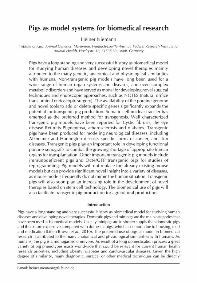
the porcine protein had the same amino acid as human diseases (Groenen et al. 2012). These genetic changes were associated with an increased risk of multifactorial traits, including obesity, diabetes or late-onset diseases such as Parkinson disease and Alzheimer disease in humans (Groenen et al. 2012). The availability of the porcine genome significantly expands the potential for transgenic pig production. This is facilitated by effective new tools to add or delete specific genes to the porcine genome, including specific nucleases such as zinc finger nucleases (Figure 1), TALEN and transposons (Hauschild et al. 2011; Flisikowska et al. 2011; Garrels et al. 2012; Pennisi 2012).
fig. 1 Mechanism of Zinc-finger-nucleases (ZFN) in the editing of DNA. Here the bi-allelic knock-out of a specific DNA sequence is shown.
The in-depth knowledge of the structure and organization of the porcine genome and further refinements of molecular tools will soon allow the production of a great variety of transgenic pig phenotypes similar to the laboratory mouse. Pigs are more expensive to keep than rodents and reproduce more slowly than rodents. However, similarities between human and pig genetics, anatomy and physiology much outweigh these limitations. Porcine eyes are similar in size with photoreceptors that are similarly distributed in the retina as in the human eye. Thus the pig became the first transgenic model for Retinitis Pigmentosa (RP) which is an important cause of human blindness. More than 4 years ago the first transgenic pig model of cystic fibrosis was reported which stimulated the interest of researchers in porcine disease models (Abbott 2012). Additional stimulation of transgenic pig production will arise from the availability of pluripotent stem cells that are superior to the currently available somatic cells for any genetic modification procedure. Germ line contribution of pluripotent cells has not yet been reported for the pig. However, significant progress has been reported towards this goal (Nowak-Imialek et al. 2011; Nowak-Imialek and Niemann 2013). The following is an update of the available literature on the production of transgenic pigs for biomedical models. The topic has also been reviewed recently (Wolf 2012; Prather et al. 2013).
Porcine models for the genetic diseases cystic fibrosis and hereditary tyrosinemia type 1
Transgenic pigs carrying a mutated CFTR (cystic fibrosis transmembrane conductance regulator) gene are present the best characterized model for a human genetic disease. Cystic fibrosis
ControlPigReprodIX.indb 272 5/23/2013 7:46:25 AM

273The pig as animal model in biomedicine
is caused by a genetic defect in the CFTR which encodes the cyclic AMP activator chloride channel in epithelial cells. This defect results in reduced epithelial fluid transportation and thus creates abnormal fluid secretion of the airway mucous glands that are the major cause for CF (cystic fibrosis) pathology (Widdicombe 2010). Mouse airways contain only few mucous glands, consequently the mouse model of CF shows little airway pathology (Widdicome 2010). The production of viable pigs carrying a homozygous knockout of the CFTR gene led to the creation of a cystic fibrosis model in domestic pigs (Rogers et al. 2008). The pigs were produced via SCNT using somatic cells with a homozygous knockout of the CFTR gene locus. The CFTR gene was disrupted by inserting an antibiotic resistance cassette in exon 10 of the CFTR gene (Rogers et al. 2008). Pigs lacking the CFTR showed defective chloride transport, developed meconium ileus, displayed exocrine pancreatic destruction and focal biliary cirrhosis, thus showing the same abnormalities observed in human CF patients (Rogers et al. 2008). These pigs provide a new source to study CF pathology and to develop novel therapies for CF patients.
The CFTR-/- pigs developed the full hallmark of the CF lung disease, including airway inflammation, remodeling mucus accumulation, and infection. Their lungs contained multiple bacterial species as consequence of the defect in the bacterial defense system (Stoltz et al., 2010). It was further found that the ΔF508 mutation induced a CF-like disease in the CFTR-/- pigs. This is an important step towards unraveling the molecular pathogenesis of common CF disease (Ostedgaard et al., 2011). The lack of functional CFTR was shown to reduce bacterial killing on the airways and to change of the pH towards a more acidic situation. This reduced the antimicrobial activity of airways surface liquid (Pezzulo et al. 2012). Airways surface liquid was thus identified as critical factor in the lung defense system and is directly linked with the initial host defense defect (Pezzulo et al., 2012). In a further attempt to unravel the etiology of CF, it was shown that pigs and humans with CF have reduced insulin-like growth factor 1 (IGF1) levels at birth (Rogan et al., 2010). IGF levels might thus serve as biomarker to predict disease severity or the response to specific therapeutics. These findings also raise the possibility that IGF1 supplementation early in development might be beneficial for CF patients (Rogan et al., 2010).
Hereditary tyrosinemia type 1 (HT1) is a human disease that is caused by deficiency in the enzyme fumary lacetoacetate hydrolase (FAH), causing hepatic failure, cirrhosis and hepatocellular carcinoma already early in childhood. The FAH gene was knocked out in porcine fibroblasts that in turn were used in SCNT. Several viable FAH+/- pigs were produced that showed a normal phenotype, but had decreased FAH transcriptional and enzymatic activity compared with wild-type pigs (Hickey et al., 2011).
Transgenic pigs for human eye diseases
Patients with Retinitis Pigmentosa (RP) develop night blindness early in life due to the loss of rod photoreceptors. Transgenic pigs were created by injecting a mutated porcine rhodopsin gene into pronuclei of zygotes and transgenic pigs showed a similar pathology as RP patients (Petters et al. 1997). Subsequently lensectomy and vitrectomy were applied which delayed photoreceptor degeneration in rhodopsin transgenic pigs (Mahmoud et al. 2003). Further research revealed that oxidative damage is a potential cause of cone cell death in a RP situation (Shen et al. 2005). These data are in line with the hypothesis that death of rods is associated with decreased oxidant consumption and hyperoxia in the outer retina followed by gradual cone cell death. In an attempt to develop a treatment for RP patients, fetal neuroretinal cells were transplanted into pig eyes with severe retina degeneration. However, graft and host retinal neurons did not form proper connections leading to reduced retinal function in the host (Ghosh et al. 2007). Ribozyme based gene therapy was tested to treat autosomal dominant RP in a transgenic pig
ControlPigReprodIX.indb 273 5/23/2013 7:46:26 AM

274 H. Niemann
model. However, the allele specific ribozyme used for the human sequence was not successful, whereas the hammerhead ribozyme had beneficial effects (Shaw et al. 2001). Further study of the RP transgenic pigs revealed that the rhodopsis PSD-95 is nearly completely lost from most rod terminals in transgenic swine. But an early postnatal PSD-95 expression continues in cone terminals even in 10 months old transgenic swine when all the rods have disappeared. This indicates that the loss of PSD-95 is not the consequence of the deteriorating cells (Blackmon et al. 2000). In transgenic porcine retina, the ectopic synapses formed between cones and rod bipolar cells were altered with impaired processing of inner retinal neurons (Ng et al. 2008).
SCNT was used to create a transgenic miniature pig model expressing a specific rhodopsin mutation. The founder animals showed abnormal full-field electroretinography and the offspring inherited the transgene with the autosomal dominant mutation. The miniature pig carrying the P23H RHO mutation is a new model to study morphology and treatment of RP (Ross et al 2012).
Obviously, these models display key features of human RP; however the time course of disease progression makes this model costly, time consuming and difficult to study. Treatment with iodoacetic acid emerged as an alternative rod dominant model of retinal damage which shared many features with the transgenic RP pig model (Wang et al. 2011).
Another model for human eye disease was recently reported. Transgenic pigs expressing the human disease causing ELOVL4 mutation revealed photoreceptor loss and disorganized inner and outer segments. These pigs are promising as new model to examine macular degeneration and STGD3 pathogenesis (Sommer et al. 2011).
Transgenic pigs in diabetes research
Transgenic pigs expressing a dominant negative receptor for the incretin hormone glucose-dependent insulinotropic polypeptide (GIP) revealed a crucial role of the GIP system for age related expansion of pancreatic β-cell mass. This model shared important characteristics with type 2 diabetes mellitus patients, including reduced glucose tolerance, insulin secretion and progressive reduction of β-cells (Renner et al. 2010). Metabolic signatures of specific amino acids and lipids were investigated in this model and several potential biomarkers of early phases of β-cell dysfunction and mass reduction were identified (Renner et al. 2012 a). Transgenic pigs with permanent diabetes were created by SCNT with a mutated insulin gene. These pigs show typical features of progressive diabetes, including cataract development and pathology of kidneys and the nervous system (Renner et al. 2012 b).
Transgenic pigs with β-cell specific expression of LEA29Y served as donors in a xenotransplantation model. Xenograft islet cell clusters from these pigs rescued diabetes and prevented rejection in a humanized mouse model (Klymiuk et al. 2012). Transgenic cloned pigs have also been produced carrying a dominant negative mutant for a hepatocyte nuclear factor 1α mutation showing obvious diabetic symptoms (Umeyama et al. 2009). The use of islets from pigs transgenic for a fluorogenic protein GFP (green fluorescent protein or Kusabira-Orange) may facilitate development of islet cell xenotransplantation (Teratani et al. 2012).
Pigs as models for neurological diseases
The survival motor neuron (SMN1) gene was mutated in fibroblasts to create a transgenic swine model for spinal muscular atrophy (SMA); but pigs have not yet been produced with this mutation (Lorson et al. 2008). In an effort to develop porcine models for Alzheimer disease, Göttingen miniature pigs were produced that carry a random integration of the Alzheimer disease causing dominant mutation APPsw. Pigs were produced by handmade cloning in cells that had a single copy of the transgene inserted in the GLIS locus. The transgene was consistently expressed and
ControlPigReprodIX.indb 274 5/23/2013 7:46:26 AM

275The pig as animal model in biomedicine
the accumulation of Alzheimer proteins is expected to occur in the brain at the age of 1 – 2 years (Kragh et al. 2008). A construct harboring the Huntington cDNA was injected into pronuclei of zygotes and transgenic minipigs were produced. However, a phenotype was not reported (Uchida et al. 2001). More recently, pigs transgenic for Huntington’s disease were produced via SCNT. These pigs expressed a mutant Huntington gene with an expanded polyglutamine tract (Yang et al. 2010). Severe postnatal death, dyskinesia and chorea-like movement were observed in some transgenic pigs with expression of the mutant huntingtin. The typical apoptotic neurons with DNA fragmentation were found in the brains of transgenic pigs (Yang et al. 2010).
Porcine cancer models
Pigs have been produced with a knockout of the breast cancer associated gene 1 (BRCA1) which predisposes for breast cancer and accounts for the majority of the cases of familial breast and ovarian cancer. Cells with a knockout of BRCA1 mediated via recombinant adeno-associated virus were produced and used in SCNT. Targeting efficiency was high, however, all BRCA1 hemizygous transgenic piglets died shortly after birth (Luo et al. 2011). The reasons of this postnatal mortality remain unclear; it illustrates the difficulty to produce a meaningful large animal model for specific diseases.
Gene targeted cloned pigs carrying a mutation in the APC (adenomatous polyposis coli) gene displayed a similar pathology in the intestine as human patients with familial adenomatous polyposis (Flisikowska et al., 2012). This large animal model is promising for the development of novel diagnostic and therapeutic strategies for colorectal cancer. Live pigs with a mutation in the tumor suppressor p53 gene were produced, that is orthologous to the oncogenic human mutant TP53R175H and mouse Trp53R172H mutation. Gene targeted MSCs were successfully employed in SCNT and viable piglets were produced with the TP53R167H mutant allele in a heterozygous form. This is the first pig model that demonstrates the feasibility of an inactivation mutation of the gatekeeper tumor suppressor gene p53 in a non-rodent mammal (Leuchs et al. 2012).
Immunodeficient pigs
A porcine model of severe combined immunodeficiency (SCID) has been produced which is largely similar to the well-known mouse model. Fibroblasts were targeted for disruption of the X-linked interleukin 2 receptor gamma chain gene (Il2rg) and were employed as donor cells to produce cloned pigs. Viable heterozygous Il2rg females were produced, whereas the IL2rg heterozygous males were athymic and showed significantly reduced immunoglobulin and T and NK cell production, clearly mimicking the human SCID situation (Suzuki et al. 2012). Moreover, allogeneic bone marrow transplantation was compatible with stable integration of heterozygous Il2rg-IY and reconstituted the Il2rg-IY lymphoid lineage. These pigs are a very important step towards the creation of pig models for the evaluation of cell based regenerative treatments. These significant advances may complement the already existing arsenal of genomically humanized mice that are a valuable tool for gaining a better understanding of basic immunological activity (Devoy et al. 2012).
Porcine atherosclerosis models
Modeling atherosclerosis in pigs has been difficult because rapid atherosclerosis could not be induced in normal pigs by high-fat feeding regimens. Promising transgenic approaches have been reported recently. Transgenic Yucatan minipigs were produced that over-expressed human catalase on the endothelial cells. The transgene was transfected into fibroblasts and transgenic
ControlPigReprodIX.indb 275 5/23/2013 7:46:26 AM

276 H. Niemann
fibroblasts were used in SCNT. Transgenic pigs showed increased activity of catalase and reduced levels of H2O2 in culture (Whyte et al. 2011). Using Sleeping Beauty DNA transposition and SCNT, Yucatan minipigs were created with liver specific expression of the human D374Y-PCSK9 gain of function mutation. The PCSK9 gene encodes human proprotein convertase subtilisin/kexin type 9 that is critically involved in cholesterol metabolism. D374Y-PCSK9 transgenic pigs displayed the typical pathology of human atherosclerosis, including reduced hepatic low-density lipoprotein (LDL) receptor levels, impaired LDL clearance, severe hypercholesterolemia and atherosclerotic lesions in the vasculatory system (Al-Mashhadi et al., 2013). Moreover, pigs with a mutated LDL receptor were created using specific TALEN, but the phenotype has not yet been reported (Carlson et al. 2012).
Porcine models of human skin diseases
The keratinocyte-specific human transgene K5-hGli2ΔN was expressed in transgenic pigs produced by SCNT. This gene is critically involved in the development of basal cell carcinomas. The transgenic pigs developed the typical skin lesions that could not be treated by antibiotics leading to an early death of the animals. This pathology has not been observed in the corresponding mouse model (McCalla-Martin et al. 2010). A model for cutaneous inflammation was produced in Göttingen miniature pigs, expressing the human β1 or α2 integrin genes under control of a keratinocyte specific promoter (Staunstrup et al. 2012). Transgenic pigs showed ectopic expression of human integrins and localization within the keratinocyte plasma membrane. This indicates that regulation of integrins β1/α2 by over-expression of the transgenes occurred via different cellular signaling pathways. Several markers of perturbed skin homeostasis were identified. These pigs are the first model with molecular markers of skin inflammation (Staunstrup et al. 2012).
Xenotransplantation of porcine organs to human patients
Today more than 250,000 people owe their lives a successful human organ transplantation (allotransplantation). Ironically, the success of organ transplantation technology has led to an acute shortage of appropriate organs, because cadaveric and live organ donation falls far short of meeting the demand in western societies. To close the growing gap between demand and availability of appropriate organs, transplant surgeons have long considered the possibility of using xenografts from domesticated pigs (Bach 1998; Platt et al. 1998; Kues and Niemann 2004). Essential prerequisites for successful xenotransplantation are: (i) overcoming the immunological hurdles, (ii) preventing the transmission of pathogens from the donor animal to the human recipient, and (iii) compatibility of the donor organs with human physiology. This requires a series of critical steps and can be time, labor and cost expensive (Figure 2).
An important advantage of xenotransplantation is the opportunity to modify the genome of the donor animals. Two modifications are required for preventing the first immunological hurdle, the hyperacute rejection (HAR), in pig-to-human transplantation: i) Elimination of antigenic sugar residues via genetic knockout of the porcine α1,3-galactosyltransferase (GGAT-1)(homozygous Gal kO), and/or ii) Suppression of the recipient’s complement system by the introduction of one or more regulators of complement activation (RCA), such as CD55, 46, or 59 (Figure 3). A tremendous amount of research focused on the production of genetically engineered pigs expressing inhibitors of the human complement cascade. The validity of this approach has been convincingly demonstrated by several groups (Schuurman et al., 2002; McCurry et al., 1995; Bhatti et al., 1999; Chen, et al. 1999). Most importantly, it has been demonstrated that organs from genetically engineered pigs lacking functional α1,3-galactosyltransferase and thus lacking
ControlPigReprodIX.indb 276 5/23/2013 7:46:26 AM

277The pig as animal model in biomedicine
expression of α-Gal epitopes (GGTA1-KO pigs), do not undergo HAR once transplanted into primates (Yamada et al., 2005; Kuwaki et al., 2005).
While induction of a knockout of a specific gene is extremely difficult and inefficient in somatic cells used in SCNT, novel approaches have been successfully explored to overcome this bottleneck in the production of pigs with inactivation of epitopes critically involved in the immunological rejection after porcine-to-primate xenotransplantation. Zinc finger nucleases (ZFNs) are a class of engineered DNA-binding proteins that facilitate editing of the genome by creating double-strand breaks in DNA at targeted loci. The ZFN mediated knock-out makes the integration of an antibiotic selection cassette superfluous that are used in conventional HR strategies for selection of the targeted cells. The first pigs with a homozygous GGTA-1KO induced with the aid of specific ZFNs were recently reported (Hauschild et al. 2011).
fig. 3 Schematic diagram of the rejection responses in a xenotransplantation setting and molecules that are involved in these processes. (for details see text)
1. Production and propagation of transgenic pigs (SCNT)2. Determination of transgenic expression patterns - mRNA -Protein(intensity,tissuespecificity,etc.)3. Selective breeding of transgenic lines4. In-vitro-testing to determine the protective properties against HAR and/or AVR (eventually
safety testing for PERV, etc.)5. Perfusion of isolated porcine organs with human blood
(Modelling the human situation)6. In-vivo-studies in primates - Physiological compatibility - Protection against immune reactions - Testing for transmission of pathogens
fig. 2 Steps of the evaluation of transgenic pigs for suitability in xenotransplantation
ControlPigReprodIX.indb 277 5/23/2013 7:46:26 AM
278 H. Niemann
Even in the case of GGTA1-KO pigs, porcine xenografts eventually failed as a consequence of the acute humoral (AHXR) or delayed (DXR) xenograft rejection, also called acute vascular rejection (AVR) (Platt et al., 1998; Bach et al., 1996). Several factors have been implicated in the pathogenesis of AHXR and pathology is primarily characterised by vascular thrombosis, blood extravasation and oedema (Platt et al., 1991). Cellular infiltrates include neutrophils, macrophages, CD8+ T cells and few NK cells (Vega et al., 2002). AVR is characterised by the progressive deposition of antibodies and complement and is associated with apoptosis and necrosis of endothelial cells, contributing to platelet aggregation and thrombosis in the graft. The current view is that long term survival of xenografts after transplantation into primates requires a specifically tailored immunosuppression regimen compliant with current clinical standards, and additional modifications of the pig genome. Several candidate genes, incl. human thrombomodulin (hTM), human heme-oxygenase 1 (hHO-1), human A20 (hA20), or CTLAIg (soluble CD28 receptor analog), have been explored in their ability to improve long term survival of porcine xenografts after transplantation into nonhuman primates (Petersen et al., 2009; Petersen et al., 2011; Oropeza et al., 2009; Phelps et al., 2009) (Figure 3).
Extensive research has revealed that the risk of porcine endogenous retrovirus (PERV) transmission to human patients is low, paving the way for preclinical testing of xenografts (Switzer et al. 2001; Irgang et al. 2003). RNA interference (RNAi) is a promising method for knocking down PERV expression in porcine somatic cells. Using RNAi mediated knockdown, PERV expression has been significantly reduced in porcine somatic cells for 4-6 months, these cells were successfully used in SCNT and gave normal piglets with long-term suppression of PERV (Dieckhoff et al., 2008; Semaan et al., 2012). RNAi knockdown thus provides an additional level of safety for porcine-to-human xenotransplantation.
Although additional refinements will always be possible, it is expected that appropriate lines of transgenic pigs will be available as organ donors within the next five to ten years. Guidelines for the clinical application of porcine xenografts already exist in the USA and are being developed in other countries. The general consensus of a worldwide debate is that the technology is ethically acceptable provided that the individual`s well-being does not compromise public health (e.g. the risk of PERV recombination). The improvement in quality of life for patients receiving conventional allotransplants is dramatic, but xenotransplantation is also economically attractive because the long term costs of maintaining patients with severe kidney disease on dialysis or treating patients with chronic heart disease can be greater than the cost of a successful transplant. Preliminary functional data on porcine kidneys and hearts in non-human primates is promising although the long term interaction between porcine organs and human physiology is to a great extent unexplored (Ibrahim et al. 2006).
Reprogramming of somatic cells and development of cell based therapies
To facilitate the use of domesticated pigs as a tool for preclinical testing of novel therapies and to facilitate the derivation of germ line competent pluripotent stem cells, Oct4-GFP transgenic pigs were recently produced from our laboratory (Nowak-Imialek et al., 2011). The transcription factor Oct4 is essential for the maintenance of pluripotency and for reprogramming somatic cells into a pluripotent state. Using a18 kb genomic sequence of the murine Oct4 gene fused to the enhanced green fluorescent (eGFP) cDNA, pluripotent cells could unequivocally identified by the green fluorescence. Expression of the EGFP reporter was confined to germ line cells in live pigs, the inner cell mass and trophectoderm in blastocysts and in testicular germ cells (Figure 4). Reprogramming of fibroblasts from these animals by fusion with pluripotent murine embryonic stem cells or viral transduction using human Oct4, Sox2, KLF4 and cMYCcDNAs revealed Oct4-EGFP reactivation (Figure 5) clearly showing the usefulness of this approach.
ControlPigReprodIX.indb 278 5/23/2013 7:46:26 AM

279The pig as animal model in biomedicine
These cells have been used for studies to isolate and characterize pluripotent stem cells in the pig (Kues et al., 2013; Petkov et al., 2013).
Transgenic pigs were also produced that expressed mitochondria localized enhanced yellow fluorescent protein under the control of the germ cell specific stimulated by retinoic acid (Stra8) promoter in the testicular tissue. Expression of the Stra EYFP transgene in spermatogenic cells could serve as a useful model for germ cell transplantation and in vitro spermatogenic studies
fig. 5 Activation of the reporter transgene Oct4-eGFP in porcine fibroblasts by over-expression of the 4 reprogramming factors. The green fluorescence indicates expression of the pluripotency marker Oct4 which in turn indicates reprogramming of the somatic cells into a pluripotent state.
fig. 4 Germ line specific expression of the Oct4/eGFP transgene, the green fluorescence indicates the presence of Oct4 expression in porcine testicular tissue, which is a marker for pluripotency.
(Analysis of 12 transgenic fetuses on days 25-30)
ControlPigReprodIX.indb 279 5/23/2013 7:46:27 AM

280 H. Niemann
(Sommer et al., 2011). Chimeric pigs derived from induced pluripotent stem cells with germ line transmission in the absence of tumor formation, were recently reported. This is a major step forward towards the establishment of a translational model to study effects and safety of stem cell therapies (West et al., 2011).
Concluding remarks and perspectives
The pig has a long and successful history in biomedical research and has thus benefitted significantly human health and well-being. The number of non-transgenic pig models is already large and the potential of the pig in biomedical research will be further enhanced with the recent availability of the porcine genome and molecular tools needed for targeted genetic modification similar to the laboratory mouse. Pig models will not replace the already existing mouse models but can provide significant novel insight into a variety of diseases, as mouse models frequently do not mimic the human situation. Transgenic pigs will also play an increasing role in the development of novel therapies based on stem cell technology. The biomedical use of pigs will also facilitate transgenic pig production for agricultural production. This will be stimulated by novel genomic knowledge and tools emerging from ongoing research.
Acknowledgements
The research in the own laboratory reported herein was supported by DFG and BMBF. The excellent assistance of Susanne Tonks in preparing this article is gratefully acknowledged.
References
Abbott A 2012 Pig geneticists go the whole hog. Nature 491 315-316.
Albert S, Kubiak BD, vieau CJ, Roy SK, DiRocco J, Gatto LA, Young JL, Tripathi S, Trikha G, Lopez C et al. 2011 Comparison of “Open Lung” modes with low tidal volumes in a porcine lung injury model. Journal of Surgical Research 166 e71-e81.
Al-Mashhadi R, Sorensen C, Kragh P, Christoffersen C, Mortensen M, Tolbod L, Thim T, Du Y, Li J, Liu Y et al. 2013 Familial hypercholesterolemia and atherosclerosis in cloned minipigs created by DNA transposition of a PCSK9 gain-of-function mutant. Science Translational Medicine 5 166ra1.
Bach fh 1998 Xenotransplantation: Problems and prospects. Annual Review of Medicine 49 301-310.
Bach fh, winkler h, ferran C, hancock ww & Robson SC 1996 Delayed xenograft rejection. Immunology Today 17 379-384.
Ballard-Croft C, wang D, Sumpter LR, Zhou X & Zwischenberger JB 2012 Large-animal models of acute respiratory distress syndrome. Ann ThoracSurg 93 1331-1339.
Bhatia R, Shaffer Th, hossain J, fisher AO, horner LM, Rodriguez ME, Penfil S & Theroux MC 2011 Surfactant administration prior to one lung ventilation: physiological and inflammatory correlates in a piglet model. Pediatric Pulmonology 46 1069-1078.
Bhatti fN, Schmoeckel M, Zaidi A, Cozzi E, Chavez G,
Goddard M, Dunning JJ, wallwork J & white DJ 1999 Three-month survival of HDAF transgenic pig hearts transplanted into primates. Transplantation Proceedings 31 958.
Blackmon SC, Peng Yw, hao Y, Moon SJ, Oliveira LB, Tatebayashi M. Petters RM & wong f 2000 Early loss of synaptic protein PSD-95 from rod terminals of rhodopsin P347L transgenic porcine retina. Brain Research 885 53-61.
Carlson Df, Tan w, Lillico SG, Stverakova D, Proudfoot C, Christian M, voytas Df, Long CR, whitelaw CBA & fahrenkrug SC 2012 Efficient TALEN-mediated gene knockout in livestock. Proceedings of the National Academy of Sciences of the United States of America 109 17382-17387.
Chang Ch, Kuo Tf, Lin fh, wang Jh, hsu YM, huang hT, Loo ST, fang hw, Liu hC & wang wC 2011 Tissue engineering-based cartilage repair with mesenchymal stem cells in a porcine model. Journal of Orthopaedic Research 29 1874-1880.
Chen Rh, Naficy S, Logan JS, Diamond LE & Adams Dh 1999 Hearts from transgenic pigs constructed with CD59/DAF genomic clones demonstrate improved survival in primates. Xenotransplantation 6 194-200.
De Castro GP, MacPhee MJ, Driscoll IR, Beall D, hsu J, Zhu S, hess JR, Scalea TM & Bochiccho Gv 2010 New hemostatic dressing (FAST Dressing) reduces blood loss and improves survival in a grade V liver
ControlPigReprodIX.indb 280 5/23/2013 7:46:27 AM

281The pig as animal model in biomedicine
injury model in noncoagulopathic swine. The Journal of TRAUMA, Injury, Infection and Critical Care 70 1408-1412.
Devoy A, Bunton-Stasyshyn RKA, Tybulewicz vLJ, Smith AJh & fisher EMC 2012 Genomically humanized mice: technologies and promises. Nature 13 14-20.
Dickson RP, hotchkin DL, Lamm wJE, hinkson C, Pierson DJ, Glenny Rw & Rubinson L 2011 A porcine model for initial surge mechanical ventilator assessment and evaluation of two limited-function ventilators. Crit Care Med 39 527-532.
Dieckhoff B, Petersen B, Kues wA, Kurth R, Niemann h & Denner J. 2008 Knockdown of porcine endogenous retrovirus (PERV) expression by PERV-specific shRNA in transgenic pigs. Xenotransplantation 15 36-45.
Eldardiri M, Martin Y, Roxburgh J, Lawrence-watt DJ & Sharpe JR 2012 Wound contraction is significantly reduced by the use of microcarriers to deliver keratinocytes and fibroblasts in an in vivo pig model of wound repair and regeneration. Tissue Engineering 18 587-597.
Ellison GM, Torella D, Dellegrottaglie S, Perez-Martinez C, Perez de Prado A, vicinanza C, Purushothaman S, Galuppo v, Iaconetti C, waring CD et al. 2011 Endogenous cardiac stem cell activation by insulin-like growth factor-1/hepatocyte growth factor intracoronary injection fosters survival and regeneration of the infarcted pig heart. Journal of the American College of Cardiology 58 977-986.
flisikowska T, Merkl C, Landmann M, Eser S, Rezaei N, Cui X, Kurome M, Zakhartchenko v, Kessler B et al. 2012 A porcine model of familial adenomatous polyposis. Gastroenterology 143 1173-1175.
flisikowska T, Thorey IS, Offner S, Ros f, Lifke v, Zeitler B, Rottmann O, vincent A, Zhang L, Jenkins S et al. 2011 Efficient immunoglobulin gene disruption and targeted replacement in rabbit using zinc finger nucleases. PLoS ONE 6 e21045.
fosse TK, Spadavecchia C, horsberg TE, haga hA & Ranheim B 2010 Pharmacokinetics and pharmacodynamic effects of meloxicam in piglets subjected to a kaolin inflammation model. Journal of Veterinary Pharmacology and Therapeutics 34 367-375.
friess Sh, Naim MY, Kilbaugh TJ, Ralston J & Margulies SS 2012 Premedication with meloxicam exacerbates intracranial haemorrhage in an immature swine model of non-impact inertial head injury. Laboratory Animals 46 164-166.
friess Sh, Ralston J, Eucker SA, helfaer MA, Smith C & Margulies SS 2011 Neucritical care monitoring correlates with neuropathology in a swine model of pediatric traumatic brain injury. Neurosurgery 69 1139-1147.
fritscher-Ravens A, Cuming T, Olagbaiye f, holland C, Milla P, Seehusenf, hadeler KG, Arlt A & Mannur K 2011 Endoscopic transesophageal vs. thoracoscopic removal of mediastinal lymph nodes: a prospective randomized trial in a long term animal survival model. Endoscopy 42, 468-474 43 1090-1096.
fritscher-Ravens A, Ghanbari A, Cuming T, Kahle E, Niemann h, Koehler P & Patel K 2008 Comparative study of NOTES alone vs. EUS-guided NOTES procedures. Endoscopy 40 925-930.
Gaines RJ, DeMaio M, Peters D, hasty J & Blanks J 2011 Management of contaminated open fractures: A comparison of two types of irrigation in a porcine model. J Trauma 72 733-736.
Garrels w, Ivics Z & Kues wA 2012 Precision genetic engineering in large mammals. Trends in Biotechnology 30 386-393.
Ghosh f, Engelsberg K, English Rv & Petters RM 2007 Long-term neuroretinal full-thickness transplants in a large animal model of severe retinitis pigmentosa. Graefe’s Archive for Clinical and Experimental Ophthalmology 245 835-846.
Girard-Misguich f, Cognie J, Delgado-Ortega M, Berthon P, Rossignol C, Larcher T. Melo S, Bruel T, Guibon R Chérel Y et al. 2011 Towards the establishment of a porcine model to study human amebiasis. PLoS ONE 6 e28795.
Giraud S, favreau f, Chatauret N, Thuillier R., Maiga S & hauet T 2011 Contribution of large pig for renal ischemia-reperfusion and transplantation studies: The preclinical model. Journal of Biomedicine and Biotechnology. Doi:10.1155/2011/532127.
Granada Jf, Milewski K, Zhao h, Stankus JJ, Tellez A, Aboodi MS, Kaluza GL, Krueger CG, virmani R, Schwartz LB et al. 2011 Vascular reponse to zotarolimus-coated balloons in injured superficial femoral arteries of the familial hypercholesterolemic swine. Circ Cardiovasc Interv 4 447-455.
Groenen MAM, Archibald AL, uenishi h, Tuggle CK, Takeuchi Y, Rothschild Mf, Rogel-Gaillard C, Park C, Milan D, Megens hJ et al. 2012 Analyses of pig genomes provide insight into porcine demography and evolution. Nature 491 393-398.
hauschild J, Petersen B, Santiago Y, Queisser AL, Carnwath Jw, Lucas-hahn A, Zhang L, Meng X, Gregory PD, Schwinzer R et al. 2011 Efficient generation of a biallelic knockout in pigs using zinc-finger nucleases. Proceedings of the National Academy of Sciences of the United States of America 108 12013-12017.
heinroth KM, unverzagt S, Buerke M, Carter J, Mahnæpf D, werdan K & Prondzinsky R 2011 Transocoronary pacing in a porcine model – impact of guidewire insulation. The Journal of Invasive Cardiology 23 108-114.
hickey RD, Lillegard JB, fisher JE, McKenzie TJ, hofherr SE, finegold MJ, Nyberg SL, Grompe M 2011 Efficient production of Fah-null heterozygote pigs by chimeric adeno-associated virus-mediated gene knockout and somatic cell nuclear transfer. Hepatology 54 1351-1359.
hitchcock KE, Invacevich NM, haworth KJ, Stamper DNC, vela DC, Sutton JT, Pyne-Geithman GJ & holland CK 2011 Ultrasound-enhanced re-PA thrombolysis in an ex vivo porcine carotid artery model. Ultrasound in Medicine and Biology 37
ControlPigReprodIX.indb 281 5/23/2013 7:46:28 AM

282 H. Niemann
1240-1251. hua Z, Sergi C, Nation PN, wizzard PR, Ball RO,
Pencharz PB, Turner JM & wales Pw 2012 Hepatic ultrastructure in a neonatal piglet model of intestinal failure-associated liver disease (IFALD). Journal of Electron Microscopy 61 179-186.
Ibrahim Z, Busch J, Awwad M, wagner R, wells K & Cooper DKC 2006 Selected physiologic compatibilities and incompatibilities between human and porcine organ systems. Xenotransplantation 13 488-499.
Irgang M, Sauer IM, Karlas A, Zeilinger K, Gerlach J, Kurth R, Neuhaus P & Denner J 2003 Porcine endogenous retroviruses: no infection in patients treated with a bioreactor based on porcine liver cells. Journal of Clinical Virology 28 141-154
Jamieson Rw, Zilvetti M, Roy D, hughes D, Morovat A, Coussios CC & friend PJ 2011 Hepatic steatosis and normothermic perfusion-preliminary experiments in a porcine model. Transplantation 92 289-295.
Jin G, deMoya MA, Duggan M, Knightly T, Mejaddam AY, hwabejire J, Lu J, Smith wM, Kasotakis G, velmahos C et al. 2012 Traumatic brain injury and hemorrhagic shock: Evaluation of different resuscitation strategies in a large animal model of combined insults. Shock 38 49-56.
Kheirabadi BS, Arnaud f, McCarron R, Murdock AD, hodge DL, Ritter B, Dubick MA & Blackbourne Lh 2011 Development of a standard swine hemorrhage model for efficacy assessment of topical hemostatic agents. The Journal of Trauma, Injury, Infection and Critical Care 71 139-S146.
Kjærgaard B, Rasmussen BS, de Neergaard S, Rasmussen Lh & Kristensen SR 2012 Extracorporeal cardiopulmonary support may be an efficient rescue of patients after massive pulmonary embolism. An experimental porcine study. Thrombosis Research 129 e147-e151.
Klymiuk N, van Buerck L, Bähr A, Offers M, Kessler B, wuensch A, Kurome M, Thormann M, Lochner K, Nagashima h et al. 2012 Xenografted islet cell clusters from INSLEA29Y transgenic pigs rescue diabetes and prevent immune rejection in humanized mice. Diabetes 61 1527-1532.
Ko Sf, Yip hK, Lee CC, Sheu JJ, Sun CK, Ng Sh, huang CC, Lin YC, Chang LT & Chen MC 2011 Immediate intramyocardial bone marrow-derived mononuclear cells implantation in minipig myocardium after permanent coronary artery ligation. Investigative Radiology 46 495-503.
Kragh PM, Nielsen AL, Li J, Du Y, Lin L, Schmidt M, Bøgh IB, holm IE, Jakobsen JE, Johansen MG et al. 2008 Hemizygous minipigs produced by random gene insertion and handmade cloning express the Alzheimer’s disease-causing dominant mutation APPsw. Transgenic Research 18 545-558.
Kues wA, Niemann h 2004 The contribution of farm animals to human health. Trends in Biotechnolology 22 286-294.
Kues w, herrmann D, Barg-Kues B, haridoss SM,
Nowak-Imialek M, Buchholz T, Streeck M, Grebe A, Grabundzija I, Merkert S et al. 2013 Derivation and characterization of Sleeping Beauty transposon-mediated porcine induced pluripotent stem cell. Stem Cells and Development 22 124-135.
Kuwaki K, Tseng YL, Dor fJ, Shimizu A, houser SL, Sanderson TM, Lancos CJ, Prabharasuth DD, Cheng J, Moran K et al. 2005 Heart transplantation in baboons using alpha1,3-galactosyltransferase gene-knockout pigs as donors: initial experience. Nature Medicine 11 29-31.
Langerhuus SN, Tønnesen EK, Jensen Kh, Damgaard BM, halekoh u & Lauridsen C 2012 Effects of dietary n-3 and n-6 fatty acids on clinical outcome in a porcine model on post-operative infection. British Journal of Nutrition 107 735-743.
LaPar DJ, Laubach vE, Emaminia A, Crosby IK, hajzus vA, Sharma AK, Sumner hM, webb Dv, Lau CL & Kron IL 2011 Pretreatment strategy with adenosine A2A receptor agonist attenuates reperfusion injury in a preclinical porcine lung transplantation model. The Journal of Thoracic and Cardiovascular Surgery 142 887-894.
Leuchs S, Saalfrank A, Merkl C, flisikowska T, Edlinger M, Durkovic M, Rezaei N, Kurome M, Zakhartchenko v, Kessler B et al. 2012 Inactivation and inducible oncogenic mutation of p53 in gene targeted pigs. PLOS ONE 7 e 43323.
Levi JR, veerappan A, Chen B, Mirkov M, Sierra R & Spiegel Jh 2011 Histologic evaluation of laser lipolysis comparing continuous wave vs pulsed lasers in an in vivo pig model. Arch Facial Plast Sur 13 41-50.
Li Y, Ristagno G, Guan J, Barbut D, Bisera J, weil Mh & Tang w 2012 Preserved heart rate variability during therapeutic hypothermia correlated to 96 hrs neurological outcomes and survival in a pig model of cardiac arrest. Crit Care Med 40 580-586.
Litten-Brown JC, Corson AM & Clarke L 2010 Porcine models for the metabolic syndrome, digestive and bone disorders: a general overview. Animal 4:6 899-920.
Lorson MA, Spate LD, Prather RS & Lorson CL 2008 Identification and characterization of the porcine (Sus scrofa) survival motor neuron (SMN1) gene: an animal model for therapeutic studies. Developmental Dynamics 237 2268-2278.
Luo Y, Li J, Liu Y, Lin L, Du Y, LI S, Yang h, vajta G, Callesen h, Bolund L et al. 2011 High efficiency of BRCA1 knockout using rAAV-mediated gene targeting: developing a pig model for breast cancer. Transgenic Research 20 975-998.
Mahmoud Th, McCuen Bw, hao Y, Moon SJ, Tatebayashi M, Stinnett S, Petters RM & wong f 2003 Lensectomy and vitrectomy decrease the rate of photoreceptor loss in rhodopsin P347L transgenic pigs. Graefe’s Archive for Clinical and Experimental Ophthalmology 241 298-308.
Martinez-Olondris P, Rigol M, Soy D, Guerrero L, Agusti C, Quera MA, Bassi GL, Esperatti M, Luque N, Liapikou M et al. 2012 Efficacy of linezolid
ControlPigReprodIX.indb 282 5/23/2013 7:46:28 AM

283The pig as animal model in biomedicine
compared to vancomycin in an experimental model of pneumonia induced by methicillin-resistant staphylococcus aureus in ventilated pigs. Crit Care Med 40 162-168.
McCalla-Martin AC, Chen X, Linder KE, Estrada JL & Piedrahita JA 2010 Varying phenotypes in swine versus murine transgenic models constitutively expressing the same human sonic hedgehog transcriptional activator, K5-HGLI2ΔN. Transgenic Research 19 869-887.
McCurry KR, Kooyman DL, Alvarado CG, Cotterell Ah, Martin MJ, Logan JS & Platt JL 1995 Human complement regulatory proteins protect swine-to-primate cardiac xenografts from humoral injury. Nature Medicine 1 423-427.
Meeker J, weinhold P & Dahners L 2011 Negative pressure therapy on primarily closed wounds improves wound healing parameters at 3 days in a porcine model. Orthop trauma 25 756-761.
Melega wP, Lacan G, Gorgulho AA, Behnke EJ & De Salles AAf 2012 Hypothalamic deep brain stimulation reduces weight gain in an obesity-animal model. PLoS ONE 7 e30672.
Meurens f, Summerfield A, Nauwynck h, Saif L & Gerdts v 2011 The pigs: a model for human infectious diseases. Trends in Microbiology 20 50-57.
Moreira-Pinto J, ferreira A, Miranda A, Rolanda C & Correla-Pinto J 2012 Transesophageal pulmonary lobectomy with single transthoracic port assistance: study with survival assessment in a porcine model. Endoscopy 44 354-361.
Morelli JN, Runge vM, Ai f, Zhang w, Li X, Schmitt P, McNeal G, Miller M, Lennox M, wusten O et al. 2012 Magnetic resonance evaluation of renal artery stenosis in a swine model. Investigative Radiology 47 376-382.
Ng Yf, Chan hhL, Chu Phw, To Ch, Gilger BS, Petters RM & wong f 2008 Multifocal electroretinogram in rhodopsin P347L transgenic pigs. Investigative Ophthalmology & Visual Science 49 2208-2214.
Ni hB, Ke L, Sun JK, Tong Zh, Ding ww, Li wQ, Li N & Li JS 2012 Beneficial Effect of hypertonic saline resuscitation in a porcine model of severe acute pancreatitis. Pancreas 41 310-316.
Noel JM, fernandez de Castro JP, DeMarcoJr PJ, franco LM, wang w, vukmanic Ev, Peng X, Sandell Jh, Scott PA, Kaplan hJ et al. 2012 Iodoacetic acid, but not sodium iodate, creates an inducible swine model of photoreceptor damage. Experimental Eye Research 97 137-147.
Nowak-Imialek M & Niemann h 2013 Pluripotent cells in farm animals: state of the art and future perspectives. Reproduction, Fertility and Development 25 103-128.
Nowak-Imialek M, Kues wA, Petersen B, Lucas-hahn A, herrmann D, haridoss S, Oropeza M, Lemme E, Schöler hR, Carnwath Jw et al. 2011 Oct4-enhanced green fluorescent protein transgenic pigs: a new large animal model for reprogramming studies. Stem Cells and Development 20 1563-1575.
Nowak-Imialek M, Kues wA, Carnwath Jw & Niemann h 2011 Pluripotent stem cells and reprogrammed
cells in farm animals. Microscopy and Microanalysis 17 474 – 497.
Okuno T, Yamaguchi M, Okada T, Takahashi T, Sakamoto N, ueshima E, Sugimura K & Sugimoto K 2012 Endovascular creation of aortic dissection in a swine model with technical considerations. Journal of Vascular Surgery 55 1410-1418.
Oropeza M, Petersen B, Carnwath Jw, Lucas-hahn A, Lemme E, hassel P, herrmann D, Barg-Kues B, holler S, Queisser AL et al. 2009 Transgenic expression of the human A20 gene in cloned pigs provides protection against apoptotic and inflammatory stimuli. Xenotransplantation 16 522-534.
Ostedgaard LS, Meyerholz DK, Chen Jh, Pezzulo AA, Karp Ph, Rokhlina T, Ernst SE, hanfland RA, Reznikov LR, Ludwig PS et al. 2011 The ΔF508 mutation causes CFTR misprocessing and cystic fibrosis-like disease in pigs. Science Translational Medicine 3 74ra24.
Patel SR, Penniston KL, Iwicki L, Saeed I, Crenshaw TD & Nakada SY 2012 Dietary induction of long-term hyperoxaluria in the porcine model. Journal of Endourology 26 433-438.
Pennesi ME, Neuringer M & Courtney RJ 2012 Animal models of age related macular degeneration. Molecular Aspects of Medicine 33 487-509.
Pennesi ME 2012 The tale of the TALEs. Science 338 1408-1411.
Petersen B, Ramackers w, Lucas-hahn A, Lemme E, hassel P, Queisser AL, herrmann D, Barg-Kues B, Carnwath Jw, Klose J et al. 2011 Transgenic expression of human heme oxygenase-1 in pigs confers resistance against xenograft rejection during ex vivo perfusion of porcine kidneys. Xenotransplantation 18 355-368.
Petersen B, Ramackers w, Tiede A, Lucas-hahn A, herrmann D, Barg-Kues B, Schuettler w, friedrich L, Schwinzer R, winkler M et al. 2009 Pigs transgenic for human thrombomodulin have elevated production of activated protein C. Xenotransplantation 16 486-495.
Petersen B, Lucas-hahn A, Oropeza M, hornen N, Lemme E, hassel P, Queisser AL, Niemann h 2008 Development and validation of a highly efficient protocol of porcine somatic cloning using preovulatory embryo transfer in peripubertal gilts. Cloning Stem Cells 10 355-362.
Petkov S, hyttel P & Niemann h 2013 The choice of expression vector promoter is an important factor in the reprogramming of porcine fibroblasts into induced pluripotent cells. Cellular Reprogramming 15 1-8.
Petters RM, Alexander CA, wells KD, Collins EB, Sommer JR, Blanton MR, Rojas G, hao Y, flowers wL, Banin E et al. 1997 Genetically engineered large animal model for studying cone photoreceptor survival and degeneration in retinitis pigmentosa. Nature Biotechnology 15 965-970.
Pezzulo AA, Tang XX, hoegger MJ, Alaiwa MhA, Ramachandran S, Moninger TO, Karp Ph, wohlford-Lenane CL, haagsman hP, van Eijk M et al. 2012 Reduced airway surface pH impairs bacterial killing in the porcine cystic fibrosis lung. Nature 487
ControlPigReprodIX.indb 283 5/23/2013 7:46:28 AM

284 H. Niemann
doi:10.1038/nature11130.Phelps CJ, Ball Sf, vaught TD, vance AM, Mendicino
M, Monahan JA, walters Ah, wells KD, Dandro AS, Ramsoondar JJ et al 2009 Production and characterization of transgenic pigs expressing porcine CTLA4-Ig. Xenotransplantation 16 477-485.
Platt JL, fischel RJ, Matas AJ, Reif SA, Bolman RM & Bach fh 1991 Immunopathology of hyperacute xenograft rejection in a swine-to-primate model. Transplantation 52 214-220.
Platt IL, Lin SS & McGregor CG 1998 Acute vascular rejection. Xenotransplantation 5 169-175.
Post ICJh, Dirkes MC, heger M, van Loon JPAM, Swildens B, huijzer GM & van Gulik TM 2012 Appraisal of the porcine kidney autotransplantation model. Frontiers in Biosciences E4 1345-1357.
Prather RS, Lorson M, Ross Jw, whyte JJ & walters E 2013 Genetically engineered pig models for human diseases. The Annual Review of Animal Biosciences 1 12.1-12.17.
Purins K, Enblad P, wiklung L & Lewén A 2012 Brain tissue oxygenation and cerebral perfusion pressure thresholds of ischemia in a standardized pig brain death model. Neurocrit Care 16 462-469.
Purins K, Sedigh A, Molnar C, Jansson L, Korsgren O, Lorant T, Tufveson G, wennberg L, wiklung L, Lewén, A et al. 2011 Standardized experimental brain death model for studies of intracranial dynamics organ preservation, and organ transplantation in the pig. Crit Care Med 39 512-517.
Rebuck DA, Nadler RB & Perry KT 2012 Trajectory image-guided percutaneous renal cryoablation in a porcine model: a pilot study. The Canadian Journal of Urology 19 6094-6099.
Regli A, Chakera J, De Keulenaer BL, Roberts B, Noffsinger B, Singh B & van heerden Pv 2012 Matching positive end-expiratory pressure to intra-abdominal pressure prevents end-expiratory lung volume decline in a pig model of intra-abdominal hypertension. Crit Care Med 40 1879-1886.
Renner S, Römisch-Margl w, Prehn C, Krebs S, Adamski J, Göke B, Blum h, Suhre K, Roscher AA & wolf E 2012a Changing metabolic signatures of amino acids and lipids during the prediabetic period in a pig model with impaired incretin function and reduced β-cell mass. Diabetes 61 2166-2175.
Renner S, Braun-Reichhart C, Blutke A, herbach N, Emrich D, Streckel E,. wünsch A, Kessler B, Kurome M, Bähr A et al. 2012b Permanent neonatal diabetes in INSC94Y transgenic pigs. Diabetes 62 1-7.
Renner S, fehlings C, herbach N, hofmann A, von waldthausen DC, Kessler B, ulrichs K, Chodnevskaja I, Moskalenko v, Amselgruber w et al. 2010 Glucose intolerance and reduced proliferation of pancreatic β-cells in transgenic pigs with impaired glucose-dependent insulinotropic polypeptide function. Diabetes 59 1228-1238.
Riha GM, Kunio NR, van PY, hamilton GJ, Anderson R, Differding JA & Schreiber MA 2011 Hextend and 7.5% hypertonic saline with dextran are equivalent
to lactated ringer’s in a swine model of initial resuscitation of uncontrolled hemorrhagic shock. The Journal of TRAUMA, Injury, Infection and Critical Care 71 1755-1760.
Rodriguez LT, friedman KA, Coffman SS & heller A 2011 Effect of the sensor site-insulin injection site distance on the dynamics of local glycemia in the minipig model. Diabetes Technology & Therapeutics 4 489-493.
Rogan MR, Reznikov LR, Pezzulo AA, Gansemer ND, Samuel M, Prather RS, Zabner J, fredericks DC, McCray PB, welsh MJ et al. 2010 Pigs and humans with cystic fibrosis have reduced insulin-like growth factor 1 (IGF1) levels at birth. Proceedings of the National Academy of Sciences of the United States of America 107 20571-20575.
Rogers CS, Stoltz DA, Meyerholz DK, Ostedgaard LS, Rokhlina T, Taft PJ, Rogan MP, Pezzulo AA, Karp Ph, Itani OA et al. 2008 Disruption of the CFTR gene produces a model of cystic fibrosis in newborn pigs. Science 321 1837-1841.
Ross Jw, de Castro JPf, Zhao J, Samuel M, walters E, Rios C, Bray-ward P, Jones Bw, Marc RE, wang w et al. 2012 Generation of an inbred miniature prig model of Retinitis Pigmentosa. Investigative Ophthalmology & Visual Science 53 501-507.
Sato T, Iso Y, uyama T, Kawachi K, wakabayashi K, Omori Y, Soda T, Shoji M, Koba S, Yokoyama SI et al 2011 Coronary vein infusion of multipotent stromal cells from bone marrow preserves cardiac function in swine ischemic cardiomyopathy via enhanced neovascularization. Laboratory Investigation 91 553-564.
Sauer M, Altrichter J, Mencke T, Klöhr S, Thomsen M, Kreutzer hJ, Nöldge-Schomburg G &Mitzner SR 2012 Plasma separation by centrifugation and subsequent plasma filtration: Impact on survival in a pig model of sepsis. Therapeutic Apheresis and Dialysis 16 205-212.
Schuurman hJ, Pino-Chavez G, Phillips MJ, Thomas L, white J & Cozzi E 2002 Incidence of hyperacute rejection in pig-to-primate transplantation using organs from hDAF-transgenic donors. Transplantation 73 1146-1151.
Semaan M, Kaulitz D, Petersen B, Niemann h & Denner J 2012 Long-term effects of PERV-specific RNA interference in transgenic pigs. Xenotransplantation 19, 112-121.
Shaw LC, Skold A, wong f, Petters R, hauswirth w. & Lewin AS 2001 An allele-specific hammerhead ribozyme gene therapy for a porcine model of autosomal dominant retinitis pigmentosa. Molecular Vision 7 6-13.
Shen J, Yang X, Dong A, Petters RM, Peng Yw, wong f &Campochiaro PA 2005 Oxidative damage is a potential cause of cone cell death in retinitis pigmentosa. J Cell Physiol 203 457-464.
Sommer JR, Jackson LR, Simpson SG, Collins EB, Piedrahita JA & Petters RM 2011 Transgenic Stra8-EYFP pigs: a model for developing male germ cell
ControlPigReprodIX.indb 284 5/23/2013 7:46:28 AM
285The pig as animal model in biomedicine
technologies. Transgenic Research DOI10.1007/s11248-011-9542-6.
Sommer JR, Estrada JL, Collins EB, Bedell M, Alexander CA, Yang Z, hughes G, Mir B, Gilger BC, Grob S et al. 2011 Production of ELOVL4 transgenic pigs: a large animal model for Stargardt-like macular degeneration. British Journal of Ophthalmology 95 1749-1754.
Staunstrup Nh, Madsen J, Primo MN, Li J, Liu Y, Kragh PM, Li R, Schmidt M, Purup S, Dagnæs-hansen f et al. 2012 Development of transgenic cloned pig models of skin inflammation by DNA transposon-directed ectopic expression of human β1 and α2 integrin. PLOS ONE 7 e36658.
Stoltz A, Meyerholz DK, Pezzulo AA, Ramachandran S, Rogan MP, Davis GJ, hanfland RA; wohlford-Lenane C, Dohrn CL, Bartlett JA et al. 2010 Cystic fibrosis pigs develop lung disease and exhibit defective bacterial eradication at birth. Science Translational Medicine 2 29-31.
Suzuki S, Iwamoto M, Saito Y, fuchimoto D, Sembon S,. Suzuki M, Mikawa S, hashimoto M, Aoki Y, Najima Y et al. 2012 Il2rg gene-targeted severe combined immunodeficiency pigs. Cell Stem Cell 10 753-758.
Suzuki Y, Yeung AC &Ikeno f 2011 The representative porcine model for human cardiovascular disease. Journal of Biomedicine and Biotechnology doi:10.1155/2011/195483.
Switzer wM, Michler RE, Shangmugam v, Matthews A, hussain AI, wright A, Sandstrom P, Chapman L, weber C, Safley S et al. 2001 Lack of cross-species transmission of porcine endogenous retrovirus infection to nonhuman primate recipients of porcine cells, tissues and organs. Transplantation 71 959-965.
Tellez A, Schuster DS, Alviar C, López-Berenstein G, Sanguino A, Ballantyne C, Perrard XYD, Schulz DG, Rousselle S, Kaluza GL et al 2011 Intramural coronay lipid injection induces atheromatous lesions expressing proinflammatory chemokines: implications for the development of a porcine model of atherosclerosis. Cardiovascular Revascularization Medicine 12 304-311.
Teratani T, Matsunari h, Kasahara N, Nagashima h, Kawarasaki T & Kobayashi E 2012 Islets from rats and pigs transgenic for photogenic proteins. Current Diabetes Reviews 8 382-389.
Turnbull IC, hadri L, Rapti K, Sadek M, Liang L, Shin hJ, Costa KD, Marin ML, hajjar RJ & faries PL 2011 Aortic implantation of mesenchymal stem cells after aneurysm injury in a porcine model. Journal of Surgical Research 170 e179-e188.
uchida M, Shimatsu Y, Onoe K, Matsuyama N, Niki R, Ikeda JE & Imai h 2001 Production of transgenic miniature pigs by pronuclear microinjection. Transgenic Research 10 577-582.
umeyama K, watanabe M, Saito h, Kurome M, Tohi S, Matsunari h, Miki K &Nagashima h 2009 Dominant-negative mutant hepatocyte nuclear factor 1β induces diabetes in transgenic-cloned pigs. Transgenic Research 18 697-706.
vega A, Garcia-Alonso D, Ramos A, Ruiz JC, Castillo
J, fleitas MG, Arias M & Pino-Chavez G 2002 Immunohistochemical study of experimental acute cellular rejection. Transplantation proceedings 34 731-732.
von Bary C, Makowski M, Preissel A, Keithahn A, warley A, Spuentrup E, Buecker A, Lazewatsky J, Cesati R, Onthank D et al 2011 MRI of coronary wall remodeling in a swine model of coronary injury using an elastin-binding contrast agent. CircCardiovasc Imaging 4 147-155.
von Renteln D, vassillou MC, McKenna D, Suriawinata AA, Swain CP & Rothstein RI 2011 Endoscopic vs. laparoscopic gastrojejunostomy for duodenal obstruction: a randomized study in a porcine model. Endoscopy 44 161-168.
wang D, Jin Y, Ding C, Zhang f, Chen M, Yang B, Shan Q, Zou J & Cao K 2011 Intracoronary delivery of mesenchymal stem cells reduces proarrhythmogenic risks in swine with myocardial infarction. Ir J Med Sci 180 379-385.
wang S, wang S & Li C 2012 Infusion of 4ºC normal saline can improve the neurological outcome in a porcine model of cardiac arrest. J Trauma 72 1213-1219.
wang w, de Castro Jf, vukmanic E, Zhou L, Emery D, DeMarco PJ, Kaplan hJ & Dean DC 2011 Selective rod degeneration and partial cone inactivation characterize an idoacetic acid model of swine retinal degeneration. Investigative Ophthalmology & Visual Science 52 7917-7923.
west fD, uhl Ew, Liu Y, Stowe h, Lu Y, Yu P, Gallegos-Cardenas A, Pratt SL & Stice SL 2011 Chimeric pigs produced from induced pluripotent stem cells demonstrate germ line transmission and no evidence of tumor formation in young pigs. Stem Cells 29 1640-1643.
whyte JJ, Samuel M, Mahan E, Padilla J, Simmons Gh, Arce-Equivel AA, Bender SB, whitworth KM, hao Yh, Murphy CN, walter EM et al. 2011 Vascular endothelium-specific overexpression of human catalase in cloned pigs. Transgenic Research 20 989-1001.
widdicombe Jh 2010 Transgenic animals may help resolve a sticky situation in cystic fibrosis. The Journal of Clinical Investigation 120 3093-3096.
wilson SM, Goldwasser MS, Clark SG, Monaco E, Bionaz M, hurley wL, Rodriguez-Zas S, feng L, Dymon Z & wheeler MB 2012 Adipose-derived mesenchymal stem cells enhance healing of mandibular defects in the ramus of swine. J Oral Maxillofac Sur 70 e193-e203.
wolf E 2012 Genetically engineered pig models for translational diabetes research. Reproduction, Fertility and Development 25 320-321.
Xie B, Li YY, Jia L, Nie YQ, Du h & Jiang SM 2011 Experimental ablation of the pancreas with high intensity focused ultrasound (HIFU) in a porcine model. International Journal of Medical Sciences 8 9-15.
Yamada K, Yazawa K, Shimizu A, Iwanaga T, hisashi Y,
ControlPigReprodIX.indb 285 5/23/2013 7:46:28 AM

286 H. Niemann
Nuhn M, O’Malley P, Nobori S, vagefi PA, Patience C et al. 2005 Marked prolongation of porcine renal xenograft survival in baboons through the use of alpha1,3-glactosyltransferase gene-knockout donors and the cotransplantation of vascularized thymic tissue. Nature Medicine 11 32-34.
Yandza T, Tauc M, Canioni D, Rogel-Gaillard C, Bernard G, Bernard A & Gugenheim J 2012 Effect of polyethylene glycon in pig intestinal allotransplantation without immunosuppression. Journal of Surgical Research 176 621-628.
Yang D, wang CE, Zhao B, Li w, Ouyang Z, Liu Z, Yang h, fan P, O’Neill A, Gu w et al. 2010 Expression
of Huntington’s disease protein results in apoptotic neurons in the brains of cloned transgenic pigs. Human Molecular Genetics 19 3893-3994.
Zetlitz E, wearing SC, Nicol A & hart AM 2012 Objective assessment of surgical training in flexor tendon repair: The utility of a low-cost porcine model as demonstrated by a single-subject research design. Journal Surgical Education 69 504-510.
Zijlmans M, Langø T, hofstad Ef, van Swol CfP & Rethy A 2012 Navigated laparoscopy – liver shift and deformation due to pneumoperitoneum in an animal model. Minimally Invasive Therapy 21 241-248.
ControlPigReprodIX.indb 286 5/23/2013 7:46:28 AM