Embed Size (px)
Citation preview
pubs.acs.org/jmcPublished on Web 06/19/2009r 2009 American Chemical Society
J. Med. Chem. 2009, 52, 4073–4086 4073
DOI: 10.1021/jm801331y
The pKBHX Database: Toward a Better Understanding of Hydrogen-Bond Basicity for Medicinal
Chemists
Christian Laurence,*,§ Ken A. Brameld,*,† J�erome Graton,§ Jean-Yves Le Questel,§ and Eric Renault§
§Universit�e de Nantes, CNRS, Chimie et Interdisciplinarit�e: Synth�ese, Analyse, Mod�elisation (CEISAM), UMR CNRS No. 6230,UFR des Sciences et des Techniques, 2 Rue de la Houssini�ere, BP 92208, 44322 Nantes Cedex 3, France, and †Roche Palo Alto LLC,3431 Hillview Avenue, Palo Alto, California 94304
Received October 21, 2008
1. Introduction
The hydrogen bond (HBa) is one of the fundamentalnoncovalent interactions between a drug molecule and itslocal environment. For drug molecules, this local environ-ment may be a biological target, a biological off-target,aqueous solution, a lipid membrane, or even a crystallinesolid. Consequently, hydrogen bonding impacts a wide rangeof molecular properties critical to drug design includingpotency,1,2 selectivity,1,3,4 and permeability and solubility.5-7
Despite its importance, it is the authors’ experience that ingeneral the medicinal chemistry community has a poor intui-tion for the relative basicity (i.e., strengths) of hydrogen-bondacceptors. In an attempt to assess the relative hydrogen-bondbasicities of functional groups, it is common practice to resortto a simple correlation with pKBH
þ,8 which is generallyincorrect and holds true only for closely related compoundsin a series (i.e., a family dependent relationship). There is alsoa tendency to view hydrogen-bond acceptors as atomic sitesand to consider them equivalent while disregarding the effectsof organic functions and substituents that define the localmolecular environment. This is evident in the lack of con-sideration for exploring hydrogen-bond basicity as an SARparameter, as is commonly done to establish preferred steric,polar, basic, and acidic moieties. This poor intuition maypartly stem from the lack of experimentally observable phy-sical properties that are directly attributed to relative hydro-gen-bondbasicities.Furthermore, despite thewell-known roleof hydrogen bonds in protein-ligand interactions and the factthat hydrogen bonds are qualitatively well understood, it isgenerally admitted that quantitative data are needed.9
In the second section of this paper we review hydrogen-bond basicity scales in general and introduce the pKBHX scalewith a brief thermodynamic discussion on the treatment ofpolyfunctional compounds. In section 3 we discuss the effectsof a medium more polar than the definition solvent CCl4 andchanges in the reference HB donor on the pKBHX scale. In
section 4, we present the pKBHX database and describe thefields of each entry,which correspond to threemain categoriesof data: HBA identification, thermodynamic, and spectro-scopic. In section 5 we show that the pKBHX scale of HBbasicity differs considerably from the pKBH
þ scale of protontransfer basicity. This is important formedicinal chemistswhohave a good knowledge of Broensted proton basicity scales andincorrectly consider HB basicity and proton basicity scales asequivalent. Section 6 reviews the hydrogen-bond basicities offunctional groups relevant to medicinal chemistry while con-sidering factors that modulate these values. Section 7 extendsthis medicinal chemistry discussion by providing examples ofthe role of hydrogen-bond basicity in properties of interest fordrug design and briefly reviews computational approaches foraddressing hydrogen bonding.
2. Introduction to Hydrogen-Bond Basicity Scales
The hydrogen bond is an attractive interaction between ahydrogen-bond donor (HBD) XH and a hydrogen-bondacceptor (HBA). A natural bond orbital analysis10 of thehydrogen bond shows that charge transfer from nonbondingor π-bonding electron pairs of the HBA to the antibondingorbital σ* of the X-H bond is a characteristic feature ofhydrogen bonding. Therefore, in the enlargedLewis definitionof acidity and basicity, where acids accept and bases donateelectron density, the HBD is a Lewis acid because it acceptselectron density and the HBA is a Lewis base since it donateselectron density. Hydrogen bonding can thus be considered aspecial class of Lewis acid-base interaction. The hydrogen-bondbasicityof a substancemust therefore be defined in termssimilar to those of the IUPAC (International Union of Pureand Applied Chemistry) definition of Lewis basicity,11 that is,as “its thermodynamic tendency to act as a [HBA]. Compara-tive measures of this property are provided by the equilibriumconstantsK for [1:1 hydrogen-bonded complex] formation fora series of [HBAs] with a common reference [HBD].”
In this way, two 1:1 hydrogen-bond basicity scales havebeen constructed from logK values for a series of bases againsta reference HBD, the pKHB scale12 against 4-fluorophenol,and the logKβ scale
13 against 4-nitrophenol. In contrast, thelogKB
H (and its linear transform β2H) scale14 has been built
against a great number of referenceHBDs.Unfortunately, thecombination of many reference acids requires a statisticaltreatment of logK values within the literature so that thedetermination of new values is not straightforward.
*To whom correspondence should be addressed. For C.L: phone,(þ33) 251125424; fax, (þ33) 251125567, e-mail, [email protected]. For K.A.B.: phone, (650) 855-5050; fax, (650) 354-7363;e-mail, [email protected].
aAbbreviations: HB, hydrogen bond; HBA, hydrogen bond accep-tor; HBD, hydrogen bond donor; IUPAC, International Union of Pureand Applied Chemistry; FTIR, Fourier transform infrared; PDE,phosphodiesterase; CYP, cytochrome P-450; LFER, linear free energyrelationship.
4074 Journal of Medicinal Chemistry, 2009, Vol. 52, No. 14 Laurence et al.
The B scale15 (formerly denotedP
β2H)16 refers to the
situation in which a solute with n HBA groups, a polyfunc-tional hydrogen-bond base, is surrounded by an excess ofHBD groups such that a hydrogen-bonded complex of1(HBA):n(HBD) stoichiometry can potentially be formed.B values are calculated mostly from partition coefficientsand chromatographic retention parameters which are equi-librium processes. While they are still logK related, they donot obey the IUPACdefinitionof basicity. Forpolyfunctionalbases, they evidently differ from 1:1 scales, since they corre-spond to 1:n complexation.
It has been claimed15 that “overall hydrogen-bondbasicity”constants B are more useful than 1:1 scales such as pKHB inanalyses of physicochemical and biochemical properties. In-deed the B scale is the most quoted hydrogen-bond basicityscale for “property-based design” in drug discovery.5 ThepKHB and logKβ scales are generally not considered bymedicinal chemists, despite the latter having been “explicitlytargeted to the needs of themedicinal chemist in the context ofpotential drug-receptor interactions”.13 There are possiblytwo reasons for this lack of interest. First, the number of datain each set (117 pKHB values, 90 logKβ values) is very limitedcompared to the number of potential hydrogen-bond accep-tors that results from the combination of atomic sites, func-tional groups, and substituents. Consider the atomic HBAsites (mainly O and N, but also Cπ, S, F, and even P, As, Cl,Br, I, Se, Te) multiplied by the number of organic functionalgroups and their possible substituents. For example, nitrogenbases may correspond to nitriles, amines, imines, amidines,pyridines, etc., and a review17 of Hammett substituent con-stants lists 530 parameters. Second, these scales do not treatthe problem of bases with multiple HBA sites. The logarithmof a global equilibrium constant is assigned to the wholemolecule, whereas each site should be characterized by anindividual equilibrium constant. This drawback is serious formedicinal chemists, since almost all drugs havemore than oneHBAsite. In the library of 2245 compounds used for establish-ing the Lipinski “rule of five”,6 12% of compounds have evenmore than 10 nitrogen and oxygen HBA sites.
Over the years 1988-2008,18,19wehavemeasured inNantesa large collection of hydrogen-bond formation constants withthe objective of keeping the simplicity of the pKHB and logKβ
definitions and to include the B advantage of treating poly-functionality. We have assembled these values in a numericaldatabase called the pKBHX database, where pKBHX has beendefined as described below.
For historical12 and technical reasons, 4-fluorophenol ischosen as the reference HBD. Thus, the hydrogen-bondbasicity of a series of bases B is measured by the 1:1 com-plexation constant K of the equilibrium 1
Bþ4-FC6H4OH ¼ 4-FC6H4OH 3 3 3B ð1Þin CCl4 at 25 �C, where the notation 3 3 3 indicates a hydrogenbond and 4-FC6H4OH 3 3 3B is the hydrogen-bonded complexof 1:1 stoichiometry. From this set of equilibrium constants,the hydrogen-bond basicity scale pKBHX (where X stands for4-FC6H4O) is defined as
pKBHX ¼ -log10KBHX ¼ þlog10K ð2ÞIn thismanner, a strongHBAwill formahydrogen-bonded
complex with a large association constant (K), that is, a lowdissociation constant (KBHX=1/K), and will correspond to alarge positive value of pKBHX. The 4-fluorophenol basicityscale was formerly denoted as pKHB, but from now on, the
clearer notation pKBHX is used. Indeed, the symbol pKBHX ishomogeneous to the notation pKBH
þ commonly used for thescale of proton basicity toward the reference acid Hþ, andclears up the ambiguity of the subscript HB, which candesignate HB acidity as well as HB basicity.
Our pKBHX scale has ameaning similar to the pKHB, logKβ,and logKB
H hydrogen-bond basicity scales. An importantdifference, however, is that we used a Fourier transforminfrared (FTIR) spectrometry methodology instead of 19FNMR, UV, and dispersive IR techniques mostly used forpKHB, logKβ, and logKB
H, respectively. As a consequence ofthe IR spectrometry time scale, bandwidths, and the ability ofFTIR todeconvolute overlapping bands, it is often possible toobserve the significant multiple HBA sites of polyfunctionalbases and to attribute a pKBHX value to each site. This FTIRadvantage is decisive for characterizing properly the hydro-gen-bond basicity of drugs and drug candidates. Moreover,the FTIR technique allows the measurement of Δν(OH), theIR shift to lower wavenumbers of theOH stretching vibrationof 4-fluorophenol (and/or methanol) upon hydrogen bond-ing. On the assumption that this shift is dominated by thestrength of the hydrogen bond, Δν(OH) can be considered aspectroscopic estimate of “basicity”.20 The main attraction ofΔν(OH) is the ease with which it can be measured. Itsusefulness arises from the existence of family dependentrelationships between pKBHX and Δν(OH), which can beapplied to the study of steric effects on hydrogen-bondbasicity,21 the detection of three-centered hydrogen bonds,22
the calculation of secondary values, and the assignment of anindividual pKBHX value to each HBA site of bases withmultiple HBA groups.
As highlighted above, a great number of organic com-pounds, and the vast majority of drugs, bear more than onepossible HBA site. They are polyfunctional bases, or poly-bases. For instance, N,N-diethylnicotinamide may be hydro-gen-bonded on the pyridine nitrogen and/or on the carbonyloxygen. In order to measure the 1:1 HB basicity of suchpolyfunctional compounds, it is necessary to determine therelative stability of the various possible isomeric 1:1 com-plexes. Surprisingly, the issue of polyfunctionality has notbeen addressed in the construction of 1:1 HB basicity scalessuch as pKHB,
12 logKβ,13 and logKB
H.14 For example, a singlevalue, 2.177 in the logKB
H scale and 2.76 in the logKβ scale, isgiven formeasuring theHBbasicity of the twoHBAsites ofN,N-diethylnicotinamide. We show below that not only doesthis single value give no useful information, but more ser-iously, it is thermodynamically incorrect.
Consider, as the simplest example, the hydrogen bonding of4-fluorophenolwithabifunctionalHBA,B.Two1:1 complexescan be formed by the hydrogen bonding of 4-fluorophenol ateither of the HBA sites, giving the isomeric 1:1 complexes C1
and C2 of equilibrium constants K1 and K2 (eqs 3 and 4).
K1 ¼ ½C1�½B�½4-FC6H4OH� ð3Þ
K2 ¼ ½C2�½B�½4-FC6H4OH� ð4Þ
A base/phenol complex with a 1:2 ratio may be formed by theaddition of a secondmolecule of 4-fluorophenol to either of the1:1 complexes. This possibility can be ruled out if the initialconcentration of base is chosen in excess of the initial concen-tration of 4-fluorophenol.
Perspective Journal of Medicinal Chemistry, 2009, Vol. 52, No. 14 4075
Most experimental methods are not able to determineseparately the equilibrium concentrations [C1] and [C2], andthey furnish only the sum [C1] þ [C2]. It follows that thesemethods yield only the total equilibrium constant Kt (eq 5)
Kt ¼ ½C1�þ½C2�½B�½4-FC6H4OH� ð5Þ
whose relationship to the individual constants K1 and K2 isgiven by eq 6.
Kt ¼ K1þK2 ð6ÞThis canbegeneralized toanynumberof isomeric1:1complexes.
Kt ¼Xn
i¼1
Ki ð7Þ
The logarithm of Kt lacks any thermodynamic signifi-cance, since no thermodynamic function can be calculatedfrom it. The term -RT lnKt is not simply related to theΔGi (-RT lnKi), since the logarithm of a sum is not equal tothe sumof logarithms.From this point of view, it is illustrativeto compare the thermodynamic data that can be calculatedfrom the logKB
H and pKBHX scales on the three polyfunctionalHBAs of Table 1, since logKB
H has been scaled to 4-fluoro-phenol as the reference HBD. Whereas no Gibbs energyvalues can be calculated from the logKB
H scale, which corres-ponds to logKt, the comparison of the pKBHX(i) values andtheir variation from the value of a parent compound do allowone to calculate, on aGibbs energy scale, the selectivity in andthe substituent effects on HB basicity toward 4-fluorophenol.For example, in nicotine, the N sp2 site is calculated to be abetter HBA than the N sp3 site by 5.3 kJ mol-1, and theelectron-withdrawing and steric effects of the pyridyl groupare calculated to weaken the 4-fluorophenol basicity ofN-methylpyrrolidine by 6.2 kJ mol-1.23
3. Domain of Validity of the pKBHX Scale
Delimitation of the range of validity of the pKBHX scale is acrucial problem for its use in hydrogen bonding studies. Boththe solvent and the reference HBD validity ranges need to bespecified.
Carbon tetrachloride has been selected for pKBHXmeasure-ments by means of the FTIR method because it is devoid ofsignificant HBA ability and is transparent to IR light in theOH stretching zone. This solvent has a relative permittivity εof 2.23 and its Onsager function, (ε- 1)/(2εþ 1), which ranksthe strength of the solvent reaction field, is 0.23. However,chemical and biological surroundings of relevance for HBinteractions can range all the way from the gas phase (ε=1) tothe aqueous phase (ε=78), corresponding to values of theOnsager function from 0 to 0.49. It is necessary to address thequestion of how the ranking order of HBA strength may beaffected by such variations. The study24 of the hydrogenbonding of 4-fluorophenol in several solvents of relativepermittivity ranging from 2.02 (cyclohexane) to 10.36 (1,2-dichloroethane) (Onsager function from 0.2 to 0.43) showsthat linear free energy relationships (logK in a given solventversus pKBHX in CCl4) are always obeyed by oxygen and sp-hybridized nitrogen bases. However, sp2 and sp3 nitrogenbases gain basicity relative to oxygen and sp nitrogen bases asthe reaction field rises. This has been explained24 by anincreased extent of proton sharing in hydrogen-bonded com-plexes permitted by the action of polar solvents. However, theresulting increase in basicity does not exceed 4 kJ mol-1.
The pKBHXdatabase has been scaled to 4-fluorophenol, butother HBDs are chemically and biochemically relevant. It isimportant toknow ifCH,NH (and evenNHþ), andotherOHdonors rank the bases in the same or in a different order than4-fluorophenol. The study12,14,19 of the existence of linear freeenergy relationships between pKBHX, or a very similar quan-tity, and logK for the complexes of various hydrogen-bonddonors shows that (i) OH donors (phenols, alcohols, andwater), (ii) NHþ donors such as n-Bu3NHþ, and (iii) strongNHdonors such as amides and imides rank all the bases in thesame way. However, CH donors such as chloroform and alk-1-ynes and weak NH donors such as aliphatic amines, ani-lines, and pyrroles rank only the oxygen and the sp nitrogenbases in the same order. The sp2 and sp3 nitrogen bases losebasicity relative to oxygen bases as the hydrogen-bond acidityof the hydrogen-bond donor falls.
Although not quite general toward all HBDs in allsolvents, the pKBHX scale is a reasonably general scaletoward many OH, NH, and NHþ hydrogen-bond donorsin a large range of medium polarity. In particular the pKBHX
scale may be applied to the hydrogen-bonded complexesof (i) water, (ii) the OH group of SER, THR, and TYRresidues, (iii) the NH group(s) of ASN, GLN, ARG, andHIS residues, and (iv) theNHgroup of the peptide backboneof proteins, all of which are frequently observed as hydro-gen-bond donors in protein-ligand complexes. The calcula-tion of absolute logK of complexation ofOHand strongNHdonors with the 1000 HBAs contained in the pKBHX data-base can be performed through the LFERs (logK vs logKB
H)listed by Abraham and co-workers.14 These LFERs can besafely used because the logKB
H values are scaled to 4-fluoro-phenol and therefore similar to pKBHX for monofunctionalbases.
4. Description of the pKBHX Database
The software used to build the database isMDL ISIS/Base,version 2.5.25 This graphics-based software allows the storageand retrieval of HBAs and their corresponding data, withsubstructure search capability. The information offeredby thedatabase is contained in a number of fields, as displayed inFigure 1 for the example of cotinine.
The HBA identification is described in five fields. Theycontain (i) its 2D structure, (ii) its empirical formula, (iii) itsname, systematic and/or common, (iv) its molecular weight,and (v) its IUPAC International Chemical Identifier (InChI),which represents the 2D chemical structure as a unique stringof characters.
Seven fields are devoted to the thermodynamics of hydro-gen bonding. The main field, “pKBHX”, containing thepKBHX value (log10 of the equilibrium constant of reaction1 in Lmol-1), is preceded by the fields “Atom”, describing theatomic site to which 4-fluorophenol is hydrogen-bonded,“Function”, the functional group containing theHBA atomicsite, and “Subfunction”, to definemore precisely the function.
Table 1. Comparison of the logKBH and pKBHX Scales for Selected
Polybases
pKBHX
polybase logKBH 1 2
nicotine 2.087 2.03 (Nsp2) 1.11 (Nsp3)
N,N-diethylnicotinamide 2.177 1.98 (O) 1.63 (N)
1,3-dimethyluracil 1.760 1.74 (O4) 0.76 (O2)
4076 Journal of Medicinal Chemistry, 2009, Vol. 52, No. 14 Laurence et al.
The field “Comment” specifies whether the value is approx-imate, secondary, or statistically corrected, and the “Refer-ence” field comprises published journal articles, Ph.D. theses,and unpublished research reports of the Nantes laboratory.The polyfunctional HBAs require a field “Kt” (with asso-ciated “Comment”and“Reference” fields). Indeed,within theconditions used for measuring by FTIR spectrometry thecomplexation constants of compounds with n HBA sites,n 1:1 complexes are formed through equilibria of individualconstants Ki. With this method only the sum of the Ki values,the global or total constant Kt, can be usually measured (seesection 2).
The other fields give IR spectroscopic information on thehydrogen bonding of 4-fluorophenol and/or methanol withthe HBA. The field “Δν” contains the shift to lower wave-numbers of the stretching vibration of the O-H bond uponhydrogen bonding, calculated from eq 8,
ΔνðOHÞ=cm-1 ¼ νðfreeOHÞ-νðhydrogen-bondedOHÞð8Þ
where ν(free OH) is 3644 cm-1 for methanol and 3614 cm-1
for 4-fluorophenol in CCl4. The field “HBD” specifieswhether theΔν(OH) values refer to 4-fluorophenol or metha-nol. In fact, 4-fluorophenolhydrogen-bonded to strongHBAsyields OH shifts that are too large to be measured easily. Inthis situation the weaker HBD methanol is more appro-priate than 4-fluorophenol. The field “Comment” is impor-tant for polyfunctional bases that usually show multiplehydrogen-bonded OH bands. These bands can be assignedto the various 1:1 complexes that are formed, since the valueof Δν(OH), as well as the band shape, often allows oneto discriminate the potential HBA sites. For example, the
comments “O complex” and “N complex” for cotinine inFigure 1 mean that Δν(OH) values correspond to the hydro-gen bonding of methanol with the amide oxygen and thepyridine nitrogen, respectively.
The pKBHX database (version 08.05) contains 1338 pKBHX
values related to 1164 HBAs. Table 2 lists the frequency ofoccurrence of the elements comprising HBA sites in thedatabase and thus provides an initial estimate of the databasediversity. The spread of the pKBHX values over the pKBHX
scale is shown graphically (Figure S1 of the SupportingInformation), arranged by functional groups and structuralmodifications that modulate the hydrogen-bond basicity.Oxygen and nitrogen atoms are by far the most encounteredsites in the database. This observation might appear tovalidate the Lipinski measurement6 of the HB basicity as thesum of nitrogens and oxygens. However the shortcomings ofthis approximation are discussed in section 7. The diversity ofHBAs can be further assessed by the variety of the relatedfunctions and subfunctions. The frequency of those corre-sponding tonitrogenandoxygenHBAsites is given inTable 3.Themost frequent functions are the carbonyl group, followedby five- and six-membered aromatic N-heterocycles andethers.
As a consequence of the large and varied selection of entriesin the database, the pKBHX scale covers an extended energetic
Figure 1. Screenshot of the pKBHX database showing the cotinine entry.
Table 2. Occurrence of HBA Sites in the pKBHX Database
group 14 n group 15 n group 16 n group 17 n
carbon 119 nitrogen 516 oxygen 624 fluorine 10
phosphorus 1 sulfur 96 chlorine 38
arsenic 2 selenium 4 bromine 18
antimony 1 tellurium 1 iodine 17
Perspective Journal of Medicinal Chemistry, 2009, Vol. 52, No. 14 4077
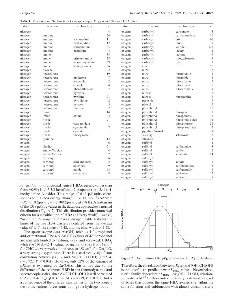
range. FormonofunctionalneutralHBAs, pKBHXvalues spanfrom-0.96 (1,1,1,3,3,3-hexafluoro-2-propanol) toþ5.46 (tri-methylamine N-oxide). This range of 6.42 pK units corre-sponds to a Gibbs energy change of 37 kJ mol-1 (ΔΔG�=-RT ln 10ΔpKBHX=-5.708ΔpKBHXat 298K).Ahistogramof the 1338pKBHXvalues in thedatabase approaches a normaldistribution (Figure 2). This distribution provides numericalcriteria for a classification of HBAs as “very weak”, “weak”,“medium”, “strong”, and “very strong”. Table 4 shows thelimits of the five HBA classes, calculated from the averagevalue of 1.17, the range of 6.42, and the class width of 1.28.
The spectroscopic data Δν(OH) refer to 4-fluorophenoland/or methanol. The 409 Δν(OH) values of 4-fluorophenolare generally limited to medium, weak, and very weakHBAs,while the 790 Δν(OH) values for methanol span from 3 cm-1
for CHCl3, a very weak chloro base, to 488 cm-1 for Oct3NO,
a very strong oxygen base. There is a statistically significantcorrelation between pKBHX and Δν(OH,CH3OH) (n=594,r=0.722, P< 0.001). However, only 52% of the variance ofpKBHX is explained by Δν(OH). This is not due to thedifference of the reference HBD in the thermodynamic andspectroscopic scales, since Δν(OH,CH3OH) is well correlatedtoΔν(OH,4-FC6H4OH) (n=164, r=0.979,P<0.001), but isa consequence of the different sensitivities of the two proper-ties to the various forces contributing to a hydrogen bond.20
Therefore, the correlation between pKBHX and (OH,CH3OH)is not useful to predict new pKBHX values. Nevertheless,useful family dependent pKBHX-Δν(OH, CH3OH) relation-ships do hold.20 In this context, a family is defined as a setof bases that possess the same HBA atomic site within thesame function and subfunction with almost constant steric
Table 3. Functions and Subfunctions Corresponding to Oxygen and Nitrogen HBA Sites
atom function subfunction n atom function subfunction n
nitrogen 4 oxygen carbonyl carbonate 5
nitrogen amidine 24 oxygen carbonyl carboxamidate 10
nitrogen amidine acetamidine 11 oxygen carbonyl ester 60
nitrogen amidine benzamidine 12 oxygen carbonyl imide 8
nitrogen amidine formamidine 35 oxygen carbonyl ketone 125
nitrogen amidine guanidine 4 oxygen carbonyl lactam 4
nitrogen amine 34 oxygen carbonyl lactone 7
nitrogen amine primary amine 28 oxygen carbonyl thiocarbamate 1
nitrogen amine secondary amine 39 oxygen carbonyl urea 8
nitrogen amine tertiary amine 56 oxygen ether 95
nitrogen diazene 2 oxygen nitro 5
nitrogen heteroarene 19 oxygen nitro nitramidate 2
nitrogen heteroarene imidazole 7 oxygen nitro nitramide 1
nitrogen heteroarene isoxazole 2 oxygen nitro nitroalkane 5
nitrogen heteroarene oxazole 4 oxygen nitro nitroalkene 5
nitrogen heteroarene phenanthroline 7 oxygen nitro nitroaromatic 9
nitrogen heteroarene pyrazole 1 oxygen nitroso 2
nitrogen heteroarene pyridine 91 oxygen nitroso nitrosamine 3
nitrogen heteroarene pyrimidine 2 oxygen peroxide 6
nitrogen heteroarene pyrrole 3 oxygen phenol 5
nitrogen heteroarene thiazole 2 oxygen phosphoryl 15
nitrogen imine 14 oxygen phosphoryl phosphate 8
nitrogen imine oxime 2 oxygen phosphoryl phosphinate 7
nitrogen nitrile 91 oxygen phosphoryl phosphine oxide 11
nitrogen nitrile cyanamidate 1 oxygen phosphoryl phosphonate 31
nitrogen nitrile cyanamide 7 oxygen phosphoryl phosphoramide 2
nitrogen nitrile cyanate 1 oxygen pyridine N-oxide 8
nitrogen nitrile thiocyanate 1 oxygen seleninyl selenoxide 2
nitrogen pyridine 12 oxygen siloxane 1
oxygen 6 oxygen sulfinyl 1
oxygen alcohol 21 oxygen sulfinyl sulfinamide 3
oxygen amine N-oxide 4 oxygen sulfinyl sulfite 5
oxygen arsine N-oxide 2 oxygen sulfinyl sulfoxide 14
oxygen carbonyl 6 oxygen sulfonyl 6
oxygen carbonyl acid anhydrid 2 oxygen sulfonyl sulfate 2
oxygen carbonyl aldehyde 9 oxygen sulfonyl sulfonamidate 4
oxygen carbonyl amide 64 oxygen sulfonyl sulfonamide 6
oxygen carbonyl carbamate 8 oxygen sulfonyl sulfonate 2
oxygen sulfonyl sulfone 8
Figure 2. Distribution of the pKBHX values in the pKBHX database.
4078 Journal of Medicinal Chemistry, 2009, Vol. 52, No. 14 Laurence et al.
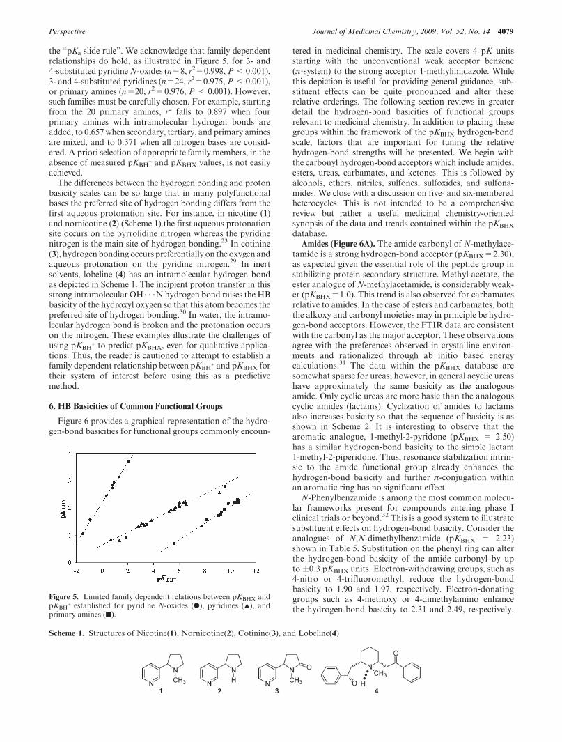
requirements. The pKBHX versus Δν(OH) plot of Figure 3shows some data that could be resolved into separate linescorresponding to the families of amides, substituted imida-zoles, 3- and 4-substituted pyridines, primary amines, andunhindered tertiary amines.
A more detailed description of the database, as well as thecontact for its appropriate access (free of charge for academicaffiliates), is available on the Web site of the CEISAM lab-oratory (http://www.sciences.univ-nantes.fr/CEISAM/en_lhmin.php). The database will be updated once a year. Theversion 09 will contain a 100 new compounds, among themanions, amines, amides, and alkaloids. A significant repre-sentative sample of the data contained in the database (aboutone-third) is provided as Supporting Information.
5. Lack of Correlation between the pKBHX and pKBHþBasicity
Scales
Previous comparisons of the 1:1 HB basicity scales pKHB,logKβ, and logKB
H with the aqueous Brønsted proton basicityscale pKBH
þ have suggested that there is noquantitative generalrelationship between HB basicity and proton basicity.12-14
Only limited family dependent relationships can be established.In the absence of other intuitive measures, many chemistsextend these family dependent relationships beyond a reason-able scope and incorrectly use the order of proton basicity toapproximate HB basicity between families. In the literature,onemay find sentences such as “an order of acidity andbasicitybasedonhydrogenbondingwouldnotdiffer radically fromonebased on acid/base equilibria in aqueous or nonaqueous solu-tion”26 or “insofar as a hydrogen bond reflects partial transferof a proton, it would seem logical for the hydrogenbond energy
to correlate closely with [proton] basicity.”27 A “pKa slide rule”has recently been proposed28 to predict the hydrogen-bondstrength from the pKa of the hydrogen-bond donor and thepKBH
þ of the hydrogen-bond acceptor. This section presents acomparison of the pKBH
þ and pKBHX scales for a sample ofbases not presented in previous studies and highlights the widevariability between these two scales. Examples are given wherethe relationship between pKBH
þ andpKBHX scales breaks downfor simple druglike molecules and becomes of little predictivevalue.
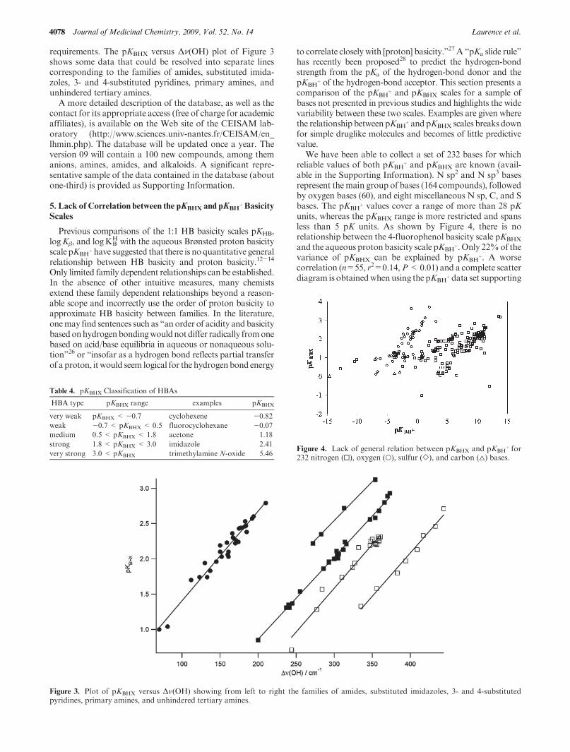
We have been able to collect a set of 232 bases for whichreliable values of both pKBH
þ and pKBHX are known (avail-able in the Supporting Information). N sp2 and N sp3 basesrepresent the main group of bases (164 compounds), followedby oxygen bases (60), and eight miscellaneous N sp, C, and Sbases. The pKBH
þ values cover a range of more than 28 pKunits, whereas the pKBHX range is more restricted and spansless than 5 pK units. As shown by Figure 4, there is norelationship between the 4-fluorophenol basicity scale pKBHX
and the aqueous proton basicity scale pKBHþ. Only 22%of the
variance of pKBHX can be explained by pKBHþ. A worse
correlation (n=55, r2=0.14,P<0.01) and a complete scatterdiagram is obtainedwhenusing the pKBH
þ data set supporting
Figure 3. Plot of pKBHX versus Δν(OH) showing from left to right the families of amides, substituted imidazoles, 3- and 4-substitutedpyridines, primary amines, and unhindered tertiary amines.
Figure 4. Lack of general relation between pKBHX and pKBHþ for
232 nitrogen (0), oxygen (O), sulfur (]), and carbon (4) bases.
Table 4. pKBHX Classification of HBAs
HBA type pKBHX range examples pKBHX
very weak pKBHX < -0.7 cyclohexene -0.82
weak -0.7 < pKBHX < 0.5 fluorocyclohexane -0.07
medium 0.5 < pKBHX < 1.8 acetone 1.18
strong 1.8 < pKBHX < 3.0 imidazole 2.41
very strong 3.0 < pKBHX trimethylamine N-oxide 5.46
Perspective Journal of Medicinal Chemistry, 2009, Vol. 52, No. 14 4079
the “pKa slide rule”. We acknowledge that family dependentrelationships do hold, as illustrated in Figure 5, for 3- and4-substituted pyridineN-oxides (n=8, r2=0.998, P<0.001),3- and 4-substituted pyridines (n=24, r2=0.975, P<0.001),or primary amines (n=20, r2=0.976, P < 0.001). However,such families must be carefully chosen. For example, startingfrom the 20 primary amines, r2 falls to 0.897 when fourprimary amines with intramolecular hydrogen bonds areadded, to 0.657when secondary, tertiary, and primary aminesare mixed, and to 0.371 when all nitrogen bases are consid-ered. A priori selection of appropriate family members, in theabsence of measured pKBH
þ and pKBHX values, is not easilyachieved.
The differences between the hydrogen bonding and protonbasicity scales can be so large that in many polyfunctionalbases the preferred site of hydrogen bonding differs from thefirst aqueous protonation site. For instance, in nicotine (1)and nornicotine (2) (Scheme 1) the first aqueous protonationsite occurs on the pyrrolidine nitrogen whereas the pyridinenitrogen is the main site of hydrogen bonding.23 In cotinine(3), hydrogenbondingoccurs preferentially on the oxygenandaqueous protonation on the pyridine nitrogen.29 In inertsolvents, lobeline (4) has an intramolecular hydrogen bondas depicted in Scheme 1. The incipient proton transfer in thisstrong intramolecular OH 3 3 3Nhydrogen bond raises the HBbasicity of the hydroxyl oxygen so that this atom becomes thepreferred site of hydrogen bonding.30 In water, the intramo-lecular hydrogen bond is broken and the protonation occurson the nitrogen. These examples illustrate the challenges ofusing pKBH
þ to predict pKBHX, even for qualitative applica-tions. Thus, the reader is cautioned to attempt to establish afamily dependent relationship between pKBH
þ and pKBHX fortheir system of interest before using this as a predictivemethod.
6. HB Basicities of Common Functional Groups
Figure 6 provides a graphical representation of the hydro-gen-bond basicities for functional groups commonly encoun-
tered in medicinal chemistry. The scale covers 4 pK unitsstarting with the unconventional weak acceptor benzene(π-system) to the strong acceptor 1-methylimidazole. Whilethis depiction is useful for providing general guidance, sub-stituent effects can be quite pronounced and alter theserelative orderings. The following section reviews in greaterdetail the hydrogen-bond basicities of functional groupsrelevant to medicinal chemistry. In addition to placing thesegroups within the framework of the pKBHX hydrogen-bondscale, factors that are important for tuning the relativehydrogen-bond strengths will be presented. We begin withthe carbonyl hydrogen-bond acceptors which include amides,esters, ureas, carbamates, and ketones. This is followed byalcohols, ethers, nitriles, sulfones, sulfoxides, and sulfona-mides. We close with a discussion on five- and six-memberedheterocycles. This is not intended to be a comprehensivereview but rather a useful medicinal chemistry-orientedsynopsis of the data and trends contained within the pKBHX
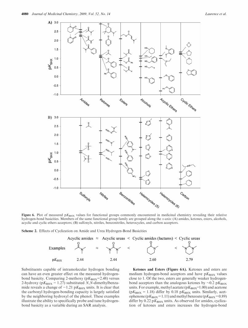
database.Amides (Figure 6A). The amide carbonyl of N-methylace-
tamide is a strong hydrogen-bond acceptor (pKBHX=2.30),as expected given the essential role of the peptide group instabilizing protein secondary structure. Methyl acetate, theester analogue of N-methylacetamide, is considerably weak-er (pKBHX=1.0). This trend is also observed for carbamatesrelative to amides. In the case of esters and carbamates, boththe alkoxy and carbonyl moieties may in principle be hydro-gen-bond acceptors. However, the FTIR data are consistentwith the carbonyl as the major acceptor. These observationsagree with the preferences observed in crystalline environ-ments and rationalized through ab initio based energycalculations.31 The data within the pKBHX database aresomewhat sparse for ureas; however, in general acyclic ureashave approximately the same basicity as the analogousamide. Only cyclic ureas are more basic than the analogouscyclic amides (lactams). Cyclization of amides to lactamsalso increases basicity so that the sequence of basicity is asshown in Scheme 2. It is interesting to observe that thearomatic analogue, 1-methyl-2-pyridone (pKBHX = 2.50)has a similar hydrogen-bond basicity to the simple lactam1-methyl-2-piperidone. Thus, resonance stabilization intrin-sic to the amide functional group already enhances thehydrogen-bond basicity and further π-conjugation withinan aromatic ring has no significant effect.
N-Phenylbenzamide is among the most common molecu-lar frameworks present for compounds entering phase Iclinical trials or beyond.32 This is a good system to illustratesubstituent effects on hydrogen-bond basicity. Consider theanalogues of N,N-dimethylbenzamide (pKBHX = 2.23)shown in Table 5. Substitution on the phenyl ring can alterthe hydrogen-bond basicity of the amide carbonyl by upto (0.3 pKBHX units. Electron-withdrawing groups, such as4-nitro or 4-trifluoromethyl, reduce the hydrogen-bondbasicity to 1.90 and 1.97, respectively. Electron-donatinggroups such as 4-methoxy or 4-dimethylamino enhancethe hydrogen-bond basicity to 2.31 and 2.49, respectively.
Figure 5. Limited family dependent relations between pKBHX andpKBH
þ established for pyridine N-oxides (b), pyridines (2), andprimary amines (9).
Scheme 1. Structures of Nicotine(1), Nornicotine(2), Cotinine(3), and Lobeline(4)
4080 Journal of Medicinal Chemistry, 2009, Vol. 52, No. 14 Laurence et al.
Substituents capable of intramolecular hydrogen bondingcan have an even greater effect on the measured hydrogen-bond basicity. Comparing 2-methoxy (pKBHX=2.48) versus2-hydroxy (pKBHX=1.27) substituted N,N-dimethylbenza-mide reveals a change of -1.21 pKBHX units. It is clear thatthe carbonyl hydrogen-bonding capacity is largely satisfiedby the neighboring hydroxyl of the phenol. These examplesillustrate the ability to specifically probe and tune hydrogen-bond basicity as a variable during an SAR analysis.
Ketones and Esters (Figure 6A). Ketones and esters aremedium hydrogen-bond acceptors and have pKBHX valuesclose to 1. Of the two, esters are generally weaker hydrogen-bond acceptors than the analogous ketones by ∼0.2 pKBHX
units. For example, methyl acetate (pKBHX=1.00) and acetone(pKBHX=1.18) differ by 0.18 pKBHX units. Similarly, acet-ophenone (pKBHX=1.11) andmethyl benzoate (pKBHX=0.89)differ by 0.22 pKBHX units. As observed for amides, cycliza-tion of ketones and esters increases the hydrogen-bond
Figure 6. Plot of measured pKBHX values for functional groups commonly encountered in medicinal chemistry revealing their relativehydrogen-bond basicities. Members of the same functional group family are grouped along the x-axis: (A) amides, ketones, esters, alcohols,acyclic and cyclic ethers acceptors; (B) sulfonyls, nitriles, benzonitriles, heterocycles, and carbon acceptors.
Scheme 2. Effects of Cyclization on Amide and Urea Hydrogen-Bond Basicities
Perspective Journal of Medicinal Chemistry, 2009, Vol. 52, No. 14 4081
basicity of the carbonyl oxygen. This effect is quite pro-nounced for esters, which have a strong preference for an s-trans geometry when acyclic. Cyclization to the lactoneforces an s-cis ester conformation, further enhancing thehydrogen-bondbasicity by∼0.4 pKBHXunits. As an exampleof these effects, compare cyclohexanone (pKBHX=1.39) andδ-valerolactone (pKBHX=1.57) to acetone and methyl acet-ate, respectively. Benzophenone, a very common scaffold indrugs, has a pKBHX=1.07 which is quite close to that ofacetone. Acetophenone behaves similarly to benzamide inresponse to substitutions on the aromatic ring, thus allowingone to adjust the hydrogen-bond basicity by (0.5 pKBHX
units (Table 5). Electron-donating groups increase whileelectron-withdrawing groups decrease hydrogen-bond basi-city. Themeasured pKBHX of 2.50 for 2,6-dimethyl-γ-pyroneis quite exceptional and represents a significant deviationfrom other ketones or esters. An examination of electrondensity from X-ray crystallography and ab initio quantummechanical calculations reveals extensive delocalization ofthe π-system electrons.33 It is this delocalization that pre-sumably enhances the hydrogen-bond basicity of theγ-pyrone carbonyl.
Alcohols and Ethers (Figure 6A). It is common dogma toconsider aliphatic ethers and alcohols as weaker hydrogen-bond acceptors than ketones or the carbonyl oxygen ofesters. However, experimental measurements are consistentwith these groups being equivalent with pKBHX values of∼1.For example, diethyl ether (pKBHX=1.01) has a pKBHX quiteclose to those of methyl acetate and acetone. Cyclic etherssuch as tetrahydrofuran (pKBHX=1.28) and tetrahydropyr-an (pKBHX= 1.23) are commonly encountered in naturalproducts and are more basic than their acyclic counterparts.The strained four-membered ring of oxetane (pKBHX=1.36)is still a stronger HBA and can be used to improve thesolubility of a molecule without compromising its metabolicstability. It is important to recognize the impact of phenylsubstitution through π-conjugation and inductive-fieldeffect, as aromatic ethers and alcohols are weak hydrogen-
bond acceptors. Anisole and phenol have the same hydro-gen-bond basicity (pKBHX=-0.07). This is consistent withthe observation that tyrosine is predominantly a hydrogen-bond donor in protein structures and rarely accepts ahydrogen bond.34
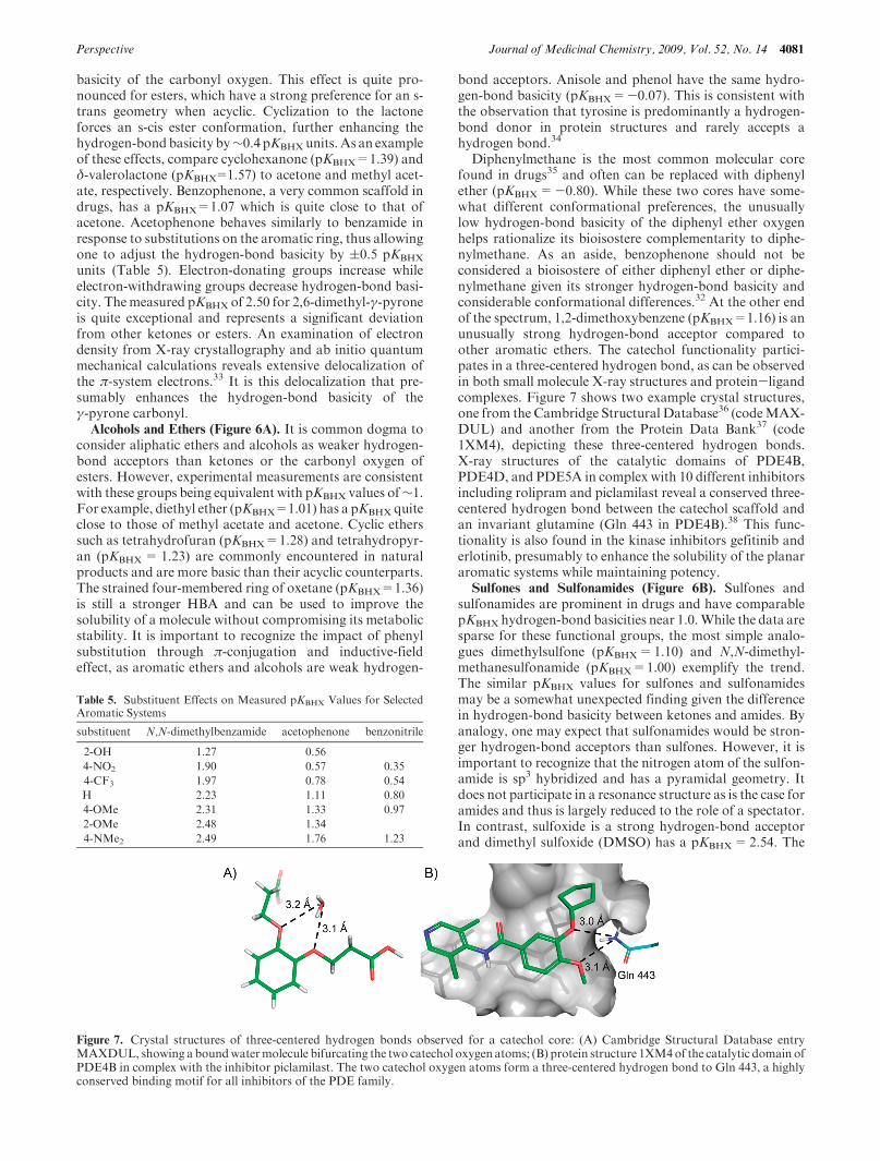
Diphenylmethane is the most common molecular corefound in drugs35 and often can be replaced with diphenylether (pKBHX=-0.80). While these two cores have some-what different conformational preferences, the unusuallylow hydrogen-bond basicity of the diphenyl ether oxygenhelps rationalize its bioisostere complementarity to diphe-nylmethane. As an aside, benzophenone should not beconsidered a bioisostere of either diphenyl ether or diphe-nylmethane given its stronger hydrogen-bond basicity andconsiderable conformational differences.32 At the other endof the spectrum, 1,2-dimethoxybenzene (pKBHX=1.16) is anunusually strong hydrogen-bond acceptor compared toother aromatic ethers. The catechol functionality partici-pates in a three-centered hydrogen bond, as can be observedin both small molecule X-ray structures and protein-ligandcomplexes. Figure 7 shows two example crystal structures,one from the Cambridge Structural Database36 (codeMAX-DUL) and another from the Protein Data Bank37 (code1XM4), depicting these three-centered hydrogen bonds.X-ray structures of the catalytic domains of PDE4B,PDE4D, and PDE5A in complex with 10 different inhibitorsincluding rolipram and piclamilast reveal a conserved three-centered hydrogen bond between the catechol scaffold andan invariant glutamine (Gln 443 in PDE4B).38 This func-tionality is also found in the kinase inhibitors gefitinib anderlotinib, presumably to enhance the solubility of the planararomatic systems while maintaining potency.
Sulfones and Sulfonamides (Figure 6B). Sulfones andsulfonamides are prominent in drugs and have comparablepKBHX hydrogen-bond basicities near 1.0.While the data aresparse for these functional groups, the most simple analo-gues dimethylsulfone (pKBHX=1.10) and N,N-dimethyl-methanesulfonamide (pKBHX=1.00) exemplify the trend.The similar pKBHX values for sulfones and sulfonamidesmay be a somewhat unexpected finding given the differencein hydrogen-bond basicity between ketones and amides. Byanalogy, one may expect that sulfonamides would be stron-ger hydrogen-bond acceptors than sulfones. However, it isimportant to recognize that the nitrogen atom of the sulfon-amide is sp3 hybridized and has a pyramidal geometry. Itdoes not participate in a resonance structure as is the case foramides and thus is largely reduced to the role of a spectator.In contrast, sulfoxide is a strong hydrogen-bond acceptorand dimethyl sulfoxide (DMSO) has a pKBHX=2.54. The
Table 5. Substituent Effects on Measured pKBHX Values for SelectedAromatic Systems
substituent N,N-dimethylbenzamide acetophenone benzonitrile
2-OH 1.27 0.56
4-NO2 1.90 0.57 0.35
4-CF3 1.97 0.78 0.54
H 2.23 1.11 0.80
4-OMe 2.31 1.33 0.97
2-OMe 2.48 1.34
4-NMe2 2.49 1.76 1.23
Figure 7. Crystal structures of three-centered hydrogen bonds observed for a catechol core: (A) Cambridge Structural Database entryMAXDUL, showing a boundwatermolecule bifurcating the two catechol oxygen atoms; (B) protein structure 1XM4of the catalytic domain ofPDE4B in complex with the inhibitor piclamilast. The two catechol oxygen atoms form a three-centered hydrogen bond to Gln 443, a highlyconserved binding motif for all inhibitors of the PDE family.
4082 Journal of Medicinal Chemistry, 2009, Vol. 52, No. 14 Laurence et al.
solubilizing properties of DMSO are well-known, and it iscommon to dissolve organic compounds with poor aqueoussolubility in a DMSO solution prior to dilution in an aqu-eous buffer for biological screening. Undoubtedly the stronghydrogen-bonding ability of the sulfoxide contributes tothese unique properties.
Nitriles (Figure 6B). Simple aromatic and aliphatic nitrilesare medium to weak hydrogen-bond acceptors. Acetonitrileand benzonitrile have pKBHX values of 0.91 and 0.80,respectively. Nitriles are particularly sensitive to substituenteffects, and the more basic nitriles are cyanamides (5),vinylogous cyanamides (6), cyanoamidines (7), and cyano-guanidines (8) (Scheme 3). Their pKBHX values span from1.56 (N,N-dimethylcyanamide) to 2.24 (N2-cyano-N1,N1-dimethylacetamidine 9).39 It is worth noting that this relativeorder of HB basicity, attributed to the increase of conjuga-tive interactions between the lone pair of the amino sub-stituent and the nitrile group, has been observed in smallorganic crystal structures and in the analysis of theoreticalcalculations at theDFT level.40 The strongHBbasicity of thecyanoguanidine moiety of cimetidine (10) suggests a role forthe nitrile group in hydrogen-bonding to the H2 receptor.
19
As observed for aromatic amides and ketones, benzonitrile(pKBHX=0.80) can bemademore basicwith the introductionof 4-methoxy (pKBHX=0.97) or 4-dimethylamino (pKBHX=1.23) substitutions. Conversely, 4-nitrobenzonitrile (pKBHX=0.35) is considerably less basic (Table 5). Perhaps ofacademic interest rather than directly applicable to medic-inal chemistry, pentafluorobenzonitrile has a pKBHX=0.01.The inductive effects of each fluorine atom are roughlyadditive, resulting in almost complete loss of hydrogen-bondaccepting ability of the nitrile group. While the data are notavailable, one can speculate that a similar trend would beobserved for other acceptors such as a benzamide.
Heterocycles (Figure 6B).Aromatic heterocycles are funda-mental tomedicinal chemistry and can impart desirable proper-ties to amolecule such as improved solubility. It is common formedicinal chemists to take into consideration potential hydro-gen-bonding interactions when deciding which heterocycle tosynthesize. However, rarely is there an appreciation of thesignificant differences in relative hydrogen-bond basicity thatcan occur upon the introduction of different heteroatoms indifferent positions. The hydrogen-bond basicities of a set ofsimple nitrogen-containing six-membered heterocycles are in-cluded in Figure 6B. Pyridine (pKBHX=1.86), with a singlenitrogen atom, is the most basic. Introduction of a secondnitrogen atom reduces the hydrogen-bond basicity to differentdegrees depending on the substitution pattern. Of these, themost pronounced change is for pyrazine with a pKBHX of only0.92. Extending this trend with the addition of a third nitrogenatom, as in 1,3,5-triazine (pKBHX = 0.32), attenuates thehydrogen-bond basicity even further.
Pyridine is particularly sensitive to aromatic substitutions,and the changes in hydrogen-bondbasicity are best describedby a combination of inductive-field and resonance Ham-mett/Taft σ descriptors.17,21 Polarizability, which is impor-tant for describing proton transfer basicity, is not significantfor describing the hydrogen-bond basicity of pyridine ana-logues. Similar to the benzamide case, 4-dimethylaminopyr-idine (pKBHX=2.80) is a considerably stronger hydrogen-bond acceptor than pyridine. Steric effects are also impor-tant for hydrogen-bonding atoms within a ring. Substitutionor annulation adjacent to the HBA site can reduce thehydrogen-bond basicity. However, these steric effects areoften counterbalanced by favorable electronic contributionsand can be difficult to predict.
Five-membered heterocycles cover a wide range of hydro-gen-bond basicities. Furan (pKBHX=-0.40) and thiophene(pKBHX=-0.50) are weak hydrogen-bond acceptors. Theyhave both nonbonding andπ-bonding pairs and give n and/orπ complexes.41 In general they should be considered analo-gues of benzene (pKBHX=-0.49) with different steric andgeometric properties. Isoxazole (pKBHX=0.81) and oxazole(pKBHX=1.30) are hydrogen-bond acceptors of mediumstrength, comparable to esters. Indeed, these rings are oftensuccessfully used as bioisosteres of esters with the benefit ofenhanced metabolic stability. For heterocycles containingboth oxygen and nitrogen atoms, a question often arises asto which atom is the preferred HBA. In the case of aromaticheterocycles, there is no ambiguity and the nitrogen atom isoverwhelmingly preferred. This trend is supported by astatistical analysis, complemented by in vacuo ab initio calcu-lations, ofHB interactions observed in the X-ray structures oforganic molecules compiled within the Cambridge StructuralDatabase.42,43 The strongest hydrogen-bond acceptors in thisseries are 1-methylpyrazole (pKBHX=1.84), 1-methylimida-zole (pKBHX=2.72), and 1,5-dicyclohexylimidazole (pKBHX=3.12). These are comparable in hydrogen-bond basicity toamides and vinylogous amides (3-dimethylamino-5,5-dimeth-yl-2-cyclohexen-1-one, pKBHX=2.92).
7. Relevance of HB Basicity to Medicinal Chemistry
Drug discovery is an extremely complex multidimensionaloptimization problem, and there is an increasing awareness inthe pharmaceutical industry that optimizing independentproperties, such as potency, absorption, metabolic stability,etc., requires a parallel approach.44 Hydrogen bonding is wellrecognized as one of the major noncovalent forces involved inprotein-ligand binding and hence is important for ligandaffinity. Indeed, nature often utilizes the variable strength ofhydrogen bonds from strong to weak to achieve specificityand control function.45 Equally well recognized, yet less oftenput into practice, is the understanding that hydrogen bondsalso strongly influence the physicochemical and transportproperties of a molecule. Despite its importance, medicinalchemists rarely probe the strength of hydrogen bonds in asystematic manner, as is often done for other properties suchas steric bulk, hydrophobicity, and proton basicity or acidity.While this shortcoming was recognized by Morris et al.nearly 2 decades ago46 and attributed to a lack of suitablehydrogen-bond scales, very little has changed following thedevelopment of such scales. One of our objectives in thisPerspective is to raise awareness and to provide access to therequired data to effect a change in this status quo with theintroduction of the pKBHX database.
Scheme 3. Structures of Superbasic Nitriles
Perspective Journal of Medicinal Chemistry, 2009, Vol. 52, No. 14 4083
While there is a recognition that parallel optimization ofproperties is desirable or even essential to success, the lure ofthe perceived benefits of high potency is difficult to resist suchthat potency and specificity are still at the forefront of mostlead optimization campaigns. In structure-based design, ex-isting protein-ligand hydrogen bonds or new sites for hydro-gen bonding are easily identified within a binding pocketdirectly from the atomic coordinates. It is common formedicinal chemists to gravitate toward these interactions inan attempt to design more potent or selective analogues.When presented with a scale such as the pKBHX, it is temptingto take a simplistic view that strengthening the HB basicity ofa critical HBA in a ligand will lead to an increase in affinity.However, the relationshipbetween ligand affinity andnumberor strength of hydrogen bonds is often complex and notpredictable. A recent study of hydrogen bonds at the interfaceof protein-ligand complexes47 found that multicentered hy-drogen bonds (e.g., bifurcation and trifurcation) are ubiqui-tous, further complicating attempts to relate hydrogen bondsdirectly with ligand affinity.
The effects of solvent, usually water, give rise to an ofteninvoked oversimplification of the free energy changes duringthe ligand-binding process. In this simplified view, the non-complexed ligand and protein site are fully solvated, withsolventwater fulfilling the full complementof hydrogenbondsfor both species. Upon complex formation, water is displacedand new hydrogen bonds are formed between the ligand andprotein with no net change in the total number of hydrogenbonds after the displaced waters form hydrogen bonds withinthe bulk solvent. This approximation ignores a multitude ofvariables that can strongly influence the energetics of hydro-gen-bond formation including hydrogen-bond geometry, sol-vent accessibility, local polarity of the binding pocket,networks of hydrogen bonds, and differences in the propertiesof bulk, surface, and bound waters and the important con-tribution of conserved water molecules in mediating ligandprotein contacts. A review of hydrogen bonds in protein-ligand complexes2 lists a dozen studies investigating theenthalpy or free energy changes attributed to hydrogen-bondformation during ligand binding. The values range from 0 to-13 kJ/mol, with no convergence to a single value. This is notsurprising, and it must be recognized that every bindingpocket presents a unique environment and simplistic rulesprovide little guidance to the value of a specific hydrogenbondin a binding pocket.
A literature search was undertaken to identify case studiesfor which hydrogen-bond basicity is reported to be correlatedwith biological activity. While we do not claim to havecompleted an exhaustive search, there is a dearth of relevantexamples, clearly suggesting that this is an area rarely system-atically explored by medicinal chemists. Apparently, wedo not hold this opinion alone. In a review, Williams andLadbury state, “moredata onΔH,ΔG, and structural changesin series of similar complexes, categorized by type of hydro-gen-bonding partners and local environmental change,are needed in order to provide a set of case histories fromwhich it may be possible to predict the effect of a proposedchange by similarity to a previous case.”2 This may be toooptimistic a view, and it ismore likely that the hydrogen-bondSAR for a given site will be unique. Thus, it is imperativethat the SAR be established early during a lead optimizationcampaign by systematically surveying the HB basicitylandscape. The pKBHX database can be useful during thisendeavor.
The parallel optimization of physicochemical propertiesduring the design of new analogues requires medicinal che-mists to consider many factors that are tangentially related tohydrogen-bond basicity. For example, low aqueous solubilityand high lipophilicity are often undesirable outcomes of apotency-driven lead optimization program and contribute tolow intestinal absorption. One commonly employed strategyto reduce lipophilicity is to substitute phenyl rings withheterocycles to introduce polarity. The choice of heterocyclecan be dictated to a large extent by the local environmentwithin the binding pocket. If the phenyl ring is believed to bepartially solvent exposed in the binding site, an ortho-sub-stituted pyridine may be a good choice. The pyridine nitrogenatom is a strong HBA and will provide a polar interface tosolvent, while the ortho-substitution can reduce undesirablebinding to Cyp3A4.48 However, in a hydrophobic environ-ment or for central nervous system indications that requiretransport across the blood-brain barrier, this is perhaps oneof the worst heterocycles to choose given its strong hydrogen-bond basicity. Instead, pyrazine or pyrimidine which formweaker hydrogen bonds are better alternatives.
Fluorine is often introduced at specific metabolically labilepositions to reduce clearance or the formation of reactiveintermediates.49,50During this process, the inductive effects offluorine can also be used to exquisitely tune the hydrogen-bond basicity of a distal group. Consider pyridine and fluor-ine-substituted analogues: pyridine (pKBHX=1.86), 3-fluoro-pyridine (pKBHX=1.35), 2-fluoropyridine (pKBHX=0.95),2,6-difluoropyridine (pKBHX=0.14), andpentafluoropyridine(pKBHX=-0.49). Walking the fluorine atom(s) around thering alters the hydrogen-bond basicity significantly. In acomprehensive study, a similar use of fluorine to modulateproton basicity has been reported.51 For fluorine-substitutedsystems, the proton basicity and HB basicity are largelyinfluenced by inductive-field effects. Therefore, it may beexpected that the other trends reported for proton basicitywill be transferable to hydrogen-bond basicity, although thisis yet to be confirmed experimentally.
The importance of relative hydrogen-bond strengths longhas been recognized within the field of computer-aided drugdesign. The initial focus was in developing descriptors for usein quantitative structure-activity relationship (QSAR) mod-els. The complexity of these descriptors ranged from simplisticcounts of donor and acceptor atoms to more sophisticatedelectronic properties derived fromquantummechanicalmeth-ods and has been reviewed by Dearden and Ghafourian.52
This remains an active area of research, and more recentreports reflect an attempt to apply these methods during thedrug designprocess.AWeb-based tool accessible tomedicinalchemists for predicting hydrogen-bond basicities has beenreported by Gancia et al.53 Application of this tool to a seriesof IKK2 kinase inhibitors showed a useful correlation bet-ween the predicted hydrogen-bond strength and enzymeIC50.
54 A similar tool based upon the electrostatic localminima (Vs,min) method55-58 has been implemented at RochePalo Alto. It is anticipated that the extensive data set con-tained within the pKBHX database can be used to improveupon these descriptors and to refine the computationalmodelsof hydrogen bonding.
With improved computational models capable of predict-ing hydrogen-bond basicities, it is reasonable to considerrefining the Lipinski “rule of 5” for intestinal absorption orpermeation.6 Lipinski et al. defined a set of rules targetedtoward medicinal chemists and their inherent strengths in
4084 Journal of Medicinal Chemistry, 2009, Vol. 52, No. 14 Laurence et al.
pattern and functional group recognition. As a consequence,the influence of hydrogen bonding on a molecule’s intestinalabsorption was reduced to a simple sum of NH andOH bondsfor HBDs and the sum of N and O atoms for HBAs. At thetime, it was recognized that this approximation for HBAs waspoor given the considerable variability of HB basicities for anitrogen or oxygen atom. Indeed, a sum of N and O atomsexplains only 26% of the variance of the pKBHX scale in asample of 1094 values calculated to take into account poly-functional HBAs (data not shown). Others have suggested theuse of HB scales as the basis for an improved intestinalabsorption model5,7 and proposed structural group constantsto rapidly predict hydrogen-bond basicities.15 We continueresearch efforts in the development of robust predictionmethods for drug molecules with 5-10 HBAs. It is anticipatedthat as these prediction methods mature, the Lipinski ruleplacing a limit of 10 N and O atoms may be replaced with amore meaningful maximum value derived from the pKBHX
scale.
8. Conclusions
The creation of the pKBHX database is an incremental stepthat provides both a framework and the associated data toallowmedicinal chemists the ability todevelop an intuition forhydrogen-bond basicity. The hydrogen-bond basicity of aselected set of functional groups deemed to be particularlyrelevant to medicinal chemists was reviewed, with a focus onthe effects of a few common substituents. Finally, a briefdiscussion highlighting the role of hydrogen-bond basicity invarious aspects of drug design was intended to stimulate thecuriosity of practicing medicinal chemists. It is the authors’hope that the data presented herein will provide the founda-tion for new experiments and inquiries that further improveour understanding of the hydrogen bond.
Acknowledgment. We thank our colleagues at Roche,particularly Martin Stahl and Klaus Mueller, for stimulatingdiscussions and critical review of this manuscript.
Biographies
Christian Laurence is Professor Emeritus of Chemistry at theUniversity of Nantes (France) where he obtained his doctoratein Chemistry. He carried out his postdoctoral studies with A. R.Katritzky at the University of East Anglia (U.K.). He was headof the laboratory of spectrochemistry of the Faculty of Sciencesof Nantes for 23 years. He published more than 140 scientificpapers in the field of physical organic chemistry (substituenteffects, solvent effects, hydrogen bonding, and halogen bond-ing). His current interests are the construction of spectroscopicand thermodynamic scales of hydrogen-bond andhalogen-bondbasicities and of electronic databases of Lewis basicity scales.Ken A. Brameld received his B.S. in Chemistry at the Uni-
versity ofWashington in 1993.He earned his Ph.D. inChemistryin 1998 from California Institute of Technology, under thesupervision of Prof. William Goddard, III. In 1998 he began apostdoctoral fellowship at University of California, San Fran-cisco, in the laboratory of Irwin “Tack” Kuntz and in 1999 wasawarded an NIH postdoctoral fellowship. He joined the phar-maceutical industry in 2000 and is currently a PrincipalResearch Scientist at Roche Palo Alto, where he is the computa-tional chemistry group leader. He has contributed to researchprojects in multiple disease areas including oncology, immunol-ogy, and virology.J�erome Graton received his Ph.D. in Physical Organic Chem-
istry from the University of Nantes (France, 2001) under the
direction of Profs.M. Berthelot andC. Laurence.He has carriedout postdoctoral studies at the University College London(U.K.) in Prof. S. L. Price’s research group “Centre for Theore-tical and Computational Chemistry”. In 2002, he joined the“Laboratoire de Spectrochimie et Mod�elisation”, currentlyCEISAM, as Lecturer. His research area concerns the charac-terization of molecular interactions and their applications toneuroactive molecules.
Jean-Yves Le Questel received his Ph.D. in Physical OrganicChemistry in 1991 from theUniversity ofNantes (France). Aftera postdoctoral period with Prof. James Milner White at Glas-gow University, U.K., he carried on another postdoctoralappointment with Dr. Serge Perez at the French NationalInstitute for Agricultural Research (INRA) of Nantes. Hebecame a Lecturer at the University of Nantes in 1993 and aProfessor in 2006. He is presently the head of the “HydrogenBonds and Neuroactive Molecules” group of the CEISAMlaboratory of Nantes University. His research is focused onthe elucidation of hydrogen-bond geometries and energiesthrough experimental (IR spectrometry, structural databaseanalyses) and computational chemistry, with a particular em-phasis on the analysis of the hydrogen bonding of small-molecule biological ligands.
Eric Renault obtained his Ph.D. in Physical Chemistry in 1996from the University Paris Descartes (France), under the super-vision of Prof. M. Gard�es-Albert and Dr. M.-P. Fontaine-Aupart. After postdoctoral research with L. Sanche at theUniversity of Sherbrooke (Canada), he joined theCommissariat�a l’Energie Atomique (Service des Photons, Atomes etMol�ecules) of Saclay (France) in M.-E. Couprie’s Super-ACOFEL team. In 2001, he became Lecturer at the University ofNantes. Apart of his researchwas devoted to time-resolved laserspectroscopy of biomolecular systems. Since his arrival in thelaboratory of spectrochemistry, currently CEISAM, his currentresearch interests include the application of computa-tional chemistry, with a special emphasis on the molecularinteractions.
Supporting Information Available: Graphic representation ofthe pKBHX scale including the different functional groupsencountered in the database; a table listing the pKBHX andpKBH
þ values used for the comparison of the HB and protonbasicity scales; a table listing a representative sample of the datacontained in the pKBHX database. This material is available freeof charge via the Internet at http://pubs.acs.org.
References
(1) Fersht, A. R.; Shi, J.-P.; Knill-Jones, J.; Lowe, D. M.; Wilkinson,A. J.; Blow, D. M.; Brick, P.; Carter, P.; Waye, M. M. Y.; Winter,G. Hydrogen bonding and biological specificity analysed by pro-tein engineering. Nature 1985, 314, 235–238.
(2) Williams, M. A.; Ladbury, J. E. Hydrogen bonds in protein-ligand complexes. Methods Princ. Med. Chem. 2003, 19, 137–161.
(3) Brzozowski, A. M.; Pike, A. C. W.; Dauter, Z.; Hubbard, R. E.;Bonn, T.; Engstrom, O.; Ohman, L.; Greene, G. L.; GustafssonJ.-A.; Carlquist,M.Molecular basis of agonism and antagonism inthe oestrogen receptor. Nature 1997, 389, 753–758.
(4) Williams, S. P.; Sigler, P. B. Atomic structure of progesteronecomplexed with its receptor. Nature 1998, 393, 392–396.
(5) Abraham, M. H.; Ibrahim, A.; Zissimos, A. M.; Zhao, Y. H.;Comer, J.; Reynolds, D. P. Application of hydrogen bondingcalculations in property based drug design. Drug Discovery Today2002, 7, 1056–1063.
(6) Lipinski, C. A.; Lombardo, F.; Dominy, B. W.; Feeney, P. J.Experimental and computational approaches to estimate solubilityand permeability in drug discovery and development settings.Adv.Drug Delivery Rev. 1997, 23, 3–25.
(7) Raevsky, O. H-Bonding Parameterization in QSAR and Drug De-sign. In Molecular Drug Properties; Mannhold, R., Ed.; Wiley-VCHVerlag GmbH & Co. KGaA: Weinheim, Germany, 2008; pp 127-154.
(8) Here, we avoid the ambiguous notation “pKa”. The aqueousproton basicity of a neutral base B is described in terms of the
Perspective Journal of Medicinal Chemistry, 2009, Vol. 52, No. 14 4085
Ka of its conjugate acidBHþ, denotedpKBH
þ (Bagno,A.; Scorrano,G. J. Am.Chem. Soc., 1998, 110, 4577-4582), clarifying the speciesfor amphoteric compounds (anilines, phenols, primary andsecondary amides and sulfonamides, etc). For example, thepKBH
þ of anilines refers to the acid-base pair ArNH3þ/ArNH2,
that is, to their basicity, and not the pair ArNH2/ArNH-
(acidity).(9) Bohm, H. J.; Schneider, G. Protein-Ligand Interactions from
Molecular Recognition to Drug Design; Wiley-VCH Verlag GmbHand Co.: Weinheim, Germany, 2003.
(10) Reed, A. E.; Curtiss, L. A.; Weinhold, F. Intermolecular interac-tions from a natural bond orbital, donor-acceptor viewpoint.Chem. Rev. 1988, 88, 899–926.
(11) Gold,V.Glossaryof termsused inphysical organic chemistry.PureAppl. Chem. 1983, 55, 1281–1371.
(12) Taft, R.W.; Gurka, D.; Joris, L.; Schleyer, P. v. R.; Rakshys, J.W.Studies of hydrogen-bonded complex formation with p-fluoro-phenol. V. Linear free energy relationshipswithOH reference acidsJ. Am. Chem. Soc. 1969, 91, 4801–4808.
(13) Abraham,M. H.; Duce, P. P.; Prior, D. V.; Barratt, D. G.; Morris,J. J.; Taylor, P. J. Hydrogen bonding. Part 9. Solute proton-donorand proton-acceptor scales for use in drug design. J. Chem. Soc.,Perkin Trans. 2 1989, 1355–1375.
(14) Abraham,M. H.; Grellier, P. L.; Prior, D. V.; Morris, J. J.; Taylor,P. J. Hydrogen bonding. Part 10. A scale of solute hydrogen-bondbasicity using logK values for complexation in tetrachloro-methane. J. Chem. Soc., Perkin Trans. 2 1990, 521–529.
(15) Abraham, M. H.; Platts, J. A. Hydrogen bond structural groupconstants. J. Org. Chem. 2001, 66, 3484–3491.
(16) Abraham,M.H.Hydrogen bonding. 31. Construction of a scale ofsolute effective or summation hydrogen-bond basicity. J. Phys.Org. Chem. 1993, 6, 660–684.
(17) Hansch, C.; Leo, A.; Taft, R.W. A survey of Hammett substituentconstants and resonance and field parameters.Chem.Rev. 1991, 91,165–195.
(18) References of journal articles and Ph.D. theses are included in thedatabase.
(19) Laurence, C.; Berthelot, M. Observations on the strength ofhydrogen bonding. Perspect. Drug Discovery Des. 2000, 18, 39–60.
(20) Joesten, M. D.; Schaad, L. J. Hydrogen Bonding; Dekker: NewYork, 1974; 622 pp.
(21) Berthelot, M.; Laurence, C.; Safar, M.; Besseau, F. Hydrogen-bond basicity pKHB scale of six-membered aromatic N-hetero-cycles. J. Chem. Soc., Perkin Trans. 2 1998, 283–290.
(22) Adcock,W.;Graton, J.; Laurence,C.; Lucon,M.; Trout,N. Three-center hydrogen bonding in the complexes of syn-2,4-difluoroada-mantane with 4-fluorophenol and hydrogen fluoride. J. Phys. Org.Chem. 2005, 18, 227–234.
(23) Graton, J.; Berthelot, M.; Gal, J.-F.; Laurence, C.; Lebreton, J.;Le Questel, J.-Y.; Maria, P.-C.; Robins, R. The nicotinic pharma-cophore: thermodynamics of the hydrogen-bonding complexationof nicotine, nornicotine, andmodels. J. Org. Chem. 2003, 68, 8208–8221.
(24) Joris, L.;Mitsky, J.; Taft, R.W.Effects of polar aprotic solvents onlinear free-energy relations in hydrogen-bonded complex forma-tion. J. Am. Chem. Soc. 1972, 94, 3438–3442.
(25) In MDL ISIS.(26) Dunitz, J. D. Weak intermolecular interactions in solids and
liquids. Mol. Cryst. Liq. Cryst. Sci. Technol., Sect. A 1996, 279,209–218.
(27) Rablen, P. R.; Lockman, J.W.; Jorgensen,W. L. Ab initio study ofhydrogen-bonded complexes of small organic molecules withwater. J. Phys. Chem. A 1998, 102, 3782–3797.
(28) Gilli, P.; Pretto, L.; Bertolasi, V.; Gilli, G. Predicting hydrogen-bond strengths fromacid-basemolecular properties. The pKa sliderule: toward the solution of a long-lasting problem. Acc. Chem.Res. 2009, 42, 33–44.
(29) Arnaud, V.; Le Questel, J.-Y.; Mathe-Allainmat, M.; Lebreton, J.;Berthelot, M. Multiple hydrogen-bond accepting capacities ofpolybasic molecules: the case of cotinine. J. Phys. Chem. 2004,108, 10740–10748.
(30) Locati, A.; Berthelot, M.; Evain, M.; Lebreton, J.; Le QuestelJ.-Y.; Mathe-Allainmat, M.; Planchat, A.; Renault, E.; Graton, J.The exceptional hydrogen-bond properties of neutral and proto-nated lobeline. J. Phys. Chem. A 2007, 111, 6397–6405.
(31) Lommerse, J. P. M.; Price, S. L.; Taylor, R. Hydrogen bonding ofcarbonyl, ether, and ester oxygen atoms with alkanol hydroxylgroups. J. Comput. Chem. 1997, 18, 757–774.
(32) Brameld, K. A.; Kuhn, B.; Reuter, D. C.; Stahl,M. Small moleculeconformational preferences derived from crystal structure data. Amedicinal chemistry focused analysis. J. Chem. Inf. Model. 2008,48, 1–24.
(33) Darakjian, Z.; Fink, W. H.; Hope, H. Comparison of X-ray andab initio deformation densities in dimethyl-[gamma]-pyroneand triazine trichloride. J. Mol. Struct.: THEOCHEM 1989, 202,111–120.
(34) Imai, Y. N.; Inoue, Y.; Yamamoto, Y. Propensities of polar andaromatic amino acids in noncanonical interactions: nonbondedcontacts analysis of protein-ligand complexes in crystal structures.J. Med. Chem. 2007, 50, 1189–1196.
(35) Bemis, G. W.; Murcko, M. A. The properties of known drugs. 1.Molecular frameworks. J. Med. Chem. 1996, 39, 2287–2893.
(36) Allen, F. H. The Cambridge Structural Database: a quarter of amillion crystal structures and rising. Acta Crystallogr. 2002, B58,380–388.
(37) Berman,H.M.;Westbrook, J.; Feng, Z.; Gilliland,G.; Bhat, T.N.;Weissig, H.; Shindyalov, I. N.; Bourne, P. E. The Protein DataBank. Nucleic Acids Res. 2000, 28, 235–242.
(38) Card, G. L.; England, B. P.; Suzuki, Y.; Fong, D.; Powell, B.;Lee, B.; Luu, C.; Tabrizizad, M.; Gillette, S.; Ibrahim, P. N.;Artis, D. R.; Bollag, G.; Milburn, M. V.; Kim, S.-H.;Schlessinger, J.; Zhang, K. Y. J. Structural basis for the activityof drugs that inhibit phosphodiesterases. Structure 2004, 12, 2233–2247.
(39) Berthelot, M.; Helbert, M.; Laurence, C.; Le Questel, J. Y.; Anvia,F.; Taft, R. W. Super-basic nitriles. J. Chem. Soc., Perkin Trans. 21993, 625–627.
(40) Ziao, N.; Graton, J.; Laurence, C.; Le Questel, J. Y. Amino andcyano N atoms in competitive situations: which is the best hydro-gen-bond acceptor? A crystallographic database investigation.Acta Crystallogr., Sect. B: Struct. Sci. 2001, 57, 850–858.
(41) Legon, A. C.; Ottaviani, P. The rotational spectrum of thio-phene 3 3 3HBr and a comparison of the geometries of thecomplexes B 3 3 3HX, where B is benzene, furan or thiopheneand X is F, Cl or Br. Phys. Chem. Chem. Phys. 2004, 6, 488–494.
(42) Nobeli, I.; Price, S. L.; Lommerse, J. P. M.; Taylor, R. Hydrogenbonding properties of oxygen and nitrogen acceptors in aromaticheterocycles. J. Comput. Chem. 1997, 18, 2060–2074.
(43) Boehm,H.-J.; Brode, S.; Hesse, U.;Klebe,G. Oxygen and nitrogenin competitive situations: which is the hydrogen-bond acceptor?.Chem.-Eur. J. 1996, 2, 1509–1513.
(44) van de Waterbeemd, H.; Smith, D. A.; Beaumont, K.; Walker, D.K. Property-based design: optimization of drug absorption andpharmacokinetics. J. Med. Chem. 2001, 44, 1313–1333.
(45) Watson, J. D.; Crick, F. H. C. A Structure for deoxyribose nucleicacid. Nature 1953, 171, 737–738.
(46) Morris, J. J.;Hughes,L.R.;Glen,A.T.; Taylor, P. J.Non-steroidalantiandrogens. Design of novel compounds based on an infraredstudy of the dominant conformation and hydrogen-bonding prop-erties of a series of anilide antiandrogens. J. Med. Chem. 1991, 34,447–455.
(47) Panigrahi, S. K.; Desiraju, G. R. Strong andweak hydrogen bondsin the protein-ligand interface. Proteins: Struct., Funct., Bioinf.2007, 67, 128–141.
(48) Riley, R. J.; Parker,A. J.; Tigg, S.;Manners, C.N.Development ofa generalized, quantitative physicochemical model of CYP3A4inhibition for use in early drug discovery. Pharm. Res. 2001, 18,652–655.
(49) Boehm, H.-J.; Banner, D.; Bendels, S.; Kansy, M.; Kuhn, B.;Muller, K.; Obst-Sander, U.; Stahl, M. Fluorine in medicinalchemistry. ChemBioChem 2004, 5, 637–643.
(50) Hagmann, W. K. The many roles for fluorine in medicinal chem-istry. J. Med. Chem. 2008, 51, 4359–4369.
(51) Morgenthaler,M.; Schweizer, E.; Hoffmann-Roder,A.; Benini, F.;Martin, R. E.; Jaeschke, G.; Wagner, B.; Fischer, H.; Bendels, S.;Zimmerli, D.; Schneider, J.; Diederich, F.; Kansy, M.; MullerK. Predicting and tuning physicochemical properties in leadoptimization: amine basicities. ChemMedChem 2007, 2, 1100–1115.
(52) Dearden, J. C.; Ghafourian, T. Hydrogen bonding parameters forQSAR: comparison of indicator variables, hydrogen bond counts,molecular orbital and other parameters. J. Chem. Inf. Comput. Sci.1999, 39, 231–235.
(53) Gancia, E.; Montana, J. G.; Manallack, D. T. Theoretical hydro-gen bonding parameters for drug design. J. Mol. Graphics Modell.2001, 19, 349–362.
(54) Bingham, A. H.; Davenport, R. J.; Gowers, L.; Knight, R. L.;Lowe, C.; Owen, D. A.; Parry, D. M.; Pitt, W. R. A novel series ofpotent and selective IKK2 inhibitors. Bioorg. Med. Chem. Lett.2004, 14, 409–412.
(55) Murray, J. S.; Ranganathan, S.; Politzer, P. Correlations bet-ween the solvent hydrogen-bond acceptor parameter beta and the
4086 Journal of Medicinal Chemistry, 2009, Vol. 52, No. 14 Laurence et al.
calculated molecular surface electrostatic potential. J. Org. Chem.1991, 56, 3734–3737.
(56) Menziani, M. C.; Cocchi, M.; De Benedetti, P. G. Electronic andelectrostatic aspects of carbonic anhydrase inhibition by sulpho-namides. J. Mol. Struct.: THEOCHEM 1992, 256, 217–229.
(57) Kenny, P.W. Prediction of hydrogen bondbasicity from computedmolecular electrostatic properties: implications for comparative
molecular field analysis. J. Chem. Soc., Perkin Trans. 2 1994, 199–202.
(58) Hagelin, H.; Murray, J. S.; Brinck, T.; Berthelot, M.; Politzer, P.Family-independent relationships between computed molecularsurface quantities and solute hydrogen bond acidity/basicity andsolute-induced methanol O-H infrared frequency shiftsCan. J. Chem. 1995, 73, 483–488.