Embed Size (px)
Citation preview

http://www.elsevier.com/locate/bba
Biochimica et Biophysica Ac
Review
The plant Golgi apparatus—Going with the flow
Chris Hawesa,*, Beatrice Satiat-Jeunemaitreb
aResearch School of Biological and Molecular Sciences, Oxford Brookes University, Oxford OX3 0BP, UKbDynamique de la Compartimentation Cellulaire, Institut des Sciences du Vegetal, CNRS, 91198 Gif sur Yvette Cedex, France
Received 7 February 2005; received in revised form 17 March 2005; accepted 22 March 2005
Available online 9 April 2005
Abstract
The plant Golgi apparatus is composed of many separate stacks of cisternae which are often associated with the endoplasmic reticulum
and which in many cell types are motile. In this review, we discuss the latest data on the molecular regulation of Golgi function. The concept
of the Golgi as a distinct organelle is challenged and the possibility of a continuum between the endoplasmic reticulum and Golgi is
proposed.
D 2005 Elsevier B.V. All rights reserved.
Keywords: Plant Golgi apparatus; Membrane flow; Protein trafficking
1. Introduction
Early electron microscopy (EM) studies revealed that
plant cells contained organelles composed of closely
apposed lamellae or cisternae, similar to the Golgi stacks
described in mammalian cells [1,2]. This initiated decades
of work on the biochemistry and the ultrastructure of the
plant Golgi apparatus (GA). Since then, the unique aspects
of the organisation and function of the plant GA has been
regularly reviewed [3–6] and the differences in its
organisation compared to other eukaryotes have often been
discussed [6–8]. However, the unravelling of the genetic
make up and molecular machineries of the plant secretory
pathway has revealed significant homology with the
mammalian and yeast counterparts [4–6,9]. Yet, despite
this presence of highly conserved genes, the mechanisms by
which the expression of these genes results in such
phenotypic differences in both the structure and function
of the plant GA has still to be unravelled. This also raises
the question as to whether there is a basic set of rules that
governs the operation of the secretory pathway across the
kingdoms that can result in the varied levels of organisation
0167-4889/$ - see front matter D 2005 Elsevier B.V. All rights reserved.
doi:10.1016/j.bbamcr.2005.03.009
* Corresponding author. Tel.: +44 1865 483266.
E-mail address: [email protected] (C. Hawes).
reported to date. In this review, we consider some of the
most recent observations on the organisation and dynamics
of the plant Golgi apparatus and attempt to reconcile these
with some of the earlier work on Golgi structure.
2. The plant Golgi apparatus: an organised profusion of
membranes with adaptive specialisations
EM studies (structural, autoradiographical, immunocyto-
chemical) have traditionally provided the basis on which the
organisation and the function of the plant Golgi apparatus
has been described [8,10,11]. More recently, the develop-
ment of specific probes (antibodies, fluorescent protein-
based Golgi markers) has permitted the visualisation of the
Golgi apparatus by fluorescence microscopy [12,13]. It is
now possible to carry out in vivo experiments on Golgi
function and dynamics [9,14–17]. Moreover, imaging
whole cells has permitted a better understanding of the
structural and functional relations between the GA and its
neighbouring membranous compartments, in particular
between the ER and the GA [18–20].
As for other eukaryotic cells, the plant GA is a pivotal
organelle in the secretory pathway, being a cross-roads in
various trafficking events. Golgi compartments receive most
ta 1744 (2005) 93 – 107
C. Hawes, B. Satiat-Jeunemaitre / Biochimica et Biophysica Acta 1744 (2005) 93–10794
of the ER-processed proteins, although export from the plant
ER may by-pass the Golgi by a number of mechanisms. In
the cereals and grasses ER, processed storage proteins may
simply concentrate (oligomerise) and segregate within the
ER lumen, and evolve into distinct storage compartments
[21–23]. In cucurbits, so-called PAC vesicles (precursor
accumulating vesicles) may carry protein cargo directly
from ER to a storage vacuole [24].
Within the GA, ER-derived cargo molecules are further
glycosylated, and the synthesis of complex polysaccharides
characteristic of plants is undertaken. The plant Golgi is
specialised in the synthesis and the export of complex
glycans to the cell surface to organise the cell walls, which
are mainly composed of polysaccharides (cellulose, hemi-
celluloses and pectins) [25,26]. Golgi-derived secretory
vesicles carry these macromolecules to the plasma mem-
brane [27,28]. Although synthesis of cellulose takes place
on the plasma membrane, it also depends on the transport of
membrane-bound enzymes, the cellulose synthases, from
the Golgi to the plasma membrane. Certain glycosylation
events are specific to the plant kingdom such as the addition
of fucose and xylose to glycan chains [29–32] and recently
a family of sugar transporters needed for complex poly-
saccharide synthesis has been described and located to the
GA [33]. Lipid modifications are also important, many tasks
taking place at the Golgi level, leading to a plant specific
plasma membrane composition [34–36].
Mature cargo molecules (membrane or soluble) are
differentially routed within the cell. Thus, another major
function of the GA is the sorting and packaging of wall
macromolecules and cargo proteins in membrane-bounded
carriers to be targeted to their final destination. They may
either reach the plasma membrane by exocytosis or other
compartments of the endomembrane system, such as the
vacuole, often via intermediate compartments such as the
prevacuolar compartment (PVC). Here again, the processes
are adapted to the cell type and the occurrence of a
pleiomorphic and multifunctional vacuolar system is unique
to plants [37,38].
Besides its pivotal role in secretion and vacuolar trans-
port, the plant Golgi apparatus may also be involved in
membrane recycling processes, either from plasma mem-
brane [39,40], from the vacuolar system [41], or in the
recycling of ER membrane [18,19].
A real challenge for the cell biologist is to understand
how this transfer of cargo molecules is achieved within the
endomembrane system. The development of cell-free
assays, allowing the reconstitution of protein transport
through the GA in mammalian systems resulted in the
concept of trafficking by vesicular carriers [42,43]. It
suggests that a membranous compartment (donor compart-
ment) is able to provide a vesicle (carrier), which will fuse
with another membranous compartment (acceptor compart-
ment). In this concept, the compartments remain static.
However, the early EM studies of Morre et al. describe the
plant Golgi apparatus as a complex tubulo-saccular struc-
ture, with some contacts with endoplasmic reticulum
[11,44–46]. Their observations raised the hypothesis of a
membrane continuum within the endomembrane compart-
ments. Finally, the description of the Golgi in some protists
[47–51] has been the basis for the so-called cisternal
maturation model. As cell wall scales were observed within
the cisternal lumen, this model proposed that cisternae
progressively move down the stack. These observations
suggest that Golgi cisternae may evolve with time into
different physiological or functional entities.
Whatever the nature of the cargo carriers in the Golgi
stack, they appear to be associated with specific molecular
machineries, and to be sensitive to the secretory inhibitor
Brefeldin A (BFA). After BFA treatment Golgi markers
have been reported to be either redistributed to the ER
[13,19] or they accumulate within ‘‘BFA compartments’’
which by electron microscopy can comprise clusters of
Golgi-derived vesicles [12,52,53]. In the former case, hybrid
membrane structures composed of ER and Golgi have been
reported in tobacco BY2 cells [54]. Such effects appear to
depend on cell type and/or physiological conditions of the
cells.
The occurrence of specific molecular machinery asso-
ciated with endomembranes is now well established. Coat
proteins (COPI, COPII, clathrin), small GTPases (Rabs, Arf,
Sar1, Rac), fusion proteins (SNARES) and many others,
appear well conserved throughout the eukaryotic kingdom,
and their identification has in many instances paralleled the
development of the vesicle carrier theory [55,56]. In plant
cells, the majority of the COP I and COP II machinery and
their associated effectors (respectively Arf and Sar1) has
been cloned [57–60], although the exact role of the coat
complexes in membrane and cargo transport has yet to be
elucidated (see Section 3.2). Clathrin coated vesicles appear,
as in mammalian cells, to be involved in the transport of
hydrolytic enzymes from export sites at the trans-Golgi, to
the vacuolar system ([43]; see Section 4.2). Regulation of
the budding and fusion events between compartments is
associated with the Rab [61–63] and SNARE [64,65]
protein families, regulating specific steps of trafficking
pathways, some of them being plant specific. Besides these
protein families involved in membrane remodelling, some
proteins have been identified as potential stabilisers for
Golgi membranes, and described as ‘‘Golgi matrix pro-
teins’’. A few homologues can be found in the Arabidopsis
genome, although a specific role in organisation of the Golgi
has not yet been shown ([6,66], see Section 5). Finally, the
families of motor proteins or their regulators, which should
assure the transport of carriers between two compartments,
are still poorly documented in plant cells [67–70].
Most of the data obtained by fluorescence microscopy
have been interpreted taking into account the knowledge
gained from earlier EM studies. However, in the latter case,
a compartment has often be identified on morphological
parameters meanwhile, in the former case, a compartment is
identified by its ability to be recognised by specific markers.
C. Hawes, B. Satiat-Jeunemaitre / Biochimica et Biophysica Acta 1744 (2005) 93–107 95
It is interesting to consider the dynamics and organisation of
the plant GA in the light of recent fluorescence studies,
ignoring all the preconceptions from many years of electron
microscopy. Ultrastructural evidence can then be mapped
onto hypotheses generated from the recent immunocyto-
chemical and live cell imaging studies.
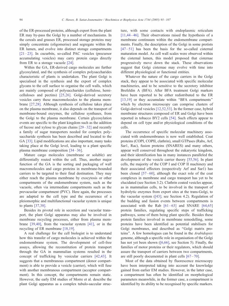
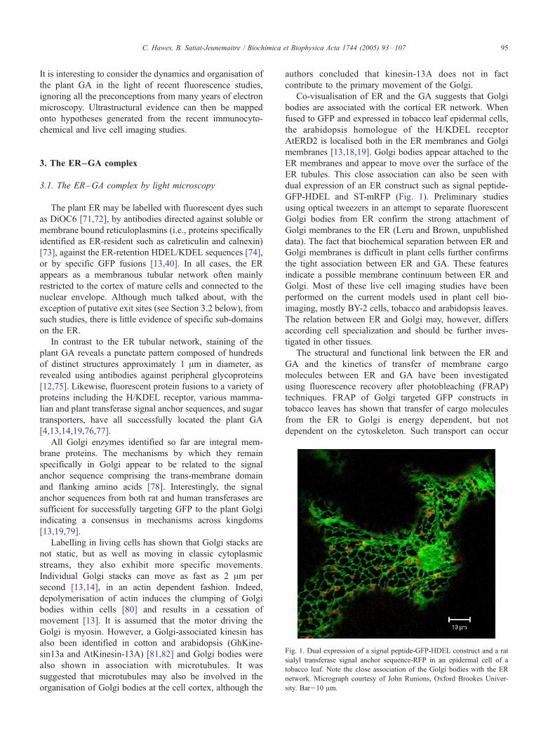
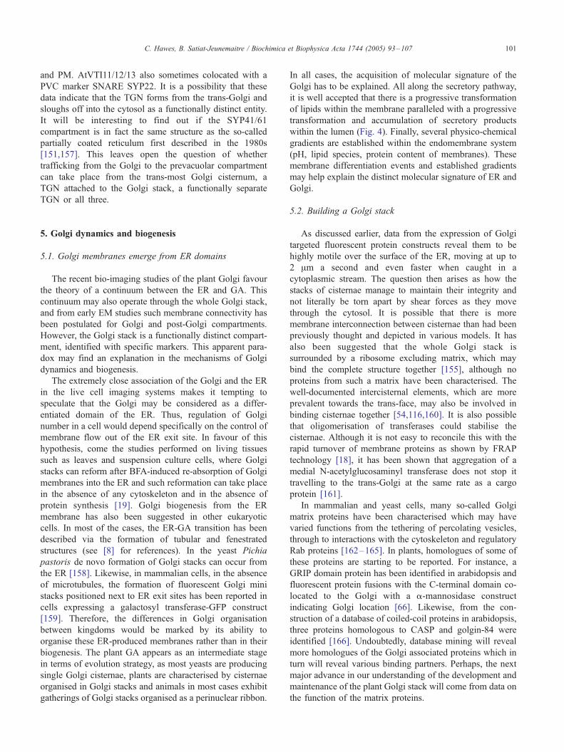
Fig. 1. Dual expression of a signal peptide-GFP-HDEL construct and a rat
sialyl transferase signal anchor sequence-RFP in an epidermal cell of a
tobacco leaf. Note the close association of the Golgi bodies with the ER
network. Micrograph courtesy of John Runions, Oxford Brookes Univer-
sity. Bar=10 Am.
3. The ER–GA complex
3.1. The ER–GA complex by light microscopy
The plant ER may be labelled with fluorescent dyes such
as DiOC6 [71,72], by antibodies directed against soluble or
membrane bound reticuloplasmins (i.e., proteins specifically
identified as ER-resident such as calreticulin and calnexin)
[73], against the ER-retention HDEL/KDEL sequences [74],
or by specific GFP fusions [13,40]. In all cases, the ER
appears as a membranous tubular network often mainly
restricted to the cortex of mature cells and connected to the
nuclear envelope. Although much talked about, with the
exception of putative exit sites (see Section 3.2 below), from
such studies, there is little evidence of specific sub-domains
on the ER.
In contrast to the ER tubular network, staining of the
plant GA reveals a punctate pattern composed of hundreds
of distinct structures approximately 1 Am in diameter, as
revealed using antibodies against peripheral glycoproteins
[12,75]. Likewise, fluorescent protein fusions to a variety of
proteins including the H/KDEL receptor, various mamma-
lian and plant transferase signal anchor sequences, and sugar
transporters, have all successfully located the plant GA
[4,13,14,19,76,77].
All Golgi enzymes identified so far are integral mem-
brane proteins. The mechanisms by which they remain
specifically in Golgi appear to be related to the signal
anchor sequence comprising the trans-membrane domain
and flanking amino acids [78]. Interestingly, the signal
anchor sequences from both rat and human transferases are
sufficient for successfully targeting GFP to the plant Golgi
indicating a consensus in mechanisms across kingdoms
[13,19,79].
Labelling in living cells has shown that Golgi stacks are
not static, but as well as moving in classic cytoplasmic
streams, they also exhibit more specific movements.
Individual Golgi stacks can move as fast as 2 Am per
second [13,14], in an actin dependent fashion. Indeed,
depolymerisation of actin induces the clumping of Golgi
bodies within cells [80] and results in a cessation of
movement [13]. It is assumed that the motor driving the
Golgi is myosin. However, a Golgi-associated kinesin has
also been identified in cotton and arabidopsis (GhKine-
sin13a and AtKinesin-13A) [81,82] and Golgi bodies were
also shown in association with microtubules. It was
suggested that microtubules may also be involved in the
organisation of Golgi bodies at the cell cortex, although the
authors concluded that kinesin-13A does not in fact
contribute to the primary movement of the Golgi.
Co-visualisation of ER and the GA suggests that Golgi
bodies are associated with the cortical ER network. When
fused to GFP and expressed in tobacco leaf epidermal cells,
the arabidopsis homologue of the H/KDEL receptor
AtERD2 is localised both in the ER membranes and Golgi
membranes [13,18,19]. Golgi bodies appear attached to the
ER membranes and appear to move over the surface of the
ER tubules. This close association can also be seen with
dual expression of an ER construct such as signal peptide-
GFP-HDEL and ST-mRFP (Fig. 1). Preliminary studies
using optical tweezers in an attempt to separate fluorescent
Golgi bodies from ER confirm the strong attachment of
Golgi membranes to the ER (Leru and Brown, unpublished
data). The fact that biochemical separation between ER and
Golgi membranes is difficult in plant cells further confirms
the tight association between ER and GA. These features
indicate a possible membrane continuum between ER and
Golgi. Most of these live cell imaging studies have been
performed on the current models used in plant cell bio-
imaging, mostly BY-2 cells, tobacco and arabidopsis leaves.
The relation between ER and Golgi may, however, differs
according cell specialization and should be further inves-
tigated in other tissues.
The structural and functional link between the ER and
GA and the kinetics of transfer of membrane cargo
molecules between ER and GA have been investigated
using fluorescence recovery after photobleaching (FRAP)
techniques. FRAP of Golgi targeted GFP constructs in
tobacco leaves has shown that transfer of cargo molecules
from the ER to Golgi is energy dependent, but not
dependent on the cytoskeleton. Such transport can occur

C. Hawes, B. Satiat-Jeunemaitre / Biochimica et Biophysica Acta 1744 (2005) 93–10796
when the Golgi stacks are static and also when they are
moving [18,20]. These data supported the hypothesis that
there may be some form of structural continuity between the
two organelles permitting direct transfer of cargo without
the requirement for vesicle vectors. It also questions the
mechanisms of putative retrograde transport by vesicle
carriers.
3.2. The ER–GA complex associated machinery
If there are in some instances direct connections between
the plant ER and Golgi bodies, even if they are transitory,
can this be reconciled with the presence of specific
molecular machinery? Presumably, a microdomain in the
ER membrane has to become competent either for produc-
ing a profusion of membrane, which either evolves into GA
membranes, or is capable of fusing to or bridging to the
existing Golgi membrane. It is therefore not unreasonable to
expect that specific molecular machinery will accumulate at
the sites where Golgi stacks are attached to the ER.
3.2.1. The COPII machinery
It has been suggested that COPII coats may be necessary
for ER to Golgi transport processes in plant cells [60,83–
86]. Recent data suggest that Sar1p, the activating GTPase
for COPII coat recruitment could play an essential role in
the formation of ER microdomains associated with Golgi
membranes. In the presence of Golgi targeted GFP
constructs Sar1p defines punctate structures on the ER
membranes, as does the GTP-locked mutant of the protein at
low levels of expression [20]. The requirement of Sar1p for
export out of the ER has been demonstrated in vivo by
inhibition of secretion after the expression of non-functional
mutant forms of the protein [20,83,87]. Double expression
of fluorescent protein Sar1p and Golgi constructs showed
them to apparently co-localise on the ER membrane and to
move together as a unit along the ER membrane. Because of
this, the concept of a motile secretory complex consisting of
Golgi and ER exit sited was proposed [20]. Over expression
of both GTP and GDP locked mutants of Sar1p resulted in a
redistribution of Golgi markers to the ER, resulting in a
phenotype similar to that observed after BFA treatment
[20,83,87].
Biochemical studies have shown that Sar1P mediates
the recruitment of two major protein complex (sec13/31p
and Sec23/24p), building up the typical COPII structures
described in mammalian cells [60]. In plant cells, no
functional studies of these proteins have been described,
nor the binding of COPII to specific motifs of Golgi
resident proteins. A recent report suggests that a
cytoplasmic dibasic motif within Sar1p may function as
a receptor for Golgi resident enzymes in mammalian cells
[88]. Thus, the occurrence of specific bonds between GA
proteins and Sar1p may contribute to the differentiation of
specific ER microdomains competent for producing Golgi
membranes.
The GTPase activity of Sar1p is regulated by an
exchange factor, Sec12p, which in tobacco leaf epidermal
cells has been shown to be uniformly distributed over the
ER network. Therefore, it should not play a role in the
differentiation of ER membranes into Golgi. This distribu-
tion of Sec12 is similar to that in Saccharomyces cerevisiae
where the Golgi exists as dispersed individual cisternae
[8,89]. However, in Pichia pastoris, which has static Golgi
stacks, Sec12p is restricted to the ER export sites [89].
Perhaps, the distribution of Sec12 is dependent on the
relative mobility of the Golgi and the export sites feeding
them.
In mammalian and yeast cells, it has been proposed that
COP II components act in a highly regulated manner to
recruit some specific ER membrane areas in coated vesicles,
which in turn would transport these membranes and their
associated cargo molecules to an intermediate compartment
forming discreet transport vesicles [90]. As can be seen
from the recent work described above, this concept may
very well not be applicable to plant cells. Data strongly
suggest that COPII components may indeed be needed for
some cargo accumulation and for regulating export out of
the ER [59]. However, there is, to date, no experimental
argument to suggest that COPII vesicles exist or are the
carriers for cargo export between the ER and GA. The fact
that in many plant cells, this ER/GA step is cytoskeleton
independent is a further argument against the involvement
of a specific vesicle carrier between the two compartments
[18,19]. Interestingly, it has also been shown that in plants,
transport from ER to GA can also be COPII independent
[91]. The concept of the biogenesis of COPII independent
carriers from ER export sites is also becoming fashionable
in mammalian cells [92,93].
3.2.2. The COPI machinery
Homologues of the proteins of the COPI coatomer
complex in plants have been cloned [57,58,60,94]. Immuno-
fluorescence studies have shown the proteins located to the
Golgi with no evidence of any location on the ER, and they
may react differently to BFA according the plant material
studied [54,95,96]. COPI components seem to interact in
vitro with the di-lysine motifs of specific ER membrane
proteins [94]. In the animal kingdom, COPI is thought to be
involved in retrograde transport from GA to the ER
although there are little data which confirm this direction-
ality in plant cells.
Interestingly, the location of Arf1P, the COPI-associated
GTPase, does not seem to be restricted to the GA. GFP
constructs have shown ARF1 to be located at the Golgi [97],
but immunofluorescence has also shown that the protein
may be located on the ER, and the plasma membrane [95],
suggesting that it may function at different levels of the
secretory pathway. It may very well have multiple roles in
that it functions in the trafficking of the H+ ATPase from the
GA to the plasma membrane, in the positioning of sialyl-
transferase to the Golgi, in the maintenance of the

C. Hawes, B. Satiat-Jeunemaitre / Biochimica et Biophysica Acta 1744 (2005) 93–107 97
endoplasmic reticulum [98,99], and with the regulation of
vacuolar secretion [97,100]. Thus, there is increasing
evidence that ARF1 acts at multiple sites in the cell and
may participate in membrane remodelling with distinct
mechanisms apart from its COPI recruitment function. Such
specific ARF1 activities may be regulated by the specificity
of effector molecules such as ARF GEFs (guanine nucleo-
tide exchange factors [96,101], or ARF-GAPs (GTPase-
activating proteins, [102]).
The question remains as to whether ARF or COPI play a
role in regulation of the ER/Golgi complex? Although there
are, to date, little functional data to suggest COPI coat
involvement in ER/GA cargo exchanges, BFA has been
shown to affect fluorescence recovery of bleached Golgi–
GFP constructs [18]. Also, a GTP-locked (Q71L) mutant of
Arf1 has been shown to inhibit the targeting of sporamin–
GFP to the vacuole in arabidopsis protoplasts, resulting in a
build up of fluorescence in the ER [97]. These data suggest
that in plant cells Arf1p may play a major role in the
construction of ER export sites and in Golgi biogenesis from
export sites, as has been proposed for mammalian cells
[103].
The exact functions of COPI and COPII complexes, as in
animals, still have to be elucidated in plant cells. Their
ability either to form coated vesicles or to define specific
recruitment domains within ER and GA membranes may be
necessary for modelling the membrane dynamics (creation
of microdomains) and maintaining the identity of each
membrane [95]. This may require such a total interdepend-
ence of the COPII and COPI pathways for the recycling of
regulatory machinery such as the SNARE fusion molecules,
that disruption of either route will result in the collapse of
the other [6].
Fig. 2. The likely location of some of the Golgi associated proteins identified to da
one cisternum although it is likely that the TGN may in cases slough off from the G
such as CASP, Golgin 84, AtGRIP and some of the SNAREs has yet to be deter
3.2.3. Regulatory machinery at the ER/Golgi interface
No matter what the exact structural nature of the ER/
Golgi interface is in plant cells, it is becoming clear that
other proteins shown to be involved in this transport step in
mammals and yeasts most likely operate in plants as well.
Recently, from an analysis of the NCBI protein database,
out of 54 SNARES in the arabidopsis genome, 15 SNARE
molecules have been identified as being located to the ER
and Golgi apparatus [104]. Of these, 6 were ER localised.
Thus, there are a number of SNARES potentially involved
in ER to Golgi transport, and their exact function are
currently under investigation.
Similarly, functional studies on plant Rab proteins are
still in their infancy. At least one Rab protein has been
shown to regulate ER to Golgi transport in plants. A
dominant inhibitory mutant of the arabidopsis homologue of
Rab1 (AtRabD2a previously AtRab1b) [62,63] has been
shown to affect transport of a secretory version of GFP out
of the ER in tobacco leaf epidermal cells [105]. This effect
was rescued by co-expression of the wild type protein but
not by other Rabs. The same construct also slowed down the
reformation of Golgi stacks in tobacco leaves after BFA
induced redistribution of Golgi membrane into the ER [19].
Likewise, it has also been suggested that an arabidopsis
homologue of Rab2 (NtRabB2) may regulate transport of
cargo between the ER and Golgi in pollen tubes [106]. The
likely locations of these plant Golgi associated proteins are
summarised in Fig. 2.
3.3. The ER/GA complex at the ultrastructural level
Electron microscopy of higher plant cells has not
revealed the geometrical organisation of the cortical
te in plant cells. The trans-Golgi and trans-Golgi network are represented as
olgi stack. The exact location of many of the proteins within the Golgi stack
mined.
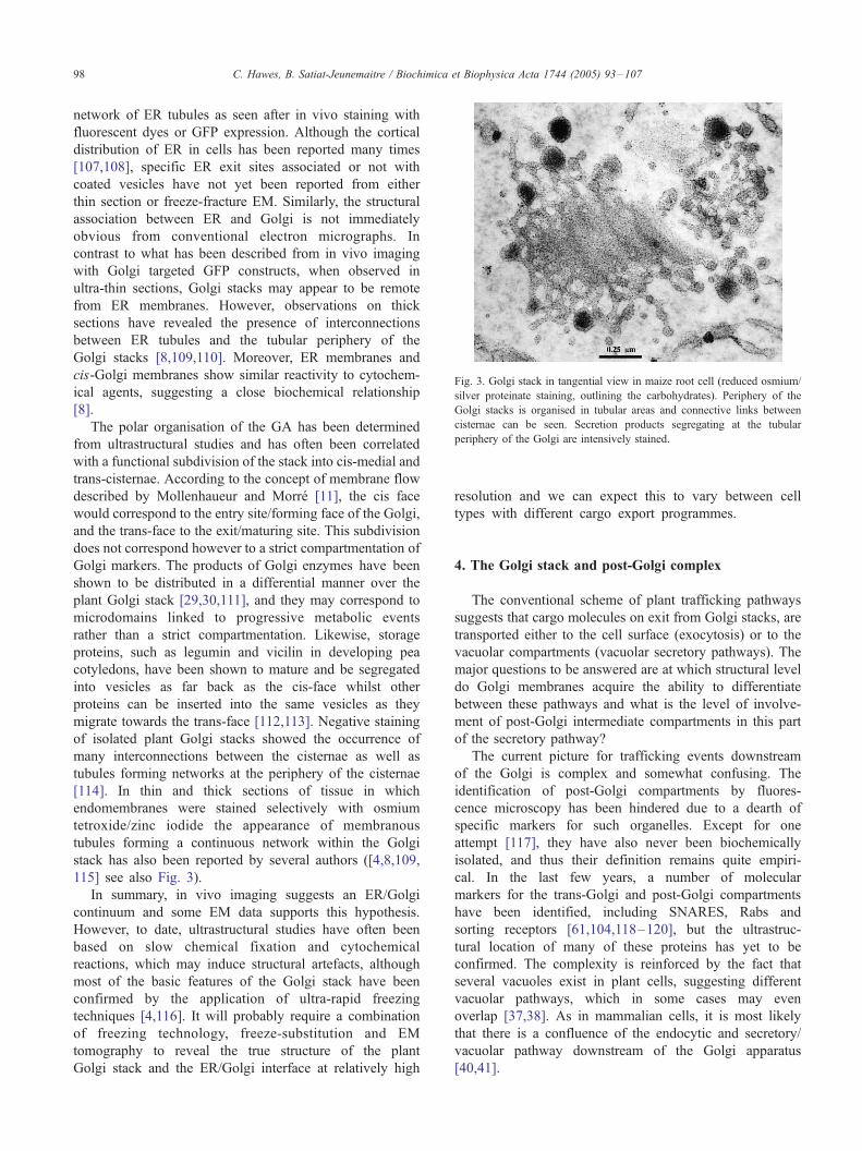
Fig. 3. Golgi stack in tangential view in maize root cell (reduced osmium/
silver proteinate staining, outlining the carbohydrates). Periphery of the
Golgi stacks is organised in tubular areas and connective links between
cisternae can be seen. Secretion products segregating at the tubular
periphery of the Golgi are intensively stained.
C. Hawes, B. Satiat-Jeunemaitre / Biochimica et Biophysica Acta 1744 (2005) 93–10798
network of ER tubules as seen after in vivo staining with
fluorescent dyes or GFP expression. Although the cortical
distribution of ER in cells has been reported many times
[107,108], specific ER exit sites associated or not with
coated vesicles have not yet been reported from either
thin section or freeze-fracture EM. Similarly, the structural
association between ER and Golgi is not immediately
obvious from conventional electron micrographs. In
contrast to what has been described from in vivo imaging
with Golgi targeted GFP constructs, when observed in
ultra-thin sections, Golgi stacks may appear to be remote
from ER membranes. However, observations on thick
sections have revealed the presence of interconnections
between ER tubules and the tubular periphery of the
Golgi stacks [8,109,110]. Moreover, ER membranes and
cis-Golgi membranes show similar reactivity to cytochem-
ical agents, suggesting a close biochemical relationship
[8].
The polar organisation of the GA has been determined
from ultrastructural studies and has often been correlated
with a functional subdivision of the stack into cis-medial and
trans-cisternae. According to the concept of membrane flow
described by Mollenhaueur and Morre [11], the cis face
would correspond to the entry site/forming face of the Golgi,
and the trans-face to the exit/maturing site. This subdivision
does not correspond however to a strict compartmentation of
Golgi markers. The products of Golgi enzymes have been
shown to be distributed in a differential manner over the
plant Golgi stack [29,30,111], and they may correspond to
microdomains linked to progressive metabolic events
rather than a strict compartmentation. Likewise, storage
proteins, such as legumin and vicilin in developing pea
cotyledons, have been shown to mature and be segregated
into vesicles as far back as the cis-face whilst other
proteins can be inserted into the same vesicles as they
migrate towards the trans-face [112,113]. Negative staining
of isolated plant Golgi stacks showed the occurrence of
many interconnections between the cisternae as well as
tubules forming networks at the periphery of the cisternae
[114]. In thin and thick sections of tissue in which
endomembranes were stained selectively with osmium
tetroxide/zinc iodide the appearance of membranous
tubules forming a continuous network within the Golgi
stack has also been reported by several authors ([4,8,109,
115] see also Fig. 3).
In summary, in vivo imaging suggests an ER/Golgi
continuum and some EM data supports this hypothesis.
However, to date, ultrastructural studies have often been
based on slow chemical fixation and cytochemical
reactions, which may induce structural artefacts, although
most of the basic features of the Golgi stack have been
confirmed by the application of ultra-rapid freezing
techniques [4,116]. It will probably require a combination
of freezing technology, freeze-substitution and EM
tomography to reveal the true structure of the plant
Golgi stack and the ER/Golgi interface at relatively high
resolution and we can expect this to vary between cell
types with different cargo export programmes.
4. The Golgi stack and post-Golgi complex
The conventional scheme of plant trafficking pathways
suggests that cargo molecules on exit from Golgi stacks, are
transported either to the cell surface (exocytosis) or to the
vacuolar compartments (vacuolar secretory pathways). The
major questions to be answered are at which structural level
do Golgi membranes acquire the ability to differentiate
between these pathways and what is the level of involve-
ment of post-Golgi intermediate compartments in this part
of the secretory pathway?
The current picture for trafficking events downstream
of the Golgi is complex and somewhat confusing. The
identification of post-Golgi compartments by fluores-
cence microscopy has been hindered due to a dearth of
specific markers for such organelles. Except for one
attempt [117], they have also never been biochemically
isolated, and thus their definition remains quite empiri-
cal. In the last few years, a number of molecular
markers for the trans-Golgi and post-Golgi compartments
have been identified, including SNARES, Rabs and
sorting receptors [61,104,118–120], but the ultrastruc-
tural location of many of these proteins has yet to be
confirmed. The complexity is reinforced by the fact that
several vacuoles exist in plant cells, suggesting different
vacuolar pathways, which in some cases may even
overlap [37,38]. As in mammalian cells, it is most likely
that there is a confluence of the endocytic and secretory/
vacuolar pathway downstream of the Golgi apparatus
[40,41].
C. Hawes, B. Satiat-Jeunemaitre / Biochimica et Biophysica Acta 1744 (2005) 93–107 99
4.1. From GA to the plasma membrane: a default pathway?
Leaving the Golgi stack for the plasma membrane is one
option for secretory molecules. All fluorescence labelling
experiments suggest a direct rapid and specific transfer to
the plasma membrane as no specific intermediate compart-
ments between the GA and plasma membrane are labelled
by the secretory products either newly synthesised or
processed in the GA [52,105,121,122]. This difficulty in
imaging secretory products in transit is most likely related to
either a low level of product, or to the speed of transport
through the system. To further reinforce this hypothesis,
blocking secretion by brefeldin A often causes an accumu-
lation of the secretory products, either within the cytoplasm
into post-Golgi ‘‘BFA compartments’’ [52,121], or within
the ER [105,122,123].
Data on the recycling of membrane proteins suggest that
the BFA compartments may represent an amplification of a
pre-existing compartment, or may be formed de novo from
Golgi or plasma membrane (see discussion in [6,41]).
Whether this compartment is an intermediate along the
secretory pathway, which also has a recycling function, has
still to be clarified.
As it stands, these data provided by light microscopy
suggest that exit from the GA to the plasma membrane
occurs by bulk flow, without other checkpoints on the way
to the cell periphery. The transport of polysaccharide
molecules from GA to the plasma membrane may be easily
mapped by EM either by specific cytochemical staining
[124] or by immunolocation of Golgi-derived products
[125,126]. This transport from GA to cell surface is
mediated by secretory vesicles of varying size which can
carry mixed cargos of polysaccharides [125] or polysac-
charides and glycoproteins [126]. The secretory vesicles
may be considered as the final processing compartment for
cargo maturation as esterification of pectins or further
polymerisation of polysaccharides may occur within the
vesicles during transport to the plasma membrane (Vian B.,
personal communication). To date, no protein coats have
been found associated with such secretory vesicle. Their
maturation can be associated with different Golgi cisternae
[8], and electron microscopy suggests that they may detach
at different levels down the Golgi stack [126]. Biochemical
studies suggested that targeting of GA-derived soluble cargo
products is by default. No positive signals have been found
associated with the transport of ER/GA-derived secretory
products to the plasma membrane [127].
The situation with membrane proteins has, however,
been controversial, as both the tonoplast and the plasma
membrane have been considered as the default pathway for
membrane proteins [128,129]. Certainly, the transmembrane
domain (TMD) of single pass membrane proteins is capable
of directing chimeric proteins to different membranes. GFP
constructs made with the membrane-spanning domain of the
human lysosomal protein LAMP1 are directed to the plasma
membrane. However, deletions in the TMD resulting in 20
or 17 amino acids resulted in the constructs being resident in
the Golgi and ER, respectively [130]. Likewise, when the
TMD of a protein usually targeted to the vacuolar pathway
(the vacuolar sorting receptor BP80) was increased to 22
amino acids the GFP reporter was transported to the plasma
membrane. However, at least for polytopic proteins like
plasma membrane H+ ATPases, it has been recently
proposed that one or more signals may be required for
sorting through the exocytic pathway [122].
In all cases, involvement of molecular equipment such as
coat complex, adaptors or receptors have never been
reported to be associated with polysaccharide or glycopro-
tein sorting to the plasma membrane. Arf1p and COPI
antibodies have shown in certain cases a staining of both
Golgi and plasma membrane [95]. However, this dual
location may reflect recycling by COPI vesicles between the
new plasma membrane being formed (made of Golgi-
derived membranes) and the Golgi stacks [95]. A total of 18
different SNARE proteins have been located to the plasma
membrane [104] which have the potential for conferring
some level of specificity on vesicle fusion. However, it has
also been suggested that some of these proteins may also act
in a signalling capacity which may explain the high number
of plasma membrane SNARES [131].
The possibility of differential targeting within the
exocytotic pathway has arisen from studies on proteins
associated with auxin efflux (PIN proteins) which have a
polar distribution in the basal plasma membrane of
Arabidopsis root cells [101,132]. However, this hypothesis
has still to be proven and preliminary studies strongly
suggest that the insertion of PIN proteins within membranes
may first occur in a non-polar fashion, and that polarity
would be the result of post-deposition organisation of the
proteins within the plasma membrane. (Boutte and Satiat-
Jeunemaitre, unpublished data). However, one specific
event in the cell cycle, that of cytokinesis, involves very
directed trafficking of Golgi-derived secretory vesicles. The
SNARE SYP111 (KNOLLE) is also specifically expressed
during mitosis and is found only on the developing cell plate
[133–136].
Another way to regulate the transport of Golgi-derived
membranes would be through the cytoskeleton, as exocy-
tosis of Golgi-derived vesicles is likely to be actin mediated
[67,68]. Interestingly, over-expression of one GFP-conju-
gated Rac protein AtRac7, which targets actin cytoskeleton
via actin depolymerising factor, but not other GFP-tagged
Rac proteins, induces a labelling both at the Golgi and at the
cell surface in pollen tubes [70].
4.2. From GA to vacuole
In contrast to the bulk flow hypothesis of transport to the
plasma membrane, trafficking to the vacuole relies on the
recognition of specific vacuolar sorting motifs. It is now
generally accepted that there are twomajor routes of secretion
from the Golgi to the vacuolar system [37,137,138]. Firstly, a
C. Hawes, B. Satiat-Jeunemaitre / Biochimica et Biophysica Acta 1744 (2005) 93–107100
direct transport of protein to the storage vacuole via dense
vesicles such as described in developing pea cotyledons
[112,113,139]. Little is known about the mode of trafficking
and targeting of these vesicles to the correct compartment,
although sorting relies on a carboxy-terminal propeptide
and may be receptor mediated as transport to the storage
vacuole is saturable [140]. These dense vesicles may form
as early as the cis-face, however, it is assumed that they
depart the Golgi only towards the trans-face ([113], see
Section 3.3).
Secondly, transport of protein to the lytic vacuole is
mediated by a family of vacuolar sorting receptors (VSR)
resident in the trans-Golgi or trans-Golgi network (TGN),
and they are carried in clathrin-coated vesicles to a
prevacuolar compartment (PVC). VSRs are related to the
receptor BP80 (VSRps-1) first cloned from peas [141,142].
Homologues to this protein have now been identified in
various species [143]. VSRs are integral membrane proteins
which can bind clathrin through adaptin-binding tyrosine
motifs in their cytosolic tails [142]. They recognise
sequence-specific NPIR (vacuolar) motifs that may be
present on either termini or internally in the cargo protein
and cycle between the Golgi and the PVC [38,144–146].
These VSRs also co-localise with the SNARE PEP12(At-
SYP21) which belongs to the syntaxin family which has
been localised on structures distinct from GA and termed
the PVC [64,117–119,147–149]. Specific Rab proteins
such as AtRabF2b (ARA7) and homologues of Ara6 have
also been located to the PVC [61,120,149]. These various
markers for PVC are BFA insensitive, and by fluorescence
microscopy reside mainly on a compartment distinct from
Golgi and vacuole. They may, however, exhibit a low
amount of colocalisation with the Golgi (10–20%),
suggesting that they are involved in the recycling of VSRs
from PVC to the GA.
This relatively simple concept of separate sorting path-
ways for storage and lytic proteins is challenged by a recent
study, which suggests that the situation may not be so
straightforward. In developing castor bean seeds, the VSR
apparently binds the proteins, proricin and pro2S albumin,
which are destined for the storage vacuole [152], suggesting
that clathrin-coated vesicles may also carry these proteins.
Likewise in arabidopsis seeds, a knock out of AtVSR1 has
been shown to result in the partial secretion of precursors of
major classes of storage proteins [153].
A recent EM study on high-pressure frozen freeze-
substituted tobacco BY2 cells has shown that VSR anti-
bodies label putative endocytic compartments, the multi-
vesicular bodies (MVBs), which may also function as PVCs
[117]. MVBs in plants are compartments, which were first
functionally defined at the EM level, as organelles on the
endocytic pathway, as they accumulated endocytosed
cationised ferritin [39,150,151]. These data suggest that
PVC may in fact function in both the endocytic and
vacuolar pathways. In all cases, both storage and lytic
pathways and their associated sorting events require the
presence of a trans-Golgi sorting compartment. Thus, the
trans-Golgi or TGN may in the appropriate tissue be able to
sort both lytic and storage cargo.
Scission of cargo laden clathrin coated vesicles at the
trans-Golgi is most likely mediated by dynamin-like
proteins. An Arabidopsis dynamin-like 6 (ADL6) has been
shown to be located at the trans-Golgi and a dominant
negative mutant caused accumulation of lytic vacuole cargo
at the Golgi but had no affect on the transport of plasma
membrane bound cargo [154].
4.3. What is the trans-Golgi Network?
The exact nature or even the presence of a functionally
distinct TGN in plants is a matter of considerable debate.
This has been made all the more confusing due to data
emerging on the location of various marker molecules, from
both immunofluorescence and fluorescent protein expres-
sion, which have yet to be reconciled with existing
ultrastructural data. The trans-most face of the Golgi stack
can exhibit various structural profiles. Some stacks may
have an obvious final cisternum which is cohesive with the
rest of the stack but which may exhibit varying degrees of
tubulation [8,116]. However, in other cases, the trans-most
cisternum may appear to be partially detached from the
Golgi and ultimately, a cisternum-like structure may appear
fully distinct from the Golgi stack and have a tubular/
vesicular profile [155]. In most cases, clathrin-coated
vesicles are associated with this last cisternum or network.
These variations are probably the result of a highly dynamic
process in the Golgi membrane transformation. The term
trans-Golgi network TGN has often been ascribed to all of
these manifestations of the final cisternum in the stack. A
number of SNARE proteins have been located to the trans-
most cisternum by gold labelling including SYP41, SYP61
AtVTI12 and AtVPS45 [119], which these authors termed
the TGN. The trans-Golgi cisternum or TGN bears a
remarkable similarity to the partially coated reticulum, first
described as a tubulo-vesicular compartment, with clathrin
buds, which can rapidly accept internalised ferritin in
protoplasts [39,150,151,156,157]. Thus, it has been sug-
gested that this compartment may function as an early
endosome, even if it may in some cases be structurally
continuous with the TGN [155].
By light microscopy, it is hard to define a structurally
distinct TGN. However, from an analysis of the arabidop-
sis SNARES, it was demonstrated that a fluorescent
protein constructs of SYP41 and SYP61 co-located and
were sometimes in close proximity to the Golgi apparatus
but most often was located to punctate structures separate
from the Golgi [104]. On BFA treatment, these structures
clumped but unlike many of the Golgi SNARES showed
no ER location. From this analysis, it was concluded that
SYP41 and 61 located to the TGN. Of the other SNARE
constructs analysed, AtVTI11/13 located to the TGN and
the vacuolar membrane and AtVTI12 located to the TGN
C. Hawes, B. Satiat-Jeunemaitre / Biochimica et Biophysica Acta 1744 (2005) 93–107 101
and PM. AtVTI11/12/13 also sometimes colocated with a
PVC marker SNARE SYP22. It is a possibility that these
data indicate that the TGN forms from the trans-Golgi and
sloughs off into the cytosol as a functionally distinct entity.
It will be interesting to find out if the SYP41/61
compartment is in fact the same structure as the so-called
partially coated reticulum first described in the 1980s
[151,157]. This leaves open the question of whether
trafficking from the Golgi to the prevacuolar compartment
can take place from the trans-most Golgi cisternum, a
TGN attached to the Golgi stack, a functionally separate
TGN or all three.
5. Golgi dynamics and biogenesis
5.1. Golgi membranes emerge from ER domains
The recent bio-imaging studies of the plant Golgi favour
the theory of a continuum between the ER and GA. This
continuum may also operate through the whole Golgi stack,
and from early EM studies such membrane connectivity has
been postulated for Golgi and post-Golgi compartments.
However, the Golgi stack is a functionally distinct compart-
ment, identified with specific markers. This apparent para-
dox may find an explanation in the mechanisms of Golgi
dynamics and biogenesis.
The extremely close association of the Golgi and the ER
in the live cell imaging systems makes it tempting to
speculate that the Golgi may be considered as a differ-
entiated domain of the ER. Thus, regulation of Golgi
number in a cell would depend specifically on the control of
membrane flow out of the ER exit site. In favour of this
hypothesis, come the studies performed on living tissues
such as leaves and suspension culture cells, where Golgi
stacks can reform after BFA-induced re-absorption of Golgi
membranes into the ER and such reformation can take place
in the absence of any cytoskeleton and in the absence of
protein synthesis [19]. Golgi biogenesis from the ER
membrane has also been suggested in other eukaryotic
cells. In most of the cases, the ER-GA transition has been
described via the formation of tubular and fenestrated
structures (see [8] for references). In the yeast Pichia
pastoris de novo formation of Golgi stacks can occur from
the ER [158]. Likewise, in mammalian cells, in the absence
of microtubules, the formation of fluorescent Golgi mini
stacks positioned next to ER exit sites has been reported in
cells expressing a galactosyl transferase-GFP construct
[159]. Therefore, the differences in Golgi organisation
between kingdoms would be marked by its ability to
organise these ER-produced membranes rather than in their
biogenesis. The plant GA appears as an intermediate stage
in terms of evolution strategy, as most yeasts are producing
single Golgi cisternae, plants are characterised by cisternae
organised in Golgi stacks and animals in most cases exhibit
gatherings of Golgi stacks organised as a perinuclear ribbon.
In all cases, the acquisition of molecular signature of the
Golgi has to be explained. All along the secretory pathway,
it is well accepted that there is a progressive transformation
of lipids within the membrane paralleled with a progressive
transformation and accumulation of secretory products
within the lumen (Fig. 4). Finally, several physico-chemical
gradients are established within the endomembrane system
(pH, lipid species, protein content of membranes). These
membrane differentiation events and established gradients
may help explain the distinct molecular signature of ER and
Golgi.
5.2. Building a Golgi stack
As discussed earlier, data from the expression of Golgi
targeted fluorescent protein constructs reveal them to be
highly motile over the surface of the ER, moving at up to
2 Am a second and even faster when caught in a
cytoplasmic stream. The question then arises as how the
stacks of cisternae manage to maintain their integrity and
not literally be torn apart by shear forces as they move
through the cytosol. It is possible that there is more
membrane interconnection between cisternae than had been
previously thought and depicted in various models. It has
also been suggested that the whole Golgi stack is
surrounded by a ribosome excluding matrix, which may
bind the complete structure together [155], although no
proteins from such a matrix have been characterised. The
well-documented intercisternal elements, which are more
prevalent towards the trans-face, may also be involved in
binding cisternae together [54,116,160]. It is also possible
that oligomerisation of transferases could stabilise the
cisternae. Although it is not easy to reconcile this with the
rapid turnover of membrane proteins as shown by FRAP
technology [18], it has been shown that aggregation of a
medial N-acetylglucosaminyl transferase does not stop it
travelling to the trans-Golgi at the same rate as a cargo
protein [161].
In mammalian and yeast cells, many so-called Golgi
matrix proteins have been characterised which may have
varied functions from the tethering of percolating vesicles,
through to interactions with the cytoskeleton and regulatory
Rab proteins [162–165]. In plants, homologues of some of
these proteins are starting to be reported. For instance, a
GRIP domain protein has been identified in arabidopsis and
fluorescent protein fusions with the C-terminal domain co-
located to the Golgi with a a-mannosidase construct
indicating Golgi location [66]. Likewise, from the con-
struction of a database of coiled-coil proteins in arabidopsis,
three proteins homologous to CASP and golgin-84 were
identified [166]. Undoubtedly, database mining will reveal
more homologues of the Golgi associated proteins which in
turn will reveal various binding partners. Perhaps, the next
major advance in our understanding of the development and
maintenance of the plant Golgi stack will come from data on
the function of the matrix proteins.
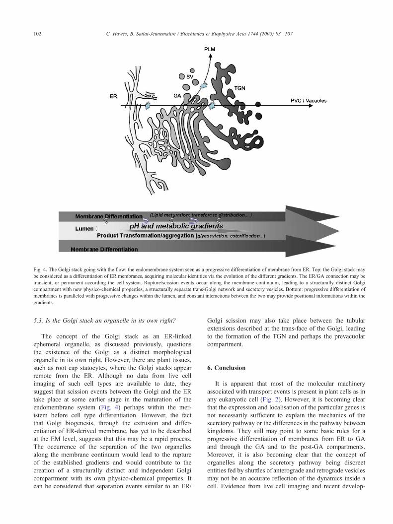
Fig. 4. The Golgi stack going with the flow: the endomembrane system seen as a progressive differentiation of membrane from ER. Top: the Golgi stack may
be considered as a differentiation of ER membranes, acquiring molecular identities via the evolution of the different gradients. The ER/GA connection may be
transient, or permanent according the cell system. Rupture/scission events occur along the membrane continuum, leading to a structurally distinct Golgi
compartment with new physico-chemical properties, a structurally separate trans-Golgi network and secretory vesicles. Bottom: progressive differentiation of
membranes is paralleled with progressive changes within the lumen, and constant interactions between the two may provide positional informations within the
gradients.
C. Hawes, B. Satiat-Jeunemaitre / Biochimica et Biophysica Acta 1744 (2005) 93–107102
5.3. Is the Golgi stack an organelle in its own right?
The concept of the Golgi stack as an ER-linked
ephemeral organelle, as discussed previously, questions
the existence of the Golgi as a distinct morphological
organelle in its own right. However, there are plant tissues,
such as root cap statocytes, where the Golgi stacks appear
remote from the ER. Although no data from live cell
imaging of such cell types are available to date, they
suggest that scission events between the Golgi and the ER
take place at some earlier stage in the maturation of the
endomembrane system (Fig. 4) perhaps within the mer-
istem before cell type differentiation. However, the fact
that Golgi biogenesis, through the extrusion and differ-
entiation of ER-derived membrane, has yet to be described
at the EM level, suggests that this may be a rapid process.
The occurrence of the separation of the two organelles
along the membrane continuum would lead to the rupture
of the established gradients and would contribute to the
creation of a structurally distinct and independent Golgi
compartment with its own physico-chemical properties. It
can be considered that separation events similar to an ER/
Golgi scission may also take place between the tubular
extensions described at the trans-face of the Golgi, leading
to the formation of the TGN and perhaps the prevacuolar
compartment.
6. Conclusion
It is apparent that most of the molecular machinery
associated with transport events is present in plant cells as in
any eukaryotic cell (Fig. 2). However, it is becoming clear
that the expression and localisation of the particular genes is
not necessarily sufficient to explain the mechanics of the
secretory pathway or the differences in the pathway between
kingdoms. They still may point to some basic rules for a
progressive differentiation of membranes from ER to GA
and through the GA and to the post-GA compartments.
Moreover, it is also becoming clear that the concept of
organelles along the secretory pathway being discreet
entities fed by shuttles of anterograde and retrograde vesicles
may not be an accurate reflection of the dynamics inside a
cell. Evidence from live cell imaging and recent develop-
C. Hawes, B. Satiat-Jeunemaitre / Biochimica et Biophysica Acta 1744 (2005) 93–107 103
ments in electron microscopy, such as tomography, are
suggesting that we have to consider that there may be a
membrane continuum between the various membrane-
bounded compartments of the secretory pathway (Fig. 4)
[6,8,93,167]. Therefore, the endomembrane system (includ-
ing the Golgi apparatus) appears as a dynamic entity in
equilibrium, maturing along the secretory gradient from the
ER to the PM or vacuolar system.
Acknowledgements
Work in the CRH lab. is supported by the BBSRC.
Work in BSJ lab is partially supported by a MENRT grant
(ACI-BDPI). Fig. 3 reproduced with permission from
Kepes et al. 2005, Int. Rev. Cytol. 242, 55–120. We are
grateful to Jean Daraspe (ISV, Gif sur Yvette) for his help
with Fig. 4.
References
[1] A.J. Dalton, M.D. Felix, Cytological and cytochemical character-
istics of the Golgi substance of epithelial cells of the epididymis-in
situ, in homogenates and after isolation, Am. J. Anat. 94 (1954)
171–208.
[2] D.G. Whaley, M. Dauwalder, The Golgi apparatus, the plasma
membrane, and functional integration, Int. Rev. Cytol. 58 (1979)
199–245.
[3] P. Dupree, D.J. Sherrier, The plant Golgi apparatus, Biochim.
Biophys. Acta 1404 (1998) 259–270.
[4] U. Neumann, F. Brandizzi, C. Hawes, Protein transport in plant cells:
in and out of the Golgi, Ann. Bot. 92 (2003) 167–180.
[5] G. Jurgens, Membrane trafficking in plants, Ann. Rev. Cell Dev.
Biol. 20 (2004) 481–504.
[6] C. Hawes, Cell biology of the plant Golgi apparatus, New Phytol.
165 (2005) 29–44.
[7] M. Pavelka, D.G. Robinson, The Golgi apparatus in mammalian and
higher plant cells: a comparison, in: D.G. Robinson (Ed.), The Golgi
Apparatus and the Plant Secretory Pathway, Blackwell, Oxford, UK,
2003, pp. 16–36.
[8] F. Kepes, A. Rambourg, B. Satiat-Jeunemaitre, Morphodynamics of
the secretory pathway, Int. Rev. Cytol. 242 (2005) 55–120.
[9] F. Brandizzi, S.L. Irons, J. Johansen, A. Kotzer, U. Neumann,
GFP is the way to glow: bioimaging of the plant endomembrane
system, J. Microsc. 214 (2004) 138–158.
[10] H.H. Mollenhauer, D.J. Morre, The Golgi apparatus, in: P. Stumpf, E.
Conn (Eds.), The Biochemistry of Plants, Academic Press, New
York, 1980, pp. 437–488.
[11] H.H. Mollenhauer, D.J. Morre, Structure of Golgi apparatus,
Protoplasma 180 (1994) 14–28.
[12] B. Satiat-Jeunemaitre, C. Hawes, Redistribution of a Golgi glyco-
protein in plant cells treated wit Brefeldin A, J. Cell Sci. 103 (1992)
1153–1166.
[13] P. Boevink, K. Oparka, S. Santa-Cruz, B. Martin, A. Betteridge, C.
Hawes, Stacks on tracks: the plant Golgi apparatus traffics on an
actin/ER network, Plant J. 15 (1998) 39.
[14] A. Nebenfuhr, L.A. Gallagher, T.G. Dunahay, J.A. Frohlick, A.M.
Mazurkiewicz, J.B. Meehl, L.A. Staehelin, Stop- and-go movements
of plant Golgi stacks are mediated by the acto-myosin system, Plant
Physiol. 121 (1999) 1127–1141.
[15] A. Nebenfuhr, L.A. Staehelin, Mobile factories: Golgi dynamics in
plant cells, Trends Plant Sci. 6 (2001) 160–167.
[16] F. Brandizzi, M. Fricker, C. Hawes, A greener world: the revolution
in plant bioimaging, Nat. Rev., Mol. Cell Biol. 3 (2002) 520–530.
[17] T.H. Ward, F. Brandizzi, Dynamics of proteins in Golgi mem-
branes: comparisons between mammalian and plant cells high-
lighted by photobleaching techniques, Cell Mol. Life Sci. 61 (2004)
172–185.
[18] F. Brandizzi, E.L. Snapp, A.G. Roberts, J. Lippincott-Schwartz, C.
Hawes, Membrane protein transport between the endoplasmic
reticulum and the Golgi in tobacco leaves is energy dependent but
cytoskeleton independent: evidence from selective photobleaching,
Plant Cell 14 (2002) 1293–1309.
[19] C.M. Saint-Jore, J. Evins, H. Batoko, F. Brandizzi, I. Moore, C.
Hawes, Redistribution of membrane proteins between the Golgi
apparatus and endoplasmic reticulum in plants is reversible and not
dependent on cytoskeletal networks, Plant J. 29 (2002) 661–678.
[20] L.L.P. daSilva, E.L. Snapp, J. Denecke, J. Lippincott-Schwartz, C.
Hawes, F. Brandizzi, ER export sites and Golgi bodies behave as
single mobile secretory units in plant cells, Plant Cell 16 (2004)
1753–1771.
[21] G. Galili, ER-derived compartments are formed by highly regulated
processes and have special functions in plants, Plant Physiol. 136
(2004) 3411–3413.
[22] I. Hara-Nishimura, R. Matsushima, T. Shimada, M. Nishimura,
Diversity and formation of endoplasmic reticulum-derived compart-
ments in plants. Are these compartments specific to plant cells? Plant
Physiol. 136 (2004) 3435–3439.
[23] A. Vitale, A. Ceriotti, Protein quality control mechanisms and protein
storage in the endoplasmic reticulum. A conflict of interests? Plant
Physiol. 136 (2004) 3420–3426.
[24] I. Hara-Nishimura, T. Shiada, K. Hatano, Y. Takeuchi, M.
Nishimura, Transport of storage proteins to protein storage vacuoles
is mediated by large precursor-accumulating vesicles, Plant Cell 10
(1998) 825–836.
[25] N.C. Carpita, M. McCann, The cell wall, in: B.B. Buchanan, W.
Gruissem, R.L. Jones (Eds.), Biochemistry and Molecular Biology of
Plants, American Society of Plant Physiology, Rockville MD, USA,
2000, pp. 52–108.
[26] W.-R. Scheible, M. Pauly, Glycosyltransferases and cell wall
biosynthesis: novel players and insights, Curr. Opin. Plant Biol. 7
(2004) 285–295.
[27] M. Pauly, P. Albersheim, A. Darvill, W.S. York, Molecular domains
of the cellulose/xyloglucan network in the cell walls of higher plants,
Plant J. 20 (1999) 629–639.
[28] S.C. Fry, Primary cell wall metabolism: tracking the careers of wall
polymers in living plant cells, New Phytol. 161 (2004) 641–675.
[29] A.C. Fitchette-Laine, V. Gomord, A. Chekkafi, L. Faye, Localization
of xylosylation ad fucosylation in the plant Golgi apparatus, Plant J.
5 (1994) 673–682.
[30] C. Hawes, L. Faye, B. Satiat-Jeunemaitre, The Golgi apparatus and
pathways of vesicle trafficking, in: M. Smallwood, P. Knox, D.
Bowles (Eds.), Membranes: Specialized Functions in Plants, Bios
Scientific Publishers, Oxford, 1996, pp. 337–365.
[31] K. Keegstra, N. Raikhel, Plant glycosyltransferases, Curr. Opin. Plant
Biol. 4 (2001) 219–224.
[32] D. Dirnberger, P. Bencur, L. Mach, H. Steinkellner, The Golgi
localization of Arabidopsis thaliana beta 1,2-xylosyltransferase in
plant cells is dependent on its cytoplasmic and transmembrane
sequences, Plant Mol. Biol. 50 (2002) 273–281.
[33] M.G. Handford, F. Sicilia, F. Brandizzi, J.H. Chung, P. Dupree,
Arabidopsis thaliana expresses multiple Golgi-localised nucleotide-
sugar transporters related to GONST1, Mol. Genet. Genomics 272
(2004) 397–410.
[34] P. Moreau, J.J. Bessoule, S. Mongrand, E. Testet, P. Vincent, C.
Cassagne, Lipid trafficking in plant cells, Prog. Lipid Res. 37 (1998)
371–391.
[35] P. Merigout, F. Kepes, A.-M. Perret, B. Satiat-Jeunemaitre, P.
Moreau, Effects of brefeldin A and nordihydroguaiaretic acid on
C. Hawes, B. Satiat-Jeunemaitre / Biochimica et Biophysica Acta 1744 (2005) 93–107104
endomembrane dynamics and lipid synthesis in plant cells, FEBS
Lett. 518 (2002) 88–92.
[36] J.-J. Bessoule, P. Moreau, Phospholipid synthesis and dynamics in
plant cells, Top. Curr. Genet. 6 (2003) 89–124.
[37] F. Marty, Vacuoles, Plant Cell 11 (1999) 587–599.
[38] L.W. Jiang, J.C. Rogers, Sorting of lytic enzymes in the plant
Golgi apparatus, in: D.G. Robinson (Ed.), The Golgi Apparatus
and the Plant Secretory Pathway, Blackwell, Oxford, UK, 2003,
pp. 114–140.
[39] L.C. Fowke, M.A. Tanchak, M.E. Galway, Ultrastructural cytology
of the endocytotic pathway in plants, in: C.R. Hawes, J.O.D.
Coleman, D.E. Evans (Eds.), Endocytosis, Exocytosis and Vesicle
Traffic in Plants, Society for Experimental Biology Seminar Series,
vol. 45, Cambridge Univ. Press, Cambridge, 1991, pp. 15–40.
[40] S. Bolte, C. Talbot, Y. Boutte, O. Catrice, N.D. Read, B. Satiat-
Jeunemaitre, FM-dyes as experimental probes for dissecting
vesicle trafficking in living plant cells, J. Microsc. 214 (2004)
159–173.
[41] N. Geldner, The plant endosomal system-ist structure and role in
signal transduction and plant development, Planta 219 (2004)
547–560.
[42] J.E. Rothman, Mechanisms of intracellular protein transport, Nature
372 (1994) 55–63.
[43] D.G. Robinson, G. Hinz, S.E.H. Holstein, The molecular character-
ization of transport vesicles, Plant Mol. Biol. 38 (1998) 49–76.
[44] H.H. Mollenhauer, D.J. Morre, Tubular connections between
dictyosomes and forming secretory vesicles in plant Golgi apparatus,
J. Cell Biol. 29 (1966) 373–376.
[45] D.J. Morre, H.H. Mollenhauer, C.E. Bracker, Origin and continuity
of Golgi apparatus, in: J. Reinsert, H. Upsprung (Eds.), Origin and
Continuity of Cell Organelles, Springer Verlag, New York, 1971,
pp. 98–117.
[46] D.J. Morre, J. Kartenbeck, W.W. Franke, Membrane flow and
interconversions among endomembranes, Biochim. Biophys. Acta
559 (1979) 71–152.
[47] R.M. Brown, H. Kleining, H. Falk, P. Sitte, W. Franke, Scale
formation in Chrysophycean algae, J. Cell Biol. 45 (1970) 246–271.
[48] G.I. MacFadden, M. Melkonian, Golgi apparatus activity and
membrane flow during scale biogenesis in the green flagellate
Scherffelia dubia (prasinophyceae) I. Flagellar regeneration, Proto-
plasma 130 (1986) 186–198.
[49] M. Melkonian, B. Becker, D. Becker, Scale formation in algae,
J. Electron Microsc. Tech. 17 (1991) 165–178.
[50] D.S. Domozych, B. Wells, P.J. Shaw, Scale biogenesis in the green
alga, Mesostigma viride, Protoplasma 167 (1992) 19–32.
[51] B. Becker, B. Bolinger, M. Melkonian, Anterograde transport of
algal scales through the Golgi complex is not mediated by vesicles,
Trends Cell Biol. 5 (1995) 305–307.
[52] B. Satiat-Jeunemaitre, C. Hawes, The distribution of secretory
products in plant cells is affected by Brefeldin A, Cell Biol. Int. 17
(1993) 183–193.
[53] A. Driouich, G.F. Zhang, A.L. Staehelin, Effect of brefeldin A on
the structure of the Golgi apparatus and on the synthesis and
secretion of proteins and polysaccharides in sycamore maple (Acer
pseudoplatanus) suspension-cultured cells, Plant Physiol. 101 (1993)
1363–1373.
[54] C. Ritzenthaler, A. Nebenfuhr, A. Movafeghi, C. Stussi-Garaud, L.
Behnia, P. Pimpl, L.A. Staehelin, D.G. Robinson, Reevaluation of the
effects of Brefeldin A on plant cells using tobacco Bright Yellow 2
cells expressing Golgi-targeted green fluorescent protein and COPI
antisera, Plant Cell 14 (2002) 237–261.
[55] A.A. Sanderfoot, N.V. Raikhel, The specificity of vesicle trafficking:
coat proteins and SNAREs, Plant Cell 11 (1999) 629–641.
[56] A. Nebenfuhr, Vesicle traffic in the endomembrane system: a tale
of COPs, Rabs and SNAREs, Curr. Opin. Plant Biol. 5 (2002)
507–512.
[57] I. Contreras, E. Ortiz-Zapater, L.M. Castilho, F. Aniento, Character-
ization of COPI coat proteins in plant cells, Biochem. Biophys. Res.
Commun. 273 (2000) 176–182.
[58] P. Pimpl, A. Movafeghi, S. Coughlan, J. Denecke, S. Hillmer, D.G.
Robinson, In situ localization and in vitro induction of plant COPI-
coated vesicles, Plant Cell 12 (2000) 2219–2235.
[59] B.A. Phillipson, P. Pimpl, L. Lamberti Pinto daSilva, A.J. Crofts, J.P.
Tayler, A. Movafeghi, D.G. Robinson, J. Denecke, Secretory bulk
flow of soluble proteins is efficient and COPII dependent, Plant Cell
13 (2001) 2005–2020.
[60] F. Aniento, B. Helms, A.R. Memon, How to make a vesicle: coat
protein–membrane interactions, in: D.G. Robinson (Ed.), The Golgi
Apparatus and the Plant Secretory Pathway, Blackwell, Oxford, UK,
2003, pp. 36–62.
[61] S. Bolte, S. Brown, B. Satiat-Jeunemaitre, The N-myristoylated Rab-
GTPase m-Rabmc is involved in post-Golgi trafficking events to the
lytic vacuole in plant cells, J. Cell Sci. 117 (2004) 943–954.
[62] S. Rutherford, I. Moore, The Arabidopsis Rab GTPase family:
another enigma variation, Curr. Opin. Plant Biol. 5 (2002) 518–528.
[63] S. Rutherford, I. Moore, Rab proteins and the Golgi apparatus, in:
D.G. Robinson (Ed.), The Golgi Apparatus and The Plant Secretory
Pathway, Blackwell, Oxford, UK, 2003, pp. 165–180.
[64] A.A. Sanderfoot, F.F. Assaad, N.V. Raikhel, The Arabidopsis
genome. An abundance of soluble N-ethylmaleimide-sensitive factor
adaptor protein receptors, Plant Physiol. 124 (2000) 1558–1569.
[65] M.R. Blatt, G. Thiel, SNARE components and mechanisms of
exocytosis in plants, in: D.G. Robinson (Ed.), The Golgi Apparatus
and the Plant Secretory Pathway, Blackwell, Oxford, UK, 2003, pp.
208–237.
[66] P.R. Gilson, C.E. Vergara, L. Kjer-Nielson, R.D. Teasdale, A. Bacic,
P.A. Gleeson, Identification of a Golgi-localised GRIP domain
protein from Arabidopsis thaliana, Planta 219 (2004) 1050–1056.
[67] M.W. Steer, D. O’Driscoll, Vesicle dynamics and membrane
turnover in plant cells, in: C. Hawes, J. Coleman, D. Evans
(Eds.), Endocytosis, Exocytosis and Vesicle Traffic in Plants,
Cambridge Univ. Press, Cambridge, 1991, pp. 129–142.
[68] J.M. Picton, M.W. Steer, The effect of cycloheximide on dictyosome
activity in Tradescantia pollen tubes determined using cytochalasin
D, Eur. J. Cell Biol. 29 (1983) 133–138.
[69] C. Hawes, C. Saint Jore, F. Brandizzi, Endomembrane and
cytoskeleton interrelationships in higher plants, in: D. Robinson
(Ed.), The Golgi Apparatus and the Plant Secretory Pathway,
Blackwell, Oxford, UK, 2003, pp. 63–75.
[70] A.Y. Cheung, C.Y.-H. Chen, L.-z. Tao, T. Andreyeva, D. Twell,
H.-m. Wu, Regulation of pollen tube growth by Rac-like
GTPases, J. Exp. Bot. 54 (2003) 73–81.
[71] H. Quader, Formation and disintegration of cisternae of the
endoplasmic reticulum visualized in live cells by conventional
fluorescence and confocal laser scanning microscopy: evidence for
involvement of calcium and the cytoskeleton, Protoplasma 155
(1990) 166–175.
[72] M. Fricker, A. Parsons, M. Tlalka, E. Blancaflor, S. Gilroy, A.
Meyer, C. Plieth, Fluorescent probes for living plant cells, in: C.
Hawes, B. Satiat-Jeunemaitre (Eds.), Plant Cell Biology A Practical
Approach, Oxford Univ. Press, 2001, pp. 35–84.
[73] C. Kluge, T. Seidel, S. Bolte, S. Sharma, M. Hanitzsch, B. Satiat-
Jeunemaitre, J. Rothmann, D. Golldack, K.-J. Dietz, Subcellular
distribution of the V-ATPase complex in plant cells, and in vivo
localisation of the 100 kDa subunit VHA-a within the complex,
BMC 5 (2004) 29.
[74] R.M. Napier, L.C. Fowke, C. Hawes, M. Lewis, H. Pelham,
Immunological evidence that plants use both HDEL and KDEL for
targeting proteins to the endoplasmic reticulum, J. Cell Sci. 102
(1992) 261–271.
[75] A.C. Fitchette, M. Cabanes-Macheteau, L. Marvin, B. Martin, B.
Satiat-Jeunemaitre, V. Gomord, K. Crooks, P. Lerouge, L. Faye, C.
Hawes, Biosynthesis and immunolocalization of Lewis a-containing
N-glycans in the plant cell, Plant Physiol. 121 (1999) 333–344.
C. Hawes, B. Satiat-Jeunemaitre / Biochimica et Biophysica Acta 1744 (2005) 93–107 105
[76] D. Essl, D. Dirnberger, V. Gomord, R. Strasser, L. Faye, J. Glossl, H.
Steinkellner, The N-terminal 77 amino acids from tobacco N-
acetylglucosoaminyltransferase I are sufficient to retain a reporter
protein in the Golgi apparatus of Nicotiana benthamiana cells, FEBS
Lett. 453 (1999) 169–173.
[77] T.C. Baldwin, M.G. Handford, M.I. Yuseff, A. Oorellana, P. Dupree,
Identification and characterization of GONST1, a Golgi-localized
GDP-mannose transporter in Arabidopsis, Plant Cell 13 (2001)
2283–2295.
[78] S. Munro, Localization of proteins to the Golgi apparatus, Trends
Cell Biol. 8 (1998) 11–15.
[79] E.G.-T. Wee, D.J. Sherrier, T.A. Prime, P. Dupree, Targeting of active
sialyl transferase to the plant Golgi apparatus, Plant Cell 10 (1998)
1759–1768.
[80] B. Satiat-Jeunemaitre, C. Steele, C. Hawes, Golgi-membrane
dynamics are cytoskeleton dependent: a study on Golgi stack
movement induced by brefeldin A, Protoplasma 191 (1996) 21–33.
[81] L. Liu, Y.R. Lee, R. Pan, J.N. Maloof, B. Liu, An internal motor
kinesin is associated with the Golgi apparatus and plays a role in
trichome morphogenesis in Arabidopsis, Mol. Biol. Cell 16 (2005)
811–823.
[82] M.L. Preuss, D.R. Kovar, Y.-R. Lee, C.T.J. Staiger, D.P. Delmer, B.
Liu, A plant-specific kinesin binds to actin microfilaments and
interacts with cortical microtubules in cotton fibers, Plant Physiol.
136 (2004) 3945–3955.
[83] A.V. Andreeva, H. Zheng, C.M. Saint-Jore, M.A. Kutuzov, D.E.
Evans, C.R. Hawes, Organization of transport from endoplasmic
reticulum to Golgi in higher plants, Biochem. Soc. Transact. 28
(2000) 505–512.
[84] M. Bar-Peled, N. Raikhel, Characterization of AtSEC12 and
AtSAR1. Proteins likely involved in endoplasmic reticulum and
Golgi transport, Plant Physiol. 114 (1997) 315–324.
[85] C. D’Enfert, M. Geusse, C. Gaillardin, Fission yeast and a plant
have functional homologues of the Sar1 and Sec12 proteins
involved in ER to Golgi traffic in budding yeast, EMBO J. 11
(1992) 4205–4210.
[86] A. Movafeghi, N. Happel, P. Pimpl, G.H. Tai, D.G. Robinson,
Arabidopsis Sec21p and Sec23p homologs. Probable coat
proteins of plant COP-coated vesicles, Plant Physiol. 119
(1999) 1437–1445.
[87] M. Takeuchi, T. Ueda, K. Sato, H. Abe, T. Nagata, A. Nakano, A
dominant negative mutant of Sar1 GTPase inhibits protein
transport from the endoplasmic reticulum to the Golgi apparatus
in tobacco and Arabidopsis suspension cultured cells, Plant J. 23
(2000) 517–525.
[88] C.G. Giraudo, J.F. Maccioni, Endoplasmic reticulum export of
glycosyl transferases depends on interaction of a cytoplasmic dibasic
motif with Sar1, Mol. Biol. Cell 14 (2003) 3753–3766.
[89] O.W. Rossanese, J.S. Soderholm, B.J. Bevis, I.B. Sears, J. O’Connor,
E.K. Williamsn, B.S. Glick, Golgi structure correlates with transi-
tional endoplasmic reticulum organisation in Pichia pastoris and
Saccharomyces cerevisia, J. Cell Biol. 145 (1999) 69–81.
[90] C. Barlowe, COPII-dependent transport from the endoplasmic
reticulum, Curr. Opin. Cell Biol. 14 (2002) 417–422.
[91] K. Tormakanagas, J.L. Hadlington, P. Pimpl, S. Hillmer, F. Brandizzi,
T.H. Teeri, J. Denecke, A vacuolar sorting domain may also influence
the way in which proteins leave the endoplasmic reticulum, Plant
Cell 13 (2001) 2021–2032.
[92] K.J. Palmer, D.J. Stephens, Biogenesis of ER-to-Golgi transport
carriers: complex roles of COPII in ER export, Trends Cell Biol. 14
(2004) 57–61.
[93] A.A. Mironov, G.V. Mironov Jr., A. Beznoussenko, P. Trucco, J.D.
Lupetti, W.J.C. Smith, A.J. Geets, K.N.J. Koster, M.E. Burger, T.J.
Martone, M.H. Deernick, A. Ellisman, Luini, ER-to-Golgi carriers
arise through direct en bloc protrusion and multistage maturation of
specialized ER exit domains, Dev. Cell 5 (2003) 1–20.
[94] I. Contreras, E. Ortiz-Zapater, F. Aniento, Sorting signals in the
cytosolic tail of membrane proteins involved in the interaction with
plant ARF1 and coatomer, Plant J. 38 (2004) 685–698.
[95] I. Couchy, S. Bolte, M.-T. Crosnier, S. Brown, B. Satiat-Jeunemaitre,
Identification and localization of a-COP-like protein involved in the
morphodynamics of the plant Golgi apparatus, J. Exp. Bot. 54 (2003)
2053–2063.
[96] N. Geldner, N. Anders, H. Wolters, J. Keicher, W. Komberge, P.
Muller, A. Delbarre, T. Ueda, A. Nakano, G. Jurgens, The
Arabidopsis GNOM ARF-GEF mediates endosomal recycling,
auxin transport and auxin-dependent plant growth, Cell 112
(2003) 219–230.
[97] M. Takeuchi, T. Ueda, N. Yahara, A. Nakano, Arf1 GTPase plays
roles in the protein traffic between the ER and the Golgi apparatus in
tobacco and Arabidopsis cultured cells, Plant J. 31 (2002) 499–515.
[98] M.H. Lee, M.K. Min, Y.J. Lee, J.B. Jin, D.H. Shin, D.H. Kim, K.-H.
Lee, I. Hwang, ADP-ribosylation factor 1 of arabidopsis plays a
critical role in intracellular trafficking and maintenance of endoplas-
mic reticulu morphology in Arabidopsis, Plant Physiol. 129 (2002)
1507–1520.
[99] N. Yahara, A. Nakano, Arf1 GTPase plays roles in the protein traffic
between the endoplasmic reticulum and the Golgi apparatus in
tobacco and Arabidopsis cultured cells, Plant J. 31 (2002) 499–515.
[100] P. Pimpl, S. Hanton, J.P. Taylor, L.L. Pinto-daSilva, J. Denecke, The
GTPase ARF1p controls the sequence-specific vacuolar sorting route
to the lytic vacuole, Plant Cell 15 (2003) 1242–1256.
[101] G. Jurgens, N. Geldner, Protein secretion in plants: from the trans-
Golgi network to the outer space, Traffic 3 (2002) 605–613.
[102] V. Vernoud, A.C. Horton, Z. Yang, E. Nielsen, Analysis of the small
GTPase gene superfamily of Arabidopsis, Plant Physiol. 131 (2003)
1191–1208.
[103] N.R. Altan-Bonnet, R. Sougrat, J. Lippincott-Schwartz, Molecular
basis for Golgi maintenance and biogenesis, Curr. Opin. Cell Biol. 16
(2004) 364–372.
[104] T. Uemera, T. Ueda, R.L. Ohniwa, A. Nakano, K. Takeyasu, M.H.
Sato, Systematic analyses of SNARE molecules in Arabidopsis:
dissection of the post-Golgi network in plant cells, Cell Struct. Funct.
29 (2004) 49–65.
[105] H. Batoko, H.Q. Zheng, C. Hawes, I. Moore, A Rab1 GTPase is
required for transport between the endoplasmic reticulum and Golgi
apparatus and for normal Golgi movement in plants, Plant Cell 12
(2000) 2201–2217.
[106] A.Y. Cheung, C.Y.-H. Chen, R.H. Glaven, B.H.J. de Graaff, L.
Vidali, P.K. Hepler, H.-M. Wu, Rab2 GTPase regulates trafficking
between the endoplasmic reticulum and the Golgi bodies and is
important for pollen tube growth, Plant Cell 14 (2002) 945–962.
[107] P.K. Hepler, B.A. Palevitz, S.A. Lancelle, M. McCauley, I.
Lichtscheidl, Cortical endoplasmic reticulum in plants, J. Cell Sci.
96 (1990) 355–373.
[108] I.K. Lichtscheidl, P.K. Hepler, Endoplasmic reticulum in the cortex
of plant cells, in: M. Smallwood, J.P. Knox, D.J. Bowles (Eds.),
Membranes Specialised Functions in Plants, Bios Scientific Publish-
ers, 1996, pp. 383–402.
[109] B. Juniper, C.R. Hawes, J.C. Horne, The relationship between
dictyosomes and the forms of endoplasmic reticulum in plant cells
with different export programs, Bot. Gaz. 143 (1982) 135–145.
[110] N. Harris, K.J. Oparka, Connections between dictyosomes, ER and
GERL in cotyledons of mung bean (Vigna radiata L.), Protoplasma
114 (1983) 93–102.
[111] H.H. Mollenhauer, W.G. Whaley, An observation on the functioning
of the Golgi apparatus, J. Cell Biol. 17 (1963) 222–225.
[112] G. Hinz, E. Herman, Sorting of storage proteins in the plant Golgi
apparatus, in: D.G. Robinson (Ed.), The Golgi Apparatus and the
Plant Secretory Pathway, Blackwell, Oxford, UK, 2003, pp. 141–164.
[113] B. Wenzel, G. Schauerman, A. von Lupke, G. Hinz, The cargo in
vacuolar storage protein transport vesicles is stratified, Traffic 6
(2005) 1–11.
[114] W.P. Cunningham, D.J. Morre, H.H. Mollenhauer, Structure of
C. Hawes, B. Satiat-Jeunemaitre / Biochimica et Biophysica Acta 1744 (2005) 93–107106
isolated plant Golgi apparatus revealed by negative staining, J. Cell
Biol. 28 (1966) 169–179.
[115] M.L. Parker, C. Hawes, The Golgi apparatus in developing endo-
sperm of wheat triticum sativum L., Planta 154 (1982) 277–283.
[116] L.A. Staehelin, T.H. Giddings Jr., J.Z. Kiss, F.D. Sack, Macro-
molecular differentiation of Golgi stacks in root tips of Arabidopsis
and Nicotiana seedlings as visualized in high pressure frozen and
freeze-substituted samples, Protoplasma 157 (1990) 75–91.
[117] Y.C. Tse, B. Mo, S. Hillmer, M. Zhao, S.W. Lo, D.G. Robinson, L.
Jiang, Identification of multivesicular bodies as prevacuolar
compartments in Nicotiana tabacum BY-2 cells, Plant Cell 16
(2004) 672–693.
[118] A.A. Sanderfoot, S.U. Ahmed, D. Marty-Mazars, I. Rapoport, T.
Kirchhausen, F. Marty, N.V. Raikhel, A putative vacuolar cargo
receptor partially colocalizes with AtPEP12p on a prevacuolar
compartment in Arabidopsis roots, Proc. Natl. Acad. Sci. U. S. A.
95 (1998) 9920–9925.
[119] A.A. Sanderfoot, V. Kovaleva, D.C. Bassham, N.V. Raikhel,
Interactions between syntaxins identify at least five SNARE com-
plexes within the Golgi/prevacuolar system of the Arabidopsis cell,
Mol. Biol. Cell 12 (2001) 3733–3743.
[120] A.M. Kotzer, F. Brandizzi, U. Neumann, N. Paris, I. Moore, C.
Hawes, AtRabF2b (Ara7) acts on the vacuolar trafficking pathway in
tobacco leaf epidermal cells, J. Cell Sci. 117 (2004) 6377–6389.
[121] B. Satiat-Jeunemaitre, L. Cole, T. Bourett, R. Howard, C.R. Hawes,
Brefeldin A effects in plant and fungal cells: something new about
vesicle trafficking? J. Microsc. 181 (1996) 162–177.
[122] B. Lefebvre, H. Batoko, G. Duby, M. Boutry, Targeting of a
Nicotiana plumbaginifolia H+-ATPase to the plasma membrane is
not by default and requires cytosolic structural determinants, Plant
Cell 16 (2004) 1772–1789.
[123] P. Boevink, B. Martin, K.J. Oparka, S. Santa Cruz, C. Hawes,
Transport of virally expressed green fluorescent protein through the
secretory pathway in tobacco leaves is inhibited by cold shock and
Brefeldin A, Planta 208 (1999) 392–400.
[124] B. Vian, Ultrastructural localisation of carbohydrates, in: J. Bailey
(Ed.), Recent Developments in Cytochemistry and Affinity Methods,
NATO ASI Series, vol. H1, Springer Verlag, Berlin Heidelberg,
1986, pp. 49–57.
[125] D.J. Sherrier, K.A. Vanden Bosch, Secretion of cell wall poly-
saccharides in Vicia root hairs, Plant J. 5 (1990) 185–195.
[126] L.A. Staehelin, T.H. Giddings, S. Levy, M.A Lynch, P.J. Moore,
K.M.M. Swords, Organisation of the secretory pathway of cell
wall glycoproteins and complex poilysaccharides in plant cells,
in: C. Hawes, J. Coleman, D. Evans (Eds.), Endocytosis,
Exocytosis and Vesicle Traffic In Plants, Cambridge Univ. Press,
1991, pp. 183–198.
[127] J.L. Hadlington, J. Denecke, Sorting of soluble proteins in the
secretory pathway of plants, Curr. Opin. Plant Biol. 3 (2000)
461–468.
[128] H. Hofte, M.J. Chrispeels, Protein sorting to the vacuolar membrane,
Plant Cell 4 (1992) 995–1004.
[129] A. Vitale, N. Raikhel, What do proteins need to reach different
vacuoles? Trends Plant Sci. 4 (1999) 149–155.
[130] F. Brandizzi, N. Frangne, S. Marc-Martin, C. Hawes, J.M. Neuhaus,
N. Paris N, The destination for single-pass membrane proteins is
influenced markedly by the length of the hydrophobic domain, Plant
Cell 14 (2002) 1077–1092.
[131] B. Leymann, D. Geelen, F.J. Quintero, M.R. Blatt, A tobaco syntaxin
with a role in hormonal control of guard cell ion channels, Science
283 (1999) 537–540.
[132] N. Geldner, J. Friml, Y.D. Stierhof, G. Jurgens, K. Palme, Auxin
transport inhibitors block PIN1 cycling and vesicle trafficking,
Nature 413 (2001) 425–428.
[133] W. Lukowitz, U. Mayer, G. Jurgens, Cytokinesis in the Arabidopsis
embryo involves the syntaxin-related KNOLLE gene products, Cell
84 (1996) 61–71.
[134] S.Y. Bednarek, T.G. Falbel, Membrane trafficking during plant
cytokinesis, Traffic 3 (2002) 621–629.
[135] G. Jurgens, T. Pacher, Cytokinesis: membrane trafficking by default?
Annu. Plant Rev. 9 (2003) 238–254.
[136] I. Muller, W. Wagner, A. Volke, S. Schellmann, P. Nacry, F. Kuttner,
Z. Schwarz-Sommer, U. Mayer, G. Jurgens, Syntaxin specificity of
cytokinesis in Arabidopsis, Nat. Cell Biol. 5 (2003) 531–534.
[137] D.N. De, Plant Cell Vacuoles, CSIRO publishing, Collinwood,
Australia, 2000.
[138] L.W. Jiang, J.C. Rogers, Integral membrane protein sorting to
vacuoles in plant cells: evidence for two pathways, J. Cell Biol.
143 (1998) 1183–1199.
[139] S. Hillmer, A. Movafeghi, D.G. Robinson, G. Hinz, Vacuolar storage
proteins are sorted in the cis-cisternae of the pea cotyledon Golgi
apparatus, J. Cell Biol. 152 (2001) 41–50.
[140] L. Frigerio, M. de Virgilio, A. Prada, F. Faoro, A. Vitale, Sorting of
phaseolin to the vacuole is saturable and requires a short C-terminal
peptide, Plant Cell 10 (1998) 1031–1042.
[141] T. Kirsch, N. Paris, J.M.N. Butler, L. Beevers, J.C. Robers,
Purification and initial characterization of a potential plant
vacuolar targeting receptor, Proc. Natl. Acad. Sci. U. S. A. 91
(1994) 3403–3407.
[142] N. Paris, J.M. Neuhaus, BP-80 as a vacuolar sorting receptor, Plant
Molec. Biol. 50 (2002) 903–914.
[143] S.U. Ahmed, E. Rokjo, S. Venkataraman, J.E. Dombrowski, K.
Matsuoka, N.V. Raikhel, The plant vacuolar sorting receptor AtELP
is involved in transport of NH2-terminal propeptide-containing
vacuolar proteins in Arabidopsis thaliana, J. Cell Biol. 149 (2000)
1335–1344.
[144] G. Hinz, S. Hillmer, M. Baumer, I. Hohl, Vacuolar storage proteins
and the putative vacuolar sorting receptor BP-80 exit the Golgi
apparatus of developing pea cotyledons in different transport
vesicles, Plant Cell 11 (1999) 1509–1524.
[145] N. Mitsuhashi, T. Shimada, S. Mano, M. Nishimura, I. Hara-
Nishimura, Characterization of organelles in the vacuolar-sorting
pathway by visualization with GFP in tobacco BY-2 cells, Plant Cell
Physiol. 41 (2000) 993–1001.
[146] N. Mitsuhashi, Y. Hayashi, Y. Koumoto, T. Shimada, T. Fukasawa-
Akada, M. Nishimura, I. Hara-Nishimura, A novel membrane protein
that is transported to protein storage vacuoles via precursor-
accumulating vesicles, Plant Cell 13 (2001) 2361–2372.
[147] A. Da Silva Conceicao, D. Maety-Mazars, D.C. Bassham, A.A.
Sanderfoot, F. Marty, N.V. Raikhel, The syntaxin homolog At-
PEP12p resides on a late post-Golgi compartment in plants, Plant
Cell 9 (1997) 571–582.
[148] D.C. Bassham, A.A. Sanderfoot, V. Kovaleva, H. Zheng, N.V.
Raikhel, AtVPS45 complex formation at the trans-Golgi network,
Mol. Biol. Cell 11 (2000) 2251–2265.
[149] Y.B. Li, S.W. Rogers, Y.C. Tse, S.W. Lo, S.S.M. Sun, G.Y. Jauh,
L.W. Jiang, BP-80 homologs are concentrated on post-Golgi,
probably lytic prevacuolar compartments, Plant Cell Physiol. 43
(2002) 726–742.
[150] M.A. Tanchak, L.C. Fowke, The morphology of multivesicular
bodies in soybean protoplasts and their role in endocytosis,
Protoplasma 138 (1987) 173–182.
[151] M.A. Tanchak, L.R. Griffing, B.G. Mersey, L.C. Fowke, Endocytosis
of cationized ferritin by coated vesicles of soybean protoplasts,
Planta 162 (1984) 481–486.
[152] N.A. Jolliffe, J.C. Brown, U. Neumann, M. Vicre, C. Hawes, A.
Ceriotti, L.M. Roberts, L. Frigerio, Transport of ricin and 2S
albumin precursors to the storage vacuoles of Ricinus comunis
endosperm involves the Golgi and VSR-like receptors, Plant J. 39
(2004) 821–833.
[153] T. Shimada, K. Fuji, K. Tamura, M. Kondo, M. Nishimura, I.
Hara-Nishimura, 2003. Vacuolar sorting receptor for seed storage
proteins in Arabidopsis thaliana, Proc. Natl. Acad. Sci. 100 (2003)
16095–16100.
C. Hawes, B. Satiat-Jeunemaitre / Biochimica et Biophysica Acta 1744 (2005) 93–107 107
[154] B.J. Jin, Y.A. Ki, S.J. Kim, S.H. Less, D.H. Kim, G.-W. Cheong, I.
Hwang, A new dynamin-like protein, ADL6, is involved in
trafficking from the trans-Golgi network to the central vacuole in
Arabidopsis, Plant Cell 13 (2001) 1511–1525.
[155] L.A. Staehelin, I. Moore, The plant Golgi apparatus: structure,
functional organization and trafficking mechanisms, Annu. Rev.
Plant Physiol. Plant Mol. Biol. 46 (1995) 261–288.
[156] T.C. Pesacreta, W.J. Lucas, Presence of a partially-coated reticulum
in angiosperms, Protoplasma 125 (1985) 173–184.
[157] S. Hillmer, H. Freundt, D.G. Robinson, The partially coated
reticulum and its relationship to the Golgi apparatus in higher plants,
Eur. J. Cell Biol. 47 (1988) 206–212.
[158] B.J. Bevis, A.T. Hammond, C.A. Reinke, B.S. Glick, De novo
formation of transitional ER sites and Golgi structures in Pichia
pastoris, Nat. Cell Biol. 4 (2002) 750–756.
[159] K.J.M. Zaal, C.L. Smith, R.S. Polishchuk, N. Altan, N.B. Cole, J.
Ellenberg, K. Hirschberg, J.F. Presley, T.H. Roberts, E. Siggia,
R.D. Phair, J. Lippincott-Schwartz, Golgi membranes are absorbed
into and reemerge from the ER during mitosis, Cell 99 (1999)
589–601.
[160] F.R. Turner, W.G. Whaley, Intercisternal elements of the Golgi
apparatus, Science 147 (1965) 1303–1304.
[161] C.L. de Graffenried, C.R. Bertozzi, The roles of enzyme localisation
and complex formation in glycan assembly within the Golgi
apparatus, Curr. Opin. Cell Biol. 16 (2004) 356–363.
[162] F.A. Barr, B. Short, Golgins in the structure and dynamics of the
Golgi apparatus, Curr. Opin. Cell Biol. 15 (2003) 403–416.
[163] J. Klumperman, The growing Golgi: in search of its independence,
Nat. Cell Biol. 2 (2000) E1–E2.
[164] J. Klumperman, Transport between ER and Golgi, Curr. Opin. Cell
Biol. 12 (2000) 445–449.
[165] A. Murshid, J.F. Presley, ER- to -Golgi transport and cytoske-
letal interactions in animal cells, Cell. Mol. Life Sci. 61 (2004)
133–145.
[166] A. Rose, M. Sankaraganesh, S.J. Schraegle, M.A. Maloy, E.A.
Stahlberg, I. Meier, Genome-wide identification of Arabidopsis
coiled-coil proteins and establishment of the ARABI-COIL database,
Plant Physiol. 134 (2004) 927–939.
[167] R.S. Polishchuk, A.A. Mironov, Structural aspects of Golgi function,
Cell Mol. Life Sci. 61 (2004) 146–158.