Embed Size (px)
Citation preview

The Potency of Semax Peptide Therapy toward MDA Level and Protein Profile in Epilepsy Rats (Rattus norvegicus)
Ratna Puspita1a*
, Dian Pratamastuti2, Anna Safitri
1, and Aulanni’am Aulanni’am
1,3,*
1 Department of Chemistry, Faculty of Sciences, Brawijaya University, Malang.
2 Neuro developmental Study Group of Airlangga University, Surabaya.
3 Faculty of Veterinary Medicine, Brawijaya University, Malang.
a)
Corresponding author: [email protected] b)
[email protected] Abstract
Epilepsy is a disruption of brain function that is characterized by abnormal depolarization of
neurons. One signs of epilepsy is seizures, which caused by brain injury. Epilepsy can cause
morbidity and mortality, thus, many drugs are used to treat epilepsy. But these drugs have negative
health effects. This research was using semax peptide as alternative therapy, because it is a
neuropeptide that directly acts on the central nervous system and free from hormonal activity,
therefore it will not cause negative health effects. Moreover, semax peptide is an antioxidant and
can synthesize some proteins in brain. The potency of semax peptide therapy on epilepsy rats can
be analyzed by MDA level and proteins profiles on the brain. The epilepsy rats were prepared by
using LiCl and pilocarpine induction. Then rats were treated with 50 µg/kg body weight of semax
peptide. Analysis of MDA level was measured using TBA test while protein profiles were
determined using SDS-PAGE method. The result showed that semax peptide reduced MDA levels
up to 40.46% and synthesize 3 kinds of proteins that were not synthesized on epilepsy rats. Those
proteins have molecular weights of 93.54 kDa, 66.76 kDa, and 59.66 kDa. In conclusion, 50 µg/kg
body weight dose of semax peptide can be used for the treatment of epilepsy. Keywords: epilepsy, semax peptide, malondyaldehyde, protein profile
INTRODUCTION
Epilepsy is a disruption of brain function that is characterized by abnormal depolarization of
neurons. One a sign of epilepsy is seizures [1]. Seizures can also be caused by conduction
abnormalities potassium ion channel defect and deficiency ATPase associated with ion transport,
thus causing the brain cell membrane fluidity. Brain cell membranes under normal condition easily
traversed by potassium ions and chloride ions, but it is very difficult to pass by sodium ions and
calcium ions [2, 3]. Brain cell damage can be caused abnormal brain cell metabolism. Brain has
oxidative stress and produce MDA. MDA levels can indicate the number of free radicals in brain,
so it can be analysis in epilepsy brain condition [4, 5]. Moreover protein is a very heterogeneous
biomacromolecules, almost all macromolecules in the body composed of protein especially in
brain. It is very unstable molecule which is sensitive to denaturized or protein structure changes by
protein reactions with chemical compounds [6], so proteins profile can be analysis in epilepsy brain
condition. Epilepsy can cause morbidity and mortality, so many drugs used to treat epilepsy. But
these drugs have negative health effects. Those can cause hypersensitivity of brain and interfere
with directly brain function [7].
Semax peptide or ACTH (4-10) is a short fragment or analog of ACTH (adrenocorticotropic
hormone). Semax is heptapeptida that composed of methionine-glutamic acid-histidine-
fenilalanine-proline-glycine-proline amino acids. Semax peptide is neuron medicine. It was tested
through human clinical trials that free from health negative effects and hormonal activity, because
294Copyright © 2018, the Authors. Published by Atlantis Press. This is an open access article under the CC BY-NC license (http://creativecommons.org/licenses/by-nc/4.0/).
1st International Conference in One Health (ICOH 2017)Advances in Health Sciences Research (AHSR), volume 5

it can directly react with CNS. It benefits in protecting brain cells, so that it can optimize the
metabolic processes of the brain cells and improve cognitive function. It is an antioxidant and can
synthesize some proteins in brain [8-10]. Due to these reasons, in this research semax peptide was
used as an alternative therapy, in order to study the potency of it in reducing MDA levels and
improvement of proteins profiles on brain of epilepsy rats.
2. METHODS
The epilepsy rats were prepared by using LiCl and pilocarpine inductions. Then rats were treated
with 50 µg/kg of body weight dose semax peptide. The rats were divided into three groups: group
A is a negative control (health rats), group B is a positive control (inducted by 254.4 mg/kg dose of
LiCl + 60 mg/kg dose of pilocarpine + 0.3 mg /kg of diazepam + placebo), and group C was
therapy (inducted by 254.4 mg/kg dose of LiCl + 60 mg/kg dose of pilocarpine + 0.3 mg /kg of
diazepam + 50 µg/kg BW dose semax peptide). The number of rats each group consisting of 8 rats.
Train surgery rats were conducted by necropsy. The MDA level were measured by TBA test [4],
and protein profile were confirmed by SDS-PAGE method [6].
3. RESULTS AND DISCUSSION
3.1 Epilepsy rats
The epilepsy rats were prepared by using LiCl and pilocarpine inductions. Epilepsy
condition in this research
confirmed by epilepticus 4th stadium. Epilepticus 4th stadium in rats when the tail of rat has stiff
[7].
3.2 MDA Levels
The results showed that LiCl and pilocarpine induction increased oxidative stress in brain
cells, with elevated
MDA levels percentage from 86.31% of 0.43 ± 0.011 µg/mL to 3.11 ± 0.028 µg/mL. After semax
peptide treatment, the MDA levels reduced up to 40.46% to 1.85 ± 0.020 µg/mL (Figure 1).
LiCl was ionized and metabolized by lipooxygenase enzyme in brain cells. The metabolism
produces hypochlorite acid (HOCl) reactive. Lipooxygenase enzyme found in many tissues of the
brain that acts as an oxidant. The enzyme utilizes hydrogen peroxide (H2O2) to oxidize chloride ion
(Cl-) into ROS. HOCl binds with oxygen to produce chlorine monoxide (ClO), which is more
reactive. The formation of radicals has continued through a chain of reactions called lipid
peroxidation, forming new radicals that are highly reactive. It brings damage to the brain tissues,
because radicals are compounds that have unpaired electron and are highly unstable makes it is
very reactive. As a result, radicals in the brain cells attack the polyunsaturated fatty acids (PUFA)
which apart from lipid membrane bilayer in order to achieve stability. Radical lipid accepts
hydrogen atoms of PUFA forming lipid hydro peroxide. Furthermore, hyper peroxide lipid releases
other lipid radicals to form MDA. The process will continue until a stable compound formed by
antioxidants [11-13].
295
Advances in Health Sciences Research (AHSR), volume 5

FIGURE 1. MDA Levels in Brain Rats Details: Group A is negative control; group B is positive control; and group C is semax peptide therapy.
Semax peptide induced the transformation of metabolic chain that significantly lowers
inflammatory factors and increasing anti-inflammatory. These reactions can reduce lipid
peroxidation [14]. Semax peptides act as an antioxidant, because it can inhibit the formation of free
radicals, preventing or inhibiting lipid peroxidation. The hydroxyl group (OH) at semax peptide act
as antioxidants. The mechanism of inhibition of free radicals by semax peptide by quickly covering
the hydroxyl group (OH) donate hydrogen atom to the lipid radicals. Semax peptides suppress the
activity of the enzyme lipooxygenase, thereby suppress oxidative damage.. Semax radicals
generated relatively stable than lipid radicals. As a result, radicals semax do not have enough
energy to react with other lipid molecules that do not form new lipid radicals. Therefore, semax
peptide can reduce the production of MDA.
3.3 Profile of Proteins
Semax peptide therapy in epilepsy rats caused changes in brain protein profile. Based on brain
protein profiles that confirmed by SDS-PAGE method (Figure 2), group A showed the protein
profiles with a specific molecular mass. Then in group B three proteins did not express and
expressed a new protein. Group C showed that semax peptide synthesize three kinds of proteins
were not synthesized before on epilepsy rats.
296
Advances in Health Sciences Research (AHSR), volume 5
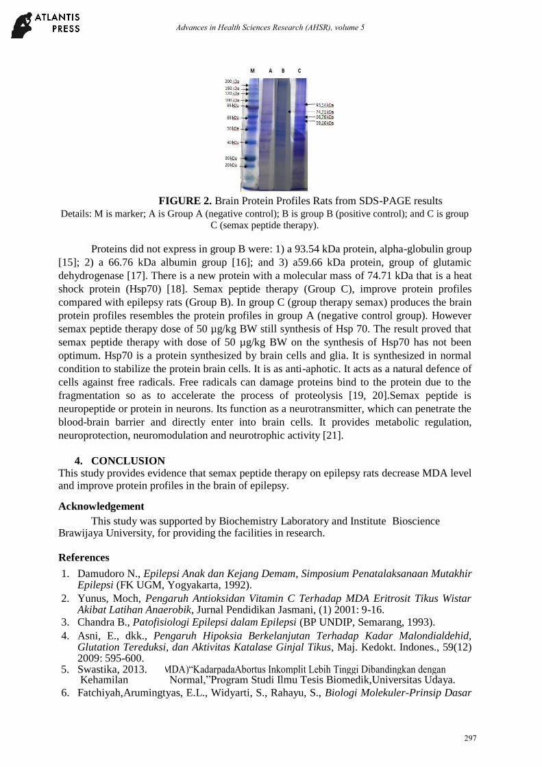
FIGURE 2. Brain Protein Profiles Rats from SDS-PAGE results Details: M is marker; A is Group A (negative control); B is group B (positive control); and C is group
C (semax peptide therapy).
Proteins did not express in group B were: 1) a 93.54 kDa protein, alpha-globulin group
[15]; 2) a 66.76 kDa albumin group [16]; and 3) a59.66 kDa protein, group of glutamic
dehydrogenase [17]. There is a new protein with a molecular mass of 74.71 kDa that is a heat
shock protein (Hsp70) [18]. Semax peptide therapy (Group C), improve protein profiles
compared with epilepsy rats (Group B). In group C (group therapy semax) produces the brain
protein profiles resembles the protein profiles in group A (negative control group). However
semax peptide therapy dose of 50 µg/kg BW still synthesis of Hsp 70. The result proved that
semax peptide therapy with dose of 50 µg/kg BW on the synthesis of Hsp70 has not been
optimum. Hsp70 is a protein synthesized by brain cells and glia. It is synthesized in normal
condition to stabilize the protein brain cells. It is as anti-aphotic. It acts as a natural defence of
cells against free radicals. Free radicals can damage proteins bind to the protein due to the
fragmentation so as to accelerate the process of proteolysis [19, 20].Semax peptide is
neuropeptide or protein in neurons. Its function as a neurotransmitter, which can penetrate the
blood-brain barrier and directly enter into brain cells. It provides metabolic regulation,
neuroprotection, neuromodulation and neurotrophic activity [21].
4. CONCLUSION
This study provides evidence that semax peptide therapy on epilepsy rats decrease MDA level and improve protein profiles in the brain of epilepsy. Acknowledgement
This study was supported by Biochemistry Laboratory and Institute Bioscience Brawijaya University, for providing the facilities in research. References 1. Damudoro N., Epilepsi Anak dan Kejang Demam, Simposium Penatalaksanaan Mutakhir
Epilepsi (FK UGM, Yogyakarta, 1992). 2. Yunus, Moch, Pengaruh Antioksidan Vitamin C Terhadap MDA Eritrosit Tikus Wistar
Akibat Latihan Anaerobik, Jurnal Pendidikan Jasmani, (1) 2001: 9-16. 3. Chandra B., Patofisiologi Epilepsi dalam Epilepsi (BP UNDIP, Semarang, 1993). 4. Asni, E., dkk., Pengaruh Hipoksia Berkelanjutan Terhadap Kadar Malondialdehid,
Glutation Tereduksi, dan Aktivitas Katalase Ginjal Tikus, Maj. Kedokt. Indones., 59(12) 2009: 595-600.
5. Swastika, 2013. Malondialdehyde.P., (MDA)“KadarpadaAbortus Inkomplit Lebih Tinggi Dibandingkan dengan Kehamilan Normal,”Program Studi Ilmu Tesis Biomedik,Universitas Udaya. 6. Fatchiyah,Arumingtyas, E.L., Widyarti, S., Rahayu, S., Biologi Molekuler-Prinsip Dasar
297
Advances in Health Sciences Research (AHSR), volume 5

Analisis (Erlangga, Jakarta, 2011). 7. Panayiotopoulos CP., The epilepsies seizures, syndromes and management (Blandon
Medical Publishing, Oxfordshire, 2005). 8. Manchenko D., Vilensky D., Levitskaya N., Kamensky A., Myasoedov N., Considerably
Different Mechanisms Underlie The Effects of The ACTH(4-Induced−10)AnalgesiaAnalogueandStress- S Induced Behavior, Federal Targeted Program-educational
“Scientificpersonnelofinnovative and Russia, 2009: (P1057).
9. Roxane, Diazepam Oral Solution 5 mg per 5 mL Diazepam IntensolTM
Oral Solution (Concentrate) 5 mg per mL (Roxane Laboratories, Inc, Columbus, Ohio 43216, 2012).
10. Eremin KO, Kudrin VS, Grivennikov IA, Miasoedov NF, Rayevsky KS., Effects Of Semax Peptide on Dopaminergic and Serotoninergic Systems of The Brain, Dokl Biol Sci;394, 2004: 1-3.
11. Carr Chloe, An Investigation of The Effects of LiCl and The -RoleCateninin LumbriculusOfΒ Variegatus Patterning (developmental Biology, 2005).
12. Marnett, L.J., Lipid Peroxidation DNA Damage by Malondyaldehyde, Mutation Research (1999): 424, 8-95.
13. Aral, H. And A. Vecchio-Sadus, Toxicity Of Lithium To Humans and The Environment—A Literature Review, Ecotoxicology and Environmental Safety 70 (2008): 349–356.
14. Gusev, E.I; Skvortsova, Brain Ischemia. Kluwer Academic/ Plenum Publishers, New
York (2003): 297-323. 15. Keren, David F., Protein Electrophoresis in Clinical Diagnosis, Holder Arnold (2003),
pp. 1-14, ISBN 0340 81213 3. 16. Lehninger A, Nelson D, Cox M M., Principles of Biochemistry 2
nd (1993): 710-711.
17. Reeds, P.J., et al., Intestinal Glutamate Metabolism, Journal of Nutrition, 130(4s) (2000): 978S-982S.
18. Murray R K, et al., Harper’s Biochemistryed.Appleton&Lange.America25 (2000): 359-373. 19. S Theresia Indah Budhy, Istiati K, Soehardjo, Peran Heat Shock Protein (HSP) terhadap
Penyakit Rongga Mulut (Bagian Biologi Oral, Fakultas Kedokteran GIgi universitas Airlangga, 2006).
20. Nollen and Richrd , Heat Shock Protein, J Set (2002); 2809. 21. Semax International Inc., Semax, www.semaxint.com (2003-2008), (7 September 2016).
298
Advances in Health Sciences Research (AHSR), volume 5