Embed Size (px)
Citation preview
JOURNAL OF VIROLOGY, Nov. 2009, p. 10993–11004 Vol. 83, No. 210022-538X/09/$12.00 doi:10.1128/JVI.01085-09Copyright © 2009, American Society for Microbiology. All Rights Reserved.
The Pre-Transmembrane Domain of the Autographa californicaMulticapsid Nucleopolyhedrovirus GP64 Protein Is Critical
for Membrane Fusion and Virus Infectivity�†Zhaofei Li and Gary W. Blissard*
Boyce Thompson Institute, Cornell University, Ithaca, New York 14853
Received 27 May 2009/Accepted 6 August 2009
The envelope glycoprotein, GP64, of the baculovirus Autographa californica multicapsid nucleopolyhedrovi-rus (AcMNPV) is a class III viral fusion protein that mediates pH-triggered membrane fusion during virusentry. Viral fusion glycoproteins from many viruses contain a short region in the ectodomain and near thetransmembrane domain, referred to as the pre-transmembrane (PTM) domain. In some cases, the PTMdomain is rich in aromatic amino acids and plays an important role in membrane fusion. Although the23-amino-acid (aa) PTM domain of AcMNPV GP64 lacks aromatic amino acids, we asked whether this regionmight also play a significant role in membrane fusion. We generated alanine scanning and single and multipleamino acid substitutions in the GP64 PTM domain. We specifically focused on amino acid positions conservedbetween baculovirus GP64 and thogotovirus GP75 proteins, as well as hydrophobic and charged amino acids.For each PTM-modified construct, we examined trimerization, cell surface localization, and membrane fusionactivity. Membrane merger and pore formation were also examined. We identified eight aa positions that areimportant for membrane fusion activity. Critical positions were not clustered in the linear sequence but weredistributed throughout the PTM domain. While charged residues were not critical or essential, three hydro-phobic amino acids (L465, L476, and L480) played an important role in membrane fusion activity and appearto be involved in formation of the fusion pore. We also asked whether selected GP64 constructs were capableof rescuing a gp64null AcMNPV virus. These studies suggested that several conserved residues (T463, G460,G462, and G474) were not required for membrane fusion but were important for budding and viral infectivity.
Baculoviruses are enveloped viruses with large double-stranded DNA genomes ranging from approximately 80 to 180kbp. They infect only invertebrates, and the majority of bacu-loviruses described are from insects in the order Lepidoptera.The type species of the family Baculoviridae is the Autographacalifornica multicapsid nucleopolyhedrovirus (AcMNPV) (11,34). Budded virions (BV) of AcMNPV enter cells via a low-pH-dependent endocytic pathway (16). During entry by endo-cytosis, the major envelope glycoprotein GP64 mediates low-pH-triggered membrane fusion (3). Baculovirus GP64 proteinsare highly conserved among the group I alphabaculoviruses,and the only known proteins with amino acid sequence simi-larities to baculovirus GP64 proteins are the GP75 envelopeglycoproteins from thogotoviruses, a subgroup of the Ortho-myxoviridae. GP64 is a type I integral membrane protein that ispresent on the infected cell surface and on the virion as ahomotrimer (22). Monomers of GP64 are associated in thetrimer by a single intermolecular disulfide bond (12, 37), andGP64 does not appear to require protease cleavage for activa-tion or function. GP64 has host cell receptor-binding activity(6), and a region important for receptor-binding was recentlymapped to the N-terminal portion of the ectodomain (39).GP64 is necessary and sufficient for pH-dependent membrane
fusion during viral entry (3, 13, 40), and in addition to itsessential role in virus entry, GP64 is also necessary for efficientbudding and production of infectious virions (20, 21). Re-cently, the crystal structure of the low-pH (postfusion) confor-mation of the AcMNPV GP64 ectodomain was reported (12).The GP64 postfusion structure is an elongated trimer com-prised largely of beta sheets and a central core consisting ofthree extended coiled coils. Each GP64 monomer can be sub-divided into five domains, and the overall structures of themonomer and trimer, as well as the linear arrangement of thedomains, bear remarkable similarities to the postfusion struc-tures of herpesvirus (herpes simplex virus type 1 and Epstein-Barr virus) gB glycoproteins and also to the structures of thevesicular stomatitis virus (VSV) G protein (1, 12). While thesethree proteins (GP64, VSV G, and herpesvirus gB) differ insize, have no apparent amino acid sequence similarity, and arefrom unrelated virus groups, they share common structuralfeatures, including the long central helix that forms a triple-stranded coiled coil at the heart of the trimer, and internalfusion loops that are likely associated with target membranesduring membrane fusion. These similarities suggested thatthese three proteins belong to a new class of viral fusion pro-teins, now referred to as class III fusion proteins (1, 12). Al-though viral fusion proteins from different classes (I, II, andIII) show a variety of molecular architectures, all are thoughtto catalyze fusion in similar manners (5). Following an initialtriggering event, the fusion protein extends and interacts withthe adjacent cellular membrane via the fusion peptide or fu-sion loop(s). This is followed by subsequent refolding to bringthe transmembrane domain and the fusion peptide or loop(s)
* Corresponding author. Mailing address: Boyce Thompson In-stitute at Cornell University, Tower Road, Ithaca, NY 14853-1801.Phone: (607) 254-1366. Fax: (607) 254-1242. E-mail: [email protected].
† Supplemental material for this article may be found at http://jvi.asm.org/.
� Published ahead of print on 19 August 2009.
10993
on Decem
ber 11, 2018 by guesthttp://jvi.asm
.org/D
ownloaded from
into close proximity. Mixing of outer leaflets of the two bilayersresults in the formation of a hemifusion stalk, and this isfollowed by mixing of the inner leaflets to form a fusion pore.In some cases (such as the influenza hemagglutinin [HA] pro-tein), the fusion pore first appears after some period (approx-imately 30 s after low-pH exposure) and begins to “flicker,”rapidly opening and closing. This is followed by a final openstate of the fusion pore (31, 32). In contrast to the flickeringpore of HA, previous studies suggest that the GP64 fusion poreopens almost immediately (�0.6 s after being triggered by lowpH) and expands rapidly with no flickering (27, 28).
Many viral fusion proteins contain a short region in theectodomain near the transmembrane domain, referred to asthe membrane-proximal region, stem region, or pre-transmem-brane (PTM) domain. This PTM domain is often rich in aro-matic amino acids (17) and in prior studies of class I viralfusion proteins, the PTM domains and particularly the con-served aromatic residues within the PTM domain, were shownto play important role(s) in membrane fusion. Examples in-clude PTM domains from the fusion proteins of human immu-nodeficiency virus type 1 (29), feline immunodeficiency virus(4), Ebola virus (26), human parainfluenza virus type 2 (35),and severe acute respiratory syndrome coronavirus (8, 27, 28).Various mechanistic roles have been proposed for these PTMdomains, including (i) contributions to the stabilization of asix-helix bundle structure, (ii) interactions with the mem-brane(s), and (iii) induction of membrane destabilization. ThePTM domain of the VSV G protein plays roles in both mem-brane fusion and virion budding (10, 25). Interestingly, whiledeletions or substitutions of residues from the VSV-G PTMdomain had dramatic negative effects on membrane fusion,only modest effects on virus infectivity were observed. In ad-dition, single or multiple substitutions of the conserved aro-matic residues (W457, F458, and W461) have only modesteffects on cell-cell fusion activity (9).
The PTM domain of the baculovirus GP64 protein was notspecifically examined for function in prior studies. However, itwas recently demonstrated that 22 amino acids (aa) (resi-dues 461 to 482) from the membrane-proximal PTM regionof GP64, together with the predicted transmembrane do-main (residues 483 to 505), cytoplasmic tail, and 38 aa from themature N terminus of the GP64 ectodomain, were sufficient forrescuing the budding defect of a gp64null virus and targetingthe protein to BV (38). Whether this region played a role inmembrane fusion, however, was unknown. To examine thefunctional role(s) of the GP64 PTM domain in detail, wegenerated a bank of mutations in the 23-aa PTM domain andanalyzed the effects of single and multiple alanine substitutionmutations in this region. Our results indicate that the PTMdomain of GP64 is not essential for stable expression, trimer-ization, or transport and localization of GP64 on the cell sur-face. However, the PTM domain was essential for GP64-me-diated membrane fusion, and three PTM leucine residues werecritical for that activity. Using recombinant baculoviruses en-coding GP64 proteins with various PTM domain mutations, wealso examined and characterized the effects of PTM domainmutations on virion budding, GP64 incorporation into virions,and virus infectivity.
MATERIALS AND METHODS
Cells, transfections, and infections. Spodoptera frugiperda (Sf9) cells andGP64-expressing cell line Sf9Op1D (23) were cultured at 27°C in TNMFH me-dium (7) containing 10% fetal bovine serum. Cells were transfected using CaPO4
precipitation (2). For viral infections, budded virus was added to cells at amultiplicity of infection (MOI) of 5 and then incubated for 1 h, and cells werewashed once in TNMFH. The zero time postinfection (p.i.) indicates the time ofviral inoculum removal.
Mutagenesis and construction of plasmids and bacmids. Modified GP64constructs were generated using an overlap PCR method with the plasmidpGEM3ZGP64 (14) as the template as described previously (15). Primer se-quences are available upon request. Briefly, the PCR products were purified anddigested with unique restriction enzymes NotI and EcoRI and used for subclon-ing into plasmid pBiepA previously digested with the same enzymes. Recombi-nant baculoviruses expressing the modified GP64 proteins were generated byfirst subcloning overlap PCR products into plasmid pGEM3ZGP64 (NotI andHindIII sites), excising the promoter and modified GP64 open reading framewith KpnI and EcoRI, and subcloning them into the same sites of pFastBac1(Invitrogen). Note that the polyhedrin promoter was removed. The constructcontaining a modified gp64 gene was inserted into the polyhedrin locus of anAcMNPV gp64null bacmid (vAc64�) by Tn7-mediated transposition (18) andconfirmed by restriction enzyme analysis and DNA sequencing.
cELISA, syncytium formation, and fusion assays. Relative levels of cell sur-face-localized GP64 protein were analyzed by a cell surface enzyme-linked im-munosorbent assay (cELISA) (14, 15). Briefly, after transfection, Sf9 cells wereincubated for 36 h and then fixed in 0.5% glutaraldehyde so that cells were notpermeabilized. Relative levels of cell surface-localized GP64 were measured bycELISA using monoclonal antibody (MAb) AcV5 as described previously indetail (15). For analysis of membrane fusion by syncytium formation assays, Sf9cells were plated in 12-well plates, transfected with plasmids encoding wild-type(WT) or modified forms of GP64, incubated 36 h at 27°C, and then exposed tophosphate-buffered saline (PBS) at pH 5.0 for 3 min as described previously (14,15). After a 4-h incubation at 27°C, cells were fixed with methanol and stainedwith a HEMA3 stain kit (Fisher Scientific LLC), and the number of nuclei foundin syncytia was scored. Syncytial masses were defined as fused cells containing �5nuclei. Five representative fields were analyzed for each construct, and relativelevels of fusion activity were determined by dividing the number of nuclei insyncytia by the total number of nuclei in a field and then normalizing the resultsto parallel data from cells expressing WT GP64 and localized to the cell surfaceat equivalent levels.
Immunofluorescence analysis of cell surface GP64. Cell surface GP64 local-ization was confirmed by immunofluorescence analysis of transfected cells. Sf9cells (4 � 105 cells/well) were transfected with plasmids expressing either WT ormodified GP64 proteins. At 36-h posttransfection (p.t.), cells were fixed with 4%paraformaldehyde in PBS (pH 7.4), washed with PBS, and then immunostainedfor GP64 with primary anti-GP64 monoclonal antibody AcV1 (1:10 dilution inPBS) and a secondary goat anti-mouse monoclonal antibody conjugated to AlexaFluor 488 (Molecular Probes, Invitrogen), as described previously (14, 15). Flo-rescence was observed and documented with an Olympus IX70 epifluorescencemicroscope.
Analysis of hemifusion and pore formation. To examine initial membranemerger (hemifusion) and pore formation, sheep red blood cells (RBCs) (Hemo-State Laboratories) were dual labeled with octadecyl rhodamine B chloride(R18) and calcein-acetoxymethyl (AM) (Molecular Probes, Invitrogen) (14, 15).Sf9 cells (4 � 105 cells/well) were transfected with plasmids expressing GP64constructs and then incubated with R18- and calcein-labeled RBCs in the fol-lowing manner. At 36-h p.t., dual-labeled RBCs were added to Sf9 cells andincubated for 20 min at room temperature, and then unbound RBCs wereremoved by washing them three times with PBS (pH 7.4). Sf9 cells were thenexposed to low pH by incubation in PBS (pH 5.0) for 3 min at room temperature.After rinsing cells in PBS (pH 7.4), cells were placed in TNMFH medium andincubated for 20 min at 27°C. Analysis of hemadsorption and the transfer offluorescence were determined by phase-contrast and epifluorescence micros-copy. For each construct, five randomly selected fields were scored for dyetransfer.
Transfection-infection assays. To determine whether GP64 constructs withmodified PTM domains were capable of supporting viral infection and rescuinga gp64null virus, bacmid DNAs encoding PTM-modified GP64 gene constructswere used to transfect Sf9 cells (or control Sf9Op1D cells) using calcium phos-phate precipitation. At 96 h p.t., the supernatant was removed, clarified bycentrifugation (10 min at 2,200 � g), and then used to infect Sf9 cells, which weresubsequently examined for �-glucuronidase (GUS) activity at 96 h p.i. as de-
10994 LI AND BLISSARD J. VIROL.
on Decem
ber 11, 2018 by guesthttp://jvi.asm
.org/D
ownloaded from
scribed previously (19). Both transfected and infected cells were stained for GUSactivity. To confirm the viability of each bacmid genome and preparation, thesame AcMNPV bacmids were used to transfect Sf9Op1D cells (which express awild-type GP64 protein), and supernatants were used to infect Sf9 cells.
Viral growth curves. To generate viral growth curves, Sf9 cells (1 � 106)were infected in triplicate with each virus (vAcGP64WT, vAcGP64G460A,vAcGP64S464A, vAcGP64S466A, vAcGP64D467A, vAcGP64D467E, andvAcGP64G474A) in six-well plates, at an MOI of 5. After a 1-h incubation period,the inoculum was removed and exchanged with TNMFH. Supernatants werecollected at the indicated times p.i., and the titers of all supernatants weredetermined by 50% tissue culture infective dose assays on Sf9 cells.
Virion budding and GP64 incorporation into virions. The effects of GP64substitutions on virion budding efficiency and incorporation into BV were exam-ined in the following manner: viruses expressing the PTM-modified GP64 con-structs were amplified, and titers of the virus in Sf9OP1D cells, which constitu-tively express OpMNPV GP64 (23), were determined. The resulting virus stockswere used to infect Sf9 cells (5 � 106 cells; MOI of 5), and virion budding andincorporation were examined as described previously (14, 15, 21). Briefly, at 15 hp.i., cells were starved for 1 h in 2 ml methionine-free Grace’s medium(Grace�met; Invitrogen), and then the medium was replaced with 2.2 ml ofGraces�met containing 200 �Ci protein labeling mix (35S EasyTag Express,1175.0 Ci/mmol; Perkin-Elmer Life Sciences). At 30 h p.i., the medium wassupplemented with methionine by adding 0.8 ml of TNMFH. Supernatants wereharvested at 40 h p.i. After clearing cell debris by centrifugation for 10 min at3,000 � g at 4°C, the virus-containing supernatant was loaded onto a 25%sucrose cushion and centrifuged at 80,000 � g for 90 min at 4°C (SW60 rotor).Virus pellets were resuspended in 200 �l Laemmli buffer (4% sodium dodecylsulfate [SDS], 20% glycerol, 10% 2-mercaptoethanol, 0.04% bromophenol blue,0.125 M Tris, pH 6.8) containing protease inhibitors (Complete; Roche AppliedScience), and proteins were separated on SDS-10% polyacrylamide gel elec-trophoresis (SDS-PAGE) gels. To identify and quantify labeled virion pro-teins, dried gels were exposed on phosphorimager screens, scanned on aphosphorimager (Molecular Dynamics), and individual protein bands werequantified using ImageQuant TL software (Amersham, GE).
Western blot analysis. Analysis of GP64 proteins by reducing and nonreducingSDS-PAGE (6% or 10% polyacrylamide gels) was described previously (21).GP64 was detected on Western blots using MAb AcV5 at a dilution of 1:1,000(14, 15).
RESULTS
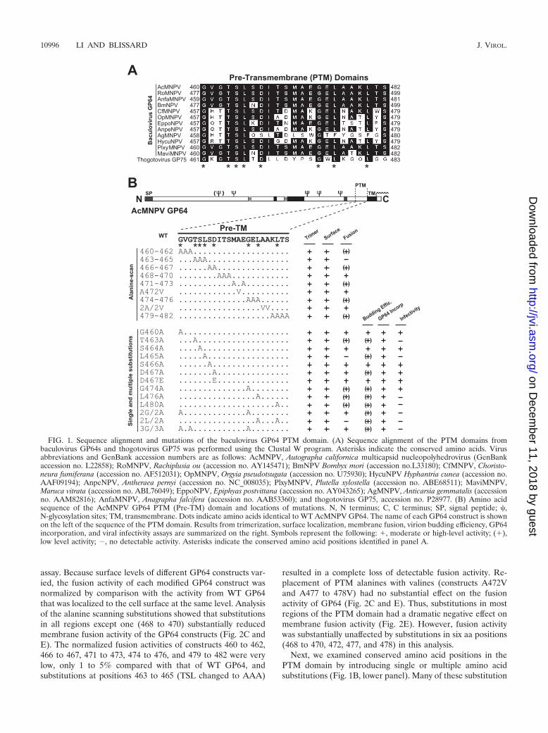
Conservation in the PTM domain of GP64 proteins. Toexamine conservation in the PTM domain of GP64, we per-formed a sequence alignment of the PTM domains from bac-ulovirus GP64 proteins and thogotovirus GP75. The PTM do-main of AcMNPV GP64 examined in this study was defined asthe 23-aa region, comprising aa 460 to 482 immediatelyupstream of the transmembrane domain. Although aromaticamino acids in the PTM domain are important for the functionof many class I viral fusion proteins, aromatic amino acids arenot conserved in the PTM domain of baculovirus GP64 pro-teins. While some GP64 proteins contain one to four aromaticresidues (F, Y, W, or H), others, such as the AcMNPV GP64protein, contain no aromatic amino acids in the PTM domain(Fig. 1A). However, by amino acid sequence alignment of thePTM domains of GP64 proteins and thogotovirus GP75 (whichshares approximately 28% overall amino acid sequence simi-larity with baculovirus GP64 proteins), we identified severalhighly conserved amino acid positions in the PTM domain.These conserved positions include G460, T463, S464, L465,D467, G474, L476, and L480 (Fig. 1A).
Substitution mutations in the GP64 PTM domain. We usedtwo strategies to examine the requirements of the GP64 PTMdomain. First, we performed an alanine scan of the GP64 PTMdomain, substituting two, three, or four alanines for aminoacids of the PTM domain sequence (Fig. 1B). Alanines in thewild-type PTM domain (positions A472, A477, and A478) were
substituted with valine residues. Next, we generated single andmultiple amino acid substitutions in various PTM positions,based primarily on sequence conservation among GP64 andGP75 proteins (Fig. 1A and B). Amino acid positions G460,T463, S464, L465, D467, S466, G474, L476, and L480 weresubstituted individually and in some cases, in combinations(Fig. 1A and B).
Expression and cell surface localization of PTM-modifiedGP64 proteins. GP64 proteins containing substitutions in thePTM domain were transiently expressed from plasmids in Sf9cells, and cell lysates were examined at 36 h p.t., by Westernblot analysis using either reducing or nonreducing conditionsfor SDS-PAGE. As GP64 trimers contain a disulfide bondbetween monomers in the trimer, nonreducing conditions per-mit the assessment of GP64 oligomerization (6, 22). Oligomersrepresenting GP64 trimers (14, 22) were detected from allGP64 constructs with substitutions in the PTM domain (seeFig. S1A and B in the supplemental material). These datasuggested that substitutions within the PTM domain of GP64did not significantly affect the expression or oligomerization ofthe resulting proteins.
Because cell surface levels of GP64 can affect the assessmentof function, we asked whether GP64 constructs containingsubstitutions in the GP64 PTM domain were transported to thecell surface and displayed on the plasma membrane. Using acELISA protocol, we measured the level of each GP64 con-struct at the cell surface relative to that detected from WTGP64. Of the nine alanine scanning constructs examined, fourresulted in cell surface GP64 levels of �20% of that fromwild-type GP64, and five resulted in surface levels below 10%(Fig. 2A). In all cases, the measured cell surface levels weresufficient for detection of membrane fusion activity from wild-type GP64. Alanine residues found in the wild-type GP64 PTMdomain were substituted with valine residues. Substitution ofalanines with valines resulted in either minor or moderatelyreduced surface levels of GP64 (Fig. 2A, constructs A472V and2A/2V). Similar to the results from alanine-scanning muta-tions, surface levels of GP64 constructs with single or multipleamino acid substitutions in the PTM domain were frequentlylower than surface levels of wild-type GP64 (Fig. 2B), althoughsome constructs (S464A, G460A, 2G/2A, and 3G/3A) had onlyminor or moderate reductions in cell surface levels. All otherconstructs were displayed at the cell surface at levels of �10%of that from WT GP64. However, the detected cell surfacelevels were sufficient for detection of membrane fusion activityfrom wild-type GP64. To confirm the presence and nativeconformation of GP64 constructs at the cell surface, we alsoused a conformation-specific MAb (MAb AcV1) to detecteach GP64 construct at the cell surface by indirect immuno-fluorescence (see Fig. S1C and D in the supplemental mate-rial). MAb AcV1 recognizes only the prefusion conformationof GP64, and the epitope is lost upon exposure of GP64 to lowpH (40). All GP64 constructs containing substitution muta-tions in the GP64 PTM domain were detected at the cellsurface by MAb AcV1, confirming cell surface localization andindicating that the GP64 constructs were in the native prefu-sion conformation.
Fusion activity of mutant GP64s. We evaluated the mem-brane fusion activity of each GP64 PTM construct by measur-ing fusion efficiency in a semiquantitative syncytium formation
VOL. 83, 2009 THE PRE-TRANSMEMBRANE DOMAIN OF AcMNPV GP64 PROTEIN 10995
on Decem
ber 11, 2018 by guesthttp://jvi.asm
.org/D
ownloaded from
assay. Because surface levels of different GP64 constructs var-ied, the fusion activity of each modified GP64 construct wasnormalized by comparison with the activity from WT GP64that was localized to the cell surface at the same level. Analysisof the alanine scanning substitutions showed that substitutionsin all regions except one (468 to 470) substantially reducedmembrane fusion activity of the GP64 constructs (Fig. 2C andE). The normalized fusion activities of constructs 460 to 462,466 to 467, 471 to 473, 474 to 476, and 479 to 482 were verylow, only 1 to 5% compared with that of WT GP64, andsubstitutions at positions 463 to 465 (TSL changed to AAA)
resulted in a complete loss of detectable fusion activity. Re-placement of PTM alanines with valines (constructs A472Vand A477 to 478V) had no substantial effect on the fusionactivity of GP64 (Fig. 2C and E). Thus, substitutions in mostregions of the PTM domain had a dramatic negative effect onmembrane fusion activity (Fig. 2E). However, fusion activitywas substantially unaffected by substitutions in six aa positions(468 to 470, 472, 477, and 478) in this analysis.
Next, we examined conserved amino acid positions in thePTM domain by introducing single or multiple amino acidsubstitutions (Fig. 1B, lower panel). Many of these substitution
FIG. 1. Sequence alignment and mutations of the baculovirus GP64 PTM domain. (A) Sequence alignment of the PTM domains frombaculovirus GP64s and thogotovirus GP75 was performed using the Clustal W program. Asterisks indicate the conserved amino acids. Virusabbreviations and GenBank accession numbers are as follows: AcMNPV, Autographa californica multicapsid nucleopolyhedrovirus (GenBankaccession no. L22858); RoMNPV, Rachiplusia ou (accession no. AY145471); BmNPV Bombyx mori (accession no.L33180); CfMNPV, Choristo-neura fumiferana (accession no. AF512031); OpMNPV, Orgyia pseudotsugata (accession no. U75930); HycuNPV Hyphantria cunea (accession no.AAF09194); AnpeNPV, Antheraea pernyi (accession no. NC_008035); PlxyMNPV, Plutella xylostella (accession no. ABE68511); MaviMNPV,Maruca vitrata (accession no. ABL76049); EppoNPV, Epiphyas postvittana (accession no. AY043265); AgMNPV, Anticarsia gemmatalis (accessionno. AAM82816); AnfaMNPV, Anagrapha falcifera (accession no. AAB53360); and thogotovirus GP75, accession no. P28977. (B) Amino acidsequence of the AcMNPV GP64 PTM (Pre-TM) domain and locations of mutations. N, N terminus; C, C terminus; SP, signal peptide; �,N-glycosylation sites; TM, transmembrane. Dots indicate amino acids identical to WT AcMNPV GP64. The name of each GP64 construct is shownon the left of the sequence of the PTM domain. Results from trimerization, surface localization, membrane fusion, virion budding efficiency, GP64incorporation, and viral infectivity assays are summarized on the right. Symbols represent the following: �, moderate or high-level activity; (�),low level activity; �, no detectable activity. Asterisks indicate the conserved amino acid positions identified in panel A.
10996 LI AND BLISSARD J. VIROL.
on Decem
ber 11, 2018 by guesthttp://jvi.asm
.org/D
ownloaded from
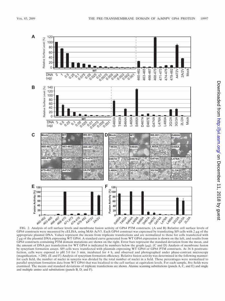
FIG. 2. Analysis of cell surface levels and membrane fusion activity of GP64 PTM constructs. (A and B) Relative cell surface levels ofGP64 constructs were measured by cELISA, using MAb AcV5. Each GP64 construct was expressed by transfecting Sf9 cells with 2 �g of theappropriate plasmid DNA. Values represent the means from triplicate transfections and are normalized to those for cells transfected with2 �g of the plasmid DNA expressing WT GP64. A standard curve generated from WT GP64 expression is shown on the left, and results fromGP64 constructs containing PTM domain mutations are shown on the right. Error bars represent the standard deviation from the mean, andthe amount of DNA per transfection for WT GP64 is indicated by numbers below the graph (�g). (C and D) Analysis of membrane fusionby syncytium formation assays. Sf9 cells were transfected with plasmids expressing WT GP64 or GP64 PTM constructs. At 36 h posttrans-fection, cells were exposed to pH 5.0 for 3 min, incubated for 4 h, and observed and photographed under phase-contrast microscopy(magnification, �200). (E and F) Analysis of syncytium formation efficiency. Relative fusion activity was determined in the following manner:for each field, the number of nuclei in syncytia was divided by the total number of nuclei in a field. Those percentages were normalized toparallel syncytium formation data from WT GP64 that was localized to the cell surface at equivalent levels. For each sample, five fields wereexamined. The means and standard deviations of triplicate transfections are shown. Alanine scanning substitutions (panels A, C, and E) and singleand multiple amino acid substitutions (panels B, D, and F).
VOL. 83, 2009 THE PRE-TRANSMEMBRANE DOMAIN OF AcMNPV GP64 PROTEIN 10997
on Decem
ber 11, 2018 by guesthttp://jvi.asm
.org/D
ownloaded from
mutations had dramatic effects on GP64 fusion activity. Themost substantial effects were observed with alanine substitu-tions L465A and 2L/2A (2L/2A represents alanine substitu-tions in positions L476 and L480), which resulted in GP64proteins with no detectable fusion activity (Fig. 2D and F). Inaddition, GP64 PTM substitutions T463A, G474A, L476A, andL480A mediated only very low levels of membrane fusion, withnormalized fusion activities of approximately 1.4% (L476Aand L480A), 3.1% (G474A), and 4.2% (T463A), comparedwith WT GP64 (Fig. 2F). Substitution of charged residue D467with alanine or glutamic acid, resulted in a 90% decrease infusion activity. In general, analysis of alanine substitutions inconserved glycine residues of the PTM domain revealed nodramatic negative effects. Construct G460A retained fusionactivity at levels near wild-type levels (�90% of wild-type ac-tivity), and double and triple glycine substitutions (2G/2A,positions G460 and G474; 3G/3A, positions G460, G462, andG474) resulted in only modestly decreased fusion activities(�70% and 60%, respectively), compared with wild-type GP64(Fig. 2D). It is of interest that the single G474A substitutionresulted in dramatically reduced GP64 fusion activity, yet whenthe same substitution was included with one or two additionalglycine substitutions (in constructs 2G/2A and 3G/3A), therewas only a minor negative effect of the G474A substitution onmembrane fusion activity. Substitutions in conserved and semi-conserved serine residues resulted in no detectable effect onfusion activity of GP64 (Fig. 1B and 2F, S464A and S466A). Intotal, our analysis of alanine scanning substitutions in combi-nation with single and multiple amino acid substitutions indi-cated that the specific amino acids at certain positions (G460,S464, S466, I468, T469, S470, A472, A477, and A478) were notcritical for membrane fusion. In contrast, the loss or dramaticreduction of fusion activity from constructs containing single ormultiple substitution mutations at other positions (T463, L465,D467, and L476, and L480) identified these residues as impor-tant for membrane fusion activity.
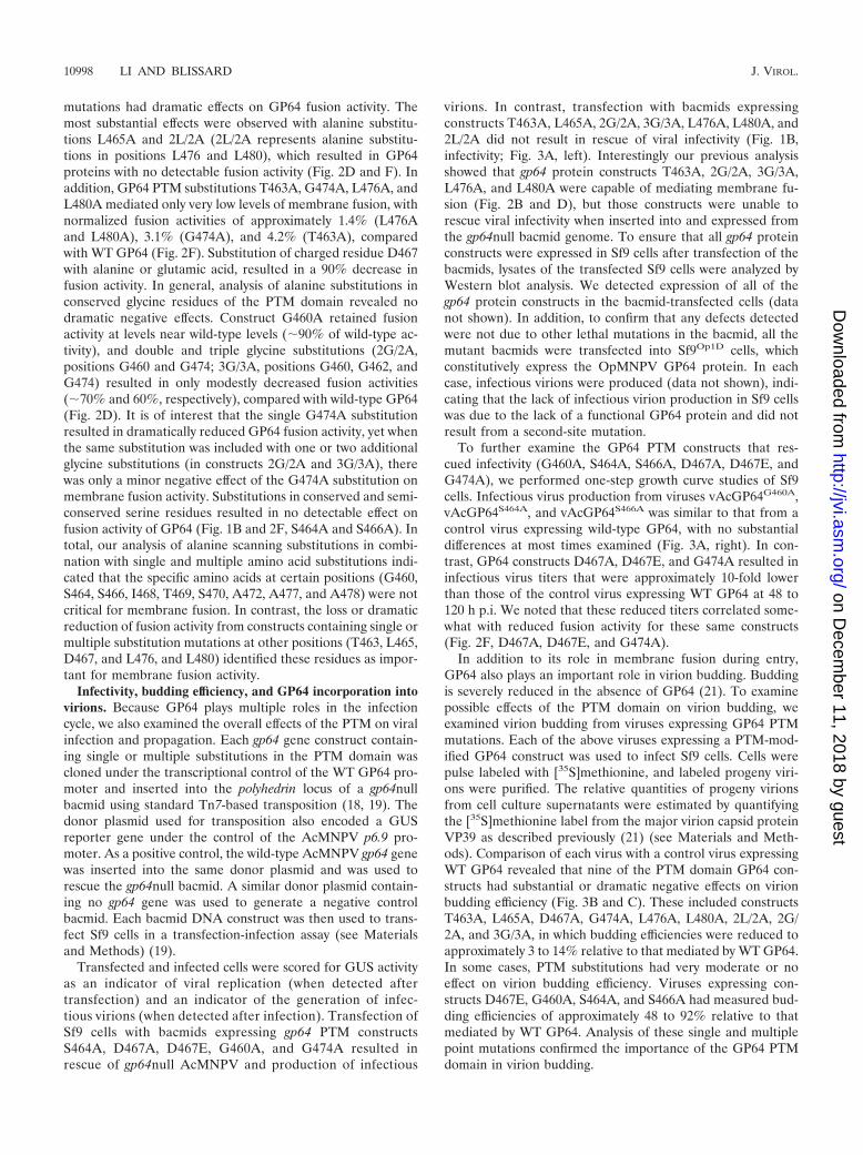
Infectivity, budding efficiency, and GP64 incorporation intovirions. Because GP64 plays multiple roles in the infectioncycle, we also examined the overall effects of the PTM on viralinfection and propagation. Each gp64 gene construct contain-ing single or multiple substitutions in the PTM domain wascloned under the transcriptional control of the WT GP64 pro-moter and inserted into the polyhedrin locus of a gp64nullbacmid using standard Tn7-based transposition (18, 19). Thedonor plasmid used for transposition also encoded a GUSreporter gene under the control of the AcMNPV p6.9 pro-moter. As a positive control, the wild-type AcMNPV gp64 genewas inserted into the same donor plasmid and was used torescue the gp64null bacmid. A similar donor plasmid contain-ing no gp64 gene was used to generate a negative controlbacmid. Each bacmid DNA construct was then used to trans-fect Sf9 cells in a transfection-infection assay (see Materialsand Methods) (19).
Transfected and infected cells were scored for GUS activityas an indicator of viral replication (when detected aftertransfection) and an indicator of the generation of infec-tious virions (when detected after infection). Transfection ofSf9 cells with bacmids expressing gp64 PTM constructsS464A, D467A, D467E, G460A, and G474A resulted inrescue of gp64null AcMNPV and production of infectious
virions. In contrast, transfection with bacmids expressingconstructs T463A, L465A, 2G/2A, 3G/3A, L476A, L480A, and2L/2A did not result in rescue of viral infectivity (Fig. 1B,infectivity; Fig. 3A, left). Interestingly our previous analysisshowed that gp64 protein constructs T463A, 2G/2A, 3G/3A,L476A, and L480A were capable of mediating membrane fu-sion (Fig. 2B and D), but those constructs were unable torescue viral infectivity when inserted into and expressed fromthe gp64null bacmid genome. To ensure that all gp64 proteinconstructs were expressed in Sf9 cells after transfection of thebacmids, lysates of the transfected Sf9 cells were analyzed byWestern blot analysis. We detected expression of all of thegp64 protein constructs in the bacmid-transfected cells (datanot shown). In addition, to confirm that any defects detectedwere not due to other lethal mutations in the bacmid, all themutant bacmids were transfected into Sf9Op1D cells, whichconstitutively express the OpMNPV GP64 protein. In eachcase, infectious virions were produced (data not shown), indi-cating that the lack of infectious virion production in Sf9 cellswas due to the lack of a functional GP64 protein and did notresult from a second-site mutation.
To further examine the GP64 PTM constructs that res-cued infectivity (G460A, S464A, S466A, D467A, D467E, andG474A), we performed one-step growth curve studies of Sf9cells. Infectious virus production from viruses vAcGP64G460A,vAcGP64S464A, and vAcGP64S466A was similar to that from acontrol virus expressing wild-type GP64, with no substantialdifferences at most times examined (Fig. 3A, right). In con-trast, GP64 constructs D467A, D467E, and G474A resulted ininfectious virus titers that were approximately 10-fold lowerthan those of the control virus expressing WT GP64 at 48 to120 h p.i. We noted that these reduced titers correlated some-what with reduced fusion activity for these same constructs(Fig. 2F, D467A, D467E, and G474A).
In addition to its role in membrane fusion during entry,GP64 also plays an important role in virion budding. Buddingis severely reduced in the absence of GP64 (21). To examinepossible effects of the PTM domain on virion budding, weexamined virion budding from viruses expressing GP64 PTMmutations. Each of the above viruses expressing a PTM-mod-ified GP64 construct was used to infect Sf9 cells. Cells werepulse labeled with [35S]methionine, and labeled progeny viri-ons were purified. The relative quantities of progeny virionsfrom cell culture supernatants were estimated by quantifyingthe [35S]methionine label from the major virion capsid proteinVP39 as described previously (21) (see Materials and Meth-ods). Comparison of each virus with a control virus expressingWT GP64 revealed that nine of the PTM domain GP64 con-structs had substantial or dramatic negative effects on virionbudding efficiency (Fig. 3B and C). These included constructsT463A, L465A, D467A, G474A, L476A, L480A, 2L/2A, 2G/2A, and 3G/3A, in which budding efficiencies were reduced toapproximately 3 to 14% relative to that mediated by WT GP64.In some cases, PTM substitutions had very moderate or noeffect on virion budding efficiency. Viruses expressing con-structs D467E, G460A, S464A, and S466A had measured bud-ding efficiencies of approximately 48 to 92% relative to thatmediated by WT GP64. Analysis of these single and multiplepoint mutations confirmed the importance of the GP64 PTMdomain in virion budding.
10998 LI AND BLISSARD J. VIROL.
on Decem
ber 11, 2018 by guesthttp://jvi.asm
.org/D
ownloaded from
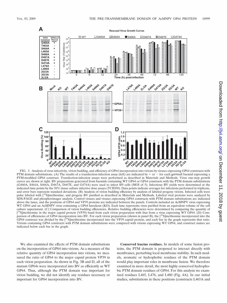
We also examined the effects of PTM domain substitutionson the incorporation of GP64 into virions. As a measure of therelative quantity of GP64 incorporation into virions, we mea-sured the ratio of GP64 to the major capsid protein VP39 ineach virion preparation. As shown in Fig. 3B and D, all of themutant GP64s were incorporated into BV as efficiently as WTGP64. Thus, although the PTM domain was important forvirion budding, we did not identify any residues necessary orimportant for GP64 incorporation into BV.
Conserved leucine residues. In models of some fusion pro-teins, the PTM domain is proposed to interact directly withmembranes, perturbing local membrane stability. In such mod-els, aromatic or hydrophobic residues of the PTM domainwould play important roles in membrane fusion. We thereforeexamined in more detail, the most highly conserved hydropho-bic PTM domain residues of GP64. For this analysis we exam-ined residues L465, L476, and L480 (Fig. 4A). In our initialstudies, substitutions in these positions (constructs L465A and
FIG. 3. Analysis of virus infectivity, virion budding, and efficiency of GP64 incorporation into virions by viruses expressing GP64 constructs withPTM domain substitutions. (A) The results of a transfection-infection assay (left) are indicated by � or � for each gp64null bacmid expressing aPTM-modified GP64 construct. Transfection-infection assays were performed as described in Materials and Methods. Virus one-step growthcurves are shown at right. BV preparations generated from bacmids containing WT GP64 or GP64 constructs with the PTM domain substitutions(G460A, S464A, S466A, D467A, D467E, and G474A) were used to infect Sf9 cells (MOI of 5). Infectious BV yields were determined at theindicated time points by the 50% tissue culture infective dose assays (TCID50). Data points indicate averages for infections performed in triplicate,and error bars represent standard deviations. (B) Analysis of virion budding efficiency by analysis of labeled progeny virions. Infected cells werepulse labeled with [35S]methionine, and progeny BV purified as described in Materials and Methods. Labeled viral proteins were analyzed bySDS-PAGE and phosphorimager analysis. Control viruses and viruses expressing GP64 constructs with PTM domain substitutions are indicatedabove the lanes, and the positions of GP64 and VP39 proteins are indicated between the panels. Controls included an AcMNPV virus expressingWT GP64 and an AcMNPV virus containing a GP64 knockout (KO). Each lane represents virus purified from an equivalent volume of the cellculture supernatant. (C) Comparison of virion budding efficiencies. Relative budding efficiencies were determined by comparing the quantity of[35S]methionine in the major capsid protein (VP39) band from each virion preparation with that from a virus expressing WT GP64. (D) Com-parison of efficiencies of GP64 incorporation into BV. For each virion preparation (shown in panel B), the [35S]methionine incorporated into theGP64 construct was divided by the [35S]methionine incorporated into the VP39 capsid protein, and each bar in the graph represents that ratio.Virions containing GP64 constructs with PTM domain substitutions were compared with virions expressing WT GP64, and construct names areindicated below each bar in the graph.
VOL. 83, 2009 THE PRE-TRANSMEMBRANE DOMAIN OF AcMNPV GP64 PROTEIN 10999
on Decem
ber 11, 2018 by guesthttp://jvi.asm
.org/D
ownloaded from
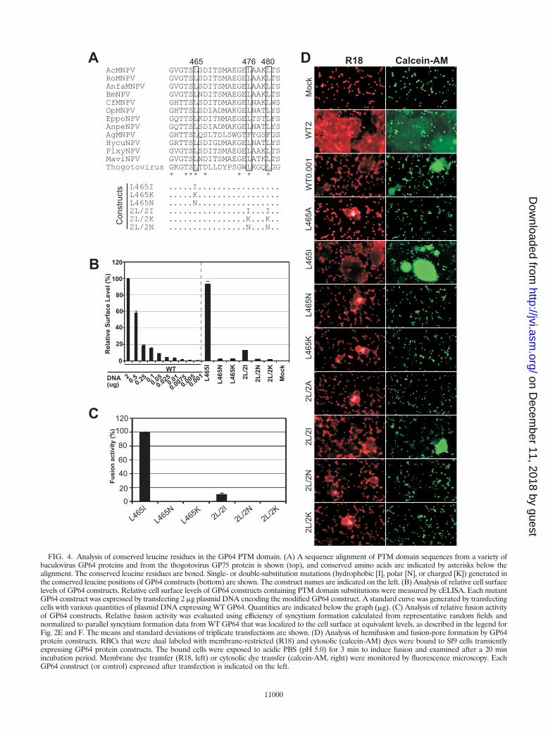
FIG. 4. Analysis of conserved leucine residues in the GP64 PTM domain. (A) A sequence alignment of PTM domain sequences from a variety ofbaculovirus GP64 proteins and from the thogotovirus GP75 protein is shown (top), and conserved amino acids are indicated by asterisks below thealignment. The conserved leucine residues are boxed. Single- or double-substitution mutations (hydrophobic [I], polar [N], or charged [K]) generated inthe conserved leucine positions of GP64 constructs (bottom) are shown. The construct names are indicated on the left. (B) Analysis of relative cell surfacelevels of GP64 constructs. Relative cell surface levels of GP64 constructs containing PTM domain substitutions were measured by cELISA. Each mutantGP64 construct was expressed by transfecting 2 �g plasmid DNA encoding the modified GP64 construct. A standard curve was generated by transfectingcells with various quantities of plasmid DNA expressing WT GP64. Quantities are indicated below the graph (�g). (C) Analysis of relative fusion activityof GP64 constructs. Relative fusion activity was evaluated using efficiency of syncytium formation calculated from representative random fields andnormalized to parallel syncytium formation data from WT GP64 that was localized to the cell surface at equivalent levels, as described in the legend forFig. 2E and F. The means and standard deviations of triplicate transfections are shown. (D) Analysis of hemifusion and fusion-pore formation by GP64protein constructs. RBCs that were dual labeled with membrane-restricted (R18) and cytosolic (calcein-AM) dyes were bound to Sf9 cells transientlyexpressing GP64 protein constructs. The bound cells were exposed to acidic PBS (pH 5.0) for 3 min to induce fusion and examined after a 20 minincubation period. Membrane dye transfer (R18, left) or cytosolic dye transfer (calcein-AM, right) were monitored by fluorescence microscopy. EachGP64 construct (or control) expressed after transfection is indicated on the left.
11000
on Decem
ber 11, 2018 by guesthttp://jvi.asm
.org/D
ownloaded from
2L/2A) resulted in no detectable membrane fusion (Fig. 2D),suggesting an important functional role for these residues. Todetermine if hydrophobicity at these positions was critical orwhether leucine residues specifically were required, we gener-ated both conservative and nonconservative substitutions atthese positions. Each leucine residue was substituted with iso-leucine, lysine, or asparagine (Fig. 4A). GP64 constructs con-taining these PTM modifications were transiently expressed inSf9 cells and examined by Western blot analysis of cell lysatesby the use of reducing and nonreducing conditions. The pro-files of the PTM domain-substituted GP64 constructs weresimilar to that of WT GP64, indicating that expression andtrimerization were not substantially affected by any of thesesubstitutions (see Fig. S1E in the supplemental material).Analysis of cell surface localization showed that substitution ofthe hydrophobic amino acid leucine by isoleucine (Fig. 4A,constructs L465I and 2L/2I) had the least effect on GP64 lo-calization to the cell surface. Surface levels for construct L465Iwere similar to those for wild-type GP64, and levels for con-struct 2L/2I were reduced to 13% of that for wild-type GP64.Substitutions of polar and charged residues for leucines at 465,476, and 480 (Fig. 4A, constructs L465N, 2L/2N, L465K, and2L/2K) had more substantial negative effects on surface local-ization, reducing surface levels of the latter constructs to lessthan 3% of that from wild-type GP64. In addition, all of theseGP64 constructs were detected at the cell surface by the mono-clonal antibody AcV1, suggesting that these constructs were inthe prefusion conformation (data not shown). Analysis ofmembrane fusion activity by GP64 constructs containing sub-stitutions in conserved leucines showed that fusion activity wassimilar to that of the wild type when the hydrophobic aminoacid isoleucine was substituted for L465, and fusion was com-pletely abolished when the same amino acid (L465) was sub-stituted with polar or charged amino acids (Fig. 4C, L465I,L465N, and L465K). These results were not caused by reducedcell surface levels since expression of WT GP64 at similarlevels resulted in readily detectable membrane fusion. Substi-tution of both leucines L476 and L480 with isoleucine had anegative effect on the fusion activity of GP64, but remainingfusion activity was estimated at approximately 10% of thatfrom wild-type GP64. As with the L465 position, substitutionof L476 and L480 by polar or charged amino acids resulted inno detectable fusion activity (Fig. 4C, 2L/2I versus 2L/2N and2L/2K). Overall, the results of substitutions in the conservedleucine residues suggest that the PTM domain of GP64 isessential for membrane fusion activity and that hydrophobicityat positions L465, L476, and L480 is critical.
Hemifusion and pore formation. To examine the step inmembrane fusion that was affected by substitutions of hydro-phobic amino acids of the PTM, we used membrane and cy-tosolic dyes to examine membrane merger and pore formation.Constructs with substitutions at positions L465, L476, andL480 were examined. Dual-labeled RBCs containing both theintegral membrane dye (R18) and a cytosolic dye (calcein AM)were bound to Sf9 cells that were previously transfected withplasmids encoding wild-type or modified GP64 constructs.Membrane fusion was initiated by exposure of the RBC-boundSf9 cells to low pH (pH 5.0) for 3 min. R18 redistribution fromRBCs to Sf9 cell membranes in the absence of calcein transferto the Sf9 cell cytosol indicates a merger of the outer mem-brane leaflets (hemifusion) and the absence of pore formation.All GP64 constructs with substitutions in the PTM domainwere observed to mediate lipid dye (R18) transfer from RBCsto Sf9 cells (Fig. 4D and data not shown). In addition, thoseconstructs positive for fusion also showed transfer of the cyto-solic dye, calcein AM (Fig. 4D, L465I and 2L/2I; see also Fig.S2 in the supplemental material). However, we did not observetransfer of the cytosolic dye calcein-AM by constructs L465A,L465N, L465K, 2L/2A, 2L/2N, and 2L/2K (Fig. 4D). Thus,although the latter constructs are capable of mediating theinitial membrane merger step (hemifusion), fusion pore for-mation did not appear to occur. Thus, these data suggest thatreduction of the hydrophobic character of positions L465,L476, and L480 resulted in GP64 constructs capable of medi-ating membrane merger but unable to form fusion pores.
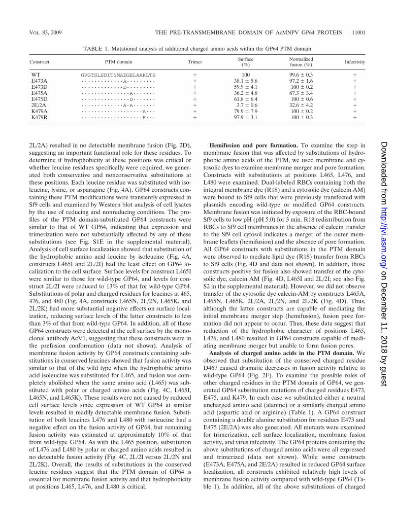
Analysis of charged amino acids in the PTM domain. Weobserved that substitution of the conserved charged residueD467 caused dramatic decreases in fusion activity relative towild-type GP64 (Fig. 2F). To examine the possible roles ofother charged residues in the PTM domain of GP64, we gen-erated GP64 substitution mutations of charged residues E473,E475, and K479. In each case we substituted either a neutraluncharged amino acid (alanine) or a similarly charged aminoacid (aspartic acid or arginine) (Table 1). A GP64 constructcontaining a double alanine substitution for residues E473 andE475 (2E/2A) was also generated. All mutants were examinedfor trimerization, cell surface localization, membrane fusionactivity, and virus infectivity. The GP64 proteins containing theabove substitutions of charged amino acids were all expressedand trimerized (data not shown). While some constructs(E473A, E475A, and 2E/2A) resulted in reduced GP64 surfacelocalization, all constructs exhibited relatively high levels ofmembrane fusion activity compared with wild-type GP64 (Ta-ble 1). In addition, all of the above substitutions of charged
TABLE 1. Mutational analysis of additional charged amino acids within the GP64 PTM domain
Construct PTM domain Trimer Surface(%)
Normalizedfusion (%) Infectivity
WT GVGTSLSDITSMAEGELAAKLTS � 100 99.6 0.3 �E473A ·············A········· � 38.1 5.6 97.2 1.6 �E473D ·············D········· � 59.9 4.1 100 0.2 �E475A ···············A······· � 36.2 4.8 87.3 3.4 �E475D ···············D······· � 61.8 6.4 100 0.6 �2E/2A ·············A·A······· � 3.7 0.6 32.6 4.2 �K479A ···················A··· � 79.9 7.9 100 0.2 �K479R ···················R··· � 97.9 3.1 100 0.3 �
VOL. 83, 2009 THE PRE-TRANSMEMBRANE DOMAIN OF AcMNPV GP64 PROTEIN 11001
on Decem
ber 11, 2018 by guesthttp://jvi.asm
.org/D
ownloaded from
amino acids resulted in GP64 proteins that were capable ofrescuing a gp64null AcMNPV bacmid in a transfection-infec-tion assay (Table 1). Thus, unlike the dramatic effects observedfrom substitution of hydrophobic residues, we observed rela-tively minor effects from substitution mutations in chargedresidues E473, E475, and K479, suggesting that these chargedresidues do not play a critical or essential role in membranefusion or other functions of GP64 in the infection cycle.
DISCUSSION
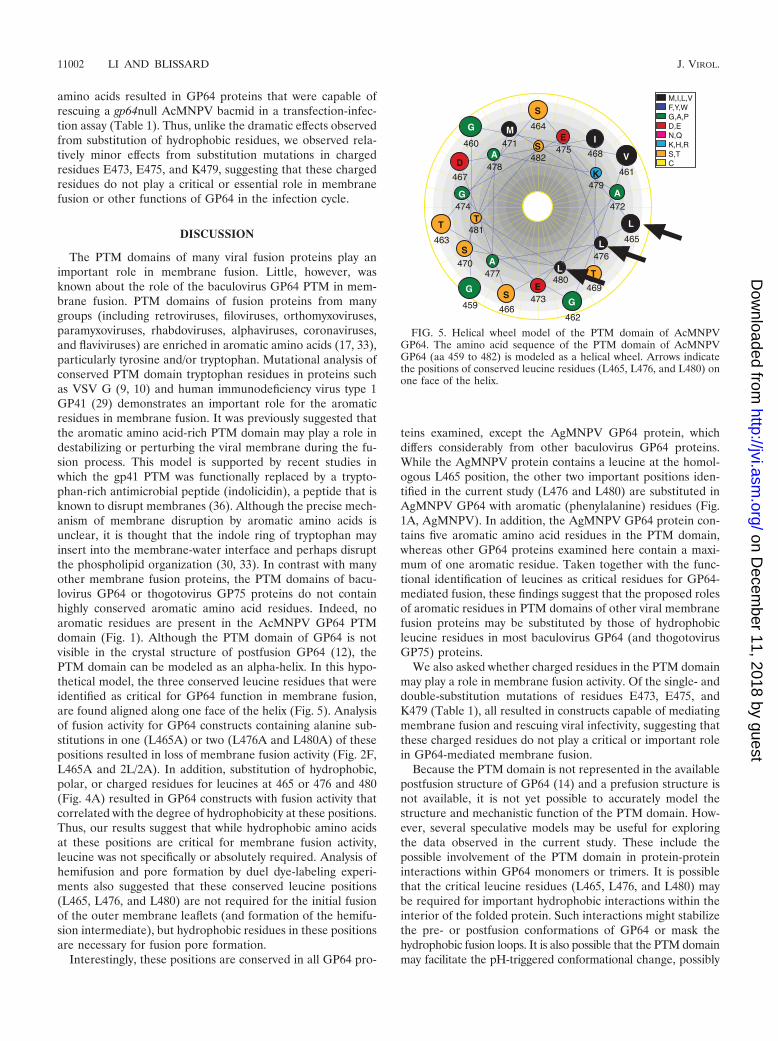
The PTM domains of many viral fusion proteins play animportant role in membrane fusion. Little, however, wasknown about the role of the baculovirus GP64 PTM in mem-brane fusion. PTM domains of fusion proteins from manygroups (including retroviruses, filoviruses, orthomyxoviruses,paramyxoviruses, rhabdoviruses, alphaviruses, coronaviruses,and flaviviruses) are enriched in aromatic amino acids (17, 33),particularly tyrosine and/or tryptophan. Mutational analysis ofconserved PTM domain tryptophan residues in proteins suchas VSV G (9, 10) and human immunodeficiency virus type 1GP41 (29) demonstrates an important role for the aromaticresidues in membrane fusion. It was previously suggested thatthe aromatic amino acid-rich PTM domain may play a role indestabilizing or perturbing the viral membrane during the fu-sion process. This model is supported by recent studies inwhich the gp41 PTM was functionally replaced by a trypto-phan-rich antimicrobial peptide (indolicidin), a peptide that isknown to disrupt membranes (36). Although the precise mech-anism of membrane disruption by aromatic amino acids isunclear, it is thought that the indole ring of tryptophan mayinsert into the membrane-water interface and perhaps disruptthe phospholipid organization (30, 33). In contrast with manyother membrane fusion proteins, the PTM domains of bacu-lovirus GP64 or thogotovirus GP75 proteins do not containhighly conserved aromatic amino acid residues. Indeed, noaromatic residues are present in the AcMNPV GP64 PTMdomain (Fig. 1). Although the PTM domain of GP64 is notvisible in the crystal structure of postfusion GP64 (12), thePTM domain can be modeled as an alpha-helix. In this hypo-thetical model, the three conserved leucine residues that wereidentified as critical for GP64 function in membrane fusion,are found aligned along one face of the helix (Fig. 5). Analysisof fusion activity for GP64 constructs containing alanine sub-stitutions in one (L465A) or two (L476A and L480A) of thesepositions resulted in loss of membrane fusion activity (Fig. 2F,L465A and 2L/2A). In addition, substitution of hydrophobic,polar, or charged residues for leucines at 465 or 476 and 480(Fig. 4A) resulted in GP64 constructs with fusion activity thatcorrelated with the degree of hydrophobicity at these positions.Thus, our results suggest that while hydrophobic amino acidsat these positions are critical for membrane fusion activity,leucine was not specifically or absolutely required. Analysis ofhemifusion and pore formation by duel dye-labeling experi-ments also suggested that these conserved leucine positions(L465, L476, and L480) are not required for the initial fusionof the outer membrane leaflets (and formation of the hemifu-sion intermediate), but hydrophobic residues in these positionsare necessary for fusion pore formation.
Interestingly, these positions are conserved in all GP64 pro-
teins examined, except the AgMNPV GP64 protein, whichdiffers considerably from other baculovirus GP64 proteins.While the AgMNPV protein contains a leucine at the homol-ogous L465 position, the other two important positions iden-tified in the current study (L476 and L480) are substituted inAgMNPV GP64 with aromatic (phenylalanine) residues (Fig.1A, AgMNPV). In addition, the AgMNPV GP64 protein con-tains five aromatic amino acid residues in the PTM domain,whereas other GP64 proteins examined here contain a maxi-mum of one aromatic residue. Taken together with the func-tional identification of leucines as critical residues for GP64-mediated fusion, these findings suggest that the proposed rolesof aromatic residues in PTM domains of other viral membranefusion proteins may be substituted by those of hydrophobicleucine residues in most baculovirus GP64 (and thogotovirusGP75) proteins.
We also asked whether charged residues in the PTM domainmay play a role in membrane fusion activity. Of the single- anddouble-substitution mutations of residues E473, E475, andK479 (Table 1), all resulted in constructs capable of mediatingmembrane fusion and rescuing viral infectivity, suggesting thatthese charged residues do not play a critical or important rolein GP64-mediated membrane fusion.
Because the PTM domain is not represented in the availablepostfusion structure of GP64 (14) and a prefusion structure isnot available, it is not yet possible to accurately model thestructure and mechanistic function of the PTM domain. How-ever, several speculative models may be useful for exploringthe data observed in the current study. These include thepossible involvement of the PTM domain in protein-proteininteractions within GP64 monomers or trimers. It is possiblethat the critical leucine residues (L465, L476, and L480) maybe required for important hydrophobic interactions within theinterior of the folded protein. Such interactions might stabilizethe pre- or postfusion conformations of GP64 or mask thehydrophobic fusion loops. It is also possible that the PTM domainmay facilitate the pH-triggered conformational change, possibly
FIG. 5. Helical wheel model of the PTM domain of AcMNPVGP64. The amino acid sequence of the PTM domain of AcMNPVGP64 (aa 459 to 482) is modeled as a helical wheel. Arrows indicatethe positions of conserved leucine residues (L465, L476, and L480) onone face of the helix.
11002 LI AND BLISSARD J. VIROL.
on Decem
ber 11, 2018 by guesthttp://jvi.asm
.org/D
ownloaded from
serving as a hinge-like region. Functional data provided in thecurrent study may argue against the latter role since substitu-tions of the three glycines, which would presumably providestructural flexibility, did not abrogate GP64-mediated mem-brane fusion. Most attractive is the possibility that the PTMdomain may serve as a membrane-disrupting domain that fa-cilitates membrane merger, as has been suggested for PTMdomains of other viral fusion proteins. The demonstrated im-portance of hydrophobic residues at positions L465, L476, andL480 is consistent with that role, and the evolutionary substi-tution of aromatic amino acids in two of those positions (po-sitions 476 and 480) provides further support for this model.
Substitutions that disrupt membrane fusion may disrupt oneor several of the steps involved in protein-mediated membranefusion. These include but are not limited to triggering of theconformational change, interaction of a fusion peptide with theadjacent membrane, apposition to bring the two membranesinto close contact, merger of the outer membrane leaflets toform a hemifusion intermediate, formation of a fusion pore bymerger of the inner membrane leaflets, and expansion of thefusion pore. As described above, our results suggest that thePTM may not be involved in the initial stage of outer mem-brane leaflet merger but instead may be required for initiatingor completing pore formation. Electrophysiological studies oftriggering and pore formation by the influenza virus HA pro-tein report a substantial delay (�30 s) after triggering, which isfollowed by an initially low conductance (suggesting formationof a small fusion pore) and rapid flickering as the fusion poreopens and closes rapidly (31, 32). In contrast, GP64 fusionpores open rapidly (�0.6 s) after low-pH triggering, with a highinitial conductance, and do not flicker (23, 24). It is possiblethat such differences in the opening and stabilization of thefusion pore may be related to differences in PTM domaininteractions within the membrane in the hemifusion interme-diate.
In prior studies, it was shown that BV budding is dramati-cally reduced in the absence of GP64 (21). A GP64 constructcontaining only 38 aa from the mature N terminus and 52 aafrom the C terminus (including only 22 aa of the PTM domain)of GP64 was capable of partially rescuing that budding defect(38). In the current study we extended the analysis of PTMdomain requirements for budding by examining the effects ofsingle and multiple amino acid substitutions in the PTM do-main of a full-length GP64 protein. Virus budding efficiencywas dramatically reduced (by 80 to 90%) when 6 of the 10single amino acid substitutions were introduced into the GP64PTM domain (Fig. 3C). The three constructs containing mul-tiple amino acid substitutions in the GP64 PTM domain alsoshowed dramatic reductions in budding efficiency. For many ofthese constructs, budding efficiency was near or only slightlyabove that of the GP64 knockout virus. Single amino acidsubstitutions that did not substantially affect budding includedsubstitutions G640A, S464A, S466A, and D467E, which hadonly slight or modestly reduced budding efficiency (reduced by10 to 50%) compared with that of WT GP64. In some cases,the effects of the PTM substitution on budding efficiency ap-peared to parallel cell surface expression levels, while in othercases, such as D467E, budding efficiency seemed to be inde-pendent of cell surface levels. We also examined the effect ofthe PTM on the incorporation of GP64 into virions by mea-
suring the ratio of GP64 to VP39 in virion preparations puri-fied from each construct. We observed no substantial effect onGP64 incorporation into virions in any of the single or multipleamino acid substitutions examined (Fig. 3D). Overall, thesedata indicate that the PTM domain is critically important forefficient virion budding. However, we did not identify any rolefor the PTM in regulating the incorporation of GP64 intovirions.
Because GP64 is involved in multiple functions during entryand egress, we also examined the effects of PTM domain sub-stitutions on overall infectivity. While membrane fusion assaysshowed that 11 of the 13 constructs retained some level offusion activity, only 6 of the 13 constructs were able to rescuevirus infectivity (Fig. 3A). Rescue of infectivity almost certainlydepends on a combination of GP64 roles in entry (binding andmembrane fusion) and egress (virion budding). Where fusionactivity was absent in GP64 constructs, no rescue was observed(Fig. 1, fusion versus infectivity). Rescue of infectivity generallycorrelated with virion budding efficiency, although in two casesin which budding efficiency was low (Fig. 3C, D467A andG474A), the constructs were able to rescue infectivity. In thosecases, however, virion production appeared to be somewhatreduced (Fig. 3A, rescued virus growth curves).
In summary, we found that the AcMNPV GP64 PTM do-main serves a critical role in GP64-mediated membrane fusion.We identified three critical leucine residues (L465, L476, andL480) that abolished GP64 fusion activity when substitutedeither alone (L465) or in combination (L476 and L480). Mu-tations in the PTM domain did not substantially affect GP64expression, trimerization, or the ability of GP64 to incorporateinto BV. However, the PTM was important for GP64 traffick-ing to the plasma membrane, and conserved amino acids in thePTM domain were important for virion budding and virusinfectivity. Because of its close proximity to the viral envelope,the importance of the PTM domain in membrane fusion islikely related to interactions of the PTM domain with the lipidbilayer of the viral envelope, although we cannot exclude otherpossibly important interactions of the PTM domain. Such in-teractions may stabilize the pre- or postfusion structure, andinteractions within the GP64 protein or between GP64 andother proteins may also be important during virion budding. Infuture studies it will be important to examine the intra- andintermolecular interactions that mediate these fundamentalprocesses. Such studies will enhance our understanding of themolecular mechanisms of membrane fusion during virus entryand will also contribute to an understanding of the complexnature of virion budding, the final step in virus assembly.
ACKNOWLEDGMENTS
We thank Gerrit Heetderks for technical assistance.This work was supported by NIH grant AI33657 and BTI project
B00103-R06-1255.
REFERENCES
1. Backovic, M., and T. S. Jardetzky. 2009. Class III viral membrane fusionproteins. Curr. Opin. Struct. Biol. 19:189–196.
2. Blissard, G. W., and G. F. Rohrmann. 1991. Baculovirus gp64 gene expres-sion: analysis of sequences modulating early transcription and transactiva-tion by IE1. J. Virol. 65:5820–5827.
3. Blissard, G. W., and J. R. Wenz. 1992. Baculovirus GP64 envelope glyco-protein is sufficient to mediate pH-dependent membrane fusion. J. Virol.66:6829–6835.
VOL. 83, 2009 THE PRE-TRANSMEMBRANE DOMAIN OF AcMNPV GP64 PROTEIN 11003
on Decem
ber 11, 2018 by guesthttp://jvi.asm
.org/D
ownloaded from
4. Garg, H., F. J. Fuller, and W. A. Tompkins. 2004. Mechanism of felineimmunodeficiency virus envelope glycoprotein-mediated fusion. Virology321:274–286.
5. Harrison, S. C. 2008. Viral membrane fusion. Nat. Struct. Mol. Biol. 15:690–698.
6. Hefferon, K., A. Oomens, S. Monsma, C. Finnerty, and G. Blissard. 1999.Host cell receptor binding by baculovirus GP64 and kinetics of virion entry.Virology 258:455–468.
7. Hink, W. F. 1970. Established insect cell line from the cabbage looper,Trichoplusia ni. Nature 226:466–467.
8. Howard, M. W., E. A. Travanty, S. A. Jeffers, M. K. Smith, S. T. Wennier,L. B. Thackray, and K. V. Holmes. 2008. Aromatic amino acids in thejuxtamembrane domain of severe acute respiratory syndrome coronavirusspike glycoprotein are important for receptor-dependent virus entry andcell-cell fusion. J. Virol. 82:2883–2894.
9. Jeetendra, E., K. Ghosh, D. Odell, J. Li, H. P. Ghosh, and M. A. Whitt. 2003.The membrane-proximal region of vesicular stomatitis virus glycoprotein Gectodomain is critical for fusion and virus infectivity. J. Virol. 77:12807–12818.
10. Jeetendra, E., C. S. Robison, L. M. Albritton, and M. A. Whitt. 2002. Themembrane-proximal domain of vesicular stomatitis virus G protein functionsas a membrane fusion potentiator and can induce hemifusion. J. Virol.76:12300–12311.
11. Jehle, J. A., G. W. Blissard, B. C. Bonning, J. S. Cory, E. A. Herniou, G. F.Rohrmann, D. A. Theilmann, S. M. Thiem, and J. M. Vlak. 2006. On theclassification and nomenclature of baculoviruses: a proposal for revision.Arch. Virol. 151:1257–1266.
12. Kadlec, J., S. Loureiro, N. G. Abrescia, D. I. Stuart, and I. M. Jones. 2008.The postfusion structure of baculovirus gp64 supports a unified view of viralfusion machines. Nat. Struct. Mol. Biol. 15:1024–1030.
13. Leikina, E., H. O. Onaran, and J. Zimmerberg. 1992. Acidic pH inducesfusion of cells infected with baculovirus to form syncytia. FEBS Lett. 304:221–224.
14. Li, Z., and G. W. Blissard. 2008. Functional analysis of the transmembrane(TM) domain of the Autographa californica multicapsid nucleopolyhedrovi-rus GP64 protein: substitution of heterologous TM domains. J. Virol. 82:3329–3341.
15. Li, Z., and G. W. Blissard. 2009. The Autographa californica multicapsidnucleopolyhedrovirus GP64 protein: analysis of transmembrane domainlength and sequence requirements. J. Virol. 83:4447–4461.
16. Long, G., X. Pan, R. Kormelink, and J. M. Vlak. 2006. Functional entry ofbaculovirus into insect and mammalian cells is dependent on clathrin-medi-ated endocytosis. J. Virol. 80:8830–8833.
17. Lorizate, M., N. Huarte, A. Saez-Cirion, and J. L. Nieva. 2008. Interfacialpre-transmembrane domains in viral proteins promoting membrane fusionand fission. Biochim. Biophys. Acta 1778:1624–1639.
18. Luckow, V. A., S. C. Lee, G. F. Barry, and P. O. Olins. 1993. Efficientgeneration of infectious recombinant baculoviruses by site-specific transpo-son-mediated insertion of foreign genes into a baculovirus genome propa-gated in Escherichia coli. J. Virol. 67:4566–4579.
19. Lung, O., M. Westenberg, J. M. Vlak, D. Zuidema, and G. W. Blissard. 2002.Pseudotyping Autographa californica multicapsid nucleopolyhedrovirus(AcMNPV): F proteins from group II NPVs are functionally analogous toAcMNPV GP64. J. Virol. 76:5729–5736.
20. Monsma, S. A., A. G. P. Oomens, and G. W. Blissard. 1996. The GP64envelope fusion protein is an essential baculovirus protein required forcell-to-cell transmission of infection. J. Virol. 70:4607–4616.
21. Oomens, A. G. P., and G. W. Blissard. 1999. Requirement for GP64 to driveefficient budding of Autographa californica multicapsid nucleopolyhedrovi-rus. Virology 254:297–314.
22. Oomens, A. G. P., S. A. Monsma, and G. W. Blissard. 1995. The baculovirusGP64 envelope fusion protein: synthesis, oligomerization, and processing.Virology 209:592–603.
23. Plonsky, I., M. S. Cho, A. G. P. Oomens, G. W. Blissard, and J. Zimmerberg.1999. An analysis of the role of the target membrane on the gp64-inducedfusion pore. Virology 253:65–76.
24. Plonsky, I., and J. Zimmerberg. 1996. The initial fusion pore induced bybaculovirus GP64 is large and forms quickly. J. Cell Biol. 135:1831–1839.
25. Robison, C. S., and M. A. Whitt. 2000. The membrane-proximal stem regionof vesicular stomatitis virus G protein confers efficient virus assembly. J. Vi-rol. 74:2239–2246.
26. Saez-Cirion, A., M. J. Gomara, A. Agirre, and J. L. Nieva. 2003. Pre-trans-membrane sequence of Ebola glycoprotein. Interfacial hydrophobicity dis-tribution and interaction with membranes. FEBS Lett. 533:47–53.
27. Sainz, B., Jr., E. C. Mossel, W. R. Gallaher, W. C. Wimley, C. J. Peters, R. B.Wilson, and R. F. Garry. 2006. Inhibition of severe acute respiratory syn-drome-associated coronavirus (SARS-CoV) infectivity by peptides analo-gous to the viral spike protein. Virus Res. 120:146–155.
28. Sainz, B., Jr., J. M. Rausch, W. R. Gallaher, R. F. Garry, and W. C. Wimley.2005. The aromatic domain of the coronavirus class I viral fusion proteininduces membrane permeabilization: putative role during viral entry. Bio-chemistry 44:947–958.
29. Salzwedel, K., J. T. West, and E. Hunter. 1999. A conserved tryptophan-richmotif in the membrane-proximal region of the human immunodeficiencyvirus type 1 gp41 ectodomain is important for Env-mediated fusion and virusinfectivity. J. Virol. 73:2469–2480.
30. Schibli, D. J., R. C. Montelaro, and H. J. Vogel. 2001. The membrane-proximal tryptophan-rich region of the HIV glycoprotein, gp41, forms awell-defined helix in dodecylphosphocholine micelles. Biochemistry40:9570–9578.
31. Spruce, A. E., A. Iwata, and W. Almers. 1991. The first milliseconds of thepore formed by a fusogenic viral envelope protein during membrane fusion.Proc. Natl. Acad. Sci. USA 88:3623–3627.
32. Spruce, A. E., A. Iwata, J. M. White, and W. Almers. 1989. Patch clampstudies of single cell-fusion events mediated by a viral fusion protein. Nature342:555–558.
33. Suarez, T., W. R. Gallaher, A. Agirre, F. M. Goni, and J. L. Nieva. 2000.Membrane interface-interacting sequences within the ectodomain of thehuman immunodeficiency virus type 1 envelope glycoprotein: putative roleduring viral fusion. J. Virol. 74:8038–8047.
34. Theilmann, D. A., G. W. Blissard, B. Bonning, J. Jehle, D. R. O’Reilly, G. F.Rohrmann, S. Thiem, and J. M. Vlak. 2005. Baculoviridae, p. 177–185. InH. V. Van Regenmortel, D. H. L. Bishop, M. H. Van Regenmortel, andC. M. Fauquet (ed.), Virus taxonomy: eighth report of the internationalcommittee on taxonomy of viruses. Elsevier Academic Press, New York, NY.
35. Tong, S., F. Yi, A. Martin, Q. Yao, M. Li, and R. W. Compans. 2001. Threemembrane-proximal amino acids in the human parainfluenza type 2 (HPIV2) F protein are critical for fusogenic activity. Virology 280:52–61.
36. Vishwanathan, S. A., and E. Hunter. 2008. Importance of the membrane-perturbing properties of the membrane-proximal external region of humanimmunodeficiency virus type 1 gp41 to viral fusion. J. Virol. 82:5118–5126.
37. Volkman, L. E. 1986. The 64K envelope protein of budded Autographacalifornica nuclear polyhedrosis virus. Curr. Top. Microbiol. Immunol. 131:103–118.
38. Zhou, J., and G. W. Blissard. 2008. Display of heterologous proteins ongp64null baculovirus virions and enhanced budding mediated by a vesicularstomatitis virus G-stem construct. J. Virol. 82:1368–1377.
39. Zhou, J., and G. W. Blissard. 2008. Identification of a GP64 subdomaininvolved in receptor binding by budded virions of the baculovirus Auto-graphica californica multicapsid nucleopolyhedrovirus. J. Virol. 82:4449–4460.
40. Zhou, J., and G. W. Blissard. 2006. Mapping the conformational epitope ofa neutralizing antibody (AcV1) directed against the AcMNPV GP64 protein.Virology 352:427–437.
11004 LI AND BLISSARD J. VIROL.
on Decem
ber 11, 2018 by guesthttp://jvi.asm
.org/D
ownloaded from