Embed Size (px)
Citation preview
Instructions for use
TitleTHE PRIMARY AND AUXILIARY AMBROSIA FUNGI ISOLATED FROM THE AMBROSIA BEETLES,SCOLYTOPLATYPUS SHOGUN BLANDFORD (COLEOPTERA SCOLYTIDAE) AND CROSSOTARSUSNIPONICUS BLANDFORD(COLEOPTERA PLATYPODIDAE)
Author(s) NAKASHIMA, Toshio; GOTO, Chie; IIZUKA, Toshihiko
Citation Journal of the Faculty of Agriculture, Hokkaido University, 63(2), 185-208
Issue Date 1987-03
Doc URL http://hdl.handle.net/2115/13057
Type bulletin (article)
File Information 63(2)_p185-208.pdf
Hokkaido University Collection of Scholarly and Academic Papers : HUSCAP
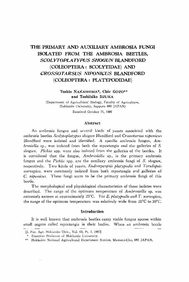
THE PRIMARY AND AUXILIARY AMBROSIA FUNGI ISOLATED FROM THE AMBROSIA BEETLES, SCOLYTOPLA TYPUS SlfOaUN BLANDFORD
(COLEOPTERA: SCOLYTIDAE) AND CROSSOTARSUS N1PON1CUS BLANDFORD
(COLEOPTERA: PLATYPODIDAE)
Toshio NAKASHIMA *, Chie GOTO** and Toshihiko IIZUKA
(Department of Agricultural Biology, Faculty of Agriculture, Hokkaido University, Sapporo 060 JAPAN)
Received October 21, 1986
Abstract
An ambrosia fungus and several kinds of yeasts associated with the ambrosia beetles Scolytoplatypus shogun Blandford and Crossotarsus niponicus Blandford were isolated and identified. A specific ambrosia fungus, Ambrosiella sp., was isolated from both the mycetangia and the galleries of S. shogun. Pichia spp. were also isolated from the galleries of the beetles. It is considered that the fungus, Ambrosiella sp., is the primary ambrosia fungus and the Pichia spp. are the auxiliary ambrosia fungi of S. shogun, respectively. Two kinds of yeasts, Endomycopsis platypodis and Torulopsis norvegica. were commonly isolated from both mycetangia and galleries of C. niponicus. These fungi seem to be the primary ambrosia fungi of this beetle.
The morphological and physiological characteristics of these isolates were described. The range of the optimum temperature of Ambrosiella sp. was extremely narrow at approximately 23°C. For E. platypodis and T. norvegica, the range of the optimum temperature was relatively wide from 20°C to 30°C.
Introduction
It is well known that ambrosia beetles carry viable fungus spores within small organs called mycetangia in their bodies. When an ambrosia beetle
[J. Fac. Agr. Hokkaido Univ., Vol. 63, Pt. 2. 1987] * Emeritus Professor of Hokkaido University.
** Hokkaido National Agricultural Experiment Station, Memuro-Cho, 082 JAPAN.
186 T. NAKASHIMA, C. GOTO AND T. IIZUKA
tunnels into wood, these viable spores are dislodged from the mycetangia, and a mass of velvety fungus soon lines the interior of the tunnel. Until the present time, many species of fungi, more than 30 genera, have been reported as the fungi associated with ambrosia beetles. Only few species of these fungi, however, have been demonstrated as the true mutualistic symbiont or ambrosia fungi of these beetles.
In this paper, we have reported that one species of ambrosia fungus and several kinds of species of yeasts were isolated from mycetangia and galleries of Scolytoplatypus shogun, and several kinds of yeasts were isolated from the mycetangia and galleries of Crossotarsus niponicus.
The morphological and physiological characteristics of each species of these isolates have been described and identified.
Materials and Methods
Collection of beech logs infested with the beetles.
The ambrosia beetles used in the present study were Scolytoplatypus shogun as Scolytidae and Crossotarsus niponicus as Platypodidae.
Beech logs (Fagw crenata Blume) infested with the beetles were collected at the Hiyama Forest Experiment Station of Hokkaido University, Kaminokuni, Hiyama in September 1980 and July 1981, and immediately carried to the campus of the Faculty of Agriculture, Hokkaido University in Sapporo. The logs collected in the fall were overwintered under snow on the campus and were used in the next spring.
Isolation of microorganisms from galleries of the beetles.
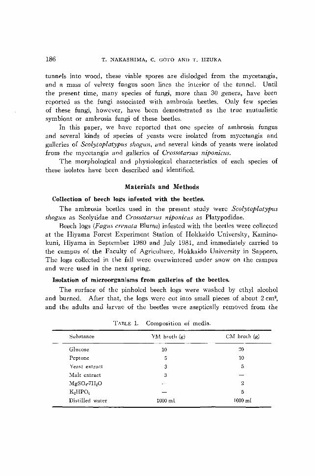
The surface of the pinholed beech logs were washed by ethyl alcohol and burned. After that, the logs were cut into small pieces of about 2 cm3,
and the adults and larvae of the beetles were aseptically removed from the
TABLE l. Composition of media.
Substance YM broth (g) eM broth (g)
Glucose 10 20
Peptone 5 10
Yeast extract 3 5
Malt extract 3
MgS04-7H2O 2
K2HP04 5
Distilled water 1000 ml 1000 ml
AMBROSIA FUNGI ISOLATED FROM AMBROSIA BEETLES 187
galleries. The inner surface of each gallery lined with velvety fungi was washed with TM and CM broth for culture. Components of both broth are shown on Table 1.
Collection of the adults.
In the overwintering period, the adults of S. shogun were collected from pinholed beech logs. In the flying period, the new adults of each species were collected within IS hours after appearance.
Culture conditions for isvlates.
i). The galleries were washed by YM and CM broth. After that, each broth was basically incubated by shaking culture at 25°C for five days, and then the cultures were incubated on YM and CM agar plates.
ii). Velvety fungus from the surface of the galleries was directly inoculated on both agar plates.
iii). The surface of each gallery was washed by sterile distilled water and drop of that water was incubated on both agar plates.
Isolation of microorganisms from the mYcetangia of beetles.
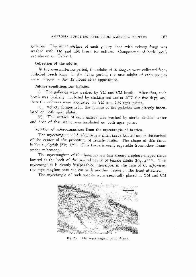
The mycetangium of S. shogun is a small tissue located under the surface of the center of the pronotum of female adults. The shape of this tissue is like a jellyfish (Fig. 1)8,9). This tissue is easily separable from other tissues under microscope.
The mycetangium of C. niponiclls is a bag around a sphere-shaped tissue located at the back of the preoral cavity of female adults (Fig. 2)6,7,9). This mycetangium is cleanly inseparabled, therefore, in the case of C. niponicu~,
the mycetangium was cut out with another tissues in the head attached. The mycetangia of each species were aseptically placed in YM and CM
Fig.!. The mycetangium of S. shogun.

188 T. NAKASHIMA, C. GOTO AND T. IIZUKA
Fig. 2. The mycetangium of C. niponiclls.
broth on slide glass and then were incubated on agar plates. Small amounts of the broth on slide glass were microscopically observed after incubation.
Isolation of microorganisms from the homogenates of larvae.
The larvae of each species were collected from pinholed beech logs onto cheese cloth and washed twice with distilled water. Each larva was homogenized gently in 1 ml of YM broth with sterile teflon homogenizer. The homogenates were spread over YM agar plates and incubated at 2.5°C up to form colonies.
Isolation of microorganisms from the digestive tracts of adult beetles in flight season.
The digestive tracts of new adults were dissected and incubated on YM agar plates.
Identification of the isolates.
Isolated yeasts were basically identified according to LODDERo), and ISO,
lated ambrosia fungi were identified according to BATRA2).
Scanning electron microscopy.
Scanning electron microscopy of the ambrosia fungi and the wall of the galleries of S. shogun was obtained by a 2% OS04 vapor fixation fol· lowed by coating with carbon and gold. Samples were observed by the scanning electron microscope Type JSM-SI.
Results
1. Fungi associated with Scolytoplatypus shogun.
Isolated fungi from mycetangia of adult beetles, homogenates of larvae,
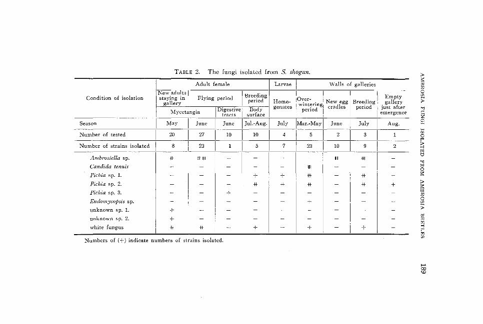
TABLE 2. The fungi isolated from S. shogun.
Adult female Larvae Walls of galleries
New.adu~ts I Flying period I Breeding Over-Condition of isolation staymg m period Homo- New egg Breeding gallery
genates wintering cradles period Mycetangia 1 Digestive 1 Body period
tracts surface
Season May June June 1 J~l.-AUg·1 July IM~.-May 1 June July
Number of tested 20 27 10 1
10 1
4 1 --5-1 2 3
Number of strains isolated 8 23 1 1
5 1 7 1 23 1 10 9
Ambrosiella sp. ffiI ffiIffiI - - - - lIIII ffiI
Candida tenuis - - - - - lIIII - -
Pichia sp. 1. - - - + + ffiI - * Pichia sp. 2. - - - * -lit * - * Pichia sp. 3. - - + - - - - -
Endomycopsis sp. - - -I
- - + - -
unknown sp. 1. + - - - - - - -
unknown sp. 2. + - - - - - - -
white fungus * * -- + - + - +
Numbers of (+) indicate numbers of strains isolated.
Empty gallery
just after emergence
Aug.
1
2
-
-
-
+ -
-
-
-
--
!l> ~ to ::<l o Ul
> 'Tj
c: z Cl H
H (fJ
o t""
~ tlj t:I 'Tj ::<l o ~
!l> ~ to ::<l o (fJ
> to tlj
~ t"" tlj Ul
f-' (Xl <.0
190 T. NAKASHIMA, C. GOTO AND T. IIZUKA
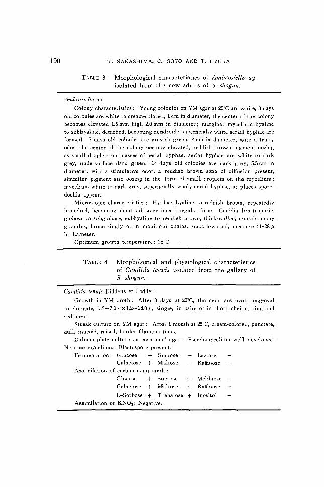
TABLE 3. Morphological characteristics of Ambrosiella sp. isolated from the new adults of S. shogun.
Ambrosiella sp.
Colony characteristics: Young colonies on YM agar at 25°C are white, 3 days
old colonies are white to cream-colored, 1 cm in diameter, the center of the colony
becomes elevated 1.5 mm high 2.0 mm in diameter; marginal mycelium hyaline
to sub hyaline, detached, becoming dendroid; superficially white aerial hyphae are
formed. 7 days old colonies are greyish green, 4 cm in diameter, with a fruity
odor, the center of the colony become elevated, reddish brown pigment oozing
as small droplets on masses of aerial hyphae, aerial hyphae are white to dark
grey, undersurface dark green. 14 days old colonies are dark grey, 5.5 cm in
diameter, with a stimulative odor, a reddish brown zone of diffusion present,
simmilar pigment also oozing in the form of small droplets on the mycelium;
mycelium white to dark grey, superficially wooly aerial hyphae, at places sporo
dochia appear.
Microscopic characteristics: Hyphae hyaline to reddish brown, repeatedly
branched, becoming dendroid sometimes irregular form. Conidia brastosporic,
globose to subglobose, subhyaline to reddish brown, thick-walled, contain many
granules, brone singly or in monilioid chains, smootb-walled, measure 11-28 fl
in diameter.
Optimum growth temperature: 23°C.
TABLE 4. Morphological and physiological characteristics of Candida tenuis isolated from the gallery of S. shogun.
Candida tenuis Diddens et Lodder
Growth in YM broth: After 3 days at 25°C, the cells are oval, long-oval
to elongate, 1.2-7.0 fl X 1.2-18.0 fl, single, in pairs or in short chains, ring and
sediment.
Streak culture on YM agar: After 1 month at 25°C, cream-colored, punctate,
dull, mucoid, raised, border filamentations.
Dalmau plate culture on corn-meal agar: Pseudomycelium well developed.
No true mycelium. Blastospore present.
Fermentation: Glucose + Sucrose
Galactose -1:- Maltose
Assimilation of carbon compounds:
Glucose + Sucrose
Galactose + Maltose
L-Sorbose + Trehalose
Assimilation of KN03 : Negative.
Lactose
Raffinose
+ Melibiose
+ Raffinose
+ Inositol
AMBROSIA FUNGI ISOLATED FROM AMBROSIA BEETLES
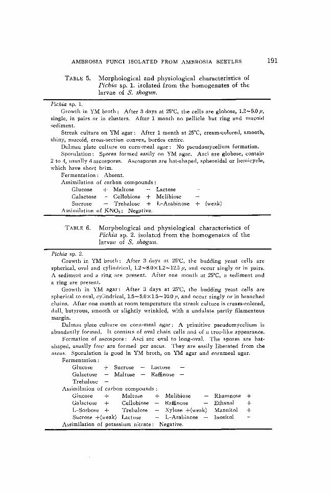
TABLE 5. Morphological and physiological characteristics of Pichia sp. 1. isolated from the homogenates of the larvae of S. shogun.
Pichia sp. 1. Growth in YM broth: After 3 days at 25°C, the cells are globose, 1.2-5.0 fl,
single, in pairs or in clusters. After 1 month no pellicle but ring and mucoid sediment.
Streak culture on YM agar: After 1 month at 25°C, cream-colored, smooth, shiny, mucoid, cross-section convex, bordes entire.
Dalmau plate culture on corn-meal agar: No pseudo mycelium formation. Sporulation: Spores formed easily on YM agar. Asci are globose, contain
2 to 4, usually 4 ascospores. Ascospores are hat-shaped, spheroidal or hemicycle, which have short brim.
Fermentation: Absent. Assimilation of carbon compounds:
Glucose + Maltose Galactose Cellobiose +
Lactose Melibiose
Sucrose Trehalose + L-Arabinose + (weak) Assimilation of KN03 : Negative.
TABLE 6. Morphological and physiological characteristics of Pichia sp. 2. isolated from the homogenates of the larvae of S. shogun.
Pichia sp. 2. Growth in YM broth: After 3 days at 25°C, the budding yeast cells are
spherical, oval and cylindrical, 1.2-S.0x1.2-12.5 fl, and occur singly or in pairs. A sediment and a ring are present. After one month at 25°C, a sediment and a ring are present.
Growth in YM agar: After 3 days at 25°C, the budding yeast cells are spherical to oval, cylindrical, 1.5-5.0x1.5-10.0 fl, and occur singly or in branched chains. After one month at room temperature the streak culture is cream-colored, dull, butyrous, smooth or slightly wrinkled, with a undulate partly filamentous margin.
Dalmau plate culture on corn-meal agar: A primitive pseudo mycelium is abundantly formed. It consists of oval chain cells and of a tree-like appearance.
Formation of ascospore: Asci are oval to long-oval. The spores are hatshaped, usually four are formed per ascus. They are easily liberated from the ascus. Sporulation is good in YM broth, on YM agar and cornmeal agar.
Fermentation: Glucose + Galactose Trehalose
Sucrose Maltose
Lactose Raffinose
Assimilation of carbon compounds: Glucose + Maltose + Melibiose Galactose + Cellobiose Raffinose L-Sorbose + Trehalose Xylose +(weak) Sucrose +(weak) Lactose L-Arabinose
Assimilation of potassium nitrate: Negative.
Rhamnose + Ethanol + Mannitol + Inositol
191
192 T. NAKASHIMA, C. GOTO AND T. IIZUKA
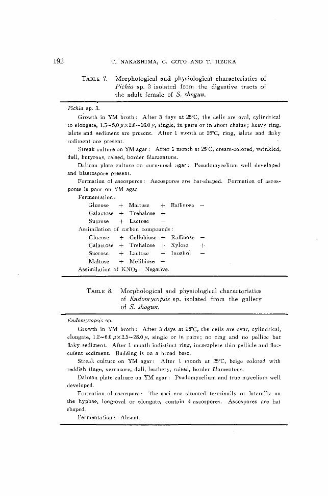
TABLE 7. Morphological and physiological characteristics of Pichia sp. 3 isolated from the digestive tracts of the adult female of S. shogun.
Pichia sp. 3.
Growth in YM broth: After 3 days at 25°C, the cells are oval, cylindrical
to elongate, 1.5-5.0 flx2.0-16.0 fl, single, in pairs or in short chains; heavy ring,
islets and sediment are present. After 1 month at 25°C, ring, islets and flaky
sediment are present.
Streak culture on YM agar: After 1 month at 25°C, cream-colored, wrinkled,
dull, butyrous, raised, border filamentous.
Dalmau plate culture on corn-meal agar: Pseudo mycelium well developed
and blastospore present.
Formation of ascospores: Ascospores are hat-shaped. Formation of ascos
pores is poor on YM agar.
Fermentation:
Glucose + Maltose + Raffinose
Galactose + Trehalose + Sucrose + Lactose
Assimilation of carbon compounds:
Glucose + Cellobiose + Raffinose
Galactose + Trehalose + Xylose + Sucrose + Lactose Inositol
Maltose + Melibiose
Assimilation of KN03 : Negative.
TABLE 8. Morphological and physiological characteristics of Endomycopsis sp. isolated from the gallery of S. shogun.
Endomycopsis sp.
Growth in YM broth: After 3 days at 25°C, the cells are ovar, cylindrical,
elongate, 1.2-6.0 fl X 2.5- 28.0 fl, single or in pairs; no ring and no pellice but
flaky sediment. After 1 month indistinct ring, incomplete thin pellicle and floc
culent sediment. Budding is on a broad base.
Streak culture on YM agar: After 1 month at 25°C, beige colored with
reddish tinge, verrucose, dull, leathery, raised, border filamentous.
Dalmau plate culture on YM agar: Psudomycelium and true mycelium well
developed.
Formation of ascospore: The asci are situated terminally or laterally on
the hyphae, long-oval or elongate, contain 4 ascospores. Ascospores are hat
shaped.
Fermentation: Absent.
AMBROSIA FUNGI ISOLATED FROM AMBROSIA BEETLES 193
and their galleries of S. shogun are shown in Table 2. Morphological and physiological characteristics of these isolates were investigated and identified as shown from Table 3 to Table S. Morphological characteristics are also indicated from Fig. 3 to Fig. 10.
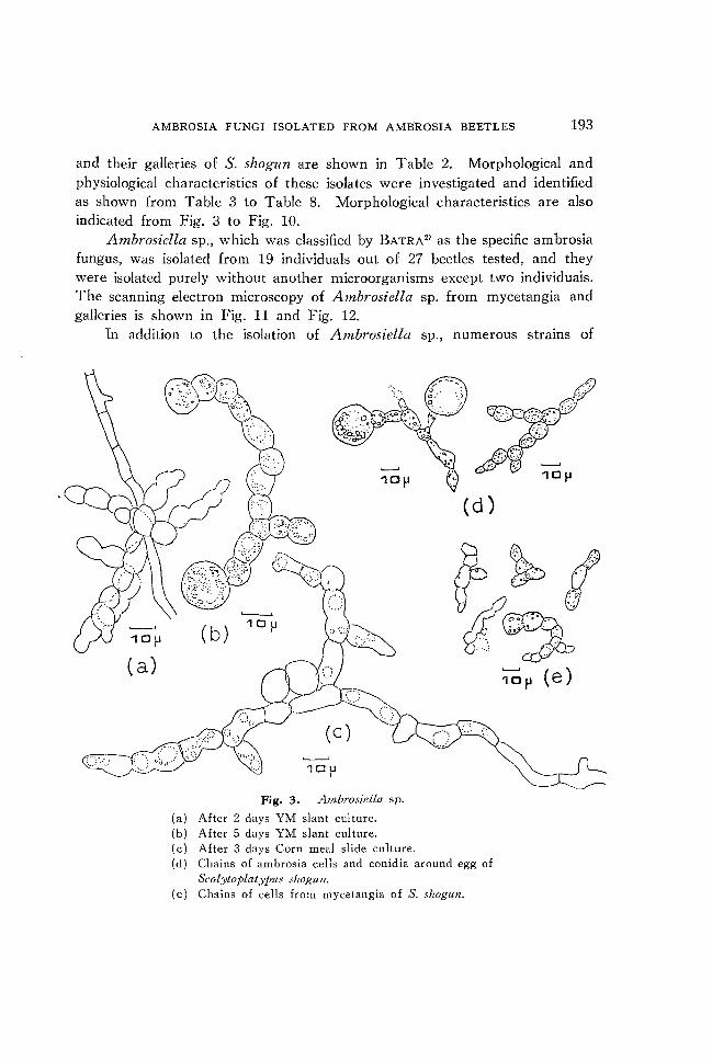
Ambrosiella sp., which was classified by BATRA2) as the specific ambrosia fungus, was isolated from 19 individuals out of 27 beetles tested, and they were isolated purely without another microorganisms except two individuais. The scanning electron microscopy of Ambrosiella sp. from mycetangia and galleries is shown in Fig. 11 and Fig. 12.
Tn addition to the isolation of Ambrosiella sp., numerous strains of
( a)
10p
Fig. 3. Ambrosiella sp.
(a) After 2 days YM slant culture. (b) After 5 days YM slant culture. (c) After 3 days Corn meal slide culture.
(d)
(d) Chains of ambrosia cells and conidia around egg of Scolytoplatyj>us shogun.
(e) Chains of cells from mycetangia of S. shogun.
10p (e)
194 T. NAKASHIMA, C. GOTO AND T. IIZUKA
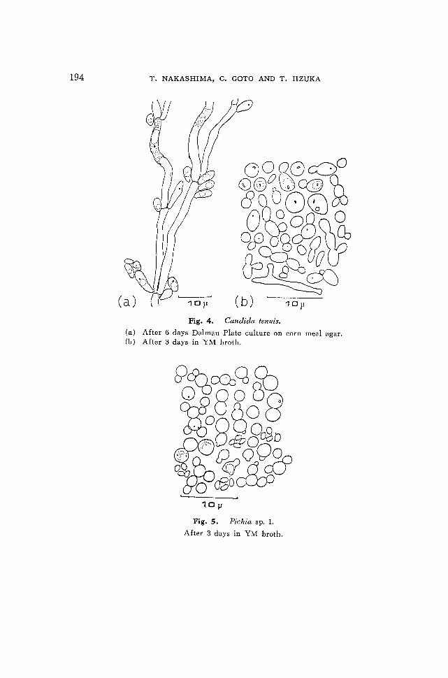
Fig. 4. Candida tenuis.
(a) After 6 days Dalmau Plate culture on corn meal agar. (b) After 3 days in YM broth.
10ll
Fig. 5. Pichia sp. 1.
After 3 days in YM broth.
AMBROSIA FUNGI ISOLATED FROM AMBROSIA BEETLES 195
(a) 10u
(c) 10u
(b)
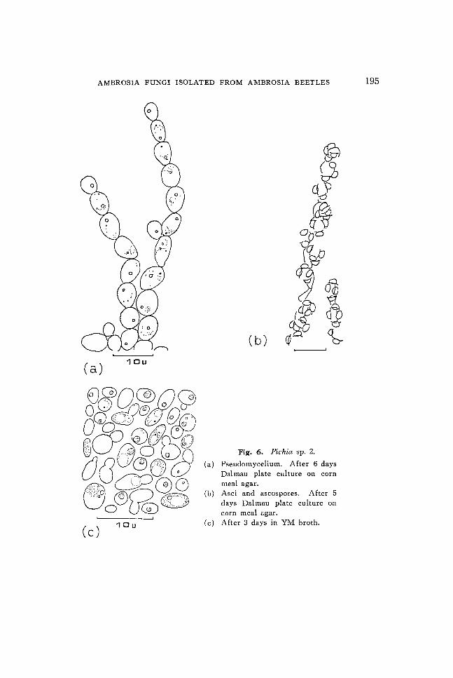
Fig. 6. Pichia sp. 2.
Pseudomycelium. After 6 days Dalmau plate culture on corn meal agar.
(b) Asci and ascospores. After 5 days Dalmau plate culture on corn meal agar.
(c) After 3 days in YM broth.
196 T. NAKASHIMA, C. GOTO AND T. IIZUKA
(a) 1°11
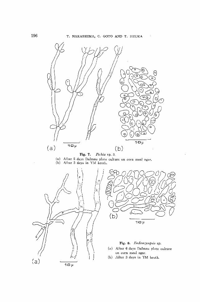
1°11 ( b) Fig. 7. Pichia sp. 3.
(a) After 5 days Dalmau plate culture on corn meal agar. (b) After 3 days in YM broth.
10fl
Fig. 8. Endomycopsis sp.
(a) After 6 days Dalmau plate culture on corn meal agar.
(b) After 3 days in YM broth.
AMBROSIA FUNGI ISOLATED FROM AMBROSIA BEETLES 197
10).l
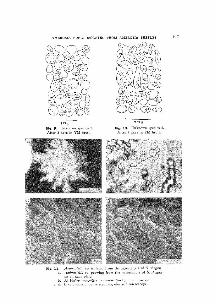
Fig. 9. Unknown species l. After 3 days in YM broth.
10).l
Fig. 10. Unknown species 2. After 3 days in YM broth.
Fig. 11. rlmbrosiella sp. isolated from the mycetangia of S. shogull. a. "lmbrosiella sp. growing from the mycetangia of S. shogun
on an agar plate. b. At higher magnification under the light microscope.
c. d. Like chains under a scanning electron microscope.
198 T. NAKASHIMA, C. GOTO AND T. IIZUKA
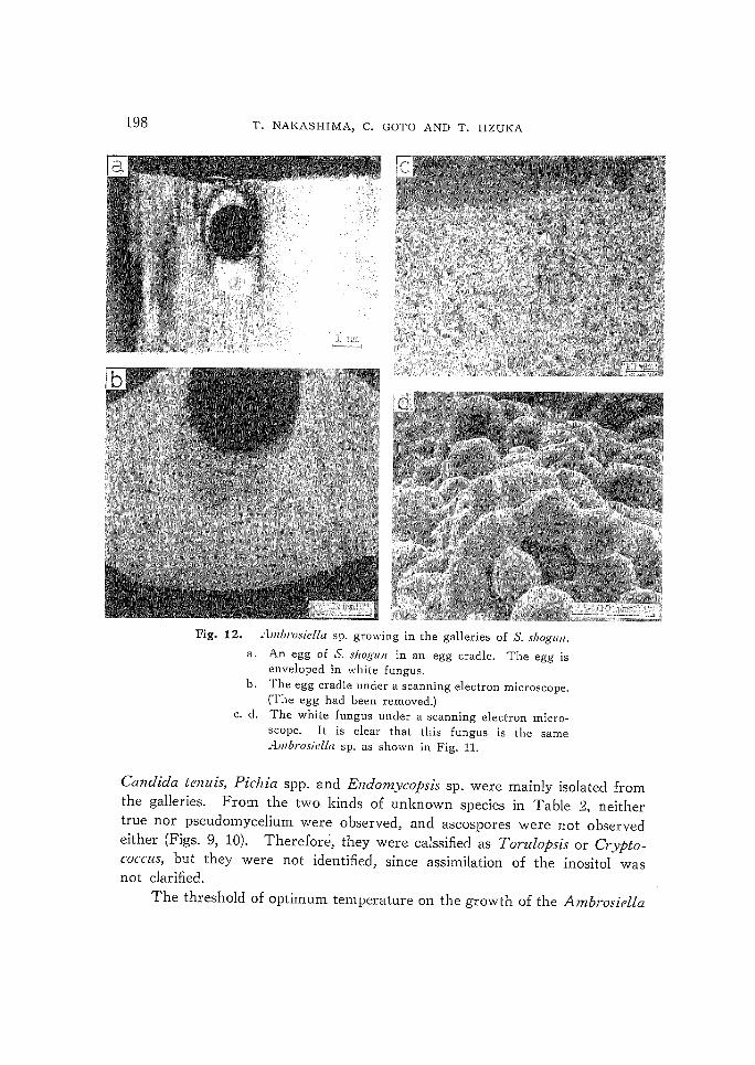
Fig. 12. .·lmbrosiella sp. growing in the galleries of S. shogun.
a. An egg of S. shogun in an egg cradle. The egg is enveloped in white fungus.
b. The egg cradle under a scanning electron microscope. (The egg had been removed.)
c. d. The white fungus under a scanning electron microscope. It is clear that this fungus is the same Alilbrosiella sp. as shown in Fig. 11.
Candida tenuis, Pic1zia spp. and Endomycopsis sp. were mainly isolated from the galleries. From the two kinds of unknown species in Table 2, neither true nor pseudomycelium were observed, and ascospores were not observed either (Figs. 9, 10). Therefore, they were calssified as Torulopsis or Cryptococcus, but they were not identified, since assimilation of the inositol was not clarified.
The threshold of optimum temperature on the growth of the Ambrosiella
1.0
o
o
AMBROSIA FUNGI ISOLATED FROM AMBROSIA BEETLES 199
23"C
11°C, 13°C, 16°C, 27°C
3UoC, 31°C, 35~C, 36°C
'1'1111': (bollr)
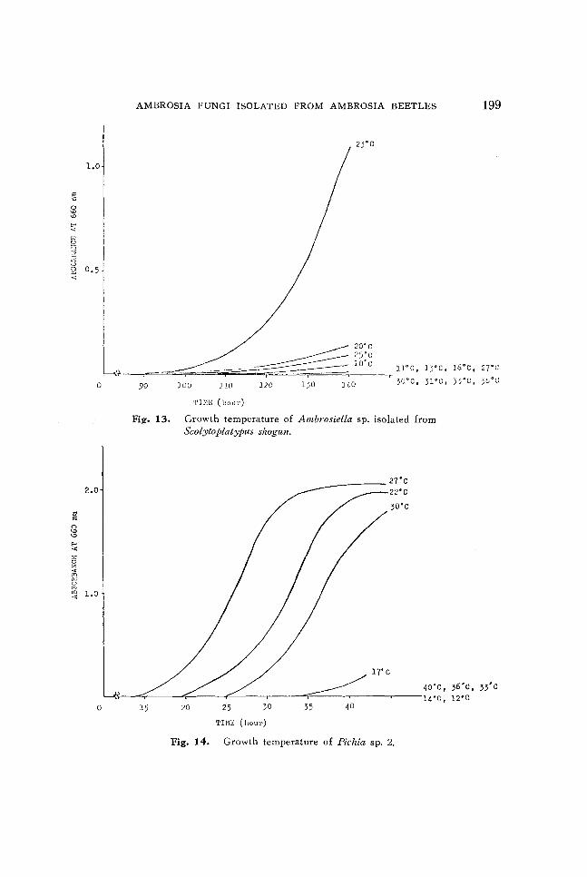
Fig. 13. Growth temperature of Ambrosiella sp. isolated from Scolytoplatyplls shogull.
40'C, 36'C, 33'C ~(?---~~----~------~-----,-------r=-----~------~----14'C, lZ'C
15 20 25 30 35 40
'1'1111<: (hour)
Fig. 14. Growth temperature of Pichia sp. 2.
200 T. NAKASHIMA, C. GOTO AND T. IIZUKA
sp. was almost 23°C, and showed extremely narrow range (Fig. 13). Tn the case of Pichia sp. 2, it was between 22°C and 30°C (Fig. 14).
2. Fungi associated with Crossotarsus niponicus.
The fungi isolated from the homogenates of the heads of adult beetles, homogenates of the larvae and their galleries of C. niponzcus are shown in
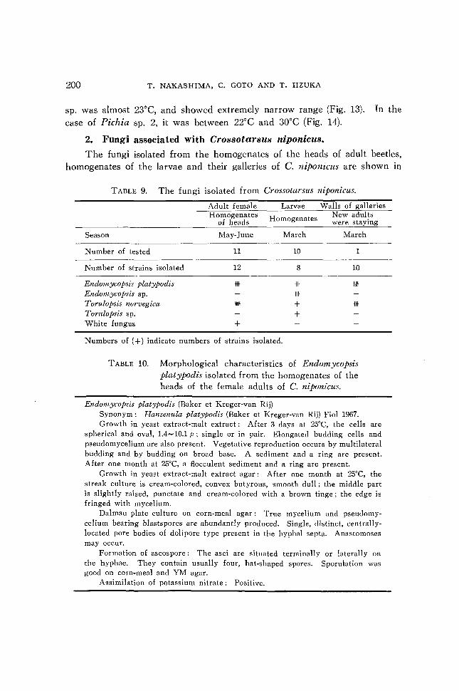
TABLE 9. The fungi isolated from Crossotarsus niponicus.
Adult female Larvae Walls of galleries Homogenates Homogenates New adults
of heads were staying
Season May-June March March
Number of tested 11 10 1
Number of strains isolated 12 8 10
Endomycopsis platypodis -Itt * lIIIf Endomycopsis sp. 1111
Torulopsis norvegica 1IIIIf + -Itt Torulopsis sp. + White fungus +
Numbers of (+) indicate numbers of strains isolated.
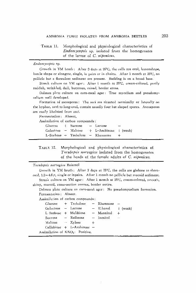
TABLE 10. Morphological characteristics of Endomycopsis platypodis isolated from the homogenates of the
heads of the female adults of C. niponicus.
Endomycopsis platypodis (Baker et Kreger-van Rij) Synonym: Hansenula platypodis (Baker et Kreger-van Rij) Fiol 1967. Growth in yeast extract-malt extract: After 3 days at 25°C, the cells are
spherical and oval, 1.4-10.1 f1; single or in pair. Elongated budding cells and pseudomycelium are also present. Vegetative reproduction occurs by multilateral budding and by budding on broad base. A sediment and a ring are present. After one month at 25°C, a flocculent sediment and a ring are present.
Growth in yeast extract-malt extract agar: After one month at 25°C, the streak culture is cream-colored, convex butyrous, smooth dull; the middle part is slightly raised, punctate and cream-colored with a brown tinge; the edge is fringed with mycelium.
Dalmau plate culture on corn-meal agar: True mycelium and pseudo mycelium bearing blasts pores are abundantly produced. Single, distinct, centrallylocated pore bodies of doli pore type present in the hyphal septa. Anastomoses may occur.
Formation of ascospore: The asci are situated terminally or laterally on the hyphae. They contain usually four, hat-shaped spores. Sporulation was good on corn-meal and YM agar.
Assimilation of potassium nitrate: Positive.
AMBROSIA FUNGI ISOLATED FROM AMBROSIA BEETLES 201
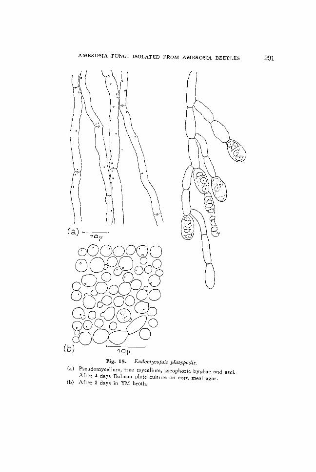
(a), 10u l
Fig. 15. Endomycopsis platypodis. (a) Pseudomycelium, true mycelium, ascophoric hyphae and asci.
After 4 days Dalmau plate culture on corn meal agar. (b) After 3 days in YM broth.
202 T. NAKASHIMA, C.
(a)
;;Jjr
:;rf; (b) , 10p ,
GOTO AND T. IIZUKA
~a8°M~, 8~08~8 ~-cJ2EJ~ ~o
(c) 10},
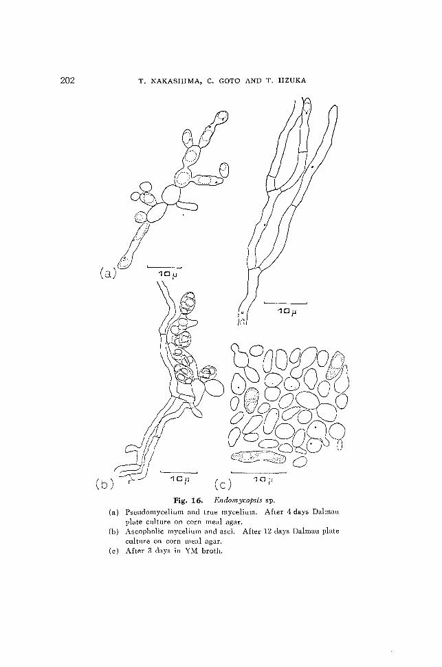
seudomyc l' . ' ndomyr:op . (a) p Fig. 16 E
1
e mm 'md SlS sp pate cu1tur ' true myce1' .
scopholic my 1.orn meal agar . ter 4 days Da1 (b) A e on c lum Af 1 ce mm d' mau
cu ture on c an asci Af (c) After 3 da o~n meal agar.' ter 12 days Da1m 1 ys III YM b au pate
roth.
AMBROSIA FUNGI ISOLATED FROM AMBROSIA BEETLES
TABLE 11. Morphological and physiological characteristics of Endomycopsis sp. isolated from the homogenates of the larvae of C. niponicus.
Endomycoj>sis sp.
Growth in YM broth: After 3 days at 25°C, the cells are oval, lemonshape,
bottle shape or elongate, single, in pairs or in chains. After 1 month at 25°C, no
pellicle but a flocculent sediment are present. Budding is on a broad base.
Streak culture on YM agar: After 1 month at 25°C, cream-colored, partly
reddish, wrinkled, dull, butyrous, raised, border erose.
Dalmau plate culture on corn-meal agar: True mycelium and pseudo my
celium well developed.
Formation of ascospores: The asci are situated terminally or laterally on
the hyphae, oval to long-oval, contain usually four hat shaped spores. Ascospores
are easily libalated from asci.
Fermentation: Absent,
Assimilation of carbon compounds:
Glucose + Sucrose Lactose
Galactose Maltose
L-Sorbose + Trehalose
+ L-Arabinose + (weak)
Rhamnose +
TABLE 12. Morphological and physiological characteristics of Torulopsis llorvegica isolated from the homogenates of the heads of the female adults of C. niponicus.
Torulopsis J/orvegica Reiersol
Growth in YM broth: After 3 days at 25°C, the cells are globose to short
oval, 1.2- 4.6/" single or in pairs. After 1 month no pellicle but mucoid sediment.
Streak culture on YM agar: After 1 month at 25°C, cream-colored, smooth,
shiny, mucoid, cross-section convex, border entire.
Dalmau plate culture on corn-meal agar: No pseudomycelium formation.
Fermentation: Absent.
Assimilation of carbon compounds:
Glucose + Trehalose Rhamnose
Galactose Lactose
L-Sorbose + Melibiose
Sucrose Raffinose
Maltose Xylose
Cellobiose + L-Arabinose
Assimilation of KN03 : Positive.
+
Ethanol
Mannitol
Inositol
+ (weak)
+
203
204 T. NAKASHIMA, C. GOTO AND T. IIZUKA
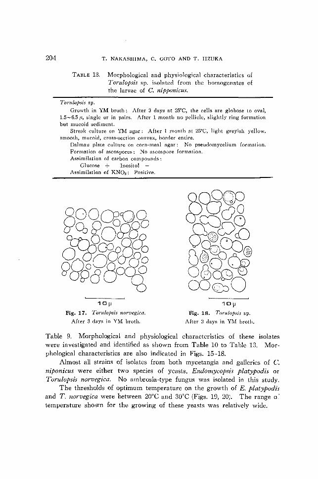
TABLE 13. Morphological and physiological characteristics of Torulopsis sp. isolated from the homogenates of the larvae of C. nipponicus.
Torulopsis sp.
Growth in YM broth: After 3 days at 25°C, the cells are globose to oval, 1.5-6.5 fl, single or in pairs. After 1 month no pellicle, slightly ring formation but mucoid sediment.
Streak culture on YM agar: After 1 month at 25°C, light greyish yellow, smooth, mucoid, cross-section convex, border entire.
Dalmau plate culture on corn-meal agar: No pseudomycelium formation. Formation of ascospores: No ascospore formation. Assimilation of carbon compounds:
Glucose + InositolAssimilation of KN03 : Positive.
101-1
Fig. 17. Torulopsis norvegica.
After 3 days in YM broth.
10ll
Fig. 18. Torulopsis sp.
After 3 days in YM broth.
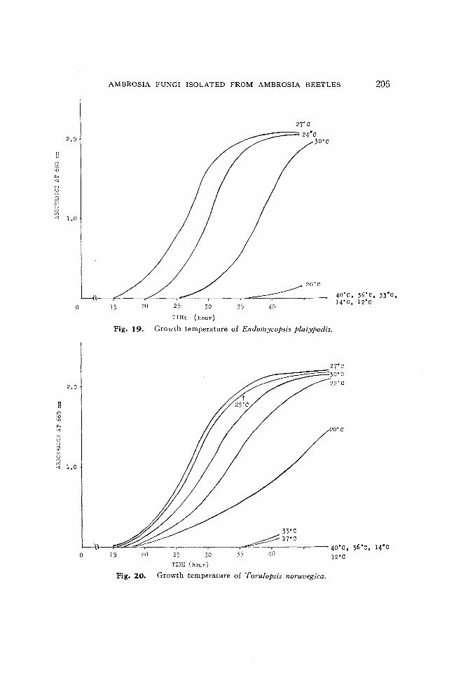
Table 9. Morphological and physiological characteristics of these isolates were investigated and identified as shown from Table 10 to Table 13. Morphological characteristics are also indicated in Figs. 15-18.
Almost all strains of isolates from both mycetangia and galleries of C. niponicus were either two species of yeasts, Endomycopsis platypodis or Torulopsis norvegica. No ambrosia-type fungus was isolated in this study.
The thresholds of optimum temperature on the growth of E. platypodis and T. norvegica were between 20°C and 30°C (Figs. 19, 20). The range o~ temperature shown for the growing of these yeasts was relatively wide.
H
'" r!~
~
~ ~!~ t:J ,n
2,0
<~ 1.0
o
2,0
@ 0
'" '" "" "'" 01 ~ :J, ;<, '" 0 UJ m
1,0 "'"
o
AMBROSIA FUNGI ISOLATED FROM AMBROSIA BEETLES 205
15 20 25 30
TIH}; ( hour)
27'C
----::=~ 24· c 30'c
35 40
40'C, 36'c, 33'C, 14'C, 12'C
Fig, 19, Growth temperature of Endomycopsis platypodis,
27'c 30'C 22'C
20'C
33'C
__ ~ • _1_7_·_C-. ____ _ ---~,- 40'C, ,6'c, 14'C
15 20 25 30 35 40 12'C TH1E (hour)
Fig. 20. Growth temperature of Torulopsis noruvegica.
206 T. NAKASHIMA, C. GOTO AND T. IIZUKA
Discussion
As shown in Table 2 and Table 9, the flora of microorganisms in the galleries of ambrosia beetles changes each season. During the periods when eggs and larvae are growing, the flora of fungi is relatively simple. Few species of ambrosia fungi appear to be predominant. These fungi seem to be deeply associated with ambrosia beetles. After pupation, however, many other species of fungi are found there.
BATRAv, BATRA & BATRA3,4) have suggested that while the young larvae of ambrosia beetles are usually associated with only one species of ambrosia fungi, adult beetles feed on more species of fungi. They have also suggested that the adult beetles contain their primary ambrosia fungus in their mycetangia predominantly even in the case of feeding on auxiliary or non-ambrosia fungi. They have also offered their opinion on primary and auxiliary ambrosia fungi, that is, each species of ambrosia beeltes is normally associated with only one species of fungus, namely the primary ambrosia fungus, and some primary ambrosia fungi of an ambrosia beetle play the role of auxiliary ambrosia fungi of another ambrosia beetles.
In the case of S. shogun, an Ambrosiella sp. which showed the shape of typical monilioid chains, was isolated from both mycetangia and galleries (Figs. 11, 12). Only this Ambrosiella sp. was isolated from the mycetangia of female adults at their flying period and from the walls of their galleries in the breeding period. It seems that this Ambrosiella sp. plays an important role in the life of S. shogun as a primary ambrosia fungus. This fungus has two forms, the ambrosial (=yeas(' phase and the mycelial phase. In the galleries, monilioid chains were observed as shown in Fig. 12. BATRA & BATRA3,4) have discovered that ambrosia fungi are pleomorphic. The fungi can easily change their form when their growth medium has been changed, and that ambrosia beetles can change the form of the fungi from a moldlike form to a yeastlike one, as in the case of some termites.
The threshold of optimum temperature for growing of this Ambrosiella sp. was near 23°C, and showed an extremely narrow range as shown in Fig. 13. It was anticipated that the Ambrosiella sp. would be isolated from the homogenates of the larvae. As shown in Table 2, however, no Ambrosiella sp. grew from the homogenates. In this examination, the growing temperature was 2°C higher than the optimum temperature. It may be the reason why no Ambrosiella sp. had grown from the homogenates.
The genus Ambrosiella was classified by BATRA2) as a specific ambrosia fungus. He has reported that 11 species of the genus Ambrosiella were
AMBROSIA FUNGI ISOLATED FROM AMBROSIA BEETLES 207
isolated from 20 species of the beetles which belong to the genera Anisandrus, Corthylus, Gnathotrichus, Monarthrum, Xyleborus, Xylosandrus, Trypodendron, Ips and Myelophilus of Scolytidae and from 1 Platypus sp. of Platypodidae.
A large amount of Pichia sp. 1. and Pichia sp. 2. were isolated from the galleries in which larvae were growing, and from the homogenates of larvae. This suggests that the fungi, especially Pichia sp. 2., are eaten by the larvae. These fungi, however, were not isolated from the mycetangia. Therefore, these fungi may be included in the group of the auxiliary ambrosia fungi of S. shogun.
It seems that Pichia sp. 1. may be the same or very nearly related to Pichia pini, which was isolated from Dendroctonus monticolae, D. brevicomis, D. ponderosae, Ips con/usus and I. oregoni by SHIFRINE & PHAFFU).
Only in winter. Candida tenuis was isolated from the galleries as the dominant species. The role of this fungus is not clear. LODDER5l reported that Candida tenuis was isolated from some beetles which belong to the genera Harpium, Rhagium, Leptura and Mycroplophorus.
In the case of C. niponicus, Endomycopsis platypodis and Torulopsis norvegica were regularly and dominantly isolated from the homogenates of the heads of female adults in their flying period, from the homogenates of larvae and from the walls of galleries. It seems that E. platypodis and T. norvegica are primary ambrosia fungi of C. niponicus. We have previously reported that E. platypodis is commonly isolated from homogenates of whole bodies of adult beetles and from galleries of C. nipon icus. 10)
According to LODDER5), E. platypodis was isolated from the beetles Xyleborus aemulus (from gallery), X. xanthophus (from gallery), Platypus cylindrus (from frass) and Crossotarsus externedentatus (from gallery).
Several strains of another fungus which belongs to the genus Endomycopsis were isolated from the homogenates of larvae. These fungi may be included in the group of auxiliary ambrOSia fungi of C. niponicu5.
Acknowledgements
We are greatful to all members of the Hiyama Forest Experiment Station of Hokkaido University for their aid in the collection of pinholed beech logs. We are also indebted to Dr. T. TANAKA, Laboratory of Applied Biology and Biotechnology, Sun tory Limited for his technical cooperation in the measurement of the growing temperature for several isolates, and for his valuable ad vice.
This work was supported in part by a Grant-in-Aid for Special Project Research on Biological Aspects of Optimal Strategy and Social Structure
208 T. NAKASHIMA, C. GOTO AND T. IIZUKA
from the Japan Ministry of Education, Science and Culture.
References
1. BATRA, L. R.: Ecology of ambrosia fungi and their dissemination by beetles,
Trans. Kan. Acad. Sci. 66 (2): 213-236. 1966
2. BATRA, L. R.: Ambrosia fungi: A taxonomic revision, and nutritional studies
of some species, Mycologia 59: 976-1017. 1967
3. BATRA, L. R. & BATRA, S. W. T.: Termite-fungus mutualism. Insect-Fungus
Symbiosis (Ed. by BATRA, L. R.), 117-163. Allanheld, Osmun & Co. Montclair,
New Jersey, 1979
4. BATRA, S. W. T. & BATRA, L. R.: The fungus gardens of insects. Scientific
American 217: 112-120. 1967
5. LODDER, J.: The yeasts: A taxonomic study (Ed. by LODDER, J.), p. 1-33.
North-Holland Pud. Co., Amsterdam, 1970
6. NAKASHIMA, T.: Notes on the associated fungi and the mycetangia of the
ambrosia bettle, Crossotarsus niponicus Blandford (Coleoptera: Platypodidae), Appl.
Ent. Zool. 6 (3): 131-137. 1971
7. NAKASHIMA, T.: Several types of the mycetangia found in platypodid ambrosia
beetles (Coleoptera: Platypodidae), Insecta MatslllllU1'ana New Series 7: 1-69. 1975
8. NAKASHIMA, T.: Ambrosia beetle - Fungus growers (in Japanese), Insectarium
15, 14-22. 1978
9. NAKASHIMA, T.: Function and location of mycetangia in ambrosia beetles. The
ultrastructure and functioning of insect cells (Ed. AKAI et al.), 87-90. Soc. for
Insect Cells Japan, 1982
10. NAKASHIMA, T., IrZUKA, T., OGURA, K., MAEDA, M. & TANAKA, T.: Isolation
of some microorganisms associated with five species of ambrosia beetles and two
kinds of antibiotics produced by Xv-3 strain in these isolates. J. Faculty Agri.
Hokkaido Univ. 61 (1): 60-72. 1982
11. SHIFRINE, M. & PHAFF, H. J.: The association of yeasts with certain bark
beetles. Mycologia 48: 41-55. 1956