Embed Size (px)
Citation preview
The Primate Working Memory Networks
Christos Constantinidis Department of Neurobiology and Anatomy,
Wake Forest University School of Medicine
Emmanuel Procyk INSERM U371, Cerveau et Vision
Institut Fédératif des Neurosciences de Lyon, Bron, France
Address correspondence to:
Christos Constantinidis, Ph.D. Department of Neurobiology and Anatomy
Wake Forest University School of Medicine
Medical Center Blvd., Winston-Salem, NC 27157-1010
Tel: 336-716-7424
Fax: 336-716-4534 E-mail: [email protected]
Abstract
Working memory has long been associated with the prefrontal cortex, as damage to this brain area can critically
impair the ability to maintain and update mnemonic information. Anatomical and physiological evidence suggest
however that the prefrontal cortex is part of a broader network of interconnected brain areas involved in working
memory. These include the parietal and temporal association areas of the cerebral cortex, cingulate and limbic areas,
and subcortical structures such as the mediodorsal thalamus and the basal ganglia. Neurophysiological studies in
primates confirm the involvement of areas beyond the frontal lobe and illustrate that working memory involves
parallel, distributed neuronal networks. The article reviews our current understanding on the anatomical
organization of networks mediating working memory and the neural correlates of memory manifested in each of
their nodes. The neural mechanisms of memory maintenance and the integrative role of the prefrontal cortex are
also discussed.
Working memory is the term commonly used for the ability to
maintain information in memory over a time span of a few seconds.
Working memory constitutes a core component of higher cognitive
functions including language, problem solving and reasoning
(Baddeley, 1992). Lesion studies first localized working memory
functions in the cortical surface of the frontal lobe (Jacobsen, 1936;
Milner, 1963). Neurophysiological recordings subsequently provided
a neural correlate of working memory, in the sustained discharges of
neurons, persisting even after the offset of brief stimuli that monkeys
were trained to remember and recall (Fuster & Alexander, 1971).
Individual neurons represent particular features and spatial locations
so that the activity of the prefrontal population can encode a
remembered stimulus. The part of the visual space where stimulus
appearance can produce sustained activation has been termed the
eu o ’s e o field , i a alog to the e epti e field of eu o s responding to sensory stimulation (Funahashi, Bruce, & Goldman-
Rakic, 1989).
Neurons with memory-related responses have since been
reported in multiple brain regions, for example the inferior temporal
and posterior parietal cortex, the end-stages of the ventral and dorsal
visual pathways, respectively (Andersen, Essick, & Siegel, 1987;
Fuster & Jervey, 1981). The prefrontal cortex is reciprocally
connected to these areas, in a well-organized and systematic fashion:
the dorsal prefrontal cortex (areas 8 and 46) is interconnected with
the posterior parietal cortex whereas the ventral prefrontal cortex
(areas 12 and 45) is linked to the inferior parietal cortex. Based on
this organization, Patricia Goldman-Rakic proposed that working
memory is mediated by the sustained activity of neurons in parallel,
distributed cortical networks (P. S. Goldman-Rakic, 1988). More
recently, imaging modalities (PET and fMRI) have verified the
concurrent activation of multiple human brain areas during
performance of cognitive tasks that engage working memory,
confirming the findings of the monkey neurophysiological studies
(Courtney, Ungerleider, Keil, & Haxby, 1997; Jonides et al., 1993;
Ungerleider, Courtney, & Haxby, 1998).
The primate working memory networks
1
Over the last decade, great progress has been made in
understanding the organization of the working memory networks
and the functional specialization of brain areas that constitute them.
Here, we will provide an update on the anatomical and physiological
details of the working memory networks, focusing on the brain of the
rhesus monkey as the best-studied model. The prefrontal cortex will
be the point of origin for our review. We will examine the
organization of the prefrontal cortex and its connections with the
sensory pathways, namely the ventral and dorsal visual pathways,
the somatosensory, auditory, gustatory and olfactory cortex. We will
also examine the function of the medial temporal system, and limbic
and subcortical structures. The differential functions of brain regions
comprising the network and the integrative role of the prefrontal
cortex will finally be discussed.
EXPERIMENTAL INVESTIGATION OF WORKING MEMORY
Working memory can be engaged and manifested in tasks that
require a subject to remember a briefly presented stimulus. For
example, an experimenter may bait one of two food wells in view of
a monkey in a Wisconsin General Testing Apparatus and lower a
s ee o st u ti g the o ke ’s ie fo a pe iod of a fe se o ds. Once the screen is raised again, the monkey is allowed to uncover
one well and retrieve the treat. For the subject to correctly perform
such a task, it is necessary to keep in memory the spatial location of
the food ite o e the dela pe iod he o sti ulus is isi le. This visual-spatial memory is mediated by neuronal discharges in a
network of brain areas that can be investigated in vivo, with
microelectrode recordings in each brain area. However, neurons also
dis ha ge i espo se to othe aspe ts of the su je t’s eha io o e the delay period, for example the planning and execution of eye
movements.
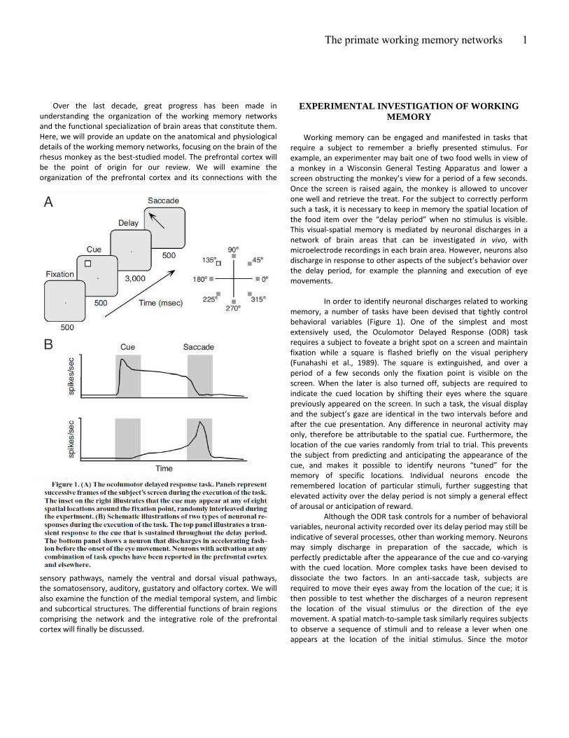
In order to identify neuronal discharges related to working
memory, a number of tasks have been devised that tightly control
behavioral variables (Figure 1). One of the simplest and most
extensively used, the Oculomotor Delayed Response (ODR) task
requires a subject to foveate a bright spot on a screen and maintain
fixation while a square is flashed briefly on the visual periphery
(Funahashi et al., 1989). The square is extinguished, and over a
period of a few seconds only the fixation point is visible on the
screen. When the later is also turned off, subjects are required to
indicate the cued location by shifting their eyes where the square
previously appeared on the screen. In such a task, the visual display
a d the su je t’s gaze are identical in the two intervals before and
after the cue presentation. Any difference in neuronal activity may
only, therefore be attributable to the spatial cue. Furthermore, the
location of the cue varies randomly from trial to trial. This prevents
the subject from predicting and anticipating the appearance of the
ue, a d akes it possi le to ide tif eu o s tu ed fo the memory of specific locations. Individual neurons encode the
remembered location of particular stimuli, further suggesting that
elevated activity over the delay period is not simply a general effect
of arousal or anticipation of reward.
Although the ODR task controls for a number of behavioral
variables, neuronal activity recorded over its delay period may still be
indicative of several processes, other than working memory. Neurons
may simply discharge in preparation of the saccade, which is
perfectly predictable after the appearance of the cue and co-varying
with the cued location. More complex tasks have been devised to
dissociate the two factors. In an anti-saccade task, subjects are
required to move their eyes away from the location of the cue; it is
then possible to test whether the discharges of a neuron represent
the location of the visual stimulus or the direction of the eye
movement. A spatial match-to-sample task similarly requires subjects
to observe a sequence of stimuli and to release a lever when one
appears at the location of the initial stimulus. Since the motor
The primate working memory networks
2
response is identical in every trial, any differences in neuronal
responses may only be due to the location of the remembered
stimulus. Even in this case however, it is difficult to distinguish
between the effects of spatially directed attention as opposed to
memory for the cued location and it is important to realize the
interplay of multiple factors that influence neuronal activity.
Tasks that require the monkey to remember the identity
rather than the spatial location of a stimulus have revealed neurons
selectively tuned for the color and form of remembered visual
stimuli, as well as for stimuli of other sensory modalities. The brain
areas activated in such a fashion during the maintenance of
information in memory are presented next.
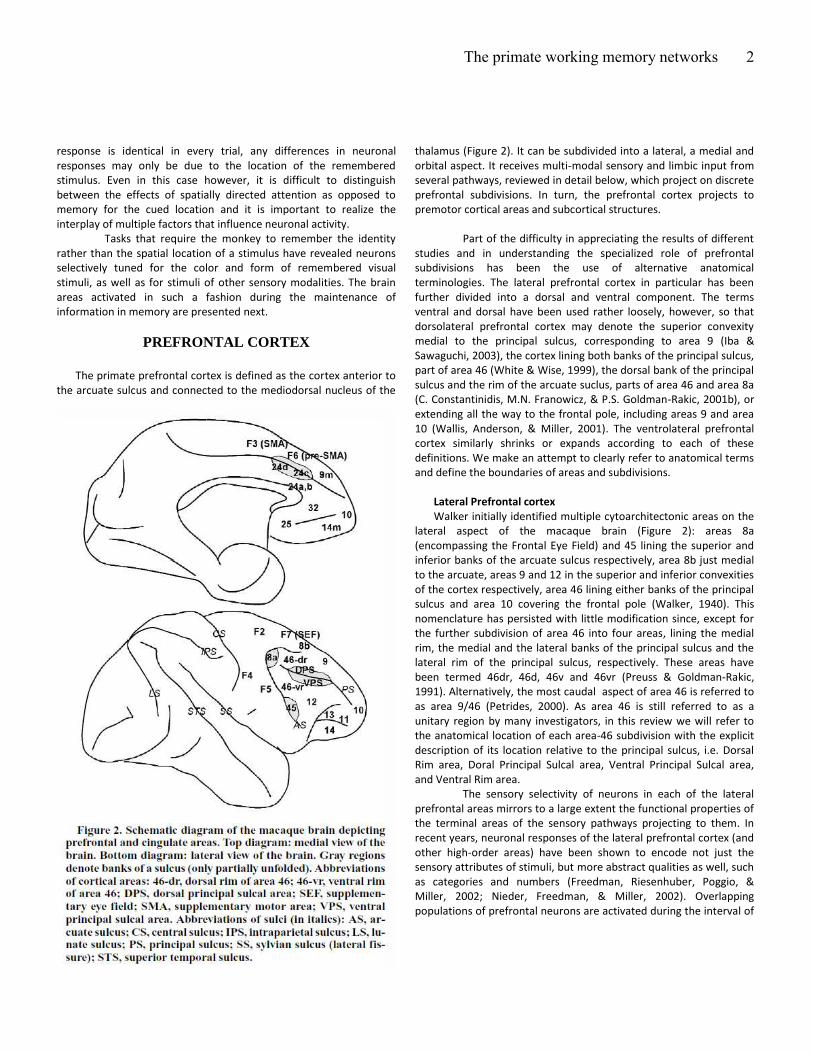
PREFRONTAL CORTEX
The primate prefrontal cortex is defined as the cortex anterior to
the arcuate sulcus and connected to the mediodorsal nucleus of the
thalamus (Figure 2). It can be subdivided into a lateral, a medial and
orbital aspect. It receives multi-modal sensory and limbic input from
several pathways, reviewed in detail below, which project on discrete
prefrontal subdivisions. In turn, the prefrontal cortex projects to
premotor cortical areas and subcortical structures.
Part of the difficulty in appreciating the results of different
studies and in understanding the specialized role of prefrontal
subdivisions has been the use of alternative anatomical
terminologies. The lateral prefrontal cortex in particular has been
further divided into a dorsal and ventral component. The terms
ventral and dorsal have been used rather loosely, however, so that
dorsolateral prefrontal cortex may denote the superior convexity
medial to the principal sulcus, corresponding to area 9 (Iba &
Sawaguchi, 2003), the cortex lining both banks of the principal sulcus,
part of area 46 (White & Wise, 1999), the dorsal bank of the principal
sulcus and the rim of the arcuate suclus, parts of area 46 and area 8a
(C. Constantinidis, M.N. Franowicz, & P.S. Goldman-Rakic, 2001b), or
extending all the way to the frontal pole, including areas 9 and area
10 (Wallis, Anderson, & Miller, 2001). The ventrolateral prefrontal
cortex similarly shrinks or expands according to each of these
definitions. We make an attempt to clearly refer to anatomical terms
and define the boundaries of areas and subdivisions.
Lateral Prefrontal cortex
Walker initially identified multiple cytoarchitectonic areas on the
lateral aspect of the macaque brain (Figure 2): areas 8a
(encompassing the Frontal Eye Field) and 45 lining the superior and
inferior banks of the arcuate sulcus respectively, area 8b just medial
to the arcuate, areas 9 and 12 in the superior and inferior convexities
of the cortex respectively, area 46 lining either banks of the principal
sulcus and area 10 covering the frontal pole (Walker, 1940). This
nomenclature has persisted with little modification since, except for
the further subdivision of area 46 into four areas, lining the medial
rim, the medial and the lateral banks of the principal sulcus and the
lateral rim of the principal sulcus, respectively. These areas have
been termed 46dr, 46d, 46v and 46vr (Preuss & Goldman-Rakic,
1991). Alternatively, the most caudal aspect of area 46 is referred to
as area 9/46 (Petrides, 2000). As area 46 is still referred to as a
unitary region by many investigators, in this review we will refer to
the anatomical location of each area-46 subdivision with the explicit
description of its location relative to the principal sulcus, i.e. Dorsal
Rim area, Doral Principal Sulcal area, Ventral Principal Sulcal area,
and Ventral Rim area.
The sensory selectivity of neurons in each of the lateral
prefrontal areas mirrors to a large extent the functional properties of
the terminal areas of the sensory pathways projecting to them. In
recent years, neuronal responses of the lateral prefrontal cortex (and
other high-order areas) have been shown to encode not just the
sensory attributes of stimuli, but more abstract qualities as well, such
as categories and numbers (Freedman, Riesenhuber, Poggio, &
Miller, 2002; Nieder, Freedman, & Miller, 2002). Overlapping
populations of prefrontal neurons are activated during the interval of
The primate working memory networks
3
sensory stimulation, over delay periods of no stimulation and around
the time of motor responses (Funahashi et al., 1989). The prefrontal
cortex projects back to all the areas from which it receives sensory
input, as well as structures with motor functions, including the
premotor cortex, the superior colliculus and the basal ganglia, though
not directly to the primary motor cortex.
The representation of sensory, mnemonic, and motor
information is generally not topographic (e.g. visuotopic) in the
prefrontal cortex. Instead, the same retinal location and eye
movement direction is represented multiple times across the cortical
surface (C. Constantinidis, M. N. Franowicz, & P.S. Goldman-Rakic,
2001a). Anatomical evidence suggests a highly regular pattern of
axonal terminations from both within the prefrontal cortex as well as
from association cortices, forming repeating, interdigitated stripes
(Kritzer & Goldman-Rakic, 1995; Pucak, Levitt, Lund, & Lewis, 1996).
Yet, the underlying principles of prefrontal functional organization
remain elusive.
One possible explanation for the absence of topological
mapping of stimulus dimensions as in the primary sensory cortex, is
that the prefrontal cortex is organized in multiple intersecting and
overlapping networks of associated attributes (Fuster, 2003). Several
lines of evidence suggest, however, at least a local organization in
neuronal coding properties. Narrow cortical lesions and local,
chemical inactivation affect only a restricted area of visual space,
typically in the contralateral hemifield, an effect known as a
e o i s oto a (Funahashi, Bruce, & Goldman-Rakic, 1993;
Sawaguchi & Goldman-Rakic, 1991, 1994). Simultaneous recordings
from electrodes spaced 0.2 – 0.3 mm apart similarly indicate that
nearby neurons represent adjacent spatial locations (Constantinidis
et al., 2001a). These results suggest at least a coarse topography,
possibly involving the entire visual hemifield represented in repeating
cortical modules.
Why would the representation of a stimulus or a motor
target be replicated multiple times across the prefrontal cortex?
Although no definitive answer is currently available, theoretical
studies provide some possible explanations. The brain is able to
flexibly generate variable responses to identical stimuli depending on
the context that they appear. The absence of a one-to-one
correspondence between sensory stimuli and motor responses
makes inevitable the replication of stimulus representations in the
prefrontal cortex. Recent theoretical studies have formally
demonstrated that a neural circuit that contains multiple
representations of the same sensory stimulus, mapped to multiple
motor outcomes but each modulated in a different fashion by
contextual factors can effectively act as a switch, producing different
motor outcomes depending on the rule or context enforced (Salinas,
2004).
The discharges of prefrontal neurons are indeed modulated
by factors other than sensory stimulation or motor responses.
Responses to the same operant stimuli vary depending on the
expectation of reward, when the later varies from trial to trial (Leon
and Shadlen, 1999). A similar modulation of neuronal responses to
identical stimulation has been observed in animals trained to
perform a number of alternative tasks, requiring association of the
same stimulus with different possible motor choices, according to
different rules (White and Wise, 1999; Asaad et al., 2000; Wallis et
al., 2001). Finally, prefrontal neurons preferentially represent the
attributes of stimuli that are behaviorally relevant and require
attention, than identical stimuli that are deemed as uninformative or
distracting for the purposes of a behavioral task (Rainer et al., 1998b;
Everling et al., 2002).
Orbitofrontal cortex
The orbital frontal cortex (OFC) includes areas 10, 11, 13 and 14
(Figure 2). It is interconnected to the amygdala, gustatory and
olfactory cortices, temporal cortex, lateral PFC and anterior cingulate,
as well as with the ventral striatum, mesencephalic structures and
hypothalamus (Barbas, 2000). This pattern of connections suggests
that the contribution of the orbital frontal cortex (OFC) to behaviour
relates to motivation and emotion (Rolls, 2002). Lesion studies in
humans and non-human primates, reveal no clear deficits in working
memory tasks after orbital frontal lesions, and thereby a strong
functional dissociation between the OFC and the lateral prefrontal
cortex (Bechara, Damasio, Tranel, & Anderson, 1998; Wallis, Dias,
Robbins, & Roberts, 2001). Indeed, OFC lesions induce deficits related
to emotional reactions, social behaviours, reversal learning,
associative memory, and adaptation to reward change (Baxter,
Parker, Lindner, Izquierdo, & Murray, 2000; Meunier, Bachevalier, &
Mishkin, 1997; R. E. Passingham, 1993).
Relation to lateral PFC and working memory
In line with lesion data, unit recordings in the OFC show activity
related to reward discrimination, expectation, and preference
(Hikosaka & Watanabe, 2000; Rosenkilde, Bauer, & Fuster, 1981;
Tremblay & Schultz, 1999; Wallis & Miller, 2003b). However, in
contrast with lesion studies, unit recordings reveal delay-related
discharges that contain information related to cues given in delayed
response tasks, and to reward expectation (Hikosaka & Watanabe,
2000; Rosenkilde et al., 1981; Wallis & Miller, 2003b). Studies that
focused on this OFC activity provide insights in its functional role in
working memory. OFC delay activity codes for expected reward even
in tasks where reward is not part of the memory component of the
task (Hikosaka & Watanabe, 2000). It has little to do with the
impending behavioural response properties, but is influenced by
motivational factors. Moreover, reward-related information appears
earlier in OFC than in lateral prefrontal cortex (Wallis & Miller,
2003b). Thus, OFC would not have a role in active maintenance of
information but could have a role in representing goals and their
motivational components (preferences), hence being a source of
target-value and reward-related information to be combined with
response–related representations in working memory. In this context
we shall note that it is the cognitive binding between memorized
items, outcome expectations (reward) and impending response plans
that enables the organization of goal directed behaviors (Fuster,
2001).
The primate working memory networks
4
Medial prefrontal cortex
Areas traditionally considered as part of the monkey medial
prefrontal cortex include the Supplementary Eye Field (alternatively
considered part of the premotor cortex – area F7 in Figure 2), and the
medial aspects of areas 9 and 14 (9m and 14m in Figure 2, top). Area
24 is also often grouped with the medial prefrontal cortex, however
here we will examine it independently as part of the anterior
cingulate cortex.
The medial prefrontal cortex has received much less
neurophysiological scrutiny than the lateral prefrontal cortex, in the
monkey. Based on its pattern of anatomical inputs, visual,
somatosensory and olfactory responses would be expected in distinct
medial frontal divisions (Barbas, Ghashghaei, Dombrowski, &
Rempel-Clower, 1999). Persistent, memory-related discharges have
been described in the Supplementary Eye Field (Russo & Bruce,
1996). This activity has been shown to be influenced by the sensory-
motor association that monkeys have been instructed to perform; for
example anti-saccades generally produce stronger responses than
pro-saccades to the same direction (Schlag-Rey, Amador, Sanchez, &
Schlag, 1997). In addition, neurons in the Supplementary Eye Field
have been shown to discharge in response to perceived errors in eye-
movement tasks (Stuphorn, Taylor, & Schall, 2000). Both of these
findings are consistent with the idea that medial frontal structures
are involved with the monitoring of self behavior and the potential
conflict between sensory stimuli, actions and expectations.
Other factors influencing neuronal responses in the medial
prefrontal cortex include the object-centered frame of reference of
eye movements, reward expectation, or rule the animals have been
trained to perform (Amador, Schlag-Rey, & Schlag, 2000; Matsumoto,
Suzuki, & Tanaka, 2003; Olson & Gettner, 1995).
FRONTAL CORTEX BEYOND PREFRONTAL
Anterior cingulate cortex
The cingulate cortex is located on the medial surface of the
hemispheres, covering the entire length of the corpus callosum. It is
divided into anterior and posterior regions. Moreover, the cortex of
the cingulate sulcus is distinguished from the cortex of the cingulate
gyrus. The monkey anterior cingulate cortex (ACC) comprises areas
24-a, b, c and d, although their precise delineations varies between
authors. Recently much attention has been drawn on area 24c, lining
the inferior and superior bank of the cingulate sulcus and bounded
posteriorly by area 24d. Area 24c contains the rostral cingulate motor
area, CMAr (Dum & Strick, 1991, 1993). Compared to caudal
cingulate motor areas, the more anterior part of the cingulate sulcus
(including CMAr) is characterised by significant connections with the
pre-supplementary motor area and lateral prefrontal areas (Hatanaka
et al., 2003; Wang, Shima, Sawamura, & Tanji, 2001). Area 24c
projects onto the median and caudal parts of the principal sulcus
(area 46, 8A, 8B, 9 and 12) (Barbas & Mesulam, 1985; Bates &
Goldman-Rakic, 1993; Lu, Preston, & Strick, 1994; McGuire, Bates, &
Goldman-Rakic, 1991). The density and significance of these
connections is debatable, especially in light of recent studies (Takada
et al., 2004), but they are consistent with a particular role of this
subdivision of ACC in higher order cognitive functions. As much of the
medial prefrontal frontal cortex, ACC receives strong dopaminergic
inputs (Williams & Goldman-Rakic, 1993).
Relation to lateral PFC
Initially, thanks to human brain imaging studies, consistent
activation of the homologue part of monkey 24c was observed during
most cognitive tasks. Among the numerous human brain imaging
studies, most of those devoted to high-order cognition reported joint
lateral prefrontal and ACC activations, in particular when tasks used
were complex or necessitated active control on behaviour (e.g. Frith,
Friston, Liddle, & Frackowiak, 1991; Koski & Paus, 2000; Paus,
Petrides, Evans, & Meyer, 1993). The most recent reviews on
prefrontal functions underline the importance of these facts (Duncan
& Owen, 2000; Miller & Cohen, 2001; Paus, 2001). Nonetheless,
activations of these two regions are dissociable in functional terms
(MacDonald, Cohen, Stenger, & Carter, 2000; Posner & DiGirolamo,
1998).
Role in working memory
It is now widely accepted that, although dissociated in some
ways, systems involved in higher brain functions (storing, monitoring
and manipulating information in memory) establish strong
relationships and interactions or even overlap with those concerned
with motivation and emotion. For instance, in her description of
distributed functions in several parallel systems, Goldman-Rakic
illustrated how interconnected prefrontal dorsolateral and parietal
areas shared common connections with the anterior cingulate (ACC),
whereby underlying a link between working memory and motivation
(Goldman-Rakic, 1988).
The involvement of monkey ACC in working memory tasks has
been suggested by brain imaging (Inoue, Mikami, Ando, & Tsukada,
2004). Among neurophysiological experiments in monkeys that
studied ACC unit activity, only a few reported neural activity recorded
during spatial delay tasks (Akkal, Bioulac, Audin, & Burbaud, 2002;
Isomura, Ito, Akazawa, Nambu, & Takada, 2003; Niki & Watanabe,
1976; Procyk & Joseph, 2001). However, although
electrophysiological studies reported tonic activity during memory
delays, the authors acknowledge that information contained in ACC
delay activity has little to do with a potential memory buffer coding
specifically for location of targets or direction of movements (Akkal et
al., 2002; Isomura et al., 2003; Procyk & Joseph, 2001). On the other
hand, ACC activity is strongly influenced by reward expectation and
various parameters related to the selection of action based on
expected rewards (Akkal et al., 2002; Matsumoto et al., 2003; Niki &
Watanabe, 1976; Procyk & Joseph, 2001; Procyk, Tanaka, & Joseph,
2000; Shidara & Richmond, 2002; Shima & Tanji, 1998). Most data,
including those related to the role of ACC in error detection,
converge toward a key function of ACC in behaviour evaluation.
Lesion studies strongly support this view (Hadland, Rushworth,
Gaffan, & Passingham, 2003; Rushworth, Hadland, Gaffan, &
Passingham, 2003). Indeed, expectation of reward associated to
The primate working memory networks
5
action and error detection are part of a unique function that
evaluates the adequacy of behaviour.
Thus, the role of ACC in working memory, and its particular
relationship with lateral prefrontal cortex remain in question. It is
possible that the unified representation of goal-directed action in
complex cognitive tasks depends on the parallel coding of integrated
motivation-action information in ACC and actively memorized
information as seen in lateral prefrontal cortex. Yet, other non-
parallel interactions may take place between these two structures, in
particular when error or conflict detection triggers action selection
adjustment (Miller & Cohen, 2001).
Premotor cortex
General connectivity.
Premotor (PM) areas are divided into mesial (areas F3 –
Supplementary Motor Area, F6 – pre-Supplementary Motor Area),
lateral dorsal (areas F2, F7 – Supplementary Eye Field) and lateral
ventral (F4, F5) territories (Matelli, Luppino, & Rizzolatti, 1991). One
main factor of subdivision of PM areas is related to effector maps.
Each PM area support at least one body representation which has
been delineated by microstimulation (Godschalk, Mitz, van Duin, &
van der Burg, 1995; Luppino, Matelli, Camarda, Gallese, & Rizzolatti,
1991; Mitz & Wise, 1987). In addition, several eye fields have been
described in lateral and medial premotor areas (Huerta, Krubitzer, &
Kaas, 1986, 1987; Schlag & Schlag-Rey, 1987; Wang, Matsuzaka,
Shima, & Tanji, 2004).
Areas F6 and F7 (anterior premotor) receive input from the
prefrontal cortex and project to the other premotor areas F2, F3, F4
and F5, which in turn project to the primary motor cortex and the
spinal cord (Dum & Strick, 1991; Rizzolatti & Luppino, 2001).
Connections between area 46 and lateral premotor areas extend the
dorso-ventral dissociation (Barbas & Pandya, 1987; Wang, Shima,
Isoda, Sawamura, & Tanji, 2002; Watanabe-Sawaguchi, Kubota, &
Arikuni, 1991).
Function
The top-to-bottom connectivity from prefrontal to primary motor
cortex reinforces a pyramidal view of the cortical organisation in the
frontal cortex as well as of the functional roles of its subdivisions.
Although recent brain imaging studies suggest that the lateral frontal
cortex is organized as a cascade of executive processes from
premotor to anterior prefrontal regions, it is also plausible that
executive networks are to some degree heterarchical instead of
purely hierarchical (Fuster, 2001; Koechlin, Ody, & Kouneiher, 2003).
Thus, whether the frontal cortex is purely hierarchically organized in
terms of cognitive function is debatable, and a description of working
memory networks is central to the debate. In particular, it is
important to compare prefrontal and premotor tonic activities and
their time of occurrence during cognitive tasks.
PreSMA and SMA have a crucial role in sequence planning, and
lateral premotor areas play a crucial role in planning visually guided
movements, and more specifically during conditional motor learning
(Nakamura, Sakai, & Hikosaka, 1998; Shima, Mushiake, Saito, & Tanji,
1996; Tanji & Shima, 1994; Wise, Murray, & Gerfen, 1996). For
instance premotor dorsal and ventral neurons respond differently to
identical stimuli that instruct different actions (Boussaoud & Wise,
1993; di Pellegrino & Wise, 1993b). This is to say that although they
process spatial or motor parameters (Crammond & Kalaska, 1994;
Jouffrais & Boussaoud, 1999; Messier & Kalaska, 2000), premotor
areas are involved in more than basic motor control.
We focus on lateral premotor areas since several attempts have
been made either to compare activity in these areas with those in
prefrontal cortex, or to detail neural activity and information coding
during memory delay period. Although delay tonic activity can be
observed in prefrontal and premotor areas, several studies report
that, on average, prefrontal cell activity is more related to stimulus
information than to motor plan and differs in that from premotor
activity (Boussaoud & Wise, 1993; di Pellegrino & Wise, 1993b; Wallis
& Miller, 2003a). In a double delay task, latencies of phasic and tonic
activities are shorter in prefrontal than in premotor cortex, which in
turn discharge more intensely later in the delay (di Pellegrino & Wise,
1991). These results support the idea of a transfer of information or
control from prefrontal cortex to premotor, and from cognitive to
motor, during delay periods that bridge instruction and motor
response. However this may not be always the case; premotor
related activity can have shorter latency than prefrontal activity
(Wallis & Miller, 2003a). Moreover in a spatial delayed response task,
prior information about distance of the target is reflected in
premotor activity during the delay period well before movement
initiation, and begins to be expressed as early as 150 ms after
presentation of target location (Messier & Kalaska, 2000).
Although most data seem to emphasize a role of premotor areas
in planning rather than in active memory, whether premotor tonic
activity has a role in the representation of information in working
memory remains in question. In most paradigms, premotor and
prefrontal activity may appear contingent, but strong differences can
appear with true comparisons and adapted behavioural tasks (di
Pellegrino & Wise, 1993b).
THE SENSORY PATHWAYS
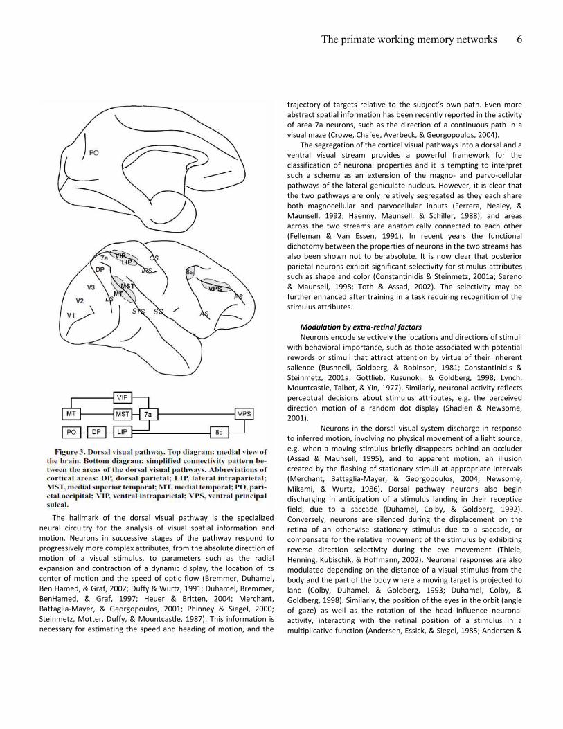
Dorsal visual pathway
General properties
The dorsal visual stream (Figure 3) includes cortical areas MT,
MST, PO, DP, VIP, LIP, and 7a (Felleman & Van Essen, 1991;
Ungerleider & Mishkin, 1982). Receptive fields of neurons in these
areas cover the visual periphery, with increasing size in successive
stages of the pathway (Blatt, Andersen, & Stoner, 1990; Komatsu &
Wurtz, 1988; Raiguel et al., 1997). The majority of neurons exhibit
contralateral receptive fields, with bilateral fields common among
neurons in area 7a, which interestingly, often exclude the fovea
(Motter & Mountcastle, 1981). Neuronal responses in higher-level
areas are also increasingly modulated by cognitive factors, such as
selective attention and reward expectation (Cook & Maunsell, 2002;
Platt & Glimcher, 1999; Treue & Maunsell, 1996).
The primate working memory networks
6
The hallmark of the dorsal visual pathway is the specialized
neural circuitry for the analysis of visual spatial information and
motion. Neurons in successive stages of the pathway respond to
progressively more complex attributes, from the absolute direction of
motion of a visual stimulus, to parameters such as the radial
expansion and contraction of a dynamic display, the location of its
center of motion and the speed of optic flow (Bremmer, Duhamel,
Ben Hamed, & Graf, 2002; Duffy & Wurtz, 1991; Duhamel, Bremmer,
BenHamed, & Graf, 1997; Heuer & Britten, 2004; Merchant,
Battaglia-Mayer, & Georgopoulos, 2001; Phinney & Siegel, 2000;
Steinmetz, Motter, Duffy, & Mountcastle, 1987). This information is
necessary for estimating the speed and heading of motion, and the
t aje to of ta gets elati e to the su je t’s o path. E e o e abstract spatial information has been recently reported in the activity
of area 7a neurons, such as the direction of a continuous path in a
visual maze (Crowe, Chafee, Averbeck, & Georgopoulos, 2004).
The segregation of the cortical visual pathways into a dorsal and a
ventral visual stream provides a powerful framework for the
classification of neuronal properties and it is tempting to interpret
such a scheme as an extension of the magno- and parvo-cellular
pathways of the lateral geniculate nucleus. However, it is clear that
the two pathways are only relatively segregated as they each share
both magnocellular and parvocellular inputs (Ferrera, Nealey, &
Maunsell, 1992; Haenny, Maunsell, & Schiller, 1988), and areas
across the two streams are anatomically connected to each other
(Felleman & Van Essen, 1991). In recent years the functional
dichotomy between the properties of neurons in the two streams has
also been shown not to be absolute. It is now clear that posterior
parietal neurons exhibit significant selectivity for stimulus attributes
such as shape and color (Constantinidis & Steinmetz, 2001a; Sereno
& Maunsell, 1998; Toth & Assad, 2002). The selectivity may be
further enhanced after training in a task requiring recognition of the
stimulus attributes.
Modulation by extra-retinal factors
Neurons encode selectively the locations and directions of stimuli
with behavioral importance, such as those associated with potential
rewords or stimuli that attract attention by virtue of their inherent
salience (Bushnell, Goldberg, & Robinson, 1981; Constantinidis &
Steinmetz, 2001a; Gottlieb, Kusunoki, & Goldberg, 1998; Lynch,
Mountcastle, Talbot, & Yin, 1977). Similarly, neuronal activity reflects
perceptual decisions about stimulus attributes, e.g. the perceived
direction motion of a random dot display (Shadlen & Newsome,
2001).
Neurons in the dorsal visual system discharge in response
to inferred motion, involving no physical movement of a light source,
e.g. when a moving stimulus briefly disappears behind an occluder
(Assad & Maunsell, 1995), and to apparent motion, an illusion
created by the flashing of stationary stimuli at appropriate intervals
(Merchant, Battaglia-Mayer, & Georgopoulos, 2004; Newsome,
Mikami, & Wurtz, 1986). Dorsal pathway neurons also begin
discharging in anticipation of a stimulus landing in their receptive
field, due to a saccade (Duhamel, Colby, & Goldberg, 1992).
Conversely, neurons are silenced during the displacement on the
retina of an otherwise stationary stimulus due to a saccade, or
compensate for the relative movement of the stimulus by exhibiting
reverse direction selectivity during the eye movement (Thiele,
Henning, Kubischik, & Hoffmann, 2002). Neuronal responses are also
modulated depending on the distance of a visual stimulus from the
body and the part of the body where a moving target is projected to
land (Colby, Duhamel, & Goldberg, 1993; Duhamel, Colby, &
Goldberg, 1998). Similarly, the position of the eyes in the orbit (angle
of gaze) as well as the rotation of the head influence neuronal
activity, interacting with the retinal position of a stimulus in a
multiplicative function (Andersen, Essick, & Siegel, 1985; Andersen &
The primate working memory networks
7
Mountcastle, 1983; Brotchie, Andersen, Snyder, & Goodman, 1995;
Galletti, Battaglini, & Fattori, 1995; Snyder, Grieve, Brotchie, &
Andersen, 1998). The combination of retinal, eye and head position
can specify the absolute location of a stimulus in relation to the head
or body, which in turn is necessary for the guidance and coordination
of eye and limb movements. The posterior parietal cortex is
anatomically connected with the superior colliculus, and populations
of parietal neurons explicitly represent motor attributes of
movements (Andersen, Snyder, Li, & Stricanne, 1993; Mountcastle,
Lynch, Georgopoulos, Sakata, & Acuna, 1975; Snyder, Batista, &
Andersen, 1997). Microstimulation, particularly in area LIP, generates
eye movements (Shibutani, Sakata, & Hyvarinen, 1984). Reaching
movements have been reported for adjoining parietal areas
(Andersen, Snyder, Batista, Buneo, & Cohen, 1998).
Memory-related properties
The posterior parietal cortex projects to the prefrontal cortex in a
systematic manner: projections from area LIP terminate mostly in the
Frontal Eye Field (area 8a), whereas area 7a projects mostly in the
Dorsal Principal Sulcal part of area 46 (Cavada & Goldman-Rakic,
1989). The corresponding parietal and prefrontal areas exhibit very
similar properties and it seems safe to assume that prefrontal
neurons exhibit the same type of location and motion selectivity as
that described for posterior parietal neurons. Functional similarities
between the two regions include memory-related activity. Indeed,
neuronal discharges in areas LIP and 7a persist after the offset of
transient visual stimuli and it is tuned for their spatial location
(Andersen et al., 1987; Chafee & Goldman-Rakic, 1998; Constantinidis
& Steinmetz, 1996; Gnadt & Andersen, 1988; Quintana & Fuster,
1992). Posterior parietal and dorsolateral prefrontal cortex are co-
active during working memory tasks (Friedman & Goldman-Rakic,
1994) and neuronal responses in the two areas are virtually
indistinguishable, at least during the execution of an oculomotor
delayed response task (Chafee & Goldman-Rakic, 1998). Neurons
active during the delay period in both the prefrontal and parietal
cortex can be grouped in two broad categories (Figure 1B). For one
population of neurons, the appearance of the visual stimulus elicits a
phasic response which is sustained after its termination or decays
slowly, as would be expected by a working memory process. A
second population of neurons only begins to discharge after the
offset of the cue with an accelerating time-course that peaks at about
the time of the response, suggesting a prospective or anticipatory
role (Quintana & Fuster, 1992). Similar percentages of neurons
display persistent activity and with the same temporal time courses
in the prefrontal and parietal cortex.
Differences between the prefrontal and parietal areas have
only emerged by the use of more complex tasks. Use of a spatial
version of delayed match to sample task, requiring subjects to
remember the spatial location of a sample stimulus and ignore
intervening, non-match stimuli revealed that neurons in posterior
parietal cortex encode the location of the most recent stimulus,
whether it is the remembered sample or the behaviorally irrelevant
non-match. Parietal neurons respond in a persistent fashion to the
presentation of irrelevant stimuli in the receptive field (Constantinidis
& Steinmetz, 1996; Powell & Goldberg, 2000), while ceasing to
represent the sample stimulus. This is so, even though the animal
continues to retain the sample in memory and successfully completes
the trial. Prefrontal neurons on the other hand encode the actively
remembered sample; they continue to exhibit elevated levels of
discharge following the presentation of a cue in the receptive field,
even after the appearance of a behaviorally irrelevant stimuli out of
the receptive field (di Pellegrino & Wise, 1993a, 1993b).
Traces of memory-related activity have been further
described in earlier areas of the dorsal visual pathway. Neurons in
area MT display continued activity shortly after the offset of the
sensory stimulus, which typically does not survive for more than a
few hundred milliseconds, but is weakly tuned for the direction of
motion of a visual stimulus (Bisley, Zaksas, Droll, & Pasternak, 2004).
This brief, sustained activation is followed by a decrease in activity
below that of the baseline. A similar, memory-related modulation of
neuronal activity has been reported in areas V3A and V1 (Nakamura
& Colby, 2000; Super, Spekreijse, & Lamme, 2001). In these areas
neuronal activation typically decreases from the background, when
an animal is actively maintaining a stimulus in memory.
In addition to the persistent responses observed in the
parietal cortex after the offset of a brief stimulus, a memory-related
phenomenon has been described in terms of the firing rate of
neurons to the match stimulus in the context of a delayed match to
sample task. After monkeys are trained to recall the spatial location
of a sample stimulus, a second presentation of the same stimulus,
which now constitutes a match, produces substantially decreased
firing in the majority of neurons in area 7a (Constantinidis &
Steinmetz, 2001b; Steinmetz, Connor, Constantinidis, & McLaughlin,
1994; Steinmetz & Constantinidis, 1995). This effect has been
interpreted as suggesting a role of the parietal cortex in redirecting
attention to stimuli appearing away from the current locus of
attention. It is impossible however to completely dissociate attention
from working memory, and it is unclear whether diminished
responses may play a role in the recall of a stimulus as being the
match sought in the trial. A related phenomenon has to do with the
novelty of a stimulus presentation. When a sample stimulus appears
at a new, unpredictable spatial location produces a robust response.
Subsequent presentations of the stimulus at the same location in a
block of trials produce progressively diminished response (Steinmetz
& Constantinidis, 1995).
Recent experiments have proposed that working memory
in the parietal lobe may be dependent not only on deviations in the
average firing rate of neurons, but in changes in their temporal
pattern of responses. During a delayed response task, an increase in
the rhythmic firing was observed in the gamma frequency range
(Pesaran, Pezaris, Sahani, Mitra, & Andersen, 2002). However, a
similar analysis in the prefrontal cortex failed to uncover significant
rhythmicity, at least in the discharges of single neurons (Compte et
al., 2003). The effect may be most evident in local field potential
recordings, suggesting that temporal entrainment of activity may
The primate working memory networks
8
emerge in the pooled activity of large number of neurons, during the
maintenance of working memory.
Ventral Visual Pathway
General Properties
The ventral visual stream (Figure 4) includes area V4 and
temporal areas alternatively known as TEO and TE, or PIT, CIT and
AIT. Receptive fields of neurons increase in size across the pathway,
with bilateral receptive fields observed in AIT, which almost
invariably include the fovea and most often display their peak
response near the center of gaze (Gross, Bender, & Gerstein, 1979;
Gross, Rocha-Miranda, & Bender, 1972; Schwartz, Desimone,
Albright, & Gross, 1983; Tovee, Rolls, & Azzopardi, 1994). As is the
case for the dorsal visual pathway, the responses of neurons in
higher areas of the pathway are increasingly modulated by attention
and other cognitive factors (Chelazzi, Duncan, Miller, & Desimone,
1998; Luck, Chelazzi, Hillyard, & Desimone, 1997; Moran &
Desimone, 1985; Richmond, Wurtz, & Sato, 1983).
Neurons in the ventral visual pathway respond to
progressively more complex stimulus features. Populations of
neurons in area V4 are tuned to color, and geometric shape defined
by local stimulus features such as orientation, length, and curvature
(Desimone, Schein, Moran, & Ungerleider, 1985; Gallant, Connor,
Rakshit, Lewis, & Van Essen, 1996; Hinkle & Connor, 2002; Pasupathy
& Connor, 2001; Zeki, 1978). On the other hand, inferior temporal
(IT) neurons are highly selective for complex features of visual stimuli
and may respond only to specific images or objects, such as faces
(Desimone, Albright, Gross, & Bruce, 1984; Fujita, Tanaka, Ito, &
Cheng, 1992; Gross et al., 1972; Tanaka, Saito, Fukada, & Moriya,
1991). Furthermore, inferior temporal responses exhibit size and
translation invariance: a neuron responds to its preferred object
regardless of where it is presented within its receptive field, and
regardless of the size of the stimulus on the retina (Ito, Tamura,
Fujita, & Tanaka, 1995; Schwartz et al., 1983; Tovee et al., 1994). IT
neurons also respond to a range of views of a 3D object rotated in
space (Logothetis & Pauls, 1995; Logothetis, Pauls, & Poggio, 1995).
These properties are essential for object recognition, as the same
object appearing at different viewing distances and positions in the
visual field must elicit activity in the same population of neurons for
it to be encoded consistently. However, the object invariance of IT
response is not absolute; although IT receptive fields are large, they
are finite and a eu o ’s p efe ed o je t ill fail to eli it a espo se if presented entirely outside the receptive field boundaries. Even
he the sti ulus is p ese ted ithi a eu o ’s e epti e field, neuronal responses can vary significantly for different positions
(DiCarlo & Maunsell, 2003; Op De Beeck & Vogels, 2000), and
neurons typically respond to only a limited range of views of a
rotated object (Logothetis & Pauls, 1995). The relative selectivity of
the neuron for different stimuli is generally preserved at each
location.
Since the segregation of the dorsal and ventral pathways is
only relative, it is no surprise that neurons in the ventral pathway
would respond to spatial properties of stimuli. For example, V4
neurons respond to moving stimuli and display no lesser tuning for
speed than MT neurons (Cheng, Hasegawa, Saleem, & Tanaka, 1994).
Although early studies emphasized location invariance of IT neurons,
recent experiments suggest that IT cortex may possess spatial
selectivity, particularly for para-foveal stimuli. Bilateral receptive
fields are frequent in IT cortex, however some neurons do exhibit
narrow receptive fields (in the order of 4°), that are distributed
evenly around the fovea. Even among neurons with larger receptive
fields, almost half respond best to a location within the central 4° of
vision, other than the fovea (Op De Beeck & Vogels, 2000). Similarly,
IT neurons can exhibit strong spatial selectivity for stimuli within 1.5°
of the center of vision, at least after training in fine discrimination
(DiCarlo & Maunsell, 2003). These results suggest that IT neurons are
capable of encoding the spatial locations of stimuli within a few
degrees from the center of vision.
Modulation by extra-retinal factors
Neuronal responses in IT cortex can be quite malleable. After
training monkeys to group stimuli together and recognize them as
belonging in the same group or category, neuronal responses often
mirror the perceptual grouping. IT neurons respond to both stimuli of
an associated pair, even when the two images were grouped
arbitrarily and have no resemblance to each other (Messinger,
Squire, Zola, & Albright, 2001; Sakai & Miyashita, 1991). Conversely,
neurons may respond differentially to visually similar stimuli that
have been grouped in different categories (Freedman, Riesenhuber,
Poggio, & Miller, 2003). Furthermore, neuronal responses are
primarily modulated by the diagnostic features that were used to
group stimuli in categories (Sigala & Logothetis, 2002).
The primate working memory networks
9
Responses of neurons in the ventral visual stream are
greatly modulated by selective attention. Neuronal responses are
generally higher to an attended stimulus, a stimulus appearing at an
attended attention, or one exhibiting a feature that the monkey is
searching for, e.g. color or orientation (Chelazzi, Miller, Duncan, &
Desimone, 1993; Haenny et al., 1988; Jagadeesh, Chelazzi, Mishkin, &
Desimone, 2001; Moran & Desimone, 1985; Motter, 1994). The effect
of spatial attention can be thought of as equivalent to a shrinking of
the eu o ’s e epti e field a ou d the atte ded sti ulus so that stimuli appearing away from the focus of attention fail to elicit an
appreciable response (Moran & Desimone, 1985; Richmond et al.,
1983; Rolls, Aggelopoulos, & Zheng, 2003). Even unattended stimuli
can elicit stronger responses, if they appear near the focus of
attention (Connor, Gallant, Preddie, & Van Essen, 1996; Connor,
Preddie, Gallant, & Van Essen, 1997). Similar to the effects of
attention, binocular rivalry suggests that IT neurons selectively
represent the image perceived by the subject under conditions of
competition between two images, presented separately to the two
eyes (Sheinberg & Logothetis, 1997).
Memory responses
The inferior temporal cortex is reciprocally connected to the
ventral aspect of the prefrontal cortex. Connections generally
maintain their anterior-posterior organization so that TEO projects
mostly to the inferior limb of the arcuate sulcus (area 45), whereas TE
targets primarily area 12 of the inferior convexity (Distler, Boussaoud,
Desimone, & Ungerleider, 1993; Kawamura & Naito, 1984;
Markowitsch, Emmans, Irle, Streicher, & Preilowski, 1985; Webster,
Bachevalier, & Ungerleider, 1994). IT and prefrontal cortex share a
number of physiological properties, including feature selectivity and
modulation by factors such as categorization. They also both exhibit
memory-related activation. IT neurons discharge in a persistent
fashion after the offset of visual stimuli and their activity encodes the
features of the remembered stimulus (Fuster & Jervey, 1981, 1982;
Miller, Li, & Desimone, 1993; Miyashita & Chang, 1988; Nakamura &
Kubota, 1995; Naya, Yoshida, & Miyashita, 2001). Neuronal activity
during the delay period may remain fairly constant, or accelerate in
anticipation of a response or another behavioral event in the trial, as
do prefrontal responses (Rainer & Miller, 2002; Yakovlev, Fusi,
Berman, & Zohary, 1998).
Memory-related responses in IT cortex exhibit many
intriguing parallels with the posterior parietal cortex. Persistent
discharges of IT neurons are interrupted by non-matching, distracting
stimuli presented in the context of a delayed match to sample task
(Miller et al., 1993). On the other hand, responses in the ventral
prefrontal cortex represent the actively remembered sample (Miller,
Erickson, & Desimone, 1996). As is the case with the posterior
parietal cortex, IT cortex appears to represent the remembered
image of the most recent stimulus to appear in the physical world. IT
neurons also exhibit match-suppression and novelty effects.
Responses to a stimulus appearing as a match in the delayed match
to sample task, after a previous presentation of the same stimulus as
a sample, produces reduced responses, compared to responses to
the same stimulus appearing as a nonmatch (Miller, Li, & Desimone,
1991). Similarly, repetition of a stimulus presentation over several
trials results in diminishing responses. Such changes in the mean
firing rate related to previous appearance of a stimulus over a time
scale of a few seconds or minutes may mediate some aspect of
memory maintenance for these stimuli.
Somatosensory pathway
Somatosensory information is processed by the primary
somatosensory cortex (SI) situated in the postcentral gyrus and
relayed to SII and higher order somatosensory areas (Figure 5)
including areas 5 and 7b in the parietal lobe and the insular cortex
(Ig). The ventral rim of area 46 is anatomically connected with
parietal lobe regions, mostly 7b (Cavada & Goldman-Rakic, 1989).
Similar to the functions of the visual cortex, somatosensory neurons
at successive stages of the cortical hierarchy progressively extract
more complex features of tactile stimuli, from the frequency of
vibration, to texture and shape (Phillips, Johnson, & Hsiao, 1988).
The primate working memory networks
10
These neurons are also modulated by cognitive factors, such as
directed attention, which produces more pronounced results at
higher stages of the cortical hierarchy (Hsiao, O'Shaughnessy, &
Johnson, 1993).
Prefrontal cortical neurons have been shown to remain
active after the presentation of vibratory stimuli that animals are
required to remember for the purposes of a subsequent
discrimination (Romo, Brody, Hernandez, & Lemus, 1999).
Furthermore, neuronal activity is graded depending on the frequency
of the vibratory stimulus. Activity in area 5 of the parietal lobe
similarly persists after the offset of the stimulus and encodes haptic
information (Koch & Fuster, 1989). Parietal and prefrontal neurons
exhibit similar time courses of activation during haptic memory, with
different populations of neurons maintaining, increasing or
decreasing their firing rates in the delay interval.
Memory related responses have been observed in earlier
areas of the somatosensory pathway. Neurons in SII typically
discharge after the offset of the stimulus, but these responses quickly
decay (Romo, Hernandez, Zainos, Lemus, & Brody, 2002). A
population of neurons exhibit sustained responses, even in the
primary somatosensory cortex (Zhou & Fuster, 1996).
Auditory pathways
The auditory cortex is organized in a central primary auditory
cortex core, lining the ventral bank of the sylvian fissure, and
surrounded by a concentric circle of higher-order auditory cortex,
known as the belt cortex (Figure 6). A tertiary auditory area, the
parabelt cortex, is located lateral to this belt. In recent years, two
auditory pathways have been identified that are thought to process
information about the identity of auditory stimuli and its location in
space, corresponding to the rostral and caudal belt and parabelt
regions, respectively (Jones, Dell'Anna, Molinari, Rausell, &
Hashikawa, 1995; Kaas & Hackett, 2000; Kosaki, Hashikawa, He, &
Jones, 1997; Rauschecker, Tian, & Hauser, 1995). The two pathways
terminate in discrete subdivisions of the frontal lobe; the rostral belt
projects to the inferior convexity (areas 12 and 45), whereas the
caudal belt cortex targets the caudal aspect of area 46 and area 8
(Hackett, Stepniewska, & Kaas, 1999; Romanski, Bates, & Goldman-
Rakic, 1999; Romanski, Tian et al., 1999).
Prefrontal cortical neurons respond to auditory stimuli and
exhibit sustained responses after stimulus offset (Azuma & Suzuki,
1984; Bodner, Kroger, & Fuster, 1996; Fuster, Bodner, & Kroger,
2000; Romanski & Goldman-Rakic, 2002; Vaadia, Benson, Hienz, &
Goldstein, 1986). Delay period responses have been shown to be
tuned to the characteristics of the sound, such as the spatial location
of the sound source and the frequency of the auditory stimulus.
Beyond the prefrontal cortex, neurons in area LIP of the posterior
parietal cortex have been shown to be active during tasks requiring
orienting to a remembered auditory target (Mazzoni et al., 1996) but
only if they have been trained to perform an eye-orienting task to
auditory stimuli (Grunewald et al., 1999). This selectivity may
therefore be the result of eye movement planning information that
has been associated with the auditory stimulus. Given the analogies
in the organization of the auditory and the visual and somatosensory
systems, it appears likely that neurons in the belt and parabelt cortex
would display persistent, memory-related discharges, though no such
responses have been reported up to this date.
Gustatory and olfactory pathways
Gustatory cortical areas include the primary gustatory cortex in
the postcentral gyrus, and secondary areas including the insula and
precentral opercular areas. These project mostly to areas 12 and 13
of the orbitofrontal cortex (Cavada, Company, Tejedor, Cruz-Rizzolo,
& Reinoso-Suarez, 2000). The same orbital frontal areas are also
targeted by the olfactory (pyriform) cortex. The entorhinal cortex
also receives olfactory input, directly from the olfactory bulb
(Carmichael, Clugnet, & Price, 1994). Although few
neurophysiological studies have employed gustatory and olfactory
cues for tasks that engage working memory, examples of memory-
related persistent responses specific for gustatory stimuli have been
clearly described in the secondary gustatory and orbitofrontal cortex
(Ifuku, Hirata, Nakamura, & Ogawa, 2003).
MEDIAL TEMPORAL CORTEX
The cortex lining the medial temporal lobe is distinguished into
perirhinal, entorhinal and parahippocampal cortex. Although initially
viewed as a singular system ancillary to the hippocampus, the areas
comprising the medial temporal cortex now appear to have distinct
functions and roles of unique importance. The perirhinal cortex
The primate working memory networks
11
(areas 35 and 36) receives extensive input from areas TE and TEO,
and perirhinal neurons exhibit highly specialized visual object
selectivity (Miller et al., 1993). In fact, perirhinal cortex is still often
grouped with, or classified as part of the inferior temporal cortex. In
addition to visual information however, the perirhinal cortex receives
auditory and somatosensory inputs and perirhinal lesions produce
deficits in tactile as well as visual recognition (Buffalo, Ramus, Squire,
& Zola, 2000). The discharge rate of perirhinal neurons is modulated
to a much greater extend by cognitive factors such as reward
expectation, than of neurons in the adjacent TE cortex (Liu &
Richmond, 2000). Perirhinal cortex also appears to be activated
before TE in the delay interval of tasks that require recall of stimulus
associations stored in long-term memory (Naya et al., 2001). The
parahippocampal cortex (areas TH and TF) receives inputs from the
posterior parietal cortex, the cingulate cortex, and the principal
sulcus area of the prefrontal cortex. Due to this pattern of anatomical
connections, the parahippocampal cortex has been thought to be
involved in spatial memory. Indeed, recent neurophysiological
investigation suggests that parahippocampal neurons exhibit mostly
peripheral receptive fields and are less selective for visual object
features than neurons in the adjacent perirhinal cortex (Sato &
Nakamura, 2003). Both the perirhinal and parahippocampal cortices
project to the entorhinal cortex, which constitutes the main input to
the hippocampus.
The projections of the dorsal and ventral visual pathways
into fairly distinct subdivisions of the medial temporal lobe, where
they intermingle with spatial or object-selective signals from other
sensory modalities mirror the organization of the prefrontal cortex.
Like neurons in the prefrontal cortex, entorhinal and perirhinal
neurons exhibit sustained discharges following the presentation of
memoranda (Erickson & Desimone, 1999; Suzuki, Miller, & Desimone,
1997), as do hippocampal neurons (Hampson, Pons, Stanford, &
Deadwyler, 2004). As is the case for the prefrontal cortex, entorhinal
cortex demonstrates resistance to interference; when monkeys are
required to remember the initial sample of a sequence of stimuli and
disregard subsequent nonmatch stimuli, sustained discharges of
entorhinal neurons represent the initial stimulus (Suzuki et al., 1997).
Entorhinal and perirhinal neurons also exhibit differential responses
to identical stimuli when they appear as match stimuli in a match-to-
sample task, as well as familiarity and novelty effects (Holscher &
Rolls, 2002; Holscher, Rolls, & Xiang, 2003; Miller et al., 1993; Suzuki
et al., 1997).
SUBCORTICAL STRUCTURES
Basal ganglia
The systems formed by the frontal cortex and the basal ganglia
have been central to models that tried to understand how the brain
manages and controls routine and non-routine behaviours (Shallice,
1988; Wise et al., 1996). In primates, the basal ganglia are formed by
several sub-structures: the striatum, the globus pallidus (Gpe, Gpi),
the substancia nigra (SNc, SNr) and the subthalamic nucleus. Within
this network, the information flows from the striatum to the output
nuclei schematically through two pathways (direct and indirect) that
function in equilibrium (Hadj-Bouziane, Meunier, & Boussaoud,
2003). The striatum, which is the main input structure of the basal
ganglia, receives massive glutamatergic cortical inputs that impose a
relative functional compartmentalization on the striatum and the
subsequent structures (Joel & Weiner, 2000). Outputs via the Gpi/SNr
and the thalamus project back to cortical territories creating cortico-
striato-thalamo-cortical loops. For instance, various subdivisions of
the PFC take part in parallel prefronto-striato-thalamo-cortical loops
(Alexander, DeLong, & Strick, 1986; Haber, Kunishio, Mizobuchi, &
Lynd-Balta, 1995; Middleton & Strick, 2002; Selemon & Goldman-
Rakic, 1985). These loops form functional entities, which are more or
less interconnected and may have a fundamental role in the control
of motor output, and in the flexibility and reinforcement of
behavioural rules (Goldman-Rakic, Bates, & Chafee, 1992; Joel &
Weiner, 2000; Passingham, 1993; Wise et al., 1996). A remarkable
feature, which might be central to their adaptive properties, is that
several nodes of these loops are targets of dopaminergic cells. The
contribution of striatal and frontal dopamine input to learning and
working memory have been shown, and is connected to major
prefrontal dysfunction in humans (Castner, Goldman-Rakic, &
Williams, 2004; Schultz, 2001; Williams & Goldman-Rakic, 1995).
Thus basal ganglia are positioned as a receptacle funnelling
and binding sensory, motor, and motivational cortical information
(Hadj-Bouziane et al., 2003). Information about visuo-motor
association, sequential planning, and substantial sustained activity
have been described in the basal ganglia, and appears as evidence
that basal ganglia perform more than motor control (Hikosaka,
Sakamoto, & Usui, 1989a, 1989b; Kawagoe, Takikawa, & Hikosaka,
1998; Kermadi & Joseph, 1995; Wise et al., 1996). As in prefrontal
cortex, visual and sustained activity during memory delay in the
caudate nucleus reflects combined visual and motivational (reward
expectation) information needed to perform working memory tasks
(Kawagoe et al., 1998). Moreover, basal ganglia activation changes
differentially during spatial and non-spatial working memory tasks
(measured by 2-Deoxy-Glucose imaging) reflecting the topographic
ordering of prefrontal connections within the striatum. These results
bring support for a place for basal ganglia in a working memory
network, which might be crucial to the cognitive dysfunction
o se ed i Pa ki so ’s disease (Dubois & Pillon, 1997). Although its
specific role may not be the maintenance of information, it might be
to feed a prefrontal-dependant maintenance system with associative
and context dependant information (Houk & Wise, 1995).
Thalamus
The thalamus is a key structure in the cortico-striato-thalamo-
cortical loops. Its functional importance also relates to the direct
cortico-thalamic systems which, combined to cortico-cortical
connections, allows for collective computation and large-scale fast
communication that may be at the base of cognitive binding (Houk &
Wise, 1995; Llinas, Ribary, Contreras, & Pedroarena, 1998; Llinas,
Leznik, & Urbano, 2002). In this regard, Goldman-Rakic suggested
that the thalamus would be a node through which functional
The primate working memory networks
12
subsystems (networks) can communicate and produce integrated
behaviour (Goldman-Rakic, 1988).
Studies of the cortico-thalamic connectivity show that several
thalamic nuclei connect with associative cortices. Among them the
mediodorsal nucleus (MD) and the medial nucleus of the pulvinar
have mostly topographically organized - but also some overlapping -
connections, with the prefrontal, premotor, anterior and posterior
cingulate, and parietal and temporal cortices, hereby creating
potential fast communications between several cortical territories
(Giguere & Goldman-Rakic, 1988; Goldman-Rakic & Porrino, 1985;
Romanski, Giguere, Bates, & Goldman-Rakic, 1997; Rouiller, Tanne,
Moret, & Boussaoud, 1999; Selemon & Goldman-Rakic, 1988). This
underlines a potentially important role for thalamic nuclei in
cognitive functions, and particularly in working memory. Indeed, unit
recordings in MD have shown typical activity during cognitive tasks
(Fuster & Alexander, 1973; Tanibuchi & Goldman-Rakic, 2003b;
Watanabe & Funahashi, 2004a). During spatial delayed response
task, MD tonic delay-related activities are similar to those found in
lateral prefrontal or in parietal cortices, although globally and
compared to these structures more MD unit activities seem to be
devoted to the prospective or motor aspects of the task (Watanabe &
Funahashi, 2004a, 2004b). Observation of these similarities and
differences reinforce the importance for further studies devoted to
cognitive processes to analyse the contribution of the cortico-
thalamic systems.
AREAL SPECIALIZATION
The manifestation of memory-related activity at multiple brain
structures poses the question of how different brain areas are
specialized in memory function. In particular it is important to
establish what role different brain areas play in the generation of
persistent responses, and the type of information represented in
memory by neuronal activity.
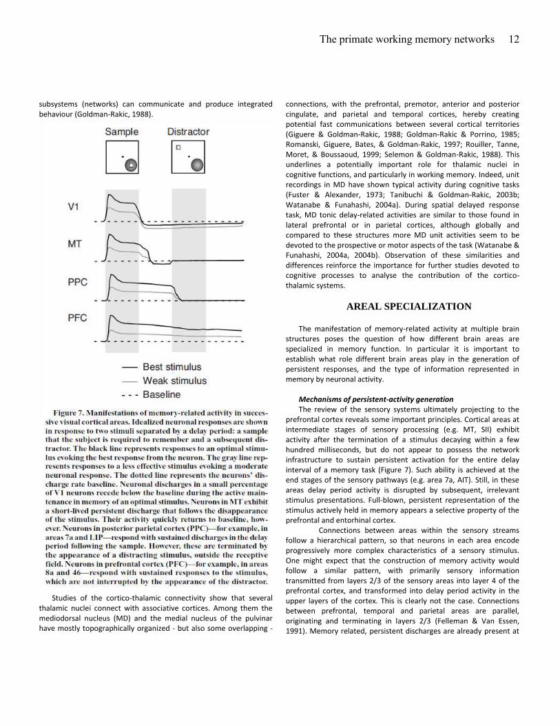
Mechanisms of persistent-activity generation
The review of the sensory systems ultimately projecting to the
prefrontal cortex reveals some important principles. Cortical areas at
intermediate stages of sensory processing (e.g. MT, SII) exhibit
activity after the termination of a stimulus decaying within a few
hundred milliseconds, but do not appear to possess the network
infrastructure to sustain persistent activation for the entire delay
interval of a memory task (Figure 7). Such ability is achieved at the
end stages of the sensory pathways (e.g. area 7a, AIT). Still, in these
areas delay period activity is disrupted by subsequent, irrelevant
stimulus presentations. Full-blown, persistent representation of the
stimulus actively held in memory appears a selective property of the
prefrontal and entorhinal cortex.
Connections between areas within the sensory streams
follow a hierarchical pattern, so that neurons in each area encode
progressively more complex characteristics of a sensory stimulus.
One might expect that the construction of memory activity would
follow a similar pattern, with primarily sensory information
transmitted from layers 2/3 of the sensory areas into layer 4 of the
prefrontal cortex, and transformed into delay period activity in the
upper layers of the cortex. This is clearly not the case. Connections
between prefrontal, temporal and parietal areas are parallel,
originating and terminating in layers 2/3 (Felleman & Van Essen,
1991). Memory related, persistent discharges are already present at
The primate working memory networks
13
the end-stages of the sensory pathways, and neurons with either
sensory-driven, motor-related or memory responses have been
encountered at all layers of the prefrontal cortex. Since this pattern
of connectivity deviates from the hierarchical model, it is not
immediately obvious how persistent activity capable of surviving the
interfering effect of distracting stimuli, emerges in the prefrontal
cortex.
Computational studies have offered insights on how
cortical networks can sustain discharges in the absence of direct
sensory stimulation and how distracting stimuli can be filtered during
the active maintenance of memory (Compte, Brunel, Goldman-Rakic,
& Wang, 2000; Lisman, Fellous, & Wang, 1998; Wang, Tegner,
Constantinidis, & Goldman-Rakic, 2004). Artificial networks can easily
generate sustained discharges following transient sensory
stimulation, if network units are densely and reciprocally connected
(Compte et al., 2000). An action potential generated by a neuron is
propagated to its synaptic targets, which in turn excite the original
neuron, allowing for the activity to reverberate in the network,
provided it does not quickly die off. Computational models have
demonstrated that persistent discharges exhibit increased signal-to-
noise ratio in networks that incorporate dopamine inputs
(Durstewitz, Seamans, & Sejnowski, 2000). The effect is generally
attributed to an enhanced NMDA conductance (Chen, Greengard, &
Yan, 2004; Seamans, Durstewitz, Christie, Stevens, & Sejnowski,
2001; Yang & Seamans, 1996), which in turn can facilitate persistent
activity by virtue of its slow time constant, leaving the postsynaptic
neuron in a depolarized state for a longer interval (Wang, 2001). The
prefrontal cortex, in contrast to its afferent inputs from the sensory
pathways, receives a significant dopaminergic innervation from the
ventral tegmental area that may render persistent activity
particularly robust and less perturbed by distracting stimulation.
Other prefrontal specializations that could have the same effect have
also been suggested, such as a differential expression of NMDA
receptors (Compte et al., 2000) or specialized interneuron cell types
(Wang et al., 2004).
After the execution of a memory-guided response in a
behavioral task, it is necessary to reset or switch off the contents of
working memory, for a new item to be stored in it. The source of
such a reset signal also remains elusive. A possible mechanism that
could achieve this goal is a non-selective burst of activity that could
equally drive all neurons and destabilize persistent discharges
(Compte et al., 2000). A burst of activity is evident in the activity of
some prefrontal neurons after the end of a single trial, or a sequence
of actions, and such activity is not specific for the preceding motor
movement (Fujii & Graybiel, 2003; Funahashi et al., 1989). Goldman-
Rakic postulated that the mediodorsal nucleus of the thalamus
constitutes the source of that signal (Goldman-Rakic et al., 1992),
however newer studies speak counter to this prediction, as most
neurons activated near the response period of the task, appear to
discharge mainly before the response initiation (Tanibuchi &
Goldman-Rakic, 2003a). At this point, the source of the reset signal
remains unclear.
Sensory Integration
Areas that make up the working memory network may also be
specialized in terms of their sensory integration. The prefrontal and
entorhinal cortex receive inputs from all sensory modalities and are
in good position to integrate these inputs and represent cross-modal
associations (Fuster et al., 2000). By some accounts, neurons
throughout the prefrontal cortex are equally potent in representing
the spatial information within 4° of the fovea and features of visual
memoranda, at least after behavioral training in a task that requires
recall of both location and identity (Rao, Rainer, & Miller, 1997).
Contrary to this account, high featural selectivity for faces was only
encountered in the inferior convexity of the prefrontal cortex which
is primarily innervated by the inferior temporal cortex (O Scalaidhe,
Wilson, & Goldman-Rakic, 1997; Wilson, O Scalaidhe, & Goldman-
Rakic, 1993). Newer results help resolve part of this discrepancy. It
now appears that the ventral visual stream already possesses
considerable spatial selectivity for stimuli within 4° of the fovea, as
discussed above (DiCarlo & Maunsell, 2003; Op De Beeck & Vogels,
2000). Similarly, the posterior parietal cortex can be highly selective
for the features of visual stimuli (Sereno & Maunsell, 1998). Although
the close proximity of the ventral- and dorsal-pathway recipient
areas in the prefrontal cortex makes the integration of all types of
information possible, it remains to be seen whether highly
specialized information converges on the same neurons.
Persistent activity may not represent strictly the sensory
attributes of stimuli, but more abstract information stored memory,
as well. Functional specialization between areas in working memory
networks may be evident in their ability to abstract information. In
recent years, the discharges of prefrontal cortical neurons have been
shown to represent rules, categories, and numerical quantities
(Asaad, Rainer, & Miller, 2000; Freedman, Riesenhuber, Poggio, &
Miller, 2001; Nieder et al., 2002; Nieder & Miller, 2003; Wallis,
Anderson et al., 2001; White & Wise, 1999). However, it is now also
evident that these are not unique prefrontal traits, as the inferior
temporal and parietal cortices demonstrate similar properties
(Freedman et al., 2003; Nieder & Miller, 2004; Sigala & Logothetis,
2002; Stoet & Snyder, 2004).
A related proposal regarding the organization of the
prefrontal cortex and working memory in general posits that distinct
subsystems process different types of information, supervised by a
central executive (Baddeley, 1992). The specialized systems can be
viewed as storage components, controlled and brought into focus by
the central executive, which has limited capacity and cannot process
all information available to the subsystems at the same time. It is
tempting to attribute neural substrates for each component of
working memory as specified by this model. It is fairly clear that
working memory for different domains of information (e.g. auditory
and visual) activate different networks. However, the functions
attributed to the central executive appear to involve multiple brain
areas, not restricted to the prefrontal cortex, and that even the
prefrontal cortex is organized in segregated domains. In the words of
Goldman-Rakic: Ou ie is that the e t al e e uti e a e composed of multiple segregated special purpose processing
The primate working memory networks
14
domains rather than one central processor served by slave systems
converging to a central processor; and that each specialized domain
consists of local and extrinsic networks with sensory, mnemonic,
motor and motivational control elements . (Gold a -Rakic, 1998)
Memory systems for action, long-term consolidation and self-
monitoring
Multisensory, memory-related discharges are present at least at
two different cortical systems, the prefrontal cortex and the medial
temporal lobe. This information may be utilized for different
purposes at each of these systems. A working memory
representation is necessary for the selection of appropriate action in
a flexible manner depending on the contingencies of the moment.
The prefrontal cortex projects to cortical and subcortical motor
structures and may guide the control of behavior depending on the
contents of the working memory buffer. The medial temporal lobe
has been thought as more involved in the consolidation of memory.
Cross-modal integration is necessary for placing information in
context before the storage into long-term memory. Finally, intelligent
eha io e ui es a o ti uous o ito i g of o e’s a tio s i relation to the internal state and values associated with the
environment. Such a function appears to be carried out by the medial
prefrontal and cingulate cortex, which requires yet a third buffer of
information. The distinct contents of each of these systems are
generally not available to introspection. One of the challenges of
neurosciences for the years ahead will be to reveal how information
in distributed cortical systems is bound together into a unitary,
conscious percept.
Authors’ note
Dedicated to the memory of Patricia S. Goldman-Rakic - our
mentor, friend, and unending source of inspiration.
Supported by the McDonnell and Whitehall Foundation (C.C.) and
the FYSSEN and NRJ Foundation (E.P.)
Bibliography
Akkal, D., Bioulac, B., Audin, J., & Burbaud, P. (2002). Comparison of
neuronal activity in the rostral supplementary and cingulate
motor areas during a task with cognitive and motor demands,
European Journal of Neuroscience 15, 887-904.
Alexander, G. E., DeLong, M. R., & Strick, P. L. (1986). Parallel
organization of functionally segregated circuits linking basal
ganglia and cortex, Annual Review of Neuroscience, 9, 357-381.
Amador, N., Schlag-Rey, M., & Schlag, J. (2000). Reward-predicting
and reward-detecting neuronal activity in the primate
supplementary eye field. Journal of Neurophysiology, 84(4), 2166-
2170.
Andersen, R. A., Essick, G. K., & Siegel, R. M. (1985). Encoding of
spatial location by posterior parietal neurons. Science, 230(4724),
456-458.
Andersen, R. A., Essick, G. K., & Siegel, R. M. (1987). Neurons of area
7 activated by both visual stimuli and oculomotor behavior.
Experimental Brain Research, 67(2), 316-322.
Andersen, R. A., & Mountcastle, V. B. (1983). The influence of the
angle of gaze upon the excitability of the light-sensitive neurons
of the posterior parietal cortex. Journal of Neuroscience, 3(3),
532-548.
Andersen, R. A., Snyder, L. H., Batista, A. P., Buneo, C. A., & Cohen, Y.
E. (1998). Posterior parietal areas specialized for eye movements
(LIP) and reach (PRR) using a common coordinate frame. Novartis
Foundation Symposium, 218, 109-122.
Andersen, R. A., Snyder, L. H., Li, C. S., & Stricanne, B. (1993).
Coordinate transformations in the representation of spatial
information. Current Opinion in Neurobiology, 3(2), 171-176.
Asaad, W. F., Rainer, G., & Miller, E. K. (2000). Task-specific neural
activity in the primate prefrontal cortex. Journal of
Neurophysiology, 84(1), 451-459.
Assad, J. A., & Maunsell, J. H. (1995). Neuronal correlates of inferred
motion in primate posterior parietal cortex. Nature, 373(6514),
518-521.
Azuma, M., & Suzuki, H. (1984). Properties and distribution of
auditory neurons in the dorsolateral prefrontal cortex of the alert
monkey. Brain Research, 298(2), 343-346.
Baddeley, A. (1992). Working memory. Science, 255(5044), 556-559.
Barbas, H. (2000). Connections underlying the synthesis of cognition,
memory, and emotion in primate prefrontal cortices. Brain
Research Bulletin, 52(5), 319-330.
Barbas, H., Ghashghaei, H., Dombrowski, S. M., & Rempel-Clower, N.
L. (1999). Medial prefrontal cortices are unified by common
connections with superior temporal cortices and distinguished by
input from memory-related areas in the rhesus monkey. Journal
of Comparative Neurology, 410(3), 343-367.
Barbas, H., & Mesulam, M. M. (1985). Cortical afferent input to the
principalis region of the rhesus monkey, Neuroscience, 15, 619-
637.
Barbas, H., & Pandya, D. N. (1987). Architecture and frontal cortical
connections of the premotor cortex (area 6) in the rhesus
monkey, Journal of Comparative Neurology, 256, 211-228.
Bates, J. F., & Goldman-Rakic, P. S. (1993). Prefrontal connections of
medial motor areas in the rhesus monkey, Journal of
Comparative Neurology, 336, 211-228).
Baxter, M. G., Parker, A., Lindner, C. C., Izquierdo, A. D., & Murray, E.
A. (2000). Control of response selection by reinforcer value
requires interaction of amygdala and orbital prefrontal cortex,
Journal of Neuroscience, 20, 4311-4319.
Bechara, A., Damasio, H., Tranel, D., & Anderson, S. W. (1998).
Dissociation Of working memory from decision making within the
human prefrontal cortex, Journal of Neuroscience, 18, 428-437.
The primate working memory networks
15
Bisley, J. W., Zaksas, D., Droll, J., & Pasternak, T. (2004). Activity of
Neurons in Cortical Area MT During a Memory for Motion Task.
Journal of Neurophysiology, 91(1), 286-300.
Blatt, G. J., Andersen, R. A., & Stoner, G. R. (1990). Visual receptive
field organization and cortico-cortical connections of the lateral
intraparietal area (area LIP) in the macaque. Journal of
Comparative Neurology, 299(4), 421-445.
Bodner, M., Kroger, J., & Fuster, J. M. (1996). Auditory memory cells
in dorsolateral prefrontal cortex. Neuroreport, 7(12), 1905-1908.
Boussaoud, D., & Wise, S. P. (1993). Primate frontal cortex: effects of
stimulus and movement. Experimental Brain Research, 95(1), 28-
40.
Bremmer, F., Duhamel, J. R., Ben Hamed, S., & Graf, W. (2002).
Heading encoding in the macaque ventral intraparietal area (VIP).
Euopeanr Journal of Neuroscience, 16(8), 1554-1568.
Brotchie, P. R., Andersen, R. A., Snyder, L. H., & Goodman, S. J.
(1995). Head position signals used by parietal neurons to encode
locations of visual stimuli. Nature, 375(6528), 232-235.
Buffalo, E. A., Ramus, S. J., Squire, L. R., & Zola, S. M. (2000).
Perception and recognition memory in monkeys following lesions
of area TE and perirhinal cortex. Learning & Memory, 7(6), 375-
382.
Bushnell, M. C., Goldberg, M. E., & Robinson, D. L. (1981). Behavioral
enhancement of visual responses in monkey cerebral cortex. I.
Modulation in posterior parietal cortex related to selective visual
attention. Journal of Neurophysiology, 46(4), 755-772.
Carmichael, S. T., Clugnet, M. C., & Price, J. L. (1994). Central
olfactory connections in the macaque monkey. Journal of
Comparative Neurology, 346(3), 403-434.
Castner, S. A., Goldman-Rakic, P. S., & Williams, G. V. (2004). Animal
models of working memory: insights for targeting cognitive
dysfunction in schizophrenia, Psychopharmacology 174(1):111-
25.
Cavada, C., Company, T., Tejedor, J., Cruz-Rizzolo, R. J., & Reinoso-
Suarez, F. (2000). The anatomical connections of the macaque
monkey orbitofrontal cortex. A review. Cerebral Cortex, 10(3),
220-242.
Cavada, C., & Goldman-Rakic, P. S. (1989). Posterior parietal cortex in
rhesus monkey: II. Evidence for segregated corticocortical
networks linking sensory and limbic areas with the frontal lobe.
Journal of Comparative Neurology, 287(4), 422-445.
Chafee, M. V., & Goldman-Rakic, P. S. (1998). Matching patterns of
activity in primate prefrontal area 8a and parietal area 7ip
neurons during a spatial working memory task. Journal of
Neurophysiology, 79(6), 2919-2940.
Chelazzi, L., Duncan, J., Miller, E. K., & Desimone, R. (1998).
Responses of neurons in inferior temporal cortex during memory-
guided visual search. Journal of Neurophysiology, 80(6), 2918-
2940.
Chelazzi, L., Miller, E. K., Duncan, J., & Desimone, R. (1993). A neural
basis for visual search in inferior temporal cortex. Nature,
363(6427), 345-347.
Chen, G., Greengard, P., & Yan, Z. (2004). Potentiation of NMDA
receptor currents by dopamine D1 receptors in prefrontal cortex.
Proceedings of the National Academy of Sciences of the United
States of America, 101(8), 2596-2600.
Cheng, K., Hasegawa, T., Saleem, K. S., & Tanaka, K. (1994).
Comparison of neuronal selectivity for stimulus speed, length,
and contrast in the prestriate visual cortical areas V4 and MT of
the macaque monkey. Journal of Neurophysiology, 71(6), 2269-
2280.
Colby, C. L., Duhamel, J. R., & Goldberg, M. E. (1993). Ventral
intraparietal area of the macaque: anatomic location and visual
response properties. Journal of Neurophysiology, 69(3), 902-914.
Compte, A., Brunel, N., Goldman-Rakic, P. S., & Wang, X. J. (2000).
Synaptic mechanisms and network dynamics underlying spatial
working memory in a cortical network model. Cerebral Cortex,
10(9), 910-923.
Compte, A., Constantinidis, C., Tegner, J., Raghavachari, S., Chafee,
M. V., Goldman-Rakic, P. S., & Wang, X. J. (2003). Temporally
irregular mnemonic persistent activity in prefrontal neurons of
monkeys during a delayed response task. Journal of
Neurophysiology, 28, 28.
Connor, C. E., Gallant, J. L., Preddie, D. C., & Van Essen, D. C. (1996).
Responses in area V4 depend on the spatial relationship between
stimulus and attention. Journal of Neurophysiology, 75(3), 1306-
1308.
Connor, C. E., Preddie, D. C., Gallant, J. L., & Van Essen, D. C. (1997).
Spatial attention effects in macaque area V4. Journal of
Neuroscience, 17(9), 3201-3214.
Constantinidis, C., Franowicz, M. N., & Goldman-Rakic, P. S. (2001a).
Coding specificity in cortical microcircuits: a multiple electrode
analysis of primate prefrontal cortex. Journal of Neuroscience,
21(10), 3646-3655.
Constantinidis, C., Franowicz, M. N., & Goldman-Rakic, P. S. (2001b).
The sensory nature of mnemonic representation in the primate
prefrontal cortex. Nature Neuroscience, 4(3), 311-316.
Constantinidis, C., & Steinmetz, M. A. (1996). Neuronal activity in
posterior parietal area 7a during the delay periods of a spatial
memory task. Journal of Neurophysiology, 76(2), 1352-1355.
Constantinidis, C., & Steinmetz, M. A. (2001a). Neuronal responses in
area 7a to multiple stimulus displays: I. Neurons encode the
location of the salient stimulus. Cerebral Cortex, 11, 581-591.
Constantinidis, C., & Steinmetz, M. A. (2001b). Neuronal responses in
area 7a to multiple stimulus displays: II. Responses are
suppressed at the cued location. Cerebral Cortex, 11(7), 592-597.
Cook, E. P., & Maunsell, J. H. (2002). Attentional modulation of
behavioral performance and neuronal responses in middle
temporal and ventral intraparietal areas of macaque monkey.
Journal of Neuroscience, 22(5), 1994-2004.
Courtney, S. M., Ungerleider, L. G., Keil, K., & Haxby, J. V. (1997).
Transient and sustained activity in a distributed neural system for
human working memory. Nature, 386(6625), 608-611.
Crammond, D. J., & Kalaska, J. F. (1994). Modulation of preparatory
neuronal activity in dorsal premotor cortex due to stimulus-
The primate working memory networks
16
response compatibility, Journal of Neurophysiology, 71, 1281-
1284.
Crowe, D. A., Chafee, M. V., Averbeck, B. B., & Georgopoulos, A. P.
(2004). Neural Activity in Primate Parietal Area 7a Related to
Spatial Analysis of Visual Mazes. Cerebral Cortex, 14(1), 23-34.
Desimone, R., Albright, T. D., Gross, C. G., & Bruce, C. (1984).
Stimulus-selective properties of inferior temporal neurons in the
macaque. Journal of Neuroscience, 4(8), 2051-2062.
Desimone, R., Schein, S. J., Moran, J., & Ungerleider, L. G. (1985).
Contour, color and shape analysis beyond the striate cortex.
Vision Research, 25(3), 441-452.
di Pellegrino, G., & Wise, S. P. (1991). A neurophysiological
comparison of three distinct regions of the primate frontal lobe,
Brain, 114, 951-978.
di Pellegrino, G., & Wise, S. P. (1993a). Effects of attention on
visuomotor activity in the premotor and prefrontal cortex of a
primate. Somatosensory & Motor Research, 10(3), 245-262.
di Pellegrino, G., & Wise, S. P. (1993b). Visuospatial versus
visuomotor activity in the premotor and prefrontal cortex of a
primate. Journal of Neuroscience, 13(3), 1227-1243.
DiCarlo, J. J., & Maunsell, J. H. (2003). Anterior inferotemporal
neurons of monkeys engaged in object recognition can be highly
sensitive to object retinal position. Journal of Neurophysiology,
89(6), 3264-3278.
Distler, C., Boussaoud, D., Desimone, R., & Ungerleider, L. G. (1993).
Cortical connections of inferior temporal area TEO in macaque
monkeys. Journal of Comparative Neurology, 334(1), 125-150.
Dubois, B., & Pillon, B. (1997). Cognitive deficits in Parkinson's
disease, Journal of Neurology, 244, 2-8.
Duffy, C. J., & Wurtz, R. H. (1991). Sensitivity of MST neurons to optic
flow stimuli. I. A continuum of response selectivity to large-field
stimuli. Journal of Neurophysiology, 65(6), 1329-1345.
Duhamel, J. R., Bremmer, F., BenHamed, S., & Graf, W. (1997). Spatial
invariance of visual receptive fields in parietal cortex neurons.
Nature, 389(6653), 845-848.
Duhamel, J. R., Colby, C. L., & Goldberg, M. E. (1992). The updating of
the representation of visual space in parietal cortex by intended
eye movements. Science, 255(5040), 90-92.
Duhamel, J. R., Colby, C. L., & Goldberg, M. E. (1998). Ventral
intraparietal area of the macaque: congruent visual and somatic
response properties. Journal of Neurophysiology, 79(1), 126-136.
Dum, R. P., & Strick, P. L. (1991). The origin of corticospinal
projections from the premotor areas in the frontal lobe, Journal
of Neuroscience, 11, 667-689.
Dum, R. P., & Strick, P. L. (1993). Cingulate motor areas. In M. Gabriel
(Ed.), Neurobiology of cingulate cortex and limbic thalamus (415-
441): Birkhaüser.
Duncan, J., & Owen, A. M. (2000). Common regions of the human
frontal lobe recruited by diverse cognitive demands, Trends in
Neurosciences, 23, 475-483.
Durstewitz, D., Seamans, J. K., & Sejnowski, T. J. (2000). Dopamine-
mediated stabilization of delay-period activity in a network model
of prefrontal cortex. Journal of Neurophysiology, 83(3), 1733-
1750.
Erickson, C. A., & Desimone, R. (1999). Responses of macaque
perirhinal neurons during and after visual stimulus association
learning. Journal of Neuroscience, 19(23), 10404-10416.
Felleman, D. J., & Van Essen, D. C. (1991). Distributed hierarchical
processing in the primate cerebral cortex. Cerebral Cortex, 1(1),
1-47.
Ferrera, V. P., Nealey, T. A., & Maunsell, J. H. (1992). Mixed
parvocellular and magnocellular geniculate signals in visual area
V4. Nature, 358(6389), 756-761.
Freedman, D. J., Riesenhuber, M., Poggio, T., & Miller, E. K. (2001).
Categorical representation of visual stimuli in the primate
prefrontal cortex. Science, 291(5502), 312-316.
Freedman, D. J., Riesenhuber, M., Poggio, T., & Miller, E. K. (2002).
Visual categorization and the primate prefrontal cortex:
neurophysiology and behavior. Journal of Neurophysiology, 88(2),
929-941.
Freedman, D. J., Riesenhuber, M., Poggio, T., & Miller, E. K. (2003). A
comparison of primate prefrontal and inferior temporal cortices
during visual categorization. Journal of Neuroscience, 23(12),
5235-5246.
Friedman, H. R., & Goldman-Rakic, P. S. (1994). Coactivation of
prefrontal cortex and inferior parietal cortex in working memory
tasks revealed by 2DG functional mapping in the rhesus monkey.
Journal of Neuroscience, 14, 2775-2788.
Frith, C. D., Friston, K. J., Liddle, P. F., & Frackowiak, R. S. (1991).
Willed action and the prefrontal cortex in man: a study with PET,
Proceedings of the Royal Society of London. Series B. Biological
Sciences, 244, 241-246.
Fujii, N., & Graybiel, A. M. (2003). Representation of action sequence
boundaries by macaque prefrontal cortical neurons. Science,
301(5637), 1246-1249.
Fujita, I., Tanaka, K., Ito, M., & Cheng, K. (1992). Columns for visual
features of objects in monkey inferotemporal cortex. Nature,
360(6402), 343-346.
Funahashi, S., Bruce, C. J., & Goldman-Rakic, P. S. (1989). Mnemonic
coding of visual space in the monkey's dorsolateral prefrontal
cortex. Journal of Neurophysiology, 61(2), 331-349.
Funahashi, S., Bruce, C. J., & Goldman-Rakic, P. S. (1993). Dorsolateral
prefrontal lesions and oculomotor delayed-response
performance: evidence for mnemonic "scotomas". Journal of
Neuroscience, 13(4), 1479-1497.
Fuster, J. M. (2001). The prefrontal cortex--an update: time is of the
essence, Neuron, 30, 319-333.
Fuster, J. M. (2003). Cortex and Mind: Unifying Cognition. Oxford:
Oxford University Press.
Fuster, J. M., & Alexander, G. E. (1971). Neuron activity related to
short-term memory. Science, 173(997), 652-654.
Fuster, J. M., & Alexander, G. E. (1973). Firing changes in cells of the
nucleus medialis dorsalis associated with delayed response
behavior, Brain Research, 61, 79-91.
The primate working memory networks
17
Fuster, J. M., Bodner, M., & Kroger, J. K. (2000). Cross-modal and
cross-temporal association in neurons of frontal cortex. Nature,
405(6784), 347-351.
Fuster, J. M., & Jervey, J. P. (1981). Inferotemporal neurons
distinguish and retain behaviorally relevant features of visual
stimuli. Science, 212(4497), 952-955.
Fuster, J. M., & Jervey, J. P. (1982). Neuronal firing in the
inferotemporal cortex of the monkey in a visual memory task.
Journal of Neuroscience, 2(3), 361-375.
Gallant, J. L., Connor, C. E., Rakshit, S., Lewis, J. W., & Van Essen, D. C.
(1996). Neural responses to polar, hyperbolic, and Cartesian
gratings in area V4 of the macaque monkey. Journal of
Neurophysiology, 76(4), 2718-2739.
Galletti, C., Battaglini, P. P., & Fattori, P. (1995). Eye position
influence on the parieto-occipital area PO (V6) of the macaque
monkey. European Journal of Neuroscience, 7(12), 2486-2501.
Giguere, M., & Goldman-Rakic, P. S. (1988). Mediodorsal nucleus:
areal, laminar, and tangential distribution of afferents and
efferents in the frontal lobe of rhesus monkeys, Journal of
Comparative Neurology, 277, 195-213.
Gnadt, J. W., & Andersen, R. A. (1988). Memory related motor
planning activity in posterior parietal cortex of macaque.
Experimental Brain Research, 70(1), 216-220.
Godschalk, M., Mitz, A. R., van Duin, B., & van der Burg, H. (1995).
Somatotopy of monkey premotor cortex examined with
microstimulation, Neuroscience Research, 23, 269-279.
Goldman-Rakic, P. S. (1988). Topography of cognition: parallel
distributed networks in primate association cortex. Annual
Review of Neuroscience, 11, 137-156.
Goldman-Rakic P (1998) The prefrontal landscape: implications of
functional architecture for understanding human mentation and
the central executive. In: The Prefrontal Cortex (Roberts AC,
Robbins TW, Weiskrantz L, eds), pp 87-102. Oxford: Oxford
University Press.
Goldman-Rakic, P. S., Bates, J. F., & Chafee, M. V. (1992). The
prefrontal cortex and internally generated motor acts. Current
Opinion in Neurobiology, 2(6), 830-835.
Goldman-Rakic, P. S., & Porrino, L. J. (1985). The primate mediodorsal
(MD) nucleus and its projection to the frontal lobe, Journal of
Comparative Neurology, 242, 535-560.
Gottlieb, J. P., Kusunoki, M., & Goldberg, M. E. (1998). The
representation of visual salience in monkey parietal cortex.
Nature, 391(6666), 481-484.
Gross, C. G., Bender, D. B., & Gerstein, G. L. (1979). Activity of inferior
temporal neurons in behaving monkeys. Neuropsychologia, 17(2),
215-229.
Gross, C. G., Rocha-Miranda, C. E., & Bender, D. B. (1972). Visual
properties of neurons in inferotemporal cortex of the Macaque.
Journal of Neurophysiology, 35(1), 96-111.
Haber, S. N., Kunishio, K., Mizobuchi, M., & Lynd-Balta, E. (1995). The
orbital and medial prefrontal circuit through the primate basal
ganglia, Journal of Neuroscience 15, 4851-4867.
Hackett, T. A., Stepniewska, I., & Kaas, J. H. (1999). Prefrontal
connections of the parabelt auditory cortex in macaque monkeys.
Brain Research, 817(1-2), 45-58.
Hadj-Bouziane, F., Meunier, M., & Boussaoud, D. (2003). Conditional
visuo-motor learning in primates: a key role for the basal ganglia,
Journal of Physiology, 97, 567-579.
Hadland, K. A., Rushworth, M. F., Gaffan, D., & Passingham, R. E.
(2003). The anterior cingulate and reward-guided selection of
actions, Journal of Neurophysiology, 89, 1161-1164.
Haenny, P. E., Maunsell, J. H., & Schiller, P. H. (1988). State
dependent activity in monkey visual cortex. II. Retinal and
extraretinal factors in V4. Experimental Brain Research, 69(2),
245-259.
Hampson, R. E., Pons, T. P., Stanford, T. R., & Deadwyler, S. A. (2004).
Categorization in the monkey hippocampus: a possible
mechanism for encoding information into memory. Proceedings
of the National Academy of Sciences of the United States of
America, 101(9), 3184-3189. Epub 2004 Feb 3120.
Hatanaka, N., Tokuno, H., Hamada, I., Inase, M., Ito, Y., Imanishi, M.,
Hasegawa, N., Akazawa, T., Nambu, A., & Takada, M. (2003).
Thalamocortical and intracortical connections of monkey
cingulate motor areas, Journal of Comparative Neurology, 462,
121-138.
Heuer, H. W., & Britten, K. H. (2004). Optic flow signals in extrastriate
area MST: comparison of perceptual and neuronal sensitivity.
Journal of Neurophysiology, 91(3), 1314-1326. Epub 2003 Oct
1318.
Hikosaka, K., & Watanabe, M. (2000). Delay activity of orbital and
lateral prefrontal neurons of the monkey varying with different
rewards, Cerebral Cortex, 10, 263-271.
Hikosaka, O., Sakamoto, M., & Usui, S. (1989a). Functional properties
of monkey caudate neurons. I. Activities related to saccadic eye
movements, Journal of Neurophysiology, 61, 780-798.
Hikosaka, O., Sakamoto, M., & Usui, S. (1989b). Functional properties
of monkey caudate neurons. III. Activities related to expectation
of target and reward, Journal of Neurophysiology, 61, 814-832.
Hinkle, D. A., & Connor, C. E. (2002). Three-dimensional orientation
tuning in macaque area V4. Nature Neuroscience, 5(7), 665-670.
Holscher, C., & Rolls, E. T. (2002). Perirhinal cortex neuronal activity is
actively related to working memory in the macaque. Neural
Plasticity, 9(1), 41-51.
Holscher, C., Rolls, E. T., & Xiang, J. (2003). Perirhinal cortex neuronal
activity related to long-term familiarity memory in the macaque.
European Journal of Neuroscience, 18(7), 2037-2046.
Houk, J. C., & Wise, S. P. (1995). Distributed modular architectures
linking basal ganglia, cerebellum, and cerebral cortex: their role in
planning and controlling action, Cerebral Cortex, 5, 95-110.
Hsiao, S. S., O'Shaughnessy, D. M., & Johnson, K. O. (1993). Effects of
selective attention on spatial form processing in monkey primary
and secondary somatosensory cortex. Journal of
Neurophysiology, 70(1), 444-447.
Huerta, M. F., Krubitzer, L. A., & Kaas, J. H. (1986). Frontal eye field as
defined by intracortical microstimulation in squirrel monkeys, owl
The primate working memory networks
18
monkeys, and macaque monkeys: I. Subcortical connections,
Journal of Comparative Neurology, 253, 415-439.
Huerta, M. F., Krubitzer, L. A., & Kaas, J. H. (1987). Frontal eye field as
defined by intracortical microstimulation in squirrel monkeys, owl
monkeys, and macaque monkeys. II. Cortical connections, Journal
of Comparative Neurology, 265, 332-361.
Iba, M., & Sawaguchi, T. (2003). Involvement of the dorsolateral
prefrontal cortex of monkeys in visuospatial target selection.
Journal of Neurophysiology, 89(1), 587-599.
Ifuku, H., Hirata, S., Nakamura, T., & Ogawa, H. (2003). Neuronal
activities in the monkey primary and higher-order gustatory
cortices during a taste discrimination delayed GO/NOGO task and
after reversal. Neuroscience Research, 47(2), 161-175.
Inoue, M., Mikami, A., Ando, I., & Tsukada, H. (2004). Functional
brain mapping of the macaque related to spatial working memory
as revealed by PET, Cerebral Cortex, 14, 106-119.
Isomura, Y., Ito, Y., Akazawa, T., Nambu, A., & Takada, M. (2003).
Neural coding of "attention for action" and "response selection"
in primate anterior cingulate cortex, Journal of Neuroscience, 23,
8002-8012.
Ito, M., Tamura, H., Fujita, I., & Tanaka, K. (1995). Size and position
invariance of neuronal responses in monkey inferotemporal
cortex. Journal of Neurophysiology, 73(1), 218-226.
Jacobsen, C. F. (1936). Studies of cerebral function in primates.
Comparative Psychology Monogaphs, 13, 1-68.
Jagadeesh, B., Chelazzi, L., Mishkin, M., & Desimone, R. (2001).
Learning increases stimulus salience in anterior inferior temporal
cortex of the macaque. Journal of Neurophysiology, 86(1), 290-
303.
Joel, D., & Weiner, I. (2000). The connections of the dopaminergic
system with the striatum in rats and primates: an analysis with
respect to the functional and compartmental organization of the
striatum. Neuroscience, 96(3), 451-474.
Jones, E. G., Dell'Anna, M. E., Molinari, M., Rausell, E., & Hashikawa,
T. (1995). Subdivisions of macaque monkey auditory cortex
revealed by calcium-binding protein immunoreactivity. Journal of
Comparative Neurology, 362(2), 153-170.
Jonides, J., Smith, E. E., Koeppe, R. A., Awh, E., Minoshima, S., &
Mintun, M. A. (1993). Spatial working memory in humans as
revealed by PET. Nature, 363(6430), 623-625.
Jouffrais, C., & Boussaoud, D. (1999). Neuronal activity related to eye-
hand coordination in the primate premotor cortex, Experimental
Brain Research, 128, 205-209.
Kaas, J. H., & Hackett, T. A. (2000). Subdivisions of auditory cortex
and processing streams in primates. Proceedings of the National
Academy of Sciences of the United States of America, 97(22),
11793-11799.
Kawagoe, R., Takikawa, Y., & Hikosaka, O. (1998). Expectation of
reward modulates cognitive signals in the basal ganglia, Nature
Neuroscience, 1, 411-416.
Kawamura, K., & Naito, J. (1984). Corticocortical projections to the
prefrontal cortex in the rhesus monkey investigated with
horseradish peroxidase techniques. Neuroscience Research, 1(2),
89-103.
Kermadi, I., & Joseph, J. P. (1995). Activity in the caudate nucleus of
monkey during spatial sequencing, Journal of Neurophysiology,
74, 911-933.
Koch, C., & Fuster, J. M. (1989). Unit activity in monkey parietal
cortex related to haptic perception and temporary memory.
Experimental Brain Research, 76(2), 292-306.
Koechlin, E., Ody, C., & Kouneiher, F. (2003). The architecture of
cognitive control in the human prefrontal cortex, Science, 302,
1181-1185.
Komatsu, H., & Wurtz, R. H. (1988). Relation of cortical areas MT and
MST to pursuit eye movements. I. Localization and visual
properties of neurons. Journal of Neurophysiology, 60(2), 580-
603.
Kosaki, H., Hashikawa, T., He, J., & Jones, E. G. (1997). Tonotopic
organization of auditory cortical fields delineated by parvalbumin
immunoreactivity in macaque monkeys. Journal of Comparative
Neurology, 386(2), 304-316.
Koski, L., & Paus, T. (2000). Functional connectivity of the anterior
cingulate cortex within the human frontal lobe: a brain-mapping
meta-analysis, Experimental Brain Research, 133, 55-65.
Kritzer, M. F., & Goldman-Rakic, P. S. (1995). Intrinsic circuit
organization of the major layers and sublayers of the dorsolateral
prefrontal cortex in the rhesus monkey. Journal of Comparative
Neurology, 359(1), 131-143.
Lisman, J. E., Fellous, J. M., & Wang, X. J. (1998). A role for NMDA-
receptor channels in working memory. Nature Neuroscience, 1(4),
273-275.
Liu, Z., & Richmond, B. J. (2000). Response differences in monkey TE
and perirhinal cortex: stimulus association related to reward
schedules. Journal of Neurophysiology, 83(3), 1677-1692.
Llinas, R., Ribary, U., Contreras, D., & Pedroarena, C. (1998). The
neuronal basis for consciousness, Philosophical Transactions of
the Royal Society of London. Series B. Biological Sciences 353,
1841-1849.
Llinas, R. R., Leznik, E., & Urbano, F. J. (2002). Temporal binding via
cortical coincidence detection of specific and nonspecific
thalamocortical inputs: a voltage-dependent dye-imaging study in
mouse brain slices, Proceedings of the National Academy of
Sciences of the United States of America, 99, 449-454.
Logothetis, N. K., & Pauls, J. (1995). Psychophysical and physiological
evidence for viewer-centered object representations in the
primate. Cerebral Cortex, 5(3), 270-288.
Logothetis, N. K., Pauls, J., & Poggio, T. (1995). Shape representation
in the inferior temporal cortex of monkeys. Current Biology, 5(5),
552-563.
Lu, M. T., Preston, J. B., & Strick, P. L. (1994). Interconnections
between the prefrontal cortex and the premotor areas in the
frontal lobe, Journal of Comparative Neurology, 341, 375-392.
Luck, S. J., Chelazzi, L., Hillyard, S. A., & Desimone, R. (1997). Neural
mechanisms of spatial selective attention in areas V1, V2, and V4
The primate working memory networks
19
of macaque visual cortex. Journal of Neurophysiology, 77(1), 24-
42.
Luppino, G., Matelli, M., Camarda, R. M., Gallese, V., & Rizzolatti, G.
(1991). Multiple representations of body movements in mesial
area 6 and the adjacent cingulate cortex: an intracortical
microstimulation study in the macaque monkey, Journal of
Comparative Neurology, 311, 463-482.
Lynch, J. C., Mountcastle, V. B., Talbot, W. H., & Yin, T. C. (1977).
Parietal lobe mechanisms for directed visual attention. Journal of
Neurophysiology, 40(2), 362-389.
MacDonald, A. W., 3rd, Cohen, J. D., Stenger, V. A., & Carter, C. S.
(2000). Dissociating the role of the dorsolateral prefrontal and
anterior cingulate cortex in cognitive control, Science, 288, 1835-
1838.
Markowitsch, H. J., Emmans, D., Irle, E., Streicher, M., & Preilowski, B.
(1985). Cortical and subcortical afferent connections of the
primate's temporal pole: a study of rhesus monkeys, squirrel
monkeys, and marmosets. Journal of Comparative Neurology,
242(3), 425-458.
Matelli, M., Luppino, G., & Rizzolatti, G. (1991). Architecture of
superior and mesial area 6 and the adjacent cingulate cortex in
the macaque monkey, Journal of Comparative Neurology, 311,
445-462.
Matsumoto, K., Suzuki, W., & Tanaka, K. (2003). Neuronal correlates
of goal-based motor selection in the prefrontal cortex. Science,
301(5630), 229-232.
McGuire, P. K., Bates, J. F., & Goldman-Rakic, P. S. (1991).
Interhemispheric integration: I. Symmetry and convergence of
the corticocortical connections of the left and the right principal
sulcus (PS) and the left and the right supplementary motor area
(SMA) in the rhesus monkey, Cerebral Cortex 1, 390-407.
Merchant, H., Battaglia-Mayer, A., & Georgopoulos, A. P. (2001).
Effects of optic flow in motor cortex and area 7a. Journal of
Neurophysiology, 86(4), 1937-1954.
Merchant, H., Battaglia-Mayer, A., & Georgopoulos, A. P. (2004).
Neural responses in motor cortex and area 7a to real and
apparent motion. Experimental Brain Research, 154(3), 291-307.
Messier, J., & Kalaska, J. F. (2000). Covariation of primate dorsal
premotor cell activity with direction and amplitude during a
memorized-delay reaching task, Journal of Neurophysiology, 84,
152-165.
Messinger, A., Squire, L. R., Zola, S. M., & Albright, T. D. (2001).
Neuronal representations of stimulus associations develop in the
temporal lobe during learning. Proceedings of the National
Academy of Sciences of the United States of America, 98(21),
12239-12244.
Meunier, M., Bachevalier, J., & Mishkin, M. (1997). Effects of orbital
frontal and anterior cingulate lesions on object and spatial
memory in rhesus monkeys, Neuropsychologia, 35, 999-1015.
Middleton, F. A., & Strick, P. L. (2002). Basal-ganglia 'projections' to
the prefrontal cortex of the primate, Cerebral Cortex, 12, 926-
935.
Miller, E. K., & Cohen, J. D. (2001). An integrative theory of prefrontal
cortex function, Annual Review of Neuroscience, 24, 167-202.
Miller, E. K., Erickson, C. A., & Desimone, R. (1996). Neural
mechanisms of visual working memory in prefrontal cortex of the
macaque. Journal of Neuroscience, 16(16), 5154-5167.
Miller, E. K., Li, L., & Desimone, R. (1991). A neural mechanism for
working and recognition memory in inferior temporal cortex.
Science, 254(5036), 1377-1379.
Miller, E. K., Li, L., & Desimone, R. (1993). Activity of neurons in
anterior inferior temporal cortex during a short-term memory
task. Journal of Neuroscience, 13(4), 1460-1478.
Milner, B. (1963). Effects of different brain lesions on card sorting.
Archives of Neurology, 9, 100-110.
Mitz, A. R., & Wise, S. P. (1987). The somatotopic organiztion of the
supplementary motor area: intracortical microstimulation
mapping, Journal of Neuroscience, 7, 1010-1021.
Miyashita, Y., & Chang, H. S. (1988). Neuronal correlate of pictorial
short-term memory in the primate temporal cortex. Nature,
331(6151), 68-70.
Moran, J., & Desimone, R. (1985). Selective attention gates visual
processing in the extrastriate cortex. Science, 229(4715), 782-
784.
Motter, B. C. (1994). Neural correlates of attentive selection for color
or luminance in extrastriate area V4. Journal of Neuroscience,
14(4), 2178-2189.
Motter, B. C., & Mountcastle, V. B. (1981). The functional properties
of the light-sensitive neurons of the posterior parietal cortex
studied in waking monkeys: foveal sparing and opponent vector
organization. Journal of Neuroscience, 1(1), 3-26.
Mountcastle, V. B., Lynch, J. C., Georgopoulos, A., Sakata, H., &
Acuna, C. (1975). Posterior parietal association cortex of the
monkey: command functions for operations within extrapersonal
space. Journal of Neurophysiology, 38(4), 871-908.
Nakamura, K., & Colby, C. L. (2000). Visual, saccade-related, and
cognitive activation of single neurons in monkey extrastriate area
V3A. Journal of Neurophysiology, 84(2), 677-692.
Nakamura, K., & Kubota, K. (1995). Mnemonic firing of neurons in the
monkey temporal pole during a visual recognition memory task.
Journal of Neurophysiology, 74(1), 162-178.
Nakamura, K., Sakai, K., & Hikosaka, O. (1998). Neuronal activity in
medial frontal cortex during learning of sequential procedures,
Journal of Neurophysiology, 80, 2671-2687.
Naya, Y., Yoshida, M., & Miyashita, Y. (2001). Backward spreading of
memory-retrieval signal in the primate temporal cortex. Science,
291(5504), 661-664.
Newsome, W. T., Mikami, A., & Wurtz, R. H. (1986). Motion
selectivity in macaque visual cortex. III. Psychophysics and
physiology of apparent motion. Journal of Neurophysiology,
55(6), 1340-1351.
Nieder, A., Freedman, D. J., & Miller, E. K. (2002). Representation of
the quantity of visual items in the primate prefrontal cortex.
Science, 297(5587), 1708-1711.
The primate working memory networks
20
Nieder, A., & Miller, E. K. (2003). Coding of cognitive magnitude.
Compressed scaling of numerical information in the primate
prefrontal cortex. Neuron, 37(1), 149-157.
Nieder, A., & Miller, E. K. (2004). A parieto-frontal network for visual
numerical information in the monkey. Proceedings of the
National Academy of Sciences of the United States of America,
101(19), 7457-7462.
Niki, H., & Watanabe, M. (1976). Cingulate unit activity and delayed
response, Brain Research, 110, 381-386.
O Scalaidhe, S., Wilson, F. A., & Goldman-Rakic, P. S. (1997). Areal
segregation of face-processing neurons in prefrontal cortex.
Science, 278(5340), 1135-1138.
Olson, C. R., & Gettner, S. N. (1995). Object-centered direction
selectivity in the macaque supplementary eye field. Science,
269(5226), 985-988.
Op De Beeck, H., & Vogels, R. (2000). Spatial sensitivity of macaque
inferior temporal neurons. Journal of Comparative Neurology,
426(4), 505-518.
Passingham, R. (1993). The frontal lobes and voluntary action, Oxford
Psychology series: Oxford University Press.
Pasupathy, A., & Connor, C. E. (2001). Shape representation in area
V4: position-specific tuning for boundary conformation. Journal
of Neurophysiology, 86(5), 2505-2519.
Paus, T. (2001). Primate anterior cingulate cortex: where motor
control, drive and cognition interface, Nature Reviews
Neuroscience 2, 417-424.
Paus, T., Petrides, M., Evans, A. C., & Meyer, E. (1993). Role of the
human anterior cingulate cortex in the control of oculomotor,
manual, and speech responses: a positron emission tomography
study, Journal of Neurophysiology, 70, 453-469.
Pesaran, B., Pezaris, J. S., Sahani, M., Mitra, P. P., & Andersen, R. A.
(2002). Temporal structure in neuronal activity during working
memory in macaque parietal cortex. Nature Neuroscience, 5(8),
805-811.
Petrides, M. (2000). The role of the mid-dorsolateral prefrontal
cortex in working memory. Experimental Brain Research, 133(1),
44-54.
Phillips, J. R., Johnson, K. O., & Hsiao, S. S. (1988). Spatial pattern
representation and transformation in monkey somatosensory
cortex. Proceedings of the National Academy of Sciences of the
United States of America, 85(4), 1317-1321.
Phinney, R. E., & Siegel, R. M. (2000). Speed selectivity for optic flow
in area 7a of the behaving macaque. Cerebral Cortex, 10(4), 413-
421.
Platt, M. L., & Glimcher, P. W. (1999). Neural correlates of decision
variables in parietal cortex. Nature, 400(6741), 233-238.
Posner, M. I., & DiGirolamo, G. J. (1998). Executive attention:
Conflict, target detection and cognitive control. In R.
Parasuraman (Ed.), The attentive brain. Cambridge: MIT Press.
Powell, K. D., & Goldberg, M. E. (2000). Response of neurons in the
lateral intraparietal area to a distractor flashed during the delay
period of a memory-guided saccade. Journal of Neurophysiology,
84(1), 301-310.
Preuss, T. M., & Goldman-Rakic, P. S. (1991). Architectonics of the
parietal and temporal association cortex in the strepsirhine
primate Galago compared to the anthropoid primate Macaca.
Journal of Comparative Neurology, 310(4), 475-506.
Procyk, E., & Joseph, J. P. (2001). Characterization of serial order
encoding in the monkey anterior cingulate sulcus, European
Journal of Neuroscience 14, 1041-1046.
Procyk, E., Tanaka, Y. L., & Joseph, J. P. (2000). Anterior cingulate
activity during routine and non-routine sequential behaviors in
macaques, Nature Neuroscience, 3, 502-508.
Pucak, M. L., Levitt, J. B., Lund, J. S., & Lewis, D. A. (1996). Patterns of
intrinsic and associational circuitry in monkey prefrontal cortex.
Journal of Comparative Neurology, 376(4), 614-630.
Quintana, J., & Fuster, J. M. (1992). Mnemonic and predictive
functions of cortical neurons in a memory task. Neuroreport, 3(8),
721-724.
Raiguel, S., Van Hulle, M. M., Xiao, D. K., Marcar, V. L., Lagae, L., &
Orban, G. A. (1997). Size and shape of receptive fields in the
medial superior temporal area (MST) of the macaque.
Neuroreport, 8(12), 2803-2808.
Rainer, G., & Miller, E. K. (2002). Timecourse of object-related neural
activity in the primate prefrontal cortex during a short-term
memory task. Eur Journal of Neuroscience, 15(7), 1244-1254.
Rao, S. C., Rainer, G., & Miller, E. K. (1997). Integration of what and
where in the primate prefrontal cortex. Science, 276(5313), 821-
824.
Rauschecker, J. P., Tian, B., & Hauser, M. (1995). Processing of
complex sounds in the macaque nonprimary auditory cortex.
Science, 268(5207), 111-114.
Richmond, B. J., Wurtz, R. H., & Sato, T. (1983). Visual responses of
inferior temporal neurons in awake rhesus monkey. Journal of
Neurophysiology, 50(6), 1415-1432.
Rizzolatti, G., & Luppino, G. (2001). The cortical motor system.
Neuron, 31(6), 889-901.
Rolls, E. T. (2002). The functions of the orbitofrontal cortex. In R. T.
Knight (Ed.), Principles of frontal lobe function (354-375). Oxford:
Oxford University Press.
Rolls, E. T., Aggelopoulos, N. C., & Zheng, F. (2003). The receptive
fields of inferior temporal cortex neurons in natural scenes.
Journal of Neuroscience, 23(1), 339-348.
Romanski, L. M., Bates, J. F., & Goldman-Rakic, P. S. (1999). Auditory
belt and parabelt projections to the prefrontal cortex in the
rhesus monkey. Journal of Comparative Neurology, 403(2), 141-
157.
Romanski, L. M., Giguere, M., Bates, J. F., & Goldman-Rakic, P. S.
(1997). Topographic organization of medial pulvinar connections
with the prefrontal cortex in the rhesus monkey, Journal of
Comparative Neurology, 379, 313-332.
Romanski, L. M., & Goldman-Rakic, P. S. (2002). An auditory domain
in primate prefrontal cortex. Nature Neuroscience, 5(1), 15-16.
Romanski, L. M., Tian, B., Fritz, J., Mishkin, M., Goldman-Rakic, P. S.,
& Rauschecker, J. P. (1999). Dual streams of auditory afferents
The primate working memory networks
21
target multiple domains in the primate prefrontal cortex. Nature
Neuroscience, 2(12), 1131-1136.
Romo, R., Brody, C. D., Hernandez, A., & Lemus, L. (1999). Neuronal
correlates of parametric working memory in the prefrontal
cortex. Nature, 399(6735), 470-473.
Romo, R., Hernandez, A., Zainos, A., Lemus, L., & Brody, C. D. (2002).
Neuronal correlates of decision-making in secondary
somatosensory cortex. Nature Neuroscience, 5(11), 1217-1225.
Rosenkilde, C. E., Bauer, R. H., & Fuster, J. M. (1981). Single cell
activity in ventral prefrontal cortex of behaving monkeys, Brain
Research 209, 375-394.
Rouiller, E. M., Tanne, J., Moret, V., & Boussaoud, D. (1999). Origin of
thalamic inputs to the primary, premotor, and supplementary
motor cortical areas and to area 46 in macaque monkeys: a
multiple retrograde tracing study, Journal of Comparative
Neurology, 409, 131-152.
Rushworth, M. F., Hadland, K. A., Gaffan, D., & Passingham, R. E.
(2003). The effect of cingulate cortex lesions on task switching
and working memory, Journal of Cognitive Neuroscience 15, 338-
353.
Russo, G. S., & Bruce, C. J. (1996). Neurons in the supplementary eye
field of rhesus monkeys code visual targets and saccadic eye
movements in an oculocentric coordinate system. Journal of
Neurophysiology, 76(2), 825-848.
Sakai, K., & Miyashita, Y. (1991). Neural organization for the long-
term memory of paired associates. Nature, 354(6349), 152-155.
Salinas, E. (2004). Fast remapping of sensory stimuli onto motor
actions on the basis of contextual modulation. Journal of
Neuroscience, 24(5), 1113-1118.
Sato, N., & Nakamura, K. (2003). Visual Response Properties of
Neurons in the Parahippocampal Cortex of Monkeys. Journal of
Neurophysiology, 90(2), 876-886.
Sawaguchi, T., & Goldman-Rakic, P. S. (1991). D1 dopamine receptors
in prefrontal cortex: involvement in working memory. Science,
251(4996), 947-950.
Sawaguchi, T., & Goldman-Rakic, P. S. (1994). The role of D1-
dopamine receptor in working memory: local injections of
dopamine antagonists into the prefrontal cortex of rhesus
monkeys performing an oculomotor delayed-response task.
Journal of Neurophysiology, 71(2), 515-528.
Schlag, J., & Schlag-Rey, M. (1987). Evidence for a supplementary eye
field, Journal of Neurophysiology, 57, 179-200.
Schlag-Rey, M., Amador, N., Sanchez, H., & Schlag, J. (1997).
Antisaccade performance predicted by neuronal activity in the
supplementary eye field. Nature, 390(6658), 398-401.
Schultz, W. (2001). Reward signaling by dopamine neurons,
Neuroscientist, 7, 293-302.
Schwartz, E. L., Desimone, R., Albright, T. D., & Gross, C. G. (1983).
Shape recognition and inferior temporal neurons. Proceedings of
the National Academy of Sciences of the United States of
America, 80(18), 5776-5778.
Seamans, J. K., Durstewitz, D., Christie, B. R., Stevens, C. F., &
Sejnowski, T. J. (2001). Dopamine D1/D5 receptor modulation of
excitatory synaptic inputs to layer V prefrontal cortex neurons.
Proceedings of the National Academy of Sciences of the United
States of America, 98(1), 301-306.
Selemon, L. D., & Goldman-Rakic, P. S. (1985). Longitudinal
topography and interdigitation of corticostriatal projections in
the rhesus monkey, Journal of Neuroscience, 5, 776-794.
Selemon, L. D., & Goldman-Rakic, P. S. (1988). Common cortical and
subcortical targets of the dorsolateral prefrontal and posterior
parietal cortices in the rhesus monkey: evidence for a distributed
neural network subserving spatially guided behavior, Journal of
Neuroscience, 8, 4049-4068.
Sereno, A. B., & Maunsell, J. H. (1998). Shape selectivity in primate
lateral intraparietal cortex. Nature, 395(6701), 500-503.
Shadlen, M. N., & Newsome, W. T. (2001). Neural basis of a
perceptual decision in the parietal cortex (area LIP) of the rhesus
monkey. Journal of Neurophysiology, 86(4), 1916-1936.
Shallice, T. (1988). From neuropsychology to mental structure:
Cambridge Univ Press.
Sheinberg, D. L., & Logothetis, N. K. (1997). The role of temporal
cortical areas in perceptual organization. Proceedings of the
National Academy of Sciences of the United States of America,
94(7), 3408-3413.
Shibutani, H., Sakata, H., & Hyvarinen, J. (1984). Saccade and blinking
evoked by microstimulation of the posterior parietal association
cortex of the monkey. Experimental Brain Research, 55(1), 1-8.
Shidara, M., & Richmond, B. J. (2002). Anterior cingulate: single
neuronal signals related to degree of reward expectancy, Science,
296, 1709-1711.).
Shima, K., Mushiake, H., Saito, N., & Tanji, J. (1996). Role for cells in
the presupplementary motor area in updating motor plans,
Proceedings of the National Academy of Sciences of the United
States of America, 93, 8694-8698.
Shima, K., & Tanji, J. (1998). Role for cingulate motor area cells in
voluntary movement selection based on reward, Science, 282,
1335-1338.
Sigala, N., & Logothetis, N. K. (2002). Visual categorization shapes
feature selectivity in the primate temporal cortex. Nature,
415(6869), 318-320.
Snyder, L. H., Batista, A. P., & Andersen, R. A. (1997). Coding of
intention in the posterior parietal cortex. Nature, 386(6621), 167-
170.
Snyder, L. H., Grieve, K. L., Brotchie, P., & Andersen, R. A. (1998).
Separate body- and world-referenced representations of visual
space in parietal cortex. Nature, 394(6696), 887-891.
Steinmetz, M. A., Connor, C. E., Constantinidis, C., & McLaughlin, J. R.
(1994). Covert attention suppresses neuronal responses in area
7a of the posterior parietal cortex. Journal of Neurophysiology,
72(2), 1020-1023.
Steinmetz, M. A., & Constantinidis, C. (1995). Neurophysiological
evidence for a role of posterior parietal cortex in redirecting
visual attention. Cerebral Cortex, 5(5), 448-456.
Steinmetz, M. A., Motter, B. C., Duffy, C. J., & Mountcastle, V. B.
(1987). Functional properties of parietal visual neurons: radial
The primate working memory networks
22
organization of directionalities within the visual field. Journal of
Neuroscience, 7(1), 177-191.
Stoet, G., & Snyder, L. H. (2004). Single neurons in posterior parietal
cortex of monkeys encode cognitive set. Neuron, 42(6), 1003-
1012.
Stuphorn, V., Taylor, T. L., & Schall, J. D. (2000). Performance
monitoring by the supplementary eye field. Nature, 408(6814),
857-860.
Super, H., Spekreijse, H., & Lamme, V. A. (2001). A neural correlate of
working memory in the monkey primary visual cortex. Science,
293(5527), 120-124.
Suzuki, W. A., Miller, E. K., & Desimone, R. (1997). Object and place
memory in the macaque entorhinal cortex. Journal of
Neurophysiology, 78(2), 1062-1081.
Takada, M., Nambu, A., Hatanaka, N., Tachibana, Y., Miyachi, S.,
Taira, M., & Inase, M. (2004). Organization of prefrontal outflow
toward frontal motor-related areas in macaque monkeys,
European Journal of Neuroscience 19, 3328-3342.
Tanaka, K., Saito, H., Fukada, Y., & Moriya, M. (1991). Coding visual
images of objects in the inferotemporal cortex of the macaque
monkey. Journal of Neurophysiology, 66(1), 170-189.
Tanibuchi, I., & Goldman-Rakic, P. S. (2003a). Dissociation of spatial-,
object-, and sound-coding neurons in the mediodorsal nucleus of
the primate thalamus. Journal of Neurophysiology, 89(2), 1067-
1077.
Tanibuchi, I., & Goldman-Rakic, P. S. (2003b). Dissociation of spatial-,
object-, and sound-coding neurons in the mediodorsal nucleus of
the primate thalamus, Journal of Neurophysiology, 89, 1067-
1077.
Tanji, J., & Shima, K. (1994). Role for supplementary motor area cells
in planning several movements ahead, Nature, 371, 413-416.
Thiele, A., Henning, P., Kubischik, M., & Hoffmann, K. P. (2002).
Neural mechanisms of saccadic suppression. Science, 295(5564),
2460-2462.
Toth, L. J., & Assad, J. A. (2002). Dynamic coding of behaviourally
relevant stimuli in parietal cortex. Nature, 415(6868), 165-168.
Tovee, M. J., Rolls, E. T., & Azzopardi, P. (1994). Translation
invariance in the responses to faces of single neurons in the
temporal visual cortical areas of the alert macaque. Journal of
Neurophysiology, 72(3), 1049-1060.
Tremblay, L., & Schultz, W. (1999). Relative reward preference in
primate orbitofrontal cortex, Nature, 398, 704-708.
Treue, S., & Maunsell, J. H. (1996). Attentional modulation of visual
motion processing in cortical areas MT and MST. Nature,
382(6591), 539-541.
Ungerleider, L. G., Courtney, S. M., & Haxby, J. V. (1998). A neural
system for human visual working memory. Proceedings of the
National Academy of Sciences of the United States of America,
95(3), 883-890.
Ungerleider, L. G., & Mishkin, M. (1982). Two cortical visual systems.
In D. J. Ingle & M. A. Goodale & R. J. W. Mansfield (Eds.), Analysis
of Visual Behavior (549-586). Cambridge, MA: MIT Press.
Vaadia, E., Benson, D. A., Hienz, R. D., & Goldstein, M. H., Jr. (1986).
Unit study of monkey frontal cortex: active localization of
auditory and of visual stimuli. Journal of Neurophysiology, 56(4),
934-952.
Walker, A. E. (1940). A cytoarchitectural study of the prefrontal area
of the macaque monkey. Journal of Comparative Neurology, 73,
59-86.
Wallis, J. D., Anderson, K. C., & Miller, E. K. (2001). Single neurons in
prefrontal cortex encode abstract rules. Nature, 411(6840), 953-
956.
Wallis, J. D., Dias, R., Robbins, T. W., & Roberts, A. C. (2001).
Dissociable contributions of the orbitofrontal and lateral
prefrontal cortex of the marmoset to performance on a detour
reaching task, European Journal of Neuroscience 13, 1797-1808.
Wallis, J. D., & Miller, E. K. (2003a). From rule to response: neuronal
processes in the premotor and prefrontal cortex, Journal of
Neurophysiology, 90, 1790-1806).
Wallis, J. D., & Miller, E. K. (2003b). Neuronal activity in primate
dorsolateral and orbital prefrontal cortex during performance of
a reward preference task, European Journal of Neuroscience 18,
2069-2081.
Wang, X. J. (2001). Synaptic reverberation underlying mnemonic
persistent activity. Trends in Neurosciences, 24(8), 455-463.
Wang, X. J., Tegner, J., Constantinidis, C., & Goldman-Rakic, P. S.
(2004). Division of labor among distinct inhibitory neurons in a
cortical microcircuit of working memory. Proceedings of the
National Academy of Sciences of the United States of America,
101, 1368-1373.
Wang, Y., Matsuzaka, Y., Shima, K., & Tanji, J. (2004). Cingulate
cortical cells projecting to monkey frontal eye field and primary
motor cortex, Neuroreport, 15, 1559-1563.
Wang, Y., Shima, K., Isoda, M., Sawamura, H., & Tanji, J. (2002).
Spatial distribution and density of prefrontal cortical cells
projecting to three sectors of the premotor cortex, Neuroreport,
13, 1341-1344.
Wang, Y., Shima, K., Sawamura, H., & Tanji, J. (2001). Spatial
distribution of cingulate cells projecting to the primary,
supplementary, and pre-supplementary motor areas: a
retrograde multiple labeling study in the macaque monkey,
Neuroscience Research, 39, 39-49.
Watanabe, Y., & Funahashi, S. (2004a). Neuronal activity throughout
the primate mediodorsal nucleus of the thalamus during
oculomotor delayed-responses. I. Cue-, delay-, and response-
period activity, Journal of Neurophysiology, 92, 1738-1755.
Watanabe, Y., & Funahashi, S. (2004b). Neuronal Activity Throughout
the Primate Mediodorsal Nucleus of the Thalamus During
Oculomotor Delayed-Responses. II. Activity Encoding Visual
Versus Motor Signal, Journal of Neurophysiology, 92, 1756-1769.
Watanabe-Sawaguchi, K., Kubota, K., & Arikuni, T. (1991).
Cytoarchitecture and intrafrontal connections of the frontal
cortex of the brain of the hamadryas baboon (Papio hamadryas),
Journal of Comparative Neurology, 311, 108-133.
The primate working memory networks
23
Webster, M. J., Bachevalier, J., & Ungerleider, L. G. (1994).
Connections of inferior temporal areas TEO and TE with parietal
and frontal cortex in macaque monkeys. Cerebral Cortex, 4(5),
470-483.
White, I. M., & Wise, S. P. (1999). Rule-dependent neuronal activity in
the prefrontal cortex. Experimental Brain Research, 126(3), 315-
335.
Williams, G. V., & Goldman-Rakic, P. S. (1995). Modulation of
memory fields by dopamine D1 receptors in prefrontal cortex,
Nature, 376, 572-575.
Williams, S. M., & Goldman-Rakic, P. S. (1993). Characterization of
the dopaminergic innervation of the primate frontal cortex using
a dopamine-specific antibody, Cerebral Cortex, 3, 199-222.
Wilson, F. A., O Scalaidhe, S. P., & Goldman-Rakic, P. S. (1993).
Dissociation of object and spatial processing domains in primate
prefrontal cortex. Science, 260(5116), 1955-1958.
Wise, S. P., Murray, E. A., & Gerfen, C. R. (1996). The frontal cortex-
basal ganglia system in primates. Critical Reviews in
Neurobiology, 10(3&4), 317-356.
Yakovlev, V., Fusi, S., Berman, E., & Zohary, E. (1998). Inter-trial
neuronal activity in inferior temporal cortex: a putative vehicle to
generate long-term visual associations. Nature Neuroscience,
1(4), 310-317.
Yang, C. R., & Seamans, J. K. (1996). Dopamine D1 receptor actions in
layers V-VI rat prefrontal cortex neurons in vitro: modulation of
dendritic-somatic signal integration. Journal of Neuroscience,
16(5), 1922-1935.
Zeki, S. M. (1978). Uniformity and diversity of structure and function
in rhesus monkey prestriate visual cortex. Journal of Physiology,
277, 273-290.
Zhou, Y. D., & Fuster, J. M. (1996). Mnemonic neuronal activity in
somatosensory cortex. Proceedings of the National Academy of
Sciences of the United States of America, 93(19), 10533-10537.