Embed Size (px)
Citation preview

RESEARCH ARTICLE
The proteome of perilymph in patients with
vestibular schwannoma. A possibility to
identify biomarkers for tumor associated
hearing loss?
Jesper Edvardsson Rasmussen1*, Goran Laurell1, Helge Rask-Andersen1,
Jonas Bergquist2, Per Olof Eriksson1
1 Department of Surgical Sciences, Otorhinolaryngology and Head and Neck Surgery, Uppsala University,
Uppsala, Sweden, 2 Department of Chemistry – BMC, Analytical Chemistry, Uppsala University, Uppsala,
Sweden
Abstract
Background
Due to the surrounding bone, the human inner ear is relatively inaccessible and difficult to
reach for cellular and molecular analyses. However, these types of investigations are
needed to better understand the etiology, pathophysiology and progression of several inner
ear disorders. Moreover, the fluid from the inner ear cannot be sampled for micro-chemical
analyses from healthy individuals in vivo. Therefore, in the present paper, we studied
patients with vestibular schwannoma (VS) undergoing trans-labyrinthine surgery (TLS).
Our primary aim was to identify perilymph proteins in patients with VS on an individual level.
Our second aim was to investigate the proteins identified at a functional level and our final
aim was to search for biological markers for tumor-associated hearing loss and tumor
diameter.
Methods and findings
Sixteen patients underwent TLS for sporadic VS. Perilymph was aspirated through the
round window before opening the labyrinth. One sample was contaminated and excluded
resulting in 15 usable samples. Perilymph samples were analyzed with an online tandem
LTQ-Orbitrap mass spectrometer. Data were analyzed with MaxQuant software to identify
the total number of proteins and to quantify proteins in individual samples. Protein function
was analyzed using the PANTHER Overrepresentation tool. Associations between peri-
lymph protein content, clinical parameters, tumor-associated hearing loss and tumor diame-
ter were assessed using Random Forest and Boruta. In total, 314 proteins were identified;
60 in all 15 patients and 130 proteins only once in 15 patients. Ninety-one proteins were
detected in at least 12 out of 15 patients. Random Forest followed by Boruta analysis con-
firmed that alpha-2-HS-glycoprotein (P02765) was an independent variable for tumor-asso-
ciated hearing loss. In addition, functional analysis showed that numerous processes were
PLOS ONE | https://doi.org/10.1371/journal.pone.0198442 June 1, 2018 1 / 16
a1111111111
a1111111111
a1111111111
a1111111111
a1111111111
OPENACCESS
Citation: Edvardsson Rasmussen J, Laurell G,
Rask-Andersen H, Bergquist J, Eriksson PO (2018)
The proteome of perilymph in patients with
vestibular schwannoma. A possibility to identify
biomarkers for tumor associated hearing loss?
PLoS ONE 13(6): e0198442. https://doi.org/
10.1371/journal.pone.0198442
Editor: Jon M. Jacobs, Pacific Northwest National
Laboratory, UNITED STATES
Received: February 20, 2018
Accepted: May 18, 2018
Published: June 1, 2018
Copyright: © 2018 Edvardsson Rasmussen et al.
This is an open access article distributed under the
terms of the Creative Commons Attribution
License, which permits unrestricted use,
distribution, and reproduction in any medium,
provided the original author and source are
credited.
Data Availability Statement: All relevant data are
within the paper and its Supporting Information
files.
Funding: Funded by Swedish Research Council,
grant 2015-4870, www.vr.se; Uppsala county ALF
grant AS-1905702; Foundation Acta
Otolaryngologica scholarship granted to Jesper
Rasmussen; Private donation from Mr and Mrs
Werner. The funders had no role in study design,

significantly increased in the perilymph. The top three enriched biological processes were:
1) secondary metabolic processes; 2) complement activation and 3) cell recognition.
Conclusions
The proteome of perilymph in patients with vestibular schwannoma has an inter-individual
stable section. However, even in a cohort with homogenous disease, the variation between
individuals represented the majority of the detected proteins. Alpha-2-HS-glycoprotein,
P02765, was shown to be an independent variable for tumor-associated hearing loss, a find-
ing that needs to be verified in other studies. In pathway analysis perilymph had highly
enriched functions, particularly in terms of increased immune and metabolic processes.
Introduction
Due to the surrounding bone, the human inner ear is relatively inaccessible and difficult to
reach for cellular and molecular analyses. However, such investigations are needed in order to
gain a better understanding of the etiology, pathophysiology and progression of several inner
ear disorders. The inner ear harbors the membranous system, consisting of the cochlear and
semicircular ducts with ampullas, the otolith organs (saccule and utricle) and the endolym-
phatic duct and sac. The labyrinth membrane separates the two extracellular inner ear fluids,
referred to as perilymph and endolymph. Endolymph is rich in potassium, and in the cochlea,
is also associated with an electric field potential (the endo-cochlear potential or EP) which is
essential for hair cell transduction and hearing [1,2].
For a variety of reasons, samples of inner ear fluid cannot be obtained for micro-chemical
analyses from healthy individuals in vivo. However, the pioneers of proteomic analysis in peri-
lymph, Arrer et al. [3], analyzed post-mortem perilymph, cerebrospinal fluid (CSF) and serum
samples and revealed different patterns of α1-antitrypsin and pre-albumin, thus suggesting
that perilymph is produced within the inner ear. Another study examined perilymph from
patients with perilymph fistula and found 100 different proteins [4]. Of these, 30 proteins were
identified and quantified; these authors used Sodium dodecyl sulfate polyacrylamide gel elec-
trophoresis or two-dimensional polyacrylamide gel electrophoresis to separate proteins, fol-
lowed by Western blot antibody staining for identification purposes.
Advancements in mass spectrometry (MS) for the identification of proteins have greatly
improved the sensitivity, dynamic range and reproducibility of protein identification. MS was
first applied to perilymph by Lysaght et al. who identified another 42 proteins in samples from
patients undergoing surgery for vestibular schwannoma (VS) or cochlear implantation [5].
At our institution, we use two surgical techniques for removal of skull-base tumors: trans-
labyrinthine (TLS) and transcochlear surgery (TCS). During surgery aspiration of perilymph
can be performed either through a cochleostomy, or through the round window membrane
(RWM). However, a general problem is to acquire samples of perilymph that are free from
contamination. Even in experimental models using optimal scientific design, CSF and blood
impurity remains a significant technical challenge [3].
In the present study, we studied patients with VS who were undergoing TLS. VS is a benign
tumor arising from the VIIIth cranial nerve, with an overall incidence of approximately 1 per
100 000 individuals [6]. The most common presenting symptom of VS is an asymmetric loss
in sensorineural hearing [7]. At the time of presentation, the growth and progression of symp-
toms are often difficult to predict. Depending on tumor diameter, a wait-and-scan principle
Proteome of perilymph in vestibular schwannoma patients
PLOS ONE | https://doi.org/10.1371/journal.pone.0198442 June 1, 2018 2 / 16
data collection and analysis, decision to publish, or
preparation of the manuscript.
Competing interests: The authors have declared
that no competing interests exist.
Abbreviations: CSF, Cerebrospinal fluid; GO, Gene
Ontology; MS, Mass spectrometry; PTA4, Pure
tone average at 500, 100, 2000, 4000 Hz; RWM,
Round window membrane; TCS, Transcochlear
surgery; TLS, Translabyrinthine surgery; VS,
Vestibular schwannoma.

may be adopted [8]. Generally, treatment options include radiation therapy or surgical
removal to decompress the brainstem and cerebellum.
The primary aim of this study was to identify perilymph proteins in patients with VS on an
individual level. Other aims were to investigate identified proteins at a functional level and
search for biological markers for tumor-associated hearing loss and tumor diameter.
Patients and methods
Patients
Sixteen patients undergoing TLS for sporadic VS, with varying degree of sensorineural hearing
loss, were included in this study. Surgery was performed at the Department of Neurosurgery,
Uppsala University Hospital, Sweden. Post-operatively, all tumors were confirmed by histolog-
ical analysis as VS. Patients with previous neurosurgery were excluded, as were those with
comorbidity.
Two filters were employed to exclude perilymph samples from accidental blood contamina-
tion. First, an optical inspection and comparison of color was made prior to mass spectrome-
try. Secondly, samples presenting with a high albumin signal, combined with a low total
number of proteins, were considered to be contaminated with blood [3], and were therefore
excluded from further proteomic analysis. In total, 16 patients were included, but the sample
from one patient was excluded after MS, resulting in the final study involving 15 patients.
Tumor-associated hearing loss was defined as the difference between the pure tone average
(PTA 4), at thresholds of 500, 1000, 2000, 4000 Hz in the affected and unaffected ear. Tumor
diameter was measured on the last preoperative magnetic resonance imaging (MRI) and
defined as the greatest extra meatal diameter (Table 1).
Sampling of perilymph
Perilymph was aspirated at TLS through the RWM after removal of the posterior bony ear
canal wall. This was combined with removal of the lateral ossicles, ear drum, and ear canal
Table 1. Clinical parameters of patients included in the study.
Gender PTA4 affected side in dBHL Tumor associated hearing loss in dBHL Tumor Ø mm
Male 48 45 17
Male 24 17 19
Male 52 47 21
Male 13 8 17
Male 61 28 40
Female 15 10 18
Female 29 10 6
Male 51 20 24
Male 60 57 27
Female 26 8 40
Female 34 34 18
Female 61 53 28
Male 110 100 14
Male 81 73 21
Female 73 39 16
dBHL, decibel hearing level; PTA4, Pure tone average at 500, 100, 2000, 4000 Hz; Tumor associated hearing loss in
dBHL was defined as the difference of PTA4 between the affected and unaffected ears; Tumor Ø mm, Tumor
diameter defined as the greatest extra meatal diameter on preoperative magnetic resonance imaging.
https://doi.org/10.1371/journal.pone.0198442.t001
Proteome of perilymph in vestibular schwannoma patients
PLOS ONE | https://doi.org/10.1371/journal.pone.0198442 June 1, 2018 3 / 16

skin, closure of the external auditory canal skin and sealing of the Eustachian tube. This proce-
dure has reduced the risk of postoperative rhino-liquorrhea at our center [9,10]. All patients
were given anti-thrombotic dalteparin, subcutaneous injection of 2500 international units, on
the morning of the day of surgery, or after the placement of external ventricular drainage
when this was indicated. Before sampling, we ensured careful hemostasis and cleaning of the
surgical field to reduce blood contamination. One perilymph sample was taken from each
patient. The RWM was perforated with a sharp needle and an Eppendorf Research1-Plus Var-
iable pipette (5–10 μL, Eppendorf AG, Hamburg, Germany) with a 20 μL tip was used to aspi-
rate a maximum of 10 μL perilymph during a period of approximately 10 seconds. The sample
was immediately frozen in liquid nitrogen and later transferred to -80˚C. All samples were col-
lected using sterile technique, including sterile vials for the fluid samples.
Preparation for mass spectrometry
Samples were freeze-dried over night to remove aqueous content and dissolved in Sodium
Dodecyl Sulfate lysis buffer (4% SDS in 100 mM Tris/HCl pH 7.6) at 70˚C for 5 minutes. Solu-
bilized proteins were separated from sample debris by centrifugation at 16,000 g for 5 minutes,
and protein concentration was determined by a detergent compatible protein assay (Bio-Rad,
Hercules, USA). Enzymatic fragmentation of proteins was performed by filter-aided sample
preparation [11]. In brief, protein disulfide bonds were reduced with 100 mM dithiothreitol at
37˚C for 30 minutes prior to sample loading onto a 30 kDa centrifugal filter unit (Millipore,
Merck, Germany). Next, samples were washed with urea buffer (8 M urea in 100 mM Tris/
HCl pH 8.5) and alkylated with 550 mM iodoacetamide. Protein digestion was performed by
Lys-C (protein-to-enzyme ratio 100:1) (Wako Chemicals GmbH, Japan) at 30˚C for 2 hours,
followed by Trypsin (protein-to-enzyme ratio 100:1) (Promega Corporation, USA) at 37˚C
overnight. The resulting peptides were eluted with 50 mM tetraethylammonium bromide,
acidified, and dried before further processing. For semi-quantitative proteome analysis, a
label-free strategy was used.
Reverse-phase liquid chromatography for peptide separation was performed using an Easy
Nano Flow System (Thermo Fisher Scientific) coupled to an LTQ-Orbitrap Velos Pro mass
spectrometer (Thermo Fisher Scientific). Peptides were separated by a pre-column (100 μm
ID, 5 μm C18-beads) and analytical columns (75 μm ID, 3 μm C18-beads) (Thermo Fisher Sci-
entific), using a linear gradient from 4% to 48% acetonitrile with 0.1% formic acid for 221 min
at a flow rate of 250 nl/min, followed by 75% acetonitrile for 10 min and 4% acetonitrile for 9
min for re-equilibration. After separation, peptides were ionized using a nano-electrospray
ionization source and transferred into the mass spectrometer. Full survey scan spectra (m/
z = 400–1750) were acquired in the Orbitrap with a resolution of 60,000 after accumulation of
1,000,000 ions. The 10 most intense peaks were isolated and fragmented in the linear ion trap
using collision-induced dissociation (35% normalized collision energy). The mass spectrome-
ter was used in a data-dependent mode.
Proteomic analysis
MS data were analyzed using Proteome Discovery 2.1.0.81, supplied by Thermo Scientific, to
identify the total number of proteins. MaxQuant 1.5.6.5, supplied by Max Plank institute, was
used to quantify the proteins in individual samples using the Andromeda search engine to cor-
relate MS/MS spectra to the Uniprot human database [12]. The following parameters were
used for data processing: maximum of two miss cleavages, mass tolerance of 4.5 ppm for main
search, trypsin as digesting enzyme, carbamidomethylation of cysteins as fixed modification,
and the oxidation of methionine and acetylation of the protein N-terminus as variable
Proteome of perilymph in vestibular schwannoma patients
PLOS ONE | https://doi.org/10.1371/journal.pone.0198442 June 1, 2018 4 / 16

modifications. Only peptides with a minimum of 7 amino acids, as well as at least one unique
peptide, were required for protein identification. Only proteins with at least two peptides, and
at least one unique peptide, were considered as being identified and used for further data
analysis.
Pathway analysis
The PANTHER Overrepresentation Test, released 2016-07-15, with annotation version 11.1,
released 2016-10-24, was used to identify all up- or down-regulated pathways [13]. This
method compares proteins within a sample with a reference of choice, for example, the human
genome. The reference used was the whole Homo sapiens genome (reference list 20972). This
was followed by an over-representation test that presents up- or down-regulations of proteins
in a selected sample. We choose to present only significant (p<0.05) up- or down-regulated
pathways in our results.
Statistical methods
Analysis was designed to assess the associations between perilymph protein content and clini-
cal parameters, tumor-associated hearing loss and tumor diameter. Only proteins detected in
at least 12 out of the 15 samples were included in the final analysis. This cut off was decided
upon after analyzing the detection frequency for each protein in the samples, to ensure that
there was consistency in terms of missing values. First, we applied univariate linear regression
with hearing loss or tumor diameter as dependent variables, and protein levels as explanatory
variables. This allowed us to create an overview of the data. Random Forest was used to deter-
mine the variable importance of all variables in the data [14] followed by the Boruta method,
which is based on repeated Random Forest analyses, to evaluate if the result was independent
from random variations [15].
Ethic statements
This study was approved by the regional ethics committee in Uppsala (ref 2013/255). Oral and
written consent was obtained from all patients prior to surgery and the study complied with
the rules of the Declaration of Helsinki.
Results
Quantification of perilymph proteins
In total, MaxQuant identified 314 different proteins; 60 of these proteins were identified in all
15 patients and 130 proteins were only identified once in 15 patients. Ninety-one proteins
were detected with a cut-off set to 12 out of 15 patients, and hereafter referred to as ‘frequently
identified proteins’. In total, 184 proteins were found in four perilymph samples or less (Fig 1).
The full list of proteins identified by MaxQuant is provided in supporting information S1
Table.
Perilymph proteins and tumor-associated hearing loss
Of the 91 frequently-identified proteins in the perilymph, four proteins were shown by univar-
iate linear regression to be significantly correlated to tumor-associated hearing loss: Ig
gamma-4 chain C region (P01857; p = 0.005); Ig kappa chain C region (P01834; p = 0.015);
Complement C3 (P01024; p = 0.016) and immunoglobulin heavy constant gamma 3 (P01860;
p = 0.023) (Table 2).
Proteome of perilymph in vestibular schwannoma patients
PLOS ONE | https://doi.org/10.1371/journal.pone.0198442 June 1, 2018 5 / 16

Random Forest and Boruta methodology, confirmed that Alpha-2-HS-glycoprotein
(P02765) was an independent variable for tumor-associated hearing loss. Alpha-2-HS-glyco-
protein was identified in all patients. However, the four proteins shown to be significant by
univariate linear regression (P01857, P01834, P01024 and P01860) were rejected by Boruta
analysis, showing that these could represent false positive results (Fig 2).
Perilymph proteins and tumor diameter
Univariate linear regression showed that tumor diameter was correlated with the expression of
N-acetylmuramoyl-L-alanine amidase (Q96PD5; 95%CI: 0, 46–8, 94; p = 0.049). However,
Random Forest followed by Boruta graded complement component C6 (P13671) as "tentative"
independent variable for tumor diameter, and rejection of Q96PD5.
Pathway analysis of perilymph
To better understand the functional roles of perilymph proteins, we applied the PANTHER
overrepresentation test. This test used the protein list from Proteome Discovery, which identi-
fied 312 different proteins in the 15 samples of perilymph. Thirty-three proteins were varia-
tions of highly variable immunoglobulin sites, and from a pathway perspective, these were
considered as variations of only one protein and were therefore excluded in further analysis.
Fig 1. The number of proteins identified in declining order of patients. This figure shows the distribution of 314 proteins identified in perilymph
samples from fifteen patients. Sixty proteins were identified in all samples, and 91 in 12 or more samples. Note that 130 proteins were only identified
once in 15 patients. Mass spectrometer data was analyzed using MaxQuant software.
https://doi.org/10.1371/journal.pone.0198442.g001
Table 2. Univariate linear regression results for proteins vs. tumor-associated hearing loss.
Protein Ids Estimate 95% CI p value Number of samples in which the protein was identified
P01857 18.16 (7.5, 28.83) 0.0053 15
P01834 16.43 (4.97, 27.9) 0.0148 15
P01024 16.3 (4.78, 27.82) 0.0158 15
P01860 15.51 (3.68, 27.35) 0.0234 15
P01857, Ig gamma-4 chain C region; P01834, Ig kappa chain C region; P01024, Complement C3; P01860, immunoglobulin heavy constant gamma 3; 95% CI, 95%
confidence intervals.
https://doi.org/10.1371/journal.pone.0198442.t002
Proteome of perilymph in vestibular schwannoma patients
PLOS ONE | https://doi.org/10.1371/journal.pone.0198442 June 1, 2018 6 / 16

In total, 279 unique proteins were included in the PANTHER overrepresentation test [13].
The functions of the 279 perilymph proteins were classified into five main categories according
to Gene Ontology (GO) [16] combined with two PANTHER subsets of ontologies: 1) biologi-
cal process; 2) molecular function and 3) cellular component, 4) PANTHER Protein Class and
5) PANTHER Pathways. These five classes are referred to as ‘PANTHER-GO-slim’ ontologies
[17]. Results are shown in logarithmic graphs to provide a comprehensive overview of the sig-
nificant up- and down-regulated processes within the perilymph.
Biological process
The biological process classification describes the biological system that a protein contributes
to within the cell or organism. For example, the insulin receptor contributes to regulate the
processes involved in carbohydrate metabolism [16]. The identified perilymph proteins were
involved in 40 significantly up- or down-regulated biological processes. Of these, 38 were up-
regulated and two were down-regulated. The three most up-regulated processes were
Fig 2. Boruta analysis for the outcome tumor-associated hearing loss. In brief Boruta analysis identifies variables performing better than random
variables based on repeated Random Forest analysis. The shadow value represents random data. Boruta analysis showed that Alpha-2-HS-glycoprotein
(P02765) was stronger than random data, while the four proteins identified as significant by univariate linear regression (P01857, P01834, P01024 and
P01860) were rejected.
https://doi.org/10.1371/journal.pone.0198442.g002
Proteome of perilymph in vestibular schwannoma patients
PLOS ONE | https://doi.org/10.1371/journal.pone.0198442 June 1, 2018 7 / 16

secondary metabolic processes, complement activation and cell recognition. RNA metabolic
processes and transcription DNA-dependent processes were the only process which was down
regulated (Fig 3). Full data are given in supporting information S2 Table.
Alpha-2-HS-glycoprotein (P02765) is part of the acute-phase response (GO:0006953), neu-
trophil degranulation (GO:0043312) and pinocytosis (GO:0006907), which all have ancestry
links to highly up-regulated biological processes [18].
Acute-phase response (GO:0006953) is a response to stress (GO:0006950) (18) and was
enriched by 3.09-fold (p = 6.64 x 10⁻⁷) in perilymph compared to the expected level. This pro-
cess is also related to complement activation (GO:0006956) which was enriched by 22.55-fold
(p = 3.38 x 10−38), and immune response (GO:0006955), which was enriched by 5.25-fold
(p = 1.04 x 10⁻¹⁹)[18].
Neutrophil degranulation (GO:0043312) is part of immune response (GO:0006955)[18]
which was enriched by 5.25-fold (p = 1.04 x 10⁻¹⁹) while pinocytosis (GO:0006907) is a form
of endocytosis (GO:0006897) and was enriched by 6.36-fold (p = 6.35 x 10⁻¹⁶)[18].
Molecular function
Molecular function refers to the functional interaction a protein carries out with its molecular
targets. For example, the insulin receptor exhibits transmembrane tyrosine kinase activity, and
can add a phosphate group to a tyrosine in another protein [16]. Molecular function analysis
revealed 13 enriched functions. The most highly up-regulated molecular functions were anti-
gen binding, serine-type endopeptidase inhibitor activity, antioxidant activity and peptidase
inhibitor activity (Fig 4). Please refer to supporting information S3 Table for the full dataset.
Alpha-2-HS-glycoprotein (P02765) is a direct part of endopeptidase inhibitor activity
(GO:0004866) and cysteine-type endopeptidase inhibitor activity (GO:0004869). These groups
prevent or reduce the activity of an endopeptidase or cysteine-type endopeptidase, a form of
enzyme that hydrolyzes non-terminal peptide bonds. The closely related molecular function
serine-type endopeptidase inhibitor activity (GO:0004867) was enriched by 16.8-fold (p = 5.43
x 10−23) [18].
Fig 3. PANTHER overrepresentation test of biological processes. Orange bars represent the proteins detected in the perilymph while gray bars
represent the expected number of proteins in each biological process group. The scale is logarithmic. In total, perilymph proteins were involved in 38
up-regulated biological processes while involved in two down-regulated biological processes.
https://doi.org/10.1371/journal.pone.0198442.g003
Proteome of perilymph in vestibular schwannoma patients
PLOS ONE | https://doi.org/10.1371/journal.pone.0198442 June 1, 2018 8 / 16
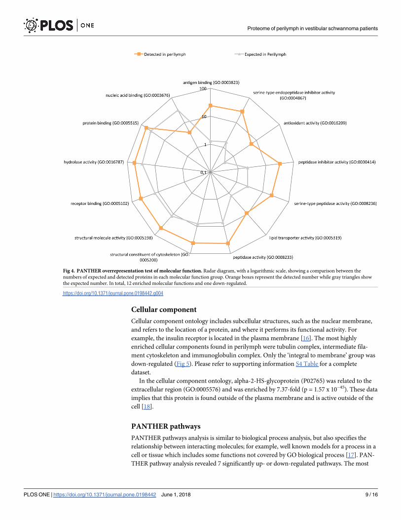
Cellular component
Cellular component ontology includes subcellular structures, such as the nuclear membrane,
and refers to the location of a protein, and where it performs its functional activity. For
example, the insulin receptor is located in the plasma membrane [16]. The most highly
enriched cellular components found in perilymph were tubulin complex, intermediate fila-
ment cytoskeleton and immunoglobulin complex. Only the ‘integral to membrane’ group was
down-regulated (Fig 5). Please refer to supporting information S4 Table for a complete
dataset.
In the cellular component ontology, alpha-2-HS-glycoprotein (P02765) was related to the
extracellular region (GO:0005576) and was enriched by 7.37-fold (p = 1.57 x 10−45). These data
implies that this protein is found outside of the plasma membrane and is active outside of the
cell [18].
PANTHER pathways
PANTHER pathways analysis is similar to biological process analysis, but also specifies the
relationship between interacting molecules; for example, well known models for a process in a
cell or tissue which includes some functions not covered by GO biological process [17]. PAN-
THER pathway analysis revealed 7 significantly up- or down-regulated pathways. The most
Fig 4. PANTHER overrepresentation test of molecular function. Radar diagram, with a logarithmic scale, showing a comparison between the
numbers of expected and detected proteins in each molecular function group. Orange boxes represent the detected number while gray triangles show
the expected number. In total, 12 enriched molecular functions and one down-regulated.
https://doi.org/10.1371/journal.pone.0198442.g004
Proteome of perilymph in vestibular schwannoma patients
PLOS ONE | https://doi.org/10.1371/journal.pone.0198442 June 1, 2018 9 / 16

enriched pathways were blood coagulation, the plasminogen activity cascade, and glycolysis
(Fig 6). Please refer to supporting information S5 Table for a complete dataset.
PANTHER protein class
PANTHER protein class analysis is similar to GO molecular function analysis but is an adapta-
tion of earlier PANTHER ontology. This includes several protein classes that are not included
in the GO molecular function ontology [17]. Twenty protein classes were up-regulated and
only one was down-regulated. The most highly enriched PANTHER protein classes were com-
plement component, tubulin, actin and actin-related proteins. Only nucleic acid binding was
down-regulated (Fig 7). Please refer to supporting information S6 Table for a complete
dataset.
Discussion
Modern surgical approaches, combined with advances in analytical techniques, have provided
us with new opportunities with which to explore perilymph in the inner ears of humans
affected by different pathological conditions. This study was performed exclusively on patients
with VS to characterize the perilymph in these patients at an individual level. We did this by
applying MS-technology for proteomic analysis. The individual approach provided new
insight of the large variations in the protein content of perilymph in VS disease. With a
Fig 5. PANTHER overrepresentation test of cellular component. Radar graph with a logarithmic scale showing a comparison between the numbers
of expected and detected proteins in each cellular component group. Within the perilymph (orange boxes), nine functions were up-regulated compared
to expected levels (gray triangles) while two were down-regulated.
https://doi.org/10.1371/journal.pone.0198442.g005
Proteome of perilymph in vestibular schwannoma patients
PLOS ONE | https://doi.org/10.1371/journal.pone.0198442 June 1, 2018 10 / 16

uniform diagnosis, it was also possible to identify inter-individual differences related to clinical
outcome. No sampling technique is currently available that allows sampling of perilymph invivo from individuals with normal inner ear function. Therefore, studies of the normal peri-
lymph proteome are not yet possible.
Sampling of perilymph is technically difficult, and several previous studies have reported
problems with blood contamination. In the present study, careful micropipette aspiration
through the RWM, before opening the labyrinth, resulted in a low incidence of contaminated
samples. The same surgical approach, but with a different sampling material and technique,
was also reported in another study [19]. Our outcome was very favorable, with only 1 out of 16
samples contaminated, compared to 16 out of 24 samples in a previous report [3].
The total number of identified proteins in the present study (314) is consistent with earlier
reports using non-selective approaches to analyze the proteome of perilymph from patients
with differential inner ear diagnosis at a group level [4][5][19]. Furthermore, 91 proteins were
identified in 12 to 15 samples, and this was validated by comparison with data arising from
previous MS studies on perilymph [5,19]. Eighty-nine of these frequently identified proteins
were also found in samples from VS patients in two earlier studies and can be presumed to rep-
resent a stable part of the perilymph proteome in patients with VS. Immunoglobulin heavy
variable 3–15 and Ig kappa chain V-II region FR, variations on immunoglobulins, are the two
proteins that were not previously identified. The list of stipulated normal proteome in peri-
lymph [5] were compared to our results, and 51 out of 71 could be found among the frequently
Fig 6. PANTHER overrepresentation test of PANTHER pathways. Image shows a logarithmic radar graph comparison between the detected (orange
boxes) and expected (gray triangles) numbers of proteins in each group. Seven significantly up-regulated PANTHER pathways were found.
https://doi.org/10.1371/journal.pone.0198442.g006
Proteome of perilymph in vestibular schwannoma patients
PLOS ONE | https://doi.org/10.1371/journal.pone.0198442 June 1, 2018 11 / 16

detected proteins in perilymph from VS patients. This discrepancy could be explained by the
large inter individual variation demonstrated here with 184 out of 314 proteins detected in 4
patients or less, even with a homogenous diagnosis group. This variation is important to take
into account for in future inner ear studies.
To date, there have been no methods developed to predict hearing loss or tumor growth in
patients with VS. This would be of significant value in the planning of clinical management.
The present work used two estimates to study these clinically important variables. The differ-
ence in PTA between the affected and unaffected ear was used as an estimate of tumor-associ-
ated hearing loss and tumor diameter was considered as an estimate of tumor growth.
The progression of hearing loss may depend on several factors such as the origin of
the tumor along the vestibular nerve, the site of auditory nerve compression, the tumors ten-
dency to infiltrate nerve axons and excretions from the tumor. The mechanisms are mostly
unknown, therefore characterization of the perilymph proteome in VS patients may provide a
valuable addition to understand the mechanisms of tumor-associated hearing loss. Recently
studies of the proteome were applied to investigate various heat shock proteins, and they
might be of importance for preservation of residual hearing after cochlear implantation [20].
However, we could not replicate this in our study since we only detected three variations of
Heat shock 70KDa proteins in only a single patient.
In contrast to earlier mass spectrometry studies on perilymph [5,19], the present study was
based on a homogenous cohort featuring individual analysis of each sample. This provided the
possibility of hypothesis-generated statistical methods and allowed us to explore the possible
association between protein expression and tumor-associated hearing loss. We hypothesized
Fig 7. PANTHER overrepresentation test of PANTHER protein class. Image shows a logarithmic radar graph of the comparison between the
number of detected (orange boxes) and expected (gray triangles) proteins in each group. Twenty up-regulated protein classes and one down-regulated
were identified.
https://doi.org/10.1371/journal.pone.0198442.g007
Proteome of perilymph in vestibular schwannoma patients
PLOS ONE | https://doi.org/10.1371/journal.pone.0198442 June 1, 2018 12 / 16

that the difference in pure tone average thresholds between the affected and unaffected ear
could represent hearing loss associated with the VS. Alpha-2-HS-glycoprotein (P02765) was
confirmed by nonlinear multivariate methods Random forest and Boruta as an independent
variable for tumor-associated hearing loss. The interpretation of the presences of alpha-2-HS-
glycoprotein is eluding since it was present in all samples, and the concentration is not directly
related to hearing threshold. But the fact it was also found in the samples from VS patients in
an study from 2017 [19] encourage to further explore if there is a causative relation.
The lack of samples of perilymph from healthy individuals is an obstacle in mapping the
pathological processes found in perilymph of VS patients, therefore an animal model is neces-
sary. Compared with perilymph from patients having cochlear implants in a previous study
shows the presence of alpha-2-HS-glycoprotein in many samples[19]. The majority of these
patients have unknown cause of sensorineural hearing loss [19], and it cannot be excluded that
the alpha-2-HS-glycoprotein could be involved in sensorineural hearing loss even in the
absence of a VS.
To understand the functions of the detected proteins, the use of pathway analysis, such as
the PANTHER overrepresentation test, provides us with an informative presentation of pro-
tein-related activities in the inner ear. In the present study, we presented functional groups,
including information relating to up- and down-regulation to present a clearer picture of their
functions in perilymph. This method relies on the reported functions of the identified proteins
in other tissues, and therefore the presented list of functional groups should be considered as
an indicator of their possible roles in the inner ear. The procedure/data-base for this type of
analysis is available at http://pantherdb.org/.
Investigating the biological process ontology in perilymph was probably the best method
with which to understand the general function. In the top 10 enriched biological processes,
three immune responses were present: complement activation, defense response to bacteria
and B-cell–mediated immunity. Furthermore, three metabolic processes were identified: sec-
ondary metabolic processes, glycolysis and cholesterol metabolic processes. The other four
processes identified were cell recognition, phagocytosis, blood coagulation and response to
biotic stimulus (Fig 3). This indicated that perilymph is much more active from an immuno-
logical and metabolic aspect than previously expected.
From the view of biological processes, alpha-2-HS-glycoprotein is part of the acute-phase
response, an ancestor to response to stress and also part of neutrophil degranulation an ances-
tor to immune response [18], which were both significant up-regulated (Fig 3). This is based
on data from other tissues and indicates that alpha-2-HS-glycoprotein is a potential inflamma-
tory and immunological mediator in perilymph. The known molecular functions of alpha-
2-HS-glycoprotein are as an endopeptidase inhibitor, cysteine-type endopeptidase inhibitor
and kinase inhibitor. These groups of inhibitors prevent or reduce the activity of enzymes that
hydrolyze non-terminal peptide bonds or catalyze the transfer of a phosphate group to a sub-
strate molecule [18]. As described by cellular component ontology, Alpha-2-HS-glycoprotein
is found in the extracellular region [18].
Hence, our hypothesis is that alpha-2-HS-glycoprotein may be excreted from the VS into
the extracellular fluid perilymph where it exerts pro inflammatory activity that contributes to
sensorineural hearing loss. An alternative hypothesis is that factors derived from the VS influ-
ence the inner ear and induce the up-regulation of alpha-2-HS-glycoprotein in perilymph.
The feasibility of using perilymph proteome as an approach for identifying biomarkers of
VS is attractive. Our present study identified alpha-2-HS-glycoprotein as an independent fac-
tor for hearing loss and since it was part of the stable section of the proteome an interesting
candidate as possible biomarker. However, further research is now needed to evaluate in an
experimental model if alpha-2-HS-glycoprotein has a pathophysiological role in the hearing
Proteome of perilymph in vestibular schwannoma patients
PLOS ONE | https://doi.org/10.1371/journal.pone.0198442 June 1, 2018 13 / 16

loss, and if it can be used as a biomarker to facilitate the decision-making process when choos-
ing between “wait and scan” strategies or surgery.
Conclusion
Using MS to analyze perilymph from 15 individual patients with VS, we found a stable section
of the proteome consisting of 91 proteins which were detected in the majority of patients. The
stable section was validated by comparison to earlier proteomic studies on perilymph and 89
proteins were confirmed. There was also a highly individual section of the proteome consisting
of 184 proteins only detected in a minority of patients. This showed an important variation in
the proteome between individuals, even in a cohort with a homogenous diagnosis.
Furthermore, Random Forest and Boruta analysis showed that alpha-2-HS-glycoprotein,
P02765, was an independent variable for tumor-associated hearing loss. This needs to be con-
firmed by future studies.
Pathway analysis, using the PANTHER overrepresentation test, showed that perilymph
exhibited numerous functions which were highly enriched compared to the level expected.
Particularly noteworthy was the highly significant increase in the activity of immune and met-
abolic processes in the perilymph from patients with vestibular schwannoma.
Supporting information
S1 Table. MaxQuant protein identification and quantification.
(XLSX)
S2 Table. PANTHER overrepresentation test of biological processes.
(XLSX)
S3 Table. PANTHER overrepresentation test of molecular function.
(XLSX)
S4 Table. PANTHER overrepresentation test of cellular component.
(XLSX)
S5 Table. PANTHER overrepresentation test of PANTHER pathways.
(XLSX)
S6 Table. PANTHER overrepresentation test of PANTHER protein class.
(XLSX)
S7 Table. Comparison with earlier mass spectrometry studies of the perilymph proteome.
(XLSX)
Acknowledgments
MSc K. Hornaeus and PhD J. Mi are acknowledged for their skillful assistance with sample
analysis and data interpretation. We also thank the Uppsala Clinical Research Center for excel-
lent statistical advice.
Author Contributions
Conceptualization: Jesper Edvardsson Rasmussen, Goran Laurell, Helge Rask-Andersen,
Jonas Bergquist, Per Olof Eriksson.
Data curation: Jesper Edvardsson Rasmussen, Jonas Bergquist, Per Olof Eriksson.
Proteome of perilymph in vestibular schwannoma patients
PLOS ONE | https://doi.org/10.1371/journal.pone.0198442 June 1, 2018 14 / 16

Formal analysis: Jesper Edvardsson Rasmussen, Jonas Bergquist.
Funding acquisition: Goran Laurell, Jonas Bergquist.
Investigation: Jesper Edvardsson Rasmussen, Per Olof Eriksson.
Methodology: Jesper Edvardsson Rasmussen, Goran Laurell, Helge Rask-Andersen, Jonas
Bergquist, Per Olof Eriksson.
Project administration: Jesper Edvardsson Rasmussen, Goran Laurell, Per Olof Eriksson.
Resources: Goran Laurell, Jonas Bergquist, Per Olof Eriksson.
Supervision: Goran Laurell, Helge Rask-Andersen, Jonas Bergquist, Per Olof Eriksson.
Validation: Jesper Edvardsson Rasmussen.
Visualization: Jesper Edvardsson Rasmussen.
Writing – original draft: Jesper Edvardsson Rasmussen, Goran Laurell, Helge Rask-Ander-
sen, Jonas Bergquist, Per Olof Eriksson.
Writing – review & editing: Jesper Edvardsson Rasmussen, Goran Laurell, Helge Rask-
Andersen, Jonas Bergquist, Per Olof Eriksson.
References
1. Bekesy Gv. DC Resting Potentials Inside the Cochlear Partition. J Acoust Soc Am. 1952; 24: 72–76.
https://doi.org/10.1121/1.1906851
2. Tasaki I, Spyropoulos CS. Stria Vascularis as Source of Endocochlear Potential. J Neurophysiol. 1959;
22: 149–155. https://doi.org/10.1152/jn.1959.22.2.149 PMID: 13642091
3. Arrer E, Oberascher G, Gibitz H-J. Protein distribution in the human perilymph: A Comparative Study
between Perilymph (Post Mortem), CSF and Blood Serum. Acta Otolaryngol (Stockh). 1988; 106: 117–
123. https://doi.org/10.3109/00016488809107378
4. Thalmann I, Kohut RI, Ryu J, Comegys TH, Senarita M, Thalmann R. Protein profile of human peri-
lymph: in search of markers for the diagnosis of perilymph fistula and other inner ear disease. Otolaryn-
gol—Head Neck Surg Off J Am Acad Otolaryngol-Head Neck Surg. 1994; 111: 273–280.
5. Lysaght AC, Kao S-Y, Paulo JA, Merchant SN, Steen H, Stankovic KM. Proteome of Human Perilymph.
J Proteome Res. 2011; 10: 3845–3851. https://doi.org/10.1021/pr200346q PMID: 21740021
6. Kshettry VR, Hsieh JK, Ostrom QT, Kruchko C, Barnholtz-Sloan JS. Incidence of vestibular schwanno-
mas in the United States. J Neurooncol. 2015; 124: 223–228. https://doi.org/10.1007/s11060-015-
1827-9 PMID: 26024654
7. Kirchmann M, Karnov K, Hansen S, Dethloff T, Stangerup S-E, Caye-Thomasen P. Ten-Year Follow-
up on Tumor Growth and Hearing in Patients Observed With an Intracanalicular Vestibular Schwan-
noma: Neurosurgery. 2016; 1. https://doi.org/10.1227/NEU.0000000000001414 PMID: 27571523
8. Rangel-Castilla L, Russin JJ, Spetzler RF. Surgical management of skull base tumors. Rep Pract Oncol
Radiother. 2016; 21: 325–335. https://doi.org/10.1016/j.rpor.2014.09.002 PMID: 27330418
9. Ekvall L, Bynke O. Prevention of cerebrospinal fluid rhinorrhea in translabyrinthine surgery. Acta Oto-
Laryngol Suppl. 1988; 449: 15–16.
10. Rask-Andersen H, Kinnefors A, Grangsjo G, Ekvall L. [Translabyrinthine surgery in tumors of the cere-
bellopontine angle. A suitable technique in large tumors]. Lakartidningen. 1994; 91: 2780–2783. PMID:
8057733
11. Wiśniewski JR, Zougman A, Nagaraj N, Mann M. Universal sample preparation method for proteome
analysis. Nat Methods. 2009; 6: 359. https://doi.org/10.1038/nmeth.1322 PMID: 19377485
12. Cox J, Matic I, Hilger M, Nagaraj N, Selbach M, Olsen JV, et al. A practical guide to the MaxQuant
computational platform for SILAC-based quantitative proteomics. Nat Protoc. 2009; 4: 698. https://doi.
org/10.1038/nprot.2009.36 PMID: 19373234
13. Mi H, Poudel S, Muruganujan A, Casagrande JT, Thomas PD. PANTHER version 10: expanded protein
families and functions, and analysis tools. Nucleic Acids Res. 2016; 44: D336–D342. https://doi.org/10.
1093/nar/gkv1194 PMID: 26578592
14. Breiman L. Random Forests. Mach Learn. 2001; 45: 5–32. https://doi.org/10.1023/A:1010933404324
Proteome of perilymph in vestibular schwannoma patients
PLOS ONE | https://doi.org/10.1371/journal.pone.0198442 June 1, 2018 15 / 16

15. Feature Selection with the Boruta Package | Kursa | Journal of Statistical Software. https://doi.org/10.
18637/jss.v036.i11
16. Ashburner M, Ball CA, Blake JA, Botstein D, Butler H, Cherry JM, et al. Gene Ontology: tool for the unifi-
cation of biology. Nat Genet. 2000; 25: 25–29. https://doi.org/10.1038/75556 PMID: 10802651
17. Mi H, Muruganujan A, Casagrande JT, Thomas PD. Large-scale gene function analysis with the PAN-
THER classification system. Nat Protoc. 2013; 8: 1551–1566. https://doi.org/10.1038/nprot.2013.092
PMID: 23868073
18. UniProt: the universal protein knowledgebase. Nucleic Acids Res. 2017; 45: D158–D169. https://doi.
org/10.1093/nar/gkw1099 PMID: 27899622
19. Schmitt HA, Pich A, Schroder A, Scheper V, Lilli G, Reuter G, et al. Proteome Analysis of Human Peri-
lymph Using an Intraoperative Sampling Method. J Proteome Res. 2017; 16: 1911–1923. https://doi.
org/10.1021/acs.jproteome.6b00986 PMID: 28282143
20. Schmitt H, Roemer A, Zeilinger C, Salcher R, Durisin M, Staecker H, et al. Heat Shock Proteins in
Human Perilymph: Implications for Cochlear Implantation. Otol Neurotol. 2018; 39: 37–44. https://doi.
org/10.1097/MAO.0000000000001625 PMID: 29227447
Proteome of perilymph in vestibular schwannoma patients
PLOS ONE | https://doi.org/10.1371/journal.pone.0198442 June 1, 2018 16 / 16