Embed Size (px)
Citation preview

THE PUZZLE OF PAIN,LOSS OF MOBILITY,
EVASIVE MOVEMENTSAND THE
SELF-MANAGEMENT
CLINICAL ASPECTS OF THE ASSESSMENT, TREATMENT AND DESIGN OFA HOME PROGRAMME FOR 2 PATIENTS. ONE WITH SEVERE SHOULDERPAIN OF NEURO-ORTHOPAEDIC ORIGIN, THE OTHER WITH PROBLEMS
DUE TO A LESION OF THE CENTRAL NERVOUS SYSTEM.
By GISELA ROLF
The Puzzle of Pain, Loss of Mobility, Evasive Movements and theSelf-Mangement. Online. Danske Fysioterapeuter, 2001. Available:
http://fysio1.inforce.dk/graphics/PDF/PDF-job.p65.PDF1. and 2. edition are eliminated, 25.th March 2002.

2
“The real purpose of scientific method is to make surenature hasn’t misled you into thinking you know so-mething you don’t actually know.”Robert M. Pirsig
INTRODUCTIONAround 1986 Maitland expanded, within his systemof assessment and treatment in manual therapy, theinvestigation of the mobility and sensitivity of neuraltissues as a possible cause of pain, loss of mobilityand muscle spasm. In many patients he claimed tobe able to demonstrate that antalgic postures andevasive movements of the trunk might result fromneural tissues being at fault.
After Elvey described what he called (1979) the Bra-chial Plexus Tension Test, 1986 the Upper Limb Ten-sion Test/ULTT (1986) and later the Upper Limb Neu-
ral Tissue Provocation Test / ULNP (Elvey 1995) phy-siotherapists could more clearly differentiate betweenjoint tissues, muscle tissues and neural tissues assources of pain and dysfunction. New clinical insightsinto the contribution of neural tissue dynamics toacute and chronic pain and its treatment were stimu-lated. The assessment and treatment of joint dysfunc-tion, the loss of neural tissue mobility and protectivemuscle spasm were primarily thought of as “mecha-nical” (Elvey R L 1986) but an attempt to link it toscientific research on neural tissues` response to irri-tation was made.
Since Mobilisation of the Nervous System (Butler 1991)was first published, physiotherapists have increasing-ly considered and included neural structures in theassessment, reassessment and treatment of thesymptoms and signs of their patients.
THE PUZZLE OF PAIN,LOSS OF MOBILITY,
EVASIVE MOVEMENTSAND THE
SELF-MANAGEMENT
CLINICAL ASPECTS OF THE ASSESSMENT, TREATMENT AND DESIGN OFA HOME PROGRAMME FOR 2 PATIENTS. ONE WITH SEVERE SHOULDERPAIN OF NEURO-ORTHOPAEDIC ORIGIN, THE OTHER WITH PROBLEMS
DUE TO A LESION OF THE CENTRAL NERVOUS SYSTEM.
By GISELA ROLF

3
Shacklock (1995) suggested, “The term neurodynamicsmay be deployed to include the link between mechanicaland physiological types of mechanisms“. Physiothera-pists have been encouraged to attempt to influencedirectly and indirectly the patho-biomechanical chan-ges of neural tissues which so often include patho-neurophysiological reactions, such as vascular andbiochemical processes which, in turn, appear to af-fect target tissues adversely. Target tissues are thosestructures such as muscles, connective tissues, joints,intervertebral discs, the vascular system, organs, andthe connective tissues of the nervous system itselfthat are directly or indirectly innervated and control-led by the nervous system.
However, it is obvious that we lack much information inthe scientific field about the nervous system and espe-cially about Neurodynamics. Even recent studies on thenervi nervorum, suggested as the missing link betweenall the other factors contributing to neuropathic pain(Bove and Light, 1997; Ochoa 1997; Sorkin 1997) canoffer only a hypothesis to support our clinical methods.
“The biomechanics of the peripheral and especially ofthe central nervous system sadly is neglected in researchand although I asked many of my colleagues on inter-national congresses who are especially involved inneurophysiology we do not know about any study in thefield of neurodynamics and its relation to clinical states”(Prof. Dr. med J. Kesselring, neurologist, director ofthe Rehabilitation Centre, clinic Valens, Switzerland,correspondence February 2000).
All physiotherapeutic techniques, either by activetreatment to improve selective muscle function andreduce pain, or passive techniques to similarly influencesigns and symptoms are in essence mechanical sincethey employ movement. They aim to ease or eliminatepain, improve mobility and regain normal muscle toneand re-educate selective muscle function, all of this inorder to restore normal function to the patient.
Shacklock (1999) writing on manual therapy reasonsthat the method of clinical management operates onall levels of the healing process: “What is important isthat treatment accommodates not just the brain andspinal cord, but all relevant stages in which input andoutput mechanisms are integrated with clinical reaso-ning. Each component as part of a reasoning model canbe assessed and treated with purpose. A focus on lear-ning (e.g. motor and cognitive) and conditioning pro-cesses should be key aspects, with the understandingthat therapy must change the culpable mechanisms,whether they be generated in the brain or the body”.
The holistic understanding of “a person in pain andwith disability“ demands from the physiotherapistmany different levels of comprehension, skills and
knowledge: e.g. an integrated scientific and clinicalapproach to biomechanics, to biochemist, to anato-my, to physiology and to pathology. Physiotherapistsshould also understand psychosocial processes, haveknowledge about body awareness in order to modifyhabits and give adequate support to the patient inthe development of confidence. They need to be in-formed about perception and significant learningstrategies, about interaction and communicationamongst their many skills.
“Whilst mobilisation of neural tissues remains a useful mo-dality in the treatment of neural disorders, refinements arenecessary. Consideration of relevant physiology in assess-ment and treatment of neurogenic pain is an importantrefinement if neural mobilisation is to become well foun-ded, safe and effective. One of the dangers in treatment ofneural tissues is the reliance on mechanics. In the not toodistant past, this reliance may have led to inappropriatephysical treatment to neural tissues“ (Shacklock, 1995).Undoubtedly every day we learn from patients that weneed more skills, experience, knowledge and furthermeans of treatment. However the physiotherapist’s abi-lity to select appropriately and find the right balancewith all the treatment tools that serve the individualneeds of the patient is an essential goal.
WHEN SELECTIVE MUSCLE ACTIVITY FAILSPATHOLOGY IS FACILITATED AND PREDISPOSINGPROCESSES DEVELOP IN TARGET AND NEURALTISSUES PRODUCING SYMPTOMS AND SIGNS.“A disorder in the nerve at one site may predispose toanother lesion either distally or proximally along the nervoussystem” (Gunn and Milbrandt, 1978). Clinical experienceconfirms that one disorder can predispose an indivi-dual to another disorder in neural tissues as well as intarget tissues and vice versa. For example the loss ofselective trunk muscle activity, especially the selectiveoblique muscle activity of the abdominal wall (Davies1990; Davies 2000), which normally pulls the ribsdown diagonally and prevents the thorax and theshoulder girdle from lifting up presents the humanbody with many problems:“ They regulate intra abdominal pressure, are involvedin respiration, stabilise the body axis during equilibriumreactions against gravity, influence selective and painfree shoulder function and participate in equilibriummovements of the pelvis while walking (S.Klein-Vogel-bach, 1986).
Even if this selective trunk activity is only slightlyweakened and therefore “imbalanced”, other targetand neural tissues will be overused proximally and di-stally due to evasive movements and postures. Thisoccurs as the body tries to compensate for the loss ofactive stabilisation proximally, which normally formsa protection for all target and neural tissues.

4
➝➝
➝ ➝
➝➝
NEURAL TISSUE DYNAMICS / NEURODYNAMICS
Neurobiomechanical Responses Neurophysiological Responses
Patho - neurophysiological ResponsesPatho - neurobiomechanical Responses
PATHO - NEURODYNAMICS(accompanying “psycho-social” reactions which can be
defined as the normal accompaniments of pain and disability)
➝➝
➝ ➝
➝➝
TARGET TISSUE DYNAMICS
Biomechanical Responses Physiological Responses
Patho - Physiological ResponsesPatho-Biomechanical Responses
TARGET TISSUE PATHO - DYNAMICS(accompanying “psycho-social” reactions, which can
be defined as the normal accompaniments of pain and disability)
➝➝➝➝ ➝
➝➝➝➝ ➝
Target Tissue Dynamics summarises in this diagram,the functional interrelationship between the biome-chanical and the physiological responses of all tis-sues. Target Tissue Patho-Dynamics describes the in-terrelationship between patho-mechanical and pat-ho-physiological mechanisms within all target tissue.
The diagram attempts to relate the clinical observa-tion of how neural tissue movement (neurodyna-mics) influences directly and indirectly non-neuraltissue activity (target tissue dynamics) and, viceverse, how target tissue dynamics influence directlyand indirectly the neurodynamics.
Fig. 1
Fig. 1
Neurodynamics, as defined by Shacklock (1995),summarises in this diagram the functional interrela-tionship between neurobiomechanical responsesand neurophysiological responses within the ner-vous system. Patho-neurobiomechanics, as
defined by Shacklock (1995), summarises theinterrelationship between patho-neurobiomechani-cal and patho-physiological responses within thenervous system.
The following diagram draws attention to the closethe interrelationship and dependence of all tissueswithin the body, whether neural or target tissues,and therefore to the possibility that tissues can pre-dispose each other to symptoms and signs.
➝➝
➝ ➝
➝➝
NEURAL TISSUE DYNAMICS / NEURODYNAMICS
Neurobiomechanical Responses Neurophysiological Responses
Patho - neurophysiological ResponsesPatho - neurobiomechanical Responses
PATHO - NEURODYNAMICS(accompanying “psycho-social” reactions which can be
defined as the normal accompaniments of pain and disability)
➝➝
➝ ➝
➝➝
TARGET TISSUE DYNAMICS
Biomechanical Responses Physiological Responses
Patho - Physiological ResponsesPatho-Biomechanical Responses
TARGET TISSUE PATHO - DYNAMICS(accompanying “psycho-social” reactions, which can
be defined as the normal accompaniments of pain and disability)
➝➝➝➝ ➝
➝➝➝➝ ➝

5
Because the nervous system is a continuous tissuetract the cervical neuraxis cannot be seen, in termsof dynamics, separately from the thoracic and thelumbar neuraxis. NEURAXIS is a term used when theCNS is considered along its length irrespective of itsbends and folds (Bowsher,1988); the neuraxis/spinalcord is a continuation of the medulla oblongata (Butler,1991). Certainly clinically these anatomical regionsof the spinal cord influence each other with regardto pathological states such as restricted mobility(elongation and accessory movements such as ante-ro-posterior, transverse and rotatory movements),pain responses to movement, evasive movementsand antalgic postures, change in muscle tone, andautonomic reactions. In this regard special attentionshould be given to the thoracic spine as a main facili-tator of lumbar and cervical vertebral pathology andmuscle-skeletal complaints in the upper and lowerlimb. Diffuse autonomic or localised symptoms suchas apparent visceral complaints in the upper and lo-wer abdomen can also be provoked by the thoracicneuraxis.The loss of selective trunk activities, depriving thetrunk of the means of effective active stabilisationagainst gravity is likely to cause progressive mechani-cal problems by overusing target and neural tissuesthereby predisposing to pathology, as the above dia-gram indicates. Therefore the re-education of selecti-ve muscle activity and at the same time the at-tempted reversal of all patho-biomechanical and pat-ho-physiolocal tissue problems is of vital importancein order to prevent recurrent problems for the pa-tient. The interdependence between a normal ner-vous system and normal target tissues cannot beemphasised enough. In order for the nervous systemto mantain its functional integrity the target tissuesmust in turn, enable it to spread out and adapt toany position and body movement (Butler, 1991). Ifthe nervous system is not mobilised and protectedby muscle activities and free joint mobility - as is sooften the case clinically in patients who have suffereda central lesion and are immobilised - it rapidly losesits normal biomechanical properties when tested andnormal physiological responses like the appropriateautonomic activation are altered. The most impor-tant of all neuro-psychological responses is the im-pulse transport which is unique to the nervoussystem.
NEURO-DYNAMIC TESTING AND TREATMENTThe neuro-dynamic tests described by Maitland(1985), by Elvey (1986) and by Butler (1991) assistin diagnosis of neurodynamic faults. The authors ex-plain how, by varying neural test components andtheir sensitising movements or by using palpationtechniques for peripheral nerves it is possible in mostcases to differentiate between neural and target tis-sues as components of a disorder. Investigation bymovements of the neuraxis in the cervical, thoracicand lumbar regions, and the addition of componentsof the Neural Tissue Provocation Tests reveal how neu-ral patho-dynamics frequently cause symptoms in
spinal areas, the trunk, the limbs and the head suchas nausea, and apparent organic pain such as attacksof angina pectoris, and gall bladder symptoms.
There are various ways in which the nervous systemmay be moved in which its forces may be modifiedand thus its patho-biomechanical properties and pat-ho-neurophysiological responses altered. The exami-ner must be sure that the diagnostic tests for tissuestructures/organs have been carried out thus indica-ting that neural tissue problems may be present.
INFLUENCING NEURAL TISSUE IN A DIRECT WAYUsing neurodynamic tests or their components (passivephysiological movements for the nervous system), indifferent sequences and combinations, adding in sen-sitising movements and including the neuraxis in theneurodynamic tests offer a clinically reliable way toassess, treat and re-assess neural tissue movementand sensitivity. This is so especially, in conditionswhere inert resistance is the main limiting factor ofmovement, with pain appearing at the limit of neuralmobility.
Palpation techniques such as local transverse move-ments of the nerve tissue (Butler, 1995) against themechanical interface of the nervous system (thistechnique can be described as passive accessory mo-vements to the nervous system) applied to peripheralnerves, nerve roots and the different parts of theplexi aid diagnosis and serve as therapeutictechniques (Butler, 1995). A second passive accessorymovement of neural tissues is a locally applied “roll-over” of a peripheral nerve with some gentle pressureapplied within its nerve bed.
(Fig. 2) Accessory movement to neural tissue (Brachialplexus, Median nerve), transverse movement of theneural tissues against the mechanical interface
INFLUENCING NEURALTISSUES IN AN INDIRECT WAYThis occurs firstly with active muscle work such as se-lective stabilisation of the trunk followed by move-ment of the limbs or, vice versa, stabilizing the limbsand moving the trunk. An indicator of the quality ofselective muscle function is the ability to rotate orcounter any rotator activity actively and then to rota-te against rotatory muscle function. Any method ofmotor learning and re-education of selective muscle

6
function may modify the biomechanics of neural tis-sues and influence therefore the physiology of thenervous system in an indirect way.
Secondly any mobilisation of interfacing tissues suchas joint mobilisation constitutes an indirect methodof influencing neural tissue dynamics e.g. passiveunilateral postero-anterior mobilisation of T5/6 verte-bral segments.
As a clinical observation, postural correction and re-gular re-positioning, as for example with a patientwho suffered a hemiplegia, can alter directly and in-directly the biodynamic reaction of neural tissues, asmay be seen in the progressive modification of allo-dynia and hyperaesthesia of neural structures.
ASPECTS OF TREATMENTAll the neurodynamic tests and test components canbe applied as treatment, according to the clinicalevaluation concept of Maitland, either in a verygentle way or more forcefully as necessary. Howeverin the case of direct mobilisation of the neural tissuepassive movement should be applied only as a rhyth-mical mobilisation and never as a prolonged stretch-ing at the limit of range. In some cases it appearsmore favourable to encourage neural tissue mobilityand ease symptoms when, for example, the trunk ismoved rhythmically against the limb, which can bepositioned in some or all components of a neurody-namic test sequence.
All such rhythmical neural mobilisations can be doneeither without pain, or by just provoking the pain, orgoing just beyond, reproducing “the pain”, or en-countering resistance with little pain, and preferablywithout pain, towards the limit of the movement.There is no justification for hurting the patient unne-cessarily. The gentleness or force of the mobilisationwill depend upon the assessment, re-assessment andthe nature of the problem (Maitland, 1986).
The accessory movements to neural tissues such asthe locally applied transverse mobilisation and “roll-over” mobilisation (palpation techniques) can be ap-plied to influence severe pain syndromes most favou-rably. Although there is as yet no scientific proof tosupport these clinical observations, experience sug-gests that they have some validity in the treatment ofsevere pain, protective muscle spasm and loss of ac-cessory and physiological neural tissue mobility inconditions with diffuse and strange symptoms e.g.:sympathetically maintained pain syndromes (McMa-hon, 1991: Melzack, R, 1991) which may develop af-ter a lesion of the central nervous system.
It is of vital importance to re-assess, after each neuraltissue mobilisation, possible changes in the quality ofmuscle activity because so often immediately afterneurodynamic treatment this can change for the bet-ter. To retrain and regain normal muscle function im-mediately after a passive mobilisation of neural tissue
is most important. Clinical experience has shownthat neural mobility is quickly lost again and thesymptoms reoccur if muscle activity and range ofmotion in both accessory and physiological joint mo-vements cannot maintain the passively regained mo-bility of the nervous system. For the same reason anyevasive movement or antalgic posture of the trunkand/or limbs has to be actively corrected to their ac-tive optimum after passive neural tissue mobilisation.
DESIGN OF A HOMEPROGRAMME ANDENCOURAGEMENT OF SELF-MANAGEMENTFrom the first treatment the patient should be helpedto understand the response of his symptoms to hisdaily activities, to his postural habits, to his activemuscle control and to his psychological state. Thepatient must learn what provokes his symptoms andsigns through a detailed and patiently maintainedteaching and learning process (on both his and thetherapists side) in order to perform a home program-me and monitor self-management with a modified li-fe style. To relearn physical awareness can take sometime especially if the patient, has been taking analge-sics, anti-inflammatory medication and mood alte-ring drugs over a long period of time.In order to protect his body tissues and to preventfurther overuse and damage to them as part of hislearning process, the patient often needs enhancedsensory input and guidance from the hand of thephysiotherapist. The guidance will facilitate aware-ness of how much movement to perform, how notoverdo movements and how to correct postures. Thefrequency and extent of tissue self-mobilisation, theexactness of active exercises, the recognition andcorrection of evasive or substitute movements andpostures have all to be learned. This learning processguided by the evaluation of each sequence by thephysiotherapist, is necessary in the beginning.As soon as possible the patient should also be encou-raged to make his own discoveries such as reducinghis pain and improving mobility by auto-mobilisationof neural and target tissues. A self-management pro-gramme can begin to be established in his daily life.
“ I keep six honest serving-men(They taught me all I knew);Their names are WHAT and WHY and WHENAnd HOW and WHERE and WHO.I send them over land and sea,I send them east and west;But after they have worked for meI give them all a rest “
R. Kipling
The patient feels safer, as they describe it, if they canuse these guidelines of knowing exactly what to dohow and when it is necessary, why they must do it,and who, if anyone, should be involved to help.
The resultant reduction of his anxiety in response tounpredictable pain attacks, discomfort and stiffness

7
and his growing confidence in his ability to modifyhis symptoms will lead to a re-organisation of his lifealong self protection and prophylactic lines. “There isincreasing evidence that people want to understand moreabout their bodies, both in the prevention of ill health, aswell as in the promotion of health and fitness” (Watson,1996).
SOME ASPECTS CONCERNING THERAPEUTICEXERCISES AS A PRE-REQUISIT FOR SELECTIVEMUSCLE FUNCTIONProgress will be more rapid if the patient is in the po-sition of contributing to the treatment with activeexercises at home as well as tissue self mobilisation.
“ A very specific type of therapeutic exercise hasbeen devised which provides effective pain relief forchronic and recurrent back pain sufferers probablythrough enhanced segmental stabilisation”.( C. A. Richardson; Jull G A, 1995). These therapeuticexercises have been of great value for patients withacute and chronic back pain, and can, if segmentalstabilisation is performed in positions which enhanceneural and target tissue mobility, improve both themuscle stabilisation effect and the neurodynamic andtarget tissue symptoms and signs.
(Fig. 3) SLR (lowerlimb neural tissueprovocation testcomponents) mobi-lisation “at limit”adding in dorsiflexion of the footand performingactive trunk stabil-isation against therestriction of theSLR neural tissues.
(Fig. 4) ULNP(upper limb neuraltissue provocationtest) position at li-mit against the wallmaintainingsegmental stabi-lisation of the trunkat the same timeand mobilising theneck away from theULNP position: onefoot is placed on astool to avoid ex-cessive lumbarextension/evasivemovement.
(Fig. 5) Activeantero-posteriorauto-mobilisation ofthe cervical spinecorrecting uppercervical extensionand lower cervicalflexion; the ULNPposition against thewall is maintainedfor the left arm,hand and fingers.One leg is positionedon a stool in orderto keep the lumbarspine in a neutralposition.
Active segmental stabilisation is much more difficult tomaintain against neuro-dynamically generated symp-toms and variations in muscle tone. However, it shouldbe progressed step by step until the neural tissues aremobile enough to allow the target tissue to performmore easily, for example muscle tone being reducedand able to work more smoothly “through range” (Fig.3, 4, 5).

8
A CASE STUDY OF A PATIENT WITH NEURO- ORTHOPAEDIC PROBLEMS
(Fig. 6)Puzzle of Pain
(Fig. 7)Body Chart ofMr. S.
ASSESSMENTHistory. Three weeks before calling for an appoint-ment, Mr. S. slipped on the stairs and banged hishead and his left shoulder against the wall. The pa-tient felt a stabbing pain in his lower neck and leftshoulder at the time of the accident, but this clearedand he did not feel any pain.
After a week he was wakened about 4 a.m. by severeshoulder pain left, when lying on his left side. He hadto get up, because he could not find any comfortableposition and about 8 a.m. the shoulder painincreased to such an extent that he could not movehis arm at all and had to consult a doctor.
In the following 3 weeks he received 9 intra-articularinjections, anti-inflammatory medicine and was sup-plied with painkillers and psycho-modifying drugsneither of which he took. After 3 weeks the shoulderpain was reduced by about 30%, and the neck feltvery stiff. He suffered headaches each morning untilabout 11a.m. He was wakened by pins and needlesin his left fingers each night about 4 a.m. He could
not lie on his left side (his normally comfortablesleeping position) and he could not perform anyshoulder nor arm movements without pain. His leftarm felt heavy, cold and very weak which concernedhim a lot. Being a carpenter, he had to lift heavy ob-jects and deliver furniture to customers.
Mr. S. has been diabetic for 21 years. He did not takeany other medication, felt well and healthy and didup to 10 hours of mountain walking each day atweekends. Once or twice a month he suffered severediffuse leg and foot pains right and left for about 5-6hours at night, which forced him to get up and walkabout. He did not know why and no doctor couldadvise him what to do in order to change these seve-re pain attacks.
He had lived alone since his wife died of cancer 9years ago and looked after his home.He suffered 2 accidents in his life: when he was 25 hefell from a tree. After falling some 8 meters he landedon his bottom fracturing his coccyx. The severe localpain at the base of the spine gradually disappearedover a period of 3 months. The second accident hap-pened when he was aged 46. He slipped on an icepatch and landed with great force in extension on hisback and head. He suffered headaches for some 6months and intermittent lumbar pain for one year,but after that he felt perfectly well, but markedly lessmobile in his spine and especially his neck.
Subjective assessment/behaviour of the main areas ofsymptoms:Symptoms in area 1 (see body chart) woke him fre-quently at night. The patient could not sleep wellbecause every position was uncomfortable after afew minutes. Area 2, 4, 6 also troubled him at night,but with shifting around these areas of pain eased fora short period.
On waking the patient felt tired and stiff all over, butafter a hot shower and moving around eased thisstiffness. Area 1 and 2 were less troublesome andareas 4, 5, 7, 8 became less intense and were morebearable with walking. The patient could not even lift2 kg, because of pain in area 1, 2, 4. Getting dressedand undressed gave more pain in area 1, 2, 3, 4, butthis settled quickly.
Selected data from ongoing records are detailedbelow.Physical assessment. Day 1, 9 June 1999: The first as-sessment procedures were carried out gently becauseof longstanding diabetes and severe left shoulderpain, which increased with every active movement.
Active movementsLeft gleno-humeral joint: all movements reproducedpain in the areas marked on the body chart 1, 2, 4,5, 6 and were severely restricted (Fig. 8-11)

9
Right gleno-humeral joint: with regard to mobilitythere was a little stiffness in all directions, but nopain, except in area 6 with 100 ° abduction.
Left rotator cuff: all resisted movements in abduction,lateral rotation, medial rotation and elbow flexionshowed markedly reduced muscle power and severepain in area 1, 2, 3, 5,
Right rotator cuff: no pain and full strength
Passive physiological movementsLeft gleno-humeral joint:Flexion: 60°, reproduced neck and upper arm symp-toms (2, 4)Abduction: 60°, reproduced (4) sharply.Extension: 0°, and gave diffuse pain (2, 4, 5)Lateral and medial rotation were not possible to per-form because of the severity of pain in area 1 and 4Left gleno-humeral joint, accessory movements:Postero-anterior (grade II- -) local pain and area 4 se-vere; antero-posterior (grade II) local pain and area 4severe; antero-posterior (grade II): local pain plus area4, severe; medial rotation (grade I): reproduced 1 and4; lateral rotation (grade I): reproduced 4 and 5.
Re-assessment: no change in the gleno-humeral acti-ve and passive movements
Active Tests: Cervical SpineAll active movements were restricted. There was nomovement in the upper and lower cervical spine forlateral flexion to the right and to the left. In the midcervical lateral flexion was 30°, reproducing areas 2,3, 4. Rotation to the left and to the right was 15°, re-producing areas 2, 3, 4. There was no extension inthe lower cervical spine, but the spine was held fromC5–T4 in 30° of flexion with a stiff upper cervical spi-ne in extension. Neither cervical positions could bereduced because of severe pain in area 2.
Cervical Palpation (Patient Prone Lying)The left arm had to be positioned alongside the trunkbecause of the severe shoulder pain.All vertebral levels right and left felt very, very stiffwith palpable scar tissue in the lamina and in the in-terspinous spaces. C3 and C5 were very deep set.The posterior neck muscles and soft tissues were verytight, hard and shortened, and thickened especiallysub-occipitally. Sharp local pain was elicited with leftunilateral postero-anterior mobilisation (grade II-) onC5/6, C3/4, C2/3 and 0/C1. All other levels on the leftwere very stiff, but painless (grade IV). From 0/C1 toT4/5 on the right (grade IV), were very stiff but no pain.
With antero-posterior mobilisation techniques ap-plied unilaterally at C3/4 and C4/5 left (grade II- -)sharp local pain was elicited radiating to areas 2, 4,5, 8. Other levels left and right anterio-posterior stiffbut gave no pain (grade IV).
(Fig 8) 1st assessment:antalgic posture of his leftshoulder girdle; theswelling over the leftshoulder is marked.
(Fig. 9 & 10) 1st assessment: G/H flexion with evasivemovements anteriorly and with cervical lateral flexion.
(Fig. 11a) 1st assessment:active test movement leftarm “hand on back”.
(Fig. 11b)1st assessment: G/H jointabduction with evasivemovements of the leftshoulder girdle into ele-vation and medial rota-tion of the G/H joint.
10
The left brachial plexus felt very tight and immobile atthe mechanical interface in its upper and lower parts.The left shoulder girdle was held in elevation; no cor-rection was possible in prone lying because of pain inareas 1, 2, 4. Accessory movements (transverse gradeII- -) on the upper and on the lower part of the brachialplexus showed no movement in the axilla, but causeddiffuse pressure and heaviness into the left axilla withpins and needles into the left hand and all fingers.
The re-assessment immediately after the testing show-ed no change in the pain nor in joint mobility of eitherthe cervical spine or the gleno-humeral joint complex.
LOWER LIMB NEURALTISSUE PROVOCATION TESTSRight Passive Straight Leg Raise (SLR) without dorsi-flexion: 35°, reproduced area 2 (diffuse and deep)and area 4, and caused evasive movement of thetrunk with a transverse shift to the left. Correction ofthe trunk to middle position gave pain in area 2, 4(sharp and severe). An evasive movement of the leftshoulder girdle into elevation increased with SLR 30°;correction was not possible because of pain in theleft arm areas 1, 2, 4, 5, 8.
Treatment: Ten neural tissue mobilisations SLR withoutdorsi-flexion 20° slowly and without pain (grade II-).
Re-assessment: a marked difference with cervical acti-ve movements in all directions. The gleno-humeralpassive joint flexion improved from 60° to 100°,reproducing a little pain in area 1, 4;Abduction from 60° to 90°, reproducing pain in area1, 4. Rotator cuff: external rotation, abduction, medialrotation and elbow flexion could all be done actively alittle, with some pain reproduced in area 1 and 4.
Left SLR without dorsi-flexion: 70°, gave a stretchingfeeling behind the knee, adding full dorsi- flexion ga-ve more of a stretching feeling. Adding adductionand medial rotation produced no more discomfort.
Right Femoral Nerve Provocation Test in left side lying:was not possible because patient could not lie on hisleft side due to severe shoulder pain.
But by positioning the patient supine with flexion ofthe cervical and thoracic spines using pillows andwith the left leg in flexion to keep the lumbar spinein neutral, hip extension showed restriction of 30°with the knee kept in 90° flexion. The patient felt astrong stretching discomfort on the anterior thigh.The attempt to add knee flexion increased thestretch of the anterior thigh markedly. Subtractingupper cervical and mid thoracic flexion abolished thestretching discomfort anteriorly on the thigh.
Left Femoral Nerve Provocation Test (first cervical andupper thoracic spine positioned in flexion withoutpain): hip extension was -20° and gave a stretchingfeeling over the anterior thigh, adding in 70° flexionof the knee (limit) provoked hyperextension of the
upper lumbar spine, but no pain. Moving the cervi-cal and thoracic spine back into neutral gave the hipmore mobility into extension and 90° knee flexionwithout pain was possible.Re-assessment: no difference in pain or mobility beha-viour in the cervical spine and left shoulder movements.
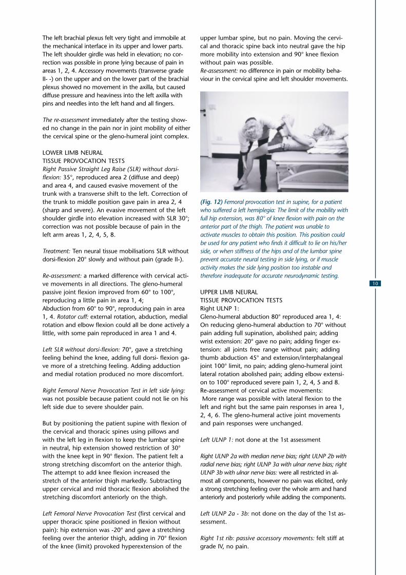
(Fig. 12) Femoral provocation test in supine, for a patientwho suffered a left hemiplegia: The limit of the mobility withfull hip extension, was 80° of knee flexion with pain on theanterior part of the thigh. The patient was unable toactivate muscles to obtain this position. This position couldbe used for any patient who finds it difficult to lie on his/herside, or when stiffness of the hips and of the lumbar spineprevent accurate neural testing in side lying, or if muscleactivity makes the side lying position too instable andtherefore inadequate for accurate neurodynamic testing.
UPPER LIMB NEURALTISSUE PROVOCATION TESTSRight ULNP 1:Gleno-humeral abduction 80° reproduced area 1, 4:On reducing gleno-humeral abduction to 70° withoutpain adding full supination, abolished pain; addingwrist extension: 20° gave no pain; adding finger ex-tension: all joints free range without pain; addingthumb abduction 45° and extension/interphalangealjoint 100° limit, no pain; adding gleno-humeral jointlateral rotation abolished pain; adding elbow extensi-on to 100° reproduced severe pain 1, 2, 4, 5 and 8.Re-assessment of cervical active movements: More range was possible with lateral flexion to theleft and right but the same pain responses in area 1,2, 4, 6. The gleno-humeral active joint movementsand pain responses were unchanged.
Left ULNP 1: not done at the 1st assessment
Right ULNP 2a with median nerve bias; right ULNP 2b withradial nerve bias; right ULNP 3a with ulnar nerve bias; rightULNP 3b with ulnar nerve bias: were all restricted in al-most all components, however no pain was elicited, onlya strong stretching feeling over the whole arm and handanteriorly and posteriorly while adding the components.
Left ULNP 2a - 3b: not done on the day of the 1st as-sessment.
Right 1st rib: passive accessory movements: felt stiff atgrade IV, no pain.

11
Left 1st rib: passive accessory movements: could not betouched because of severity of pain.
Right acromio-clavicular joint: passive accessory move-ments: felt restricted in all directions with grade IV,no pain.
Left acromio-clavicular joint: passive accessory move-ments: marked swelling on the joint and very warm.It could not be moved because of severity of pain.
Shoulder horizontal adduction was very restricted andgave severe pain in areas 1, 4
(Fig. 13) 1st assessment: horizontal adduction of the leftshoulder, stressing the acromio-clavicular and sterno-clavicular joints.
Left sterno-clavicular joint movements: in all directionslocal pain with grade II
Right sterno-clavicular joint movements: in all direct-ions locally sensitive with grade IV
On observation no selective abdominal muscle workcould be seen during the examination. The patient laysupine with excessive lumbar extension and the ribcage was extremely elevated (fig. 14).
Treatment and Teaching session for the home program-me/supine: selective activity of the abdominal mus-cles short of pain or discomfort, legs positioned inhip and knee flexion 90° (fig. 15).
(Fig. 14) 1st assessment Mr. S. no abdominal muscleactivity
(Fig. 15) Selective muscle activity/supine: legs supported,heels compressed against each other to encourage lowerabdominal muscle activity; 3rd re-assessment andtreatment
Re-assessment of right SLR without dorsi-flexion: hadincreased to 60° with only slight reproduction of painin area 1, 2, 4, 5. Gleno-humeral active joint move-ments unchanged but far less evasive movement ofthe left shoulder girdle into elevation occurred.
Treatment and Teaching session for the home program-me: the patient was shown how to mobilise the rightSLR without dorsi-flexion and without evasive move-ments of the left hip into flexion and using active ab-dominal bracing and thus avoiding lumbar and lowthoracic hyperextension and elevation of the rib cage.
Re-assessment: the neck and the shoulder pain felteasier; no physical testing was doneWarning: the patient was asked for a detailed report ofthe pain behaviour over the next 2 days; the patientwas given the telephone number because he shouldreturn the next day if any pain areas worsened.
Home programme for the following two days:1. Abdominal muscle activities in lying with the legs
supported in 90° flexion of the hips over a chairand with 90° knee flexion: 20 times in the morningbefore getting up and 20 times during the after-noon or evening, making sure that the rib cagemoved diagonally down towards the navel. Theseactive contractions of the abdominal muscles wereto be carried out without provoking any pain. Thepatient had to re-assess his active shoulder move-ments for pain and mobility after each session.
2. Right SLR mobilisation in standing: the patientstood in the doorway and swung the right leg intohip flexion with firm extension of the right kneeand some dorsi-flexion of the foot. Evasive move-ments of the left hip and pelvis were to be activelycountered and the trunk actively stabilised with ab-dominal muscles. The patient was asked to per-form a minimum of 20 movements with active sta-bilisation of the trunk, every day.
3. An ice pack was applied for a few minutes to theleft acromio-clavicular joint for a minimum of 10 ti-mes a day by the patient.

12
2nd re-assessment and treatment: day 3The patient reported that the pain in areas 1, 2, 4, 5,6, 8 had lessened about 40%. He had followed hishome programme as taught over the two days.Body chart: pain in symptom area 2 was now inter-mittent, 1 less burning, 4 only when lying on the leftshoulder. The patient was still wakened by pain inarea 4 during the night when turning in his sleep onhis left side. Pain in area 8 did not occur and area 7felt cold and heavy only at times. The condition inarea 6 was unchanged and the whole neck felt stiff.
The physical assessmentActive movements left gleno-humeral joint:Flexion 130°, reproduced pain in area 1, 4; abduction100°, reproduced pain in area 1, 2, 4, but less sharp;lateral rotation 20° reproduced pain in area 1 and 4;medial rotation 20° without pain; hand on back 30%less than right only reproducing a little pain in area1, 2, 4Left rotator cuff: the patient could counter mediumresistance, but pain in area 1 and 4 was reproducedTreatment: left gleno-humeral accessory movementsleft: 20 times postero-anterior mobilisation grade III- -without pain, 20 oscillations.
Re-assessment: gleno-humeral flexion 130°, but lesspain in area 1, 4; gleno-humeral abduction 110°(im-proved) and less pain in area 1, 4. Repeat treatment(same): no change in gleno-humeral active move-ments.Cervical active mobility has become less in all directionsand the lateral flexion to the right was more painfulin area 1, 2, 4. The cervical rotation to the left and tothe right was unchanged.
Treatment of the cervical spine:Right and left postero-anterior unilateral and centralmobilisations grade III- -, at each level from 0/C1 toTH 6/7 with re-assessment of the cervical mobility af-ter each mobilisation set at each level. The local painresponses reduced quickly during passive accessorycentral and unilateral mobilisations. The antero-po-sterior unilateral mobilisation grade III - - was perfor-med at each level from C2/3 to C6/7 and cervicalmobility was re-assessed after each mobilisation sessi-on on each level. The local pain response to move-ment lessened and reproduction of pain areas 2, 4,5, 8 was only diffuse.
Re-assessment: the patient felt less stiff in the neck;the quality of the movements in all directions hadimproved and the lateral flexion to the left was thesame as to the right, but reproduced slight pain inarea 2 and 4. Left gleno- humeral range and pain re-sponse was the same.
Treatment and teaching session for the home program-me: sitting on a firm chair. Active mobilisation of thelower cervical spine in an antero-posterior directionwith chin tucking and with an active stabilisation ofthe trunk, keeping the left shoulder girdle down (cor-
recting actively the evasive movement). All compo-nents of this sequence are done without pain aggra-vation, but into the feeling of resistance.
Re-assessment: “my neck feels free”. Active cervicalmovements: rotation to the left 35°, no pain; rotationto the right 35°, gave no pain. Left lateral flexion(mid cervical) was 30° (upper and lower cervical late-ral flexion was still very stiff) and no pain; overpressu-re was not applied. Lower cervical extension: virtuallyno movement but fixed in 30° flexion; upper cervicalextension: full range, no pain. Right SLR without dor-si-flexion: 80° reproducing very slight pain in area 1,2, 4.
Treatment: 20 times right SLR 70° without dorsi-flexi-on; mobilisation with the hip in some degree ofmedial rotation grade III without pain, then 20 timesinto adduction III without pain; then adding in dorsalflexion at limit 20 times touching the beginning ofpain (P1) of areas 1, 2, 4.
Re-assessment: The shoulder “feels good and free”;cervical mobility in all directions improved and lateralflexion to the left, rotation to the left and to the rightdid not reproduce any pain.
Repeat treatment: right SLR 70° right with some dor-si-flexion without pain: 30 times grade III- - -, then to70° with dorsi-flexion and with very slight pain inarea 2 and 4, 20 times SLR grade III. The evasive tho-racic movements to the left were less marked andcould be corrected and overcorrected without anypain. The antalgic posture into elevation of the leftshoulder girdle could be corrected to the mid linewithout pain.
Re-assessment: the cervical active movements werewithout pain responses, however the through rangequality of each movement did not look smooth.Oscillatory overpressure grade IV - - very slowly atthe end of the available range of lateral flexion to theright and to the left and of rotation to the right andto the left reproduced, only with lateral flexion to theright, slight pain in area 2, 4, 5.
Slight oscillation (grade IV- -) into lower cervical ex-tension at limit of the active range reproduced a sharppain in area 6 and “pins and needles” in area 8.Left acromio-clavicular joint: the swelling and the heatover the joint were less severe. The antero-posteriormobilisation on the joint line, on the clavicle and onthe acromion with grade II - - produced a local pain,which the patient could accept.
Treatment: antero-posterior mobilisation (grade I), 20oscillations with angulations to the right and to theleft with a bearable local pain response, but after 18mobilisations the joint started to develop more heat.Re-assessment: with horizontal adduction of theshoulder and the shoulder girdle the elbow passedthe middle line of the body, but at the limit of the

13
available active range the patient felt a sharp twingeof pain over the A/C joint.Advice: continued use of ice treatment at home.
Treatment: Left 1st rib: 20 longitudinal caudal mobil-isations grade II without pain; then moving along the1st rib with anterior posterior and posterior anteriormobilisations (grade III-) without pain for 5 minutes.Re-assessment: the antalgic posture of the left shoul-der into elevation was reduced; the cervical activemovements were the same; the range of gleno-hu-meral active joint movements were the same butnow without pain.Right ULNP 1: range of each component remainedthe same, but the pain response was “40%“ less.
Treatment: mobilisation for 10 minutes of all compo-nents separately of the right ULNP 1 with grade III-reproducing a slight stretching discomfort.Re-assessment: all active movements of the cervicalspine were the same, but there was no pain now. Allgleno-humeral movements to the limit of the activerange were without pain: flexion 160°, abduction130°, lateral rotation in adduction 30°, medial rotati-on 25°.Left ULNP 1: gleno-humeral abduction 90° withoutpain, 100° gave pain in area 1, 2, 4. Going back to90° of abduction and adding supination: full range,no pain. Adding wrist extension, 30°, no pain. Ad-ding finger extension, very restricted, no pain. Ad-ding lateral rotation in the gleno-humeral joint painin areas 1, 2, 4, at 10° lateral rotation. Eliminatinggleno-humeral lateral rotation, no pain. Adding el-bow extension to 50° gave strong pain in area 1, 2, 4and a strong stretching feeling in the anterior part ofthe elbow.
Treatment: mobilising the left ULNP 1 in individualcomponents with grade II- without pain, 10 minutesRe-assessment: active gleno-humeral joint movementsnow were without pain. Flexion 170° and abductionwas full (similar to the right side), but with evasivemovements into abduction with flexion and medialrotation with abduction.
Treatment: left ULNP 1 was repeated for 10 minutes,then the individual components of ULNP 1 were re-versed in their sequence and an elbow extension of110° without pain, was achieved.Re-assessment: cervical active movements and gleno-humeral active movements were the same, but sub-jectively more comfortable for the patient.
Treatment: the reversed components of the left ULNP1 were accumulated to 110° elbow extension with 50°of gleno-humeral joint abduction and full lateral rotati-on, without pain. This position was held and the pa-tient performed lumbar and thoracic rotation move-ments to the left with right SLR “swinging” 20 times.Re-assessment: the cervical active movements impro-ved in all directions without any pain. Very gentleoscillatory overpressure movements did not hurt and
the resistance at limit felt less rigid. The active leftgleno-humeral joint movements seemed to be lesslaborious and there was no pain response at limit ofthe active range. The antalgic posture of elevation ofthe left shoulder girdle had disappeared. The acro-mio-clavicular joint was less warm and the swellingreduced by 70%.Right ULNP 1 had lost the strong stretching sensati-on, but the range of all components was the same.Left ULNP1 had increased mobility to a 100° abduc-tion without pain and a 150° elbow extensionwithout pain.
Teaching the home programme:1. Ensuring the exactness of the SLR, swing grade III
in the doorway without evasive movements of thepelvis and with active stabilisation of the trunkwhilst keeping the chin “tucked” in.
2. Practising the right and left SLR in lying with the helpof a towel and stabilising with the trunk muscles.
(Fig. 16) Right SLR mobilisation with the help of a bathtowel. Active trunk stabilisation whilst countering anyevasive movement into flexion of the lumbar spine and ofthe contra -lateral hip and knee (patient with a lefthemiplegia)
3.Practising the right SLR supine with lumbar rotati-on to the left, adding dorsi-flexion of the rightfoot.
4.Left ULNP 1 position against the wall and stabil-ising the trunk actively. To prevent lumbar hype-rextension one leg was put unto a stool with hipand knee flexion 90° (see Fig. 5); rhythmical cervi-cal lateral flexion away from the left ULNP position(see Fig. 4.).
5.Practising the selective activity of the abdominalmuscles and adding in bilateral SLR (BSLR) whilstpressing the heels firmly together to enhance ab-dominal activity.
Warning: the patient was asked to give a detailed re-port about any pain response to these manoeuvres.
Follow- up treatments:Every second day, a total of 7 sessions. Treatmentwas as described above with varying emphasis ontarget tissue (e.g. left 1st rib: left shoulder girdle com-plex; cervical zygapophyseal joints) and neural tis-

14
sues. Neural tissue mobilisation was progressed; thehome programme was increased with an emphasison postural correction whilst sitting in the car orwatching television and working in the workshop.Correction of evasive movements by activating selec-tive trunk muscles when carrying loads was emphasi-sed.
After four weeks without treatment:1 re-assessment and a treatment session: The pa-tient’s only complaint was that he was sometimeswoken at night when he was lying on his left shoul-der with his neck in flexion.Positioning of his neck on a pillow at night was de-monstrated and a folded towel put, under his upperthorax when lying on his left side in order to mini-mise shoulder compression.The treatment of all neural and target tissues couldbe done at limit grade IV without reproducing pain.The active movements of the left gleno-humeral jointwere now equal to the other side.
(Fig. 17) Active test:left G/H flexion after9 treatment sessionsand daily auto mobil-isation of Neuraltissues and activeexercises of theabdominal musclesby the patient.
(Fig. 18) Active test"hand on back" after9 treatment sessions.
(Fig. 19) Horizontal adduction of the left shoulder girdleafter 9 treatment sessions.
(Fig. 20) Home programme with both legs in 90° hipflexion, knee extension and 90° dorsi flexion of both thehint foot. The trunk is mobilised into thoracic extensionwhile the arms rest with elbow extension on the table.Antero-posterior mobilisation of the cervical spine,correcting upper cervical extension and lower cervicalflexion is then initiated as an auto-mobilisation at thelimit of thoracic extension.
The SLR plus DF at the limit of range reached 80° hipflexion without pain. The patient worked full timeand suffered no pain while working, felt much moremobile than a few years ago and did not suffer underany night pain in both legs. After 6 months the pa-tient phoned and said he feels o.k. and did not haveany night pain. He thought this is due to the SLRmobilisation right with lumber rotation to the leftand building up trunk muscle activity.
Part 2CLINICAL OBSERVATIONS REGARDING NEURAL PAT-HO-DYNAMIC FEATURES IN THE ASSESMENT ANDTREATMENT OF PATIENTS WHO HAVE SUFFERED ALESION OF THEIR CENTRAL NERVOUS SYSTEMIt is the purpose of the following remarks to make phy-siotherapists aware that testing and treating the neuro-dynamic problems of patients with a lesion of their cen-tral nervous system requires different handling to thosewith other neuro-musculo-skeletal problems, such asthe example described above.
15
Physiotherapists can rely on patients with neuro-muscular-skeletal problems to report their symptomsin detail. The physiotherapist can detect resistance inpassive movements and its behaviour through thetest movements; evaluate changes in protective mus-cle tone, analyse antalgic postures and evasive move-ments and can define the quality of muscle activityas well as the autonomic response.
Re-assessing meticulously gives the therapist guidan-ce on how accurately the problem-solving processhas been as well as the effectiveness of treatment.Non-verbal and verbal communication is available,although it needs to be interpreted carefully. And thepsychological reactions in individuals appear, in mostcases, in proportion to their suffering and disabilityand to their efforts to maintain a normal life.
With patients, who have been affected by a centralnervous system lesion the physiotherapist is facedwith completely different input, output and feedback process on all levels of their physical, psycholo-gical and social state. It is obvious that the physio-therapists have therefore to modify their physical,psychological, social and communicative approach.Also the interpretation of symptoms and signs, thegoals set for treatment in relation to the extent of acentral lesion and the approach to the partners andfamily of the patient may also be difficult.Over the last 50 years a large amount of scientific ar-ticles and clinical studies have been published in thisfield for those involved in the assessment and treatmentof patients who have suffered a lesion of their centralnervous system. More recent literature has becomeavailable that stresses the importance of assessing theneurodynamic state of each patient and integratingthese findings into the assessment and treatment at allstages of the rehabilitation process (Davies, 1994,2000; McKibbin, 1995; Consensus Report, 1998).
The sensory system, which initiates “feed forward”and “feed back” for body functions, and especiallypostural and selective muscle activity, have complete-ly changed and are somehow “distorted” for the pa-tient. Muscle function therefore no longer protects thenervous system or guarantees neural mobility. Thenervous system loses its control over the target tissuesand these in turn lose their ability to co-ordinate withother target tissues and with the nervous system. Thevicious circle of biomechanical and physiological pat-hology in neural and target tissues is powerfully facili-tated. In addition memory and learning as well as ver-bal and non-verbal communication abilities changeand tactile- kinaesthetic modalities have been to someextend disengaged. The patient with a central lesionhas, so to speak, lost his physical world.Whether muscle tone is high, provoking muscle hy-per-activity or muscle spasm or whether the muscletone is low, resulting in muscle hypo-activity the ner-vous system as a plastic, continuous tissue tract ap-pears to loose a great deal of mobility (i.e. elongationand transverse gliding at the interface) to varying ex-
tents, in different body areas, pulling the whole bodyout of alignment.
In the case of hemiplegia a SLR test (without dorsi-flexion) of the so- called “better side” may be far lessmobile than of the more affected side. When deter-mining the point of onset of neural tissue resistanceduring this test the trunk may rotate backward onthe more effected side. At the same time elevation ofthe shoulder girdle and excessive gleno-humeral sub-luxation (on the more affected side) can be seen withthe cervical spine becoming laterally flexed to that si-de. The whole arm and hand with the fingers can beobserved to move into “associated reactions” (evasi-ve movements).Likewise a ULNP test movement of the “better side”can provoke evasive movement of the trunk, of theleg and foot, of the arm and hand of the more af-fected side. Often extreme upper cervical extensionoccurs at the same time.When investigating the upper cervical spine it is feltto be almost fixed in hyperextension, combined withsub-occipital tissue “thickening". All sub-occipital andneck muscles show marked hyper- tonicity and lossof elasticity. This situation has to be reversed urgentlyto create a more mobile spine with free movement ofnerve roots, plexi and peripheral nerves in additionto re-educating selective muscle activity using targetand neural tissue mobilisation. If this is not underta-ken, poor balance reactions will be perpetuated andcontractures will develop. In addition the face andthe oral tract will suffer permanent damage and theshoulder girdle and all joints of the upper limb willbecome more and more contracted. This in itselfdistorts sensory feedback and makes the recovery ofactive selective muscle function much more difficult.Without such corrections normal alignment of thehead and neck, and of the trunk, and pelvis and lo-wer limbs likewise will not be possible, especiallywhen the vertebral column and neuraxis are sub-jected to less and less normal movement.
“ It seems that the measured mobility of theM.Trapezius descendens stands in direct relationshipwith the mobility of neural structures” (Dale E, Jull G,Sutton S,1997). This study was done with asympto-matic subjects and supports the clinical observationthat target tissues are only as good as their nervesupply. Therefore continuous assessment andre-assessment, mobilising all neural and target tissuesguarantees the best prophylaxis against adaptiveshortening of target tissues when neural tissuesguarantees their normal biomechanical characteri-stics following a lesion of the central nervous system.To maintain the mobility of the nervous system and allits target tissues is prophylaxis against soft tissue andjoint contractures – that once allowed to develop –cause great discomfort and pain for the patient andprolong markedly the rehabilitation process.
Devising a home programme with tissue self-mobil-isation demands regular re-assessment of all target

16
and neural tissues for the rest of the patients‘ life.This will assure optimal physical mobility and facilita-te the recovery of muscle function. The progressiverecovery of muscle function is the only way to furtherimprove altered neurodynamics. Therefore the reacti-vation and re-education of selective muscle functionagainst patho-mechanical resistance of any targetand neural tissues should be undertaken as early aspossible. “In order for the nervous system to main-tain its functional integrity the target tissues must inturn, enable it to spread out and adapt to any positi-on and body movement” (Butler 1991), (see Fig. 5).
Neurodynamic testing and treatment as well as jointmobilisation should be performed very slowly anddeliberately, searching for the beginning of musclespasm or inert tissue resistance, avoiding any painor discomfort and stopping when the onset ofspasm is reached and when any evasive movement-reactions and muscle tone changes begin (Rolf,1997). Attempting to override resistance associatedwith neural tissue sensitivity (which is often ac-companied by protective muscle spasm in the ab-sence of pain) with accumulated neural test compo-nents only provokes discomfort afterwards and con-sequently makes muscle tone rise. Forcefultechniques to neural tissues of the limbs and espe-cially to the neuraxis are contraindicated. Further-more, the evasive movement reactions are likely toincrease when passive movement ignores and mo-ves into spasm and inert tissue resistances, aggrava-ting neural and target tissue reactions.
In this situation the nervous system is only “shifted”(pushed from one evasive movement into another)rather than be mobilised/elongated. Any successfulrhythmical neurodynamic mobilisation with an ade-quate grade should result in a better ability of theneural tissues to elongate and the peripheral nervesshould move more easily transversely at their mecha-nical interface. However, the treatment of neural tis-sues in patients who have suffered a central lesion oftheir nervous system needs to be performed towardsthe limit of range of movement of neural tissues inorder to enhance full normal elongation.
Because of the distorted sensory modalities, whichoccur in any central lesion, pain variations in respon-se to movement, as well as activities and time are ve-ry difficult if not impossible for the patient to assess.Therefore the physiotherapist cannot, in most cases,rely on the patient as a witness of the detailed beha-viour of their pain.
It is therefore also essential for the physiotherapist togive, with the flat and comfortable hand sensory in-put (2–3 seconds) by way of intermittent gentle pres-sure on the contra- lateral trunk and limb. This facili-tates sensory awareness before and during neurody-namic or joint testing and treatment. Thisintermittent sensory input is repeated after 30 se-conds when the sensory stimulus begins to fade
away. In this way tactile-kinaesthetic communicationcan be stimulated throughout the less affected sideas well as to the other side of the body. The impor-tance of sensory stimulation while treating a patientwith a lesion of the central nervous system shouldnot be underestimated. “The motor disturbances areaggravated by sensory impairment. Patients withsensory deficit lack the urge to move and do not knowhow to move limbs or segments of limbs which they donot feel properly” (B. Bobath, 1990).
There have been various ways discussed and develo-ped how to stimulate and facilitate active musclefunction in central lesions. However the means ofsensory input in order to improve tactile- kinaestheticcommunication which is essential for selective musclework, has received little attention and discussion.One system, the Affolter concept, offers a way to im-prove “perceptional modalities” (Affolter 1980) by ac-tively guiding the patient physically with any particu-lar activity, emphasising sensory input. Readers maybe interested to explore this method, which is theapproach used by the author.
In most patients with a lesion of the central nervoussystem each component of neurodynamic test proce-dure has to be freed in some way, especially after along period of immobility. The vertebral columnlooses mobility at almost all its levels and in all direct-ions and therefore target tissues (e.g. the vertebraljoint structure, the costo-transverse joints, proximaland distal limb joint complexes) lack their normal ran-ges of passive physiological and passive accessory mo-tion. All muscular and connective tissues feel hard andinelastic when palpated. Adding the neuraxis as a sen-sitising movement (spinal flexion) to neurodynamictesting reveals marked limitation of the elongation ofneural tissues and restrictions in adapting to normalmovement reactions. Therefore, in many cases, thepatient when he wants to move actively or change po-sition has continuously to struggle against resistance.
The normal transverse gliding movement of peripheralnerves when adapting to tensile forces during posturalchanges or movements appear also to become veryrestricted. The peripheral nerves frequently are felt likeimmobile plastic tubes within their interfacing tissues.It is possible to imagine that the nervous system suf-fers an abnormally high proportion of compensatoryintra- neural movement because the extra-neural mo-vements are extremely restricted. This can provoke ex-treme autonomic reactions when for example positio-ning the patient, such as waves of sweating, difficul-ties in breathing as well as diffuse physical discomfort.Sadly in many patients a complete loss of extra- andintra-neural mobility can be found when contractureshave been allowed to develop.
If a single neurodynamic test component has to bemobilised, localised techniques of mobilising the pe-ripheral nerve, the plexus and the nerve roots withaccessory movements are clinically beneficial:

17
e.g. transverse mobilisation against the mechanical in-terface of the lower part of the brachial plexus in orderto achieve more gleno-humeral flexion, or rolling theperipheral nerve with gentle compression in its nervebed, e.g. the median nerve at the anterior aspect ofthe elbow in order to achieve more elbow extension.
(Fig. 21a) Hemiplegia (left): “roll-over” technique for aperipheral nerve in its nerve bed, accessory movement toneural tissue, the peroneal nerve in this case.
(Fig. 21b) Localised passive mobilisation of the peronealnerve with accessory mobilisation technique of “roll-over”releases the protective spasm of the toe flexors (resistancerelated to neural tissue sensitivity) on the more affectedside and improves sensory awareness at the same time.
Another example is moving at the mechanical inter-face with a transverse or/and rotatory tissuetechnique against the nerve: e.g. moving the adduc-tor muscles against the obturator nerve (interfacemobilisation against the nerve) in order to reducemuscle tone and to achieve more hip abduction.
The restricted movement should then be slowly ad-ded in without encountering pain or resistance. Theaccessory mobilisation techniques for neural tissueare especially indicated in patients with severe painsyndromes such as SMP/sympathetically maintainedpain (McMahon, 1991; Melzack 1991; Davies 1994;CRPS Consensus Report, 1998). Pain and the autono-mic signs like swelling, sweating as well as feelings ofhot-cold, heaviness, electrical shocks and other stran-ge sensations are also reduced. At the same time thesensory awareness of that part of the limb or even ofthe whole limb can improve rapidly.
If movement of one of the neurodynamic test com-ponents has been restored another can be introdu-ced. Again only to the point of onset of resistancewhich often is identical with spasm onset and alwayswithout pain. It is clinically impressive when thewhole of a neurodynamic test sequence in a limb isgradually restored. With patients who have a greatdeal of pain it is advisable to introduce passive physi-ological movements of neurodynamic test compo-nents only when approximately 50% of the normalrange of each test component has been achieved.But active stabilisation without pain is encouraged ata much earlier stage ensuring rhythmical active mus-cle work without evasive movements.
After local accessory neural mobilisation an immediateactive muscle contraction, for instant of the peronealmuscles (see Fig.21B) or of the wrist and finger ex-tensors, is possible. This activation should then be rhy-thmically repeated interspersed with the same neuralmobilisation techniques of accessory movementsalong the peripheral nerve that supplies these mus-cles. Should the regained muscle activity become mo-re accentuated with this treatment and show a betterfunctional quality then other neurodynamic test com-ponents can be very slowly introduced continuing ac-cessory mobilisation to the respective nerve.
Any neural, articular, connective tissue and muscleresistance (e.g. increased muscle tone) will counter-act the regained selective muscle function in centrallesions. Therefore the physiotherapist has to restorepain- free range of movement in all target tissues al-so. The mobilisation techniques used for joints needto be adapted to the sensory disturbance of a centrallesion and be slow, never staccato-like at the limit ofthe available range, avoiding painful contact pressurefrom the therapist’s thumb tips and avoiding painand discomfort. It is important to add intermittentsensory stimulation.
Normalising muscle hyper- activity on the so-called“better side” is another important aspect in the reha-bilitation of patients with a central lesion. It is possi-ble that gentle neural mobilisation with SLR, PKB(prone knee bend), ULNP (grades of II and III-) withall the sensitising movements progressively addedwith intermittent sensory stimulation can reduce hy-per-activity spontaneously.
Because patients with a central lesion of their ner-vous system in most of the cases tend to sit behindthe transverse axis of their hips and have great diffi-culties in bringing their trunk and weight forwards insitting as well as in standing and walking, the verte-bral column suffers progressive stiffening. Thereforethe lumbar, thoracic and cervical neuraxis should al-ways be included in the mobilisation of neural tis-sues. For example positioning the vertebral columnin rotation and lateral flexion to either side, mobil-ising into flexion and extension as well as in combi-ned physiological movement are useful measures.

18
However intermittent sensory input, rhythmical acti-ve movements and the correction of evasive move-ments are essential also to reduce the hyperactivity ofthe less affected side, which so often provokes hyper-tonicity of the more affected side.
Frequently the shoulder complex (gleno-humeral andacromio-clavicular joints as well as the scapulo-thora-cic structures and the ribs 1-5 included) of the moreaffected side shows extensive loss of mobility. A mar-ked reduction of neural tissue mobility and a lack ofany muscle activity in the arm and hand are, sadly,often seen in patients with a central lesion of theirnervous system. The complete loss of gleno-humerallateral rotation and of passive accessory movementspredisposes the shoulder to rotator cuff and nervelesions if the affected arm is not positioned andhandled with great care. Many authors havecommented in case studies on the important clinicalaspects of treatment and some scientific studies havebeen published on the subject (Basmajian, 1979; Cal-liet, 1980; Smith et al.,1982; Roper, 1982; Najensonet al. 1971; Davies 1985, 1990, 2000). Pain- free mo-bility has to be restored and maintained in all neuraland target tissues of the shoulder complex, of thetrunk and rib cage, of the arm and hand in these ca-ses. It is laborious, but essential work to slowly altertheir patho-dynamics in order to facilitate the reco-very of muscular activity.
It is particularly difficult to restore passive and activelateral rotation of the gleno-humeral joint. This mo-vement encourages thoracic extension, stimulatestrunk activity, scapula thoracic movements andseems to contribute to neural tissue mobility of thebrachial plexus. Firstly the gleno-humeral joint needsto be mobilised with accessory movements at the li-mit of the available range of lateral rotation. Thisshould be done without pain, in a fully adducted po-sition and with mobilising techniques that use thewhole palm of the hand of the physiotherapist ratherthan the thumbs alone, thus avoiding painful localpressure. In most cases the A/C joint, the ribs 1-5,the clavicle and the S/C joint as well as the scapula/thoracic tissues need slow rhythmical mobilisationnear the limit of their range and intermittent sensorystimulation.
At the same time intermittent sensory input on thecontra-lateral side should be carried out. To achievefull lateral rotation of the gleno-humeral joint thecervical nerve roots, the brachial plexus and the peri-pheral nerves should be mobilised with accessorymovements, adding in shoulder depression, elevati-on, protraction, retraction, as well as all cervical phy-siological movements and components of ULNP mo-vements.
Selective trunk activity with the emphasis of pullingthe rib cage down to the umbilicus is then initiatedwith the shoulder held by the physiotherapist in de-pression and adduction at the limit of the obtained
lateral rotation of the gleno-humeral joint. SLR andPKB positioning can be added in at any stage of thetreatment. Should any muscle activity occur in thearm while this treatment procedure is being carriedout (e.g. activity of the muscles deltoid, biceps, tri-ceps, rotator cuff muscles) it is encouraged by acces-sory mobilisation to the respective nerve.In patients with central lesions, the mobilisation ofthe neuraxis is an enormously important aspect oftreatment. It is possible that, in long sitting with thetrunk actively stabilised then mobilising into morehip flexion, and adding in abduction of the legs canprovoke spontaneously muscle activity in the hand ofthe more effected side. Long sitting with and withoutabduction, mobilising the trunk into maximum flexi-on with large and rhythmical amplitudes grade IIIwithout pain and into the slump position and addingthoracic rotation away from the more effected sidecan also provide spontaneous selective arm, handand finger activity.At some stage the ULNP on the more effected sidewill become passively free without pain. This will aidthe recovery of active shoulder, arm and hand func-tion.
With respect to the prognosis for the patient with acentral lesion all tissue mobility has to be restored toan optimal level to encourage muscle activity and inorder to maintain mobility of all target and neural tis-sues. Therefore a home programme should be estab-lished at a very early stage of the treatment workingon specific areas where the patient is most affected.The physiotherapist can then re-evaluate specificaspects of the home programme over time, teachand encourage rhythmical self-mobilisation, correctevasive movements and postures, and teach how toavoid hyper- activity. It is important to make surethat the patient can do the home programme himselfwithout the help of another person.
(Fig. 22) Patient with left hemiplegia. Home programme:the patient cannot yet avoid pelvic and lumbar evasivemovements and some knee flexion with mobilising theleft SLR (hands clasped under her left knee)

19
(Fig. 23) Patient with left hemiplegia. Home programme:the patient can actively stabilise the left trunk retraction bystabilising the trunk muscles and avoiding lumbar andpelvic flexion.
(Fig. 24a & 24b) Left hemiplegia: home programme: thepatient with a left hemiplegia has learned to brace themuscles of her trunk; antero-posterior mobilisation of thecervical spine/neuraxis with the trunk stabilised, is carriedout without evasive movements. At the same time the SLRposition at limit of range on the less affected side ismaintained; the left hip of the more affected side iscorrected actively into more extension, avoiding the eva-sive movements of flexion and medial rotation of the lefthip and lumbar extension. The photo is marked for the pa-tient’s home programme.
Correct postures need also to be taught to every pa-tient with a central lesion. It seems to be of help forthe patient if such postures, self-mobilisationtechniques and selective muscle activities are docu-mented with photographs which are clearly marked(e.g.Fig.24 a). He can use these to be sure that thehome programme he performs is exact.
The speed and rhythm of neural and target tissueauto-mobilisation and active muscle work sequencesrequire a long time for the patient to learn and getright. However, this is a very necessary aspect of thetreatment because these individuals have lost theirrhythm of movement and are always forced to try tomove against the resistance of their neural and target
tissues. Therefore activity in groups or developing li-ke golf and skiing/”Langlauf” should also be encou-raged (Consensus Report, 1998; Gerber, 1995; Ras-mussen, 1995; Malström, Johansson, Sallnäs, 1995;Davies, 2000). The more his sensory awareness willimprove the more he will initiate movement. Thepractise of a home programme is often the begin-ning of the adaptation of the patient to his future lifewith a central nervous system lesion.
Great care should be taken right from the beginningto teach the activities of daily life without evasive mo-vements since a patient with a lesion of his centralnervous system will spontaneously perform move-ments with evasive movements (associated reacti-ons). Moving from lying to sitting and from sitting tolying, getting up from sitting to standing, sittingdown, dressing and undressing – as well as other dai-ly and often repeated activities - should be carefullyanalysed. Abnormal resistance against movementsand positional changes in target and neural tissues,hyper- activity and reduced sensory feedback provo-ke evasive movements. These in themselves give thewrong information to the patient and can facilitatefurther patho-mechanical and patho-physiologicalchanges in neural and target tissues.
The spontaneous activities of daily life offer an excel-lent chance for analysis by the physiotherapist as towhy, where, how and when movements go wrongand which kind of therapeutic intervention is therefo-re needed. A carefully devised home programme haslittle result as regard to neural and target tissue mobi-lity and selective muscle function if the activities ofdaily life are performed with massive evasive move-ments and hyper-activity of the so-called better side.
There are many different ways to dress for instance,but in whichever way it is taught and performed itmust not provoke any evasive movement or hyper-activity. With the relearning of daily movement se-quences the patient needs intensive guiding in thebeginning, as practised in the Affolter concept (Affol-ter, Stricker (eds) 1980). Also sensory input to thecontra-lateral side should always be applied to ens-ure tactile-kinaesthetic awareness. The patient shouldbe enabled to perform an activity without any helpand recognize immediately if a movement goeswrong whilst he moves. He should then be able toeliminate all evasive movements. Once patients un-derstand why and how they have to perform activi-ties of their daily living in certain ways to achieve im-provement and avoid adaptive shortening of tissues(e.g. Achilles tendon), they usually try conscientious-ly to perform the postural and movement sequenceswith the best possible quality.

20
REFERENCES• Affolter F, Stricker (ed.) Perceptual processes as pre-
requisites for complex human behaviour, HuberBern 1980
• Basmajian J, (1971) Muscles alive. Their functionsrevealed by electromyography, 4th edn. Williamsand Wilkins, Baltimore
• Bobath, Berta (1990) Adult Hemiplegia, Evaluationand Treatment, 3rd ed., Heinemann Medical Books
• Bove Geoffrey M, Light Alan R, The Nervi Nervo-rum. Missing Link For Neuropathic Pain? Forum 6/3, 181-190, 1997
• Bove G M, Light A R, The Nerve Of These Nerves!Pain Forum 6 (3): 199-201, 1997
• Bowsher D 1988 Introduction to the anatomy andphysiology of the nervous system, 5th ed. Black-well, Oxford
• Butler D, The Mobilisation of the Nervous System,Churchill Livingstone, 1991
• Cailliet R, (1980) The Shoulder in Hemiplegia,Davis, Philadelphia
• Cranenburgh (SVMP,ASPM,ASFM,Bulletin 3-4,1995)
• Dale E, Jull G, Sutton S, The Relationship BetweenUpper Trapezius Muscle Length and Upper Qua-drant Neural Tissue Extensibility, SVMP-ASPMO-ASF, Mo. Journal Vol No 1, March, 1997, p 3
• Davies P, Steps To Follow, A Guide To The Treat-ment Of Adult Hemiplegia Springer Heidelberg,1985, 2000
• Davies P, Right In The Middle, Selective Trunk Acti-vity In The Treatment Of Adult Hemiplegia, Sprin-ger Heidelberg, 1990
• Davies P, Starting Again, Early Rehabilitation AfterTraumatic Brain Injury Or Other Severe Brain Les-ions, Springer Heidelberg, 1994
• Elvey R L 1979, Brachial plexus tension tests andthe patho-anatomical origin of arm pain in: Glas-gow E F, Twomey L (ed.) Aspects of manipulativetherapy. Lincoln Institute of Health Sciences, Mel-bourne: 105 - 110.
• Elvey R L 1986, Treatment of arm pain associatedwith abnormal brachial plexus tension, AustralianJournal of Physiotherapy 32: 224-229
• Elvey R L 1995, Peripheral Neuropathic Disordersand Neuro-musculoskeletal pain Moving in onPain, edited by M.O.Shacklock, Butterworth-Heine-mann 1995
• Gerber M, Cross Country Skiing And The BobathConcept, Churchill Livingstone,1995
• Grieve G P 1994 The autonomic nervous system invertebral pain syndroms. In Boyling J Palastanga N(ed.) Grieve‘s Modern Manual Therapy, end ed.Churchill Livingstone, Edinburgh
• Grieve G, Lecture: Scrutinising Tacit Assumptions inManual Therapy, 10th Anniversary Congress in Co-operation with IFOMT - ECE, Lausanne 1995
• Kesselring J, Director of the rehabilitation centreclinic Valens, Switzerland, personal correspondenceFebruary, 2000
The home programme should be re-evaluated fromtime to time. So too should the detailed neurodyna-mics and condition of the target tissues and the acti-vities of daily living in order to avoid any secondarychanges such as adaptive shortening of tissues (e.g.loss of shoulder movement mobility, loss of Achillestendon elasticity) that so easily can develop in thesepatients.
Patients who are mentally or physically not capableof performing a daily home programme or of initia-ting selective muscle activities of their daily life, willrequire someone to be taught how to help themmaintain their mobility, prevent contractures and im-prove muscle activity.
CONCLUSIONThis article has stressed the importance of the assess-ment and treatment of disordered neurodynamicsand the associated changes in the muscular system,both in neuro-orthopaedic patients and those withcentral lesion disorders has been emphasised. Thehome programme to maintain and progress the im-provements gained is an essential aspect of physi-otherapy in these cases. Intermittent sensory stimula-tion needs to become an important aspect in thetreatment of patients with a central lesion disorder.
I would like to thank Peter Wells BA, senior instructorof the Maitland concept, London, for his unrelentinghelp with the English language for this article and hisconstructive criticism and advice on the outlay, orga-nisation and structure of this article, which I couldnot have published without his kind encouragement.
Gisela Rolf MAMaitland Instructor IMTA, Bobath Instructor IBITAPost Box 143CH – 6855 STABIO/TESSINSwitzerland
Fig. 25a. Dressing withevasive movements andhyperactivity.
Fig. 25b. Home program-me: dressing withoutevasive movements andno hyperactivity.

21
• Kleinrensink G J, Stoeckart R, Mulder P G H, HoekG v D, Broek Th, Vleeming A, Snijders C J UpperLimb Tension Tests as Tools In The Diagnosis OfNerve And Plexus Lesions, Anatomical And Biome-chanical Aspects, The Orthopaedic Letter, Feb.2000, Clinical Biomechanics 15 (2000), 9-14
• Klein-Vogelbach S, Funktionelle Bewegungslehre,Rehabilitation und Prävention, Springer 1989
• Maitland G.D. 1985, The Slump Test: an examinati-on and treatment. Austr. J. Physiother 31: 215-219
• Maitland G D, Peripheral Manipulation, Butter-worth-Heinemann, 1986
• Maitland G D, Vertebral Manipulation, Butterworth- Heinemann, 1991
• Malström, Johansson, Sallnäs, Volleyball, MusicAnd Balance, Archery and Riding With Stroke Pa-tients, Churchill Livingstone, 1995
• McKibbin, Neurodynamics Related To The Treat-ment Of Patients Following A Cerebro Vascular Ac-cident, Physiotherapy, Stroke Management, Chur-chill Livingstone, 1995
• McMahon S B, (1991), Mechanisms of Sympathe-tic Pain, In: Wells J C D, Woolf L J (ed.) Pain Mecha-nisms and Management, Churchill Livingstone,Edinburgh (British Medical BulletinSeries, Vol 47,No 3) pp 584-600
• Melzack R, (1991) Central Pain Syndromes AndTheories Of Pain, In: Casey K L (ed) Pain And Cen-tral Nervous System Disease: The Central PainSyndroms, Raven, New York
• Najenson T, Yacubovich E, Pikielni S S, (1971) Rota-tor Cuff Injury In Shoulder Joints Of Hemiplegic Pa-tients, Scand J Rehabil Med 3: 131-137
• Ochoa J L, Valid Versus Redundant Links In TheTheory For” Neuropathic Pains”, Commentary,Pain Forum 6 (3): 196-198, 1997
• Pirsig R M, Zen And The Art Of Motor Cycle Main-tenance, Vintage, 1974
• C.A. Richardson, G.A. Jull, Muscle Control - Paincontrol. What exercises would you prescribe? Ma-nual Therapy, Volume 1, Number 1, Novem-ber,1995. Churchill Livingstone
• Rasmussen, Aerobic With Hemiplegic Patients: Re-sult Of Physical Aerobic Fitness Training In StrokeRehabilitation, Churchill Livingstone, 1995
• Rolf, Gisela, The Treatment Of Patients WithSudeck‘s Atrophy Syndrome Includin The Use OfPassive Mobilisation Techniques As Described ByMaitland, I.F.O.M.T. Congress 1988, Paper and Po-ster Abstracts, International Federation Of Orthop-aedic Manipulative Therapists
• Rolf, Gisela, Bedeutung der Mobilität des Nerven-systems für ein gesundes Bewegungsverhalten Zeit-schrift für Physiotherapeuten, Sonderdruck 49. Jah-rg.(4/1997) S.608 - 613; Richard Pflaum VerlagGmbH & Co. KG
• Roper B A, ( 1982), Rehabilitation After A Stroke, J.Bone Joint Surg 64 B (2): 156-163
• Shacklock M O, Case Study, Positive Upper LimbTension Test In A Case Of Surgically Proven Neuro-pathy: Analysis And Validity, Manual Therapy(1996) 1, 154-161
• Shacklock M O, Neurodynamics, Physiotherapy, Ja-nuary 1995, Vol 81, No 1
• Shacklock M O, ed. Moving in on Pain, Clinical Ap-plication on Neurodynamics, Butterworth-Heine-mann, Australia 1995
• Shacklock M O, The Clinical Application Of CentralPain Mechanisms In Manual Therapy, AustralianJournal of Physiotherapy 1999, Vol. 45
• Shacklock M O, Central Pain Mechanisms: A NewHorizon In Manual Therapy, Australian Journal OfPhysiotherapy 1999, Vol. 45
• Smith R G, Cruikshank J D, Dunbar S, Akhtar A J,(1982) Malalignment Of The Shoulder After Stroke,Br. Med. J 284: 1224-1226
• Sorkin L S, Wagner R, Myers R R, Role Of The NerviNervorum In Neuropathic Pain. Innocent Until Pro-ven Guilty, Pain Forum 6 (3): 191-192, 1997
• Stanton-Hicks M, M.B.,B.S., Baron R, Dr.med., BoasR, M.D., Gordh T, M.D. Ph.D. Harden N, M.D.Hendler N, M.D., Koltzenburg M, M.D., Raj P,M.D. Wilder R, M.D., Ph.D., Consensus Report,Complex Regional Pain Syndromes: Guidelines ForTherapy, The Clinical Journal of Pain, Vol. 14, No.2, 1998
• Van der Heide B, Auswirkung von Muskeldehnungauf neurale Strukturen, Manuelle Therapie 3(1999): 176 181, Georg Thieme Verlag
• Wells, Peter, Clinical Aspects Of Pain, Managementand Control in Physiotherapy, Heinemann 1988
• William D, Willis Jr., An Alternative Mechanism ForNeuropathic Pain, Commentary, Pain Forum 6 (3):193-195