Embed Size (px)
Citation preview
THE RARE COAGULATIONDISORDERSPaula HB Bolton-MaggsDepartment of HaematologyManchester Royal InfirmaryManchester, United Kingdom
TREATMENT Of HEMOPHILIA
April 2006 · No. 39
Published by the World Federation of Hemophilia (WFH) © World Federation of Hemophilia, 2006 The WFH encourages redistribution of its publications for educational purposes by not-for-profit hemophilia organizations. In order to obtain permission to reprint, redistribute, or translate this publication, please contact the Communications Department at the address below. This publication is accessible from the World Federation of Hemophilia’s web site at www.wfh.org. Additional copies are also available from the WFH at:
World Federation of Hemophilia 1425 René Lévesque Boulevard West, Suite 1010 Montréal, Québec H3G 1T7 CANADA Tel. : (514) 875-7944 Fax : (514) 875-8916 E-mail: [email protected] Internet: www.wfh.org
The Treatment of Hemophilia series is intended to provide general information on the treatment and management of hemophilia. The World Federation of Hemophilia does not engage in the practice of medicine and under no circumstances recommends particular treatment for specific individuals. Dose schedules and other treatment regimes are continually revised and new side effects recognized. WFH makes no representation, express or implied, that drug doses or other treatment recommendations in this publication are correct. For these reasons it is strongly recommended that individuals seek the advice of a medical adviser and/or to consult printed instructions provided by the pharmaceutical company before administering any of the drugs referred to in this monograph. Statements and opinions expressed here do not necessarily represent the opinions, policies, or recommendations of the World Federation of Hemophilia, its Executive Committee, or its staff. Treatment of Hemophilia Monographs Series Editor Dr. Sam Schulman Acknowledgement This monograph is based on a review prepared by the UK Haemophilia Centre Doctors’ Organisation rarer hemostatic disorders working party and I therefore acknowledge the co-authors of this paper [1]. This monograph was originally written as a chapter in Haemostasis and Thrombosis: Principles and Clinical Practice, D. Perry and KJ Pasi, eds., published by Imperial College Press, London (not yet published), and has been modified for an international audience. It is reprinted with the publisher’s permission.
Table of Contents Introduction................................................................................................................................................................ 1
Defects of fibrinogen ................................................................................................................................................. 2
Prothrombin deficiency ............................................................................................................................................ 3
Factor V deficiency .................................................................................................................................................... 3
Combined deficiency of factors V and VIII ............................................................................................................ 4
Factor VII deficiency.................................................................................................................................................. 4
Factor X deficiency .................................................................................................................................................... 5
Deficiency of vitamin K-dependent factors (II, VII, IX, X) ................................................................................... 5
Factor XI deficiency ................................................................................................................................................... 6
Factor XII deficiency.................................................................................................................................................. 6
Factor XIII deficiency ................................................................................................................................................ 7
Conclusion .................................................................................................................................................................. 7
References ................................................................................................................................................................... 8
Appendix 1: Clotting factor concentrates for rare bleeding disorders ............................................................. 10
Appendix 2: Prothrombin complex concentrates................................................................................................ 11


The Rare Coagulation Disorders
Paula HB Bolton-Maggs
Introduction The rare coagulation disorders are inherited abnormalities of hemostasis that may present significant difficulties in diagnosis and management. The overall frequency of these disorders in the general population is low (with the exception of factor XI deficiency). Homozygous deficiency varies from 1 in 500,000 for factor VII deficiency to 1 in 2 million for prothrombin [2]. The prevalence of these disorders is strongly influenced by the racial mix in the population. Consequently, diagnosis and monitoring of affected individuals may require specialist phenotypic and molecular investigations that are not widely available. There may be considerable variation in bleeding pattern between affected individuals resulting at least in part from variability at the molecular level in the rare coagulation disorders. All the disorders are autosomally inherited and, with the exception of factor XI deficiency, generally have no significant clinical manifestations in heterozygotes. Severe deficiencies are more likely to be found in populations where marriage between blood relatives is common, and in rare cases individuals may inherit more than one disorder [3]. Systematic reporting (case series) has been done from Iran for several disorders [4-12], although it is not clear how representative the clinical findings are for other populations and mutations. Blood products available for each disorder are listed in Appendices 1 and 2. Management of pregnancy in women with rare disorders Pregnancy in women with severe rare disorders is best managed in an obstetric unit in a hospital that has a hemophilia centre. If this is not possible, close collaboration between the obstetric unit and hemophilia centre is required. Good communication between pediatricians, hematologists, and obstetricians is also
important to ensure proper investigation and management of a potentially affected newborn, for example where the parents are related and already have one affected child or are known to be carriers for one of these disorders. Several of the severe disorders are associated with a significant risk of intracranial hemorrhage (ICH) in the first week of life. Pediatricians and neonatologists need to be aware of the increased risk of the rare severe coagulation defects presenting in offspring of parents who are related. It is very important that newborns who present with unexpected bleeding be investigated urgently, and then the bleeding symptoms treated vigorously to raise the level of the missing coagulation factor. Inadequate or delayed treatment of ICH in a newborn leads to death or significant long-term disability. It is also important that appropriate normal ranges for factor levels are used for infants and children [13]. Many of the coagulation factors are low in newborns due to liver immaturity and/or vitamin K deficiency (which affects factors II, VII, IX, X, XI) so that where there is doubt, levels may need to be measured again after 6 months. Laboratory tests Laboratory tests used for investigation and diagnosis can be affected by methods of collection and processing, as well as by the choice and execution of the assays. A good venipuncture with free flowing blood is essential; blood should be drawn into anticoagulant (trisodium citrate 0.105-0.109M) taking care to fill the container correctly. Poor or difficult venipuncture may result in tissue activation in the sample, and false normal results, even in a severe coagulation disorder. Samples should be centrifuged as soon as possible and either analysed or frozen within 4 hours of collection. Frozen samples should be thawed for assay rapidly.
2 The Rare Coagulation Disorders
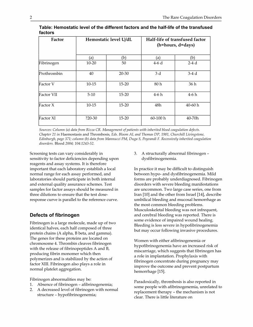
Table: Hemostatic level of the different factors and the half-life of the transfused factors
Sources: Column (a) data from Rizza CR. Management of patients with inherited blood coagulation defects. Chapter 21 in Haemostasis and Thrombosis, Eds. Bloom AL and Thomas DP, 1981, Churchill Livingstone, Edinburgh, page 371; column (b) data from Mannucci PM, Duga S, Peyvandi F. Recessively inherited coagulation disorders. Blood 2004; 104:1243-52.
Screening tests can vary considerably in sensitivity to factor deficiencies depending upon reagents and assay systems. It is therefore important that each laboratory establish a local normal range for each assay performed, and laboratories should participate in both internal and external quality assurance schemes. Test samples for factor assays should be measured in three dilutions to ensure that the test dose-response curve is parallel to the reference curve. Defects of fibrinogen Fibrinogen is a large molecule, made up of two identical halves, each half composed of three protein chains (A alpha, B beta, and gamma). The genes for these proteins are located on chromosome 4. Thrombin cleaves fibrinogen with the release of fibrinopeptides A and B, producing fibrin monomer which then polymerizes and is stabilized by the action of factor XIII. Fibrinogen also plays a role in normal platelet aggregation. Fibrinogen abnormalities may be: 1. Absence of fibrinogen – afibrinogenemia; 2. A decreased level of fibrinogen with normal
structure – hypofibrinogenemia;
3. A structurally abnormal fibrinogen – dysfibrinogenemia.
In practice it may be difficult to distinguish between hypo- and dysfibrinogenemia. Mild forms are probably underdiagnosed. Fibrinogen disorders with severe bleeding manifestations are uncommon. Two large case series, one from Iran [10] and the other from Israel [14], describe umbilical bleeding and mucosal hemorrhage as the most common bleeding problems. Musculoskeletal bleeding was not infrequent, and cerebral bleeding was reported. There is some evidence of impaired wound healing. Bleeding is less severe in hypofibrinogenemia but may occur following invasive procedures. Women with either afibrinogenemia or hypofibrinogenemia have an increased risk of miscarriage, which suggests that fibrinogen has a role in implantation. Prophylaxis with fibrinogen concentrate during pregnancy may improve the outcome and prevent postpartum hemorrhage [15]. Paradoxically, thrombosis is also reported in some people with afibrinogenemia, unrelated to replacement therapy – the mechanism is not clear. There is little literature on
Hemostatic level U/dL Half-life of transfused factor (h=hours, d=days)
Factor
(a) (b) (a) (b) Fibrinogen 10-20 50 4-6 d 2-4 d
Prothrombin 40 20-30 3 d 3-4 d
Factor V 10-15 15-20 80 h 36 h
Factor VII 5-10 15-20 4-6 h 4-6 h
Factor X 10-15 15-20 48h 40-60 h
Factor XI ?20-30 15-20 60-100 h 40-70h
The Rare Coagulation Disorders 3 dysfibrinogenemia and what there is mainly consists of case reports or molecular analyses. The clinical picture is very variable; a compilation of 250 cases reported hemorrhage in 26%, thrombosis in 21% and no symptoms in 53%. An analysis of patients with dysfibrinogenemia and thrombosis demonstrated an unequivocal association of thrombosis with 26 different mutations [16]. Laboratory investigation Coagulation tests will be prolonged in proportion to the reduced fibrinogen. It is important to exclude acquired causes of hypofibrinogenemia. Family studies are often helpful. The thrombin time is the most sensitive test for dysfibrino-genemia. Diagnosis depends upon documenting a difference between functional and antigenic fibrinogen assays. In patients with thrombosis, other causes of thrombophilia should be excluded by a thrombophilia screen. Genetic testing can be performed in some research laboratories. A database of mutations can be viewed at http://www.geht.org/databaseang/fibrinogen/. Treatment Fibrinogen concentrates are detailed in recent UK treatment guidelines [17]. The half-life of infused fibrinogen is 3-5 days (based on adult data). Cryoprecipitate is a good source of fibrinogen but has the major disadvantage of not being treated to inactivate blood-borne viruses. Replacement therapy is recommended before surgery in people with afibrinogenemia (post-operative hemorrhage occurred in 40% of those untreated in one series [10]) and should be sufficient to produce a rise of fibrinogen to at least 1 g/L to ensure hemostasis. Further doses will depend upon clinical and laboratory monitoring, and should aim to achieve a trough level of >0.5 g/L. It is not clear whether infants diagnosed with afibrinogenemia require primary prophylaxis, but the occurrence of ICH in newborns may be an indication. The management of dysfibrinogenemia is less clear [18] and the issues are discussed in the UK guidelines for the management of rare coagulation disorders [1]. In individuals with thrombotic risk, anticoagulant prophylaxis may be indicated in addition to replacement therapy, depending upon the clinical circumstances.
Prothrombin deficiency Factor II (FII) is a vitamin K-dependent carboxylase synthesized in the liver. It is a single chain glycoprotein with four domains. Factor Xa (FXa) activates it on the surface of platelets releasing an activation peptide (fragment 1.2) on cleavage. FII deficiency is very rare, estimated to be 1 in 2 million of the general population. Deficiency may be hypoprothrombinemia (reduced level of a normal molecule, Type 1) or dysprothrombinemia (activity reduced but antigen normal, Type 2). A complete deficiency may be incompatible with life (lethal in knockout mice). Only a small number of cases are reported worldwide [19], and the largest series (14 patients) is from Iran [6]. Severe deficiency was associated with levels of 4-10% and the most common bleeding manifestations were hemarthrosis and muscle hematomata. Life-threatening umbilical bleeding occurred in two patients, and intracranial hemorrhage (ICH) in one. Five other cases of ICH are found in the literature. The clinical picture in dysprothrom-binemia is more variable. Laboratory investigation Both the prothrombin time (PT) and the activated partial thromboplastin time (APTT) will be prolonged but this may be minimal and is reagent dependent. FII assays may be performed by a variety of methods and particular caution is required in infants [1]. Treatment There are no products licensed for use in prothrombin deficiency but several factor concentrates contain FII [17]. In the absence of these, viral-inactivated fresh frozen plasma (FFP) is a potential source of FII. Factor V deficiency Factor V (FV) is a large glycoprotein with 40% sequence homology to factor VIII (FVIII) in the A and C domains and a similar overall structure. FV is encoded on chromosome 1 and produced in hepatocytes and megakaryocytes. Platelets contain about 20% of circulating FV. Both quantitative and qualitative defects are reported. FV deficiency is rare, occurring in 1 in 1 million of the general population. Severely deficient individuals have FV levels from <1 to 10 IU/dL
4 The Rare Coagulation Disorders
(normal range 71-125 IU/dL), and have a moderately severe bleeding tendency which presents in childhood with easy bruising and mucous membrane bleeding, especially epistaxis. Joint and muscle bleeding may also occur but usually less than in hemophilia A. ICH has been reported in infancy and several of the cases in the literature have been complicated by the development of inhibitors after treatment with plasma. Laboratory investigation Both the PT and APTT are prolonged, and the diagnosis is confirmed by performing a FV assay. A FVIII assay should also be performed to exclude combined deficiency (see below). Treatment There are no FV-containing concentrates. FFP, preferably viral inactivated, is the treatment of choice [20]. The minimum hemostatic level has been reported as 15 IU/dL [6]. Large volumes of plasma may be required [20]. Platelet transfusions (with FV in the granules) may be of benefit. Neonates with ICH have been reported; it is therefore prudent to watch infants with care and perform a cranial ultrasound within the first few days of life. Combined deficiency of factors V and VIII The combined deficiency of FV and FVIII is of particular interest as it is the first coagulation disorder attributable to gene defects outside the coagulation factor genes themselves, as inheritance patterns had suggested. The disorder is caused by abnormal transport through the endoplasmic reticulum due to a defect in ERGIC-53 coded on chromosome 18 [21,22]. The factor levels are not usually below 1 IU/dL so spontaneous bleeding is rare. Bleeding occurs after surgery and dental extractions; women may have menorrhagia and postpartum hemorrhage. Laboratory investigation Both PT and APTT are prolonged with the latter being disproportionately long. FV and FVIII levels are generally between 5 and 20 IU/dL.
Treatment Both FV and FVIII levels must be corrected, using FFP for FV to achieve a level of >25 IU/dL, and FVIII concentrate as a source of FVIII to raise the level to 25 IU/dL for minor procedures and >50 IU/dL for major procedures or bleeding episodes. Neonatal ICH has not been reported. Factor VII deficiency Factor VII (FVII) is one of the vitamin K-dependent glycoproteins and is encoded on chromosome 13. A mutation database can be viewed at http://www.193.60.222.13/index.htm. FVII deficiency is the commonest of the rare coagulation disorders excluding factor XI deficiency (severe FVII deficiency occurs in 1 in 500,000 of the general population), but diagnosis of the heterozygous state is complicated by the considerable variation of levels in the normal population, due to both inherited (F7 gene polymorphisms [23]) and acquired (dietary fat, age, obesity, etc.) causes. In addition, reagent (thromboplastin source) can markedly affect the assay result. There is a relatively poor correlation between FVII level and the wide variety of bleeding manifestations [7]. Mucous membrane bleeding, including epistaxis and menorrhagia, is common. Some patients with severe deficiency have suffered ICH, often in the neonatal period, or joint bleeding. Occasionally patients have paradoxical thrombosis, which is not understood [23]. Laboratory investigation The PT is prolonged but all other screening tests are normal. FVII is assayed in a one-stage prothrombin-based assay. Human thromboplastin may give a better reflection of in vivo levels than animal thromboplastins. Blood samples should not be stored on ice before the assay is done as this may induce cold activation of FVII and cause an overestimate of the level. Treatment People with heterozygous deficiency do not have an abnormal bleeding risk. Viral-inactivated plasma-derived FVII concentrates have been available and are effective (see [1] and Appendix 1). Factor IX (FIX) and prothrombin complex concentrates containing FVII have also been used, but carry a risk of thrombosis and are
The Rare Coagulation Disorders 5 no longer recommended. Recombinant factor VIIa (FVIIa) is the treatment of choice [17], and is now licensed for use in this condition [1]. The dose required is much lower than that used for patients with FVIII inhibitors; 15-30 ug/kg seems to be effective. The half-life of FVII is short, and treatment needs to be given every 2-4 hours. The hemostatic level is probably 10-15 IU/dL. For treatment strategies, please see the UKHCDO guidelines [1]. In families where both parents are known to have heterozygous deficiency, preparation for the delivery of a potentially affected newborn includes making a careful management plan with the obstetrician and pediatrician, and avoidance of instrumental delivery. A severely affected newborn is at risk for ICH in the neonatal period. Factor X deficiency Factor X (FX) is a vitamin K-dependent protease and has a key role in the coagulation pathway, being the first enzyme in the common pathway and the most important activator of prothrombin. In association with factor Va and phospholipid membranes, FXa accelerates the conversion of prothrombin to thrombin 280,000-fold. The gene is located on chromosome 13 near the FVII gene to which it is closely related. FX is synthesized in the liver. The overall frequency of severe deficiency is estimated to be 1 in 1 million of the general population. Although most heterozygotes do not have any symptoms, some have a significant bleeding tendency. Severe FX deficiency (FX <1 IU/dL) is generally a severe bleeding disorder [8]. It carries a particular risk of ICH in the neonatal period, therefore, where both parents are known to be heterozygous a delivery management plan should be prepared and the infant watched closely for evidence of ICH. Epistaxis is particularly common, and menorrhagia occurs in 50% of women. Joint bleeding can result in severe arthropathy. Prophylactic treatment for severe FX deficiency should be considered. Mild FX deficiency is defined as 6-10 IU/dL and may be discovered incidentally. Individuals with more than 10 IU/dL and no bleeding
history despite hemostatic challenge may not require replacement therapy. Laboratory investigation The PT and APTT are both prolonged, and the deficiency confirmed by FX assay. Several different assay methods are available (PT- or APTT-based, chromogenic, immunological). The results may vary depending upon the source of thromboplastin used, and chromogenic assays may give normal results in some dysfunctional FX variants. Treatment The hemostatic level for FX post-operatively is thought to be 10-20 IU/dL, and the half-life of infused FX is 60 hours. There is no FX concentrate but prothrombin complex concentrates contain FX and are effective. 1 IU/kg of FX raises the FX level by 1.5%. These concentrates are associated with a thrombotic risk and should be used with caution in those patients with additional risk factors [24]. Children with repeated joint bleeds may benefit from prophylaxis once or twice a week. FFP, preferably viral inactivated, is an alternative. As the half-life in vivo varies in different individuals, regular treatment and post-operative dosing should always be guided by measurement of levels. Deficiency of vitamin K-dependent factors (II, VII, IX, X) FII, FVII, FIX, and FX require a critical gamma-carboxylation step during synthesis to become activated. Defects in the carboxylation steps, caused by enzyme deficiencies, can produce combined deficiency of these four factors. Gene defects have been reported in gamma glutamyl carboxylase and the vitamin K epoxide reductase complex [25,26]. The defect is rare, and is inherited as an autosomal recessive disorder. It has been reported in only about 20 kindreds with variable severity between them. Severe deficiency is associated with levels of <5 IU/dL, and may present in the neonatal period (with umbilical cord bleeding or even ICH) at which time it must be distinguished from vitamin K deficiency. Milder types may present with mucocutaneous or post-surgical bleeding.
6 The Rare Coagulation Disorders
The defect also affects the other vitamin K-dependent factors, which are protein C and S, matrix Gla protein and osteocalcin. Thus, severely deficient children may also have other clinical features similar to warfarin embryopathy, such as nasal hypoplasia, distal digital hypoplasia, epiphyseal stippling, and mild conductive hearing loss. Laboratory investigation The PT and APTT are prolonged and the four factor levels reduced. Vitamin K deficiency and coumarin exposure must be excluded. Treatment Oral vitamin K therapy produces a significant improvement in many individuals, but severely affected people may require replacement therapy prior to surgery with prothrombin complex concentrates, bearing in mind the prothrombotic risks. FFP, preferably viral inactivated, has also been used for some acute bleeds. Factor XI deficiency Factor XI (FXI) is a dimeric serine protease whose function in coagulation is to recruit the intrinsic factor pathway after the tissue factor pathway has generated thrombin. Bleeding tendency may depend upon the levels of other coagulation factors such as FVIII:C and von Willebrand factor (vWF). The gene is on chromosome 4. Factor XI deficiency is the commonest of the rare disorders. The deficiency is particularly common in Ashkenazi Jews where the carrier rate is 8-9%. In this population most individuals have one or both of two particular mutations, a stop codon in exon 5 (type II) and a missense mutation in exon 9 (type III) leading to reduced secretion of the molecule. In other populations the mutations are more variable, but founder mutations have been noted in the English (in as many as 1-2% of the population) and Basques. Heterozygotes with FXI often have a bleeding risk that is not well predicted by the FXI:C level [27]. Women may have menorrhagia and bleeding after childbirth. Severely deficient individuals (FXI <10 IU/dL) have a mild bleeding tendency after surgery, especially in
areas with high fibrinolytic potential such as the mouth and nose and the genitor-urinary tract. Spontaneous bleeding is rare, and hemarthroses are not a feature. The disorder rarely presents in the neonatal period. ICH has not been reported, but bleeding may occur after circumcision. Laboratory investigation The APTT is prolonged (reagent dependent) and the diagnosis established by FXI assay. The lower limit of the normal range is in the region of 60-70 IU/dL, therefore, the defect may be missed by the APTT. However, caution is required since heterozygotes may bleed excessively after surgery and childbirth. Treatment Treatment is not straightforward partly because of the variable bleeding tendency and the risks associated with the currently available FXI concentrates. These have been associated with a risk of thrombosis mainly in older patients with other risk factors. FFP, preferably viral inactivated, is an alternative but large volumes may be required. It is not clear what the hemostatic level is, although a level of 30 IU/dL is probably adequate for surgery in severe deficiency. For those with mild deficiency who have a bleeding history at baseline levels above this, it is reasonable to aim at a level of 70 IU/dL. Optimal management will, therefore, vary with individual circumstances. Another alternative treatment is rVIIa, but in a pilot study thrombotic events were also reported [28]. Dental extractions can be managed with oral tranexamic acid alone, even in those with severe deficiency. FXI concentrate is the appropriate therapy for severely deficient women during labour. Circumcision should be delayed in infants with FXI:C <10 IU/dL at birth and the level checked at 6 months. If it is still <10 IU/dL the procedure should be performed in hospital with appropriate cover, co-ordinating with the Mohel if necessary. Factor XII deficiency Factor XII (FXII) deficiency does not give rise to a bleeding disorder. FXII deficiency (heterozygous) is common in the general Caucasian population (2.3% of blood donors
The Rare Coagulation Disorders 7 [29]) and is the commonest cause of an unexpected prolongation of the APTT in pre-surgical screening. Severe FXII deficiency is most common in Asians where it is usually completely asymptomatic. There is the possibility that FXII deficiency is related to thrombotic events, but recent analysis suggests that there is no association [30,31]. Factor XIII deficiency FXIII deficiency is rare, estimated at 1 per million of the general population. As with the other rare disorders, heterozygotes are asymptomatic. Factor XIII (FXIII) is a tetramer with two ‘a‘ chains (containing the thrombin cleavage site and a calcium binding site) and two ‘b’ chains which are cleaved away when FXIII is activated, in other words the ‘b’ unit is a carrier for the activatable ‘a’ unit. The activated molecule then stabilizes fibrin by cross-linking the gamma and alpha chains by the formation of lysine-glutamine links. Alpha2-plasmin inhibitor is also linked to the ‘a’ chains of fibrin by XIIIa. Interestingly, the subunits have different sites of synthesis and location. The ‘a’ subunits are located in platelets and megakaryocytes, placenta, uterus, and macrophages whereas the ‘b’ units are synthesized in the liver. Thus liver transplantation changes the ‘b’ subunit to that of the donor, leaving the ‘a’ unit unchanged and bone marrow transplantation does the reverse. The FXIII subunit ‘a’ gene is located on chromosome 6, and the ‘b’ gene on chromosome 1. The majority of mutations associated with FXIII deficiency have been described for the ‘a’ unit. [32]. The disorder shows considerable molecular heterogeneity and therefore variable clinical severity, which is discussed by Anwar et al. [32]. Affected individuals tend to bleed excessively from the umbilical stump and are at risk for ICH, which may occur in the neonatal period. Extensive skin bruising and bleeding is also common and patients may suffer muscle and joint bleeding. Pregnancy is often associated with miscarriage unless prophylaxis is given. Laboratory investigation All standard coagulation tests give normal results. Clot solubility is increased after
coagulation with thrombin and suspension in 2% acetic acid, which is more sensitive than urea solubility. Various methods for measurement of FXIII are commercially available but the rarity of this disorder means that laboratories are unfamiliar with this test and it may be advisable to send samples to a specialist laboratory for confirmation. Treatment Because of the high risk of ICH, people with severe deficiency should be offered prophylaxis. FXIII concentrate is available and due to the long half-life of FXIII this only needs to be given every 4-6 weeks. Other sources of FXIII are FFP, cryoprecipitate, and stored plasma. Because of the difficulty of measuring FXIII levels it is hard to make recommendations about trough levels or hemostatic levels for surgery [1]. Conclusion The clinical expression of the rare coagulation disorders is more variable than hemophilia and may present challenges in both diagnosis and management. Awareness of the increased risk of these disorders in appropriate population groups will prompt a higher index of suspicion and thus earlier diagnosis of severely affected infants who are at risk of serious bleeding, particularly ICH.
8 The Rare Coagulation Disorders
References 1. Bolton-Maggs PH, Perry D, Chalmers EA, et
al. The rare coagulation disorders - review with guidelines for management from the UKHCDO. Haemophilia 2004; 10:593-628.
2. Mannucci PM, Duga S, Peyvandi F. Recessively inherited coagulation disorders. Blood 2004; 104:1243-52.
3. Menegatti M, Karimi M, Garagiola I, Mannucci P, Peyvandi F. A rare inherited coagulation disorder: combined homozygous factor VII and factor X deficiency. Am J Hematol 2004; 77:90-1.
4. Peyvandi F, Asselta R, Mannucci PM. Autosomal recessive deficiencies of coagulation factors. Rev Clin Exp Hematol 2001; 5:369-88.
5. Peyvandi F, Duga S, Akhavan S, Mannucci PM. Rare coagulation deficiencies. Haemophilia 2002; 8:308-21.
6. Peyvandi F, Mannucci PM. Rare coagulation disorders. Thromb Haemost 1999; 82:1207-14.
7. Peyvandi F, Mannucci PM, Asti M, et al. Clinical manifestations in 28 Italian and Iranian patients with severe factor VII deficiency. Haemophilia 1997; 3:242-246.
8. Peyvandi F, Mannucci PM, Lak M, et al. Congenital factor X deficiency: spectrum of bleeding symptoms in 32 Iranian patients. Br J Haematol 1998; 102:626-8.
9. Peyvandi F, Tuddenham EG, Akhtari AM, Lak M, Mannucci PM. Bleeding symptoms in 27 Iranian patients with the combined deficiency of factor V and factor VIII. Br J Haematol 1998; 100:773-6.
10. Lak M, Keihani M, Elahi F, Peyvandi F, Mannucci PM. Bleeding and thrombosis in 55 patients with inherited afibrinogenaemia. Br J Haematol 1999; 107:204-6.
11. Lak M, Peyvandi F, Ali Sharifian A, Karimi K, Mannucci PM. Pattern of symptoms in 93 Iranian patients with severe factor XIII deficiency. J Thromb Haemost 2003; 1:1852-3.
12. Lak M, Sharifian R, Peyvandi F, Mannucci PM. Symptoms of inherited factor V deficiency in 35 Iranian patients. Br J Haematol 1998; 103:1067-9.
13. Williams MD, Chalmers EA, Gibson BE. The investigation and management of neonatal haemostasis and thrombosis. Br J Haematol 2002; 119:295-309.
14. Fried K, Kaufman S. Congenital afibrinogenemia in 10 offspring of uncle-niece marriages. Clin Genet 1980; 17:223-7.
15. Kobayashi T, Kanayama N, Tokunaga N, Asahina T, Terao T. Prenatal and peripartum management of congenital afibrinogenaemia. Br J Haematol 2000; 109:364-6.
16. Haverkate F, Samama M. Familial dysfibrinogenemia and thrombophilia. Report on a study of the SSC Subcommittee on Fibrinogen. Thromb Haemost 1995; 73:151-61.
17. United Kingdom Haemophilia Centre Doctors’ Organisation. Guidelines on the selection and use of therapeutic products to treat haemophilia and other hereditary bleeding disorders. Haemophilia 2003; 9:1-23.
18. Roberts HR, Stinchcombe TE, Gabriel DA. The dysfibrinogenaemias. Br J Haematol 2001; 114:249-57.
19. Girolami A, Scarano L, Saggiorato G, et al. Congenital deficiencies and abnormalities of prothrombin. Blood Coagul Fibrinolysis 1998; 9:557-69.
20. Horowitz MS, Pehta JC. SD Plasma in TTP and coagulation factor deficiencies for which no concentrates are available. Vox Sang 1998; 74 (Suppl 1):231-5.
21. Nichols WC, Seligsohn U, Zivelin A, et al. Mutations in the ER-Golgi intermediate compartment protein ERGIC-53 cause combined deficiency of coagulation factors V and VIII. Cell 1998; 93:61-70.
22. Neerman-Arbez M, Johnson KM, Morris MA, et al. Molecular analysis of the ERGIC-53 gene in 35 families with combined factor V-factor VIII deficiency. Blood 1999; 93:2253-60.
23. Perry DJ. Factor VII Deficiency. Br J Haematol 2002; 118:689-700.
24. Kohler M. Thrombogenicity of prothrombin complex concentrates. Thromb Res 1999; 95:S13-7.
The Rare Coagulation Disorders 9 25. Brenner B, Sanchez-Vega B, Wu SM, et al.
Missense mutation in gamma-glutamyl carboxylase gene causes combined deficiency of all vitamin K-dependent blood coagulation factors. Blood 1998; 92:4554-9.
26. Oldenburg J, von Brederlow B, Fregin A, et al. Congenital deficiency of vitamin K dependent coagulation factors in two families presents as a genetic defect of the vitamin K-epoxide-reductase-complex. Thromb Haemost 2000; 84:937-41.
27. Bolton-Maggs PH, Patterson DA, Wensley RT, Tuddenham EG. Definition of the bleeding tendency in factor XI-deficient kindreds–a clinical and laboratory study. Thromb Haemost 1995; 73:194-202.
28. O'Connell N, G P, et al. Prevention of surgical bleeding with recombinant factor VIIa in patients with factor XI deficiency. Blood 2002; 100:697a.
29. Halbmayer WM, Mannhalter C, Feichtinger C, Rubi K, Fischer M. [Factor XII (Hageman factor) deficiency: a risk factor for development of thromboembolism. Incidence of factor XII deficiency in patients after recurrent venous or arterial thromboembolism and myocardial infarction]. Wien Med Wochenschr 1993; 143:43-50.
30. Girolami A, Morello M, Girolami B, Lombardi AM, Bertolo C. Myocardial infarction and arterial thrombosis in severe (homozygous) FXII deficiency: No apparent causative relation. Clin Appl Thromb Hemost 2005; 11:49-53.
31. Koster T, Rosendaal FR, Briet E, Vandenbroucke JP. John Hageman's factor and deep-vein thrombosis: Leiden thrombophilia study. Br J Haematol 1994; 87:422-4.
32. Anwar R, Miloszewski KJ. Factor XIII deficiency. Br J Haematol 1999; 107:468-84.
10 The Rare Coagulation Disorders
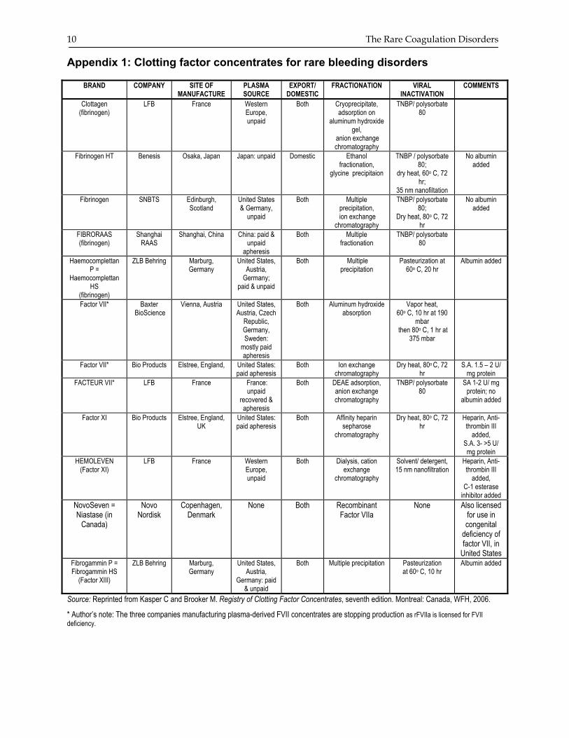
Appendix 1: Clotting factor concentrates for rare bleeding disorders
BRAND COMPANY SITE OF MANUFACTURE
PLASMA SOURCE
EXPORT/ DOMESTIC
FRACTIONATION VIRAL INACTIVATION
COMMENTS
Clottagen (fibrinogen)
LFB France Western Europe, unpaid
Both Cryoprecipitate, adsorption on
aluminum hydroxide gel,
anion exchange chromatography
TNBP/ polysorbate 80
Fibrinogen HT Benesis Osaka, Japan Japan: unpaid Domestic Ethanol fractionation,
glycine precipitaion
TNBP / polysorbate 80;
dry heat, 60o C, 72 hr;
35 nm nanofiltation
No albumin added
Fibrinogen SNBTS Edinburgh, Scotland
United States & Germany,
unpaid
Both Multiple precipitation,
ion exchange chromatography
TNBP/ polysorbate 80;
Dry heat, 80o C, 72 hr
No albumin added
FIBRORAAS (fibrinogen)
Shanghai RAAS
Shanghai, China China: paid & unpaid
apheresis
Both Multiple fractionation
TNBP/ polysorbate 80
Haemocomplettan P =
Haemocomplettan HS
(fibrinogen)
ZLB Behring Marburg, Germany
United States, Austria,
Germany; paid & unpaid
Both Multiple precipitation
Pasteurization at 60o C, 20 hr
Albumin added
Factor VII*
Baxter BioScience
Vienna, Austria United States, Austria, Czech
Republic, Germany, Sweden:
mostly paid apheresis
Both Aluminum hydroxide absorption
Vapor heat, 60o C, 10 hr at 190
mbar then 80o C, 1 hr at
375 mbar
Factor VII*
Bio Products Elstree, England, United States: paid apheresis
Both Ion exchange chromatography
Dry heat, 80o C, 72 hr
S.A. 1.5 – 2 U/ mg protein
FACTEUR VII*
LFB France France: unpaid
recovered & apheresis
Both DEAE adsorption, anion exchange chromatography
TNBP/ polysorbate 80
SA 1-2 U/ mg protein; no
albumin added
Factor XI Bio Products Elstree, England, UK
United States: paid apheresis
Both Affinity heparin sepharose
chromatography
Dry heat, 80o C, 72 hr
Heparin, Anti-thrombin III
added, S.A. 3- >5 U/ mg protein
HEMOLEVEN (Factor XI)
LFB France Western Europe, unpaid
Both Dialysis, cation exchange
chromatography
Solvent/ detergent, 15 nm nanofiltration
Heparin, Anti-thrombin III
added, C-1 esterase
inhibitor added NovoSeven = Niastase (in
Canada)
Novo Nordisk
Copenhagen, Denmark
None Both Recombinant Factor VIIa
None Also licensed for use in congenital
deficiency of factor VII, in
United States Fibrogammin P = Fibrogammin HS
(Factor XIII)
ZLB Behring Marburg, Germany
United States, Austria,
Germany: paid & unpaid
Both Multiple precipitation Pasteurization at 60o C, 10 hr
Albumin added
Source: Reprinted from Kasper C and Brooker M. Registry of Clotting Factor Concentrates, seventh edition. Montreal: Canada, WFH, 2006.
* Author’s note: The three companies manufacturing plasma-derived FVII concentrates are stopping production as rFVIIa is licensed for FVII deficiency.
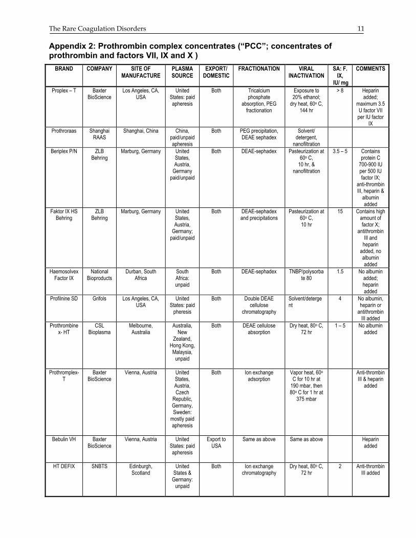
The Rare Coagulation Disorders 11 Appendix 2: Prothrombin complex concentrates (“PCC”; concentrates of prothrombin and factors VII, IX and X )
BRAND COMPANY SITE OF MANUFACTURE
PLASMA SOURCE
EXPORT/ DOMESTIC
FRACTIONATION VIRAL INACTIVATION
SA: F. IX,
IU/ mg
COMMENTS
Proplex – T Baxter BioScience
Los Angeles, CA, USA
United States: paid apheresis
Both Tricalcium phosphate
absorption, PEG fractionation
Exposure to 20% ethanol;
dry heat, 60o C, 144 hr
> 8 Heparin added;
maximum 3.5 U factor VII per IU factor
IX Prothroraas Shanghai
RAAS Shanghai, China China,
paid/unpaid apheresis
Both PEG precipitation, DEAE sephadex
Solvent/ detergent,
nanofiltration
Beriplex P/N ZLB Behring
Marburg, Germany United States, Austria,
Germany paid/unpaid
Both DEAE-sephadex Pasteurization at 60o C,
10 hr, & nanofiltration
3.5 – 5 Contains protein C
700-900 IU per 500 IU factor IX;
anti-thrombin III, heparin &
albumin added
Faktor IX HS Behring
ZLB Behring
Marburg, Germany United States, Austria,
Germany; paid/unpaid
Both DEAE-sephadex and precipitations
Pasteurization at 60o C, 10 hr
15 Contains high amount of factor X;
antithrombin III and
heparin added, no albumin added
Haemosolvex Factor IX
National Bioproducts
Durban, South Africa
South Africa: unpaid
Both DEAE-sephadex TNBP/polysorbate 80
1.5 No albumin added; heparin added
Profilnine SD Grifols Los Angeles, CA, USA
United States: paid
pheresis
Both Double DEAE cellulose
chromatography
Solvent/detergent
4 No albumin, heparin or
antithrombin III added
Prothrombinex- HT
CSL Bioplasma
Melbourne, Australia
Australia, New
Zealand, Hong Kong, Malaysia,
unpaid
Both DEAE cellulose absorption
Dry heat, 80o C, 72 hr
1 – 5 No albumin added
Prothromplex-T
Baxter BioScience
Vienna, Austria United States, Austria, Czech
Republic, Germany, Sweden:
mostly paid apheresis
Both Ion exchange adsorption
Vapor heat, 60o C for 10 hr at
190 mbar, then 80o C for 1 hr at
375 mbar
Anti-thrombin III & heparin
added
Bebulin VH Baxter BioScience
Vienna, Austria United States: paid apheresis
Export to USA
Same as above Same as above Heparin added
HT DEFIX SNBTS Edinburgh, Scotland
United States &
Germany: unpaid
Both Ion exchange chromatography
Dry heat, 80o C, 72 hr
2 Anti-thrombin III added
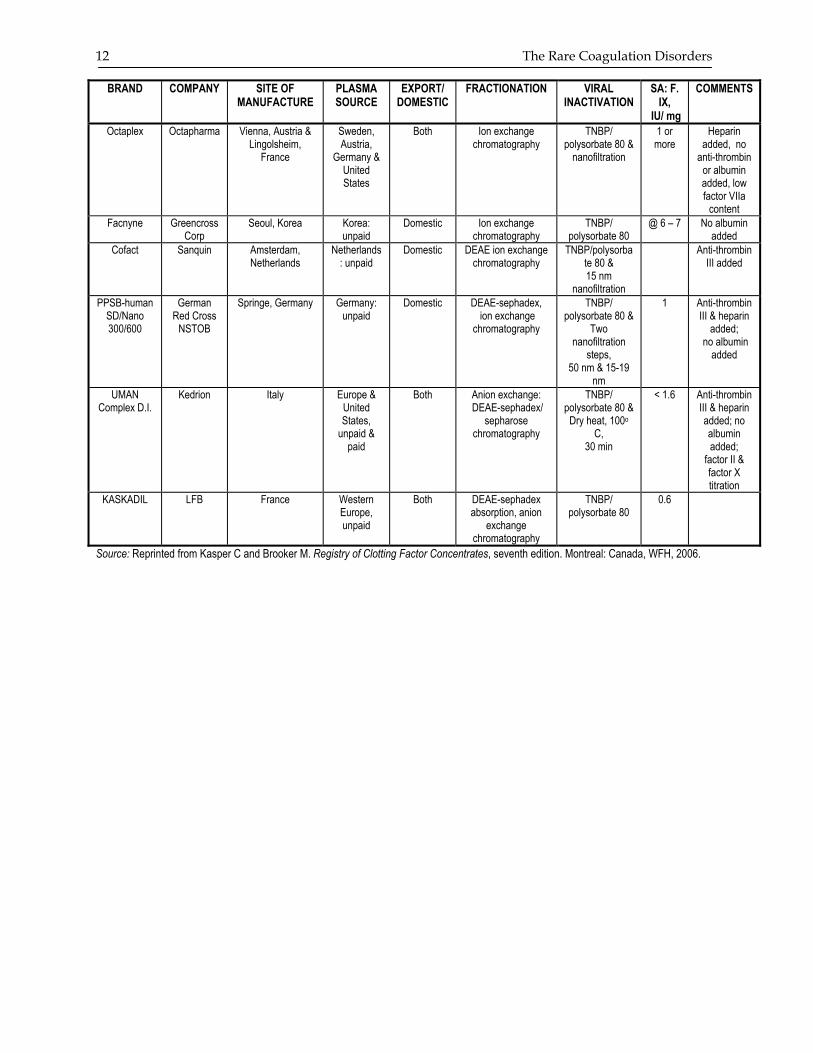
12 The Rare Coagulation Disorders
BRAND COMPANY SITE OF MANUFACTURE
PLASMA SOURCE
EXPORT/ DOMESTIC
FRACTIONATION VIRAL INACTIVATION
SA: F. IX,
IU/ mg
COMMENTS
Octaplex
Octapharma Vienna, Austria & Lingolsheim,
France
Sweden, Austria,
Germany & United States
Both Ion exchange chromatography
TNBP/ polysorbate 80 &
nanofiltration
1 or more
Heparin added, no
anti-thrombin or albumin added, low factor VIIa
content Facnyne Greencross
Corp Seoul, Korea Korea:
unpaid Domestic Ion exchange
chromatography TNBP/
polysorbate 80 @ 6 – 7 No albumin
added Cofact Sanquin Amsterdam,
Netherlands Netherlands
: unpaid Domestic DEAE ion exchange
chromatography TNBP/polysorba
te 80 & 15 nm
nanofiltration
Anti-thrombin III added
PPSB-human SD/Nano 300/600
German Red Cross
NSTOB
Springe, Germany Germany: unpaid
Domestic DEAE-sephadex, ion exchange
chromatography
TNBP/ polysorbate 80 &
Two nanofiltration
steps, 50 nm & 15-19
nm
1 Anti-thrombin III & heparin
added; no albumin
added
UMAN Complex D.I.
Kedrion Italy Europe & United States,
unpaid & paid
Both Anion exchange: DEAE-sephadex/
sepharose chromatography
TNBP/ polysorbate 80 &
Dry heat, 100o C,
30 min
< 1.6 Anti-thrombin III & heparin added; no albumin added;
factor II & factor X titration
KASKADIL LFB France Western Europe, unpaid
Both DEAE-sephadex absorption, anion
exchange chromatography
TNBP/ polysorbate 80
0.6
Source: Reprinted from Kasper C and Brooker M. Registry of Clotting Factor Concentrates, seventh edition. Montreal: Canada, WFH, 2006.


The printing of this publication was supported by an unrestricted
educational grant from
LfB(Laboratoire français du fractionnement
et des biotechnologies)