Embed Size (px)
Citation preview

The Regulation of Multidrug Resistance Phosphoglycoprotein
(MDR1/P-gp) and Breast Cancer Resistance Protein (BCRP) in the
Human Placenta
Jenna Rainey
This thesis is submitted as a partial fulfillment of the M.Sc. program in Cellular
and Molecular Medicine
January 2011
University of Ottawa
Ottawa, Ontario
© Jenna Rainey, Ottawa, Canada, 2011
ii
ABSTRACT
Multidrug resistance phosphoglycoprotein (MDR1/P-gp) and breast cancer resistance
protein (BCRP) were first isolated in chemoresistant cancer cells and have since been found in a
variety of normal tissue, including the placenta. The potential function of MDR1/P-gp and
BCRP in the human placenta is to protect the fetus from maternally circulating endogenous
steroids and hormones, therapeutic drugs and toxins. Studies from our laboratory have shown
that there is a significant decrease in MDR1/P-gp levels and an increase in BCRP levels in the
syncytiotrophoblast of the human placenta with advancing gestation; however, the regulation of
both proteins is not known. The objective of this study was to examine the role of maternal
steroids in the regulation of MDR1/P-gp and BCRP in the human placenta. Term (38-40 weeks)
placenta tissues were collected after scheduled, non-complicated cesarean sections at the Ottawa
General Hospital and a modified version of the Kliman’s technique was used to isolate
trophoblast cells. Immunohistochemistry (IHC), western blot analysis and transport studies were
used to determine the effect of maternal steroids on MDR1/P-gp and BCRP regulation. Maternal
steroids (estrogen, progesterone and cortisol), present at high concentrations in maternal serum,
did not have an effect on BCRP in human syncytiotrophoblast. Estrogen and progesterone did
not alter MDR1/P-gp levels in human syncytiotrophoblast, but cortisol significantly decreased
MDR1/P-gp levels. Dexamethasone, an agonist of the glucocorticoid receptor, did not have an
effect on MDR1/P-gp levels. Furthermore, co-incubation with RU486, a synthetic
glucocorticoid, did not reverse the inhibitory effect of cortisol on MDR1/P-gp levels. The
decrease in MDR1/P-gp levels with advancing gestation could be due to the increase in cortisol
levels in maternal circulation towards term. It would appear that cortisol is not acting via the
glucocorticoid receptor to exert its regulatory effect on MDR1/P-gp.

iii
TABLE OF CO(TE(TS
ABSTRACT………………………………………………………………………………..ii
LIST OF TABLES………………………...………………………………………………vi
LIST OF FIGURES……………………………………………………………………….vii
LIST OF ABBREVIATIO(S…………………………………………………………….x
ACK(OWLEDGEME(TS………………………………………………………………xii
CHAPTER 1: I(TRODUCTIO(………………………………………………………..1
1.1 ABC Transporters……………………………………………………………………1
1.1.1 Overview……………………………………………………………………...1
1.1.2 MDR1/P-gp…………………………………………………………………...2
1.1.3 BCRP………………………………………………………………………….4
1.1.4 MDR1/P-gp and BCRP Protein Structure…………………………………….6
1.1.5 Regulation of Human MDR1/P-gp and BCRP……………………………….9
1.2 The Human Placenta…………………………………………………………………10
1.2.1 Overview……………………………………………………………………...10
1.2.2 Structure………………………………………………………………………11
1.3 ABC Transporters in Human Placenta……………………………………………….14
1.3.1 Maternal Pharmacotherapy……………………………………………………14
1.3.2 MDR1/P-gp in the Human Placenta…………………………………………..15
1.3.3 BCRP in the Human Placenta………………………………………………...16
1.3.4 Regulation of MDR1/P-gp and BCRP by Steroids…………………………...19
1.4 Choriocarcinoma Cell Lines………………………………………………………….21
1.4.1 BeWo Cell Line……………………………………………………………….21
1.4.2 ABC Transporter in the BeWo Cell Line……………………………………..22

iv
1.5 Rationale……………………………………………………………………………...23
1.6 Purpose………………………………………………………………………………..24
1.7 Hypotheses……………………………………………………………………………24
1.8 Objectives…………………………………………………………………………….25
CHAPTER 2: MATERIALS A(D METHODS…………………………………………27
2.1 Tissue Collection……………………………………………………………………...27
2.2 Trophoblast Primary Cell Culture…………………………………………………….27
2.3 BeWo Cell Culture……………………………………………………………………28
2.4 Immunohistochemistry………………………………………………………………..28
2.5 Western blotting………………………………………………………………………31
2.6 Functional Transport Assay…………………………………………………………..34
2.7 Statistical Analysis……………………………………………………………………34
CHAPTER 3: REGULATIO( OF MULTIDRUG RESISTA(CE
PHOSPHOGLYCOPROTEI( (MDR1/P-GP) A(D BREAST CA(CER RESISTA(CE
PROTEI( (BCRP) I( THE HUMA( PLACE(TA…………………………………….36
3.1 RESULTS…………………………………………………………………………….36
3.1.1 General Characteristics of Syncytiotrophoblast………………………………….....36
3.1.2 Estrogen and Progesterone Regulation of MDR1/P-gp…………………………….40
3.1.3 Glucocorticoid Regulation of MDR1/P-gp…………………………………………44
3.1.4 Function of MDR1/P-gp……………………………………………………………51
3.1.5 Estrogen and Progesterone Regulation of BCRP…………………………………...53
3.1.6 Glucocorticoid Regulation of BCRP……………………………………………….57
3.2 DISCUSSION………………………………………………………………………...60
3.2.1 Syncytialization and Purity of Trophoblast Cells in Culture……………………….60
3.2.2 MDR1/P-gp in Human Placental Tissue and Trophoblast Primary Cell Culture…..61
3.2.3 Steroid Regulation of MDR1/P-gp…………………………………………………62

v
3.2.4 Steroid Regulation of BCRP……………………………………………………….68
CHAPTER 4: EXAMI(ATIO( OF THE BEWO CELL LI(E AS A POTE(TIAL
MODEL FOR MDR1/P-GP REGULATIO(……………………………………………71
4.1 RESULTS…………………………………………………………………………….71
4.1.1 Verification of Purity and Syncytialization of the BeWo Cell Line………………..71
4.1.2 MDR1/P-gp and BCRP in the BeWo Cell Line……………………………………71
4.1.3 Forskolin Regulation of MDR1/P-gp and BCRP in the BeWo Cell Line………….72
4.2 DISCUSSION………………………………………………………………………...78
CHAPTER 5: SUMMARY………………………………………………………………..81
REFERE(CES…………………………………………………………………………….84
vi
LIST OF TABLES
Table 1: Immunoblotting antibodies and dilutions................................................................30
Table 2: Immunohistochemistry antibodies and dilutions.....................................................33
vii
LIST OF FIGURES
Figure 1: Proposed structure of MDR1/P-gp………………………………………………8
Figure 2: Proposed structure of BCRP……………………………………………………..8
Figure 3: The structure of the human placenta…………………………………………......13
Figure 4: The potential function of MDR1/P-gp and BCRP in the human placenta……....18
Figure 5: Schematic diagram of conversion of Calcein-AM into fluorescent Calcein by
cytoplasmic esterase…………………………………………………………………………35
Figure 6: Immunohistochemistry localization of MDR1/P-gp in human placental tissue....37
Figure 7: Trophoblast cells in culture……………………………………………………....38
Figure 8: Immunohistochemistry of syncytialized trophoblast primary cell culture…….....38
.
Figure 9: Verification of syncytialized trophoblast primary cell culture…………………...39
Figure 10: Typical western blot of MDR1/P-gp in human placental tissue and placental primary
cell culture……………………………………………………………………………………39
Figure 11: MDR1/P-gp levels in human syncytiotrophoblast after 24 hours of estrogen
treatment…………………………………………………………………………………......41
Figure 12: MDR1/P-gp levels in human syncytiotrophoblast after 24 hours of progesterone
treatment………………………………………………………………………………..........42
viii
Figure 13: MDR1/P-gp levels in human syncytiotrophoblast after 24 hours of co-incubation with
estrogen and progesterone……………………………………………………………….......43
Figure 14: MDR1/P-gp levels in human syncytiotrophoblast after 24 hours of cortisol
treatment……………………………………………………………………………………..46
Figure 15: Syncytialization of trophoblast primary cells after 96 hours in culture……........47
Figure 16: MDR1/P-gp levels in human syncytiotrophoblast after 24 hours of dexamethasone
treatment………………………………………………………………………………….….48
Figure 17: MDR1/P-gp levels in human syncytiotrophoblast after 24 hours of cortisol and
cortisol + RU486 treatment…………………………………………………………...……..49
Figure 18: MDR1/P-gp levels in human syncytiotrophoblast after 24 hours of cortisol and
cortisol + spironolactone treatment…………………………………………………..……...50
Figure 19: Fluorescence accumulation of calcein in syncytiotrophoblast treated with cortisol for
24 hours………………………………………………………………………………….......52
Figure 20: Typical western blot of BCRP in placental primary cell culture and placental
tissue……………………………………………………………………………………..…..54
Figure 21: BCRP levels in human syncytiotrophoblast after 24 hours of estrogen
treatment……………………………………………………………………………….…….55
Figure 22: BCRP levels in human syncytiotrophoblast after 24 hours of progesterone
treatment……………………………………………………………………………….….....56
ix
Figure 23: BCRP levels in human syncytiotrophoblast after 24 hours of cortisol
treatment……………………………………………………………………………………..58
Figure 24: BCRP levels in human syncytiotrophoblast after 24 hours of dexmathesone
treatment…………………………………………………………………………………......59
Figure 25: Verification of purity of BeWo cells after 72 hours in culture………………….73
Figure 26: Immunohistochemistry of BeWo cells in culture……………………………….73
Figure 27: Typical western blot of MDR1/P-gp in BeWo cell line and placental primary cell
culture…………………………………………………………………………………..........74
Figure 28: Typical western blot of MDR1/P-gp in the BeWo cell line…………………….74
Figure 29: Typical western blot of BCRP in the BeWo cell line……………………….….74
Figure 30: Immunohistochemistry of BeWo cells after treatment with forskolin…….........75
Figure 31: MDR1/P-gp levels in the BeWo cell line after 72 hours of forskolin treatment..76
Figure 32: BCRP levels in the BeWo cell line after 72 hours of forskolin treatment……....77
x
LIST OF ABBREVIATIO(S
ABC ATP-Binding Cassette
ABCB1 ATP-Binding Cassette, sub-family B
ABCC ATP-Binding Cassette, sub-family C
ABCG2 ATP-Binding Cassette, sub-family G
ABCP ABC transport in the placenta
ATCC American Type Culture Collection
AZT Zidovudine
BCRP Breast Cancer Resistance Protein
BSA Bovine serum albumin
cAMP Cyclic adenosine monophosphate
CAR Constitutive androsterone receptor
DAB Diaminobenzidine
DNAse Deoxyribonuclease 1
DMEM Dulbecco’s modified eagles medium
ECL Enhanced chemiluminescence
ERα Estrogen receptor alpha
ERE Estrogen response element
FBS Fetal bovine serum
FDA Food and Drug Administration
GR Glucocorticoid receptor
GRE Glucocorticoid response element
Gβ G protein beta subunit
hCG Human chorionic gonadotrophin

xi
HIV Human immunodeficiency virus
HRE Hypoxia response elements
IHC Immunohistochemistry
MDR1/P-gp Multidrug Resistance Phosphoglycoprotein
MR Mineralocorticoid receptor
mRNA Messenger ribonucleic acid
MRP1-9 Multidrug Resistance-Associated Protein 1-9
MXR Mitoxantrone resistance protein
NBD Nucleotide binding domain
NCS Newborn calf serum
PBS Phosphate buffered saline
PhIP Carcinogen 2-amino-1-methyl-6-phenylimidazo [4,5-b] pyridine
PR Progesterone receptor
PRA Progesterone receptor A
PRB Progesterone receptor B
PRE Progesterone response element
PXR Pregnane xenobiotic receptor
RU486 Mifepristone
RT-PCR Reverse transcription polymerase chain reaction
SDS Sodium dodecyl sulfate
TMD Transmembrane domain

xii
ACK(OWLEDGME(TS
This thesis is dedicated to my family, Eric, Sandy, Kelsey and Russell Rainey, for their
undying support and love. I would especially like to thank my mother for her continued
optimism, support, advice and tri-daily phone calls. I am eternally grateful.
I would like to thank my supervisor, Dr. William Gibb, for his guidance, encouragement,
and above all his patience throughout my studies in his laboratory. I also appreciated his
allowing me to take time to pursue volunteering opportunities, particularly Science Travels and
The Olympic Winter Games in Vancouver, and being very supportive and enthusiastic about
these endeavours. I am also grateful to Dr. Stephen Matthews at the University of Toronto, who
is the collaborator and co-grant owner, for his advice regarding my project.
I wish to show my appreciation to my advisory committee, Dr. Gruslin, Dr. Cooke, and
Dr. Tsang for their suggestions and providing me with direction regarding my project. Thank
you for taking time out of your busy schedules to read my reports and come to my meetings.
Thank you to Meihua Sun, for her technical expertise and her advice in Dr. Gibb’s lab. I
would also like to thank Dorothy Yeboah, for teaching me laboratory techniques, especially for
her patience in demonstrating human trophoblast cell culture.
I am indebted to Pauline Shields and Emma Webster for consenting tissues at the Ottawa
General Hospital. My research was very much dependent on them and I am grateful.
Many thanks to my extended family, particularly my Aunt Nicole, Uncle John, and my
Nama, for providing me with accommodations in Ottawa throughout my studies.
Finally, I would like to thank my fabulous friends for listening to me when I was stressed,
for your endless encouragement and always picking me up when I’m down. I would especially
like to thank Deana Conway and Danny Richardson for all their support in the past two and a
half years.
1
CHAPTER 1: I(TRODUCTIO(
1.1 ABC TRA(SPORTERS
1.1.1 Overview
ATP-binding cassette (ABC) proteins are a large and functionally varied superfamily of proteins
that use energy generated from ATP hydrolysis to move substrates across a membrane, usually
against a concentration gradient (Dean, Hamon, & Chimini, 2001; Higgins, 1992; Childs & Ling,
1994; Dean & Allikmets, 1995). ABC transporters are present in all phyla and can either import
or export substrates across cell membranes. In humans, these proteins export substrates from the
cytoplasm or cell membrane to the exterior of the cell (Saurin, Hofnung, & Dassa, 1999). There
are 49 known ABC proteins encoded by the human genome, which are divided into 7
subfamilies, A to G (Sheps et al., 2004; Dean & Annilo, 2005). Three of these proteins are
involved in drug transport in the human body; Multidrug Resistance Phosphoglycoprotein
(MDR1/P-gp or ABCB1; encoded by the MDR1 gene), Multidrug Resistance-Associated Protein
1-9 (MRP1-9; encoded by the ABCC gene), and Breast Cancer Resistance Protein (BCRP or
ABCG2; encoded by the BCRP gene) (Litman et al., 2001). The substrates of these proteins are
diverse and overlapping, therefore it is thought that they collaborate to form a network to protect
the body from xenobiotics (Sharom, 2008). ABC transporters can cause resistance to
chemotherapy drugs in cancer cells and can also act as pharmacological barriers in normal tissue.
MDR1/P-gp and BCRP will be the focus of this thesis.

2
1.1.2 MDR1/P-gp
Multidrug Resistance Phosphoglycorprotein, MDR1/P-gp, was first discovered by Juliano and
Ling (1976). They found high levels of a 170 kDA protein on the membrane of Chinese hamster
ovarian cells, which extruded colchicine out of the cells, causing drug resistance (Juliano &
Ling, 1976). In 1987, the protein was named MDR1/P-gp (Ueda et al., 1987).
MDR1/P-gp was initially studied in chemoresistant tumour cells (Juliano & Ling, 1976), but was
later found in a variety of normal tissue, where the protein is localized at important barrier sites
to limit the absorption of pharmacological agents and promote excretion of these substrates
(Thiebaut et al., 1987; Cordon-Cardo et al., 1990). For example, on the intestinal epithelium,
MDR1/P-gp extrudes substrates back into the gut lumen (Thiebaut et al., 1987). The protein
transports substrates out of the proximal tubule cells of the kidney into the urine and out of the
liver into the bile (Schinkel & Jonker, 2003). MDR1/P-gp is also expressed at high levels in the
blood-brain barrier and in the blood-testis barrier (Cordon-Cardo et al., 1989). Our laboratory
has shown MDR1/P-gp to be localized on the apical membrane of the syncytiotrophoblast of the
placenta, where the protein transports substrates from the syncytiotrophoblast back into maternal
blood, to limit substrate distribution to the fetus (Sun et al., 2006).
MDR1/P-gp can efflux a large variety of structurally different substrates (Wang et al., 2003).
Substrates are typically, but not always, between 200 Da-1900 Da in size, have an aromatic ring
in their structure and are uncharged or basic. However, these properties do not hold true for all
substrates of the protein, as some are non-aromatic linear or circular in structure and acidic in pH

3
(Schinkel & Jonker, 2003). Pharmacological substrates of MDR1/P-gp include cardiac
glycosides, antibiotics, and human immunodeficiency virus (HIV) protease inhibitors (Schinkel
et al., 1995; Schuetz et al., 1996; Choo et al., 2000). Furthermore, endogenous steroids such as
cortisol (Ueda et al., 1992) and synthetic steroids, such as dexamethasone and betamethasone,
are also substrates of the protein (Meijer, Karssen, & de Kloet, 2003). Anticancer drugs that are
unique substrates for MDR1/P-gp, but not BCRP or MRP, include cisplatin, taxols, and
verapamil (Sharom, 2008). The large variety and number of substrates of MDR1/P-gp indicates
a potential function of the protein in protecting the body from toxic xenobiotics, as well as a
possible physiological function in the regulation of endogenous steroids levels.
One gene, MDR1, encodes MDR1/P-gp in humans (Chen et al., 1986). Interestingly, two genes,
Mdr1a and Mdr1b, encode two forms of the Mdr1/P-gp in the mouse. The two isoforms of the
protein, Mdr1a/P-gp and Mdr1b/P-gp, have both unique and overlapping substrate specificities
and localizations. For example, in the mouse, the heart, lungs, and kidney have similar levels of
both Mdr1a and Mdr1b mRNA, however, the liver and brain have higher levels of Mdr1a
mRNA and the placenta and uterus have higher levels of Mdr1b mRNA (Devault & Gros, 1990;
Gros et al., 1991; Kalabis et al., 2005).
Homozygous Mdr1a knock-out mice, Mdr1a (-/-), were found to be healthy and fertile, and have
normal lifespans. However, when the cages were sprayed with ivermectin, to treat a mite
infestation, it was found that all Mdr1a (-/-) mice died, while the normal and heterozygous mice
lived. Upon further investigation, it was found that there was 100-fold increase of invermectin in
4
the brain, compared to wild-type mice (Schinkel et al., 1994). This study indicated that
Mdr1a/P-gp plays an important role in limiting entry of toxins into the brain.
1.1.3 BCRP
BCRP was originally discovered and cloned from the breast carcinoma cell line, MCF-7/AdrVp,
by Doyle et al. (1998). The cell line was resistant to the chemotherapeutic agent doxorubicin, as
well as daunorubicin and mitoxantrone (Doyle et al., 1998). Shortly afterwards, two other
groups independently cloned proteins that were almost identical to BCRP. ABCP (ABC
transporter in the placenta) was isolated from the human placenta (Allikmets et al., 1998) and
MXR (mitoxantrone resistance protein) was isolated from S1-M1–80, a human colon cancer cell
line that is resistant to mitoxantrone (Miyake et al., 1999). Other than a few amino acid
differences, BCRP, ABCP, and MXP are the same protein.
BCRP has been found in cancer cells and in normal tissue. Northern blot studies determined that
BCRP mRNA was expressed at high levels in the human placenta, as well as being expressed in
the liver, small intestine, brain, and testis (Doyle et al., 1998). BCRP was localized to the apical
membrane of syncytiotrophoblast of the placenta, the liver canalicular membrane, the apical
membrane of the epithelium of the small intestine, the breast, and the blood-brain barrier using
immunohistochemistry (IHC) (Maliepaard et al., 2001). BCRP has also been proposed to
provide protection against hypoxia via the efflux of toxic heme metabolites from the cytoplasm
(Krishnamurthy et al., 2004).
5
BCRP extrudes a broad range of xenobiotic and endogenous substrates, which are hydrophobic
or hydrophilic in nature and can be either conjugated or unconjugated, including sulphate
conjugates (Imai et al., 2003). Many chemotherapeutic agents that BCRP transport are also
substrates of MDR1/P-gp. For example, daunorubicin, doxorubicin and mitoxantrone are anti
cancer drugs that are transported by both proteins (Doyle & Ross, 2003). However, the substrate
specificity of BCRP only partially overlaps with MDR1/P-gp. Substrates unique to BCRP
include flavopiridiol and SN-38, a topoisomerase 1 inhibitor (Mao & Unadkat, 2005).
BCRP also transports a variety of therapeutic drugs. Nitrofurantoin, an antibiotic, is a substrate
of the protein (Merino et al., 2005). Glyburide, an anti-diabetic drug, and the HIV drug
zidovudine (AZT) are also pharmacological agents that are transported by BCRP (Gedeon et al.,
2006; X. Wang et al., 2003). Cimetidine, a histamine H2-receptor antagonist, was also found to
be a substrate (Pavek et al., 2005). Certain toxins are transported by BCRP, such as 2-amino-1-
methyl-6-phenylimidazo [4,5-b] pyridine (PhIP), a carcinogen found in food and cigarette smoke
(Pavek et al., 2005). Furthermore, certain endogenous steroids are also known to be substrates
of the protein, such as sulfo- conjugates of estrone or 17β-estradiol (Imai et al., 2003).
Interestingly, BCRP is not implicated in the transport of free estrogen (Pavek et al., 2005). The
protein is also known to transport folate, associating BCRP with folic acid homeostasis in cells
(Hooijberg et al., 2006). Considering the localization and substrate specificity of the protein, the
potential function of BCRP is to protect the body from foreign BCRP substrates, such as toxins
and pharmaceutical agents, and regulate endogenous BCRP substrates.

6
1.1.4 MDR1/P-gp and BCRP Protein Structure
The basic core structure of the human ABC protein consists of two hydrophilic nucleotide
binding domains (NBDs), which are believed to bind and hydrolyze ATP, and two hydrophobic
transmembrane domains (TMDs), which are thought to recognize and provide a translocation
passageway for the substrate to cross the membrane (Higgins et al., 1986). This is referred to as
a full transporter and is typically organized TMD1-NBD1-TMD2-NBD2 (Loo & Clarke, 1999).
Human ABC proteins can also be half transporters, containing one TMD and one NBD. The
domain sequences of half transporters are NBD1-TMD1 or TMD1-NBD1 (Doyle et al., 1998).
In order to be functional, human ABC proteins require two TMDs and two NBDs; therefore, half
transporters must form homo- or heterodimers to operate (Hyde et al., 1990).
As previously stated, the MDR1 gene encodes MDR1/P-gp in humans. The human protein is
composed of 1280 amino acids; with molecular mass of approximately 170 kDa (Juliano & Ling,
1976; Hyde et al., 1990), although some publications have suggested a molecular mass of 140-
170 kDa (Ling et al., 1983). MDR1/P-gp is a full transporter composed of two NBDs and two
TMDs, with 6 α-helices per TMD (Figure 1) (Schinkel & Jonker, 2003; Borst & Elferink, 2002).
The BCRP gene encodes BCRP in humans. BCRP is 72 kDa in size and composed of 665 amino
acids (Doyle et al., 1998). The configuration of BCRP is that of a half-transporter, NBD1-
TMD1 (Figure 2); therefore the protein is thought to homodimerize or forms a complex to
become functional (Ozvegy, Varadi, & Sarkadi, 2002). It has been suggested that BCRP forms a
homodimer through S-S bonds. This disulphide bond bridge has been considered necessary for

7
drug transport function, due to the fact that function was abolished by a dominant negative
mutant (Kage et al., 2002). Yeboah et al. (2006b) observed a protein of approximately 200 kDa,
under non-reducing conditions in human placental tissue. These results indicate that BCRP may
function as a multimer, instead of a homodimer, in the human placenta (Yeboah et al. 2006b).

8
Figure 1| Proposed structure of MDR1/P-gp. MDR1/P-gp is composed of two transmembrane domains (TMDs),
with 6 transmembrane α-helices each, and two NBDs. MDR1/P-gp is considered a full transporter. Modified from
Shinkel and Jonker (2003).
Figure 2| Proposed structure of BCRP. BCRP is composed of one NBD and one TMD. BCRP is considered a
half transporter. Adapted from Schinkel and Jonker (2003).
OUT
IN

9
1.1.5 Regulation of Human MDR1/P-gp and BCRP
The human MDR1 gene promoter has been characterized; however transcriptional regulation is
still not entirely understood. The MDR1 gene promoter is TATA-less, therefore, it has multiple
start sites (Labialle et al., 2002), with transcription potentially beginning upstream of a major
start site (Ueda, Pastan, & Gottesman, 1987). The promoter contains a GC-box, a GC-rich
region where the transcription factor Sp1 binds (Cornwell & Smith, 1993), and a CCAAT, or Y-
box, where the protein NF-Y has been shown to bind in several studies (Jin & Scotto, 1998;
Sundseth et al., 1997). It is thought that the GC-box and the Y-box may work together to
regulate MDR1 gene transcription (Roder et al., 1999). The promoter of the MDR1 gene also
contains an AP-1 element, where c-Jun and c-Fos proteins may be involved in the complex
(Daschner et al., 1999). Furthermore, in the mouse a putative glucocorticoid response element
(GRE) has been characterized in the Mdr1b gene promoter. This indicates that glucocorticoids
could potentially regulate MDR1 gene transcription (Cohen et al., 1991). It remains to be
determined whether the human MDR1 gene promoter contains a GRE. MDR1/P-gp can also be
regulated by mRNA stability or post-translational processing (Schinkel et al., 1993).
The human BCRP gene extents over 66 kb and has 16 exons and 15 introns. Similar to the
MDR1 gene promoter, the promoter is TATA-less. Other characteristics of the BCRP promoter
include several Sp1 sites (GC boxes), AP-1 and AP-2 binding sites, a CCAATT box and a
putative CpG island (Bailey-Dell et al., 2001). Interestingly, the BCRP promoter may contain
one or more hypoxia response elements (HRE) (Krishnamurthy et al., 2004), indicating that
BCRP has the potential to be transcriptionally regulated by hypoxic conditions. An estrogen
response element (ERE) (Ee et al., 2004) and a progesterone response element (PRE) (Wang et
10
al., 2008) have also been identified upstream of the transcription start site and they have been
shown to be functional. Consequently, sex steroids, such as estrogen and progesterone, have
been suggested as possible regulators of BCRP.
The regulation of MDR1 and BCRP still remains unclear.
1.2 The Human Placenta
1.2.1 Overview
The human placenta plays an integral role in pregnancy and the healthy development of the fetus.
It acts as the pulmonary, renal, and hepatic system of the fetus in utero. The placenta brings the
blood of mother and fetus in close proximity, to exchange gas, ions, and nutrients from mother to
fetus and to remove toxic waste products from fetal circulation (Ceckova-Novotna, Pavek, &
Staud, 2006). Furthermore, the placenta is known to possess drug metabolizing enzymes
involved in biotransformation and detoxification reactions and drug transporters that influence
the absorption, distribution and metabolism of xenobiotics (Pasanen, 1999; Syme, Paxton, &
Keelan, 2004).
An important example of substances traversing the placenta was thalidomide, which was
prescribed to pregnant women as a sedative drug in the 1960s. Thalidomide produced
teratogenicity, such as skeletal malformation of the limbs, when given at certain time points
during pregnancy (Botting, 2002). Following this discovery, the idea that the placenta was

11
impermeable was disproven and it has now been shown that most pharmaceutical agents that are
administered to the mother during pregnancy will cross the placenta, to some extent, and reach
the fetus (Syme et al., 2004; Pacifici & Nottoli, 1995; Audus, 1999a).
1.2.2 Structure
The feto-placenta-maternal circulation does not develop until approximately the 10th week of
pregnancy, after organogenesis of the embryo. Therefore, during this time, xenobiotics that are
present in maternal circulation can only reach the embryo by way of diffusion through
extracellular fluid (Syme et al., 2004). After the 10th week of pregnancy, maternal blood enters
the intervillous space, through spiral arteries in the decidua basalis. In the intervillous space
maternal blood comes in contact with syncytiotrophoblast, a multinucleated barrier composed of
fusion of the underlying cytotrophoblast cells. The syncytiotrophoblast is polarized epithelial-
like layer, with an apical, brush-bordered membrane that is in contact with maternal blood and a
basolateral membrane that is in contact with cytotrophoblast, stroma cells, or endothelial cells of
the fetal blood vessels (Young, Allen, & Audus, 2003). The syncytiotrophoblast layer is the
rate-limiting barrier for substances crossing the placenta. Oxygen, nutrients, and xenobiotics
from maternal blood first cross the syncytiotrophoblast, then cytotrophoblast and finally the
endothelial cells of fetal blood vessels into the fetal blood (Leslie, Deeley, & Cole, 2005). The
placenta and the developing fetus are attached by the umbilical cord, containing one umbilical
vein and two umbilical arteries (Figure 3A). Oxygen rich blood, carrying nutrients and possibly
xenobiotics, is transported to the fetus by the umbilical vein. Deoxygenated blood carries waste
12
products through two umbilical arteries, to reach maternal blood through the reverse pathway
(Ceckova-Novotna et al., 2006).
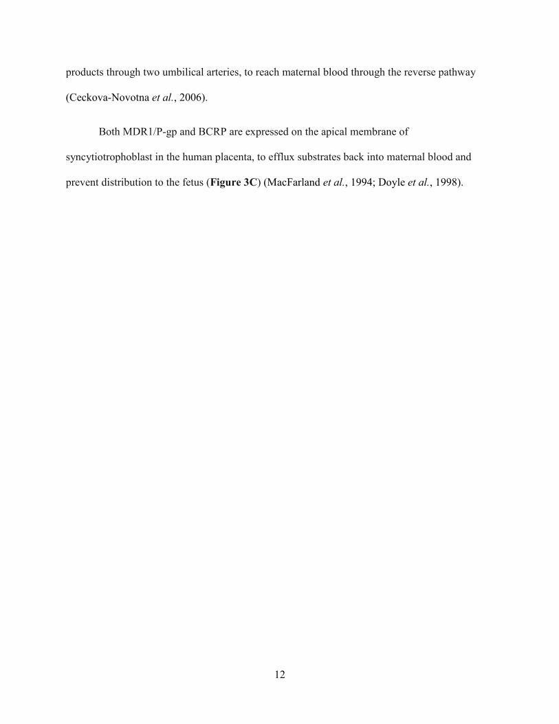
Both MDR1/P-gp and BCRP are expressed on the apical membrane of
syncytiotrophoblast in the human placenta, to efflux substrates back into maternal blood and
prevent distribution to the fetus (Figure 3C) (MacFarland et al., 1994; Doyle et al., 1998).

13
Figure 3| The structure of the human placenta. (A) A cross section of the fetus at term, inside the uterus of the
mother, as well as a cross section of the placenta. (B) Hematoxylin–eosin stained paraffin sections of terminal villi.
(C) A proposed model of drug transporters on syncytiotrophoblast and fetal blood vessels. Orange: BCRP, pink:
MDR1/P-gp. Adopted from Ceckova-Novotna et al. (2006).

14
1.3 ABC Transporters in the Human Placenta
1.3.1 Maternal Pharmacotherapy
Women can be treated for a variety of pre-existing or acquired conditions throughout pregnancy.
For example, hypertension, diabetes, and depression are disease states for which it is crucial for
pregnant women to take medication. In one study, 96% of 578 women interviewed reported
taking a prescription drug during pregnancy. The same study reported that 93% of pregnant
women had taken over-the-counter medication (Glover et al., 2003). These alarmingly high
statistics suggest that a majority of women obtain prescription and over-the-counter medication
during pregnancy. In addition, it has also been estimated that 5-10% of pregnant women have
taken Food and Drug Administration (FDA) drugs classified as category D or category X.
Category D include those drugs where there is evidence of fetal risk but the benefits may
outweigh the risks, and category X, such as warfarin derivatives and benzodiazepines sedatives,
include drugs where there is fetal teratogenic evidence and the risk of taking the drug clearly
outweighs any benefits (Andrade et al., 2004; Andrade et al., 2006; Cooper, Hickson, & Ray,
2004). The health of both the mother and fetus must be considered independently and the health
benefits must outweigh the potential risk of toxicity to the fetus (Gedeon & Koren, 2006).
Transplacental transfer of potentially harmful substances cannot be studied in vivo in pregnant
women, due to ethical, moral and legal restrictions. Furthermore, clinical trials are infrequently
carried out in pregnant women due to the same reasons (Behravan & Piquette-Miller, 2007). On
account of this, animal models and in vitro studies are often used to study drug transmission
across the placenta.

15
There are multiple ways in which maternally administered drugs can cross the placenta from
maternal blood to the fetus, or vice versa, which include passive diffusion, facilitated diffusion,
and active transport. For the most part, drugs cross the placenta from maternal circulation via
passive diffusion (Audus, 1999b). The major determinants for transplacental passive diffusion of
drugs is molecular weight (>500 Da), lipophilicity, ionization, and protein binding (Pacifici &
Nottoli, 1995; Syme et al., 2004). In opposition to passive diffusion, ABC transporters in the
syncytiotrophoblast were identified to participate in active transport of substrates out of the
placenta. MDR1/P-gp was the first ABC transporter to be found in the human placenta (Cordon-
Cardo et al., 1990). BCRP is also expressed at high levels and therefore it may play an
important role in fetal protection (Doyle et al., 1998; Allikmets et al., 1998). Studies in the
human placenta are limited, however in vitro and in vivo studies in animals have helped to
identify the protective role that MDR1/P-gp and BCRP play in pregnancy.
1.3.2 MDRI/P-gp in the Human Placenta
Animal models have shown the importance of MDR1/P-gp in the placenta. CF-1 mice, in which
25% of the population have spontaneous mutations in the Mdr1a gene, can be divided into -/-
homozygous negative (phenotypically comparable to Mdr1a knockout mice), +/- heterozygous,
or +/+ homozygous positive for Mdr1a. Fetuses with genotype Mdr1a (-/-) had 100% occurrence
of cleft palate, when exposed to avermectin, used for parasite control in veterinary medicine.
Heterozygous fetuses were less sensitive and homozygous positive had no adverse effects when
exposed to the toxin (Lankas et al., 1998). Even though Mdr1b/P-gp is the main isoform in the
placenta, this indicates a function of Mdr1a/P-gp in protecting the body from toxic xenobiotics.

16
Our laboratory localized of MDR1/P-gp on the apical membrane of the syncytiotrophoblasts in
the human placenta and examined its level in human placental tissue throughout gestation. We
have shown that there is a significant decrease in MDR1/P-gp levels in the syncytiotrophoblast
of the human placenta with increasing gestation. Levels of MDR1/P-gp are high in mid-
gestation, but dramatically decrease at term (Sun et al., 2006). Similar results have been found
by our group in the mouse (Kalabis et al., 2005) and this decrease in MDR1/P-gp correlated with
an increase in [3H]digoxin, a MDR1/P-gp substrate, crossing the placenta (Petropoulos et al.,
2007). MDR1/P-gp may protect the developing fetus from high levels of maternal steroids,
maternally administered drugs and environmental toxins and could be one of the primary means
of fetal protection (Figure 4). The dramatic decrease in the level of this efflux protein in human
placenta towards term can subject the term fetus to these substances in the maternal circulation.
1.3.3 BCRP in the Placenta
Animal models have also shown the importance of BCRP in the placenta. For example,
glyburide, a substrate of BCRP, was injected in wild-type and Bcrp -/- pregnant mice. Both
groups were compared at different time point after glyburide injection. Glyburide concentration
in the maternal plasma samples of both wild-type and Bcrp -/- pregnant mice were comparable.
However, glyburide concentrations in the fetal tissue homogenate of Bcrp knock-out mice were
two times greater than the concentration in the fetal tissue homogenate of the wild-type group
(Zhou et al., 2008). One of the potential functions of BCRP is to limit the entry of
pharmaceutical agents into fetal circulation.

17
BCRP is present at high levels in the human placenta (Doyle et al., 1998). Our laboratory has
localized BCRP to the apical membrane of the syncytiotrophoblasts. Furthermore, we have
shown that BCRP levels increase at term, compared to earlier time points of gestation (Yeboah et
al., 2006a). This increase in protein levels at term may provide enhanced fetal protection
towards the end of pregnancy. Similar to MDR1/P-gp, the potential function of BCRP in the
human placenta is to increase fetal protection from pharmaceuticals, toxins, and endogenous
steroids in maternal circulation (Figure 4).

18
Figure 4| The potential function of MDR1/P-gp and BCRP in the human placenta. Adapted from Dr S.
Matthews, The University of Toronto, 2011.
AAppiiccaall
mmeemmbbrraannee BBaasseemmeenntt mmeemmbbrraannee
SSyynnccyyttiioottrroopphhoobbllaasstt
Maternal blood
Cytotrophoblast
Mesenchyme
Fetal blood
vessel
Epithelial cells
P-glycoprotein
Toxins
Therapeutics
Steroids
Adapted from: S. Matthews, The University of Toronto
BCRP
19
1.3.4 Regulation of MDR1/P-gp and BCRP by Sex Steroids
There is little known about the physiological regulation of MDR1/P-gp and BCRP in the human
placenta. Estrogen and progesterone levels in maternal serum increase throughout pregnancy (H.
Wang et al., 2006), coinciding with the decrease in MDR1/P-gp. Estrogen has been shown to
regulate MDR1/P-gp in human cell lines, although the results are conflicting (Biing et al., 1994)
(Kim & Benet, 2004; Zampieri et al., 2002; Mutoh et al., 2006). Furthermore, one study has
implicated estrogen in upregulating MDR1/P-gp in human trophoblast cell culture (Evseenko,
Paxton, & Keelan, 2007). However, in this study only one concentration of the steroid was used.
Previous studies have shown that progesterone regulated MDR1/P-gp levels in cell lines (Mallick
& Horwitz, 1997; Piekarz, Cohen, & Horwitz, 1993), the mouse (Yang et al., 1989), and human
trophoblast cell culture (Evseenko et al., 2007), however Petropoulos et al.(2007) found that
progesterone did not have an effect on Mdr1/P-gp in the mouse placenta (Petropoulos et al.,
2007). In combination, progesterone and estrogen has been shown to increase Mdr1/P-gp in the
secretory epithelium of the uterus of the mouse in early pregnancy (Arceci et al., 1990; Yang et
al., 1989). Although previous studies are conflicting, estrogen and progesterone may be
potential regulators of MDR1/P-gp in the human placenta, alone or in combination.
Studies regarding the regulation of placental BCRP during pregnancy are extremely limited. The
increase in estrogen and progesterone during pregnancy parallels the increase in BCRP, which
may implicate these steroids in the protein’s regulation. Previous studies have shown that
estrogen regulated BCRP in human cell lines (Imai et al., 2005; H. Wang et al., 2006; Zhang et
al., 2006) and human trophoblast cell culture (Evseenko et al., 2007), however, again, results are

20
contradictory. Progesterone up-regulated BCRP in the human choricarcinoma BeWo cell line
(H. Wang et al., 2008), but had no effect on BCRP in the mouse placenta or human trophoblast
cell culture (Kalabis et al., 2007; Evseenko et al., 2007). Due to conflicting regulatory effects of
estrogen and progesterone on BCRP in previous studies, these steroids must be further
investigated in human trophoblast culture.
Endogenous cortisol levels also increase in late gestation and are believed to be involved in the
onset of labour (Fowden, Li, & Forhead, 1998), coinciding with the decrease in MDR1/P-gp and
an increase in BCRP. In addition, pregnant women could potentially be exposed to synthetic
glucocorticoids, such as dexamethasone, which are given to approximately 10% of mothers who
are at risk of preterm pregnancy to aid in fetal lung development (Koenen et al., 2007). Studies
regarding glucocorticoid regulation of MDR1/P-gp and BCRP are extremely limited. In regards
to MDR1/P-gp, in the mouse placenta, dexamethasone increased the protein in late pregnancy
(Petropoulos, Gibb, & Matthews, 2010). A few studies have implicated dexamethasone in the
down-regulation of BCRP in breast cancer cell lines (Elahian, Kalalinia, & Behravan, 2009;
Elahian, Kalalinia, & Behravan, 2010; Honorat et al., 2008), however, there are no studies in
which glucocorticoid regulation of BCRP has been investigated in the placenta. Glucocorticoid
regulation of ABC proteins, MDR1/P-gp and BCRP, in the placenta is a subject that has not been
thoroughly investigated and warrants further study.
21
1.4 Choriocarcinoma Cell Lines
1.4.1 BeWo Cell Lines
Pattillo and Gey (1968) established the BeWo cell line from a human carcinoma of the placenta.
This cell line is an extensively used model for the study of trophoblast cell function in vitro, due
to the fact that BeWo cells show comparable morphological characteristics, such as multiple
nuclei per cytoplasm and “brush border” appearance. Biochemical markers in the cell line, for
instance the production of human chorionic gonadotrophin (hCG) and placental specific proteins
SP1 and placental lactogen (Wice et al., 1990), are also similar to human trophoblast cells.
Furthermore, BeWo cells produce hormones, such as progesterone, that are also produced by
syncytiotrophoblast of the human placenta in vivo (Pattillo & Gey, 1968; Prouillac et al., 2009).
The BeWo cell line is also appealing for in vitro use because it is consistent, maintained without
difficulty, and grows quickly to confluency when plated in culture (Liu, Soares, & Audus, 1997).
It is controversial within the literature whether BeWo spontaneously syncytialize in culture,
without the aid of agents which increase intracellular levels of cyclic adenosine monophosphate
(cAMP), for example, forskolin. The majority of publications agree that spontaneous fusion of
BeWo cells in culture is low but syncytialization is augmented by forskolin. For example,
Borges et al. (2003) used fluorescence microscopy to study the syncytialization of BeWo cells in
culture. The BeWo cells were divided into two separate groups, one group injected with
cytoplasmic green-fluorescent protein and the other with cytoplasmic red-fluorescent protein.
Fluorescence microscopy was used to determine if BeWo cells syncytialize, thus mixing the red
and green colours. Untreated BeWo cells fusion index was less the percent, calculated by the
22
number of nuclei in the syncytia/total number of nuclei x 100. However, when BeWo cells were
treated with forskolin for 48 hours, the fusion index increased to 11 percent (Borges et al., 2003).
Rote et al. (2005) have repeatedly observed that 5-10% of BeWo cells spontaneously fused in
media alone, whereas 70-80% of BeWo cells syncytialized after 24 hours treatment with
forksolin (Rote, 2005). Similarly, Orendi et al. (2010) compared spontaneous syncytialization of
BeWo cells to syncytialization when treated with forskolin. Quantification of
immunofluoresence staining for biomarker protein of syncytialization, the β subunit of hCG,
showed that BeWo cells had a spontaneous fusion rate of 4.9% and treatment with forskolin
raised this rate to 50.5% (Orendi et al., 2010). However, some research groups have questioned
whether BeWo cells spontaneously fuse (Evseenko, Paxton, & Keelan, 2006a; Coutifaris et al.,
1991). Coutifaris et al. (1991) concluded that BeWo cells “aggregate” under standard settings,
but do not syncytialize unless cAMP analogues or substances which increase cAMP levels are
administered (Coutifaris et al., 1991).
1.4.2 ABC Transporters in the BeWo Cell Line
MDR1/P-gp is found in the placenta; however, it is controversial in the literature whether
MDR1/P-gp is present in BeWo cells. Magnarin et al. (2008) could not detect MDR1 mRNA
expression and protein in the BeWo cell line; using RT-PCR and Western blot analysis
respectively (Magnarin et al., 2008). Low levels of MDR1/P-gp were found in the BeWo cell
line by other groups (Atkinson et al., 2003; Evseenko, Paxton, & Keelan, 2006a). Ushigome et
al. (2000) found MDR1/P-gp, using Western blot analysis in the BeWo cell line (Ushigome et
al., 2000). Furthermore, the efflux of tritiated substrates (vinblastine, vincristine, and digoxin) of

23
the protein was decreased by inhibitors of MDR1/P-gp, including cyclosporine A and verapamil,
in BeWo cells (Ushigome et al., 2000). Utoguchi et al. (2000) also found MDR1/P-gp in the
BeWo cell line using Western analysis (Utoguchi et al., 2000). Magnarin et al. (2008) suggested
that the differences between the findings may be due to differences in conditions of cell cultures
(Magnarin et al., 2008). Interestingly, Mark and Waddell (2006) observed an increase level of
MDR1/P-gp when BeWo cells syncytialized (Mark & Waddell, 2006).
High levels of endogenous BCRP in the BeWo cell line have been repeatedly observed at the
mRNA (Ceckova et al., 2006; Evseenko, Paxton, & Keelan, 2006b) and protein level (Bailey-
Dell et al., 2001; Ceckova et al., 2006; Evseenko, Paxton, & Keelan, 2006a). Evseenko, Paxton
and Keelan (2006) found significantly higher levels of BCRP mRNA and protein in the BeWo
cell line, compared to cultured human trophoblast cells. Furthermore, BeWo cells showed
functionally active BCRP, similar to that found in cultured trophoblast cells, using the Hoechst
33342 fluorescence assay.
1.5 RATIO(ALE
Pregnant women may require medication during pregnancy for a variety of conditions, for
example hypertension, diabetes or even cancer. An alarmingly high number of women have
reported taking prescription or over the counter medications throughout pregnancy and
furthermore, synthetic glucocorticoids are given to approximately 10% of mothers who threaten
preterm delivery.
24
ABC transporters, such as MDR1/P-gp and BCRP, are known to actively transport substrates out
of the placenta, potentially functioning to protect the fetus from maternally circulating
endogenous steroids, drugs and toxins. Understanding how MDR1/P-gp and BCRP are regulated
in the human placenta will enable the regulation of fetal protection against maternally circulating
therapeutic agents and toxins, develop treatment options for mother and fetus, and increase fetal
protection in pathological pregnancies. Progesterone, estrogen and glucocorticoids are present at
high levels in maternal plasma and have been shown to regulate MDR1/P-gp and BCRP in other
systems.
1.6 PURPOSE
The purpose of this study was to examine the role of steroids in the regulation of ABC
transporters, MDR1/P-gp and BCRP, in the human placenta.
1.7 HYPOTHESES
1) Steroids are responsible for the decrease in MDR1/P-gp levels that occur in the human
placenta at term.
2) Steroids are responsible for the increase in BCRP levels that occur in the human placenta
at term.
25
1.8 OBJECTIVES
Objective 1: To determine the effects of steroids on the regulation of MDR1/P-gp levels in the
human placenta.
1) Localize MDR1/P-gp in human placental tissue.
2) Investigate the presence of MDR1/P-gp levels in human placental tissue and
trophoblast primary cell culture.
3) Determine the effect of progesterone and estrogen, alone and in combination, on
MDR1/P-gp.
4) Examine the regulatory effects of glucocorticoids on MDR1/P-gp
Objective 2: To determine the effects of steroids on the regulation of BCRP levels in the
human placenta.
1) Investigate the presence of BCRP levels in human placental tissue.
2) Examine the effects of estrogen and progesterone on BCRP.
3) Examine the regulatory effects of glucocorticoids on BCRP.
Objective 3: To examine MDR1/P-gp and BCRP in the BeWo cells line as a potential model
for human trophoblasts.
1) Investigate MDR1/P-gp and BCRP in the BeWo cell line.

26
2) Examine the effect forskolin on syncytialization of BeWo cells in culture and
MDR1/P-gp and BCRP levels.
27
CHAPTER 2: MATERIALS A(D METHODS
2.1 Tissue Collection
Term (38-40 weeks) placenta tissue was used as a source of trophoblast cells. Placenta tissues,
with no indication of infection, were collected after scheduled, non-complicated cesarean
sections at the Ottawa General Hospital, preceding labour. Approval was obtained from the
ethics committee.
2.2 Trophoblast Primary Cell Culture
A modified (Premyslova et al., 2003) version of the Kliman’s method (Kliman et al., 1986) was
used to isolate trophoblast cells. In short, 60 g of syncytiotrophoblast tissue was rinsed with
0.9% NaCl (EM Science, NJ, USA). The tissue was coarsely minced and transferred to
Dulbecco’s modified eagles medium, DMEM (Wisent Inc, QC, Canada), with 0.125% trypsin
and 0.02% deoxyribonuclease 1 (DNase; Sigma-Aldrich MO, USA), and shaken in a 37OC water
bath for three 30 minute digestions. Newborn calf serum, NCS (Wisent) was used to stop
enzyme activity. Cells pooled from the digests were layered over a 5-70% Percoll (Sigma)
gradient and centrifuged for 20 minutes at 2500xg. Trophoblast cells were isolated between
density markers of 1.049 g/l and 1.06 g/l. Isolated cells were cultured in a DMEM culture
medium supplemented with 10% fetal bovine serum, FBS (Wisent), and 1% antibiotic-
antimycotic solution (Sigma). 2.5 million cells/mL were plated on a six well plate, 2.5 mL per
well, for western analysis. 1.3 million cells/mL were plated on an eight-well chamber slides, 3.2
mL per slide, for IHC, and 1 million cells/mL were plated on a 24 well plate, 1 mL per well, for
functional transport assay. The cells were incubated at 37OC, with 5% CO2, for 96 hours to allow
28
complete syncytialization. Culture medium was replaced every 48 hours. For steroid studies,
cortisol (500 nM-5µM; Sigma), dexamethasone (500 nM-5µM; Sigma), estrogen (500 nM-5µM;
Sigma), and progesterone (500 nM-5µM; Sigma) were introduced into culture medium after 96
hours. Protein was isolated and functional activity was assessed after 24 hours.
2.3 BeWo Cell Culture
The BeWo cell line was obtained from American Type Culture Collection (ATCC). BeWo cells
were maintained in 75 cm2 flasks, in F-12K medium, Kaighn’s Modification of Ham’s F-12
medium (ATCC, Manassas,VA; #CCL-98), supplemented with 10% FBS and 1% antibiotic-
antimycotic solution. The cells were incubated at 37OC, with 5% CO2. At 70% confluence, cells
were plated in six-well plates for western analysis or eight-well chamber slides for IHC. BeWo
cells were seeded in six-well plates for 24 hours, after which forkolin (20 µM; Sigma) was added
to the F-12K medium. Protein was isolated after 72 hours of forskolin treatment.
2.4 Immunohistochemistry
Cells were fixed onto slides with 10% buffered formalin phosphate (Fisher Scientific, NJ, USA),
after being washed twice with PBS, and dehydrated in 75% ethanol before being stored in 95%
ethanol at 4 OC. IHC methods were executed as previously described (Sun et al., 2006; Yeboah
et al., 2006a). In short, slides were taken out of storage and rehydrated in 75% and 50% ethanol,
for 5 minutes respectively. Slides were washed twice in phosphate buffered saline (PBS; 5
minutes/wash), before natural endogenous peroxidase activity was blocked with H2O2 (BDH

29
Canada) in methanol, for 30 minutes. Non-specific binding was obstructed by incubation with
PBS containing 1.5% normal horse serum (Vector Laboratories, CA, USA), for 20 minutes, after
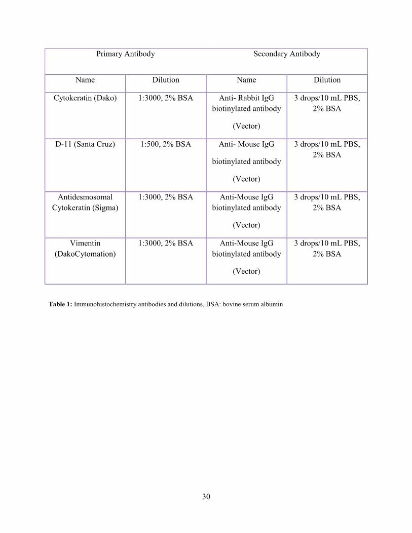
slides were washed twice in PBS (10 minutes/wash). Slides were incubated with cytokeratin
(1:3000; Dako CA, USA), vimentin (1:3000; DakoCytomation, Denmark), antidesmosomal
cytokeratin (1:500-1000; Sigma), or D-11 (1:500; Santa-Cruz Biotechnology, CA, USA),
summarized in Table 1, and washed twice in PBS (5 minutes/wash). Rabbit anti mouse
biotinylated antibody (Vector Laboratories) was used to incubate slides for 30 minutes. The
avidin-biotin-peroxidase technique, from the Vectastain Elite ABC kit (Vector Laboratories),
was used to visualize protein, using diaminobenzidine, DAB (Sigma), as a substrate.
Haemotoxylin (EMD), which stained the nucleus and washed off non-specific staining, was used
as a counterstain. Slides were dehydrated in serial dilutions of ethanol, cleared in xylene (EM
Science), and mounted in permount (Fisher Scientific).
30
Primary Antibody Secondary Antibody
Name Dilution Name Dilution
Cytokeratin (Dako) 1:3000, 2% BSA Anti- Rabbit IgG
biotinylated antibody
(Vector)
3 drops/10 mL PBS,
2% BSA
D-11 (Santa Cruz) 1:500, 2% BSA Anti- Mouse IgG
biotinylated antibody
(Vector)
3 drops/10 mL PBS,
2% BSA
Antidesmosomal
Cytokeratin (Sigma)
1:3000, 2% BSA Anti-Mouse IgG
biotinylated antibody
(Vector)
3 drops/10 mL PBS,
2% BSA
Vimentin
(DakoCytomation)
1:3000, 2% BSA Anti-Mouse IgG
biotinylated antibody
(Vector)
3 drops/10 mL PBS,
2% BSA
Table 1: Immunohistochemistry antibodies and dilutions. BSA: bovine serum albumin

31
2.5 Western blotting
PBS, containing 13.7mM NaCl, 8mM Na2HPO4 (Sigma), 2.67mM KCl (Sigma), and 1.48mM
KH2PO4 (Aldrich WS, USA), was used to wash cells, twice. Cells were scraped in 1mL of cold
PBS, collected, and centrifuged at 18500 xg. The supernatant was discarded and the pellet was
resuspended in 50 µL of cell lysis buffer, containing PBS, 1% Nonidet P-40 (Sigma), 0.1%
sodium dodecyl sulfate (SDS; Sigma), 0.5% sodium deoxycholate (Sigma), and Pefabloc SC
Plus inhibitors (Roche Molecular Biochemicals; Dorval, Quebec). The samples were sonicated
and then centrifuged at 25 200 xg, at 4 OC, for 20 minutes. The protein containing supernatant
was collected, and protein concentration was determined using the Bradford assay (Bio-Rad,
Richmond, CA, USA). Samples were diluted at a 1:1 ratio in Laemmli sample buffer (Bio-Rad),
with β-mercaptoethanol (Bio-Rad) added as a reducing agent (1:50). Protein (40 µg) was
separated on 4-15% polyacrylamide Ready Gel (Bio-Rad) and transferred to a nitrocellulose
membrane (Bio-Rad) at 100V for 1 hour. First, the membrane was blocked for 4 hours with 5%
non-fat dry milk (Bio-Rad) dissolved in a mixture PBS and 0.05% tween (Bio-Rad). The
membrane was then incubated overnight, at 4 OC, with a mouse monoclonal anti human MDR1
antibody, D-11 (1:200; Santa-Cruz), in 2% non-fat dry milk, preceded by incubation with a horse
peroxidase linked anti-mouse secondary antibody (1:1000; GE Healthcare, UK) in 2% non-fat
dry milk, for 1 hour. The enhanced chemiluminescence (ECL) system (GE Healthcare) was used
to detect proteins on Kodak BioMax MR-1 film (Kodak) and densitometry was used to quantify
bands. G beta (Gβ; Santa-Cruz Biotechnology) was used as an internal control (1:1000). The
membrane was then stripped for 30 minutes with Western Blot Stripping Buffer (ThermoFisher
Scientific, NJ, USA), and blocked overnight with 5% non-fat dry milk dissolved in a mixture
PBS and 0.05% Tween. This was followed by incubation with a rabbit polyclonal anti human
32
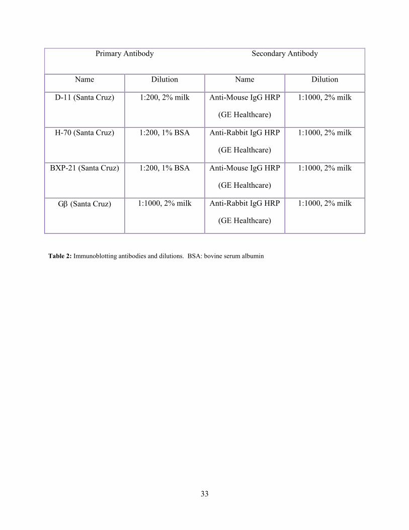
BCRP antibody, H-70 (1:200; Santa-Cruz Biotechnology) or BXP-21 (1:200; Santa Cruz) in 1%
bovine serum albumin (BSA), overnight at 4 OC. The membrane was then incubated with horse
radish peroxidase linked anti-rabbit secondary antibody (1:1000; GE Healthcare), for 1 hour, and
bands were quantified using densitometry with Gβ. A list of primary and secondary antibodies
and their respective dilutions are provided in Table 2.
33
Primary Antibody Secondary Antibody
Name Dilution Name Dilution
D-11 (Santa Cruz) 1:200, 2% milk Anti-Mouse IgG HRP
(GE Healthcare)
1:1000, 2% milk
H-70 (Santa Cruz) 1:200, 1% BSA Anti-Rabbit IgG HRP
(GE Healthcare)
1:1000, 2% milk
BXP-21 (Santa Cruz) 1:200, 1% BSA Anti-Mouse IgG HRP
(GE Healthcare)
1:1000, 2% milk
Gβ (Santa Cruz) 1:1000, 2% milk Anti-Rabbit IgG HRP
(GE Healthcare)
1:1000, 2% milk
Table 2: Immunoblotting antibodies and dilutions. BSA: bovine serum albumin

34
2.6 Functional Transport Assay
Efflux activity of MDR1/P-gp was quantified as previously described by Evseenko, Paxton and
Keelan (2006), with slight modifications (Evseenko, Paxton, & Keelan, 2006a). Briefly, 1
million cells/mL of trophoblast cells were cultured in DMEM culture medium supplemented
with 10% FBS and 1% antibiotic-antimycotic solution, for 96 hours until complete
syncytialization, after which cortisol (500 nM-5µM) was added to the culture medium. After 24
hours of treatment, cells were washed with room temperature HEPES-buffered Tyrode solution,
containing Tyrode’s salts (Sigma) and 1g NaHCO3 (Sigma). Calcein-AM (Fluka Chemical
Corp., Milwaukee, WI, USA) was added to HEPES-buffered Tyrode solution, at a final
concentration of 0.4 µM, and cells were incubated for 1 hour, at 37 OC. When calcein-AM
entered into the cell, it was cleaved by esterases in the cytoplasm to form fluorescent calcein, a
specific substrate of MDR1/P-gp (Figure 5). Cells were washed twice with ice cold HEPES-
buffered Tyrode solution and lysed with 10mM Tris•HCl (Sigma)-1% Triton X-100 (Fisher
Chemicals) for 15 minutes. Fluorescent calcein accumulation within cells was measured on a
fluorescence reader (Spectro Max 2) at 485/535 nm. Cells from individual tissues were cultured
in quadruplicate for all treatments. Previous studies in our laboratory have optimized
concentration of Calcein-AM and treatment time (Anne Marie Downey, Honours Thesis).
2.7 STATISTICAL A(ALYSIS
Data were analyzed and displayed using GraphPad Prism (version 5.04; San Diego, CA, USA).
Multiple comparisons were analyzed using one-way ANOVA followed by the Tukey method of
post-hoc analyses. Two samples were analyzed using unpaired Student’s t-test. Significance
was set at P<0.05.

35
Figure 5| Schematic diagram of conversion of Calcein-AM into fluorescent Calcein by cytoplasmic esterase.
Adopted from Anne Marie Downey (Honours Thesis 2008)
36
CHAPTER 3: STEROID REGULATIO( OF MULTIDRUG RESISTA(CE
PHOSPHOGLYCOPROTEI( (MDR1/P-GP) A(D BREAST CA(CER RESISTA(CE
PROTEI( (BCRP) I( THE HUMA( PLACE(TA
3.1 RESULTS
3.1.1 General Characterization of Syncytiotrophoblast
Following initial experiments in our laboratory with the G-1 antibody (Santa Cruz), which
recognized MDR1/P-gp and MDR3/P-gp, we began using the more specific D-11 antibody, a
monoclonal antibody raised against human MDR1/P-gp. To verify the location of MDR1/P-gp
in placental tissue, immunohistochemistry was performed using the D-11 antibody. MDR1/P-gp
was localized to the syncytiotrophoblast of human placental tissue at both 38 weeks (Figure 6a)
and 40 weeks of gestation (Figure 6c). After 24 hours, trophoblast cells did not completely
syncytialize in culture, confirmed by Haemotoxylin staining of the nucleus of individual cells
(Figure 7a), but they were found to syncytialize after 96 hours in culture (Figure 7b). Initial
studies were carried out to verify the purity of human trophoblast cells in culture. After 72 hours
in culture, IHC was performed using an anti-cytokeratin antibody (Figure 8b), which stained
trophoblast cells. An anti-vimentin antibody, which detects non-trophoblast cells, did not stain
the cells (Figure 8c). Syncytialization was confirmed by the presence of multinucleic, anti-
desmosomal structures. Desmosomes are cell structures in intracellular junctions. The absence
of staining around individual cells and the presence of staining around multiple nuclei indicate
syncytialization (Figure 9b). MDR1/P-gp was identified in human placental tissue and primary
cell culture, by Western blot with the D-11 antibody (Figure 10).

37
Figure 6| Immunohistochemistry localization of MDR1/P-gp in human placental tissue. Immunohistochemistry
localization of MDR1/p-glycoprotein in the human placenta with D-11, a monoclononal anti-MDR1 antibody
diluted to 1:500. s:syncytiotrophoblast. A) C. section (38 weeks), b) control C. section (38 weeks, mouse IgG), c)
C. section (40 weeks), d) control (mouse IgG).
38 weeks
40 weeks
38 weeks
40 weeks
a b
c d
s
38
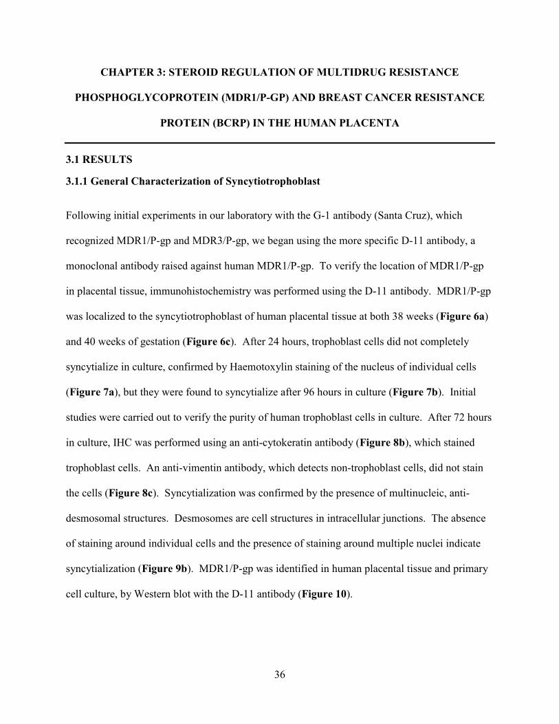
Figure 7| Trophoblast cells in culture. a) Haemotoxylin staining of trophoblast cells after 24 hours in culture. b)
staining of trophoblast cells after 96 hours in culture. c: cytotrophoblast, s: syncytiotrophoblast. Figure 6a adopted
from Yeboah et al. (2007, unpublished data).
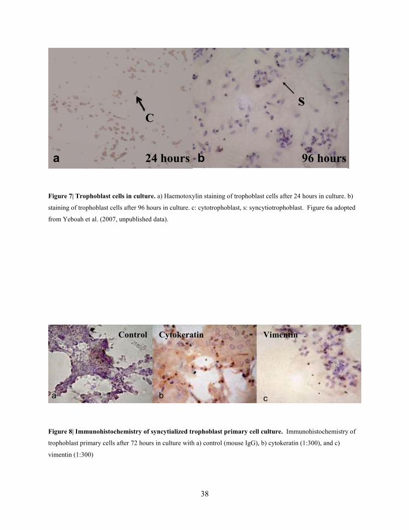
Figure 8| Immunohistochemistry of syncytialized trophoblast primary cell culture. Immunohistochemistry of
trophoblast primary cells after 72 hours in culture with a) control (mouse IgG), b) cytokeratin (1:300), and c)
vimentin (1:300)
Control Cytokeratin Vimentin
a b c
96 hours b 24 hours a
C

39
Figure 9| Verification of syncytialized trophoblast cell culture. Immunohistochemistry of trophoblast primary
cells after 72 hours in culture with a) control (mouse IgG) and b) anti-desmosomal antibody (1:500)
Figure 10| Typical western blot of MDR1/P-gp in human placental tissue and placental primary cell culture.
D-11, a monoclonal anti-MDR1/P-gp antibody, was used. 120 µg of tissue protein was loaded and 40 µg of primary
cell culture was loaded. Gβ was used as an internal control. T: human placental tissue, P: primary cell culture.
b
Control
a
Desmosome

40
3.1.2 Estrogen and Progesterone Regulation of MDR1/P-gp
Studies from our laboratory have shown that there is a significant decrease in MDR1/P-gp in the
human placenta with advancing gestation (Sun et al., 2006); however, how MDR1/P-gp
expression is regulated is not known. Estrogen and progesterone levels in maternal serum
increase throughout pregnancy (Wang et al., 2006) and glucocorticoids levels also increase in
late gestation (Fowden et al., 1998). These steroids have been found to regulate MDR1/P-gp in
other cell lines and tissues.
In this study, estrogen and progesterone were investigated as potential regulators of MDR1/P-gp
in the human trophoblast cell culture. Western analysis was performed to assess MDR1/P-gp,
treated with 0.5 µM, 1 µM, and 5 µM of estradiol for 24 hours, as illustrated in Figure 11.
Under reducing conditions, the molecular weight of the protein was 150kDa. The level of the
150 kDa protein was quantified by densitometry. Estradiol did not alter MDR1/P-gp levels in
human syncytiotrophoblast. Furthermore, Western analysis was also performed to assess
MDR1/P-gp in human syncytiotrophoblast, treated with progesterone (500 nM-5 µM) for 24
hours (Figure 12). Progesterone did not have an effect on MDR1/P-gp levels. In combination,
estrogen and progesterone were found to regulate MDR1/P-gp in the mouse endometrium
(Arceci et al., 1990). To determine the effects of estrogen and progesterone in combination on
human syncytiotrophoblast, cells were incubated with a combination of 0.1 µM of both estrogen
and progesterone and 5 µM of both estrogen and progesterone for 24 hours. A combination of
estrogen and progesterone did not alter MDR1/P-gp levels (Figure 13).

41
Figure 11| MDR1/P-gp levels in human syncytiotrophoblast after 24 hours of estrogen treatment. A) A typical
western blot of MDR1/P-gp and Gβ protein levels after estrogen treatment of 0 (n=4), 0.5 µM (n=4), 1 µM (n=4),
and 5 µM (n=4) for 24 hours, using D-11 antibody. B) Quantification of MDR1/P-gp in syncytiotrophoblast.
MDR1/P-gp was quantified after 0, 0.5 µM, 1 µM, and 5 µM of estrogen treatment for 24 hours. ANOVA, Bar
represents mean +/-SEM, standardized against Gβ and expressed as relative optical density (ROD).

42
Figure 12| MDR1/P-gp levels in human syncytiotrophoblast after 24 hours of progesterone treatment. A) A
typical western blot of MDR1/P-gp and Gβ protein levels after progesterone treatment of 0 (n=4), 0.5 µM (n=4), 1
µM (n=4), and 5 µM (n=4) for 24 hours, using D-11 antibody. B) Quantification of MDR1/P-gp in
syncytiotrophoblast. MDR1/P-gp was quantified after 0, 0.5 µM, 1 µM, and 5 µM of progesterone treatment for 24
hours. ANOVA, Bar represents mean +/-SEM, standardized against Gβ and expressed as relative optical density
(ROD).

43
Figure 13| MDR1/P-gp levels in human syncytiotrophoblast after 24 hours of co-incubation with estrogen and
progesterone. A) A typical western blot of MDR1/P-gp and Gβ protein levels after a combination of 0 (n=3), 0.1
µM (n=3) of both estrogen and progesterone and 5 µM (n=3) of both estrogen and progesterone for 24 hours, using
D-11 antibody. B) Quantification of MDR1/P-gp in syncytiotrophoblast. MDR1/P-gp was quantified after 0, 0.1
µM, and 5 µM of co-incubation of estrogen and progesterone for 24 hours. ANOVA, Bar represents mean +/-SEM,
standardized against Gβ and expressed as relative optical density (ROD). E: Estrogen, P: Progesterone.

44
3.1.3 Glucocorticoid Regulation of MDR1/P-gp
Cortisol levels increase at the end of pregnancy (Fowden et al., 1998) and glucocorticoids have
been implicated in MDR1/P-gp regulation other tissues and in vivo. To investigate the effect of
cortisol on MDR1/P-gp in human syncytiotrophoblast, cells were treated with 0.5 µM, 1 µM, and
5 µM of cortisol for 24 hours. A high level of cortisol (5 µM) significantly decreased MDR1/P-
gp levels (p<0.05), compared to the control (Figure 14). Other groups have suggested that
cortisol may affect the syncytialization of trophoblast cells in culture (J. Challis, personal
communication), which could potentially alter MDR1/P-gp. To examine if cortisol altered
syncytialization of trophoblast cells, we incubated cells with a high concentration of cortisol (10
µM) from 24-96 hours of culture. After 96 hours in culture, syncytialization of trophoblast cells
was not affected by 10 µM of cortisol, as verified by Haemotoxylin staining (Figure 15).
The mechanism by which cortisol regulates MDR1/P-gp in the placenta was investigated. Cells
were incubated with dexamethasone, a synthetic glucocorticoid and a known agonist of the
glucocorticoid receptor (GR) receptor (Frego & Davidson, 2006), to determine if the effects of
dexamethasone paralleled the effects of cortisol on MDR1/P-gp regulation under the same
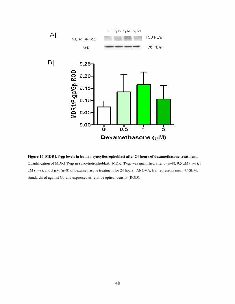
conditions that cortisol caused a decrease in MDR1/P-gp levels. Dexamethasone (0.5-5 µM) did
not affect MDR1/P-gp (Figure 16). Furthermore, cells were co-incubated with cortisol and
RU486, a competitive antagonist of the GR receptor (Beck et al., 1993). Co-incubation of
cortisol (5 µM) and RU486 (5 µM) did not reverse the inhibitory effect of cortisol (p<0.01), as
illustrated in Figure 17.
45
Another potential mechanism by which cortisol could regulate MDR1/P-gp in human
syncytiotrophoblast is via the mineralocorticoid receptor (MR). Cell were co-incubated with
spironolactone, a mineralocorticoid receptor antagonist (Delyani, 2000), and cortisol, to
determine if spironolactone would alter cortisol’s regulation of MDR1/P-gp. Western analysis
was performed to assess MDR1/P-gp in human syncytiotrophoblast treated with 5 µM of cortisol
for 24 hours and a combination of 5 µM of spironolactone and 5 µM of cortisol for 24 hours
(Figure 18). In this study the results were variable and MDR1/P-gp cortisol alone, nor the
combination of cortisol and spironolactone, significantly altered MDR1/P-gp levels. However,
in all instances the levels of MDR1/P-gp in the treated cells appeared lower.

46
Figure 14| MDR1/P-gp levels in human syncytiotrophoblast after 24 hours of cortisol treatment. A) A typical
western blot of MDR1/P-gp and Gβ protein levels after cortisol treatment of 0 (n=4), 0.5 µM (n=4), 1 µM (n=4),
and 5 µM (n=4) for 24 hours, using D-11 antibody. B) Quantification of MDR1/P-gp in syncytiotrophoblast.
MDR1/P-gp was quantified after 0, 0.5 µM, 1 µM, and 5 µM of cortisol treatment for 24 hours. ANOVA, Bar
represents mean +/-SEM, standardized against Gβ and expressed as relative optical density (ROD).*=p<0.05.

47
Figure 15| Syncytialization of trophoblast primary cells after 96 hours in culture. s: syncytiotrophoblast.
Immunostaining of syncytiotrophoblasts in culture a) with mouse IgG b) after treatment with 10 µM of cortisol,with
mouse IgG.
48
Figure 16| MDR1/P-gp levels in human syncytiotrophoblast after 24 hours of dexamethasone treatment.
Quantification of MDR1/P-gp in syncytiotrophoblast. MDR1/P-gp was quantified after 0 (n=8), 0.5 µM (n=8), 1
µM (n=8), and 5 µM (n=8) of dexamethasone treatment for 24 hours. ANOVA, Bar represents mean +/-SEM,
standardized against Gβ and expressed as relative optical density (ROD).

49
Figure 17| MDR1/P-gp levels in human syncytiotrophoblast after 24 hours of cortisol and cortisol + RU486
treatment. A) A typical western blot of MDR1/P-gp and Gβ protein levels after cortisol treatment 5 µM (n=4) and
a combination of cortisol, 5 µM (n=4), and RU486, 5 µM (n=4), for 24 hours, using D-11 antibody. B)
Quantification of MDR1/P-gp in syncytiotrophoblast. MDR1/P-gp was quantified after 5 µM of cortisol treatment
and 5 µM of both cortisol and RU486 treatment for 24 hours. C: cortisol, R:RU486. ANOVA, Bar represents mean
+/-SEM, standardized against Gβ and expressed as relative optical density (ROD). *=p<0.01.

50
Figure 18| MDR1/P-gp levels in human syncytiotrophoblast after 24 hours of cortisol and cortisol +
spironolactone treatment. A) A typical western blot of MDR1/P-gp and Gβ protein levels after cortisol treatment
5 µM (n=4) and a combination of cortisol, 5 µM (n=4), and spironolactone, 5 µM (n=4), for 24 hours, using D-11
antibody. B) Quantification of MDR1/P-gp in syncytiotrophoblast. MDR1/P-gp was quantified after 5 µM of
cortisol treatment and 5 µM of both cortisol and spironolactone treatment for 24 hours. C:cortisol,
SP:spironolactone. ANOVA, Bar represents mean +/-SEM, standardized against Gβ and expressed as relative
optical density (ROD).
51
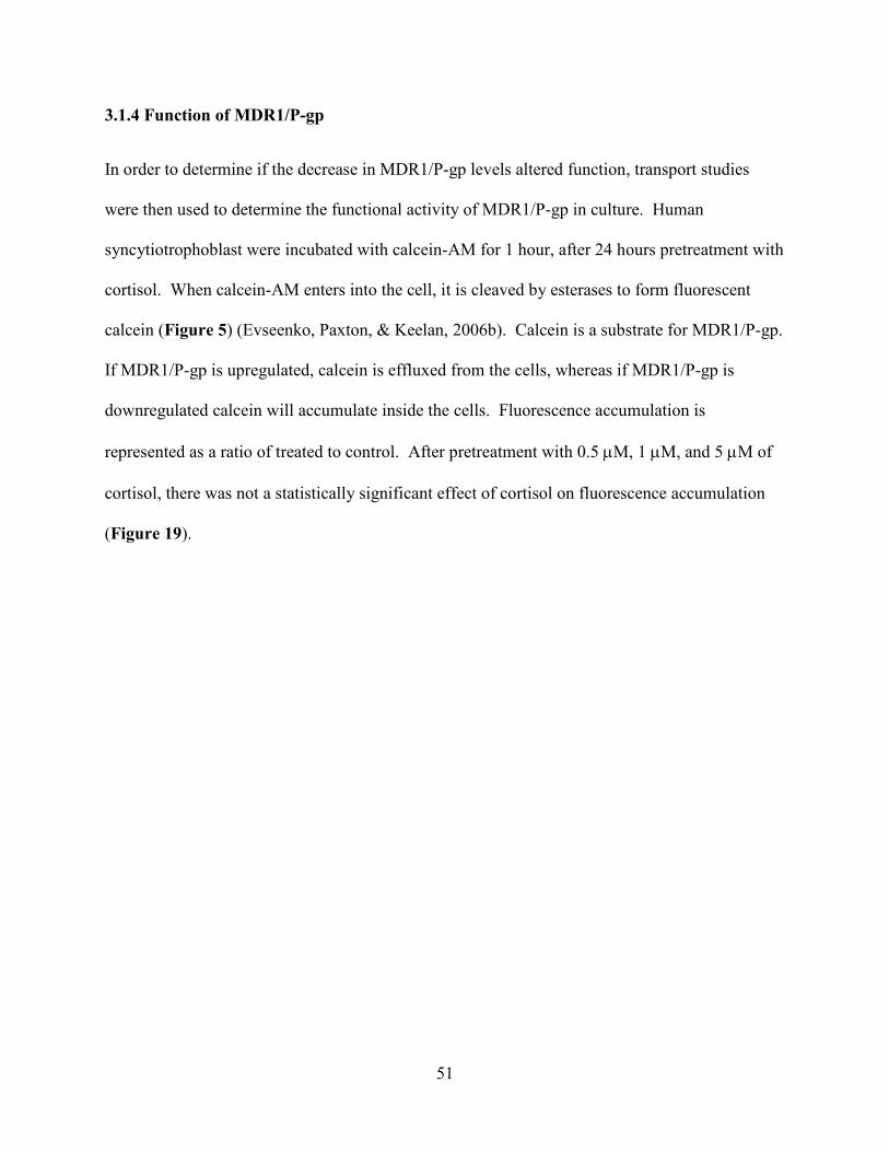
3.1.4 Function of MDR1/P-gp
In order to determine if the decrease in MDR1/P-gp levels altered function, transport studies
were then used to determine the functional activity of MDR1/P-gp in culture. Human
syncytiotrophoblast were incubated with calcein-AM for 1 hour, after 24 hours pretreatment with
cortisol. When calcein-AM enters into the cell, it is cleaved by esterases to form fluorescent
calcein (Figure 5) (Evseenko, Paxton, & Keelan, 2006b). Calcein is a substrate for MDR1/P-gp.
If MDR1/P-gp is upregulated, calcein is effluxed from the cells, whereas if MDR1/P-gp is
downregulated calcein will accumulate inside the cells. Fluorescence accumulation is
represented as a ratio of treated to control. After pretreatment with 0.5 µM, 1 µM, and 5 µM of
cortisol, there was not a statistically significant effect of cortisol on fluorescence accumulation
(Figure 19).
52
Cortisol (µµµµM)
Ratio of treated fluorescence
to control fluorescence
CTL 0.5 1 50.0
0.5
1.0
1.5
2.0
2.5
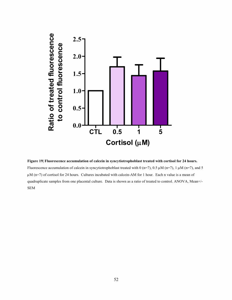
Figure 19| Fluorescence accumulation of calcein in syncytiotrophoblast treated with cortisol for 24 hours.
Fluorescence accumulation of calcein in syncytiotrophoblast treated with 0 (n=7), 0.5 µM (n=7), 1 µM (n=7), and 5
µM (n=7) of cortisol for 24 hours. Cultures incubated with calcein-AM for 1 hour. Each n value is a mean of
quadruplicate samples from one placental culture. Data is shown as a ratio of treated to control. ANOVA, Mean+/-
SEM

53
3.1.5 Estrogen and Progesterone Regulation of BCRP
BCRP was found in primary trophoblast cell culture and placental tissue, using the H-70
antibody (Figure 20). Studies from our laboratory have shown that there is a significant increase
in BCRP in the human placenta with advancing gestation (Yeboah et al., 2006a). The
physiological regulator of BCRP is not known. Estrogen, progesterone, and glucocorticoids
increase throughout pregnancy (H. Wang et al., 2006; Fowden et al., 1998), and may potentially
regulate BCRP in the human placenta. To investigate if estrogen and progesterone regulates
BCRP in human syncytiotrophoblast, cells were incubated with 0.5 µM, 1 µM, and 5 µM of
estrogen for 24 hours (Figure 21). Under reducing condition, the molecular weight of the
protein was 72 kDa. The level of the 72 kDa protein was quantified by densitometry. Estrogen
did not have a significant effect on BCRP levels in human syncytiotrophoblast after 24 hours.
Western analysis was performed after cells were treated with 0.5 µM, 1 µM, and 5 µM of
progesterone for 24 hours, as illustrated in Figure 22. Progesterone did not have a significant
effect on BCRP levels in human syncytiotrophoblast after 24 hours. Occasionally, a double band
was observed, with one band at approximately 72 kDa and one between 75-80 kDa.

54
Figure 20| Typical western blot of BCRP in placental primary cell culture and placental tissue. H-70, a
polyclonal anti-BCRP antibody, was used. 20 µg of protein was loaded in each lane. Gβ was used as an internal
control. A: Primary cell culture, B: Placental Tissue.

55
Figure 21| BCRP levels in human syncytiotrophoblast after 24 hours of estrogen treatment. A) A typical
western blot of BCRP and Gβ protein levels after estrogen treatment of 0 (n=4), 0.5 µM (n=4), 1 µM (n=4), and 5
µM (n=4) for 24 hours, using H-70 antibody. B) Quantification of BCRP in syncytiotrophoblast. BCRP was
quantified after 0, 0.5 µM, 1 µM, and 5 µM of estrogen treatment for 24 hours. ANOVA, Bar represents mean +/-
SEM, standardized against Gβ and expressed as relative optical density (ROD).

56
Figure 22| BCRP levels in human syncytiotrophoblast after 24 hours of progesterone treatment. A) A typical
western blot of BCRP and Gβ protein levels after progesterone treatment of 0 (n=4), 0.5 µM (n=4), 1 µM (n=4), and
5 µM (n=4) for 24 hours, using H-70 antibody. B) Quantification of BCRP in syncytiotrophoblast. BCRP was
quantified after 0, 0.5 µM, 1 µM, and 5 µM of progesterone treatment for 24 hours. ANOVA, Bar represents mean
+/-SEM, standardized against Gβ and expressed as relative optical density (ROD).
57
3.1.6 Glucocorticoid Regulation of BCRP
To investigate glucocorticoid regulation of BCRP, western analysis was performed after human
syncytiotrophoblast cells were incubated with cortisol or dexamethasone. Cortisol (0.5-5 µM)
did not have a significant effect on BCRP levels in human syncytiotrophoblast after 24 hours
(Figure 23). Similarly, dexamethasone did not have a significant effect on BCRP levels, as
shown in Figure 24. A double band was inconsistently observed, with one band at
approximately 72 kDa and one between 75-80 kDa.
58
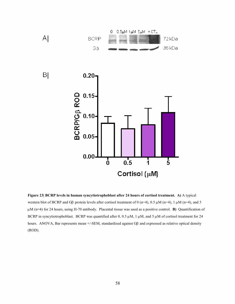
Figure 23| BCRP levels in human syncytiotrophoblast after 24 hours of cortisol treatment. A) A typical
western blot of BCRP and Gβ protein levels after cortisol treatment of 0 (n=4), 0.5 µM (n=4), 1 µM (n=4), and 5
µM (n=4) for 24 hours, using H-70 antibody. Placental tissue was used as a positive control. B) Quantification of
BCRP in syncytiotrophoblast. BCRP was quantified after 0, 0.5 µM, 1 µM, and 5 µM of cortisol treatment for 24
hours. ANOVA, Bar represents mean +/-SEM, standardized against Gβ and expressed as relative optical density
(ROD).

59
Figure 24| BCRP levels in human syncytiotrophoblast after 24 hours of dexmathesone treatment. A) A typical
western blot of BCRP and Gβ protein levels after dexamethasone treatment of 0 (n=4), 0.5 µM (n=4), 1 µM (n=4),
and 5 µM (n=4) for 24 hours, using H-70 antibody. B) Quantification of BCRP in syncytiotrophoblast. BCRP was
quantified after 0, 0.5 µM, 1 µM, and 5 µM of dexamethasone treatment for 24 hours. ANOVA, Bar represents
mean +/-SEM, standardized against Gβ and expressed as relative optical density (ROD).

60
3.2 DISCUSSIO(
There is limited information regarding the physiological regulation of ABC transporters,
MDR1/P-gp and BCRP, in the human placenta. Therefore, our main objectives were to
determine if maternal steroids, which are known to increase throughout pregnancy (Wang et al.,
2006; Fowden et al., 1998), regulate MDR1/P-gp and/or BCRP in the human placenta.
Specifically, the regulatory effects of progesterone, estrogen, and glucocorticoids were studied in
human syncytiotrophoblast cultures. Our results indicate that estrogen and progesterone did not
alter MDR1/P-gp levels in human syncytiotrophoblast, but cortisol decreased MDR1/P-gp levels.
It did not appear that cortisol acted via the GR or MR to exert its regulatory effect. Furthermore,
MDR1/P-gp function decreased, but this decrease was not statisically significant. We also
demonstrated that maternal steroids did not have a regulatory effect on BCRP levels. Human
studies relied on in vitro cultures, using a well defined system.
3.2.1 Syncytialization and purity of trophoblast cells in culture
Kliman et al. (1986) found that trophoblast cells in culture form syncytium at 24-48 hours
(Kliman et al., 1986). Other groups have used 72 hours for syncytialization of trophoblast cells
in culture, before extracting protein for western blot analysis (Evseenko et al., 2007) . We
wanted to establish a timeframe in our laboratory, in which we could ensure that trophoblast cells
were completely syncytialized in culture, and this timeframe would be consistently used
throughout the study. Our results indicate, by staining cells with Haemotoxylin and IHC using
an anti-desmosomal cytokeratin antibody, that 72-96 hours was a sufficient amount of time for
syncytialization of trophoblast cells in vitro. Therefore, steroid treatment began at 96 hours,
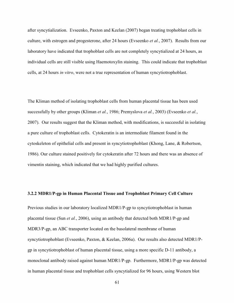
61
after syncytialization. Evseenko, Paxton and Keelan (2007) began treating trophoblast cells in
culture, with estrogen and progesterone, after 24 hours (Evseenko et al., 2007). Results from our
laboratory have indicated that trophoblast cells are not completely syncytialized at 24 hours, as
individual cells are still visible using Haemotoxylin staining. This could indicate that trophoblast
cells, at 24 hours in vitro, were not a true representation of human syncytiotrophoblast.
The Kliman method of isolating trophoblast cells from human placental tissue has been used
successfully by other groups (Kliman et al., 1986; Premyslova et al., 2003) (Evseenko et al.,
2007). Our results suggest that the Kliman method, with modifications, is successful in isolating
a pure culture of trophoblast cells. Cytokeratin is an intermediate filament found in the
cytoskeleton of epithelial cells and present in syncytiotrophoblast (Khong, Lane, & Robertson,
1986). Our culture stained positively for cytokeratin after 72 hours and there was an absence of
vimentin staining, which indicated that we had highly purified cultures.
3.2.2 MDR1/P-gp in Human Placental Tissue and Trophoblast Primary Cell Culture
Previous studies in our laboratory localized MDR1/P-gp to syncytiotrophoblast in human
placental tissue (Sun et al., 2006), using an antibody that detected both MDR1/P-gp and
MDR3/P-gp, an ABC transporter located on the basolateral membrane of human
syncytiotrophoblast (Evseenko, Paxton, & Keelan, 2006a). Our results also detected MDR1/P-
gp in syncytiotrophoblast of human placental tissue, using a more specific D-11 antibody, a
monoclonal antibody raised against human MDR1/P-gp. Furthermore, MDR1/P-gp was detected
in human placental tissue and trophoblast cells syncytialized for 96 hours, using Western blot
62
and the same antibody. Throughout this study, MDR1/P-gp was consistently detected at 150
kDa, by Western blot, and some studies have suggested that MDR1/P-gp can be found between
140-170 kDa (Ling et al., 1983). MDR1/P-gp is also N-glycosylated at the first extracellular
loop (Schinkel & Jonker, 2003) and this may account for the differences in molecular weight
found between these studies.
3.2.3 Steroid Regulation of MDR1/P-gp
There is limited information regarding estrogen and progesterone regulation of MDR1/P-gp in
humans. Estrogen and progesterone increase towards the end of pregnancy, in maternal
circulation (Wang et al., 2006), and could possibly be implicated in the decrease of MDR1/P-gp
levels described towards term in the human placenta (Sun et al., 2006). There are a few studies
that implicate estrogen in the regulation of MDR1/P-gp in cell lines (Biing et al., 1994; Kim &
Benet, 2004; Zampieri et al., 2002; Mutoh et al., 2006), however these results are contradictory.
For example, Kim et al. (2004) found that 50 µM of ethynyl estradiol induced MDR1/P-gp levels
in the LS180, a human colon cancer cell line (Kim & Benet, 2004). However, in another study,
MDR1/P-gp levels were down-regulated in two MDR1/P-gp induced, estrogen receptor
α positive human breast cancer cell lines, MCF-7/MDR and T-47D/MDR, by 100 pM of
estradiol (Mutoh et al., 2006). These divergences between studies could be attributed to the
differences in concentrations of estradiol or cell lines used, yet there are no consistent findings
related to estrogen regulation of MDR1/P-gp. In regards to the human placenta, one study has
shown that estrogen (100 nM) upregulated MDR1/P-gp in human trophoblast cell culture, after
48 hours treatment (Evseenko et al., 2007). Our results do not support these findings; we
63
observed that estrogen (500 nM-5µM) did not have a regulatory effect on MDR1/P-gp levels, in
human syncytiotrophoblast. The differences in the results could be contributed to the fact that
Evseenko, Paxton and Keelan (2007) treated trophoblast cells after 24 hours in culture, which we
have found to be an insufficient amount of time for syncytialization to occur and may not have
been a true representation of syncytiotrophoblast in vitro. The concentration of estrogen used by
Evseenko, Paxton and Keelan (2007) was based on the concentration of estrogen in maternal
serum during pregnancy; however, we used a higher concentration of estrogen based on the fact
that the concentration of steroids in steroid-producing tissue is probably much higher than in
maternal serum (Yang et al., 1989).
As opposed to humans, where maternally circulating progesterone levels increase towards term,
progesterone levels in the mouse decrease significantly from mid-gestation to term. This
decrease in progesterone concentration in maternal serum positively correlated with the decrease
in Mdr1b mRNA (the main isoform in the mouse placenta), which implies a potential
progesterone regulation of Mdr1b mRNA and Mdr1b (Kalabis et al., 2005). However, in vivo,
progesterone was found to have no effect on Mdr1 mRNA expression and Mdr1/P-gp levels in
the mouse placenta (Petropoulos et al., 2007). These results were confirmed in the rat placenta,
where Mdr1a and Mdr1b mRNA expression was not affected by partial progesterone withdrawal
(Mark et al., 2009). Similarly, our results indicate that progesterone does not regulate MDR1/P-
gp in human syncytiotrophoblast. Progesterone has been shown to regulate MDR1/P-gp in
transfected T47D, a human breast cancer cell line, and HeLa, human cervical cell line (Mallick
& Horwitz, 1997; Piekarz et al., 1993) and in the uterine secretory epithelium of the mouse
(Yang et al., 1989). The differences in these findings and our results may be due to cell or

64
tissue-specific regulation of MDR1/P-gp. A concentration of 10 nM-1 µM was shown to up-
regulate MDR1/P-gp in granulosa cells from porcine ovarian follicles (Fukuda et al., 2006),
while a progesterone concentration of 5-50 µM down-regulates MDR1/P-gp in a murine
macrophage-like cell line (Yang et al., 1989) , in vitro. Progesterone was found to upregulate
MDR1/P-gp levels in human trophoblast culture (Evseenko et al., 2007), however, as previously
stated, they treated cultured trophoblast cells after 24 hours in culture, which may account for the
disparity between findings. Furthermore, the differences in concentration of progesterone used
in the two studies could be a source of discrepancy. Evseenko, Paxton and Keelan (2007) used a
concentration of 100 nM of progesterone, whereas 500 nM-5µM of progesterone was used in this
study. This range of progesterone concentrations was decided upon based past in vitro studies.
Estrogen and progesterone, in combination, have been found to upregulate Mdr1/P-gp in the
mouse endometrium (Arceci et al., 1990). In the present study, co-incubation of human
syncytiotrophoblast with both estrogen and progesterone did not regulate MDR1/P-gp levels.
Endogenous cortisol levels increase exponentially in late pregnancy (Fowden et al., 1998) and
are believed to be involved in fetal lung maturation and the onset of labour. The increase in
cortisol coincides with the down-regulation in MDR1/P-gp levels in the human placenta,
previously described by our laboratory (Sun et al., 2006), indicating a potential role of cortisol in
the physiological regulation of MDR1/P-gp. The present study found that cortisol
downregulated MDR1/P-gp in human trophoblast culture. Other groups have found that the
placenta becomes progressively more sensitive to glucorticoids towards the end of pregnancy

65
(Speirs, Seckl, & Brown, 2004; Thompson, Han, & Yang, 2002); therefore MDR1/P-gp
regulation may be dependent on the period of gestation. Cortisol regulation of MDR1/P-gp may
be limited to late gestation, when maternal cortisol levels are drastically elevated and the
placenta is most sensitive to glucocorticoids.
Other groups have suggested that cortisol effects syncytialization of trophoblast cells in culture
(J. Challis, personal communication). We wanted to ensure that cortisol was not altering
syncytialization of trophoblast cells in culture and potentially causing an effect on MDR1/P-gp.
A higher concentration of cortisol was used and treatment started from 24 hours, when cells were
not yet completely syncytialized. Our results show that cortisol did not affect syncytialization of
trophoblast cells in culture, using Haemotoxylin staining of the nucleus. Therefore, the decrease
in MDR1/P-gp produced by cortisol is most likely attributed to regulation, rather than a
disruption in syncytialization.
We found that cortisol down-regulated MDR1/P-gp in human syncytiotrophoblast, however, the
mechanism of this regulation is unknown. One possible explanation may be the regulation of
MDR1/P-gp cortisol through the GR. The GR is a nuclear receptor that is in the placenta (Mark
et al., 2009)(Wong & Burton, 1974). The Mdr1b gene promoter is known to contain a GRE in
the mouse (Cohen et al., 1991), where the GR can potentially homodimerize and bind to directly
activate gene transcription. Furthermore, the GR can also “cross-talk” signal though the AP-1
element (Gottlicher, Heck, & Herrlich, 1998), which has been found in the human MDR1
promoter, and can form complexes with c-Jun and c-Fos proteins. GR has been shown to

66
activate or repress AP-1 activity, based on if AP-1 is in complex with c-Jun/c-Jun or c-Jun/c-Fos
(Diamond et al., 1990). Dexamethasone is a synthetic glucocorticoid and a known agonist of the
GR (Koenen et al., 2007). In our study, dexamethasone did not appear to regulate MDR1/P-gp,
providing evidence that cortisol may not regulate MDR1/P-gp through the GR. To investigate
the GR further, we also used RU486, which is an antagonist of the receptor (Beck et al., 1993).
When cells were co-incubated with cortisol and RU486, the inhibitory effect of cortisol on
MDR1/P-gp was not reversed. This implies that cortisol’s effect does not occur through the GR.
Cortisol can also bind to the mineralocorticoid receptor (MR). Spironolactone, a competitive
antagonist of the MR (Delyani, 2000), was co-incubated with cortisol to determine if it would
interfere with cortisol’s effects on MDR1/P-gp. There was variation in MDR1/P-gp levels,
however, in both cortisol and a combination of cortisol and spironolactone, protein levels were
lower, compared to control. In future studies, this experiment must be repeated in order to better
understand the potential MR’s involvement in the regulation of MDR1/P-gp by cortisol.
Orphan nuclear receptors are also mechanisms by which cortisol may regulate MDR1/P-gp in
placental trophoblast cells. Orphan nuclear receptors are located in the cytoplasm and when
bound to a ligand, move into the nucleus to regulate gene transcription of target genes. Cortisol
can bind to the orphan nuclear receptor, pregnane xenobiotic receptor (PXR) (Schuster &
Langer, 2005). PXR is known to regulate MDR1/P-gp in the brain endothelium of other species
(Bauer et al., 2004; Narang et al., 2008; Ott, Fricker, & Bauer, 2009; Synold, Dussault, &
Forman, 2001), however whether PXR is expressed in the placenta is yet to be determined.
67
Glucocorticoids are also ligands for another orphan nuclear receptor, the constitutive
androsterone receptor (CAR) (Pascussi et al., 2000). CAR and MDR1/P-gp expression
positively correlate in other tissues (Burk et al., 2005) and in the mouse small intestine, CAR
activation upregulated the Mdr1a isoform but did not affect the Mdr1b isoform (Maglich et al.,
2002). It has yet to be established whether CAR is present in the placenta. Furthermore, there is
evidence of cross-talk between the GR, PXR, and CAR in regulating metabolizing enzyme (C.
Chen, Staudinger, & Klaassen, 2003; J. D. Schuetz et al., 1996), further complicating the
potential mechanistic pathway involved in cortisol regulation of MDR1/P-gp. Cortisol may also
have non-receptorial effects on MDR1/P-gp, for example, by altering the shuttling of the inactive
protein to the membrane where it is functional. In future studies, the PXR and CAR should be
studied as potential mechanisms by which cortisol regulate MDR1/P-gp and non-receptorial
regulation should also be investigated.
We found that the inhibition of MDR1/P-gp levels, potentially regulated by cortisol showed a
tendency for decreased transport but did not translate to a significant change in transport activity.
This disconnect between protein levels and active protein has also been found in other studies
(Vahakangas & Myllynen, 2009; Petropoulos et al., 2010). For example, dexamethasone
exposure in late gestation significantly increased Mdr1/P-gp in the mouse placenta; however it
did not change the ‘fetal-unit’ tritiated digoxin accumulation (Petropoulos et al., 2010). This
discrepancy between protein and function could be due to the location of the protein measured.
In our present study MDR1/P-gp levels were measured from whole cell extract, yet, functional
protein is only present at the cell membrane. Therefore, cortisol may decrease MDR1/P-gp in
the whole cell; however, this protein may not be integrated into the cell membrane to become
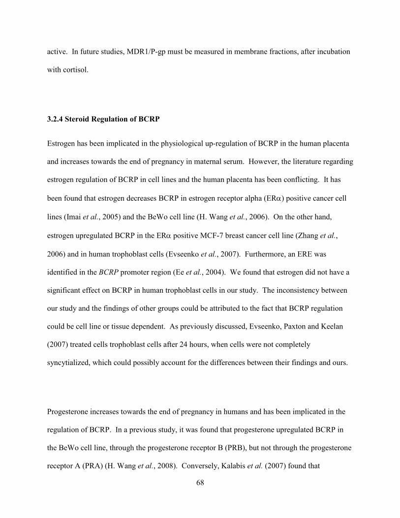
68
active. In future studies, MDR1/P-gp must be measured in membrane fractions, after incubation
with cortisol.
3.2.4 Steroid Regulation of BCRP
Estrogen has been implicated in the physiological up-regulation of BCRP in the human placenta
and increases towards the end of pregnancy in maternal serum. However, the literature regarding
estrogen regulation of BCRP in cell lines and the human placenta has been conflicting. It has
been found that estrogen decreases BCRP in estrogen receptor alpha (ERα) positive cancer cell
lines (Imai et al., 2005) and the BeWo cell line (H. Wang et al., 2006). On the other hand,
estrogen upregulated BCRP in the ERα positive MCF-7 breast cancer cell line (Zhang et al.,
2006) and in human trophoblast cells (Evseenko et al., 2007). Furthermore, an ERE was
identified in the BCRP promoter region (Ee et al., 2004). We found that estrogen did not have a
significant effect on BCRP in human trophoblast cells in our study. The inconsistency between
our study and the findings of other groups could be attributed to the fact that BCRP regulation
could be cell line or tissue dependent. As previously discussed, Evseenko, Paxton and Keelan
(2007) treated cells trophoblast cells after 24 hours, when cells were not completely
syncytialized, which could possibly account for the differences between their findings and ours.
Progesterone increases towards the end of pregnancy in humans and has been implicated in the
regulation of BCRP. In a previous study, it was found that progesterone upregulated BCRP in
the BeWo cell line, through the progesterone receptor B (PRB), but not through the progesterone
receptor A (PRA) (H. Wang et al., 2008). Conversely, Kalabis et al. (2007) found that

69
progesterone treatment of pregnant mice did not change Bcrp1 levels in the placenta (Kalabis et
al., 2007). Furthermore, progesterone did not change BCRP levels in human trophoblast culture,
at 48 hours at 100 nM (Evseenko et al., 2007). The progesterone receptor (PR) is expressed in
the human placenta (Cudeville et al., 2000) and PRE was identified in the BCRP promoter,
indicating a potential role of progesterone in BCRP regulation (H. Wang et al., 2008). In the
present study, progesterone did not have an effect of BCRP in human trophoblast culture, which
is consistent with findings in the mouse placenta.
There is limited information regarding the glucocorticoid regulation of BCRP. In a few studies,
dexamethasone was shown to decrease BCRP in breast cancer cell lines, in vitro (Elahian et al.,
2009; Elahian et al., 2010; Honorat et al., 2008). There is no information regarding the
glucocorticoid regulation of BCRP in the placenta. Glucocorticoids are administered to 10% of
pregnant women who are at risk of premature labour, in order to aid in fetal lung development
(Koenen et al., 2007), therefore women can be exposed to synthetic glucocorticoids, as well as
endogenous glucocorticoids that are known to increase drastically towards the end of gestation.
Our studies indicate that both cortisol and dexamethasone do not have significant effects on
BCRP. Furthermore, GRE has yet to be found on the BCRP promoter.
On occasion, a double line was seen using western blot with the H-70 antibody, with one band at
approximately 72 kDa and the other band at approximately 75-80kDa (Figure 23). Interestingly,
when the syncytialized trophoblast cells were treated with cortisol this was consistently
observed. A double line was also observed by others, at approximately the same molecular

70
weights, and it has been suggested that the higher molecular weight band represents a complex-
glycosylated form of the protein (Mohrmann et al., 2005). Therefore, it is possible that cortisol
can alter the glycosylated state of the protein and affect control in the same yet undefined
manner.
71
Chapter 4: EXAMI(ATIO( OF THE BEWO CELL LI(E AS A POTE(TIAL MODEL
FOR MDR1/P-GP A(D BCRP REGULATIO(
4.1 RESULTS
4.1.1 Verification of Purity and Syncytialization of the BeWo Cell Line
The BeWo cell line is a human choriocarcinoma cell line that our laboratory was attempting to
use as a primary model for the study of MDR1/P-gp regulation, in addition to human trophoblast
cells. Initial studies were carried out to determine the purity of BeWo cells in culture and
immunohistochemistry was performed on the BeWo cell line after 72 hours in culture, to
determine if the cells aggregate, similar to cytotrophoblast fusing together to form
syncytiotrophoblast in primary cell culture (Figure 25a). BeWo cells stained positively with
cytokeratin, which stains trophoblast cells (Figure 25b). Vimentin, a stain for non-trophoblast
cells, did not stain BeWo cells (Figure 25c).
4.1.2 MDR1/P-gp and BCRP in the BeWo Cell Line
BeWo cells stained positively for MDR1/P-gp after 72 hours in culture (Figure 26b). However,
using western analysis, MDR1/P-gp could not be detected in BeWo cells, as shown in Figure 27.
This experiment was repeated numerous times and with different methods protocols and various
MDR1/P-gp antibodies (Table 2, pg. 33). After approximately 12 months a new batch of BeWo
cells was obtained from the ATCC and MDR1/P-gp was subsequently identified using western
analysis, with D-11 antibody (Figure 28). BCRP was identified in the BeWo cell line, using
western blot with BXP-21, a monoclonal antibody raised against human BCRP (Figure 29).
72
It is controversial in the literature as to whether BeWo cells spontaneously syncytialize in culture
or if forksolin is needed for syncytialization. To investigate this, BeWo cells were incubated
with forskolin (20 µM), after 24 hours in culture, for an additional 72 hours. Syncytialization
was investigated using an anti-desmosomal antibody, where the absence of staining around
individual cells, and the presence of staining around multiple nuclei, indicate the formation of
syncytialization. BeWo cells, treated with forskolin (Figure 30d), showed markedly reduced
staining, compared to control (Figure 30b).
4.1.3 Forskolin Regulation of MDR1/P-gp and BCRP in the BeWo Cell Line
Other groups have suggested that increased syncytialization upregulated MDR1/P-gp in the
BeWo cell line (Mark & Waddell, 2006). We investigated MDR1/P-gp regulation by forskolin,
in order to potentially use the cell line as a model for MDR1/P-gp regulation. After 24 hours in
culture, BeWo cells were treated for 72 hours with forskolin (20 µM), used by other research
groups to induce syncytialization (Mark & Waddell, 2006). Western analysis was performed to
assess the protein level of MDR1/P-gp after forskolin treatment. Forskolin (20 µM) has a
tendency to increase MDR1/P-gp levels after 72 hours; however this increase was not
statistically significant (Figure 31). The effect of forskolin on BCRP levels was also
investigated. BeWo cells were treated for 72 hours with forskolin (20 µM), after 24 hours in
culture, as previously described. BCRP protein levels were evaluated using Western analysis
and forskolin did not have an effect after 48 hours (Figure 32).
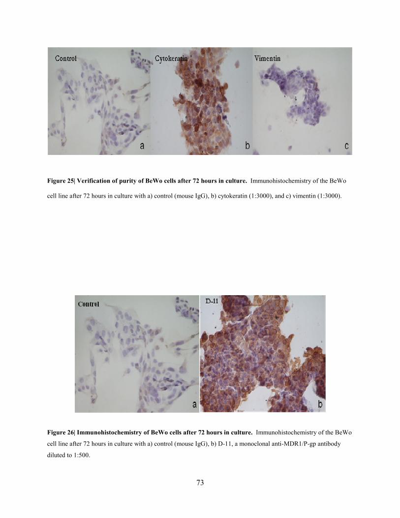
73
Figure 25| Verification of purity of BeWo cells after 72 hours in culture. Immunohistochemistry of the BeWo
cell line after 72 hours in culture with a) control (mouse IgG), b) cytokeratin (1:3000), and c) vimentin (1:3000).
Figure 26| Immunohistochemistry of BeWo cells after 72 hours in culture. Immunohistochemistry of the BeWo
cell line after 72 hours in culture with a) control (mouse IgG), b) D-11, a monoclonal anti-MDR1/P-gp antibody
diluted to 1:500.

74
Figure 27| Typical western blot of MDR1/P-gp in BeWo cell line and placental primary cell culture. D-11, a
monoclonal anti-MDR1/P-gp antibody, was used. 40 µg of protein was loaded in each lane. Gβ was used as an
internal control.
Figure 28| Typical western blot of MDR1/P-gp in the BeWo cell line. D-11, a monoclonal anti-MDR1/P-gp
antibody was used. 40 µg of protein was loaded in each lane. Gβ was used as an internal control.
Figure 29| Typical western blot of BCRP in the BeWo cell line. BXP-21, a monoclonal anti-BCRP antibody, was
used. 25 µg of protein was loaded in each lane. Gβ was used as an internal control.

75
Figure 30| Immunohistochemistry of BeWo cells after treatment with forskolin. Immunohistochemistry of the
BeWo cell line after 72 hours in culture with a) control (mouse IgG), b) a monoclonal anti-desmosomal cytokeratin
antibody diluted to 1:500, c) pretreated with 20 µM of forskolin for 48 hours, control (mouse IgG), and d)
pretreatment with 20 µM of forskolin for 48 hours, anti-desmosomal antibody diluted to 1:500.
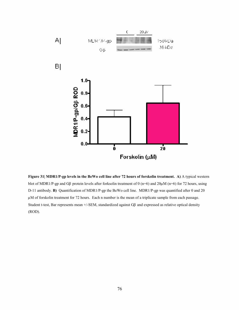
76
Figure 31| MDR1/P-gp levels in the BeWo cell line after 72 hours of forskolin treatment. A) A typical western
blot of MDR1/P-gp and Gβ protein levels after forksolin treatment of 0 (n=6) and 20µM (n=6) for 72 hours, using
D-11 antibody. B) Quantification of MDR1/P-gp the BeWo cell line. MDR1/P-gp was quantified after 0 and 20
µM of forskolin treatment for 72 hours. Each n number is the mean of a triplicate sample from each passage.
Student t-test, Bar represents mean +/-SEM, standardized against Gβ and expressed as relative optical density
(ROD).
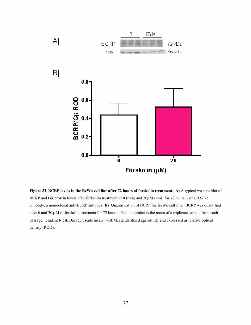
77
Figure 32| BCRP levels in the BeWo cell line after 72 hours of forskolin treatment. A) A typical western blot of
BCRP and Gβ protein levels after forksolin treatment of 0 (n=4) and 20µM (n=4) for 72 hours, using BXP-21
antibody, a monoclonal anti-BCRP antibody. B) Quantification of BCRP the BeWo cell line. BCRP was quantified
after 0 and 20 µM of forskolin treatment for 72 hours. Each n number is the mean of a triplicate sample from each
passage. Student t-test, Bar represents mean +/-SEM, standardized against Gβ and expressed as relative optical
density (ROD).
78
4.2 DISCUSSIO(
There are several limitations associated with using human placenta as an experimental model.
Along with the issue of ethics, there is a limited quantity of tissue available. Additionally, the
quality and quantity of cells obtained from the placenta is inconsistent and from a genetically
varied population. Our laboratory attempted to use the BeWo cell line, a human
choriocarcinoma cell line, as a primary model for the study of MDR1/P-gp regulation. We
initially hoped to use the BeWo cell line to optimize concentration and time curves to study the
regulation of MDR1/P-gp and BCRP with steroids, before carrying out studies in human
trophoblast cells. Initial studies were centered on verifying purity, syncytialization, and if
MDR1/P-gp was present in the cell line. BeWo cells stained positively with cytokeratin and did
not stain with vimentin, which indicated that cell culture was a pure culture of trophoblast cells,
similar to results with trophoblast cells.
The majority of publications propose that BeWo cells spontaneously syncytialize at low levels in
culture and syncytialization is augmented by increasing intracellular cAMP, for example, by
adding agents such as forskolin (Borges et al., 2003; Orendi et al., 2010; Rote, 2005). However,
other groups argue that BeWo cells “aggregate” in culture, unless treated with an agent that
increases cAMP (Coutifaris et al., 1991). In the present study, cultured BeWo cells stained
darkly with an antibody that detects desmosomal cell structures located in intracellular junctions,
which indicates that cells were not completely syncytialized. When BeWo cells were incubated
with forksolin and then stained with the demsosomal antibody, staining was markedly reduced,
indicating an increase in syncytialization. Our results indicate that BeWo cells in culture do not

79
spontaneously syncytialize, unless treated with an agent, such as forskolin, which increases
intracellular levels of cAMP.
Whether or not MDR1/P-gp is present in BeWo cells is controversial. A small number of studies
have identified MDR1/P-gp in the BeWo cell line using western analysis (Ushigome et al., 2000;
Utoguchi et al., 2000), whereas, other studies have not found the protein in BeWo cells
(Magnarin et al., 2008). In initial studies, we detected immunoreactive MDR1/P-gp in the BeWo
cell line, using IHC, but could not confirm its presence using western analysis. For months,
western analysis was repeated using different passages of BeWo cells, with the D-11 and G-1
antibody. MDR1/P-gp could not be identified. The BeWo cell line was then reobtained from
ATCC and MDR1/P-gp was detected, using western blot with the D-11 antibody. Our results
indicate that MDR1/P-gp is inconsistently found in the BeWo cell line. In contrast, BCRP was
found at high levels in BeWo cells, in the present study. This is consistent with other published
studies, which have reported BCRP in the BeWo cell line (Bailey-Dell et al., 2001)(Ceckova et
al., 2006)(Evseenko, Paxton, & Keelan, 2006a).
Mark and Waddell (2006) found that increased syncytialization of BeWo cells upregulated
MDR1/P-gp (Mark & Waddell, 2006). This was investigated in our study, to determine if
MDR1/P-gp could be induced and potentially used as a consistent model to study the protein,
using western analysis, in the future. Forskolin treatment slightly increased MDR1/P-gp, but this
was not statistically significant, in BeWo cells. The results of the western blot were still
inconsistent and the increase in MDR1/P-gp was not great enough to consider the BeWo cell line

80
as a useful model for studying the regulation of the protein. BCRP levels are highly expressed in
the BeWo cell line (Evseenko, Paxton, & Keelan, 2006a); but syncytialization did not alter
protein levels. At the present time we would suggest that the BeWo cell line has limited utility
for studying the regulation of MDR1/P-gp but may be of use for the study of BCRP.

81
CHAPTER 5: SUMMARY
The purpose of this thesis was to study the physiological regulation of ABC proteins, MDR1/P-
gp and BCRP, in the human placenta. Previous studies from our laboratory have shown there is
a significant decrease in MDR1/P-gp in human syncytiotrophoblast with increasing gestation and
slight increase in BCRP. The decrease in MDR1/P-gp could cause the fetus to become exposed
to toxins, drugs, steroids and hormones from the maternal blood in the latter part of gestation but
it is possible that the increase in BCRP compensates for this. Furthermore, we previously have
shown that BCRP increases at term. There is limited published data on the regulation of either
protein in the placenta. Estrogen and progesterone have been proposed as potential regulators of
MDR1/P-gp and BCRP in cell lines, animal tissue, and human trophoblast cells. These
hormones are produced by the placenta and increase towards term. Glucocorticoids are also
known to increase exponentially in maternal serum at term and have been implicated in
MDR1/P-gp and BCRP regulation in cell lines and animal models.
Studies in this thesis have shown that estrogen and progesterone, alone and in combination, do
not have an effect on MDR1/P-gp in human trophoblast cells. Cortisol plays a potential role in
down regulating MDR1/P-gp in human syncytiotrophoblast, but does not appear to regulate the
protein through the GR or MR. Other potential mechanisms of regulation could be orphan
nuclear receptors or by non-receptorial regulation. Transport studies indicate that cortisol does
not have a significant effect on protein function. The disconnect between protein levels and
protein function has been observed in other studies and could be due to the fact that in this thesis,
total cell protein was measured instead of protein on the cell membrane, where MDR1/P-gp is
82
active. In future studies, MDR1/P-gp should be measured in cell membrane extract to get a
better understanding of functional protein levels. It is possible that cortisol may be increasing
MDR1/P-gp levels in the total cell; however, this protein is not being incorporated into the
membrane, where the protein activity is localized. Our data suggest that BCRP is not regulated
by maternal steroids, namely progesterone, estrogen, and glucocorticoids, in the human
syncytiotrophoblast. Evseenko, Paxton and Keelan (2007) found that estrogen upregulated
BCRP in trophoblast cells; however in this study cells were treated at 24 hours (Evseenko et al.,
2007). We found that at 24 hours in culture, cytotrophoblast had not completely differentiated
into syncytiotrophoblast, which would not be a true representation of syncytiotrophoblast in the
human placenta and could account for the differences in the findings. Furthermore, Evseenko,
Paxton and Keelan (2007) only used one concentration of steroid and therefore their study was
extrememly limited.
There are limitations with using human placental tissue for trophoblast cell culture. Tissue was
obtained from the Ottawa General Hospital from non-complicated, scheduled cesareans. The
patient had to consent and the c-section had to be on time, without complication, in order to
receive the tissue. Furthermore, the quality of tissue was not always consistent and it was
unpredictable as to the amount of cells yielded from each culture. The population was
genetically varied making results inconsistent. Therefore, in this study we were seeking an
appropriate cell line to use as a model to study the regulation of MDR1/P-gp and BCRP,
primarily to optimize concentration and time curves to use in trophoblast cell culture. We found
that MDR1/P-gp was inconsistently present in the BeWo cell line. MDR1/P-gp could not be
detected, using western blot, in the first batch of BeWo cells ordered from ATCC. This

83
experiment was repeated using different antibodies and by different individuals in the laboratory.
MDR1/P-gp was detected in the second batch of BeWo cells that were obtained at a later date
from the same company; however, protein levels were still inconsistent between repeated
western blots. This is an indication that the BeWo cell line might not be an appropriate model in
which to study MDR1/P-gp. Due to the low levels of the protein in the BeWo cell line,
Evseenko, Paxton and Keelan (2006) have also suggested that the BeWo cell line was not an
appropriate model to study MDR1/P-gp (Evseenko, Paxton, & Keelan, 2006a).
Due to the limitations previously discussed, it is debatable as to whether human cell culture is a
worthwhile model to study MDR1/P-gp and BCRP regulation. In regards to MDR1/P-gp, levels
are higher in placental tissue from earlier gestation; however, access to such tissue is limited by
availability and size of tissue would limit the studies that could be carried out. Other laboratories
have used a BeWoMDR cell line, a virally transduced daughter line with elevated levels of
MDR1/P-gp (Mark & Waddell, 2006). In future studies, the BeWoMDR cell line might be a
better alternative to studying MDR1/P-gp, due to the increased level of protein.
In conclusion, we found that estrogen and progesterone do not have an effect on ABC
transporters, MDR1/P-gp and BCRP in trophoblast cells. Cortisol does not regulate BCRP, but
significantly inhibits MDR1/P-gp levels in human syncytiotrophoblast. Through this project we
have made contributions concerning the regulation of MDR1/P-gp and BCRP in the human
placenta.

84
REFERE(CES
Allikmets, R., Schriml, L. M., Hutchinson, A., Romano-Spica, V., & Dean, M. (1998). A human
placenta-specific ATP-binding cassette gene (ABCP) on chromosome 4q22 that is involved
in multidrug resistance. Cancer Research, 58(23), 5337-5339.
Andrade, S. E., Gurwitz, J. H., Davis, R. L., Chan, K. A., Finkelstein, J. A., Fortman, K., et al.
(2004). Prescription drug use in pregnancy. American Journal of Obstetrics and
Gynecology, 191(2), 398-407.
Andrade, S. E., Raebel, M. A., Morse, A. N., Davis, R. L., Chan, K. A., Finkelstein, J. A., et al.
(2006). Use of prescription medications with a potential for fetal harm among pregnant
women. Pharmacoepidemiology and Drug Safety, 15(8), 546-554.
Arceci, R. J., Baas, F., Raponi, R., Horwitz, S. B., Housman, D., & Croop, J. M. (1990).
Multidrug resistance gene expression is controlled by steroid hormones in the secretory
epithelium of the uterus. Molecular Reproduction and Development, 25(2), 101-109.
Atkinson, D. E., Greenwood, S. L., Sibley, C. P., Glazier, J. D., & Fairbairn, L. J. (2003). Role of
MDR1 and MRP1 in trophoblast cells, elucidated using retroviral gene transfer. American
Journal of Physiology.Cell Physiology, 285(3), C584-91.
Audus, K. L. (1999a). Controlling drug delivery across the placenta. European Journal of
Pharmaceutical Sciences : Official Journal of the European Federation for Pharmaceutical
Sciences, 8(3), 161-165.
85
Audus, K. L. (1999b). Controlling drug delivery across the placenta. European Journal of
Pharmaceutical Sciences : Official Journal of the European Federation for Pharmaceutical
Sciences, 8(3), 161-165.
Bailey-Dell, K. J., Hassel, B., Doyle, L. A., & Ross, D. D. (2001). Promoter characterization and
genomic organization of the human breast cancer resistance protein (ATP-binding cassette
transporter G2) gene. Biochimica Et Biophysica Acta, 1520(3), 234-241.
Bauer, B., Hartz, A. M., Fricker, G., & Miller, D. S. (2004). Pregnane X receptor up-regulation
of P-glycoprotein expression and transport function at the blood-brain barrier. Molecular
Pharmacology, 66(3), 413-419.
Beck, C. A., Estes, P. A., Bona, B. J., Muro-Cacho, C. A., Nordeen, S. K., & Edwards, D. P.
(1993). The steroid antagonist RU486 exerts different effects on the glucocorticoid and
progesterone receptors. Endocrinology, 133(2), 728-740.
Behravan, J., & Piquette-Miller, M. (2007). Drug transport across the placenta, role of the ABC
drug efflux transporters. Expert Opinion on Drug Metabolism & Toxicology, 3(6), 819-830.
Biing, J. T., Yang, Y. F., Liu, H. S., Ye, C. T., & Chao, C. F. (1994). The induction of multidrug
resistance in human cervical carcinoma cell lines by estrogenic hormones. Proceedings of
the 1ational Science Council, Republic of China.Part B, Life Sciences, 18(2), 64-70.
Borges, M., Bose, P., Frank, H. G., Kaufmann, P., & Potgens, A. J. (2003). A two-colour
fluorescence assay for the measurement of syncytial fusion between trophoblast-derived cell
lines. Placenta, 24(10), 959-964.

86
Borst, P., & Elferink, R. O. (2002). Mammalian ABC transporters in health and disease. Annual
Review of Biochemistry, 71, 537-592.
Botting, J. (2002). The history of thalidomide. Drug 1ews & Perspectives, 15(9), 604-611.
Burk, O., Arnold, K. A., Geick, A., Tegude, H., & Eichelbaum, M. (2005). A role for
constitutive androstane receptor in the regulation of human intestinal MDR1 expression.
Biological Chemistry, 386(6), 503-513.
Ceckova, M., Libra, A., Pavek, P., Nachtigal, P., Brabec, M., Fuchs, R., et al. (2006). Expression
and functional activity of breast cancer resistance protein (BCRP, ABCG2) transporter in
the human choriocarcinoma cell line BeWo. Clinical and Experimental Pharmacology &
Physiology, 33(1-2), 58-65.
Ceckova-Novotna, M., Pavek, P., & Staud, F. (2006). P-glycoprotein in the placenta: Expression,
localization, regulation and function. Reproductive Toxicology (Elmsford, 1.Y.), 22(3), 400-
410.
Chen, C., Staudinger, J. L., & Klaassen, C. D. (2003). Nuclear receptor, pregname X receptor, is
required for induction of UDP-glucuronosyltranferases in mouse liver by pregnenolone-16
alpha-carbonitrile. Drug Metabolism and Disposition: The Biological Fate of Chemicals,
31(7), 908-915.
Chen, C. J., Chin, J. E., Ueda, K., Clark, D. P., Pastan, I., Gottesman, M. M., et al. (1986).
Internal duplication and homology with bacterial transport proteins in the mdr1 (P-
glycoprotein) gene from multidrug-resistant human cells. Cell, 47(3), 381-389.

87
Childs, S., & Ling, V. (1994). The MDR superfamily of genes and its biological implications.
Important Advances in Oncology, , 21-36.
Choo, E. F., Leake, B., Wandel, C., Imamura, H., Wood, A. J., Wilkinson, G. R., et al. (2000).
Pharmacological inhibition of P-glycoprotein transport enhances the distribution of HIV-1
protease inhibitors into brain and testes. Drug Metabolism and Disposition: The Biological
Fate of Chemicals, 28(6), 655-660.
Cohen, D., Piekarz, R. L., Hsu, S. I., DePinho, R. A., Carrasco, N., & Horwitz, S. B. (1991).
Structural and functional analysis of the mouse mdr1b gene promoter. The Journal of
Biological Chemistry, 266(4), 2239-2244.
Cooper, W. O., Hickson, G. B., & Ray, W. A. (2004). Prescriptions for contraindicated category
X drugs in pregnancy among women enrolled in TennCare. Paediatric and Perinatal
Epidemiology, 18(2), 106-111.
Cordon-Cardo, C., O'Brien, J. P., Boccia, J., Casals, D., Bertino, J. R., & Melamed, M. R.
(1990). Expression of the multidrug resistance gene product (P-glycoprotein) in human
normal and tumor tissues. The Journal of Histochemistry and Cytochemistry : Official
Journal of the Histochemistry Society, 38(9), 1277-1287.
Cordon-Cardo, C., O'Brien, J. P., Casals, D., Rittman-Grauer, L., Biedler, J. L., Melamed, M. R.,
et al. (1989). Multidrug-resistance gene (P-glycoprotein) is expressed by endothelial cells at
blood-brain barrier sites. Proceedings of the 1ational Academy of Sciences of the United
States of America, 86(2), 695-698.

88
Cornwell, M. M., & Smith, D. E. (1993). SP1 activates the MDR1 promoter through one of two
distinct G-rich regions that modulate promoter activity. The Journal of Biological
Chemistry, 268(26), 19505-19511.
Coutifaris, C., Kao, L. C., Sehdev, H. M., Chin, U., Babalola, G. O., Blaschuk, O. W., et al.
(1991). E-cadherin expression during the differentiation of human trophoblasts.
Development (Cambridge, England), 113(3), 767-777.
Cudeville, C., Mondon, F., Robert, B., Rebourcet, R., Mignot, T. M., Benassayag, C., et al.
(2000). Evidence for progesterone receptors in the human fetoplacental vascular tree.
Biology of Reproduction, 62(3), 759-765.
Daschner, P. J., Ciolino, H. P., Plouzek, C. A., & Yeh, G. C. (1999). Increased AP-1 activity in
drug resistant human breast cancer MCF-7 cells. Breast Cancer Research and Treatment,
53(3), 229-240.
Dean, M., & Allikmets, R. (1995). Evolution of ATP-binding cassette transporter genes. Current
Opinion in Genetics & Development, 5(6), 779-785.
Dean, M., & Annilo, T. (2005). Evolution of the ATP-binding cassette (ABC) transporter
superfamily in vertebrates. Annual Review of Genomics and Human Genetics, 6, 123-142.
Dean, M., Hamon, Y., & Chimini, G. (2001). The human ATP-binding cassette (ABC)
transporter superfamily. Journal of Lipid Research, 42(7), 1007-1017.
Delyani, J. A. (2000). Mineralocorticoid receptor antagonists: The evolution of utility and
pharmacology. Kidney International, 57(4), 1408-1411.

89
Devault, A., & Gros, P. (1990). Two members of the mouse mdr gene family confer multidrug
resistance with overlapping but distinct drug specificities. Molecular and Cellular Biology,
10(4), 1652-1663.
Diamond, M. I., Miner, J. N., Yoshinaga, S. K., & Yamamoto, K. R. (1990). Transcription factor
interactions: Selectors of positive or negative regulation from a single DNA element.
Science (1ew York, 1.Y.), 249(4974), 1266-1272.
Doyle, L. A., & Ross, D. D. (2003). Multidrug resistance mediated by the breast cancer
resistance protein BCRP (ABCG2). Oncogene, 22(47), 7340-7358.
Doyle, L. A., Yang, W., Abruzzo, L. V., Krogmann, T., Gao, Y., Rishi, A. K., et al. (1998). A
multidrug resistance transporter from human MCF-7 breast cancer cells. Proceedings of the
1ational Academy of Sciences of the United States of America, 95(26), 15665-15670.
Ee, P. L., Kamalakaran, S., Tonetti, D., He, X., Ross, D. D., & Beck, W. T. (2004). Identification
of a novel estrogen response element in the breast cancer resistance protein (ABCG2) gene.
Cancer Research, 64(4), 1247-1251.
Elahian, F., Kalalinia, F., & Behravan, J. (2009). Dexamethasone downregulates BCRP mRNA
and protein expression in breast cancer cell lines. Oncology Research, 18(1), 9-15.
Elahian, F., Kalalinia, F., & Behravan, J. (2010). Evaluation of indomethacin and dexamethasone
effects on BCRP-mediated drug resistance in MCF-7 parental and resistant cell lines. Drug
and Chemical Toxicology, 33(2), 113-119.

90
Evseenko, D. A., Paxton, J. W., & Keelan, J. A. (2006a). ABC drug transporter expression and
functional activity in trophoblast-like cell lines and differentiating primary trophoblast.
American Journal of Physiology.Regulatory, Integrative and Comparative Physiology,
290(5), R1357-65.
Evseenko, D. A., Paxton, J. W., & Keelan, J. A. (2006b). ABC drug transporter expression and
functional activity in trophoblast-like cell lines and differentiating primary trophoblast.
American Journal of Physiology.Regulatory, Integrative and Comparative Physiology,
290(5), R1357-65.
Evseenko, D. A., Paxton, J. W., & Keelan, J. A. (2007). Independent regulation of apical and
basolateral drug transporter expression and function in placental trophoblasts by cytokines,
steroids, and growth factors. Drug Metabolism and Disposition: The Biological Fate of
Chemicals, 35(4), 595-601.
Fowden, A. L., Li, J., & Forhead, A. J. (1998). Glucocorticoids and the preparation for life after
birth: Are there long-term consequences of the life insurance? The Proceedings of the
1utrition Society, 57(1), 113-122.
Frego, L., & Davidson, W. (2006). Conformational changes of the glucocorticoid receptor ligand
binding domain induced by ligand and cofactor binding, and the location of cofactor binding
sites determined by hydrogen/deuterium exchange mass spectrometry. Protein Science : A
Publication of the Protein Society, 15(4), 722-730.
91
Fukuda, H., Arai, M., Soh, T., Yamauchi, N., & Hattori, M. A. (2006). Progesterone regulation
of the expression and function of multidrug resistance type I in porcine granulosa cells.
Reproductive Toxicology (Elmsford, 1.Y.), 22(1), 62-68.
Gedeon, C., Behravan, J., Koren, G., & Piquette-Miller, M. (2006). Transport of glyburide by
placental ABC transporters: Implications in fetal drug exposure. Placenta, 27(11-12), 1096-
1102.
Gedeon, C., & Koren, G. (2006). Designing pregnancy centered medications: Drugs which do
not cross the human placenta. Placenta, 27(8), 861-868.
Glover, D. D., Amonkar, M., Rybeck, B. F., & Tracy, T. S. (2003). Prescription, over-the-
counter, and herbal medicine use in a rural, obstetric population. American Journal of
Obstetrics and Gynecology, 188(4), 1039-1045.
Gottlicher, M., Heck, S., & Herrlich, P. (1998). Transcriptional cross-talk, the second mode of
steroid hormone receptor action. Journal of Molecular Medicine (Berlin, Germany), 76(7),
480-489.
Gros, P., Dhir, R., Croop, J., & Talbot, F. (1991). A single amino acid substitution strongly
modulates the activity and substrate specificity of the mouse mdr1 and mdr3 drug efflux
pumps. Proceedings of the 1ational Academy of Sciences of the United States of America,
88(16), 7289-7293.
Higgins, C. F. (1992). ABC transporters: From microorganisms to man. Annual Review of Cell
Biology, 8, 67-113.

92
Higgins, C. F., Hiles, I. D., Salmond, G. P., Gill, D. R., Downie, J. A., Evans, I. J., et al. (1986).
A family of related ATP-binding subunits coupled to many distinct biological processes in
bacteria. 1ature, 323(6087), 448-450.
Honorat, M., Mesnier, A., Di Pietro, A., Lin, V., Cohen, P., Dumontet, C., et al. (2008).
Dexamethasone down-regulates ABCG2 expression levels in breast cancer cells.
Biochemical and Biophysical Research Communications, 375(3), 308-314.
Hooijberg, J. H., de Vries, N. A., Kaspers, G. J., Pieters, R., Jansen, G., & Peters, G. J. (2006).
Multidrug resistance proteins and folate supplementation: Therapeutic implications for
antifolates and other classes of drugs in cancer treatment. Cancer Chemotherapy and
Pharmacology, 58(1), 1-12.
Hyde, S. C., Emsley, P., Hartshorn, M. J., Mimmack, M. M., Gileadi, U., Pearce, S. R., et al.
(1990). Structural model of ATP-binding proteins associated with cystic fibrosis, multidrug
resistance and bacterial transport. 1ature, 346(6282), 362-365.
Imai, Y., Asada, S., Tsukahara, S., Ishikawa, E., Tsuruo, T., & Sugimoto, Y. (2003). Breast
cancer resistance protein exports sulfated estrogens but not free estrogens. Molecular
Pharmacology, 64(3), 610-618.
Imai, Y., Ishikawa, E., Asada, S., & Sugimoto, Y. (2005). Estrogen-mediated post transcriptional
down-regulation of breast cancer resistance protein/ABCG2. Cancer Research, 65(2), 596-
604.

93
Jin, S., & Scotto, K. W. (1998). Transcriptional regulation of the MDR1 gene by histone
acetyltransferase and deacetylase is mediated by NF-Y. Molecular and Cellular Biology,
18(7), 4377-4384.
Juliano, R. L., & Ling, V. (1976). A surface glycoprotein modulating drug permeability in
chinese hamster ovary cell mutants. Biochimica Et Biophysica Acta, 455(1), 152-162.
Kage, K., Tsukahara, S., Sugiyama, T., Asada, S., Ishikawa, E., Tsuruo, T., et al. (2002).
Dominant-negative inhibition of breast cancer resistance protein as drug efflux pump
through the inhibition of S-S dependent homodimerization. International Journal of
Cancer.Journal International Du Cancer, 97(5), 626-630.
Kalabis, G. M., Kostaki, A., Andrews, M. H., Petropoulos, S., Gibb, W., & Matthews, S. G.
(2005). Multidrug resistance phosphoglycoprotein (ABCB1) in the mouse placenta: Fetal
protection. Biology of Reproduction, 73(4), 591-597.
Kalabis, G. M., Petropoulos, S., Gibb, W., & Matthews, S. G. (2007). Breast cancer resistance
protein (Bcrp1/Abcg2) in mouse placenta and yolk sac: Ontogeny and its regulation by
progesterone. Placenta, 28(10), 1073-1081.
Khong, T. Y., Lane, E. B., & Robertson, W. B. (1986). An immunocytochemical study of fetal
cells at the maternal-placental interface using monoclonal antibodies to keratins, vimentin
and desmin. Cell and Tissue Research, 246(1), 189-195.

94
Kim, W. Y., & Benet, L. Z. (2004). P-glycoprotein (P-gp/MDR1)-mediated efflux of sex-steroid
hormones and modulation of P-gp expression in vitro. Pharmaceutical Research, 21(7),
1284-1293.
Kliman, H. J., Nestler, J. E., Sermasi, E., Sanger, J. M., & Strauss, J. F.,3rd. (1986). Purification,
characterization, and in vitro differentiation of cytotrophoblasts from human term placentae.
Endocrinology, 118(4), 1567-1582.
Koenen, S. V., Dunn, E. A., Kingdom, J. C., Ohlsson, A., & Matthews, S. G. (2007).
Overexposure to antenatal corticosteroids: A global concern. Journal of Obstetrics and
Gynaecology Canada : JOGC = Journal d'Obstetrique Et Gynecologie Du Canada : JOGC,
29(11), 879.
Krishnamurthy, P., Ross, D. D., Nakanishi, T., Bailey-Dell, K., Zhou, S., Mercer, K. E., et al.
(2004). The stem cell marker Bcrp/ABCG2 enhances hypoxic cell survival through
interactions with heme. The Journal of Biological Chemistry, 279(23), 24218-24225.
Labialle, S., Gayet, L., Marthinet, E., Rigal, D., & Baggetto, L. G. (2002). Transcriptional
regulators of the human multidrug resistance 1 gene: Recent views. Biochemical
Pharmacology, 64(5-6), 943-948.
Lankas, G. R., Wise, L. D., Cartwright, M. E., Pippert, T., & Umbenhauer, D. R. (1998).
Placental P-glycoprotein deficiency enhances susceptibility to chemically induced birth
defects in mice. Reproductive Toxicology (Elmsford, 1.Y.), 12(4), 457-463.
95
Leslie, E. M., Deeley, R. G., & Cole, S. P. (2005). Multidrug resistance proteins: Role of P-
glycoprotein, MRP1, MRP2, and BCRP (ABCG2) in tissue defense. Toxicology and
Applied Pharmacology, 204(3), 216-237.
Ling, V., Kartner, N., Sudo, T., Siminovitch, L., & Riordan, J. R. (1983). Multidrug-resistance
phenotype in chinese hamster ovary cells. Cancer Treatment Reports, 67(10), 869-874.
Litman, T., Druley, T. E., Stein, W. D., & Bates, S. E. (2001). From MDR to MXR: New
understanding of multidrug resistance systems, their properties and clinical significance.
Cellular and Molecular Life Sciences : CMLS, 58(7), 931-959.
Liu, F., Soares, M. J., & Audus, K. L. (1997). Permeability properties of monolayers of the
human trophoblast cell line BeWo. The American Journal of Physiology, 273(5 Pt 1),
C1596-604.
Loo, T. W., & Clarke, D. M. (1999). Merck frosst award lecture 1998. molecular dissection of
the human multidrug resistance P-glycoprotein. Biochemistry and Cell Biology = Biochimie
Et Biologie Cellulaire, 77(1), 11-23.
MacFarland, A., Abramovich, D. R., Ewen, S. W., & Pearson, C. K. (1994). Stage-specific
distribution of P-glycoprotein in first-trimester and full-term human placenta. The
Histochemical Journal, 26(5), 417-423.
Maglich, J. M., Stoltz, C. M., Goodwin, B., Hawkins-Brown, D., Moore, J. T., & Kliewer, S. A.
(2002). Nuclear pregnane x receptor and constitutive androstane receptor regulate

96
overlapping but distinct sets of genes involved in xenobiotic detoxification. Molecular
Pharmacology, 62(3), 638-646.
Magnarin, M., Rosati, A., De Iudicibus, S., Bartoli, F., & Decorti, G. (2008). Role of ABC
transporters in the BeWo trophoblast cell line. Toxicology Mechanisms and Methods, 18(9),
763-769.
Maliepaard, M., Scheffer, G. L., Faneyte, I. F., van Gastelen, M. A., Pijnenborg, A. C., Schinkel,
A. H., et al. (2001). Subcellular localization and distribution of the breast cancer resistance
protein transporter in normal human tissues. Cancer Research, 61(8), 3458-3464.
Mallick, S., & Horwitz, S. B. (1997). Transcriptional regulation of the murine multidrug
resistance gene mdr1b by progesterone occurs via an indirect mechanism. D1A and Cell
Biology, 16(7), 807-818.
Mao, Q., & Unadkat, J. D. (2005). Role of the breast cancer resistance protein (ABCG2) in drug
transport. The AAPS Journal, 7(1), E118-33.
Mark, P. J., Augustus, S., Lewis, J. L., Hewitt, D. P., & Waddell, B. J. (2009). Changes in the
placental glucocorticoid barrier during rat pregnancy: Impact on placental corticosterone
levels and regulation by progesterone. Biology of Reproduction, 80(6), 1209-1215.
Mark, P. J., & Waddell, B. J. (2006). P-glycoprotein restricts access of cortisol and
dexamethasone to the glucocorticoid receptor in placental BeWo cells. Endocrinology,
147(11), 5147-5152.
97
Meijer, O. C., Karssen, A. M., & de Kloet, E. R. (2003). Cell- and tIssue-specific effects of
corticosteroids in relation to glucocorticoid resistance: Examples from the brain. The
Journal of Endocrinology, 178(1), 13-18.
Merino, G., Jonker, J. W., Wagenaar, E., van Herwaarden, A. E., & Schinkel, A. H. (2005). The
breast cancer resistance protein (BCRP/ABCG2) affects pharmacokinetics, hepatobiliary
excretion, and milk secretion of the antibiotic nitrofurantoin. Molecular Pharmacology,
67(5), 1758-1764.
Miyake, K., Mickley, L., Litman, T., Zhan, Z., Robey, R., Cristensen, B., et al. (1999).
Molecular cloning of cDNAs which are highly overexpressed in mitoxantrone-resistant
cells: Demonstration of homology to ABC transport genes. Cancer Research, 59(1), 8-13.
Mohrmann, K., van Eijndhoven, M. A., Schinkel, A. H., & Schellens, J. H. (2005). Absence of
N-linked glycosylation does not affect plasma membrane localization of breast cancer
resistance protein (BCRP/ABCG2). Cancer Chemotherapy and Pharmacology, 56(4), 344-
350.
Mutoh, K., Tsukahara, S., Mitsuhashi, J., Katayama, K., & Sugimoto, Y. (2006). Estrogen-
mediated post transcriptional down-regulation of P-glycoprotein in MDR1-transduced
human breast cancer cells. Cancer Science, 97(11), 1198-1204.
Narang, V. S., Fraga, C., Kumar, N., Shen, J., Throm, S., Stewart, C. F., et al. (2008).
Dexamethasone increases expression and activity of multidrug resistance transporters at the
rat blood-brain barrier. American Journal of Physiology.Cell Physiology, 295(2), C440-50.
98
Orendi, K., Gauster, M., Moser, G., Meiri, H., & Huppertz, B. (2010). The choriocarcinoma cell
line BeWo: Syncytial fusion and expression of syncytium-specific proteins. Reproduction
(Cambridge, England), 140(5), 759-766.
Ott, M., Fricker, G., & Bauer, B. (2009). Pregnane X receptor (PXR) regulates P-glycoprotein at
the blood-brain barrier: Functional similarities between pig and human PXR. The Journal of
Pharmacology and Experimental Therapeutics, 329(1), 141-149.
Ozvegy, C., Varadi, A., & Sarkadi, B. (2002). Characterization of drug transport, ATP
hydrolysis, and nucleotide trapping by the human ABCG2 multidrug transporter.
modulation of substrate specificity by a point mutation. The Journal of Biological
Chemistry, 277(50), 47980-47990.
Pacifici, G. M., & Nottoli, R. (1995). Placental transfer of drugs administered to the mother.
Clinical Pharmacokinetics, 28(3), 235-269.
Pasanen, M. (1999). The expression and regulation of drug metabolism in human placenta.
Advanced Drug Delivery Reviews, 38(1), 81-97.
Pascussi, J. M., Gerbal-Chaloin, S., Fabre, J. M., Maurel, P., & Vilarem, M. J. (2000).
Dexamethasone enhances constitutive androstane receptor expression in human hepatocytes:
Consequences on cytochrome P450 gene regulation. Molecular Pharmacology, 58(6), 1441-
1450.
Pattillo, R. A., & Gey, G. O. (1968). The establishment of a cell line of human hormone-
synthesizing trophoblastic cells in vitro. Cancer Research, 28(7), 1231-1236.
99
Pavek, P., Merino, G., Wagenaar, E., Bolscher, E., Novotna, M., Jonker, J. W., et al. (2005).
Human breast cancer resistance protein: Interactions with steroid drugs, hormones, the
dietary carcinogen 2-amino-1-methyl-6-phenylimidazo(4,5-b)pyridine, and transport of
cimetidine. The Journal of Pharmacology and Experimental Therapeutics, 312(1), 144-152.
Petropoulos, S., Gibb, W., & Matthews, S. G. (2010). Effect of glucocorticoids on regulation of
placental multidrug resistance phosphoglycoprotein (P-gp) in the mouse. Placenta, 31(9),
803-810.
Petropoulos, S., Kalabis, G. M., Gibb, W., & Matthews, S. G. (2007). Functional changes of
mouse placental multidrug resistance phosphoglycoprotein (ABCB1) with advancing
gestation and regulation by progesterone. Reproductive Sciences (Thousand Oaks, Calif.),
14(4), 321-328.
Piekarz, R. L., Cohen, D., & Horwitz, S. B. (1993). Progesterone regulates the murine multidrug
resistance mdr1b gene. The Journal of Biological Chemistry, 268(11), 7613-7616.
Premyslova, M., Li, W., Alfaidy, N., Bocking, A. D., Campbell, K., Gibb, W., et al. (2003).
Differential expression and regulation of microsomal prostaglandin E(2) synthase in human
fetal membranes and placenta with infection and in cultured trophoblast cells. The Journal
of Clinical Endocrinology and Metabolism, 88(12), 6040-6047.
Prouillac, C., Videmann, B., Mazallon, M., & Lecoeur, S. (2009). Induction of cells
differentiation and ABC transporters expression by a myco-estrogen, zearalenone, in human
choriocarcinoma cell line (BeWo). Toxicology, 263(2-3), 100-107.
100
Roder, K., Wolf, S. S., Larkin, K. J., & Schweizer, M. (1999). Interaction between the two
ubiquitously expressed transcription factors NF-Y and Sp1. Gene, 234(1), 61-69.
Rote, N. S. (2005). Intercellular fusion of BeWo. Placenta, 26(8-9), 686; author reply 687.
Saurin, W., Hofnung, M., & Dassa, E. (1999). Getting in or out: Early segregation between
importers and exporters in the evolution of ATP-binding cassette (ABC) transporters.
Journal of Molecular Evolution, 48(1), 22-41.
Schinkel, A. H., & Jonker, J. W. (2003). Mammalian drug efflux transporters of the ATP binding
cassette (ABC) family: An overview. Advanced Drug Delivery Reviews, 55(1), 3-29.
Schinkel, A. H., Kemp, S., Dolle, M., Rudenko, G., & Wagenaar, E. (1993). N-glycosylation and
deletion mutants of the human MDR1 P-glycoprotein. The Journal of Biological Chemistry,
268(10), 7474-7481.
Schinkel, A. H., Smit, J. J., van Tellingen, O., Beijnen, J. H., Wagenaar, E., van Deemter, L., et
al. (1994). Disruption of the mouse mdr1a P-glycoprotein gene leads to a deficiency in the
blood-brain barrier and to increased sensitivity to drugs. Cell, 77(4), 491-502.
Schinkel, A. H., Wagenaar, E., van Deemter, L., Mol, C. A., & Borst, P. (1995). Absence of the
mdr1a P-glycoprotein in mice affects tissue distribution and pharmacokinetics of
dexamethasone, digoxin, and cyclosporin A. The Journal of Clinical Investigation, 96(4),
1698-1705.
Schuetz, E. G., Schinkel, A. H., Relling, M. V., & Schuetz, J. D. (1996). P-glycoprotein: A major
determinant of rifampicin-inducible expression of cytochrome P4503A in mice and humans.

101
Proceedings of the 1ational Academy of Sciences of the United States of America, 93(9),
4001-4005.
Schuetz, J. D., Schuetz, E. G., Thottassery, J. V., Guzelian, P. S., Strom, S., & Sun, D. (1996).
Identification of a novel dexamethasone responsive enhancer in the human CYP3A5 gene
and its activation in human and rat liver cells. Molecular Pharmacology, 49(1), 63-72.
Schuster, D., & Langer, T. (2005). The identification of ligand features essential for PXR
activation by pharmacophore modeling. Journal of Chemical Information and Modeling,
45(2), 431-439.
Sharom, F. J. (2008). ABC multidrug transporters: Structure, function and role in
chemoresistance. Pharmacogenomics, 9(1), 105-127.
Sheps, J. A., Ralph, S., Zhao, Z., Baillie, D. L., & Ling, V. (2004). The ABC transporter gene
family of caenorhabditis elegans has implications for the evolutionary dynamics of
multidrug resistance in eukaryotes. Genome Biology, 5(3), R15.
Speirs, H. J., Seckl, J. R., & Brown, R. W. (2004). Ontogeny of glucocorticoid receptor and
11beta-hydroxysteroid dehydrogenase type-1 gene expression identifies potential critical
periods of glucocorticoid susceptibility during development. The Journal of Endocrinology,
181(1), 105-116.
Sun, M., Kingdom, J., Baczyk, D., Lye, S. J., Matthews, S. G., & Gibb, W. (2006). Expression of
the multidrug resistance P-glycoprotein, (ABCB1 glycoprotein) in the human placenta
decreases with advancing gestation. Placenta, 27(6-7), 602-609.
102
Sundseth, R., MacDonald, G., Ting, J., & King, A. C. (1997). DNA elements recognizing NF-Y
and Sp1 regulate the human multidrug-resistance gene promoter. Molecular Pharmacology,
51(6), 963-971.
Syme, M. R., Paxton, J. W., & Keelan, J. A. (2004). Drug transfer and metabolism by the human
placenta. Clinical Pharmacokinetics, 43(8), 487-514.
Synold, T. W., Dussault, I., & Forman, B. M. (2001). The orphan nuclear receptor SXR
coordinately regulates drug metabolism and efflux. 1ature Medicine, 7(5), 584-590.
Thiebaut, F., Tsuruo, T., Hamada, H., Gottesman, M. M., Pastan, I., & Willingham, M. C.
(1987). Cellular localization of the multidrug-resistance gene product P-glycoprotein in
normal human tissues. Proceedings of the 1ational Academy of Sciences of the United
States of America, 84(21), 7735-7738.
Thompson, A., Han, V. K., & Yang, K. (2002). Spatial and temporal patterns of expression of
11beta-hydroxysteroid dehydrogenase types 1 and 2 messenger RNA and glucocorticoid
receptor protein in the murine placenta and uterus during late pregnancy. Biology of
Reproduction, 67(6), 1708-1718.
Ueda, K., Cardarelli, C., Gottesman, M. M., & Pastan, I. (1987). Expression of a full-length
cDNA for the human "MDR1" gene confers resistance to colchicine, doxorubicin, and
vinblastine. Proceedings of the 1ational Academy of Sciences of the United States of
America, 84(9), 3004-3008.

103
Ueda, K., Okamura, N., Hirai, M., Tanigawara, Y., Saeki, T., Kioka, N., et al. (1992). Human P-
glycoprotein transports cortisol, aldosterone, and dexamethasone, but not progesterone. The
Journal of Biological Chemistry, 267(34), 24248-24252.
Ueda, K., Pastan, I., & Gottesman, M. M. (1987). Isolation and sequence of the promoter region
of the human multidrug-resistance (P-glycoprotein) gene. The Journal of Biological
Chemistry, 262(36), 17432-17436.
Ushigome, F., Takanaga, H., Matsuo, H., Yanai, S., Tsukimori, K., Nakano, H., et al. (2000).
Human placental transport of vinblastine, vincristine, digoxin and progesterone:
Contribution of P-glycoprotein. European Journal of Pharmacology, 408(1), 1-10.
Utoguchi, N., Chandorkar, G. A., Avery, M., & Audus, K. L. (2000). Functional expression of P-
glycoprotein in primary cultures of human cytotrophoblasts and BeWo cells. Reproductive
Toxicology (Elmsford, 1.Y.), 14(3), 217-224.
Vahakangas, K., & Myllynen, P. (2009). Drug transporters in the human blood-placental barrier.
British Journal of Pharmacology, 158(3), 665-678.
Wang, H., Lee, E. W., Zhou, L., Leung, P. C., Ross, D. D., Unadkat, J. D., et al. (2008).
Progesterone receptor (PR) isoforms PRA and PRB differentially regulate expression of the
breast cancer resistance protein in human placental choriocarcinoma BeWo cells. Molecular
Pharmacology, 73(3), 845-854.
Wang, H., Zhou, L., Gupta, A., Vethanayagam, R. R., Zhang, Y., Unadkat, J. D., et al. (2006).
Regulation of BCRP/ABCG2 expression by progesterone and 17beta-estradiol in human
104
placental BeWo cells. American Journal of Physiology.Endocrinology and Metabolism,
290(5), E798-807.
Wang, R. B., Kuo, C. L., Lien, L. L., & Lien, E. J. (2003). Structure-activity relationship:
Analyses of p-glycoprotein substrates and inhibitors. Journal of Clinical Pharmacy and
Therapeutics, 28(3), 203-228.
Wang, X., Furukawa, T., Nitanda, T., Okamoto, M., Sugimoto, Y., Akiyama, S., et al. (2003).
Breast cancer resistance protein (BCRP/ABCG2) induces cellular resistance to HIV-1
nucleoside reverse transcriptase inhibitors. Molecular Pharmacology, 63(1), 65-72.
Wice, B., Menton, D., Geuze, H., & Schwartz, A. L. (1990). Modulators of cyclic AMP
metabolism induce syncytiotrophoblast formation in vitro. Experimental Cell Research,
186(2), 306-316.
Wong, M. D., & Burton, A. F. (1974). Studies on corticosterone-receptor complexes from mouse
placenta. Canadian Journal of Biochemistry, 52(3), 190-195.
Yang, C. P., DePinho, S. G., Greenberger, L. M., Arceci, R. J., & Horwitz, S. B. (1989).
Progesterone interacts with P-glycoprotein in multidrug-resistant cells and in the
endometrium of gravid uterus. The Journal of Biological Chemistry, 264(2), 782-788.
Yeboah, D., Sun, M., Kingdom, J., Baczyk, D., Lye, S. J., Matthews, S. G., et al. (2006a).
Expression of breast cancer resistance protein (BCRP/ABCG2) in human placenta
throughout gestation and at term before and after labor. Canadian Journal of Physiology
and Pharmacology, 84(12), 1251-1258.

105
Yeboah, D., Sun, M., Kingdom, J., Baczyk, D., Lye, S. J., Matthews, S. G., et al. (2006b).
Expression of breast cancer resistance protein (BCRP/ABCG2) in human placenta
throughout gestation and at term before and after labor. Canadian Journal of Physiology
and Pharmacology, 84(12), 1251-1258.
Young, A. M., Allen, C. E., & Audus, K. L. (2003). Efflux transporters of the human placenta.
Advanced Drug Delivery Reviews, 55(1), 125-132.
Zampieri, L., Bianchi, P., Ruff, P., & Arbuthnot, P. (2002). Differential modulation by estradiol
of P-glycoprotein drug resistance protein expression in cultured MCF7 and T47D breast
cancer cells. Anticancer Research, 22(4), 2253-2259.
Zhang, Y., Zhou, G., Wang, H., Zhang, X., Wei, F., Cai, Y., et al. (2006). Transcriptional
upregulation of breast cancer resistance protein by 17beta-estradiol in ERalpha-positive
MCF-7 breast cancer cells. Oncology, 71(5-6), 446-455.
Zhou, L., Naraharisetti, S. B., Wang, H., Unadkat, J. D., Hebert, M. F., & Mao, Q. (2008). The
breast cancer resistance protein (Bcrp1/Abcg2) limits fetal distribution of glyburide in the
pregnant mouse: An obstetric-fetal pharmacology research unit network and university of
washington specialized center of research study. Molecular Pharmacology, 73(3), 949-959.