Embed Size (px)
Citation preview
Universität Trier - Fachbereich I - Psychobiologie
Dissertation zur Erlangung des
Doktorgrades der Naturwissenschaften (Dr. rer. nat.)
The Relation between Stress and Aggression
and
The Role of Inhibitory Control and Social Information Processing
within
A Psychophysiological Approach
Autorin:
Dipl.-Psych., Dipl. Umweltwiss. Angelika Margarete Dierolf
Eingereicht im April 2014
Gutachter:
Dr. rer. nat. Ewald Naumann
Prof. Dr. med. Hartmut Schächinger

The research leading to this doctoral thesis was carried out at the
Psychophysiological Laboratory
Department of Psychology - University of Trier
Affiliation of the Supervisors:
Dr. rer. nat. Ewald Naumann
Psychophysiological Laboratory - Department of Psychology
University of Trier
Prof. Dr. med. Hartmut Schächinger
Department of Clinical Psychophysiology- Institute of Psychobiology
University of Trier
Funded by
The German Research Foundation
(Deutsche Forschungsgemeinschaft: DFG), Project GRK 1389/1:
International Research Training Group
“Psychoneuroendocrinology of Stress:
From Molecules and Genes to Affect and Cognition”
Project H: “On the Relationship of Stress and Aggression”
and by
The Ministry of Science of the State Rhineland-Palatinate
(Wissenschaftsministerium Rheinland-Pfalz):
Research Focus “Psychobiology of Stress”
within the Research Initiative of the State Rhineland-Palatinate
(Forschungsinitiative des Landes Rheinland-Pfalz)
i
Acknowledgement
Recalling the time from the beginning of my PhD studies up to now, close to its finalization, I consider
myself incredibly lucky to have had such a good time, probably the best so far. Nearly every day I had
the chance to work on interesting and challenging topics, learning new and fascinating things – science
is never boring! – but most importantly, this work was done accompanied and supported by a number
of wonderful people whom I cannot thank enough for their encouragement and help in stressful, and
sometimes tough, times.
First and foremost, I am deeply grateful to my supervisor Dr. Ewald Naumann, who constantly
supported and encouraged me, always took the time to share his expert knowledge with me, and
opened so many chances to gain great experiences and insight at conferences, in teaching, and in the
scientific ado. Thank you for the countless fruitful and encouraging discussions and conversations, your
positivity and your trust in me – Thank you for your incredibly great supervision! I am looking forward
to the possibility to continue working with you.
Next, I would particularly like to thank my fantastic colleagues in the Psychophysiological Laboratory:
Dr. Julia Fechtner, for the wonderful cooperation within the project in every respect and
becoming such a good friend. Thank you for the great times – I miss you here a lot!
Renate Freudenreich – the Lady Bountiful of the lab, for being so cordial and helpful in getting
the data collection done; and Joachim Paulus, for the excellent technical support within the Laboratory
and for the patient and more than perfect transformation of our complex designs and special wishes
into Eprime, Python, etc.
All of the research students at the lab: - Juliane Dietrich, Lea Kistemaker, Leander Schütte, Felix
Müller, and especially Lea Arlt, Olga Rapoport, Björn Christians, and Brian Schwartz. Thank you for your
commitment, your dedicated work, and the energy you put in every project.
My former colleagues Dr. Robina Böhnke and Dr. Katja Bertsch, for making the start of my PhD
time so very nice and easy, for sharing your knowledge and scientific experience with me, and for your
motivation and support; and Dr. Florian Strelzyk, for being a good colleague and ready to lend a hand
learning new methods of analysis.
And Mareike Hülsemann, thank you for being such a good-humored and committed colleague
and a great companion at conferences.
Furthermore, I am deeply grateful to Prof. Dr. med. Hartmut Schächinger for his great support,
especially for giving me the chance to finish this project within the Research Focus Psychobiology of
Stress. Also, I would like to thank my colleagues of the Clinical Physiology and the Research Focus, in
particular Immo Curio, Diana de Sá, Lisa Pramme, Fabian Friedrich, Philipp Röhrig, and Daniel Best for
ii
great times at meetings, autumn schools, and of course for terrific hours at the soccer field. Moreover,
I wish to thank Dr. André Schulz, for great workshops, taking such good care for students’ and junior
PhDs’ interests and always being positive and enthusiastic about science.
Moreover, a very special thanks must go to my friends: Katharina M., Julia F., Lea A., Julia B., Ana L.,
Olja R., Björn C., Fabian F., Yorick J., – and especially Holger & Gunnar E. - L., Matthias J., and Martin
D., who know me so astonishingly well – for your invaluable support, encouragement, cordiality, and
for taking care for me and my sometimes askew work-life balance. And of course thanks to those of
them, together with Lua, who took the time to review my writing, for the very elaborate, sometimes
humorous and always motivating comments on it.
And finally, I owe my deepest gratitude to my family for their constant support, interest in my work,
solace in challenging situations, and patience within the last months, especially to my twin Bernhard,
for coping together with everyday hassles and more and taking the load off me as often and much as
possible!

iii
To my mother
Prof. Dr. Susanne Dierolf
General Abstract
iv
General Abstract
Stress has been considered one of the most relevant factors promoting aggressive behavior. Animal
and human pharmacological studies revealed the stress hormones corticosterone in rodents and
cortisol in humans to constitute a particularly important neuroendocrine determinate in facilitating
aggression and beyond that, assumedly in its continuation and escalation. Moreover, cortisol-induced
alterations of social information processing, as well as of cognitive control processes, have been
hypothesized as possible influencing factors in the stress-aggression link. So far, the immediate impact
of a preceding stressor and thereby stress-induced rise of cortisol on aggressive behavior as well as
higher-order cognitive control processes and social information processing in this context have gone
mostly unheeded.
The present thesis aimed to extend the hitherto findings of stress and aggression in this regard.
For this purpose two psychophysiological studies with healthy adults were carried out, both using the
socially evaluated-cold pressor test as an acute stress induction. Additionally to behavioral data and
subjective reports, event related potentials were measured and acute levels of salivary cortisol were
collected on the basis of which stressed participants were divided into cortisol-responders and –
nonresponders.
Study 1 examined the impact of acute stress-induced cortisol increase on inhibitory control
and its neural correlates. 41 male participants were randomly assigned to the stress procedure or to a
non-stressful control condition. Beforehand and afterwards, participants performed a Go Nogo task
with visual letters to measure response inhibition. The effect of acute stress-induced cortisol increase
on covert and overt aggressive behavior and on the processing of provoking stimuli within the
aggressive encounter was investigated in study 2. Moreover, this experiment examined the combined
impact of stress and aggression on ensuing affective information processing. 71 male and female
participants were either exposed to the stress or to the control condition. Following this, half of each
group received high or low levels of provocation during the Taylor Aggression Paradigm. At the end of
the experiment, a passive viewing paradigm with affective pictures depicting positive, negative, or
aggressive scenes with either humans or objects was realized.
The results revealed that men were not affected by a stress-induced rise in cortisol on a
behavioral level, showing neither impaired response inhibition nor enhanced aggressive behavior. In
contrast, women showed enhanced overt and covert aggressive behavior under a surge of endogenous
cortisol, confirming previous results, albeit only in case of high provocation and only up to the level of
the control group. Unlike this rather moderate impact on behavior, cortisol showed a distinct impact
on neural correlates of information processing throughout inhibitory control, aggression-eliciting
stimuli, and emotional pictures for both men and women. At this, stress-induced increase of cortisol
General Abstract
v
resulted in enhanced N2 amplitudes to Go stimuli, whereas P2 amplitudes to both and N2 to Nogo
amplitudes retained unchanged, indicating an overcorrection and caution of the response activation
in favor of successful inhibitory control. The processing of aggression-eliciting stimuli during the
aggressive encounter was complexly altered by stress differently for women and men. Under increased
cortisol levels, the frontal or parietal P3 amplitude patterns were either diminished or reversed in the
case of high provocation compared to the control group and to cortisol-nonresponders, indicating a
desensitization towards aggression-eliciting stimuli in males, but a more elaborate processing of those
in women. Moreover, stress-induced cortisol and provocation jointly altered subsequent affective
information processing at early as well as later stages of the information processing stream. Again,
increased levels of cortisol led opposite directed amplitudes in the case of high provocation relative to
the control group and cortisol-nonresponders, with enhanced N2 amplitudes in men and reduced P3
and LPP amplitudes in men and women for all affective pictures, suggesting initially enhanced
emotional reactivity in men, but ensuing reduced motivational attention and enhanced emotion
regulation in both, men and women.
As a result, these present findings confirm the relevance of HPA activity in the elicitation and
persistence of human aggressive behavior. Moreover, they reveal the significance of compensatory
and emotion regulatory strategies and mechanisms in response to stress and provocation, indorsing
the relevance of social information and cognitive control processes. Still, more research is needed to
clarify the conditions which lead to the facilitation of aggression and by which compensatory
mechanisms this is prevented.

Index
vi
Content
Acknowledgement.................................................................................................................................... i
General Abstract...................................................................................................................................... iv
Index of Figures ....................................................................................................................................... ix
Index of Tables ...................................................................................................................................... xiii
Index of Abbreviations .......................................................................................................................... xiv
I. Chapter: General Introduction and Outline of the Thesis ........................................................... 1
1.1 Introduction ............................................................................................................................. 1
1.2 Aggression ............................................................................................................................... 2
1.2.1 Theoretical Frameworks .................................................................................................. 3
1.3 Stress ....................................................................................................................................... 6
1.3.1 Definition ......................................................................................................................... 6
1.3.2 Physiological Stress Response - HPA axis ........................................................................ 7
1.3.3 Glucocorticoid Effects on the Brain- Target Tissues ........................................................ 8
1.4 Stress and Aggression .............................................................................................................. 9
1.4.1 Animal Research .............................................................................................................. 9
1.4.2 Human Research ........................................................................................................... 10
1.4.3 The Role of Cognitive Control and Social Information Processing in the Relationship of
Stress and Aggression .................................................................................................................... 11
1.5 Aims of this Thesis ................................................................................................................. 14
II. Chapter: Influence of Stress on Inhibitory Control ................................................................... 16
2.1 Introduction ........................................................................................................................... 17
2.2 Material and Methods ........................................................................................................... 19
2.2.1 Participants .................................................................................................................... 19
2.2.2 Procedure ...................................................................................................................... 20
2.2.3 Go Nogo Task ................................................................................................................. 20
2.2.4 Socially Evaluated Cold-Pressor Test - SECPT ................................................................ 21
2.2.5 Salivary Cortisol Measurement ..................................................................................... 21
2.2.6 EEG Recording and Quantification ................................................................................ 22
2.2.7 Statistical Analyses ........................................................................................................ 23
2.3 Results ................................................................................................................................... 25
2.3.1 Demographics ................................................................................................................ 25
2.3.2 Stress Induction ............................................................................................................. 25
Index
vii
2.3.3 Impact of Stress on Response Inhibition - Behavioral Data .......................................... 26
2.3.4 Impact of Stress on Response Inhibition - Electrophysiological Data ........................... 26
2.4 Discussion .............................................................................................................................. 33
2.4.1 Impact of Stress on Response Inhibition Performance ................................................. 33
2.4.2 Impact of Stress on Electrophysiological Data of Response Inhibition ......................... 36
2.4.3 Strengthens and Limitations ......................................................................................... 40
2.4.4 Conclusion ..................................................................................................................... 41
III. Chapter: Gender-specific Effects of Stress on Aggressive Behavior and Processing of
Provoking Stimuli ................................................................................................................................... 42
3.1 Introduction ........................................................................................................................... 43
3.2 Materials and Methods ......................................................................................................... 47
3.2.1 Participants .................................................................................................................... 47
3.2.2 Socially Evaluated Cold-Pressor Test - SECPT ................................................................ 47
3.2.3 Taylor Aggression Paradigm - TAP ................................................................................. 48
3.2.4 Subjective Measures ..................................................................................................... 50
3.2.5 Salivary Cortisol Measurement ..................................................................................... 51
3.2.6 Procedure ...................................................................................................................... 51
3.2.7 EEG Recording and Quantification ................................................................................ 53
3.2.8 Statistical Analyses ........................................................................................................ 54
3.3 Results ................................................................................................................................... 56
3.3.1 Stress Induction ............................................................................................................. 56
3.3.2 Subjective Measures ..................................................................................................... 57
3.3.3 Aggressive Behavior in the Taylor Aggression Paradigm .............................................. 59
3.3.4 Effects of Stress on Processing of Provoking Stimuli - Electrophysiological Data ......... 63
3.4 Discussion .............................................................................................................................. 72
3.4.1 Aggressive Behavior - Impact of Stress and Provocation .............................................. 73
3.4.2 Electrophysiological Data – Impact of Stress and Provocation on Processing of
Provoking Stimuli ........................................................................................................................... 77
3.4.3 Limitations and Future Directions ................................................................................. 83
3.4.4 Conclusion ..................................................................................................................... 84
IV. Chapter: Stress-induced Cortisol and Aggression Alter Subsequent Social Information
Processing .............................................................................................................................................. 85
4.1 Introduction ........................................................................................................................... 86
4.2 Materials and Methods ......................................................................................................... 90
4.2.1 Participants .................................................................................................................... 90
Index
viii
4.2.2 Socially Evaluated Cold-Pressor Test- SECPT ................................................................. 91
4.2.3 Taylor Aggression Paradigm- TAP .................................................................................. 91
4.2.4 Presentation of Affective Pictures ................................................................................. 92
4.2.5 Salivary Cortisol Measurement ..................................................................................... 94
4.2.6 Procedure ...................................................................................................................... 94
4.2.7 EEG Recording and Quantification ................................................................................ 95
4.2.8 Statistical Analyses ........................................................................................................ 96
4.3 Results ................................................................................................................................... 98
4.3.1 Stress Induction ............................................................................................................. 98
4.3.2 Aggressive Behavior in the Taylor Aggression Paradigm .............................................. 99
4.3.3 Electrophysiological Data of Affective Picture Processing .......................................... 100
4.4 Discussion ............................................................................................................................ 112
4.4.1 Effects of Picture Content on Processing of Affective Pictures ................................... 113
4.4.2 Combined Effects of Stress and Provocation on Processing of Affective Pictures ...... 113
4.4.3 Distinct Effects of Stress and Provocation on Processing of Affective Pictures .......... 120
4.4.4 Limitations and Future Directions ............................................................................... 124
4.4.5 Conclusion ................................................................................................................... 125
V. Chapter: General Discussion ................................................................................................... 126
5.1 Summary of the Findings ..................................................................................................... 127
5.2 Discussion and Integration .................................................................................................. 129
5.2.1 Stress, Aggression, and Inhibitory Control .................................................................. 129
5.2.2 Stress and Aggression – Impact on Information processing ....................................... 130
5.3 Strengths and Limitations.................................................................................................... 133
5.4 Future Research - Suggestions ............................................................................................ 135
5.5 Conclusion ........................................................................................................................... 137
References ........................................................................................................................................... 138

Index
ix
Index of Figures
Figure 1. Mean levels of free salivary cortisol during the experimental session of study 1
for cortisol-responders, cortisol-nonresponders and the warm water control
group.............................................................................................................................. 26
Figure 2. (a) Grand average ERP waveforms at F and FC for Go and Nogo trials, averaged
over Block and SECPT groups. (b) Maps for Go and Nogo trials (c) difference maps
Nogo – Go trials for the time domains of P2, N2 and P3 averaged over Block and
SECPT groups. ................................................................................................................ 28
Figure 3. P2 peak amplitudes (µV) to Go and Nogo stimuli for the three SECPT groups
(warm water control group, cortisol-nonresponders, cortisol-responders) before
(Block 1) and after the SECPT or control procedure (Block 2) at F and FC. ................... 29
Figure 4. N2 amplitudes to Go and Nogo stimuli for the three SECPT groups (warm water
control group, cortisol-nonresponders, cortisol-responders) before (Block 1) and
after the SECPT or control procedure (Block 2) at F and FC. ......................................... 31
Figure 5. Grand average ERP waveforms at F and FC for Go and Nogo trials for the three
SECPT groups (warm water control group, cortisol-nonresponders and cortisol-
responders) before the SECPT (Block 1, left panel) and after the SECPT (Block 2,
right panel). ................................................................................................................... 32
Figure 6. Exemplary trial of the Taylor Aggression Paradigm, showing an example of low
and high provocation. .................................................................................................... 49
Figure 7. Exemplary possible feedback of participants' and opponents' settings for the
noise, left: gentle settings - low provocation, right: loud and lengthy noise - high
provocation. ................................................................................................................... 50
Figure 8. Timeline of the experimental session of study 2. ................................................... 52
Figure 9. Mean levels of free salivary cortisol during the experimental session of study 2
for cortisol-responders, cortisol-nonresponders, and the warm water control
group.............................................................................................................................. 57
Figure 10. Mean overt and covert aggressive behavior (i.e., mean of noise duration vs.
mean noise volume settings) over the three blocks of the Taylor Aggression
Paradigm (TAP) in male and female participants, exposed to either low or high
provocation. ................................................................................................................... 60

Index
x
Figure 11. Mean overt and covert aggressive behavior, i.e., mean of noise duration vs.
mean noise volume settings, over the three blocks of the Taylor Aggression
Paradigm (TAP) in male and female participants exposed to either low or high
provocation separated for the three SECPT groups (warm water control group,
cortisol-nonresponders, cortisol-responders). .............................................................. 62
Figure 12. Mean overt and covert aggressive behavior, i.e., mean of noise duration vs.
mean noise volume settings, over the three blocks of the Taylor Aggression
Paradigm (TAP) in male and female participants of the three SECPT groups (warm
water control group, cortisol-nonresponders, cortisol-responders) separated for
high (upper graphs) and low provocation (lower graphs). ............................................ 63
Figure 13. (a) Grand average ERP waveforms for the provoking stimuli for high (n=35)
versus low provocation (n=36) averaged over trials of TAP Block 2 and 3 at frontal
(FFC3, FFCz, FFC4) and parietal sides (CPP3, CPPz, CPP4). (b) Difference map high –
low provocation for the time domain of P3. ................................................................. 64
Figure 14. (a) Grand average ERP waveforms to the provoking stimuli averaged over
trials of Tap Block 2 and 3 and (b) head maps for the time domain of the P3 for
highly and mildly provoked male and female participants of each of the three
SECPT groups (i.e., warm water control group, cortisol – nonresponders, cortisol –
responders) at frontal (FFC) and parietal (CPP) sites. ................................................... 65
Figure 15. Mean frontal (upper row) and parietal (lower row) P3 amplitudes (µV) locked
to the provoking stimuli, averaged over block 2 and 3 of the Taylor Aggression
Paradigm (TAP) for highly and mildly provoked male and female participants of the
three SECPT groups (warm water control group, cortisol-nonresponders, cortisol-
responders). ................................................................................................................... 66
Figure 16. Difference maps high – low provocation for the time domain of P3 for the
provoking stimuli for male (left panel) and female (right panel) of each of the three
SECPT groups (i.e., warm water control group, cortisol – nonresponders, cortisol –
responders). ................................................................................................................... 67
Figure 17. Timeline of the experimental session of study 2. ................................................. 95
Figure 18. Mean levels of free salivary cortisol during the experimental session of study
2 for cortisol-responders, cortisol-nonresponders, and the warm water control
group.............................................................................................................................. 99
Index
xi
Figure 19. Mean aggressive behavior in block 1, 2, and 3 of the Taylor Aggression
Paradigm (TAP) for high versus low provocation. ....................................................... 100
Figure 20. Grand average ERP waveforms at Fz, FCz, and Pz for the six different
categories of picture content (positive human, positive nonhuman, negative
human, negative nonhuman, aggressive human, aggressive nonhuman) averaged
over gender, SEPCT groups, and provocation. ............................................................. 101
Figure 21. Grand grand mean topographic maps of the stimulus-locked P2, N2, P3, and
LPP averaged over valence, object, gender, SEPCT groups, and provocation. ............ 101
Figure 22. Mean P2, N2, P3 and LPP 500 – 1000 ms amplitude (µV) to affective pictures
at Fz, FCz, Cz, CPz, and Pz for valence of picture content (positive, negative,
aggressive). .................................................................................................................. 103
Figure 23. Mean P2 and LPP 500 – 1000 ms amplitude (µV) to affective pictures at Fz,
FCz, Cz, CPz and Pz for depicted object (human, nonhuman). .................................... 104
Figure 24. Grand average ERP waveforms to all emotional pictures at Fz, FCz, Cz, CPz,
and Pz for three SECPT groups (warm water control group, cortisol-nonresponders,
cortisol-responders) averaged over valence, object, gender, and provocation. ......... 106
Figure 25. Grand average ERP waveforms at Fz, Cz, and Pz in response to all emotional
pictures for three SECPT groups (warm water control group, cortisol-
nonresponders, cortisol-responders) exposed to either low or high provocation,
averaged over valence, object, and gender. ................................................................ 107
Figure 26. N2 peak amplitudes (µV) for highly vs. mildly provoked male (left panel) and
female (right panel) participants of the three SECPT groups (warm water control
group, cortisol-nonresponders, cortisol-responders), elicited in response the six
categories of affective pictures (positive human, positive nonhuman, negative
human, negative nonhuman, aggressive human, aggressive nonhuman) averaged
over electrode position. ............................................................................................... 109
Figure 27. P3 peak amplitudes (µV) for highly vs. mildly provoked participants of the
three SECPT groups (warm water control group, cortisol-nonresponders, cortisol-
responders), elicited in response the six categories of affective pictures (positive
human, positive nonhuman, negative human, negative nonhuman, aggressive
human, aggressive nonhuman) averaged over electrode position and gender. ......... 110
Index
xii
Figure 28. LPP peak amplitudes (µV) for highly vs. mildly provoked participants of the
three SECPT groups (warm water control group, cortisol-nonresponders, cortisol-
responders), elicited in response to all affective pictures, averaged over electrode
position, valence, object, and gender. Dark bars refer to high provocation, whereas
lighter bars refer to low provocation. ......................................................................... 111
Figure 29. LPP peak amplitudes (µV) for highly vs. mildly provoked male (left panel) and
female (right panel) participants of the three SECPT groups (warm water control
group, cortisol-nonresponders, cortisol-responders), elicited in response to all
affective pictures, averaged over electrode position, valence, object and gender. .... 112
Index
xiii
Index of Tables
Table 1 Characteristics and behavioral data in the Go Nogo task of participants in the
three SECPT groups (study 1) ......................................................................................... 25
Table 2 Sample sizes in each condition of study 2 .................................................................. 54
Table 3 Mean amplitude at left, central and right electrode sites of highly and mildly
provoked participants for 200 – 300 ms, P3 peak, 400 – 500 ms and 500 – 600 ms
following the provoking stimulus .................................................................................. 68
Table 4 Correlation of P3 amplitudes at FFC3, FFCz, FFC4, CPP3, CPPz, and CPP4
(averaged over TAP Block 2 and 3) with aggressive behavior in TAP Block 1, 2 and
3, regarding high provocation versus low provocation ................................................. 69
Table 5 Correlation of P3 amplitudes at FFC3, FFCz, FFC4, CPP3, CPPz, and CPP4
(averaged over TAP Block 2 and 3) with aggressive behavior in TAP Block 1, 2, and
3, regarding high provocation versus low provocation for male and female
participants of the warm water control group. ............................................................. 70
Table 6 Correlation of P3 amplitudes at FFC3, FFCz, FFC4, CPP3, CPPz, and CPP4
(averaged over Tap Block 2 and 3) with aggressive behavior in TAP Block 1, 2 and
3, regarding high provocation versus low provocation for male and female cortisol-
nonresponders ............................................................................................................... 71
Table 7 Correlation of P3 amplitudes at FFC3, FFCz, FFC4, CPP3, CPPz, and CPP4
(averaged over TAP Block 2 and 3) with aggressive behavior in TAP Block 1, 2 and
3, regarding high provocation versus low provocation for male and female cortisol-
responders ..................................................................................................................... 72
Table 8 Sample sizes in each condition of study 2 .................................................................. 97
Index
xiv
Index of Abbreviations
AC alternating current
ACC anterior cingulate cortex
ACTH adrenocorticotrophic hormone
Ag/AgCl silver/silver chloride
a.m. ante meridiem, "before midday"
ANOVA Analysis of Variance
ASTS Aktuelle Stimmungsskala
AUCG Area under the curve with respect to the ground
AVP arginine vasopressin
BMI Body-Mass-Index
CAR cortisol awakening response
CRH corticotrophin-releasing hormone
dB Decibels
DLPFC dorsolateral prefrontal cortex
ECG electrocardiogram
EEG electroencephalogram
EF executive function
EOG electrooculogram
ERP event-related potential
fMRI functional magnetic resonance imaging
GAM general aggression model
GC glucocorticoid
GR glucocorticoid receptor
HAA hypothalamic attack area
HPA hypothalamic-pituitary-adrenal
Hz Hertz
IAPS International Affective Picture System
ICA independent component analysis
ISI interstimulus interval
kΩ kiloohm
LPP late positive potential
M mean

Index
xv
MBDF Mehrdimensionale Befindlichkeitsskala
mg milligram
min minutes
ml milliliter
MΩ megaohm
mPFC medial prefrontal cortex
MR mineralocorticoid receptor
ms milliseconds
µV microvolt
N1 first negative stimulus-locked ERP component
N2 second negative stimulus-locked ERP component
nmol/l nanomol per liter
no. number
P1 first positive stimulus-locked ERP component
P2 second positive stimulus-locked ERP component
P3 third positive stimulus-locked ERP component
PFC prefrontal cortex
p.m. post meridiem, "after midday"
PVN Paraventricular nucleus
s second
SD standard deviation
SE standard error of the mean
SECPT socially evaluated cold-pressor test
SNS Sympathetic nervous system
TAP Taylor Aggression Paradigm
TMS Transcranial magnetic stimulation
TSST Trier Social Stress Test
1
I. Chapter:
General Introduction and Outline of the Thesis
I. Chapter: General Introduction and Outline of the Thesis
1
1.1 Introduction
“Don't push me ‘cause I'm close to the edge”
(Fletcher, Melle, & Robinson, 1982/1982)
“Auch Wasser wird unter Druck aggressiv”
(GFZ, 2013, March 13)
Stress is almost allegorical for life in the 21st century. Virtually everybody knows stress and most
people associate it with the feeling of being overwhelmed, pushed for time, or faced with a mountain
of work. Under such pressure, we might sleep fitfully, feel uncomfortable, and snack or smoke more.
Furthermore, with each additional appointment or assignment the strain accumulates and we become
jittery, edgy, thin-skinned, and huffy (Fink, 2010). Daily hassles, censure, and criticism are more and
more difficult to swallow. Usually, we are able to control our temper and suppress impulsive reactions,
but every so often, an insult, a wrongful treatment or any other form of (putative) provocation can be
the final straw, and we lose our self-control and go ballistic, as the two quotations above vividly
describe.
Accordingly, stress is considered a crucial factor promoting aggressive behavior (e.g., Barnett,
Fagan, & Booker, 1991; Natvig, Albrektsen, & Qvarnstrøm, 2001; Tonelli, Hoshino, Katz, & Postolache,
2008; van Goozen & Fairchild, 2006; Verona, Reed, Curtin, & Pole, 2007). However, despite a
considerable amount of supporting evidence, the neurobiological underpinnings and mechanisms of
this relationship had gone mostly unheeded. Only over the last decades, animal and human studies
revealed the stress hormone cortisol to constitute a particular relevant neuroendocrine determinate
in facilitating aggression and, furthermore, presumably in its preservation and escalation (Böhnke,
Bertsch, Kruk, & Naumann, 2010; Böhnke, Bertsch, Kruk, Richter, & Naumann, 2010; Gordis, Granger,
Susman, & Trickett, 2006; Kruk, Halász, Meelis, & Haller, 2004; Lopez-Duran, Olson, Hajal, Felt, &
Vazquez, 2009; McBurnett, Lahey, Rathouz, & Loeber, 2000). Moreover, beyond the mere effect of
cortisol on aggressive behavior with regard of underlying mechanism, cortisol-induced alteration of
social information processing as well as of executive functions, like cognitive control, have been
hypothesized as possible moderators and/or mediators in the stress-aggression relationship (Bertsch,
Böhnke, Kruk, Richter, & Naumann, 2011; Kruk et al., 2004; Sprague, Verona, Kalkhoff, & Kilmer, 2011).
Still, these studies focused either mainly on trait aspects of both the neuroendocrinology of stress and
aggressive behavior or administered cortisol exogenously. The immediate impact of a preceding
stressful event (and in this way stress-induced rise of endogenous cortisol) on aggressive behavior has
I. Chapter: General Introduction and Outline of the Thesis
2
hardly been investigated, yet. The same holds true for social information processing and higher-order
cognitive (control) processes in the context of stress and aggression.
In view of that, the present thesis aims to extend the hitherto findings of the promoting impact of
stress on aggressive behavior and of possible contributing factors in healthy humans, including
psychophysiological as well as endocrinological techniques and measurements. More specifically, the
present thesis intends to investigate the impact of acute stress-induced cortisol increase on (1)
cognitive control and its neural correlates, (2) provoked aggression and the processing of the provoking
stimuli during this aggressive encounter, and (3) the combined effect of both stress and aggression on
ensuing affective information processing.
In the following, an overview of aggression and stress is given, with special regard of
neurobiological and/or neuroendocrinological circuits, followed by a presentation of hitherto empirical
evidence of stress effects on aggression. Furthermore, the role of inhibitory control and social
information processing in this context will be outlined. The subsequent chapters cover two event-
related potential (ERP) studies of effects of acute stress on inhibitory control processes (Chapter II,
study 1), the influence of acute stress on subsequent experimentally provoked aggressive behavior
and concurrent processing of the provoking stimuli (Chapter III, study 2), as well as the combined effect
of acute stress and provoked aggressive behavior on later affective information processing (Chapter
IV, study 21). Chapter V provides the summary, a general discussion and an integration of the findings
of the preceding Chapters II-IV, as well as implications for future research.
1.2 Aggression
Basically, aggression is an innate and adaptive behavior with the objective to acquire or defend
essential resources, such as food and shelter and to ensure reproduction (Baron & Richardson, 2004;
Geen & Donnerstein, 1998; Miczek, Fish, & Bold, 2003). However, in our modern society aggressive
behavior is in general not socially acceptable and is considered a substantial problem if it is misplaced,
excessive, or persistent (Nelson & Trainor, 2007). Reports of World Health Organization show that
violence is one of the main causes of death worldwide among adolescent to middle-aged people,
claiming the life of more than 1.6 million people annually and involving enormous economic costs
(Krug, 2002; Waters et al., 2004).
While the term violence characterizes extreme forms of aggression (Anderson & Bushman,
2001), aggression itself is defined as “any form of behavior directed toward the goal of harming or
1 Chapter II comprises the results of study 1, whereas Chapter III and IV both concern with different results of study 2. In favour of a comprehensive picture of the experimental designs and applied tasks within each chapter, repetitions in this respect are accepted.

I. Chapter: General Introduction and Outline of the Thesis
3
injuring another living being who is motivated to avoid such treatment” (Baron & Richardson, 2004,
p. 7). Similarly, Bushman and Anderson (2001) states that human aggression comprises any action
“directed toward another individual that is carried out with the proximate (immediate) intent to cause
harm. In addition, the perpetrator must believe that the behavior will harm the target and that the
target is motivated to avoid the behavior.” (p. 274). Such definitions distinguish aggression from any
other behavior causing harm as medical treatment or accidents as well as damage of inanimate objects
and underline the immediate purpose to cause harm, whilst subsuming different subtypes of
aggressive behavior (for an overview of subtypes see Parrott & Giancola, 2007). Commonly, with
regard to the underlying motive of aggressive behavior, reactive aggression is often contrasted with
proactive aggression, with the first representing a defensive impulsive reaction to perceived threat or
provocation, the latter being more calculated and instrumental to achieve another goal (e.g., money,
promotion) (Brendgen, Vitaro, Tremblay, & Lavoie, 2001; Bushman & Anderson, 2001; Dodge & Crick,
1990). Moreover, in terms of different forms of aggression, overt or direct aggressive behavior (e.g.,
kicking, insult) has to be differentiated from covert or indirect aggressive behavior (e.g., manipulation,
gossiping, or backbiting), the latter being more often used by females (Archer, 2004; Björkqvist, 1994;
Frodi, Macaulay, & Thome, 1977). These dichotomous categorizations were frequently considered to
oversimplify the complexity of aggressive behavior, especially with regard of multiple motives or the
way of information processing (Bushman & Anderson, 2001; Parrott & Giancola, 2007). Despite these
objections, current research and literature still work with and refer to these dichotomous subtypes of
aggression.
1.2.1 Theoretical Frameworks
Aggression and its causes and consequences are one of the most researched topics in
psychology (Geen & Donnerstein, 1998) and a number of theories, guiding current research, were
developed to explain the occurrence and elicitation of aggression. These rather domain-specific
theoretical approaches put the emphasis on different aspects. The cognitive-neoassociation theory by
Berkowitz (1989, 1990, 1993), for instance, concentrates on negative affect. Others, as the social
learning theory (e.g., Bandura, 1978) or the script theory (Huesmann, 1998) focus on how aggressive
behaviors are acquired and learned. Zillmann, Katcher, and Milavsky (1972), on the other hand,
suggested nonspecific physiological arousal (excitation transfer theory) as a promoter of aggression.
In order to integrate existing theories, Anderson and Bushman (2002) proposed a unifying framework,
the general aggression model (GAM). In the following, brief descriptions of the cognitive-
neoassociation theory and the GAM are given.
Berkowitz (1989, 1990, 1993) assumes in his cognitive-neoassociation theory that aggression
results from a process of spreading activation in cognitive networks due to negative affect. More
I. Chapter: General Introduction and Outline of the Thesis
4
precisely, negative affect which is elicited by aversive events (e.g., frustration, provocation,
uncomfortable temperatures), automatically activates cognitive-associative networks compromising
cognitions, physiological responses, expressive-motor reactions, and memories linked to both fight
and flight tendencies simultaneously. These response tendencies result in rudimentary feelings of
anger or fear. Appraisals, attributions, and other higher order cognitive processes can alter or even
control these first affective responses. If a component of a network is triggered or processed, by a cue
for instance, other contents of its network are primed or activated as well. Moreover, activation
spreads to related networks priming those as well. In short, this theory states that there is an
associative connection between negative affect, unconnected to anger and anger-related feelings,
memories, and aggressive tendencies, whereby aggressive behavior becomes more likely.
The integrative GAM postulates that both situational factor and personal factors determine
aggressive behavior, mediated by cognition, affect, and arousal (Anderson & Bushman, 2002). Person
factors subsume personality traits, gender, genetic predispositions, and knowledge structures as
attitudes or scripts. Situational factors comprise features of the situation as aggressive cues,
provocation, or frustration. These input variables have an impact on cognition (e.g., aggressive
thoughts), affect (e.g., emotions as anger or hostile feelings), and/or arousal and create thereby a
present internal state. Immediate and ensuing, more elaborate appraisal and decision processes follow
and result in either impulsive behavior or thoughtful action within the situation (Anderson & Bushman,
2002). Taken together, the GAM integrates the other domain-specific theories on every step (Breckler,
Olson, & Wiggins, 2005). For instance, situational factors encompass central elements of the
frustration-aggression hypothesis (i.e., frustration), cognitive-neoassociation theory (aggression cues,
provocation), and the excitation transfer theory (e.g., exercise). Similarly, internal state and the
outcomes incorporate key features of the other theories. In summary, the GAM offers a valuable –
although rather broad – framework for integrating and organizing previous knowledge and insights
about human aggression and suggests directions for further research.
However, in none of the above outlined models are biological aspects of aggression discussed;
especially the possible role of underlying neurobiological and neuroendocrinological mechanisms is
disregarded. Recent studies emphasize the relevance of those in the development, expression, and
therapeutic interventions of aggressive behavior (for reviews see Bertsch, 2012; Nelson & Trainor,
2007; Patrick, 2008; Siever, 2008; Trainor & Nelson, 2012).
Animal as well as human research revealed a neural network including cortical and subcortical
structures controlling and modulating aggression (Gregg, 2003; Nelson & Trainor, 2007; Siever, 2008;
Trainor & Nelson, 2012). In respect of cortical structures, this network includes the prefrontal cortex
(PFC), in particular the orbitofrontal and medialfrontal subareas, parts of the limbic lobe, specifically
the anterior cingulated cortex (ACC), and the hippocampus, which is part of temporal lobes (Potegal,

I. Chapter: General Introduction and Outline of the Thesis
5
2012; Siever, 2008). Regarding subcortico-limbic structures, the amygdala, the hypothalamus, and the
periaqueductal gray are of particular relevance (Gregg, 2003; Kruk et al., 2004; Nelson & Trainor, 2007;
Siever, 2008). Evidence for the involvement of most of these structures in human aggression and
violence is mainly based on lesion studies or structural imaging in pathological groups. Concerning the
PFC, the most prominent example is probably the case of Phineas Gage, a formally reliable railroad
worker who, after suffering major destruction of the orbital and medial prefrontal cortices by a taming
rod, became hostile and verbally aggressive (Damasio, Grabowski, Frank, Galaburda, & Damasio, 1994;
New et al., 2002). Also, neuroimaging studies showed structural modifications or altered activity in
orbital frontal cortex, ACC, temporal cortex, hippocampus, amygdala, and hypothalamus in individuals
with pathological antisocial and aggressive behavior (Coccaro, McCloskey, Fitzgerald, & Phan, 2007;
George et al., 2004; Hazlett et al., 2005; Narayan et al., 2007; Raine, Lencz, Bihrle, LaCasse, & Colletti,
2000; Volkow et al., 1995; Zetzsche et al., 2007, for reviews see Blair, 2010; Lee, Coccaro, Flannery,
Vazsonyi, & Waldman, 2007; Siever, 2008; Struber, Luck, & Roth, 2008). In short, these studies suggest
that impaired frontal cortex increases aggression, indicating that these structures provide inhibitory
inputs in this network. In contrast, (hyper-) activity of the hypothalamus and the amygdala might
promote aggressive behavior (Davidson, Putnam, & Larson, 2000; Nelson & Trainor, 2007). In line with
this, Siever (2008) proposed that aggression emerges when the hyperactivity of limbic parts of the
circuits encounter dysfunctional frontal and temporal structures, for what reasons the “bottom-up
drive” is not controlled by “top-down brakes” (p. 431). This assumption is in accordance with
neuroanatomical pathways of aggression described in rodents and non-human primates (Nelson
& Trainor, 2007). Besides, a few studies investigated online neural responses in healthy individuals
while being engaged in aggressive behavior. For instance, Lotze, Veit, Anders, and Birbaumer (2007)
revealed increased activity in the medial prefrontal cortex (mPFC) during retaliation in participants
performing a laboratory aggression paradigm. In addition, Krämer and colleagues, concentrating on
the decision to respond aggressively during a similar aggression paradigm, found altered activity in the
anterior insula and rostral and dorsal ACC as a function of the amount of provocation (Krämer, Jansma,
Tempelmann, & Münte, 2007). Thus, these studies show that beyond different mediating and
modulating functions of the various components of the neural circuitry, distinct patterns are involved
in the different phases of provoked reactive aggression.
Within the neurobiology of aggression, several different neuroendocrine substances are
assumed to be involved in aggression and in the modulation of its neural circuits. Particularly
testosterone and serotonin gained special attention and have been extensively investigated in a wide
variety of species (serotonin: Alekseyenko, Lee, Kravitz, & McCabe, 2010; Holmes, Murphy, & Crawley,
2002; Lesch & Merschdorf, 2000; Seo, Patrick, & Kennealy, 2008; Takahashi, Quadros, Almeida, &
Miczek, 2011, testosterone: Archer, 1995; Arnold, 1975; Book, Starzyk, & Quinsey, 2001; Carre &
I. Chapter: General Introduction and Outline of the Thesis
6
McCormick, 2008; Weiss & Moore, 2004; Wingfield, 1994). While serotonin seems to be consistently
inversely associated in particular with impulsive aggression across species (including human) (for
reviews see Coccaro, 1989; Miczek et al., 2007; Montoya, Terburg, Bos, & van Honk, 2012), the findings
with respect to testosterone are mixed and less than convincing. As reviewed by Archer (2006) and
Trainor and Nelson (2012), testosterone was positively associated with aggression in several species,
as in certain birds and fishes. However, in other species this effect was season- or context-dependent.
Most importantly, in humans, associations between this steroid and aggression were proved to be
weak and inconsistent.
Astonishingly, in most overviews on neuroendocrinological aspects of (human) aggression
hitherto, stress, HPA axis, or its end product cortisol have been only briefly brought up, if at all. Nelson
and Trainor (2007), in their review on neural mechanism of aggression, for instance, listed
glucocorticoids only amongst a variety of other relevant classes of molecules. Similarly, Siever (2008)
mentioned low cortisol levels in aggressive individuals only in passing, while it was not discussed by
Miczek et al. (2007) or Lee et al. (2007). This is at odds not only with the face validity of aggression-
promoting characteristics of stress, but also with the considerable overlap of neural circuits of the
glucocorticoid stress response as well as its target structures with the neural basis of (reactive)
aggression. And most importantly, there is increasing empirical evidence for the involvement of stress
and glucocorticoids in aggression, as outlined below after a brief overview of stress and the physiology
of the stress response.
1.3 Stress
1.3.1 Definition
Although in everyday life we are familiar with the term stress and how it subjectively feels to be
stressed, various scientific definitions exist. For instance, McEwen (2000) defines stress “as a threat,
real or implied, to the psychological or physiological integrity of an individual” (p. 108). Alike, Miller
and O'Callaghan (2002) characterize stress “as any disruption of homeostasis” (p. 5). Hence, a core
feature of stress is the disturbance of the balanced state of an individual. This threat or disruption of
the homeostasis is caused by a so-called stressor, an internal or external real or perceived stimulus,
which is evaluated by the individual as stressful (e.g., de Kloet, Holsboer, & Joëls, 2005; Greenberg,
2002; Sapolsky, Romero, & Munck, 2000). Thus, what is considered a stressor in a given case is highly
subjective (Lupien, Maheu, Tu, Fiocco, & Schramek, 2007; Thiel & Dretsch, 2011). Nevertheless, as
reviewed by Dickerson and Kemeny (2004), there are a number of laboratory stressors, possessing the
features of uncontrollability and social evaluation, which reliably induce stress in the majority of
subjects. Experienced stress causes a highly adaptive stress response that comprises emotional,
I. Chapter: General Introduction and Outline of the Thesis
7
cognitive, physiological, and behavioral components, aiming not only to face the stressor, but also to
restore homeostasis (or allostasis2) (Campbell & Ehlert, 2012; Cannon, 1929; Conrad, 2011; de Kloet,
et al., 2005; Greenberg, 2002; Taylor, S. E. et al., 2000).
1.3.2 Physiological Stress Response - HPA axis
On the physiological level, disturbances of the homeostasis (i.e., stress) are met by the activation of
two systems, the hypothalamic-pituitary-adrenal (HPA) axis and the sympathetic nervous system
(SNS). This stressor-induced activation results in a series of neuroendocrinological modulations and
changes known as the stress cascade, which enables the organism to (re)establish homeostasis or
allostasis (McEwen, 2004; Miller & O'Callaghan, 2002), predominantly via energy mobilization and
inhibiting interfering and nonessential body functions. Accordingly, the first acute stress response
constitutes the rapid activation of the SNS, which directs the autonomic processes via norepinephrine
and epinephrine, resulting in increased blood flow and oxygen and glucose availability to prepare and
initiate a prompt adaptive behavioral response (de Kloet, et al., 2005).
The second stress response involves the HPA axis, which comprises the hypothalamus, the
pituitary gland, and the adrenal gland and eventually results in the synthesis and secretion of
glucocorticoids. By HPA axis activation in response to a stressor, neurons of the paraventricular nucleus
(PVN) of the hypothalamus start the secretion of hypothalamic-releasing hormones, precisely
corticotrophin-releasing hormones (CRH) and arginine vasopressin (AVP), into the hypophysial portal
blood system. This system feeds into the hypophysis, i.e., the pituitary gland, whereby the synthesis
and release of adrenocorticotrophic hormone (ACTH) from the anterior pituitary into the bloodstream
are stimulated. ACTH in turns binds to receptors of the adrenal glands, stimulating the synthesis and
release of glucocorticoids (GC), primarily cortisol in humans and corticosterone in rodents, from the
adrenal cortex into the blood stream, where it binds reversibly to carriers. About 2 to 15% of cortisol
remains unbound. This so-called “free” cortisol is biologically active and can be assessed in saliva,
among other body fluids (Kirschbaum & Hellhammer, 1994). GCs act on the stress response, for
instance, by stimulating gluconeogenesis, enhancing glucose transport to areas requiring a high
energy, regulating immune response and suppressing inflammation (Thiel & Dretsch, 2011). Besides,
GCs are able to react in a regulatory manner on their own production. Via negative-feedback
mechanisms at each level of the axis the activity and production of respective components are
suppressed in order to facilitate the return of the organism to a balanced state (Herman, 2011; Miller
& O'Callaghan, 2002).
2 McEwen (2000) took into consideration that the organism is capable of maintaining homeostasis for a period of time despite ongoing challenges, referring to this as allostasis.
I. Chapter: General Introduction and Outline of the Thesis
8
Under basal conditions independently of acute stressors, the HPA axis is controlled by a
circadian rhythm, generating a cortisol secretion approximately every hour (Walker, Terry, & Lightman,
2010), with a characteristic rapid rise in cortisol concentration in the early morning right before and
subsequent awakening, the so-called cortisol awakening response (CAR) (Federenko et al., 2004; van
Cauter et al., 1994; Wilhelm, Born, Kudielka, Schlotz, & Wüst, 2007). This feature has been shown to
be a useful indicator for dispositional basal HPA axis activity and reactivity and to be associated to
psychological factors and altered in several psychological syndromes and illnesses (Chida & Steptoe,
2009; Clow, Thorn, Evans, & Hucklebridge, 2004; Fries, Dettenborn, & Kirschbaum, 2009; Hellhammer
et al., 2007; Huber, Issa, Schik, & Wolf, 2006; Pruessner, Hellhammer, & Kirschbaum, 1999; van Santen
et al., 2011; Wessa, Rohleder, Kirschbaum, & Flor, 2006).
1.3.3 Glucocorticoid Effects on the Brain- Target Tissues
Due to their lipophilic features, Glucocorticoids (GCs) easily pass the blood-brain barrier, and
affect besides peripheral tissues the brain as well (Lupien et al., 2007), initiating both rapid and delayed
effects via genomic and non-genomic mechanisms (de Kloet, et al., 2005; Makara & Haller, 2001;
Sutter-Dub, 2002; Tasker, Di, & Malcher-Lopes, 2006). The genomic pathway is rather slow, its full
effects becoming apparent the earliest 15 min after the stressful event (Makara & Haller, 2001). This
slow genomic action of cortisol/corticosterone is mediated for most parts by two cytoplasmic
receptors, the glucocorticoid (GRs) and the mineralocorticoid receptors (MRs). GCs bind to MRs and in
case of stress-induced elevated GCs levels especially to GRs, where a receptor-ligand complex
emerges, which influences eventually transcription of different glucocorticoid-regulated genes (de
Kloet, et al., 2005; Tasker et al., 2006). In contrast, rapid mechanisms lead to effects, and in certain
cases their washout as well, within a time frame of seconds or minutes, respectively. Moreover, they
are for most parts independent of MR/GR interaction and do not require genomic mechanisms (Haller,
Mikics, & Makara, 2008; Makara & Haller, 2001; Tasker et al., 2006).
Essential brain structures containing corticosteroid receptors are amongst others the frontal
lobes, including the PFC, the hippocampus, and the amygdala, as well as the PVN (hypothalamus)
(Lupien et al., 2007; Thiel & Dretsch, 2011). Additionally, Makara and Haller (2001) and Tasker et al.
(2006) list the cerebral cortex, the hippocampus, the hypothalamus, the raphe, the locus coeruleus,
the peripheral ganglia, and the brainstem reticular formation as structures which are affected by rapid
non-genomic effects. High density of GR and MR especially in the hippocampus (Lupien & Lepage,
2001; Lupien et al., 2007) and the PFC (de Kloet, et al., 2005; Patel et al., 2000) led to the assumption
that cortisol alters human cognitive performance, which was supported by extensive research
regarding declarative memory as well as recently working memory (for a review, see Lupien et al.,
2007) and other executive functions (e.g., Elzinga & Roelofs, 2005; Oei, Everaerd, Elzinga, van Well, &

I. Chapter: General Introduction and Outline of the Thesis
9
Bermond, 2006; Oei, Tollenaar, Spinhoven, & Elzinga, 2009; Plessow, Kiesel, & Kirschbaum, 2012).
More importantly, the listed target structures of cortisol overlap with the neural circuits of aggression
reviewed above. Furthermore, there is increasing evidence for aggression-promoting impact of stress
and cortisol as outlined in the following.
1.4 Stress and Aggression
1.4.1 Animal Research
The first well-founded evidence for a causal relation of acute HPA axis activation and aggression is
based on animal research, particularly on experiments with rodents (Haller, Do, & Makara, 1996;
Hayden-Hixson & Ferris, 1991; Kruk et al., 2004; Mikics, Kruk, & Haller, 2004; Tonelli et al., 2008;
Wommack & Delville, 2007). Most importantly, in a series of experiments, Kruk et al. (2004) identified
a fast positive feedback loop between the glucocorticoid stress response and brain structures engaged
in aggressive behavior. The researchers used adrenalectomized male rats and evoked aggressive
behavior by means of invasive brain stimulation of the hypothalamic attack area (HAA), a brain area
underlying both offensive and defensive aggression (Halasz, Liposits, Kruk, & Haller, 2002; Siegel,
Roeling, Gregg, & Kruk, 1999). To avoid interference with endogenous glucocorticoid production,
adrenal glands were replaced by a slow release corticosterone pellet before the actual experimental
session, maintaining levels about 30% of the normal level. An acute administration of corticosterone,
similar to an increase evoked by a natural stressor, prior to the stimulation of the HAA, facilitated the
aggressive response, i.e., the threshold for elicitation was reduced by approximately one third (study
3). Moreover, in a next step, Kruk et al. (2004) revealed that this was only the case if the administration
was within 10 min, but not 60 or 240 min before the HAA stimulation, indicating that non-genomic
mechanisms mediated this impact. This assumption is in line with studies reviewed by Makara and
Haller (2001) and is supported by findings of Mikics et al. (2004), who could show that the fast
aggression-promoting effect of corticosterone was unaffected by administration of protein synthesis
inhibitor. Beyond that, further experiments in this series of Kruk et al. (2004) revealed that stimulation
of the hypothalamic attack area itself led to an HPA axis activation and thereby a surge of
corticosterone. An actual performance of aggressive behavior against an intruder was not necessary
(Kruk et al., 2004, study 1 and 2). Hence, the circle between glucocorticoid stress response and
aggressive behavior as well as involved brain structures of both circuits was closed. Based on these
findings, the authors concluded that “such mutual facilitation could constitute a vicious circle, which
would explain why aggressive behavior escalates so easily, and why it is so difficult to stop once it has
started” (Kruk et al., 2004, p. 1068).
I. Chapter: General Introduction and Outline of the Thesis
10
Beside effects of acute rise in corticosterone, chronic low levels and low variation of plasma
glucocorticoids have been found to be positively associated with (abnormal) aggressive behavior in
rodents as well (Halasz et al., 2002; Haller, Halasz, Mikics, & Kruk, 2004; Haller & Kruk, 2006; Haller,
van de Schraaf, & Kruk, 2001).
1.4.2 Human Research
In humans, the importance of the HPA axis in the context of aggressive and aggressive-related behavior
has been investigated mostly in correlational and quasi-experimental studies (e.g., Gerra et al., 2007;
Gordis et al., 2006; McBurnett et al., 2000; Poustka et al., 2010; Rudolph, Troop-Gordon, & Granger,
2010; van Goozen & Fairchild, 2006; Victoroff et al., 2011), as well as in several more controlled
experimental studies (e.g., Böhnke, Bertsch, Kruk, & Naumann, 2010; Cote, McCormick, Geniole, Renn,
& MacAulay, 2013; Geniole, Carre, & McCormick, 2011; Hirvikoski, Lindholm, Nordenstrom,
Nordstrom, & Lajic, 2009; Kempes, Vries, Matthys, van Engeland, & van Hooff, 2008; Lopez-Duran et
al., 2009). For instance, Poustka et al. (2010) and McBurnett et al. (2000) both observed negative
associations of basal cortisol levels and reported antisocial and aggressive behavior in children and
adolescents. In an elaborate study, Böhnke, Bertsch, Kruk, and Naumann (2010) could confirm this
negative relationship between low basal HPA axis activity, quantified via the CAR over three
consecutive days, and reactive aggression in a well-validated laboratory paradigm in healthy adults.
Furthermore, Hirvikoski et al. (2009) found cortisol levels after a cognitive stress test to be positively
correlated with self-rated impulsivity according to DSM-IV.
Even though these above listed and outlined studies revealed rather consistently an
association between the stress system (i.e., cortisol) and aggressive and aggressive-related behavior
in humans, they often either rely on (self-) reported aggressive behavior, related traits (i.e.,
impulsivity), or symptoms of psychiatric disorders, respectively, or focused on basal HPA axis activity.
Hence, the causal connection of stress, more precisely cortisol, and aggressive behavior, which was
found in rodents, could not be confirmed this way. However, a series of experiments by Verona and
colleagues constitute an exception, investigating the impact of acute stress on (concurrent) aggressive
behavior under laboratory conditions (Verona & Curtin, 2006; Verona, Joiner, Johnson, & Bender,
2006; Verona & Kilmer, 2007; Verona et al., 2007). Male and female participants were stressed with a
physical stressor while (or subsequently, respectively) performing a teacher-learner paradigm in which
aggressive behavior was measured in the form of administered electric shocks. Predominantly, their
studies showed that stressed men react with enhanced aggressive behavior, while females did not
(Verona & Curtin, 2006; Verona et al., 2006; Verona & Kilmer, 2007, but see Verona et al., 2007).
Unfortunately, the authors did not include cortisol measurements in their studies, for what reason the
actual impact of HPA axis activation remains unclear. In contrast, Böhnke, Bertsch, Kruk, and Richter
I. Chapter: General Introduction and Outline of the Thesis
11
et al. (2010) focused on the impact of the stress hormone cortisol on aggressive behavior. They
administered either an oral dose of cortisol or a placebo to healthy male and female adults, who were
subsequently exposed to high or low provocation in a laboratory aggression paradigm. They found
increasing aggressive behavior over the course of the paradigm in the cortisol group, which was
independent of the amount of provocation. Unlike Verona et al., Böhnke, Bertsch, Kruk, and Richter et
al. (2010) could show that females who received exogenous cortisol reacted significantly more
aggressively relative to females in the placebo group, irrespectively of the amount of provocation.
Hence, both studies found supporting evidence for a causal enhancing effect of stress or exogenous
cortisol on aggressive behavior, albeit with contradicting results for men and women. In summary,
there is preliminary evidence that stress and cortisol promote aggressive behavior in males and
females, respectively. Nevertheless, the influence of acute increase of cortisol due to a stressor on
aggression has not been investigated so far, which is of particular interest from the perspective of
ecological validity.
1.4.3 The Role of Cognitive Control and Social Information Processing in the Relationship
of Stress and Aggression
In accordance with Siever’s assumption that aggression arises from a malfunction of “top-down”
control systems (Siever, 2008), aggression has been repeatedly associated with impaired impulse
control, self-control, and self-regulation, (e.g., Denson, Capper, Oaten, Friese, & Schofield, 2011;
Denson, Pedersen, Friese, Hahm, & Roberts, 2011; DeWall, Baumeister, Stillman, & Gailliot, 2007;
DeWall, Finkel, & Denson, 2011; Struber et al., 2008; Wilkowski, Robinson, & Troop-Gordon, 2010),
which rely on cognitive control processes or executive functions, respectively (Barkley, 2001; Hofmann,
Schmeichel, & Baddeley, 2012; Morgan & Lilienfeld, 2000; Ochsner & Gross, 2005; Patrick, 2008). Of
particular interest is inhibitory control, or more precisely response inhibition, as it is assumed to be a
qualification for self-regulation and impulse control by which means aggressive drives and motivations
can be regulated (Barkley, 2001; DeWall et al., 2007; Logan, Schachar, & Tannock, 1997). In line with
this, deficits in tasks demanding behavioral inhibition were linked to reactive aggression (e.g., Ellis,
Weiss, & Lochman, 2009) and trait aggression (Pawliczek et al., 2013; Raaijmakers et al., 2008).
Neuroanatomically, cognitive control processes rely on PFC functioning (e.g., Krämer et al.,
2013; for a review, see Miller & Cohen, 2001), a structure, as mentioned above, with a high density of
mineralocorticoid and glucocorticoid receptors (de Kloet, et al., 2005; Patel et al., 2000). Accordingly,
both acute and chronic stress as well as exogenous cortisol have been shown to affect and impair its
neural structure and function (Barsegyan, Mackenzie, Kurose, McGaugh, & Roozendaal, 2010;
Cerqueira, Mailliet, Almeida, Jay, & Sousa, 2007; Liston, 2006; Radley et al., 2004; Wellman, 2001 for
a review, see Arnsten, 2009). Hence, impairment of cognitive control processes due to a stress-induced

I. Chapter: General Introduction and Outline of the Thesis
12
surge of cortisol constitutes a promising key role in the link between stress and aggression. This notion
is supported by a recent study of Sprague et al. (2011), who found executive functions (EFs) including
inhibitory control to moderate the association between stress and aggression. Participants with
deficits in EFs showed a stronger connection between self-reported stress and aggression, while high
EF abilities diminished the strength of this association.
However, while the influence of cognitive control on aggressive and antisocial behavior is
widely supported, the effect of stress on this form of executive function has been hardly investigated
so far. Only within the last decade, studies examined the impact of acute stress or cortisol
administration on cognitive control (e.g., Liston, McEwen, & Casey, 2009; Steinhauser, Maier, &
Hübner, 2007) or inhibitory control (Oei et al., 2009; Zwissler, Koessler, Engler, Schedlowski, & Kissler,
2011) and only few studies examined the effects on response inhibition in (healthy) human subjects
(Schlosser et al., 2013; Scholz et al., 2009; Wolf et al., 2001), reporting divergent findings regarding the
occurrence of an impact and its direction. Moreover, these studies relied on different behavioral
measurements, i.e., errors vs. reaction times vs. interference score, and did not include
neurophysiological measurements, even so there is profound knowledge, in particular with regard to
the electrophysiological correlates of response inhibition, which allow a more elaborate examination
of underlying cognitive processes (e.g., Falkenstein, Hoormann, & Hohnsbein, 1999; Huster, Enriquez-
Geppert, Lavallee, Falkenstein, & Herrmann, 2013). Beside impaired cognitive control, the processing
of aggression-related stimuli might constitute a crucial factor regarding the relationship between
stress and aggression. Based on their findings of the fast positive feedback loop between glucocorticoid
stress response and aggressive behavior in rodents reviewed above, Kruk et al. (2004) proposed that
this causal relationship between stress and aggression is mediated by a change in the processing of
social conflict signals and aggression-promoting stimuli. On the side of aggression, this notion is
supported by the fact that social information is considered a crucial factor in the development and
occurrence of aggressive behavior (e.g., Anderson & Bushman, 2002; Crick & Dodge, 1994; Dodge
& Crick, 1990; Huesmann, 1988). Crick and Dodge (1994), for instance, postulated a model of social
information processing, according to which selective attention or hypervigilance to hostile cues and
hostile attribution biases amongst other steps prone aggressive behavior. Supporting this, studies with
children and adolescents showed that those individuals who react with reactive aggression in social
situations, were more likely to display hostile attribution biases when interpreting ambiguous
provocation situations (Crick & Dodge, 1996; Dodge & Crick, 1990; Nelson & Crick, 1999). Similarly,
Calvete and Orue (2012) found different social information components to mediate the link between
diverse cognitive schemata and aggressive behavior. Moreover, the impact of violent cues on
subsequent aggressive behavior varied with the experience and knowledge of the respective cue
(Bartholow, Anderson, Carnagey, & Benjamin, 2005). Beyond that, in the last decade a few studies

I. Chapter: General Introduction and Outline of the Thesis
13
investigated online-information processing during an aggressive encounter. These studies examined
event-related potentials (ERPs) to the decision to retaliate, i.e., to react aggressively, and reported
altered early frontal positive or negative ERP components, respectively, as a function of the amount of
provocation for individuals with high trait aggressiveness (Krämer, Büttner, Roth, & Münte, 2008) or a
history of violence (Wiswede et al., 2011).
Regarding stress and cortisol, there is profound evidence that a preceding stressful event or
cortisol alter the processing of social relevant information across various tasks (e.g., Buchanan &
Lovallo, 2001; Ellenbogen, Schwartzman, Stewart, & Walker, 2002; Oei et al., 2012; Putman & Roelofs,
2011; Roelofs, Elzinga, & Rotteveel, 2005; van Marle, Hermans, Qin, & Fernandez, 2009). In particular
threat-related stimuli are affected by increased levels of cortisol, even so there are, similar to results
concerning cognitive control, inconsistencies regarding the direction of this effect. While several
studies report a preferential processing of threat-related material after exogenous cortisol
administration (Putman, Hermans, & van Honk, 2007; van Peer et al., 2007; van Peer, Spinhoven, van
Dijk, & Roelofs, 2009) or an acute stressor (Akinola & Mendes, 2012; Roelofs, Bakvis, Hermans, van
Pelt, & van Honk, 2007; Weymar, Schwabe, Low, & Hamm, 2012), others found a reduced bias for
emotional material (Oei et al., 2009) or fearful faces (Putman, Hermans, Koppeschaar, van Schijndel,
& van Honk, 2007) and increased inhibition for angry faces (Taylor, V. A., Ellenbogen, M. A., Washburn,
D., & Joober, R., 2011).
Taken together, there is considerable evidence that (stress-induced) cortisol alters the
processing of aggression-related information and that the development and likelihood of aggressive
behavior itself is mediated by the processing of social information and perceived violent cues.
However, to my knowledge, the processing of the actual aggression-eliciting stimuli, i.e., the
provocation, has not been investigated so far, nor is the influence of stress on the processing of this
during an aggressive encounter.
Furthermore, Kruk et al. (2004) suggest that the identified mutual facilitation of stress and
aggression might form a “vicious cycle” (p. 1068). Hence, the stress system may play a key role in the
escalation of aggression and its persistence, once it has started. Consequently, the impact of stress as
well as of aggression on information processing should last beyond the scope of the actual aggressive
encounter. So far, previous studies investigating the influence of aggression on social information
processing have focused mainly on maltreated children, trait aspects of anger and aggression, self-
reported aggressive experience, and samples with pathological aggression-related behavior (e.g.,
Anderson & Stanford, 2012; Calvete & Orue, 2011; Coccaro et al., 2007; Crick & Dodge, 1996; Houston
& Stanford, 2001; Verona, Sprague, & Sadeh, 2012; Zelli, Dodge, Lochman, & Laird, 1999). Coccaro et
al. (2007), for instance, found enhanced amygdala activity accompanied with reduced orbitofrontal
cortex activation in response to angry faces in participants diagnosed with intermittent explosive
I. Chapter: General Introduction and Outline of the Thesis
14
disorder. Alike, Anderson and Stanford (2012) reported altered processing of affective pictures in
psychopaths compared to healthy controls. However, the role of information processing in the
persistence of aggressive behavior and motivation beyond the actual encounter into another context
remain open, as these studies did not examine information processing following an aggressive
encounter.
Pioneering work in this regard was carried out by Bertsch and colleagues (2009, 2011). In two
ERP studies, the authors examined the influence of provoked aggressive behavior by itself and in
combination with exogenous cortisol on subsequent processing of emotional and neutral faces in an
emotional Stroop task in healthy males and females (Bertsch, Böhnke, Kruk, & Naumann, 2009;
Bertsch, Böhnke, Kruk, Richter et al., 2011). Enhanced aggressive behavior due to high provocation
resulted in rather enhanced interference for emotional facial expressions (Bertsch, Böhnke, Kruk, &
Naumann, 2009), while cortisol in combination with high provocation facilitates the response to all
kind of facial expressions (Bertsch, Böhnke, Kruk, Richter et al., 2011). Regarding electrocortical
correlates, high provocation and hence, enhanced aggressive behavior, caused a preferential
processing for facial expressions, with the greatest effect for threat-related faces (Bertsch, Böhnke,
Kruk, & Naumann, 2009), whereas participants who received an oral dose of cortisol showed a
diminished attentional bias for angry faces, reflected in reduced early positive frontocentral ERPs.
Thus, there is preliminary evidence, that preceding cortisol manipulation before as well as provocation
and aggressive behavior itself led to altered social information processing beyond the aggressive
encounter, albeit with distinct rather opposite effects. Still, it remains an open question if an acute
stressful event would have the same impact. Moreover, as the emotional Stroop task used by Bertsch
and colleagues (2009, 2011) was limited to facial expression, it is standing reason if alterations in
information processing can be generalized to scenes and stimuli, in particular to scenes of assault and
weapons. Especially the latter have been be considered to enhance aggressive behavior, as described
by the so-called “weapons effect” (e.g., Anderson, Benjamin, & Bartholow, 1998; Bartholow et al.,
2005; Berkowitz & LePage, 1967).
1.5 Aims of this Thesis
The above reviewed findings provide experimental evidence that cortisol has a promoting effect on
aggressive behavior in healthy participants as well. Besides, there are notable indications that
inhibitory control and social information processing constitute promising mediating factors in this
causal stress-aggression relationship. Beyond that, cortisol and provoked aggressive behavior seem to
affect subsequent social information processing, suggesting a crucial involvement of the stress
hormone in the persistence and escalation of aggression. At the same time, the hitherto encouraging
results point out several limitations and open questions, outlined above.

I. Chapter: General Introduction and Outline of the Thesis
15
Taking these into account, the present thesis aims to further elucidate these aspects of the
stress-aggression relationship from a psychophysiological perspective, approaching the following
issues. (1) Does an endogenous increase of cortisol due to an acute stressor lead to heightened
aggressive behavior in healthy participants? (2) Does this involve an alteration of processing of the
aggression-eliciting cues? (3) Does the combination of stress-induced increase of cortisol and ensuing
aggressive behavior alter processing of subsequent affective information? (4) Does a stress-induced
increase of cortisol impair response inhibition and how are neural correlates of inhibitory control
affected thereby?
To address these questions, two psychophysiological studies with healthy participants were
conducted. Study 1, described in Chapter II, sought to analyze the impact of an acute stressor on
response inhibition using a Go/Nogo paradigm. Study 2 aimed to explore the impact of an acute
stressor on aggressive behavior and information processing within (Chapter III) and beyond the
aggressive encounter (Chapter IV), extending the findings of Böhnke, Bertsch, Kruk, and Richter et al.
(2010) and Bertsch, Böhnke, Kruk, and Richter et al. (2011). Aggression was induced by means of the
Taylor Aggression Paradigm (Taylor, 1967). In both studies stress and HPA axis activation was induced
via the socially evaluated cold-pressor test (Schwabe, Haddad, & Schächinger, 2008), and several
salivary samples were collected over the course of the experiments to capture cortisol levels.
Furthermore, in both studies the neural correlates of cognitive processes, i.e., either inhibitory control
(study 1) or processing of aggression-eliciting stimuli or affective information (study 2), were assessed
by event-related potentials, a non-invasive measurement which allows the online-registration of
information processing with a very high temporal resolution (Hillyard & Kutas, 1983).

16
II. Chapter:
Influence of Stress on Inhibitory Control
II. Chapter: Influence of Stress on Inhibitory Control
17
2.1 Introduction
Stress is considered a crucial factor promoting aggression and aggressive behavior (e.g., Craig, 2007;
Gerra et al., 1998; Gordis et al., 2006; Kruk et al., 2004; Natvig et al., 2001; van Goozen & Fairchild,
2006). However, usually we are able to pull ourselves together and restrain aggressive impulses. This
ability is often referred to as self-regulation, self-control or impulse control (Baumeister & Heatherton,
1996; DeWall et al., 2007), and strongly relies on cognitive control processes or so-called executive
functions (Barkley, 2001; Denson, Capper et al., 2011; Denson, Pedersen et al., 2011; DeWall et al.,
2011; Hofmann et al., 2012; Ochsner & Gross, 2005).
Cognitive control processes constitute a promising key role in the link between stress and
aggression. Neuroanatomically, cognitive control processes rely on prefrontal cortex (PFC) functioning
(e.g., Krämer et al., 2013; for a review, see Miller & Cohen, 2001), a structure particularly sensitive to
effects of both acute and chronic stress (Barsegyan et al., 2010; Radley et al., 2004; Wellman, 2001 for
a review, see Arnsten, 2009). Perceiving a stressor leads to, not only an acute autonomic nervous
response, but also an activation of the hypothalamus-pituitary-adrenal (HPA) axis which causes in
humans the release of the stress hormone cortisol by the adrenal glands. Cortisol enters the brain and
binds to mineralocorticoid (MR) and glucocorticoid receptors (GR), the latter occurring especially in
high densities in the hippocampus and PFC (de Kloet, et al., 2005; Patel et al., 2000). In this way, cortisol
alters neuronal structures and responses crucial for cognitive control processes (e.g., Cerqueira et al.,
2007; Liston, 2006). On the other hand, aggressive behavior has repeatedly been associated with
dysfunctions of the PFC and poor executive functions (e.g., Anderson, Bechara, Damasio, Tranel, &
Damasio, 1999; Best, Williams, & Coccaro, 2002; Pardini et al., 2011; Patrick, 2008; Patrick, Verona,
Flannery, Vazsonyi, & Waldman, 2007; Siever, 2008). Additionally, deficits in tasks demanding
behavioral inhibition were linked to reactive aggression (e.g., Ellis et al., 2009). Thus, stress might
promote aggressive behavior by impairing cognitive control. This assumption is supported by a recent
study of Sprague et al. (2011), who could show that executive functions (EFs) including inhibitory
control moderate the association between stress and aggression. Participants with deficits in EFs
showed a stronger connection between stress and aggression, while high EF abilities diminished
strength of this association.
However, while the influence of cognitive control on aggressive behavior is widely supported,
the effect of stress on cognitive control is less clear. So far, research of stress and cognitive control or
executive functions, respectively, has mainly focused on working memory (Elzinga & Roelofs, 2005;
Oei et al., 2006; Porcelli et al., 2008), goal-directed behavior (Plessow, Fischer, Kirschbaum, & Goschke,
2011; Plessow et al., 2012) or cognitive control in the sense of task switching (e.g., Steinhauser et al.,
II. Chapter: Influence of Stress on Inhibitory Control
18
2007). Inhibitory control in the proper sense of impulse control or response inhibition, defined by Aron,
Robbins, and Poldrack (2004) as “the cognitive process required to cancel an intended movement”
(p. 170), is of particular interest since it is assumed to be a prerequisite of self-regulation (Barkley,
2001). To my knowledge, only two studies investigated the effect of stress or acute elevated cortisol
levels on inhibitory control (Oei et al., 2009; Zwissler et al., 2011) and only three studies examined the
effects on response inhibition in (healthy) human subjects (Schlosser et al., 2013; Scholz et al., 2009;
Wolf et al., 2001). Zwissler et al. (2011) found no effects of a psychosocial stressor (Trier Social Stress
Test - TSST) on inhibitory control of memory in a directed forgetting task. Similarly, acute cortisol
administration did not impair performance in a Stroop Color and Word Test (Wolf et al., 2001). In
contrast, Oei et al. (2009) found an enhancing effect of hydrocortisone on inhibitory performance
when examining emotional distracter interference in working memory. Scholz et al. (2009) and
Schlosser et al. (2013) both used a Go Nogo paradigm, a standard task to measure response inhibition.
In this task participants are instructed to respond to one kind of stimuli (Go) only, while withholding
the response to another stimuli (Nogo). Scholz et al. (2009) found impaired response inhibition after
stress induction via TSST, whereas Schlosser et al. (2013) revealed enhancing effects on inhibitory
performance in an emotional Go Nogo task in healthy control participants after administration of
hydrocortisone.
In summary, these previous studies found rather inconsistent results concerning the influence
of stress and cortisol on inhibitory control and, in particular, on response inhibition. This might be
caused at least in some parts by methodological differences. Additionally, these studies concentrated
on effects on a behavioral level, i.e., reaction times and accuracy, although, there is profound
knowledge and extensive literature on the neurophysiological basis of inhibitory control and response
inhibition (e.g., event-related potentials: Bokura, Yamaguchi, & Kobayashi, 2001; Falkenstein et al.,
1999; Fallgatter & Strik, 1999; functional magnetic resonance imaging (fMRI): Aron, Behrens, Smith,
Frank, & Poldrack, 2007; Garavan, Ross, & Stein, 1999; Huster et al., 2013; Krämer et al., 2013; Liddle,
Kiehl, & Smith, 2001). Moreover, event-related potentials (ERPs) allow a more distinct and specific
examination of the underlying cognitive information processing, especially concerning the chronology
of processing and the cortical resources included therein (Hillyard & Kutas, 1983). ERPs measured in
Go Nogo tasks consistently revealed typical differences in Nogo compared to Go stimuli: stimulus-
locked N2 and P3 ERP components are larger and more frontally distributed in Nogo trials compared
to Go trials, and are associated with inhibitory processes and inhibition itself (e.g., Bruin, Wijers, & van
Staveren, 2001; Falkenstein et al., 1999; Jodo & Kayama, 1992; Kiefer, Marzinzik, Weisbrod, Scherg, &
Spitzer, 1998; Kropotov, Ponomarev, Hollup, & Mueller, 2011; Pfefferbaum & Ford, 1988; Pfefferbaum,
Ford, Weller, & Kopell, 1985, but see Donkers & van Boxtel, 2004; Nieuwenhuis, Yeung, van den
Wildenberg, Wery, & Ridderinkhof, 2003 for an alternative explanation). Moreover, Go trials showed
II. Chapter: Influence of Stress on Inhibitory Control
19
a P2 ERP component, which is non-existent in Nogo trials (Gajewski & Falkenstein, 2013). These brain
potentials have been shown to be sensitive to task characteristics and demands (e.g., Benikos,
Johnstone, & Roodenrys, 2013; Eimer, 1993; Gajewski & Falkenstein, 2013; Lavric, Pizzagalli, &
Forstmeier, 2004; Nakata, Sakamoto, & Kakigi, 2012) as well as substances (Wit, Enggasser, & Richards,
2002) and symptoms of (sub)-clinical populations (Fallgatter et al., 2004; Oddy & Barry, 2009; Ruchsow
et al., 2008) which are linked to reduced inhibitory control, and therefore might be particularly useful
to detect stress- and cortisol-induced alternations in response inhibition.
The present ERP study aimed to investigate the influence of acute stress and the thereby
caused increase in cortisol levels on response inhibition in consideration of cortical processing. Stress
was induced via the socially evaluated cold-pressor test (Schwabe et al., 2008) and several salivary
cortisol measurements were taken in the course of the experiment for validation purpose. Response
inhibition was measured with a simple non-emotional Go Nogo task before, as well as after, the
stressor. In addition to behavioral data, stimulus-locked ERPs (N2, P3) for Go and Nogo stimuli were
analyzed. In addition, the P2 component was exploratively examined. I expected stress and in
particular stress-induced increase in cortisol levels to impair response inhibition, which should be
reflected in larger reaction times and less accuracy as well as altered ERPs- specifically, in reduced N2
and P3 components.
2.2 Material and Methods
2.2.1 Participants
Forty-one male students recruited from the University of Trier, Germany, participated in the study.
Due to extreme (over ±3 SD) or missing salivary cortisol values after the stress induction, respectively,
two participants had to be removed from the analysis, leaving 39 participants for analysis with a mean
age of 23.44 years (SE = .43, range 19-30 years) and a mean BMI of 23.01 kg/m² (SE =.40). Participants
had to meet the following criteria: no acute or chronic physical disease or mental disorder (including a
history of the latter), no use of medication and native German speaker. Only non-smokers were
allowed to participate due to the fact that cigarette smoking influences the HPA axis activity (Granger
et al., 2007). Since the EEG was measured, only right-handed students were included, as handedness
affects hemispheric specialization (Galin, Ornstein, Herron, & Johnstone, 1982). Additionally, students
taking classes in psychology were excluded to ensure an unbiased behavior during the experiment. The
experiment was conducted in accordance with the Declaration of Helsinki. The Research Ethics
Committee of the University of Trier approved the study, and all participants gave their written
informed consent. Participation was compensated with €35 (approximately US $47) or optional with
course credit.
II. Chapter: Influence of Stress on Inhibitory Control
20
2.2.2 Procedure
Prior to the experimental session, participants were invited to an informational interview, during which
exclusion criteria were checked and information about the aim and procedure of the study, i.e., the
investigation of the relationship between stress and different cognitive functions, was given.
Participants were informed at full length that they might be exposed to a stress procedure comprising
cold water, videotaping and observation. Furthermore, the electroencephalogram (EEG) and the
sampling of cortisol were described. Besides a battery of personality questionnaires to fill out at home,
they received sampling devices for salivary cortisol and a corresponding protocol for measuring the
cortisol awakening response (CAR), as described in (Fechtner, 2012). Moreover, participants were
required to refrain from physical exercise on the day prior, as well as alcohol, caffeinated drinks and
meals within 1 h prior to the date fixed for the experimental session. The completed questionnaires
and cortisol samples for the CAR had to be returned on this occasion. All participants gave their written
informed consent being aware that participation was voluntary and that they may withdraw at any
time without any consequences and without having to give reasons.
The actual experiment was conducted between 12:00 noon and approximately 07:00 p.m.,
starting at 12:00 noon, 02:30 p.m. and 05:00 p.m., where endogenous cortisol levels are low (Schreiber
et al., 2006). All participants were examined individually and were randomly assigned to the stress or
control procedure. They were seated in a dimly lit sound-attenuated room, 1 m from the monitor (20
in. Eizo FlexScan S2031W) and electroencephalogram (EEG), electrooculogram (EOG) and
electrocardiogram (ECG) recording devices were prepared. The participants received all instructions
via the computer screen. Before and after the socially evaluated cold pressor test (SECPT) or the warm
water control condition, participants performed a block of two cognitive tasks each, a Go Nogo
paradigm and a Task Switching paradigm (for a description and results of the latter see Fechtner, 2012).
The order of these tasks was balanced across participants. During the course of the experiment,
participants filled out short state questionnaires several times (description and results reported
elsewhere), and provided seven saliva samples for cortisol analysis. After removal of the physiological
recording devices, participants were extensively debriefed and compensated for their participation.
The experiment, from arrival to debriefing, had a duration of about 120 min.
2.2.3 Go Nogo Task
Cognitive control was measured using a Go Nogo paradigm. The letters “X” and “Y” served as Go or
Nogo stimuli, respectively. The letters were presented in white front Courier Newsize 36 in the middle
of a black screen. Each trial started with a white fixation cross in the center of the screen. Then the
letter appeared for 400 ms, followed by a black screen. The interstimulus interval (ISI) was set to 2500
II. Chapter: Influence of Stress on Inhibitory Control
21
ms. Two blocks with 180 trials each were realized in the experiment. Before the first block, a practice
block with 16 trials (half Go) was carried out. Stimuli were presented equiprobably and in random
order. The assignment of the letter to the Go and Nogo condition was counterbalanced across
participants: For half of the participants “X” served as a Go and “Y” as a Nogo stimulus, and vice versa
for the other half. Participants were instructed to press a button with the index finger of their right
hand as quickly as possible if the Go stimulus appeared and to withhold the response to the Nogo
stimulus.
E-Prime presentation software (Eprime 2.0, Psychological Software Tools, Pittsburgh, PA) was
used to present the stimuli and record the reaction times during the tasks.
2.2.4 Socially Evaluated Cold-Pressor Test - SECPT
Participants who were assigned to the stress condition, were exposed to the socially evaluated cold-
pressor test (SECPT, Schwabe et al., 2008), an economic and efficient stress induction causing
significant activation of the HPA axis and the adrenergic system and, moreover, an increase in
subjective stress experience (e.g., Schwabe & Wolf, 2009; Weymar et al., 2012). Namely, an unfamiliar
female experimenter who acted neutrally and distanced asked them to immerse their left hand up to
the wrist into ice water (0-3 °C) and to look at a camera throughout the whole procedure, as their facial
expressions would be analyzed. Meanwhile, the experimenter watched them closely, took notes and
stopped the time. At the end of three minutes, they were asked to remove their hand. No further
communication between experimenter and participants was permitted and participants were unaware
about the elapsed time. Participants in the non-stressful control condition underwent the same
procedure with warm water (37-39 °C) instead of ice water. No participant removed his hand from the
ice water before the expiration of the term.
2.2.5 Salivary Cortisol Measurement
Saliva samples for cortisol analysis were obtained using Salivette® collection devices (Sarstedt,
Nürnbrecht, Germany). Samples were collected at seven assessment points over the course of the
experiment: before the start of the experiment (C0, about -65 min with reference to the beginning of
the SECPT), before the first blocks of both cognitive tasks (C1, about-35 min), before the SECPT (C2, -3
min), after the SECPT (C3, +7 min), after the second block of the first cognitive task (C4, +25 min), after
the second block of the second cognitive task (C5, +40 min), and at the end of the experiment (C6, +5
5min) (cf. Figure 1). Sampling instructions were given via computer and Salivettes® were positioned
on the table in front of the participants. Immediately after the experiment, samples were frozen for
biochemical analysis. Salivary cortisol was analyzed with a time-resolved immunoassay with
II. Chapter: Influence of Stress on Inhibitory Control
22
fluorescence detection as described in detail elsewhere (Dressendörfer, Kirschbaum, Rohde, Stahl, &
Strasburger, 1992). Intra- and interassay variability was less than 10 and 12%, respectively. Beside the
measurement of salivary cortisol in the course of the experiment, further samples of native saliva were
collected on three consecutive weekdays at awakening and 30, 45, and 60 min later prior to the
experiment to determine a reliable measure of HPA axis activity via the cortisol awakening response
(CAR) (Hellhammer et al., 2007, for details of the procedure, see Böhnke, Bertsch, Kruk, & Naumann,
2010; Fechtner, 2012).
2.2.6 EEG Recording and Quantification
The EEG was recorded from 32 electrode sites including the mastoids according to the 10–10 electrode
reference system (Chatrian, Lettich, & Nelson, 1988) with the Easy-Cap electrode system (Falk Minow
Services, Munich). All sites were referenced to FCz. A bipolar horizontal EOG was recorded from the
epicanthus of each eye, and a bipolar vertical EOG was recorded from supra- and infra-orbital positions
of the left eye. Ag/AgCl electrodes were used for EEG and EOG recording. Prior to the electrode
placement, the electrode sites on the participant's scalp and face were cleaned with alcohol and gently
abraded. The conduction was facilitated using Abralyt-light (FMS, Munich) electrode gel for the EEG
and EC2® Genuine Grass Electrode Cream (Grass Products, Natus Neurology) for the EOG, respectively.
A BrainAmp amplifier (input impedance: 10 MΩ; Brain Products, GmbH) in AC mode was used to record
the EEG and EOG at 1000 Hz using a pass-band set to 0.016 to 499 Hz (−12 dB/octave roll-off). All
impedances of the EEG electrodes were maintained below 5 kΩ. Data was stored to hard disk for later
analysis using BrainVision Analyzer 2 (Brain Products, Munich, Germany).
The EEG was re-referenced offline to linked mastoids. The data was resampled at 200 Hz and
low pass filtered using a digital filter with high cutoff of 12 Hz, 24 dB/oct. Artifacts due to eye
movements were corrected semiautomatically via the algorithm developed by Gratton, Coles, and
Donchin, 1983. If necessary, blinks were detected and marked using Ocular Correction with
Independent Component Analysis (ICA) beforehand. EEG and EOG of trials with accurate responses
were epoched off-line into periods of 1200 ms, starting 200 ms prior to stimulus onset (i.e., Go and
Nogo stimuli, respectively) and ending 1000 ms after stimulus onset. A baseline correction was
performed using the first 200-ms interval as a reference. Trials with non-physiological artifacts were
excluded from analysis via semiautomatic artifact rejection. Separate averages were computed for
each electrode and individual for Go and Nogo trials before (Block 1) and after (Block 2) the SECPT or
control condition, respectively. Using the grand average across participants to guide window selection,
ERP maximum peak amplitude (µV) and latency (ms) for the stimulus-locked P2, N2 and P3 components
were detected semiautomatically for F3, Fz, F4, FC3, FCz and FC4 within windows of 150–210 ms post
stimulus for the P2, 210–310 ms for the N2, and 270–370 ms for the P3. For statistical analyses, peak

II. Chapter: Influence of Stress on Inhibitory Control
23
amplitudes were averaged over an interval of ± 3 data points (i.e., 35 ms). New electrode sites F and
FC were built by averaging F3, Fz and F4 as well as FC3, FCz and FC4, respectively.
2.2.7 Statistical Analyses
The data was edited with Microsoft Excel 2003 and analyzed with SPSS 17.0 and IBM SPSS Statistics 20.
Using Q-Q plots and Shapiro-Wilk tests of normality or Levene test, respectively, the data was checked
for non-normality of sampling distribution and violation of homogeneity of variance. These analyses
revealed that the error rates and cortisol data were skewed and showed slight heterogeneity of
variance. However, as the analysis of variance is known to be robust against these violations if degrees
of freedom for error are greater than 20 and if sample sizes are large and fairly equal (Eid, Gollwitzer,
& Schmitt, 2010; Tabachnick & Fidell, 2007), I refrained from transformation of this data.
Stress Manipulation. Based on their cortisol reaction in response to the SECPT, participants of
the stress condition were post-hoc allocated to a cortisol-responder group or a cortisol-nonresponder
group: The stress-induced cortisol response of each individual was computed by calculating the
difference of the cortisol levels C4 and C3, which reflected the HPA axis activity right before and after
the stressor. A median split (1.09 nmol/l) of this cortisol change divided the participants of the stress
condition (n=27) into cortisol-responders (n=14) and cortisol-nonresponders (n=13). The warm water
control group comprised 12 participants. A 3 x 7 analysis of variance with the between-subjects factor
SECPT groups (cortisol-responders, cortisol-nonresponders, warm water control group) and the within-
subjects factor time of cortisol measurement (C0 - C6) was conducted to check whether the stress
induction was successful and how long the cortisol increase lasted3. Finally, a one-way analysis of
variance with the factor SECPT groups (cortisol-responders, cortisol-nonresponders, warm water
control group) and difference of cortisol at time points C4 and C3 as the dependent variable was used
to test significance of the stress groups’ categorization.
Behavioral data.
Number of Errors. Numbers of errors of omission (i.e., withholding a response when a Go
stimulus is presented) and errors of commission (i.e., false alarm - responding to a Nogo stimulus) were
counted and summed for each individual. Descriptive statistics revealed a ceiling effect of task
performance: Participants of all SECPT groups showed a very high accuracy before and after the SECPT
or control procedure (see Table 1). Hence, no further analyses were carried out.
3 This analysis was calculated on the basis of 38 participants, as one participant of the warm water control group had a missing value at C2. All other analyses were based on the whole sample of N=39.

II. Chapter: Influence of Stress on Inhibitory Control
24
Reaction Times. Only trials with correct responses were analyzed. Outliers were removed on
an individual basis by visual inspection of the frequency distribution of the reaction times of each
participant. For the statistical analysis a median was calculated separately for each participant in block
1 and 2. In order to analyze the effect of the acute cortisol rise in response to the SECPT on reaction
times in Go trials, a 3 x 2 mixed-design analysis of variance was conducted, including the between-
subjects factor SECPT groups (cortisol-responders, cortisol-nonresponders, warm water control group)
and the within-subjects factor Block (before SECPT (Block 1) vs. after SECPT (Block 2)).
Electrophysiological data. To investigate the effects of the acute cortisol rise in response to the SECPT
on the peak amplitude means of the three ERPs (P2, N2, P3) during the Go Nogo task, separated 3 x 2
x 2 x 2 mixed-design analyses of variance were calculated, including the between-subjects factor SECPT
groups (cortisol-responders, cortisol-nonresponders, warm water control group), and the within-
subjects factors Go Nogo (Go vs. Nogo stimuli), Block (before vs. after SECPT) and electrode position
(F, FC). The same analyses were performed to capture the effect on latency of the components P2, N2,
and P3.
Additional analyses. I considered checking whether the basal HPA axis activity had an influence
on behavioral and electrophysiological measurements of the Go Nogo task. Hence, the area under the
curve with respect to the ground (AUCG) of the cortisol awakening response was calculated using the
formula reported in Pruessner, Kirschbaum, Meinlschmid, and Hellhammer (2003). I recalculated all
analyses of reaction times and electrophysiological data including the AUCG as a continuous factor,
which was z-standardized beforehand (Aiken, West, & Reno, 1991). The results showed a non-linear
relationship between AUCG and the dependent variables in the different levels of the factors and their
combinations. As these non-linear relationships cannot be explained with an ANOVA (analysis of
variance), I further left the AUCG out of the analyses and do not report the results of these analyses.
The calculation of the sample size prior to the experiment showed that with sample size of
N=39 and a power of 1-ß=.80 an effect Ω² of at least .03 (for highest order interactions of the ERP data)
or .17 (for the main effect of RTs), respectively, can be revealed. However, only effects greater than or
equal to .05 were deemed relevant and are reported. Hays’ ω² (Hays, 1974) was calculated as an effect
size measure, with .01 considered a small effect, .05 considered a medium and .14 a large effect
(Cohen, 1988). For main effects of within-subject factors or interaction with those, ω² was corrected
for mean correlation r̄ of the respective levels or combination of those. In case the assumption of
sphericity was violated, the degrees of freedom for all ANOVAs were Huynh-Feldt corrected (Huynh &
Feldt, 1976). The statistical significance level was set to alpha = .05 (two-tailed). Where appropriate,
Dunn's Multiple Comparison Tests were used as post hoc tests.
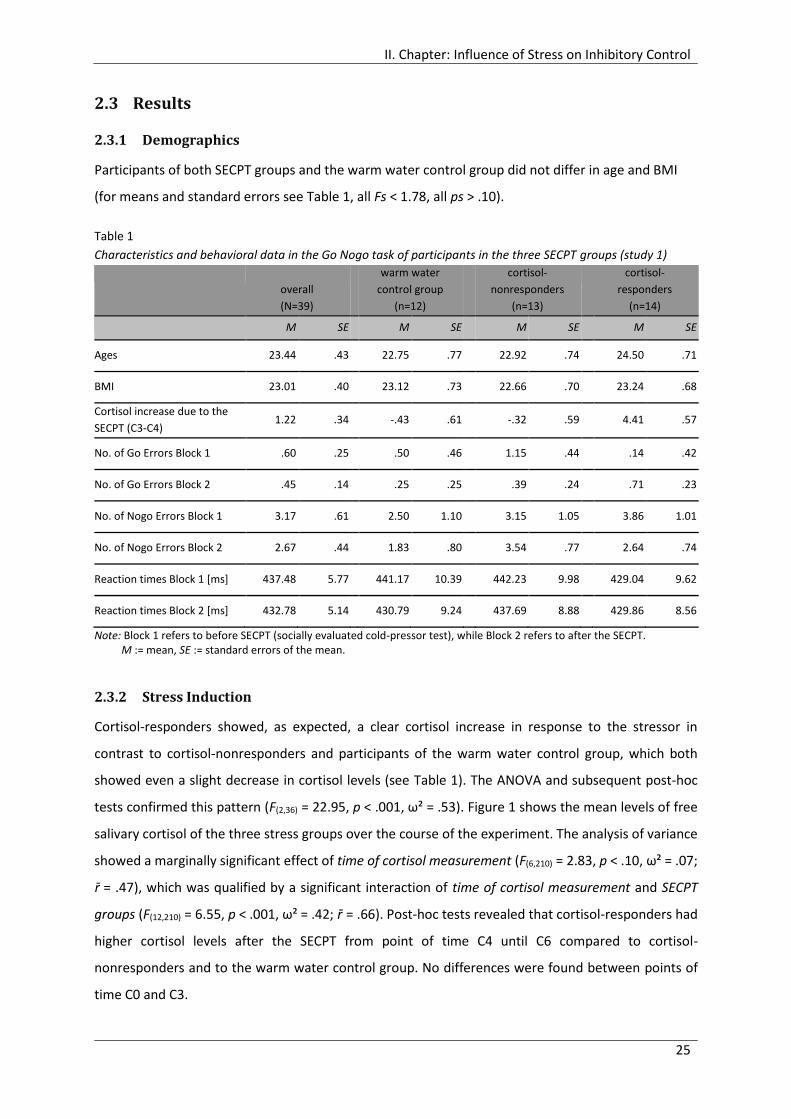
II. Chapter: Influence of Stress on Inhibitory Control
25
2.3 Results
2.3.1 Demographics
Participants of both SECPT groups and the warm water control group did not differ in age and BMI
(for means and standard errors see Table 1, all Fs < 1.78, all ps > .10).
Table 1
Characteristics and behavioral data in the Go Nogo task of participants in the three SECPT groups (study 1)
overall
(N=39)
warm water
control group
(n=12)
cortisol-
nonresponders
(n=13)
cortisol-
responders
(n=14)
M SE M SE M SE M SE
Ages 23.44 .43 22.75 .77 22.92 .74 24.50 .71
BMI 23.01 .40 23.12 .73 22.66 .70 23.24 .68
Cortisol increase due to the
SECPT (C3-C4) 1.22 .34 -.43 .61 -.32 .59 4.41 .57
No. of Go Errors Block 1 .60 .25 .50 .46 1.15 .44 .14 .42
No. of Go Errors Block 2 .45 .14 .25 .25 .39 .24 .71 .23
No. of Nogo Errors Block 1 3.17 .61 2.50 1.10 3.15 1.05 3.86 1.01
No. of Nogo Errors Block 2 2.67 .44 1.83 .80 3.54 .77 2.64 .74
Reaction times Block 1 [ms] 437.48 5.77 441.17 10.39 442.23 9.98 429.04 9.62
Reaction times Block 2 [ms] 432.78 5.14 430.79 9.24 437.69 8.88 429.86 8.56
Note: Block 1 refers to before SECPT (socially evaluated cold-pressor test), while Block 2 refers to after the SECPT. M := mean, SE := standard errors of the mean.
2.3.2 Stress Induction
Cortisol-responders showed, as expected, a clear cortisol increase in response to the stressor in
contrast to cortisol-nonresponders and participants of the warm water control group, which both
showed even a slight decrease in cortisol levels (see Table 1). The ANOVA and subsequent post-hoc
tests confirmed this pattern (F(2,36) = 22.95, p < .001, ω² = .53). Figure 1 shows the mean levels of free
salivary cortisol of the three stress groups over the course of the experiment. The analysis of variance
showed a marginally significant effect of time of cortisol measurement (F(6,210) = 2.83, p < .10, ω² = .07;
r̄ = .47), which was qualified by a significant interaction of time of cortisol measurement and SECPT
groups (F(12,210) = 6.55, p < .001, ω² = .42; r̄ = .66). Post-hoc tests revealed that cortisol-responders had
higher cortisol levels after the SECPT from point of time C4 until C6 compared to cortisol-
nonresponders and to the warm water control group. No differences were found between points of
time C0 and C3.

II. Chapter: Influence of Stress on Inhibitory Control
26
Figure 1. Mean levels of free salivary cortisol during the experimental session of study 1 for cortisol-responders,
cortisol-nonresponders and the warm water control group. Error bars indicate standard errors of the mean.
SECPT := socially evaluated cold-pressor test. Note that the different orders of the task switching (TS) and Go
Nogo task were balanced across participants. := p < .05.
2.3.3 Impact of Stress on Response Inhibition - Behavioral Data
Reaction times. The mean reaction times for correct responses in Go trials are depicted in Table
1. Descriptively, participants of the three SECPT groups showed hardly any differences in their reaction
times before (Block 1) or after the SECPT or the warm water control procedure (Block 2), respectively.
The ANOVA confirmed this pattern, revealing no significant influence, neither by Block, nor by SECPT
groups, nor by the interaction of both (all Fs < 2.63, all ps > .10).
2.3.4 Impact of Stress on Response Inhibition - Electrophysiological Data
Figure 2a shows grand average ERP responses to Go and Nogo stimuli averaged over Block 1 and 2 and
SECPT groups. In Go trials, the general morphology of the waveform included an early positive peak at
about 180 ms (P2), followed by a negative peak at approximately 260 ms (N2) and a less pronounced
positive peak at approximately 330 ms (P3). The waveform in Nogo trials showed a positive peak at
about 170 ms (P2), followed by a distinct negative peak at approximately 240 ms (N2) and a
pronounced positive peak at approximately 340 ms (P3).
0
1
2
3
4
5
6
7
8
9
10
C0 -65 C1 -35 C2 -3 C3 +7 C4 +25 C5 +40 C6 + 55 min
salivary
cort
isol[n
mol/
l]warmwater control group
cortisol-nonresponder
cortisol-responder
SECPTBlock 2
TS/Go Nogo
Block 1
TS/Go Nogo

II. Chapter: Influence of Stress on Inhibitory Control
27
P2 component.
Latency. The mean latency of the P2 peak amplitude was 181.29 ms (SE = 2.93). Go trials led
to a delayed P2 (M = 187.82 ms, SE = 3.53) compared to Nogo trials (M = 174.76 ms, SE = 3.32)
independent of Block and electrode position (F(1,36) = 13.65, p < .001, ω² = .24, r̄ = .48). Besides, the
ANOVA revealed a significant main effect of Block, showing a delayed P2 peak before the SECPT (Block
1, M = 182.87 ms; SE = 3.12) compared to afterwards (Block 2, M = 179.71 ms; SE = 2.93; F(1,36) = 4.41,
p < .001, ω² = .27, r̄ = .88). No further effects reached significance (all Fs< 1.45, all ps >.10).
Peak. The P2 amplitude was more positive in Go trials (M = 4.67 µV, SE = .38) compared to
Nogo trials (M = 3.75 µV, SE = .36), as shown in Figure 2a-c (F(1,36) = 14.88, p < .001, ω² = .47, r̄ = .80).
This pattern will be referred to with the term Go>Nogo hereafter. Moreover, the P2 amplitude was
influenced by an interaction between electrode position and Block (F(1,36) = 4.87, p < .05, ω² = .20, r̄ =
.90). Post-hoc test showed that the P2 amplitude was greater at frontocentral sites compared to frontal
sites after the SECPT (Block 2), while no difference was found beforehand (Block 1). This effect was
qualified by a marginally significant four way interaction between electrode positions, Go Nogo, Block,
and SECPT groups (F(2,36) = 2.63, p < .10, ω² = .05, r̄ = .79). According to the post-hoc test, all three SECPT
groups showed greater P2 amplitude in Go trials compared to Nogo trials at both electrode sites before
the SECPT (Block 1). However, after the SECPT (Block 2), the P2 amplitude in Go and Nogo trials did not
differ any longer in cortisol-nonresponders at frontal and frontocentral sites, while this effect was still
found at both electrode sites in participants of the warm water control group and was descriptively
even more distinct in cortisol-responders (see Figure 3 and Figure 5) . Apparently, in cortisol-
nonresponders the P2 amplitudes for Go stimuli decreased after the SECPT, whereas Nogo trials
remained unaffected. On the other hand, cortisol-responders showed the reversed pattern with
slightly reduced Nogo-P2 amplitudes and unchanged Go-P2 amplitudes.
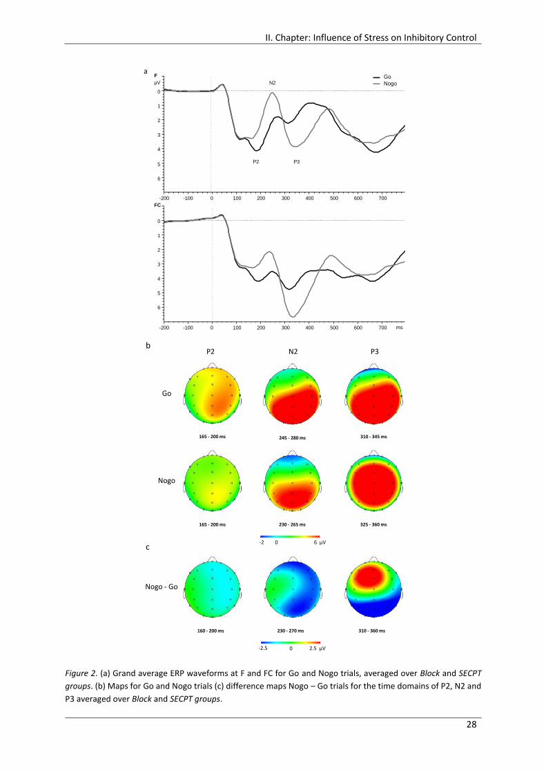
II. Chapter: Influence of Stress on Inhibitory Control
28
Figure 2. (a) Grand average ERP waveforms at F and FC for Go and Nogo trials, averaged over Block and SECPT
groups. (b) Maps for Go and Nogo trials (c) difference maps Nogo – Go trials for the time domains of P2, N2 and
P3 averaged over Block and SECPT groups.
F
FC
-200 -100 0 100 200 300 400 500 600 700
0
1
2
3
4
5
6
-200 -100 0 100 200 300 400 500 600 700
0
1
2
3
4
5
6
Go
Nogo
P2 P3
N2µV
a
ms
-2.5 0 2.5 µV
-2.5 µV 0 µV 2.5 µV
310 ms - 350 ms
-2.5 µV 0 µV 2.5 µV
230 ms - 270 ms
-2.5 µV 0 µV 2.5 µV
160 ms - 200 ms160 - 200 ms 230 - 270 ms 310 - 360 ms
-2 µV 0 µV 6 µV
325 ms - 360 ms
-2 µV 0 µV 6 µV
230 ms - 265 ms
-2 µV 0 µV 6 µV
165 ms - 200 ms
-2 µV 0 µV 6 µV
310 ms - 345 ms
-2 µV 0 µV 6 µV
245 ms - 280 ms
-2 µV 0 µV 6 µV
165 ms - 200 ms
-2 0 6 µV
165 - 200 ms 245 - 280 ms 310 - 345 ms
165 - 200 ms 230 - 265 ms 325 - 360 ms
P2 N2 P3
Go
Nogo
Nogo - Go
b
c
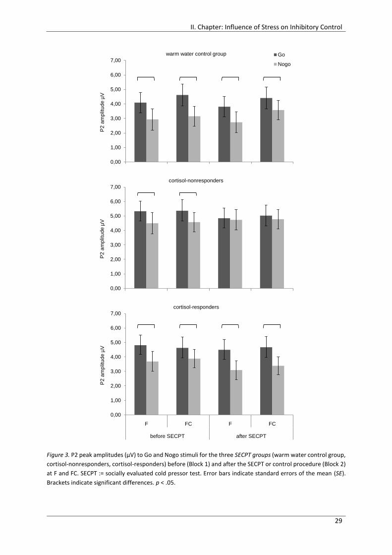
II. Chapter: Influence of Stress on Inhibitory Control
29
Figure 3. P2 peak amplitudes (µV) to Go and Nogo stimuli for the three SECPT groups (warm water control group,
cortisol-nonresponders, cortisol-responders) before (Block 1) and after the SECPT or control procedure (Block 2)
at F and FC. SECPT := socially evaluated cold pressor test. Error bars indicate standard errors of the mean (SE).
Brackets indicate significant differences. p < .05.
0,00
1,00
2,00
3,00
4,00
5,00
6,00
7,00
F FC F FC
before SECPT after SECPT
P2
am
plit
ud
e µ
V
warm water control group Go
Nogo
0,00
1,00
2,00
3,00
4,00
5,00
6,00
7,00
F FC F FC
before SECPT after SECPT
P2
am
plit
ud
e µ
V
cortisol-nonresponders
0,00
1,00
2,00
3,00
4,00
5,00
6,00
7,00
F FC F FC
before SECPT after SECPT
P2
am
plit
ud
e µ
V
cortisol-responders
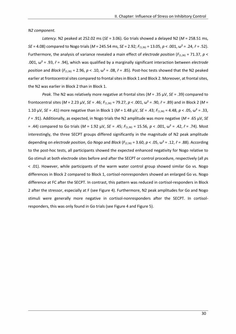
II. Chapter: Influence of Stress on Inhibitory Control
30
N2 component.
Latency. N2 peaked at 252.02 ms (SE = 3.06). Go trials showed a delayed N2 (M = 258.51 ms,
SE = 4.08) compared to Nogo trials (M = 245.54 ms, SE = 2.92; F(1,36) = 13.05, p < .001, ω² = .24, r̄ = .52).
Furthermore, the analysis of variance revealed a main effect of electrode position (F(1,36) = 71.37, p <
.001, ω² = .93, r̄ = .94), which was qualified by a marginally significant interaction between electrode
position and Block (F(1,36) = 2.96, p < .10, ω² = .08, r̄ = .85). Post-hoc tests showed that the N2 peaked
earlier at frontocentral sites compared to frontal sites in Block 1 and Block 2. Moreover, at frontal sites,
the N2 was earlier in Block 2 than in Block 1.
Peak. The N2 was relatively more negative at frontal sites (M = .35 µV, SE = .39) compared to
frontocentral sites (M = 2.23 µV, SE = .46; F(1,36) = 79.27, p < .001, ω² = .90, r̄ = .89) and in Block 2 (M =
1.10 µV, SE = .41) more negative than in Block 1 (M = 1.48 µV, SE = .43; F(1,36) = 4.48, p < .05, ω² = .33,
r̄ = .91). Additionally, as expected, in Nogo trials the N2 amplitude was more negative (M = .65 µV, SE
= .44) compared to Go trials (M = 1.92 µV, SE = .45; F(1,36) = 15.56, p < .001, ω² = .42, r̄ = .74). Most
interestingly, the three SECPT groups differed significantly in the magnitude of N2 peak amplitude
depending on electrode position, Go Nogo and Block (F(2,36) = 3.60, p < .05, ω² = .12, r̄ = .88). According
to the post-hoc tests, all participants showed the expected enhanced negativity for Nogo relative to
Go stimuli at both electrode sites before and after the SECPT or control procedure, respectively (all ps
< .01). However, while participants of the warm water control group showed similar Go vs. Nogo
differences in Block 2 compared to Block 1, cortisol-nonresponders showed an enlarged Go vs. Nogo
difference at FC after the SECPT. In contrast, this pattern was reduced in cortisol-responders in Block
2 after the stressor, especially at F (see Figure 4). Furthermore, N2 peak amplitudes for Go and Nogo
stimuli were generally more negative in cortisol-nonresponders after the SECPT. In cortisol-
responders, this was only found in Go trials (see Figure 4 and Figure 5).

II. Chapter: Influence of Stress on Inhibitory Control
31
Figure 4. N2 amplitudes to Go and Nogo stimuli for the three SECPT groups (warm water control group, cortisol-
nonresponders, cortisol-responders) before (Block 1) and after the SECPT or control procedure (Block 2) at F and
FC. SECPT := socially evaluated cold pressor test. Error bars indicate standard errors of the mean. Brackets
indicate significant differences. p < .05.
-3
-2
-1
0
1
2
3
4
5
6
N2 a
mp
litu
de
µV
warm water control group Go
Nogo
-3
-2
-1
0
1
2
3
4
5
6
N2 a
mp
litu
de
µV
cortisol-nonresponders
-3
-2
-1
0
1
2
3
4
5
6
F FC F FC
before SECPT after SECPT
N2 a
mp
litu
de
µV
cortisol-responders
II. Chapter: Influence of Stress on Inhibitory Control
32
Figure 5. Grand average ERP waveforms at F and FC for Go and Nogo trials for the three SECPT groups (warm
water control group, cortisol-nonresponders and cortisol-responders) before the SECPT (Block 1, left panel) and
after the SECPT (Block 2, right panel). SECPT := socially evaluated cold pressor test.
F
FC
-200 -100 0 100 200 300 400 500 600 700
0
1
2
3
4
5
6
-200 -100 0 100 200 300 400 500 600 700
0
1
2
3
4
5
6
P2 P3
N2µV
F
FC
-200 -100 0 100 200 300 400 500 600 700
0
1
2
3
4
5
6
-200 -100 0 100 200 300 400 500 600 700
0
1
2
3
4
5
6
P2 P3
N2
F
FC
-200 -100 0 100 200 300 400 500 600 700
0
1
2
3
4
5
6
-200 -100 0 100 200 300 400 500 600 700
0
1
2
3
4
5
6
F
FC
-200 -100 0 100 200 300 400 500 600 700
0
1
2
3
4
5
6
-200 -100 0 100 200 300 400 500 600 700
0
1
2
3
4
5
6
P2 P3
N2µV
block 1 block 2w
arm
wa
ter
co
ntr
ol g
rou
pF
FC
-200 -100 0 100 200 300 400 500 600 700
0
1
2
3
4
5
6
-200 -100 0 100 200 300 400 500 600 700
0
1
2
3
4
5
6
Go
NogoµV
F
FC
-200 -100 0 100 200 300 400 500 600 700
0
1
2
3
4
5
6
-200 -100 0 100 200 300 400 500 600 700
0
1
2
3
4
5
6
F
FC
-200 -100 0 100 200 300 400 500 600 700
0
1
2
3
4
5
6
-200 -100 0 100 200 300 400 500 600 700
0
1
2
3
4
5
6
F
FC
-200 -100 0 100 200 300 400 500 600 700
0
1
2
3
4
5
6
-200 -100 0 100 200 300 400 500 600 700
0
1
2
3
4
5
6
Go
NogoµV
F
FC
-200 -100 0 100 200 300 400 500 600 700
0
1
2
3
4
5
6
-200 -100 0 100 200 300 400 500 600 700
0
1
2
3
4
5
6
µV
F
FC
-200 -100 0 100 200 300 400 500 600 700
0
1
2
3
4
5
6
-200 -100 0 100 200 300 400 500 600 700
0
1
2
3
4
5
6
F
FC
-200 -100 0 100 200 300 400 500 600 700
0
1
2
3
4
5
6
-200 -100 0 100 200 300 400 500 600 700
0
1
2
3
4
5
6
F
FC
-200 -100 0 100 200 300 400 500 600 700
0
1
2
3
4
5
6
-200 -100 0 100 200 300 400 500 600 700
0
1
2
3
4
5
6
µV
F
FC
-200 -100 0 100 200 300 400 500 600 700
0
1
2
3
4
5
6
-200 -100 0 100 200 300 400 500 600 700
0
1
2
3
4
5
6
µV
F
FC
-200 -100 0 100 200 300 400 500 600 700
0
1
2
3
4
5
6
-200 -100 0 100 200 300 400 500 600 700
0
1
2
3
4
5
6
F
FC
-200 -100 0 100 200 300 400 500 600 700
0
1
2
3
4
5
6
-200 -100 0 100 200 300 400 500 600 700
0
1
2
3
4
5
6
F
FC
-200 -100 0 100 200 300 400 500 600 700
0
1
2
3
4
5
6
-200 -100 0 100 200 300 400 500 600 700
0
1
2
3
4
5
6
µV
co
rtis
ol-
no
nre
sp
on
de
rs
F
FC
-200 -100 0 100 200 300 400 500 600 700
0
1
2
3
4
5
6
-200 -100 0 100 200 300 400 500 600 700
0
1
2
3
4
5
6
µV
F
FC
-200 -100 0 100 200 300 400 500 600 700
0
1
2
3
4
5
6
-200 -100 0 100 200 300 400 500 600 700
0
1
2
3
4
5
6
F
FC
-200 -100 0 100 200 300 400 500 600 700
0
1
2
3
4
5
6
-200 -100 0 100 200 300 400 500 600 700
0
1
2
3
4
5
6
F
FC
-200 -100 0 100 200 300 400 500 600 700
0
1
2
3
4
5
6
-200 -100 0 100 200 300 400 500 600 700
0
1
2
3
4
5
6
µV
F
FC
-200 -100 0 100 200 300 400 500 600 700
0
1
2
3
4
5
6
-200 -100 0 100 200 300 400 500 600 700
0
1
2
3
4
5
6
µV
F
FC
-200 -100 0 100 200 300 400 500 600 700
0
1
2
3
4
5
6
-200 -100 0 100 200 300 400 500 600 700
0
1
2
3
4
5
6
F
FC
-200 -100 0 100 200 300 400 500 600 700
0
1
2
3
4
5
6
-200 -100 0 100 200 300 400 500 600 700
0
1
2
3
4
5
6
F
FC
-200 -100 0 100 200 300 400 500 600 700
0
1
2
3
4
5
6
-200 -100 0 100 200 300 400 500 600 700
0
1
2
3
4
5
6
µV
co
rtis
ol-
resp
on
de
rs
msms
msms
msms

II. Chapter: Influence of Stress on Inhibitory Control
33
P3 component.
Latency. The latency of the P3 peak averaged 335.50 ms (SE = 3.67). Go trials peaked earlier
(M = 330.81 ms, SE = 4.25) compared to Nogo trials (M = 340.19 ms, SE = 4.23; F(1,36) = 4.87, p < .05, ω²
= .09, r̄ = .51). This main effect was qualified by an interaction between electrode position and Go Nogo
(F(1,36) = 13.82, p < .001, ω² = .20, r̄ = .67) and by an interaction between Block and Go Nogo (F(1,36) =
7.59, p < .01, ω² = .07, r̄ = .45), revealing that the advantage in Go trials compared to Nogo trials was
found at F (Go: M = 329.26 ms, SE = 4.46, Nogo: M = 344.04 ms, SE = 4.45) and in Block 1 (Go: M =
325.32 ms, SE = 3.98, Nogo: M = 344.86 ms, SE = 5.32), but not at FC (Go: M = 332.36 ms, SE = 4.32,
Nogo: M = 336.34 ms, SE = 4.26) and Block 2 (Go: M = 336.30 ms, SE = 5.76, Nogo: M = 335.53 ms, SE
= 4.19), respectively. No further effects reached significance (all Fs< 2.36, all ps >.10).
Peak. The P3 peak amplitude was relatively more positive at the frontocentral site (M = 6.23
µV, SE = .41) compared to the frontal site (M = 3.57 µV, SE = .32) (F(1,36) = 109.99, p < .001, ω² = .86, r̄ =
.78). Besides, Nogo trials showed a greater P3 peak amplitude than Go trials (Go: M = 3.88 µV, SE =
.39), Nogo: M = 5.92 µV, SE = .42; F(1,36) = 21.62, p < .001, ω² = .31, r̄ = .42). No further effects (ω² ≥ .05)
reached significance (all Fs< 1.00, all ps >.10).
2.4 Discussion
The present study sought to investigate the influence of a psychosocial stressor and the thereby caused
increase of cortisol on cognitive control, specifically response inhibition, comprising both behavioral
and electrocortical measurements.
The stress induction via the socially evaluated cold-pressor test was successful. Cortisol-
responders showed a considerable increase of free cortisol in response to the stressor, similar to which
was found in other studies using this stressor (e.g., Lass-Hennemann et al., 2011; Schwabe & Wolf,
2009; Smeets, 2011).
2.4.1 Impact of Stress on Response Inhibition Performance
Contrary to my hypothesis, acute stress had no influence on performance. In general, accuracy
in the Go Nogo task was very high throughout both blocks and all participants. Descriptively,
participants of the warm water control group improved their performance slightly in Go as well as in
Nogo trials from block 1 to 2, while cortisol-nonresponders and cortisol-responders made only slightly
more mistakes after the SECPT in Nogo trials (i.e., errors of commission) or in Go trials (i.e., errors of
omission), respectively. As numbers of errors were too low for analysis in the present study, I cannot
draw conclusion whether the performance regarding the accuracy would have been statistically
significantly influenced by the stress induction or the thereby caused increase in cortisol or not. In any
II. Chapter: Influence of Stress on Inhibitory Control
34
case, the impact can be considered as rather negligible. Accordingly, previous research on
stress/cortisol and response inhibition found no significant influence on accuracy, too, neither after a
psychosocial stressor (Scholz et al., 2009; Zwissler et al., 2011), nor after intravenous (Wolf et al.,
2001), nor oral hydrocortisone administration (Schlosser et al., 2013).
Concerning the reaction times, neither cortisol-responders nor cortisol-nonresponders
differed in their performance after the stressor compared to beforehand or to the warm water control
group. This finding is in contrast with altered reactions times in the studies examining response
inhibition after stress exposure or glucocorticoid treatment (Schlosser et al., 2013; Scholz et al., 2009).
Scholz et al. (2009) found slower reaction times in a Go Nogo task in participants who completed the
Trier Social Stress Test (TSST) beforehand compared to the control group who was assigned to a rest
condition. On the contrary, in the study of Schlosser et al. (2013) healthy participants who received a
dose of 10 mg hydrocortisone reacted faster in an emotional Go Nogo task, indicating enhanced
response inhibition.
Different reasons may account for the missing stress effect on response inhibition in the
present study. First, the task characteristics may have been inappropriate to reproduce stress effects
on performance. A simple task with letters was used, which was performed twice, before and after the
experimental manipulation. Participants showed a very high accuracy and hardly any further
improvement from Block 1 to Block 2, which indicates that the task was easy to accomplish. Hockey
(1997) proposed in his cognitive-energetical framework that regulatory processes required for coping
with stress allocate resources at the expense of performance. Several studies found supporting
evidence for this assumption for working memory but only in the case of high workload (e.g., Lupien,
Gillin, & Hauger, 1999; Oei et al., 2006; Schoofs, Preuss, & Wolf, 2008). Thus, the Go Nogo task used in
the present study probably did not produce a sufficient workload to reveal the impairing effect of
stress. This is supported by the fact that the stress induction influenced reaction times in the more
complex and demanding task switching paradigm which was accomplished in balanced order by
participants of the present study as well (see Fechtner, 2012, Chapter 2). Hockey (1997) supposed
further that the cognitive system is able to adapt to restricted resources under stress or high workload
and still maintain performance by adopting less capacity-demanding performance-protection strategy
(Hockey, 1997, p. 78). In line with this, Scholz et al. (2009) could demonstrate that implementation
intentions, a cognitive strategy in form of simple if-then plans which have been shown to make action
initiation automatic and less effortful (Gollwitzer, 1999), offset the negative effects of stress on
response inhibition. Participants who were instructed to form implementation intentions for the Go
Nogo task showed no detrimental effects of the stress induction in their reaction times (Scholz et al.,
2009). In a way, participants in the present study were provided with if-then rules via the instruction,
and it is possible that they form implicitly an implementation intention during the first block of the Go

II. Chapter: Influence of Stress on Inhibitory Control
35
Nogo by telling the rules to themselves (i.e., If X [Y] appears, then I press the button. If Y [X] appears,
then I withhold the response.). As only two stimuli were used, the rules would have been as specific as
the implementation-intention instruction used by Scholz et al. (2009), described in Brandstätter,
Lengfelder, and Gollwitzer, 2001. However, as I did not ask whether and which strategies participants
used, this is so far only speculative. Nevertheless, it is quite plausible that participants used any kind
of cognitive strategy to maintain performance throughout the two blocks.
Furthermore, there is increasing evidence that the impact of cortisol is greater for emotional
compared to neutral material across different cognitive processes (memory: Abercrombie, Speck, &
Monticelli, 2006; Buchanan & Lovallo, 2001; Roozendaal, 2000, working memory: Luethi, 2008,
attention: van Honk et al., 1998). Hence, the use of single letters might additionally account for the
absence of an effect of stress on response inhibition performance.
Second, the quantity and quality of the cortisol manipulation seems to be crucial for its possible
effect on cognitive control. The SECPT induces a relatively moderate endogenous cortisol increase,
compared to the TSST, which generally leads to an increase in cortisol levels about two to three times
higher (Elzinga & Roelofs, 2005; Luethi, 2008; Plessow et al., 2011; Plessow et al., 2012; Scholz et al.,
2009). Consequently, the detrimental effects on response inhibition found by Scholz et al. (2009) could
be the result of the higher increase in cortisol achieved by the use of the TSST. On the other hand, the
fact that Schlosser et al. (2013), in contrast to the present study as well as in contrast to the one by
Scholz et al. (2009), found enhancing effects of cortisol on response inhibition, might be due to
qualitative aspects of cortisol manipulation. In their study, participants received an oral dose of
exogenous glucocorticoids. While a psychological stressor leads to an activation of the HPA axis which
is accompanied by the activation of the sympathetic nervous system, hydrocortisone artificially raises
cortisol levels, lacking the quality of a real-life stressor. The absence of a general arousal might alter
the impact on prefrontal cognitive functions. Elzinga and Roelofs (2005), for example, found acute
sympathetic activation to be necessary for cortisol-induced impairments on working memory.
Supporting this, Oei et al. (2009) reported a reduced distraction of emotional stimuli in a Sternberg
item-recognition task in participants who received an oral dose of 35 mg hydrocortisone. Beside these
qualitative aspects, exogenous cortisol administration generally leads to substantially higher levels of
cortisol compared to a stressor including the TSST, causing an allocation of all high affinity receptors.
Various studies focusing on different aspects of cognition found evidence that the influence of cortisol
is dose dependent, forming an inverted-U shape relationship (e.g., Abercrombie, Kalin, Thurow,
Rosenkranz, & Davidson, 2003; Lupien et al., 1999; Mateo, 2008; Salehi, Cordero, & Sandi, 2010;
Schilling et al., 2013). In a similar vein, this may be the case for cognitive control. More precisely,
moderate levels of cortisol do not alter performance of response inhibition, while higher levels cause
deterioration of performance and the saturation of glucocorticoid receptors even an enhancement.
II. Chapter: Influence of Stress on Inhibitory Control
36
Thus, the relationship between performance and level of cortisol would result in a U curve or J curve,
respectively.
Third, Schlosser et al. (2013) hypothesized that response inhibition might be generally less
affected by cortisol manipulation compared to working memory performance. They argued that
although both of these cognitive functions rely on the prefrontal cortex, different subregions (e.g.,
dorsolateral prefrontal cortex (DLPFC)) and structures (e.g., anterior cingulate cortex (ACC)) might be
task-specifically activated, which in turn are differentially affected by cortisol. Further, the authors
pointed out the interaction of cortisol with other neurotransmitters like dopamine and
norepinephrine, which influence executive functions as well and, hence, might count additionally for
the differing sensitivity of various executive functions to cortisol. Supporting this, Alexander, Hillier,
Smith, Tivarus, and Beversdorf (2007) found performance in cognitive flexibility, but not in cognitive
control tasks, to be impaired when exposed to psychosocial stress. Moreover, they could show that
this was related to adrenergic activity, as a non-specific beta-adrenergic antagonist reversed the
impairment.
In addition to these methodological differences, i.e., endogenous vs. exogenous cortisol
manipulation, the amount of resultant cortisol increase, different paradigms, and the valence of
selected stimuli, timing of glucocorticoid manipulation has to be considered. Het, Ramlow, and Wolf
(2005) pointed out in their review that the “time of day” significantly influences the impact of cortisol
administration on human memory. The circadian rhythm of cortisol leads to a peak in the morning,
followed by a continuous decrease in the course of the day. Thus, the number of unoccupied mineralo-
and glucocorticoid receptors differs with the time of day, which may influence the effect of
administered or stress-induced glucocorticoids on cognitive functions, such as working memory and
likewise inhibitory control. This might serve as an additional explanation why Wolf et al. (2001) did not
find altered performance in a Stroop Color Word Test after intravenous administration of a rather high
dosage of hydrocortisone, as they tested response inhibition about noontime.
2.4.2 Impact of Stress on Electrophysiological Data of Response Inhibition
Established effects within the event-related potentials (ERPs) of response inhibition could be
replicated: Nogo stimuli elicited a pronounced frontal negativity (N2), followed by a frontal positivity
(P3) compared to Go stimuli (e.g., Benikos et al., 2013; Bokura et al., 2001; Bruin & Wijers, 2002; Eimer,
1993; Falkenstein et al., 1999; Jodo & Kayama, 1992; Kiefer et al., 1998; Kopp, Mattler, Goertz, & Rist,
1996). Moreover, in accordance with Gajewski and Falkenstein (2013) and Benikos et al. (2013), Go
trials caused a P2 component reaching to frontal and frontocentral sites, while no such distinct
positivity was found in Nogo trials. Besides amplitudes, in line with previous research, latency varied
with stimulus type: Go trials lead to a delayed P2 as well as N2 (N2: Nakata et al., 2012), whereas the
II. Chapter: Influence of Stress on Inhibitory Control
37
P3 was earlier compared to Nogo trials (Benikos & Johnstone, 2009; Jodo & Kayama, 1992). Unlike
performance of response inhibition, stress influenced the early processing of Go and Nogo stimuli as a
function of the induced cortisol increase. Cortisol-responders and –nonresponders showed each
altered P2 and N2 amplitudes after the stress-induction, while the P3 component remained
unaffected.
P2. Stress altered the P2 peak amplitude to Go stimuli, albeit in opposite direction depending
on the extent of stress-induced HPA axis activation. Namely, by tendency, cortisol-nonresponders
showed diminished P2 amplitudes for Go stimuli after the SECPT, while the Go>Nogo4 pattern was
descriptively even more pronounced in cortisol-responders. To my knowledge, only very few studies
in the field of response inhibition considered this component or reported the appearance of a so-called
Go-P2. Gajewski and Falkenstein (2013) investigated the effects of task complexity on ERP components
using Go Nogo tasks. They reported a delayed P2 in more complex tasks, while no influence was found
concerning the amplitude. Similarly, task difficulty in the Go Nogo task was studied by Benikos et al.
(2013), who found in contrast to Gajewski and Falkenstein (2013) no effects of task difficulty on P2
latency. However, they found a reduction of the P2 amplitudes with increasing time pressure that was
more salient in Nogo trials. Moreover, another study showed that caffeine leads to globally enhanced
P2 amplitudes to Go stimuli in an auditory Go Nogo task (Barry et al., 2007). The authors interpreted
this as an improvement of processing associated with response production by the substance. In line
with this suggestion, Gajewski and Falkenstein (2013) conclude that “the Go-P2 reflects [...] stimulus-
response activation in Go trials.” (p. 278). Thus, the reduced P2 in Go trials found in cortisol-
nonresponders of the present study indicates that stress without a noticeable HPA axis activation
impairs response activation, whereas neuronal processing of Nogo stimuli remains unaffected. Stress-
induced cortisol increase, as found in cortisol-responders, however, seems to rather promote the
maintenance of response execution, as the Go-P2 remained invariant across both blocks.
Alternatively, Benikos and Johnstone (2009) and Benikos et al. (2013) argue that the P2 is
positively associated with suppression of interference from distracting and irrelevant information
(Hegerl, Gallinat, & Mrowinski, 1994; Oades, 1998 as cited in Benikos & Johnstone, 2009), “giving the
imperative stimulus a clear path for further processing (Oades, 1998).” (Benikos et al., 2013, p. 270).
In line with this, Gajewski, Stoerig, and Falkenstein (2008) quote that the P2 is related to task relevant
stimulus evaluation (Potts, 2004 as cited in Gajewski et al., 2008, p. 132), and may constitute a crucial
factor in optimizing current performance. Benikos and Johnstone (2009), Benikos et al. (2013) and
Gajewski et al. (2008) did not find P2 amplitude alterations specific for Go stimuli, but did for Nogo
stimuli as a function of task difficulty (Benikos et al., 2013), and for both stimuli in Attention-
4 i.e., P2 amplitude was more positive in Go trials compared to Nogo trials.

II. Chapter: Influence of Stress on Inhibitory Control
38
Deficit/Hyperactivity disorder diagnosed children performing a cued Go Nogo task (Gajewski et al.,
2008). Nevertheless, the investigated stimuli still demanded a response or were in the context of
response preparation. Hence, their interpretation may extend the idea of altered response action by
successful recognition of relevant response requiring stimulus. Accordingly, the preservation of
response activation due to HPA axis activation might be ascribed to elaborate stimulus processing,
allocating more cognitive resources.
N2. Similar to the P2 component, cortisol-nonresponders and cortisol-responders showed
altered N2 amplitudes after the stress-induction, in contrast to the warm water control group. All three
groups showed a more negative N2 to Nogo stimuli compared to Go stimuli before and after the
SECPT/control condition. However, in cortisol-nonresponders this difference was enlarged and N2
amplitudes of both, Go and Nogo trials, were shifted to more negative values. In contrast, cortisol-
responders showed a reduced Go vs. Nogo pattern and N2 amplitudes in Go trials were more negative
after the SECPT relative to Block 1, before the SECPT. Thus, stress and stress-induced cortisol increase
did not affect Nogo trials in particular.
A profound amount of research deals with the role of the N2 to Nogo stimuli considering it
reflecting response inhibition itself (Fallgatter & Strik, 1999; Jodo & Kayama, 1992; Kaiser et al., 2003;
Kopp et al., 1996). However, the present results suggest that stress with and without a noticeable HPA
axis activation does not seem to particularly effect response inhibition. The present data rather suggest
a general effect, independent of the stimulus, in stressed participants without an increase in cortisol.
Augmented negative N2 amplitudes were repeatedly interpreted as enhanced neural activity (e.g.,
Chen, Tien, Juan, Tzeng, & Hung, 2005; Euser & Franken, 2012; Jodo & Kayama, 1992). Therefore,
cortisol-nonresponders might need more cognitive effort to maintain their performance. In contrast,
stress-induced HPA axis activation specifically altered N2 amplitudes to Go stimuli. Consequently,
stress-induced increase in cortisol did not impair the response inhibition by Nogo stimuli, but rather
affected the processing of response requiring stimuli in this time window. Alternatively to the
inhibition hypothesis, Nieuwenhuis et al. (2003), Donkers and van Boxtel (2004) and Enriquez-Geppert,
Konrad, Pantev, and Huster (2010) proposed that the N2 might rather reflect a response conflict
between Go and Nogo response tendencies, revealing the component to be sensitive to stimulus
frequency independently of whether a response was required or to suppress it. Following this
assumption, more negative N2 amplitudes to Go stimuli found in cortisol-responders indicate that
stimuli triggering response performance tendencies allocated enhanced cognitive resources and
demanded heightened conflict monitoring. Similar to the present results Yang et al. (2009),
investigating response inhibition in heroin addicts compared to healthy controls in a visual Go Nogo
task, found no group differences in behavior and N2 for Nogo stimuli, but enlarged Go N2 amplitudes
II. Chapter: Influence of Stress on Inhibitory Control
39
in heroin addicts relative to controls. The authors interpreted this in line with the conflict monitoring
hypothesis and concluded that heroin addicts show an overactivation towards response signals.
P3. In contrast to P2 and N2 amplitudes and contrary to my hypothesis, the P3 remained
unaffected by stress and cortisol, which is unexpected regarding the fact that N2 and P3 were initially
interpreted as a single complex N2/P3 (Simson, Vaughan, & Ritter, 1977). Notwithstanding, there is
evidence that both components are modulated by different neurobiological pathways (Beste, Saft,
Andrich, Gold, & Falkenstein, 2008; Beste, Willemssen, Saft, & Falkenstein, 2010; Bokura et al., 2001;
Huster, Westerhausen, Pantev, & Konrad, 2010). Moreover, Smith, Johnstone, and Barry (2008) found
supporting evidence that the Nogo P3 is associated with motor response inhibition and further
research suggests that this component might reflect the evaluation or finalization of the inhibitory
process (Band & van Boxtel, 1999; Bruin et al., 2001). The present findings show that these processes
are not affected by stress or stress-induced cortisol increase, which is supported by the fact that the
inhibitory performance was successful in all groups.
The present electrophysiological results suggest that stress without HPA axis activation interfered with
cognitive processing during response inhibition, making the task more difficult for respective
participants, reflected in reduced P2 amplitudes in Go trials and diminished Go>Nogo pattern. More
cognitive resources were required for an accurate performance as shown by generally reduced N2
amplitudes. On the other hand, stress-induced increase of cortisol levels did not impair neural
processes of response inhibition, but rather maintained these, albeit affecting electrophysiological
correlates of stimuli requiring a response. Together with the tendency towards a slightly increased
number of errors of omission, these findings suggest that cortisol led to a pronounced caution or an
overcorrection in response inhibition, reflected by a distinct Go>Nogo P2 pattern and a more negative
Go-N2 after the SECPT. The assumption, that acute cortisol affects response activation, is supported
by the results of Tops et al. (2005) who investigated the effect of exogenous cortisol administration on
resting frontal EEG power asymmetry in healthy participants. They found in the cortisol group a relative
increase in right frontal activity in the alpha band and drew the conclusion that high levels of cortisol
inhibit approach motivation.
Taking into account the negligible impact of stress and cortisol on response inhibition
performance, the present findings indicate that both SECPT groups were not visibly impaired in their
inhibitory control, but were able to perform with high accuracy. Thus, revisiting the cognitive-
energetical framework by Hockey (1997), both SECPT groups seem to have compensatory control
mechanisms at their disposal. These allowed them to maintain manifest performance, but probably
imply so-called “latent performance decrements” (Hockey, 1997, p. 82), which might be reflected in
cortisol-dependently altered electrophysiological correlates of response inhibition. Consequently, the
II. Chapter: Influence of Stress on Inhibitory Control
40
present study suggests that neither stress itself nor stress-induced moderate cortisol increase impairs
inhibitory control performance. However, it is not clear whether these conclusions can be transferred
to real-life situations, as participants might be particularly able to control themselves for the short
period of the experiment in favor of an accurate task performance. Especially, as the laboratory
surroundings are very likely to promote the participants’ desire to perform well.
2.4.3 Strengthens and Limitations
The present study was the first to investigate effects of an acute stressor on response inhibition
including electrophysiological measurements.
Still, some limitations should be mentioned. First, the study only included healthy young men
with an academic background. Hence, the present results cannot be generalized to women or
individuals of higher age, different socioeconomic status or with a history or presence of a physical or
mental disorder.
Second, even though the stress manipulation had the quality of a real-life stressor, the
achieved cortisol increase was rather low. Against the background of previous research, which
indicated the importance of quality and quantity of cortisol, a dose-response study or the usage of
stressors inducing a stronger HPA axis activation, as the TSST (Kirschbaum, Pirke, & Hellhammer, 1993),
could provide evidence to integrate the results of the present study and those of others (e.g., Schlosser
et al., 2013; Scholz et al., 2009). Besides, the post-hoc classification of the SECPT group in cortisol-
responders and –nonresponders provides the opportunity to disentangle the specific effect of
endogenous stress-induced cortisol increase from further impacts of the stress test. However,
concomitantly, this quasi-experimental approach may have implied confounding variables. For
instance, as other physiological aspects as heart rate or blood pressure were not included, the two
SECPT groups may differ in their sympathetic arousal as well. Moreover, it is possible that the
physiological response to a stressor is rather stable over time and situations which may be linked to
different personality traits. Thus, future studies should consider a more elaborate characterization of
these groups, especially with regard to the adrenergic system to detangle mutual effects of the
autonomic stress response and HPA axis activation.
Third, the pre-post design ensured that the three groups did not differ in their response
inhibition before the stressor and made it possible to analyze the change due to the intervention.
Nevertheless, as mentioned above, this design may have caused bottom effects, as for instance in
accuracy because of the extensive training. Accordingly, future studies might include a group who does
not perform the task before the stress manipulation. Similarly, the equiprobable Go Nogo task was
easy to accomplish, as reaction preparation was not prepotent compared to response inhibition and
no reaction time deadline was realized. Additionally, neutral stimuli may not be as suitable as

II. Chapter: Influence of Stress on Inhibitory Control
41
emotional material preferably with a relation to the stress situation to reveal the influence of stress
on response inhibition. Hence, next studies should induce a higher workload or task difficulty, use
emotional stimuli and take into account measurements of potential compensatory strategies, for
example by including an appropriate questionnaire.
2.4.4 Conclusion
The present study showed that stress influenced the neurophysiological basis of response inhibition
as a function of induced HPA-axis activation, whilst performance maintained unimpaired. Stress
without HPA axis activation caused reduced Go-P2 and augmented N2 amplitudes, indicating impaired
response activation and enhanced allocation of cognitive resources to perform accurately. An acute
rise in cortisol, in contrast, led to a distinct P2 Go>Nogo pattern and more negative Go-N2 amplitudes,
reflecting an elaborate processing and conflict monitoring of Go stimuli that indicates an
overcorrection of response activation. The evaluation and finalization of the inhibitory process were
affected neither by stress nor by cortisol, as shown by unimpaired P3 components. Taken together,
the results indicate that stress alters cognitive control processes; nevertheless, stressed participants
seemed to be equipped with compensatory mechanisms to overcome the impairment on a behavioral
level. The study provides insight in stress and cortisol effects on cognitive control, underlining the
advantage of electrocortical measurements to capture a comprehensive picture of those.

42
III. Chapter:
Gender-specific Effects of Stress
on Aggressive Behavior and Processing of
Provoking Stimuli
III. Chapter: Gender-specific Effects of Stress on Aggressive Behavior and Processing of Provoking Stimuli
43
3.1 Introduction
Aggression and aggressive behavior are – though natural and adaptive - considered as a substantial
problem in society if it is misplaced, excessive or persistent (Nelson & Trainor, 2007). In total, violence
is one of the main causes of death worldwide among adolescent to middle-aged people (Krug, 2002),
and involves enormous economic costs (Waters et al., 2004). Thus, it is hardly surprising that
aggression and its causes and consequences are one of the most researched topics in psychology (Geen
& Donnerstein, 1998). Anderson and Bushman (2002) suggest the following definition: “Human
aggression is any behavior directed toward another individual that is carried out with the proximate
(immediate) intent to cause harm. In addition, the perpetrator must believe that the behavior will
harm the target, and that the target is motivated to avoid the behavior.” (p. 28). These researchers
have formulated the general aggression model (GAM); a unifying framework, which postulates that
both, situational (i.e., provocation) as well as personal factors (i.e., gender), determine aggressive
behavior, mediated by cognition, affect and arousal. However, it does not include the detailed role of
underlying neurobiological mechanisms. Recent studies emphasize the relevance of these mechanisms
in the development, expression, and therapeutic interventions of aggressive behavior (for reviews see
Bertsch, 2012; Nelson & Trainor, 2007; Patrick, 2008).
Regarding underlying neural processes of aggression, previous research revealed alterations in
cortical activity not only in pathological groups (e.g., Blair, 2010; Gao & Raine, 2009; Zetzsche et al.,
2007), but in healthy individuals as well. Studies investigating electrocortical responses during an
aggressive encounter are of particular interest, as they offer the opportunity to examine elicitation
and development of aggressive behavior. A frequently used and extensively validated laboratory
paradigm to induce and measure aggression is the Taylor Aggression Paradigm (TAP, Taylor, 1967, for
data concerning validity see Anderson & Bushman, 1997; Bernstein, Richardson, & Hammock, 1987;
Giancola & Parrott, 2008; Giancola & Zeichner, 1995; Phillips, 2011). In this paradigm, participants are
informed that they are engaged in a reaction time task with a team-mate, the opponent. The slower
person in each trial will receive a punishment, the intensity and duration of which are set for the
opponent by the other player before each trial. In fact, wins and losses are predetermined and the
participant receives a series of fixed punishments during the course of the experiment, which can be
a shock or a noxious noise. Occasionally, intensity and duration settings for the punishment are
analyzed separately as a measurement for direct and indirect aggressive behavior, respectively. Lotze
et al. (2007) used a modified version of the TAP to provoke healthy male participants, giving them the
opportunity to retaliate against the opponent. Functional magnetic resonance imaging (fMRI) showed
increased activity in the medial prefrontal cortex (mPFC) during retaliation. Krämer and colleagues

III. Chapter: Gender-specific Effects of Stress on Aggressive Behavior and Processing of Provoking Stimuli
44
concentrated on the decision to respond aggressively, using a very similar version of the TAP. They
found altered activity in the anterior insula and rostral and dorsal anterior cingulate cortex (ACC) as a
function of the amount of provocation (Krämer et al., 2007). Furthermore, in two event-related
potentials (ERPs) studies, early frontal positive (P2) or negative (N2) components, respectively, were
affected by high provocation for individuals with trait aggressiveness (Krämer et al., 2008) or a history
of violence (Wiswede et al., 2011). Another ERP, the P3, has been frequently linked to aggressive
behavior (for a review see Patrick, 2008). A reduced P3 was consistently found in participants showing
antisocial and aggressive behavior. Bartholow, Bushman, and Sestir (2006), for example, compared
violent video game players with nonviolent video game players in a picture viewing task and measured
aggressive behavior afterwards in a modified version of the TAP. They found reduced P3 amplitudes
for violent images in violent video games players, which predicted aggressive behavior. The authors
suggest that the reduced ERP reflects desensitization to violent images. Others focused on trait
aggressiveness or self-reported measures of aggression. Bernat, Hall, Steffen, and Patrick (2007) found
that violent offenses predicted a reduced P3 amplitude in a standard two-stimulus visual oddball task
in a sample of male prisoners. Similarly, reduced P3 amplitudes in an auditory oddball task were
associated with self-reported aggression in undergraduate students (Gerstle, Mathias, & Stanford,
1998). On account of the consistency of the results, Gao and Raine (2009) suggest a reduced P3
amplitude as a neurobiological marker in the context of antisocial, externalizing and other aggression-
related behaviors. Nevertheless, to my knowledge the P3 amplitude to aggression-provoking stimuli
during an aggressive encounter has not been investigated so far.
Concerning psychoneuroendocrinological mechanisms, stress has been identified as an
important factor in precipitating and promoting aggressive behavior (cf. Barnett et al., 1991; Craig,
2007). In particular, animal research in rodents revealed that the hypothalamic-pituitary-adrenal (HPA)
axis, the so-called stress axis, and the stress hormones cortisol and corticosterone 5 are causally
involved in the genesis, elicitation and reinforcement of aggressive behavior (Hayden-Hixson & Ferris,
1991; Kruk et al., 2004; Wommack & Delville, 2007). Evoking aggression by electrical activation of the
hypothalamic attack area in rats, Kruk et al. (2004) found that “an experimentally induced acute surge
in corticosterone facilitates the aggressive response to hypothalamic stimulation.” (p. 1066), by
lowering the threshold for attack behavior. In addition, they revealed that aggressive behavior and
stimulation of the hypothalamic attack area itself led to a strong activation of the HPA axis. Thus, a fast
positive feedback loop between the glucocorticoid stress response and brain structures engaged in
aggressive behavior was identified. In humans, the importance of the HPA axis in the context of
5 Corticosterone is the primary glucocorticoid within rodents, whereas cortisol is the most important glucocorticoid hormone in humans.

III. Chapter: Gender-specific Effects of Stress on Aggressive Behavior and Processing of Provoking Stimuli
45
aggressive and aggressive-related behavior has been studied in some correlational and quasi-
experimental studies (e.g., Gerra et al., 2007; McBurnett et al., 2000; Poustka et al., 2010; Rudolph et
al., 2010; van Goozen & Fairchild, 2006; Victoroff et al., 2011), as well as in several more controlled
experimental studies (e.g., Cote et al., 2013; Geniole et al., 2011; Hirvikoski et al., 2009; Kempes et al.,
2008; Lopez-Duran et al., 2009; Verona & Curtin, 2006; Verona et al., 2006; Verona & Kilmer, 2007;
Verona, Sadeh, & Curtin, 2009). Even though these results revealed a clear association between the
stress system and aggressive as well as aggressive-related behavior in humans, the mutual causal
interaction, which was found in rodents, could not be entirely confirmed so far. In a series of
experiments, Verona and colleagues stressed healthy male and female participants with a physical
stressor and subsequently measured aggressive behavior in the form of administered electric shocks
in a teacher-learner paradigm. Predominantly, their studies showed that stressed men react with
enhanced aggressive behavior, while females did not (Verona & Curtin, 2006; Verona et al., 2006;
Verona & Kilmer, 2007, but see Verona et al., 2007). However, as the authors did not include
measurements of cortisol, it remains unclear whether the hormone accounted for these results.
Böhnke, Bertsch, Kruk, and Richter et al. (2010) examined the effect of exogenous cortisol on
aggressive behavior. In contrast to male participants, female participants who received an oral dose of
20 mg hydrocortisone, reacted more aggressively in a retaliation paradigm (TAP) compared to the
control group, who received a placebo. Hence, both studies found supporting evidence for a causal
enhancing effect of stress or cortisol on aggressive behavior, albeit with contradicting results for men
and women.
Typically, men are supposed to be more aggressive than women (as outlined in Baron
& Richardson, 2004; Eagly, 2013; Hyde, 1984). Yet, past research emphasized the need of taking into
account different forms of aggression (e.g., physical aggression, verbal aggression, indirect forms of
aggression) when investigating gender differences in the research area of aggression and violence
(Archer, 2004; Björkqvist, 1994; Cross & Campbell, 2011; Eagly & Steffen, 1986). Archer (2004), for
example, concluded that men show more direct or physical aggression, whereas (young) women prefer
indirect forms of aggression. These differences are often explained with gender roles and cultural
norms (Baron & Richardson, 2004) or neurobiological differences (Staniloiu & Markowitsch, 2012;
Struber et al., 2008). However, in their meta-analysis Bettencourt and Miller (1996) come to the
conclusion that provocation moderates gender differences in aggression, diminishing impact of gender
on nature and degree of aggressive behavior. That is, provoked females act similarly to men, although
perception and appraisal of provoking cues can be different for men and women as well as the
respective reaction (Bettencourt & Kernahan, 1997; Knight, Guthrie, Page, & Fabes, 2002). Similarly,
responses to stress are supposed to differ in males and females (Burns & Katkin, 1993; Kajantie &
Phillips, 2006; Stroud, Salovey, & Epel, 2002). In line with this, Taylor and colleagues proposed that, in

III. Chapter: Gender-specific Effects of Stress on Aggressive Behavior and Processing of Provoking Stimuli
46
contrast to men who respond to stress with fight-or-flight behavior, women react with affiliation and
seek for social support, a so-called “tend-and-befriend” behavior pattern (Taylor, S. E. et al., 2000;
Taylor, 2006). Though pellucid and easily comprehensible, it stands in contrast to the findings of
Böhnke, Bertsch, Kruk, and Richter et al. (2010).
To summarize, there is preliminary evidence that stress and cortisol enhance reactive aggressive
behavior in both females and males. However, this needs further investigations, not only against the
background of gender differences in aggressive behavior and in response to stress, but also in respect
to the interaction of stress and cortisol. Taking these issues into account, as a first aim, the present
study sought to further explore the relationship between acute stress and aggressive behavior in
healthy males and females. In common with Böhnke, Bertsch, Kruk, and Richter et al. (2010) and
Verona and colleagues (Verona & Curtin, 2006; Verona et al., 2006; Verona & Kilmer, 2007; Verona et
al., 2007) a modified version of the TAP was chosen to induce and measure aggressive behavior.
Considering different forms of aggression, volume (i.e., intensity) and duration setting of the punitive
noise were analyzed separately. Half of the participants were highly provoked, whereas the other half,
as a control group, were only mildly provoked. Prior to this retaliation paradigm, participants were
either exposed to an acute stressor, the socially evaluated cold-pressor test (Schwabe et al., 2008), or
to a control procedure with warm water. Several salivary cortisol measurements were taken in the
course of the experiment for the purpose of validation. It was expected that participants who respond
to the stressor with a rise in cortisol levels to react more aggressively, especially when being provoked
beforehand. In males, this effect should be most pronounced in direct aggressive behavior (i.e., volume
settings), while stressed females with an increase in cortisol should respond with enhanced indirect
aggression (i.e., duration settings). According to Bettencourt and Miller (1996), I expected that this
difference between male and female participants should be less distinct, when being highly provoked.
Furthermore, as an exploratory approach, the present study aimed to include the processing
of the aggression-eliciting stimuli. Within an aggressive encounter, provoking actions of others elicit
impulsive reactive aggressive behavior (van Goozen, Fairchild, Snoek, & Harold, 2007). Moreover, Kruk
et al. (2004) proposed that the relationship between stress and aggression is mediated by a change in
the processing of social conflict signals and aggression-promoting stimuli. Thus, I wanted to test if
neural correlates (P3) to the provoking stimuli are altered through stress or stress-induced rise in
cortisol and provocation and whether the magnitude of the P3 is related to aggressive behavior. I
expected the P3 amplitude in the high provocation group to be reduced and negatively correlated with
the aggressive behavior during the task. Furthermore, the influence of gender and stress as well as
stress-induced increase in cortisol on the P3 amplitude was tested as an explorative hypothesis. Lastly,

III. Chapter: Gender-specific Effects of Stress on Aggressive Behavior and Processing of Provoking Stimuli
47
preceding and succeeding intervals were explored as well to see whether effects were specific for the
P3 component.
3.2 Materials and Methods
3.2.1 Participants
Participants were recruited from the University of Trier, Germany. Out of 75 Participants, who
completed the experiment, four individuals had to be excluded due to abnormal high salivary cortisol
values (one female) or incorrect experimental procedure (three individuals), leaving 71 participants
(36 males, 35 females) with a mean age of 23.96 years (SD = 2.27, range 20-31 years) and a mean BMI
of 22.77 kg/m² (SD=2.54). Criteria for exclusion were (1) acute or chronic physical disease, (2) a mental
disorder or a history of such, (3) use of medication, (4) smoking, as it is known to influence HPA axis
activity (Granger et al., 2007), and (5) being not a native German speaker. Only right-handed students
were included, as handedness affects hemispheric specialization, thus altering EEG measurements
(Galin et al., 1982). To ensure no problems with the experimental manipulations, individuals who
reported to suffer from dyschromatopsia or stated to be sensitive to loud noises or to cold were
excluded. Additionally, students taking classes in psychology were excluded to guarantee an unbiased
behavior during the experiment. In order to control for hormonal status, only non-pregnant women
who used hormonal contraceptives6 were included in the study. The experiment was conducted in
accordance with the Declaration of Helsinki. The Research Ethics Committee of the University of Trier
approved all parts of the study, and all participants gave written informed consent. Participation was
compensated with 45 € (approximately US $57).
3.2.2 Socially Evaluated Cold-Pressor Test - SECPT
Participants who were assigned to the stress condition were exposed to the socially evaluated cold-
pressor test (SECPT, Schwabe et al., 2008), an economic and efficient stress induction causing
significant activation of the HPA axis, thereby a rise in cortisol levels, as well as an activation of the
adrenergic system and an increase in subjective stress experience (e.g., Schwabe & Wolf, 2009;
Weymar et al., 2012). More precisely, an unfamiliar experimenter of the opposite sex, who acted
neutrally and distanced, asked them to immerse their left hand up to the wrist into ice water (0-3 °C)
and to look at a camera throughout the whole procedure as they would be videotaped in order to
analyze their facial expressions. Meanwhile, the experimenter watched them closely, took notes, and
6 Except the contraceptives pills containing Drospirenone, which is an antagonist for the mineralocorticoid
receptor, and therefore might have skewed the cortisol measurements Genazzani, Mannella, and Simoncini
(2007); namely Yasmine, Yasminelle, Petibelle, Aida, Angeliq or Yaz
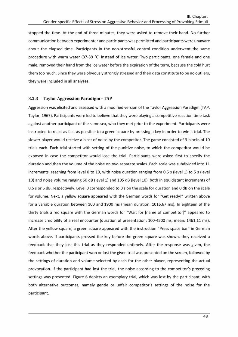
III. Chapter: Gender-specific Effects of Stress on Aggressive Behavior and Processing of Provoking Stimuli
48
stopped the time. At the end of three minutes, they were asked to remove their hand. No further
communication between experimenter and participants was permitted and participants were unaware
about the elapsed time. Participants in the non-stressful control condition underwent the same
procedure with warm water (37-39 °C) instead of ice water. Two participants, one female and one
male, removed their hand from the ice water before the expiration of the term, because the cold hurt
them too much. Since they were obviously strongly stressed and their data constitute to be no outliers,
they were included in all analyses.
3.2.3 Taylor Aggression Paradigm - TAP
Aggression was elicited and assessed with a modified version of the Taylor Aggression Paradigm (TAP,
Taylor, 1967). Participants were led to believe that they were playing a competitive reaction time task
against another participant of the same sex, who they met prior to the experiment. Participants were
instructed to react as fast as possible to a green square by pressing a key in order to win a trial. The
slower player would receive a blast of noise by the competitor. The game consisted of 3 blocks of 10
trials each. Each trial started with setting of the punitive noise, to which the competitor would be
exposed in case the competitor would lose the trial. Participants were asked first to specify the
duration and then the volume of the noise on two separate scales. Each scale was subdivided into 11
increments, reaching from level 0 to 10, with noise duration ranging from 0.5 s (level 1) to 5 s (level
10) and noise volume ranging 60 dB (level 1) and 105 dB (level 10), both in equidistant increments of
0.5 s or 5 dB, respectively. Level 0 corresponded to 0 s on the scale for duration and 0 dB on the scale
for volume. Next, a yellow square appeared with the German words for “Get ready!” written above
for a variable duration between 100 and 1900 ms (mean duration: 1016.67 ms). In eighteen of the
thirty trials a red square with the German words for “Wait for [name of competitor]” appeared to
increase credibility of a real encounter (duration of presentation: 100-4500 ms, mean: 1461.11 ms).
After the yellow square, a green square appeared with the instruction “Press space bar” in German
words above. If participants pressed the key before the green square was shown, they received a
feedback that they lost this trial as they responded untimely. After the response was given, the
feedback whether the participant won or lost the given trial was presented on the screen, followed by
the settings of duration and volume selected by each for the other player, representing the actual
provocation. If the participant had lost the trial, the noise according to the competitor’s preceding
settings was presented. Figure 6 depicts an exemplary trial, which was lost by the participant, with
both alternative outcomes, namely gentle or unfair competitor’s settings of the noise for the
participant.
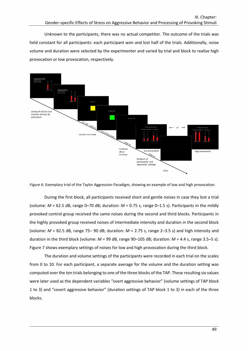
III. Chapter: Gender-specific Effects of Stress on Aggressive Behavior and Processing of Provoking Stimuli
49
Unknown to the participants, there was no actual competitor. The outcome of the trials was
held constant for all participants: each participant won and lost half of the trials. Additionally, noise
volume and duration were selected by the experimenter and varied by trial and block to realize high
provocation or low provocation, respectively.
Figure 6. Exemplary trial of the Taylor Aggression Paradigm, showing an example of low and high provocation.
During the first block, all participants received short and gentle noises in case they lost a trial
(volume: M = 62.5 dB, range 0–70 dB; duration: M = 0.75 s, range 0–1.5 s). Participants in the mildly
provoked control group received the same noises during the second and third blocks. Participants in
the highly provoked group received noises of intermediate intensity and duration in the second block
(volume: M = 82.5 dB, range 75– 90 dB; duration: M = 2.75 s, range 2–3.5 s) and high intensity and
duration in the third block (volume: M = 99 dB, range 90–105 dB; duration: M = 4.4 s, range 3.5–5 s).
Figure 7 shows exemplary settings of noises for low and high provocation during the third block.
The duration and volume settings of the participants were recorded in each trial on the scales
from 0 to 10. For each participant, a separate average for the volume and the duration setting was
computed over the ten trials belonging to one of the three blocks of the TAP. These resulting six values
were later used as the dependent variables “overt aggressive behavior” (volume settings of TAP block
1 to 3) and “covert aggressive behavior” (duration settings of TAP block 1 to 3) in each of the three
blocks.

III. Chapter: Gender-specific Effects of Stress on Aggressive Behavior and Processing of Provoking Stimuli
50
Figure 7. Exemplary possible feedback of participants' and opponents' settings for the noise, left: gentle settings
- low provocation, right: loud and lengthy noise - high provocation.
3.2.4 Subjective Measures
Measurements of mood. Self-reported momentary mood was assessed before and after the SECPT or
the control procedure, respectively, as well as after the Taylor Aggression Paradigm with a German
mood questionnaire (Mehrdimensionaler Befindlichkeitsfragebogen, MDBF, Steyer, Schwenkmezger,
Notz, & Eid, 1994). This questionnaire measures the current mood state on three dimensions: good
versus bad mood, wakefulness versus sleepiness, and calmness versus restlessness. Parallel short
version A and B, each containing twelve items, were implemented alternating within each subject.
Hopelessness and Anger. The MDBF questionnaires were completed with the scale
hopelessness and anger of the German Aktuelle Stimmungsskala (ASTS, Dalbert, 1992), each consisting
of three items (Hoffnungslosigkeit (hopelessness): hoffnungslos (hopeless), entmutigt (discouraged),
verzweifelt (desperate); Zorn (anger): zornig (irate), verärgert (angry), wütend (furious)).
Stress. Immediately after the SECPT, participants were asked to rate their feelings of stress and
pain during the stress or control procedure. This comprised six statements, which had to be evaluated
on a six point Likert scale (1: “strongly disagree”, 6: “strongly agree”): “Ich war sehr aufgewühlt.” (“I
was very upset.“), “Ich fühlte mich stark gestresst.“ (“I felt very stressed.“), “Das Wasserbad war
besonders stressig.“ (“The water bath was particular stressful.“), “Die Beobachtung und Aufzeichnung
waren besonders stressig.“ (“The surveillance and the recording were particular stressful.”), “Die
Schmerzen waren sehr unangenehm.” (“The pain was very unpleasant.”), “Ich war sehr
angespannt.”(“I was very tensed up.”).

III. Chapter: Gender-specific Effects of Stress on Aggressive Behavior and Processing of Provoking Stimuli
51
3.2.5 Salivary Cortisol Measurement
Saliva samples for cortisol analysis were obtained using Salivette® collection devices (Sarstedt,
Nürnbrecht, Germany). Samples were collected at seven assessment points over the course of the
experiment: before the start of the experiment (C0, -15 min, with reference to the beginning of the
SECPT), before the SECPT (C1, -2 min), after the SECPT (C2, +5 min), after the TAP (C3, +20 min), before
the emotional picture task (C4, +30 min), after the emotional picture task (C5, +40 min), and before
debriefing (C6, +60 min). Sampling instructions were given via computer and Salivettes® were
positioned on the table in front of the participants. Immediately after the experiment, samples were
frozen for biochemical analysis. Salivary cortisol was analyzed with a time-resolved immunoassay with
fluorescence detection as described in detail elsewhere (Dressendörfer et al., 1992). Intra- and
interassay variability was less than 10 and 12%, respectively. Beside the measurement of salivary
cortisol in the course of the experiment, further samples of native saliva were collected prior to the
experiment on three consecutive weekdays at awakening and 30, 45, and 60 min later to determine a
reliable measure of HPA axis activity via the cortisol awakening response (CAR) (Hellhammer et al.,
2007).
3.2.6 Procedure
Prior to the experimental session, participants were invited individually to an informational interview
to check exclusion criteria and to inform them about the aim and procedure of the study, i.e., the
investigation of the relationship between stress, reaction time, and cognitive functions. They were
informed at full length that they might be exposed to a stress procedure involving cold water,
videotaping, and observation, as well as to loud noises. Additionally, the electroencephalogram (EEG)
and the sampling of cortisol were described. Then, the dates and times for the sampling of the CAR
and the experiment were arranged. Finally, besides a battery of personality questionnaires to fill out
at home, they received sampling devices for salivary cortisol and a corresponding protocol, which is
described in detail by Böhnke, Bertsch, Kruk, and Naumann (2010) and Fechtner (2012). The examiner
further emphasized the necessity to adhere to the written instructions and sampling times. Moreover,
participants were required to refrain from physical exercise on the day prior, as well as alcohol,
caffeinated drinks, and meals within 1 h prior to the date fixed for the experimental session. The
completed questionnaires and cortisol samples had to be returned on this occasion. All participants
gave their written informed consent being aware that participation was voluntary and that they may
withdraw at any time without any consequences and without having to give reasons.
The actual experiment was conducted between 01:30 p.m. and approximately 07:30 p.m.,
starting at 01:30 p.m., 03:30 p.m., and 05:30 p.m., when endogenous cortisol levels are low (Schreiber

III. Chapter: Gender-specific Effects of Stress on Aggressive Behavior and Processing of Provoking Stimuli
52
et al., 2006). All participants were examined individually. Participants were randomly assigned to the
stress or control procedure, as well as to the highly provoked or mildly provoked control group, while
sex was balanced across conditions. On arrival, the experimenter acquainted the participant with
another participant of the same sex, who was in fact a confederate of the investigator, with whom he
or she was to play a computer game during the experiment. The participant and the confederate
handed over the filled out questionnaires and the salivary cortisol samples they collected at home.
Next, the participant was led in the EEG laboratory, where he or she was seated in a dimly lit sound-
attenuated room, 1 m from the monitor (20 in. Eizo FlexScan S2031W). To further increase credibility
of the cover story, the experimenter left the laboratory to ostensibly lead the confederate into another
EEG laboratory. After preparation of EEG, electrooculogram (EOG), and electrocardiogram (ECG)
recording devices, the actual experiment started. The participants received all instructions for the
different tasks, saliva samples, and questionnaires via the computer screen. First, they worked on an
Approach Avoidance Task (description and results reported in Fechtner, 2012), then they were
exposed to SECPT or the warm water control procedure. Afterwards, they played the TAP, followed by
a second block of the Approach Avoidance task. Finally, participants viewed passively pictures with
different emotional content for about 10 min (for details see Chapter 4.2.4, p.92 ff.). During the course
of the experiment, participants filled out short state questionnaires several times (see Chapter 3.2.4)
and provided seven saliva samples for cortisol (C). After removal of the physiological recording devices,
participants were extensively debriefed, thanked, and compensated for their participation.
E-Prime presentation software (Eprime 2.0, Psychological Software Tools, Pittsburgh, PA) was
used to present the stimuli and record the reaction times during the tasks. The experiment, from arrival
to debriefing had a duration of about 100 min. Figure 8 presents the timeline of the experimental
procedure.
Figure 8. Timeline of the experimental session of study 2. SECPT:= Socially evaluated cold-pressor test. C := saliva
samples for cortisol analyses. Time information refers to the beginning of the SECPT. Experimental parts printed
in bold are the topic of the present chapter.
Approach Avoidance
Task
SECPT
Taylor Aggression Paradigm
Approach Avoidance
Task
emotional pictures
C0
-15 min
C1
-2 min
C2
+5 min
C3
+20 min
C4
+30 min
C5
+40 min
C6
+60 min
EEG, EOG, ECG
preparation
III. Chapter: Gender-specific Effects of Stress on Aggressive Behavior and Processing of Provoking Stimuli
53
3.2.7 EEG Recording and Quantification
The EEG was recorded from 32 electrode sites including the mastoids according to the 10–10 electrode
reference system (Chatrian et al., 1988) with the Easy-Cap electrode system (Falk Minow Services,
Munich). All sites were referenced to FCz. A bipolar horizontal EOG was recorded from the epicanthus
of each eye and a bipolar vertical EOG was recorded from supra- and infra-orbital positions of the left
eye. Ag/AgCl electrodes were used for EEG and EOG recording. Prior to the electrode placement, the
electrode sites on the participant's scalp and face were cleaned with alcohol and gently abraded. The
conduction was facilitated using Abralyt-light (FMS, Munich) electrode gel or EC2® Genuine Grass
Electrode Cream (Grass Products, Natus Neurology) for the EOG, respectively. A BrainAmp amplifier
(input impedance: 10 MΩ; Brain Products, GmbH) in AC mode was used to record the EEG and EOG at
1000 Hz using a pass-band set to 0.016 to 499 Hz (−12 dB/octave roll-off). All impedances of the EEG
electrodes were maintained below 10 kΩ. Data was stored to hard disk for later analysis using
BrainVision Analyzer 2 (Brain Products, Munich, Germany).
The EEG was re-referenced offline to linked mastoids. The data was resampled at 200 Hz and
low pass filtered using a digital filter with high cutoff of 5 Hz, 48 dB/oct. Artifacts due to eye movements
were corrected semiautomatically via the algorithm developed by Gratton et al. (1983). If necessary,
blinks were detected and marked using Ocular Correction with Independent Component Analysis (ICA)
beforehand. EEG and EOG of trials were epoched off-line into 1200 ms periods, starting 200 ms prior
to stimulus onset (i.e., feedbacks of participants' and opponents' settings, see Figure 7) and ending
1000 ms after stimulus onset. A baseline correction was performed using the first 200 ms interval as a
reference. Trials with non-physiological artifacts were excluded from analysis via semiautomatic
artifact rejection. Separate averages were computed for each electrode and individual over the ten
trials of each of the three TAP blocks. Using the grand average across participants to guide window
selection, ERP maximum peak amplitude (µV) and latency (ms) for the stimulus-locked frontal P3
component were detected semiautomatically at Fz as reference channel within window of 250 - 450
ms post stimulus and for the parietal P3 at Pz as a reference channel within window of 250 - 500 ms
post stimulus. For statistical analyses, peak amplitudes were averaged over an interval of ± 10 data
points (i.e., 105 ms) and new electrode sites FFC3, FFCz, FCC4 and CPP3, CPPz, CCP4 were built by
averaging F3, FC3, FCz, Fz, as well as F4 and FC4 for the frontal P3, and CP3, P3, CPz, Pz as well as CP4
and P4 for the parietal P3, respectively. Additionally, preceding (200 - 300 ms) and succeeding
stimulus-locked intervals (400 - 500 ms, 500 - 600 ms) were exported for analyses.
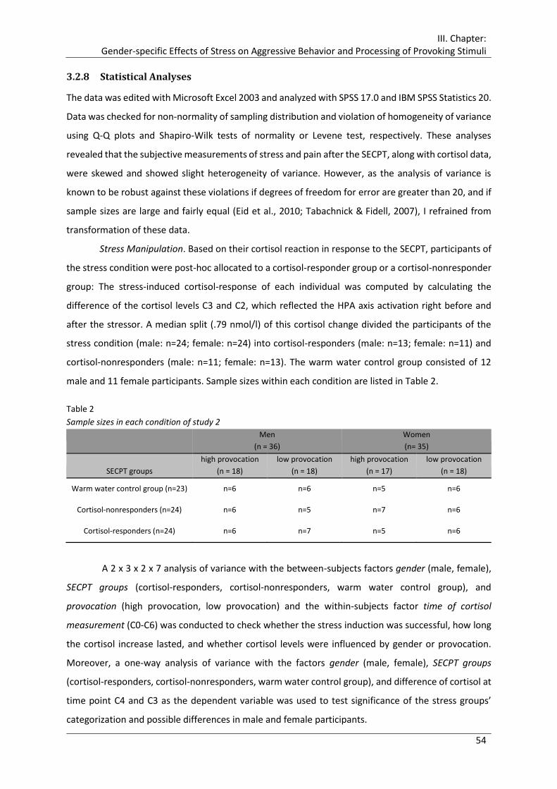
III. Chapter: Gender-specific Effects of Stress on Aggressive Behavior and Processing of Provoking Stimuli
54
3.2.8 Statistical Analyses
The data was edited with Microsoft Excel 2003 and analyzed with SPSS 17.0 and IBM SPSS Statistics 20.
Data was checked for non-normality of sampling distribution and violation of homogeneity of variance
using Q-Q plots and Shapiro-Wilk tests of normality or Levene test, respectively. These analyses
revealed that the subjective measurements of stress and pain after the SECPT, along with cortisol data,
were skewed and showed slight heterogeneity of variance. However, as the analysis of variance is
known to be robust against these violations if degrees of freedom for error are greater than 20, and if
sample sizes are large and fairly equal (Eid et al., 2010; Tabachnick & Fidell, 2007), I refrained from
transformation of these data.
Stress Manipulation. Based on their cortisol reaction in response to the SECPT, participants of
the stress condition were post-hoc allocated to a cortisol-responder group or a cortisol-nonresponder
group: The stress-induced cortisol-response of each individual was computed by calculating the
difference of the cortisol levels C3 and C2, which reflected the HPA axis activation right before and
after the stressor. A median split (.79 nmol/l) of this cortisol change divided the participants of the
stress condition (male: n=24; female: n=24) into cortisol-responders (male: n=13; female: n=11) and
cortisol-nonresponders (male: n=11; female: n=13). The warm water control group consisted of 12
male and 11 female participants. Sample sizes within each condition are listed in Table 2.
Table 2
Sample sizes in each condition of study 2
Men
(n = 36)
Women
(n= 35)
SECPT groups
high provocation
(n = 18)
low provocation
(n = 18)
high provocation
(n = 17)
low provocation
(n = 18)
Warm water control group (n=23) n=6 n=6 n=5 n=6
Cortisol-nonresponders (n=24) n=6 n=5 n=7 n=6
Cortisol-responders (n=24) n=6 n=7 n=5 n=6
A 2 x 3 x 2 x 7 analysis of variance with the between-subjects factors gender (male, female),
SECPT groups (cortisol-responders, cortisol-nonresponders, warm water control group), and
provocation (high provocation, low provocation) and the within-subjects factor time of cortisol
measurement (C0-C6) was conducted to check whether the stress induction was successful, how long
the cortisol increase lasted, and whether cortisol levels were influenced by gender or provocation.
Moreover, a one-way analysis of variance with the factors gender (male, female), SECPT groups
(cortisol-responders, cortisol-nonresponders, warm water control group), and difference of cortisol at
time point C4 and C3 as the dependent variable was used to test significance of the stress groups’
categorization and possible differences in male and female participants.
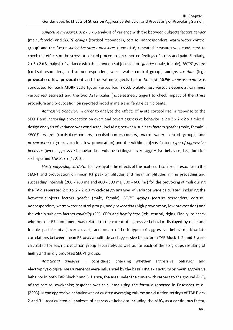
III. Chapter: Gender-specific Effects of Stress on Aggressive Behavior and Processing of Provoking Stimuli
55
Subjective measures. A 2 x 3 x 6 analysis of variance with the between-subjects factors gender
(male, female) and SECPT groups (cortisol-responders, cortisol-nonresponders, warm water control
group) and the factor subjective stress measures (Items 1-6, repeated measure) was conducted to
check the effects of the stress or control procedure on reported feelings of stress and pain. Similarly,
2 x 3 x 2 x 3 analysis of variance with the between-subjects factors gender (male, female), SECPT groups
(cortisol-responders, cortisol-nonresponders, warm water control group), and provocation (high
provocation, low provocation) and the within-subjects factor time of MDBF measurement was
conducted for each MDBF scale (good versus bad mood, wakefulness versus sleepiness, calmness
versus restlessness) and the two ASTS scales (hopelessness, anger) to check impact of the stress
procedure and provocation on reported mood in male and female participants.
Aggressive Behavior. In order to analyze the effects of acute cortisol rise in response to the
SECPT and increasing provocation on overt and covert aggressive behavior, a 2 x 3 x 2 x 2 x 3 mixed-
design analysis of variance was conducted, including between-subjects factors gender (male, female),
SECPT groups (cortisol-responders, cortisol-nonresponders, warm water control group), and
provocation (high provocation, low provocation) and the within-subjects factors type of aggressive
behavior (overt aggressive behavior, i.e., volume settings; covert aggressive behavior, i.e., duration
settings) and TAP Block (1, 2, 3).
Electrophysiological data. To investigate the effects of the acute cortisol rise in response to the
SECPT and provocation on mean P3 peak amplitudes and mean amplitudes in the preceding and
succeeding intervals (200 - 300 ms and 400 - 500 ms, 500 - 600 ms) for the provoking stimuli during
the TAP, separated 2 x 3 x 2 x 2 x 3 mixed-design analyses of variance were calculated, including the
between-subjects factors gender (male, female), SECPT groups (cortisol-responders, cortisol-
nonresponders, warm water control group), and provocation (high provocation, low provocation) and
the within-subjects factors caudality (FFC, CPP) and hemisphere (left, central, right). Finally, to check
whether the P3 component was related to the extent of aggressive behavior displayed by male and
female participants (covert, overt, and mean of both types of aggressive behavior), bivariate
correlations between mean P3 peak amplitude and aggressive behavior in TAP Block 1, 2, and 3 were
calculated for each provocation group separately, as well as for each of the six groups resulting of
highly and mildly provoked SECPT groups.
Additional analyses. I considered checking whether aggressive behavior and
electrophysiological measurements were influenced by the basal HPA axis activity or mean aggressive
behavior in both TAP Block 2 and 3. Hence, the area under the curve with respect to the ground AUCG
of the cortisol awakening response was calculated using the formula reported in Pruessner et al.
(2003). Mean aggressive behavior was calculated averaging volume and duration settings of TAP Block
2 and 3. I recalculated all analyses of aggressive behavior including the AUCG as a continuous factor,
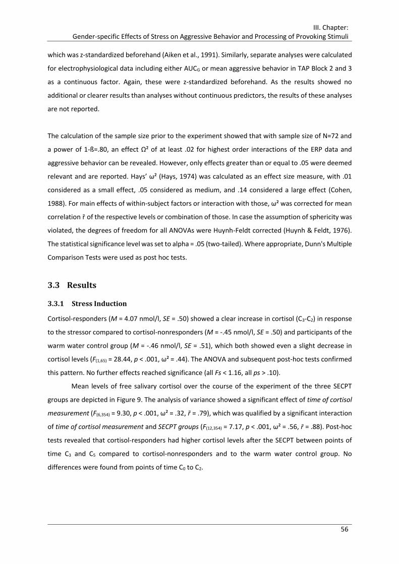
III. Chapter: Gender-specific Effects of Stress on Aggressive Behavior and Processing of Provoking Stimuli
56
which was z-standardized beforehand (Aiken et al., 1991). Similarly, separate analyses were calculated
for electrophysiological data including either AUCG or mean aggressive behavior in TAP Block 2 and 3
as a continuous factor. Again, these were z-standardized beforehand. As the results showed no
additional or clearer results than analyses without continuous predictors, the results of these analyses
are not reported.
The calculation of the sample size prior to the experiment showed that with sample size of N=72 and
a power of 1-ß=.80, an effect Ω² of at least .02 for highest order interactions of the ERP data and
aggressive behavior can be revealed. However, only effects greater than or equal to .05 were deemed
relevant and are reported. Hays’ ω² (Hays, 1974) was calculated as an effect size measure, with .01
considered as a small effect, .05 considered as medium, and .14 considered a large effect (Cohen,
1988). For main effects of within-subject factors or interaction with those, ω² was corrected for mean
correlation r̄ of the respective levels or combination of those. In case the assumption of sphericity was
violated, the degrees of freedom for all ANOVAs were Huynh-Feldt corrected (Huynh & Feldt, 1976).
The statistical significance level was set to alpha = .05 (two-tailed). Where appropriate, Dunn's Multiple
Comparison Tests were used as post hoc tests.
3.3 Results
3.3.1 Stress Induction
Cortisol-responders (M = 4.07 nmol/l, SE = .50) showed a clear increase in cortisol (C3-C2) in response
to the stressor compared to cortisol-nonresponders (M = -.45 nmol/l, SE = .50) and participants of the
warm water control group (M = -.46 nmol/l, SE = .51), which both showed even a slight decrease in
cortisol levels (F(1,65) = 28.44, p < .001, ω² = .44). The ANOVA and subsequent post-hoc tests confirmed
this pattern. No further effects reached significance (all Fs < 1.16, all ps > .10).
Mean levels of free salivary cortisol over the course of the experiment of the three SECPT
groups are depicted in Figure 9. The analysis of variance showed a significant effect of time of cortisol
measurement (F(6,354) = 9.30, p < .001, ω² = .32, r̄ = .79), which was qualified by a significant interaction
of time of cortisol measurement and SECPT groups (F(12,354) = 7.17, p < .001, ω² = .56, r̄ = .88). Post-hoc
tests revealed that cortisol-responders had higher cortisol levels after the SECPT between points of
time C3 and C5 compared to cortisol-nonresponders and to the warm water control group. No
differences were found from points of time C0 to C2.
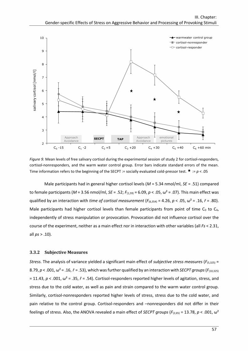
III. Chapter: Gender-specific Effects of Stress on Aggressive Behavior and Processing of Provoking Stimuli
57
Figure 9. Mean levels of free salivary cortisol during the experimental session of study 2 for cortisol-responders,
cortisol-nonresponders, and the warm water control group. Error bars indicate standard errors of the mean.
Time information refers to the beginning of the SECPT := socially evaluated cold-pressor test. := p < .05
Male participants had in general higher cortisol levels (M = 5.34 nmol/ml, SE = .51) compared
to female participants (M = 3.56 nmol/ml, SE = .52; F(1,59) = 6.09, p < .05, ω² = .07). This main effect was
qualified by an interaction with time of cortisol measurement (F(6,354) = 4.26, p < .05, ω² = .16, r̄ = .80).
Male participants had higher cortisol levels than female participants from point of time C0 to C4,
independently of stress manipulation or provocation. Provocation did not influence cortisol over the
course of the experiment, neither as a main effect nor in interaction with other variables (all Fs < 2.31,
all ps > .10).
3.3.2 Subjective Measures
Stress. The analysis of variance yielded a significant main effect of subjective stress measures (F(5,325) =
8.79, p < .001, ω² = .16, r̄ = .53), which was further qualified by an interaction with SECPT groups (F(10,325)
= 11.43, p < .001, ω² = .35, r̄ = .54). Cortisol-responders reported higher levels of agitation, stress, and
stress due to the cold water, as well as pain and strain compared to the warm water control group.
Similarly, cortisol-nonresponders reported higher levels of stress, stress due to the cold water, and
pain relative to the control group. Cortisol-responders and –nonresponders did not differ in their
feelings of stress. Also, the ANOVA revealed a main effect of SECPT groups (F(2,65) = 13.78, p < .001, ω²
2
3
4
5
6
7
8
9
10
C0 -15 C1 -5 C2 +5 C3 +20 C4 +30 C5 +40 C6 +60 min
salivary
cort
isol [n
mol/
l]warmwater control group
cortisol-nonresponder
cortisol-responder
SECPT TAPemotional pictures
Approach Avoidance
Approach Avoidance
C0 -15 C1 -2 C2 +5 C3 +20 C4 +30 C5 +40 C6 +60 min
III. Chapter: Gender-specific Effects of Stress on Aggressive Behavior and Processing of Provoking Stimuli
58
= .26), as well as a two-way interaction between gender and SECPT groups (F(2,65) = 4.34, p < .05, ω² =
.09). Female cortisol-responders and –nonresponders reported higher levels of stress relative to
female participants of the warm water control group. In contrast, male participants of the three SECPT
groups did not differ significantly in their reported stress.
Mood.
Good versus bad mood. The ANOVA showed a significant main effect of time of subjective
MDBF mood measures (F(1,58) = 12.52, p < .001, ω² = .06, r̄ = .60). Participants reported worse temper
after the TAP compared to beforehand. Besides a significant main effect of the covariate baseline good
versus bad mood (F(1,58) = 44.95, p < .001, ω² = .38), the ANOVA revealed main effects of SECPT groups
(F(2,58) = 4.85, p < .05, ω² = .10) and provocation (F(1,58) = 8.86, p < .01, ω² = .10), as well as a two way
interaction between these variables (F(2,58) = 7.16, p < .01, ω² = .15), which was further qualified by a
three-way interaction between gender, SECPT groups, and provocation (F(2,58) = 4.72, p < .05, ω² = .09).
Descriptively, highly provoked female cortisol-responders reported the lowest level of good mood.
Post-hoc tests confirmed this, revealing significant comparison between this group and mildly
provoked female cortisol-responders, as well as highly provoked male cortisol-responders. Within
male participants, only highly provoked participants of the warm water control group reported less
good mood compared to mildly provoked participants of this group, while cortisol-responders and –
nonresponders did not differ in their reported temper.
Wakefulness versus sleepiness. Besides a significant main effect of the covariate baseline
wakefulness versus sleepiness (F(1,58) = 37.52, p < .001, ω² = .34), the ANOVA revealed main effects of
SECPT groups (F(2,58) = 4.25, p < .05, ω² = .08) and a two way interaction between gender and
provocation (F(1,58) = 9.71, p < .01, ω² = .11). The warm water control group reported lower levels of
wakefulness compared to cortisol-responders and –nonresponders, which did not differ from each
other. Highly provoked male participants reported higher levels of wakefulness compared to mildly
provoked men, while female participants showed descriptively the reversed pattern. Accordingly,
mildly provoked women reported higher levels of wakefulness relative to mildly provoked men.
Calmness versus restlessness. The analysis of variance yielded a main effect of the covariate
baseline calmness versus restlessness (F(1,58) = 72.39, p < .001, ω² = .50), as well as main effects of gender
(F(1,58) = 8.26, p < .01, ω² = .09), SECPT groups (F(2,58) = 5.34, p < .01, ω² = .11), and provocation (F(1,58) =
4.91, p < .05, ω² = .05) and a two way interaction between gender and SECPT groups (F(2,58) = 3.56, p <
.05, ω² = .07), which was further qualified by a three-way interaction between gender, SECPT groups,
and provocation (F(1,58) = 8.07, p < .001, ω² = .17). Descriptively, highly provoked female cortisol-
responders reported the lowest levels of calmness. Subsequent post-hoc test confirmed that this group
reported significantly lower values of calmness compared to highly provoked male cortisol-responders

III. Chapter: Gender-specific Effects of Stress on Aggressive Behavior and Processing of Provoking Stimuli
59
and mildly provoked female cortisol-responders. Highly provoked male participants of the warm water
control group reported marginally significant lower levels of calmness compared to mildly provoked
men of the control group.
Hopelessness. The ANOVA revealed, besides a main effect of the covariate baseline
hopelessness (F(1,58) = 28.47, p < .001, ω² = .28), a significant interaction between gender and SECPT
groups (F(2,58) = 3.86, p < .05, ω² = .07), which was further qualified by a three way interaction between
gender, SECPT groups, and provocation (F(2,58) = 3.42, p < .05, ω² = .06). Subsequent post-hoc tests
showed that highly provoked female cortisol-responders reported higher levels of hopelessness than
male highly provoked cortisol-responders.
Anger. The ANOVA revealed a main effect of the covariate baseline anger (F(1,58) = 15.81, p <
.001, ω² = .17), a main effect of SECPT groups (F(2,58) = 4.33, p < .05, ω² = .09) and provocation (F(1,58) =
4.37, p < .05, ω² = .05), as well as a significant interaction between these two variables (F(2,58) = 5.01, p
< .01, ω² = .10). Highly provoked cortisol-responders reported higher levels of anger relative to mildly
provoked cortisol-responders, highly provoked cortisol-nonresponders and participants of the warm
water control group.
3.3.3 Aggressive Behavior in the Taylor Aggression Paradigm
3.3.3.1 Effects of Provocation
As intended, participants responded to high provocation with generally more aggressive behavior (M
= 2.88, SE = .19) compared to those of the low provocation group (M = 1.84, SE = .19) (F(1,59) = 15.05, p
< .001, ω² = .17), independent of type of aggressive behavior (i.e., noise duration or volume settings).
Moreover, the analysis revealed a main effect of Tap Block (F(2,118) = 16.73, p < .001, ω² = .31, r̄ = .68),
which was qualified by an interaction of TAP Block and provocation (F(2,118) = 27.95, p < .001, ω² = .50,
r̄ = .75). Descriptively, participants who were subjected to increasing provocation in TAP Block 2 and 3
showed more aggressive behavior, while their behavior did not differ from mildly provoked
participants in TAP Block 1. (TAP Block 1: high provocation: M = 1.92, SE = .14; low provocation: M =
2.00, SE = .14; TAP Block 2: high provocation: M = 2.93, SE = .20; low provocation: M = 1.78, SE = .20;
TAP Block 3: high provocation: M = 3.79, SE = .30; low provocation: M = 1.76, SE = .20). Post-hoc tests
showed that high provocation led to more aggressive behavior in TAP Block 2 and 3 compared to low
provocation. Moreover, participants responded to high provocation with increasing aggressive
behavior over all TAP Blocks.

III. Chapter: Gender-specific Effects of Stress on Aggressive Behavior and Processing of Provoking Stimuli
60
3.3.3.2 Effects of Overt and Covert Aggressive Behavior- Duration and Volume Settings
Female and male participants differed in their type of aggressive behavior as a function of provocation,
TAP Block, and interaction of both. The analysis of variance revealed a significant three-way interaction
of type of aggressive behavior, gender, and provocation (F(1,59) = 7.11, p < .01, ω² = .18, r̄ = .81), as well
as a four way interaction between type of aggressive behavior, TAP Block, gender, and provocation
(F(2,118) = 6.75, p < .05, ω² = .08, r̄ = .71). As depicted in Figure 10, post-hoc tests showed that male
participants showed less covert aggressive behavior (i.e., noise duration settings) in of the TAP Block 2
and 3 than women.
Figure 10. Mean overt and covert aggressive behavior (i.e., mean of noise duration vs. mean noise volume
settings) over the three blocks of the Taylor Aggression Paradigm (TAP) in male and female participants, exposed
to either low or high provocation. Values are means ± SE. Different letters indicate significant difference between
men and women, brackets indicate significant differences within men or women. p < .05. Note: range of possible
settings was 0-10.
Female participants who were highly provoked acted more aggressively with both covert and overt
aggressive behavior in TAP Block 2 and 3 compared to female participants of the low provocation group
(Figure 10, right panel). In contrast, men who were highly provoked showed more overt aggressive
behavior in TAP block 2 and 3 compared to mildly provoked male participants (Figure 10, left panel),
while covert aggressive behavior only increased in block 3. Moreover, male participants reacted to
high provocation with significantly more overt aggression than covert aggressive behavior in TAP Block
2 and 3, while mildly provoked males used similar amounts (Figure 10, left panel). Female participants,
however, did not differ in the type of aggressive behavior used in response to high or low provocation.
0
1
2
3
4
5
6
7
1 2 3 1 2 3
Duration Volume
me
an a
ggre
ssiv
e b
eh
avio
r
Men
0
1
2
3
4
5
6
7
1 2 3 1 2 3
Duration Volume
me
an a
ggre
ssiv
e b
eh
avio
r
Women high provocation
low provocation
a a a a b b b b

III. Chapter: Gender-specific Effects of Stress on Aggressive Behavior and Processing of Provoking Stimuli
61
Effects of stress and stress-induced cortisol on aggressive behavior. Most interestingly, the
analysis revealed a significant five-way interaction between type of aggressive behavior, TAP Block,
gender, provocation, and SECPT groups (F(4,118) = 2.75, p < .05, ω² = .07, r̄ = .80). Figure 11 shows mean
values of covert (i.e., mean noise duration settings) and overt (i.e., mean noise volume settings)
aggressive behavior for female and male participants of the warm water group, cortisol-
nonresponders, and cortisol-responders in TAP block 1 to 3, comparing low versus high provocation.
Figure 12 shows the same data, albeit sorted by the three SECPT groups, separated for gender and
provocation.
Descriptively, enhanced overt and covert aggressive behavior in response to high provocation
was particularly pronounced in female participants of the warm water control group and female
cortisol-responders (cf. Figure 11, right panel). On the other hand, male participants of these two
groups showed by trend less aggressive behavior, particularly with regard of covert aggressive
behavior (cf. Figure 11, left panel). In contrast, cortisol-nonresponders showed less overt aggressive
behavior in response to high provocation. No typical provocation pattern with increased settings was
found for covert aggression in this group (cf. Figure 12, upper graphs). Post-hoc tests showed that
female participants of the warm water control group reacted to high provocation in TAP Block 2 and 3
with increasing overt and covert aggressive behavior, while male participants of this SEPCT group
showed this pattern only in overt aggressive behavior (see Figure 11, upper graphs of left and right
panels).
Female cortisol-nonresponders reacted less aggressively and overt and covert aggressive
behavior only exceeded the response of mildly provoked female cortisol-nonresponders in TAP Block
3 (see Figure 11, middle left graphs of right panel). Male cortisol-nonresponders, in contrast, only
showed increased overt aggressive behavior to high provocation in TAP Block 3 compared to low
provocation, while the reversed pattern was found in TAP Block 1 (see Figure 11, middle left graphs of
left panel). Highly provoked female and male cortisol-responders showed increased overt and covert
aggression in TAP Block 2 and 3 compared to mildly provoked participants, with similar amount of
aggressive behavior as the warm water control group (see Figure 11, bottom graphs of left and right
panels). Moreover, highly provoked female cortisol-nonresponders showed less covert (TAP Block 2
and 3) and overt aggressive behavior (TAP Block 3) than women in the warm water group and female
cortisol-responders (Figure 12, upper right graph).

III. Chapter: Gender-specific Effects of Stress on Aggressive Behavior and Processing of Provoking Stimuli
62
Highly provoked male participants of the three SECPT groups did not differ in their amount of
aggressive behavior, except for male cortisol-nonresponders who showed less covert aggressive
behavior in TAP Block 1 compared to male participants of the warm water group (Figure 12, upper left
graph). Mildly provoked male cortisol-responders showed less covert aggressive behavior than male
cortisol-nonresponders (TAP Block 2) and male participants of the warm water control group (TAP
Block 3) (Figure 12, lower left graph).
Figure 11. Mean overt and covert aggressive behavior, i.e., mean of noise duration vs. mean noise volume
settings, over the three blocks of the Taylor Aggression Paradigm (TAP) in male and female participants exposed
to either low or high provocation separated for the three SECPT groups (warm water control group, cortisol-
nonresponders, cortisol-responders). Values are means ± SE. Different letters indicate significant difference
between men and women, brackets indicate significant differences within men or women. p < .05. Note: range
of possible settings was 0-10.
0
1
2
3
4
5
6
7
1 2 3 1 2 3
Duration Volume
me
anag
gre
ssiv
e b
eh
avio
r
Men - Warm Water Control Group
0
1
2
3
4
5
6
7
1 2 3 1 2 3
Duration Volume
me
anag
gre
ssiv
e b
eh
avio
r
Men - Cortisol-Nonresponder
0
1
2
3
4
5
6
7
1 2 3 1 2 3
Duration Volume
me
anag
gre
ssiv
e b
eh
avio
r
Men - Cortisol-Responder
0
1
2
3
4
5
6
7
1 2 3 1 2 3
Duration Volume
me
anag
gre
ssiv
e b
eh
avio
r
Women - Warm Water Control Grouphigh provocation
low provocation
0
1
2
3
4
5
6
7
1 2 3 1 2 3
Duration Volume
me
anag
gre
ssiv
e b
eh
avio
r
Women - Cortisol-Nonresponder
0
1
2
3
4
5
6
7
1 2 3 1 2 3
Duration Volume
me
anag
gre
ssiv
e b
eh
avio
r
Women - Cortisol-Responder
a a a a b b b b
a b
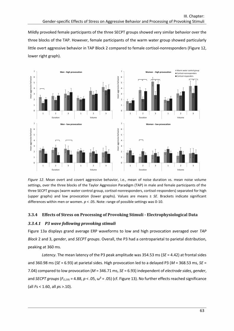
III. Chapter: Gender-specific Effects of Stress on Aggressive Behavior and Processing of Provoking Stimuli
63
Mildly provoked female participants of the three SECPT groups showed very similar behavior over the
three blocks of the TAP. However, female participants of the warm water group showed particularly
little overt aggressive behavior in TAP Block 2 compared to female cortisol-nonresponders (Figure 12,
lower right graph).
Figure 12. Mean overt and covert aggressive behavior, i.e., mean of noise duration vs. mean noise volume
settings, over the three blocks of the Taylor Aggression Paradigm (TAP) in male and female participants of the
three SECPT groups (warm water control group, cortisol-nonresponders, cortisol-responders) separated for high
(upper graphs) and low provocation (lower graphs). Values are means ± SE. Brackets indicate significant
differences within men or women. p < .05. Note: range of possible settings was 0-10.
3.3.4 Effects of Stress on Processing of Provoking Stimuli - Electrophysiological Data
3.3.4.1 P3 wave following provoking stimuli
Figure 13a displays grand average ERP waveforms to low and high provocation averaged over TAP
Block 2 and 3, gender, and SECPT groups. Overall, the P3 had a centroparietal to parietal distribution,
peaking at 360 ms.
Latency. The mean latency of the P3 peak amplitude was 354.53 ms (SE = 4.42) at frontal sides
and 360.98 ms (SE = 6.93) at parietal sides. High provocation led to a delayed P3 (M = 368.53 ms, SE =
7.04) compared to low provocation (M = 346.71 ms, SE = 6.93) independent of electrode sides, gender,
and SECPT groups (F(1,59) = 4.88, p < .05, ω² = .05) (cf. Figure 13). No further effects reached significance
(all Fs < 1.60, all ps >.10).
0
1
2
3
4
5
6
7
1 2 3 1 2 3
Duration Volume
mea
n a
ggre
ssiv
e b
ehav
ior
Men - high provocation
0
1
2
3
4
5
6
7
1 2 3 1 2 3
Duration Volume
mea
n a
ggre
ssiv
e b
ehav
ior
Men - low provocation
0
1
2
3
4
5
6
7
1 2 3 1 2 3
Duration Volumem
ean
agg
ress
ive
beh
avio
r
Women - high provocationWarm water control group
Cortisol-nonresponders
Cortisol-responders
0
1
2
3
4
5
6
7
1 2 3 1 2 3
Duration Volume
mea
n a
ggre
ssiv
e b
ehav
ior
Women - low provocation

III. Chapter: Gender-specific Effects of Stress on Aggressive Behavior and Processing of Provoking Stimuli
64
Figure 13. (a) Grand average ERP waveforms for the provoking stimuli for high (n=35) versus low provocation
(n=36) averaged over trials of TAP Block 2 and 3 at frontal (FFC3, FFCz, FFC4) and parietal sides (CPP3, CPPz,
CPP4). (b) Difference map high – low provocation for the time domain of P3.
Peak. The analysis revealed a main effect of hemisphere (F(2,118) = 15.49, p < .001, ω² = .52, r̄ =
.87), which was qualified by an interaction between hemisphere and provocation (F(2,118) = 5.13, p < .01,
ω² = .26, r̄ = .89). Subsequent post-hoc tests showed that the P3 amplitude was more positive at central
electrode sides (M = 5.69 µV, SE = .50) compared to the right (M = 4.78 µV, SE = .43) and the left
hemisphere (M = 4.67 µV, SE = .46), which did not differ. Participants who were subjected to high
provocation showed a more positive P3 at central and left sides compared to participants who were
mildly provoked (see Figure 13a, b, Table 3). Moreover, P3 amplitudes were more positive at parietal
sides (M = 6.02 µV, SE = .49) compared to frontal sides (M = 4.08 µV, SE = .48, F(1,59) = 26.28, p < .001,
ω² = .33, r̄ = .64). This main effect was further qualified by an interaction with SECPT groups (F(2,59) =
3.50, p < .05, ω² = .09, r̄ = .65), with gender and SECPT groups (F(2,59) = 3.30, p < .05, ω² = .09, r̄ = .69),
and most important by a four-way interaction with SECPT groups, gender, and provocation (F(2,59) =
3.24, p < .05, ω² = .12, r̄ = .77). Figure 14 and Figure 15 display ERP waveforms and P3 amplitudes,
respectively, at frontal and parietal sites for highly and mildly provoked male and female participants
of each of the three SECPT groups. Figure 16 depicts corresponding difference maps high – low-
provocation for the time domain of the P3.
FFC3
0 500
-1
0
1
2
3
4
5
6
7
8
FFCz
0 500
-1
0
1
2
3
4
5
6
7
8
FFC4
0 500
-1
0
1
2
3
4
5
6
7
8
CPPz
0 500
-1
0
1
2
3
4
5
6
7
8
CPP4
0 500
-1
0
1
2
3
4
5
6
7
8
CPP3
0 500
-1
0
1
2
3
4
5
6
7
8
High provocation
Low provocation
P3
-2.5 0 2.5 µV
310 ms - 410 ms
high provocation
–
low provocation
a
b
ms
µV

III. Chapter: Gender-specific Effects of Stress on Aggressive Behavior and Processing of Provoking Stimuli
65
Figure 14. (a) Grand average ERP waveforms to the provoking stimuli averaged over trials of Tap Block 2 and 3
and (b) head maps for the time domain of the P37 for highly and mildly provoked male and female participants
of each of the three SECPT groups (i.e., warm water control group, cortisol – nonresponders, cortisol –
responders) at frontal (FFC) and parietal (CPP) sites.
7 Precise start and end point of 105 ms time domain of P3 for each group with respect to P3 peak latency: warm water group: male 320 - 425 ms, female 305 - 410 ms; cortisol-nonresponder: male 300 - 400 ms, female 315 - 420 ms; cortisol-responder: male 305 - 410 ms, female 300 - 405 ms.
FFC
CPP
-200 0 200 400 600 800
0
2
4
6
8
10
-200 0 200 400 600 800
0
2
4
6
8
10
µV
ms
MenFFC
CPP-200 0 200 400 600 800
0
2
4
6
8
10
-200 0 200 400 600 800
0
2
4
6
8
10
µV
ms
Women
Warm water control group – low provocationWarm water control group – high provocationCortisol-nonresponders – low provocationCortisol-nonresponders – high provocationCortisol-responders – low provocationCortisol-responders – high provocation
a
high provocation low provocation high provocation low provocation
Co
rtis
ol-
no
nre
spo
nd
ers
War
m w
ater
co
ntr
ol g
rou
pC
ort
iso
l-re
spo
nd
ers
Men Women
-8 µV 0 µV 8 µV
305 ms - 410 ms
-8 µV 0 µV 8 µV
305 ms - 410 ms
-8 µV 0 µV 8 µV
315 ms - 420 ms
-8 µV 0 µV 8 µV
315 ms - 420 ms
-8 µV 0 µV 8 µV
300 ms - 405 ms
-8 µV 0 µV 8 µV
300 ms - 405 ms
-8 µV 0 µV 8 µV
320 ms - 425 ms
-8 µV 0 µV 8 µV
320 ms - 425 ms
-8 µV 0 µV 8 µV
300 ms - 405 ms
-8 µV 0 µV 8 µV
305 ms - 410 ms
-8 µV 0 µV 8 µV
305 ms - 410 ms
-8 µV 0 µV 8 µV
305 ms - 410 ms
-8 0 8 µV
b

III. Chapter: Gender-specific Effects of Stress on Aggressive Behavior and Processing of Provoking Stimuli
66
Descriptively, highly provoked male participants of the warm water control group and male cortisol-
nonresponders had more positive frontal P3 amplitude than those male participants who were
subjected to low provocation (cf. Figure 14, left graph; Figure 15, left upper graph; Figure 16, left
panel). Male cortisol-responders, however, showed the reversed pattern with less positive P3
amplitude in response to high provocation compared to low provocation and highly provoked male
participants of the other two SECPT groups (see Figure 15, left upper graph). The parietal P3 was more
positive in male participants of the warm water control group in response to high provocation, while
male cortisol-nonresponders did not differ regarding the amount of provocation. Again, male cortisol-
responders showed the reversed pattern with less positive parietal P3 amplitude in the case of high
compared to low provocation (Figure 15, left lower graph).
Figure 15. Mean frontal (upper row) and parietal (lower row) P3 amplitudes (µV) locked to the provoking stimuli,
averaged over block 2 and 3 of the Taylor Aggression Paradigm (TAP) for highly and mildly provoked male and
female participants of the three SECPT groups (warm water control group, cortisol-nonresponders, cortisol-
responders). Values are means ± SE. Brackets indicate significant differences. p < .05.
-2
0
2
4
6
8
10
12
Warm watercontrol group
Cortisol -nonresponder
Cortisol -responder
fro
nta
l P
3 A
mp
litu
de
µV
Men
-2
0
2
4
6
8
10
12
Warm watercontrol group
Cortisol -nonresponder
Cortisol -responder
par
ieta
l P
3 A
mp
litu
de
µV
Men
-2
0
2
4
6
8
10
12
Warm watercontrol group
Cortisol -nonresponder
Cortisol -responder
fro
nta
l P
3 A
mp
litu
de
µV
Women
high provocation
low provocation
-2
0
2
4
6
8
10
12
Warm watercontrol group
Cortisol -nonresponder
Cortisol -responder
par
ieta
l P
3 A
mp
litu
de
µV
Women
CP
PFF
C

III. Chapter: Gender-specific Effects of Stress on Aggressive Behavior and Processing of Provoking Stimuli
67
Female participants showed a distinct different pattern throughout all SECPT groups (cf. Figure 14,
right graph; Figure 15, right panel; Figure 16, right panel). Female participants of the warm water group
and female cortisol-nonresponders did not differ between high and low provocation at frontal sides.
In contrast, highly provoked female cortisol-responders showed the expected pattern with more
positive frontal P3 amplitudes compared to mildly provoked female cortisol-responders (Figure 15,
right upper graph). Parietal P3 amplitudes were more positive in highly provoked female cortisol-
nonresponders compared to mildly provoked ones. Female cortisol-responders showed the same
tendency, but less marked. Female participants of the warm water group, however, showed the
reversed pattern with less positive parietal P3 amplitudes in highly provoked compared to mildly
provoked participants (Figure 15, right lower graph).
Figure 16. Difference maps high – low provocation for the time domain of P38 for the provoking stimuli for male
(left panel) and female (right panel) of each of the three SECPT groups (i.e., warm water control group, cortisol
– nonresponders, cortisol – responders).
8 Precise start and end point of 105 ms time domain of P3 for each group with respect to P3 peak latency: warm water group: male 320 - 425 ms, female 305 - 410 ms; cortisol-nonresponder: male 300 - 400 ms, female 315 - 420 ms; cortisol-responder: male 305 - 410 ms, female 300 - 405 ms.
Co
rtis
ol-
no
nre
spo
nd
ers
War
m w
ater
con
tro
lgro
up
Co
rtis
ol-
resp
on
der
s
-4 µV 0 µV 4 µV
305 ms - 410 ms
-4 µV 0 µV 4 µV
320 ms - 425 ms
-4 µV 0 µV 4 µV
300 ms - 400 ms
-4 µV 0 µV 4 µV
315 ms - 420 ms
-4 µV 0 µV 4 µV
300 ms - 405 ms
-4 µV 0 µV 4 µV
305 ms - 410 msMen Women-4 0 4 µV
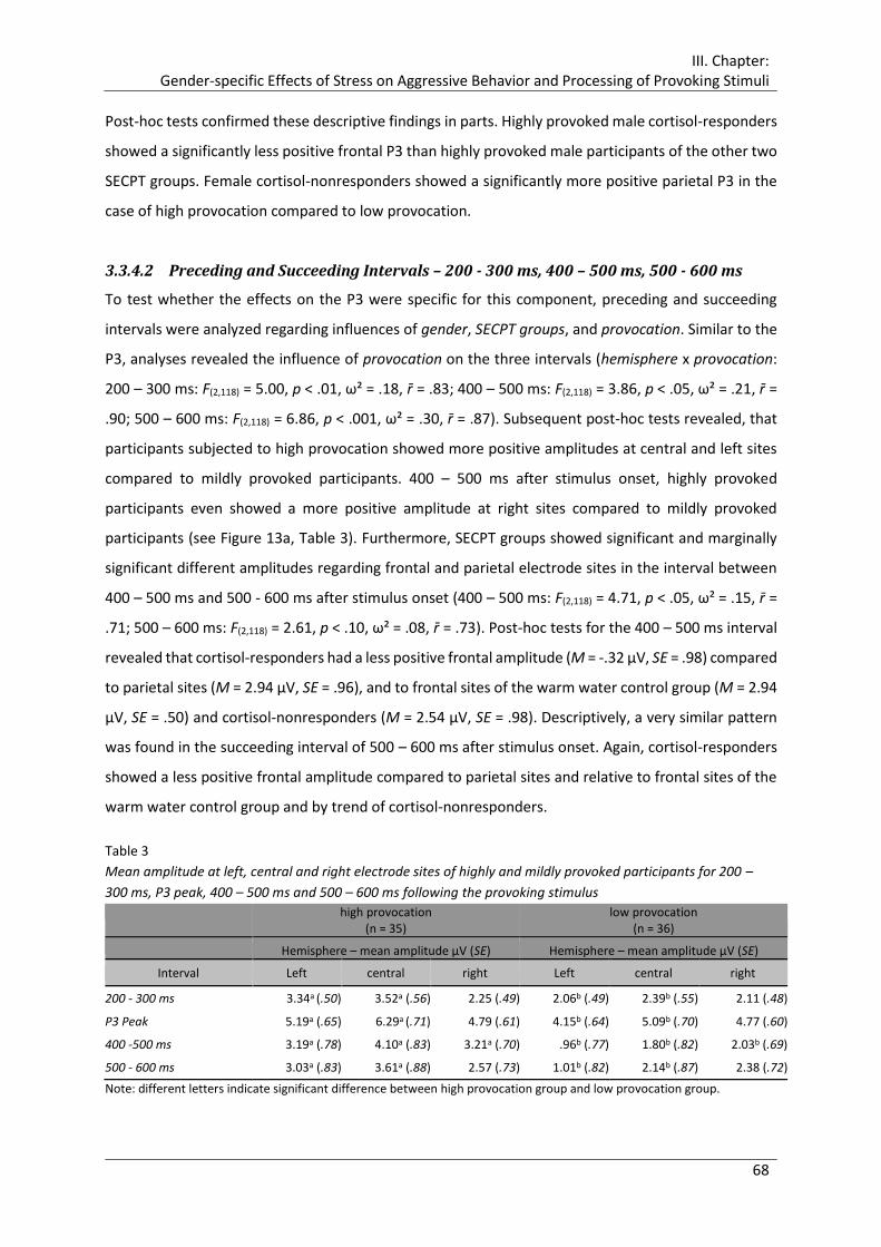
III. Chapter: Gender-specific Effects of Stress on Aggressive Behavior and Processing of Provoking Stimuli
68
Post-hoc tests confirmed these descriptive findings in parts. Highly provoked male cortisol-responders
showed a significantly less positive frontal P3 than highly provoked male participants of the other two
SECPT groups. Female cortisol-nonresponders showed a significantly more positive parietal P3 in the
case of high provocation compared to low provocation.
3.3.4.2 Preceding and Succeeding Intervals – 200 - 300 ms, 400 – 500 ms, 500 - 600 ms
To test whether the effects on the P3 were specific for this component, preceding and succeeding
intervals were analyzed regarding influences of gender, SECPT groups, and provocation. Similar to the
P3, analyses revealed the influence of provocation on the three intervals (hemisphere x provocation:
200 – 300 ms: F(2,118) = 5.00, p < .01, ω² = .18, r̄ = .83; 400 – 500 ms: F(2,118) = 3.86, p < .05, ω² = .21, r̄ =
.90; 500 – 600 ms: F(2,118) = 6.86, p < .001, ω² = .30, r̄ = .87). Subsequent post-hoc tests revealed, that
participants subjected to high provocation showed more positive amplitudes at central and left sites
compared to mildly provoked participants. 400 – 500 ms after stimulus onset, highly provoked
participants even showed a more positive amplitude at right sites compared to mildly provoked
participants (see Figure 13a, Table 3). Furthermore, SECPT groups showed significant and marginally
significant different amplitudes regarding frontal and parietal electrode sites in the interval between
400 – 500 ms and 500 - 600 ms after stimulus onset (400 – 500 ms: F(2,118) = 4.71, p < .05, ω² = .15, r̄ =
.71; 500 – 600 ms: F(2,118) = 2.61, p < .10, ω² = .08, r̄ = .73). Post-hoc tests for the 400 – 500 ms interval
revealed that cortisol-responders had a less positive frontal amplitude (M = -.32 µV, SE = .98) compared
to parietal sites (M = 2.94 µV, SE = .96), and to frontal sites of the warm water control group (M = 2.94
µV, SE = .50) and cortisol-nonresponders (M = 2.54 µV, SE = .98). Descriptively, a very similar pattern
was found in the succeeding interval of 500 – 600 ms after stimulus onset. Again, cortisol-responders
showed a less positive frontal amplitude compared to parietal sites and relative to frontal sites of the
warm water control group and by trend of cortisol-nonresponders.
Table 3
Mean amplitude at left, central and right electrode sites of highly and mildly provoked participants for 200 –
300 ms, P3 peak, 400 – 500 ms and 500 – 600 ms following the provoking stimulus
high provocation (n = 35)
low provocation (n = 36)
Hemisphere – mean amplitude µV (SE) Hemisphere – mean amplitude µV (SE)
Interval Left central right Left central right
200 - 300 ms 3.34a (.50) 3.52a (.56) 2.25 (.49) 2.06b (.49) 2.39b (.55) 2.11 (.48)
P3 Peak 5.19a (.65) 6.29a (.71) 4.79 (.61) 4.15b (.64) 5.09b (.70) 4.77 (.60)
400 -500 ms 3.19a (.78) 4.10a (.83) 3.21a (.70) .96b (.77) 1.80b (.82) 2.03b (.69)
500 - 600 ms 3.03a (.83) 3.61a (.88) 2.57 (.73) 1.01b (.82) 2.14b (.87) 2.38 (.72)
Note: different letters indicate significant difference between high provocation group and low provocation group.
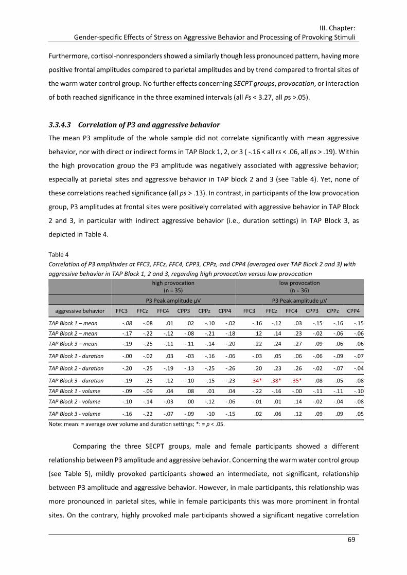
III. Chapter: Gender-specific Effects of Stress on Aggressive Behavior and Processing of Provoking Stimuli
69
Furthermore, cortisol-nonresponders showed a similarly though less pronounced pattern, having more
positive frontal amplitudes compared to parietal amplitudes and by trend compared to frontal sites of
the warm water control group. No further effects concerning SECPT groups, provocation, or interaction
of both reached significance in the three examined intervals (all Fs < 3.27, all ps >.05).
3.3.4.3 Correlation of P3 and aggressive behavior
The mean P3 amplitude of the whole sample did not correlate significantly with mean aggressive
behavior, nor with direct or indirect forms in TAP Block 1, 2, or 3 ( -.16 < all rs < .06, all ps > .19). Within
the high provocation group the P3 amplitude was negatively associated with aggressive behavior;
especially at parietal sites and aggressive behavior in TAP block 2 and 3 (see Table 4). Yet, none of
these correlations reached significance (all ps > .13). In contrast, in participants of the low provocation
group, P3 amplitudes at frontal sites were positively correlated with aggressive behavior in TAP Block
2 and 3, in particular with indirect aggressive behavior (i.e., duration settings) in TAP Block 3, as
depicted in Table 4.
Table 4
Correlation of P3 amplitudes at FFC3, FFCz, FFC4, CPP3, CPPz, and CPP4 (averaged over TAP Block 2 and 3) with
aggressive behavior in TAP Block 1, 2 and 3, regarding high provocation versus low provocation
high provocation (n = 35)
low provocation (n = 36)
P3 Peak amplitude µV P3 Peak amplitude µV
aggressive behavior FFC3 FFCz FFC4 CPP3 CPPz CPP4 FFC3 FFCz FFC4 CPP3 CPPz CPP4
TAP Block 1 – mean -.08 -.08 .01 .02 -.10 -.02 -.16 -.12 .03 -.15 -.16 -.15
TAP Block 2 – mean -.17 -.22 -.12 -.08 -.21 -.18 .12 .14 .23 -.02 -.06 -.06
TAP Block 3 – mean -.19 -.25 -.11 -.11 -.14 -.20 .22 .24 .27 .09 .06 .06
TAP Block 1 - duration -.00 -.02 .03 -03 -.16 -.06 -.03 .05 .06 -.06 -.09 -.07
TAP Block 2 - duration -.20 -.25 -.19 -.13 -.25 -.26 .20 .23 .26 -.02 -.07 -.04
TAP Block 3 - duration -.19 -.25 -.12 -.10 -.15 -.23 .34* .38* .35* .08 -.05 -.08
TAP Block 1 - volume -.09 -.09 .04 .08 .01 .04 -.22 -.16 -.00 -.11 -.11 -.10
TAP Block 2 - volume -.10 -.14 -.03 .00 -.12 -.06 -.01 .01 .14 -.02 -.04 -.08
TAP Block 3 - volume -.16 -.22 -.07 -.09 -10 -.15 .02 .06 .12 .09 .09 .05
Note: mean: = average over volume and duration settings; *: = p < .05.
Comparing the three SECPT groups, male and female participants showed a different
relationship between P3 amplitude and aggressive behavior. Concerning the warm water control group
(see Table 5), mildly provoked participants showed an intermediate, not significant, relationship
between P3 amplitude and aggressive behavior. However, in male participants, this relationship was
more pronounced in parietal sites, while in female participants this was more prominent in frontal
sites. On the contrary, highly provoked male participants showed a significant negative correlation
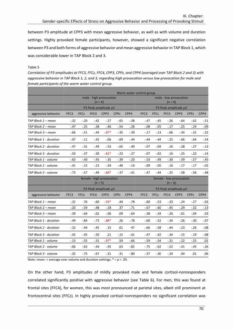
III. Chapter: Gender-specific Effects of Stress on Aggressive Behavior and Processing of Provoking Stimuli
70
between P3 amplitude at CPP3 with mean aggressive behavior, as well as with volume and duration
settings. Highly provoked female participants, however, showed a significant negative correlation
between P3 and both forms of aggressive behavior and mean aggressive behavior in TAP Block 1, which
was considerable lower in TAP Block 2 and 3.
Table 5
Correlation of P3 amplitudes at FFC3, FFCz, FFC4, CPP3, CPPz, and CPP4 (averaged over TAP Block 2 and 3) with
aggressive behavior in TAP Block 1, 2, and 3, regarding high provocation versus low provocation for male and
female participants of the warm water control group.
Warm water control group
male - high provocation (n = 6)
male - low provocation (n = 6)
P3 Peak amplitude µV P3 Peak amplitude µV
aggressive behavior FFC3 FFCz FFC4 CPP3 CPPz CPP4 FFC3 FFCz FFC4 CPP3 CPPz CPP4
TAP Block 1 – mean -.32 -.20 -.42 -.27 -.65 -.38 -.47 -.45 -.26 -.64 -.62 -.51
TAP Block 2 – mean -.47 -.25 -.38 -.46 -.54 -.28 -.08 -.05 -.17 -.25 -.24 -.09
TAP Block 3 – mean -.66 -.51 -.44 -.87* -.35 -.39 -.17 -.13 -.06 -.34 -.31 -.22
TAP Block 1 - duration -.07 -.11 -.42 -.06 -.69 -.44 -.44 -.44 -.25 -.66 -.64 -.54
TAP Block 2 - duration -.47 -.31 -.49 -.53 -.65 -.40 -.07 -.04 .16 -.28 -.27 -.13
TAP Block 3 - duration -.50 -.37 -.30 -.81* -.23 -.27 -.07 -.02 .16 -.25 -.22 -.14
TAP Block 1 - volume -.63 -.40 -.45 -.35 -.39 -.20 -.53 -.49 -.30 -.59 -.57 -.45
TAP Block 2 - volume -.41 -.15 -.23 -.34 -.40 -.14 -.09 -.05 .16 -.17 -.17 -.02
TAP Block 3 - volume -.73 -.57 -.49 -.84* -.37 -.41 -.47 -.44 -.25 -.58 -.56 -.48
female - high provocation (n = 5)
female - low provocation (n = 6)
P3 Peak amplitude µV P3 Peak amplitude µV
aggressive behavior FFC3 FFCz FFC4 CPP3 CPPz CPP4 FFC3 FFCz FFC4 CPP3 CPPz CPP4
TAP Block 1 – mean -.31 -76 -.60 -.93* .44 -.78 -.60 -.53 -.33 -.24 -.27 -.03
TAP Block 2 – mean -.20 -.59 -.48 -.18 .37 -.71 -.67 -.60 -.45 -.29 -.32 -.13
TAP Block 3 – mean -.39 -.64 -.62 -.06 .09 -.64 -.38 -.34 -.26 -.01 -.04 .03
TAP Block 1 - duration -.49 -.84 -.73 -.88* .26 -.78 -.60 -.52 -.34 -.26 -.30 -.07
TAP Block 2 - duration -.32 -.44 -.45 .15 .01 -47 -.66 -.58 -.44 -.23 -.26 -.08
TAP Block 3 - duration -.42 -.45 -.50 .21 -.15 -.41 -.47 -.42 -.34 -.15 -.19 -.08
TAP Block 1 - volume -.13 -.55 -.31 -.97* .59 -.66 -.59 -.54 -.31 -.22 -.25 .01
TAP Block 2 - volume -.06 -.63 -.44 -.45 .63 -.82 -.75 -.62 -.52 -.41 -.45 -.26
TAP Block 3 - volume -.32 -.75 -.67 -.31 .31 -.80 -.37 -.30 -.24 .04 .01 .06
Note: mean := average over volume and duration settings; * = p < .05.
On the other hand, P3 amplitudes of mildly provoked male and female cortisol-nonresponders
correlated significantly positive with aggressive behavior (see Table 6). For men, this was found at
frontal sites (FFC4), for women, this was most pronounced at parietal sites, albeit still prominent at
frontocentral sites (FFCz). In highly provoked cortisol-nonresponders no significant correlation was

III. Chapter: Gender-specific Effects of Stress on Aggressive Behavior and Processing of Provoking Stimuli
71
revealed. While highly provoked male cortisol-nonresponders still showed a weaker positive
correlation, highly provoked female participants showed even a negative relationship, albeit not
significant.
Table 6
Correlation of P3 amplitudes at FFC3, FFCz, FFC4, CPP3, CPPz, and CPP4 (averaged over Tap Block 2 and 3) with
aggressive behavior in TAP Block 1, 2 and 3, regarding high provocation versus low provocation for male and
female cortisol-nonresponders
Cortisol-nonresponders
male - high provocation (n = 6)
male - low provocation (n = 5)
P3 Peak amplitude µV P3 Peak amplitude µV
aggressive behavior FFC3 FFCz FFC4 CPP3 CPPz CPP4 FFC3 FFCz FFC4 CPP3 CPPz CPP4
TAP Block 1 – mean .15 .17 .66 .67 .69 .52 -.62 -.50 -.51 -.47 -.30 -.29
TAP Block 2 – mean .12 .07 .31 .67 .57 .46 .72 .70 .93* .44 .40 .38
TAP Block 3 – mean .11 04 .31 .67 .57 .45 .78 .74 .95* .47 .43 .41
TAP Block 1 - duration .11 .17 .70 .57 .63 .45 -.34 -.15 -.35 -.03 .11 .11
TAP Block 2 - duration .15 .07 .32 .68 .58 .47 .66 .62 .90* .31 .31 .29
TAP Block 3 - duration .04 -.06 .28 .62 .51 .40 .72 .70 .94* .43 .42 .41
TAP Block 1 - volume .21 .22 .59 .72 .70 .55 -.57 -.35 -.54 -.18 .00 .02
TAP Block 2 - volume .11 .07 .31 .66 .56 .46 .80 .82 .98** .57 .56 .54
TAP Block 3 - volume .19 .15 .31 .65 .56 .47 .31 .51 .11 .73 .73 .70
female - high provocation (n = 7)
female - low provocation (n = 6)
P3 Peak amplitude µV P3 Peak amplitude µV
aggressive behavior FFC3 FFCz FFC4 CPP3 CPPz CPP4 FFC3 FFCz FFC4 CPP3 CPPz CPP4
TAP Block 1 – mean .01 -.12 -.17 -.01 -.21 -.21 .57 .60 .63 .82* .83* .86*
TAP Block 2 – mean -.27 -.37 -.18 -.28 -.50 -.49 .67 .74 .72 .80 .77 .81
TAP Block 3 – mean -.11 -.21 -.01 -.16 -.40 -.39 .81 .84* .76 .82* .83* .90*
TAP Block 1 - duration .15 .02 .06 -.10 -.14 -.18 .57 .57 .61 .77 .81 .85*
TAP Block 2 - duration -.44 -.47 -.21 -.47 -.64 -.66 .74 .76 .70 .87* .83* .88*
TAP Block 3 - duration -.21 -.26 .01 -.30 -.51 -.53 .81 .83* .75 .82* .85* .91*
TAP Block 1 - volume -.08 -.20 -.29 .03 -.26 -.23 .56 .58 .56 .83* .80 .80
TAP Block 2 - volume -.10 -.24 -.09 -.08 -.31 -.29 .64 .71 .65 .73 .70 .72
TAP Block 3 - volume -.02 -.16 -.02 -.04 -.29 -.27 .84* .86* .74 .83* .83* .89*
Note: mean := average over volume and duration settings; * = p < .05.
Mildly provoked cortisol-responders, on the other hand, showed a weak negative correlation, which
was more prominent in females at frontal sites (see Table 7). Highly provoked male cortisol-responders
showed a significant positive correlation for indirect aggressive behavior in TAP Block 1 at parietal sites.
High provocation reduced the negative relationship slightly in female cortisol-responders compared to
low provocation.
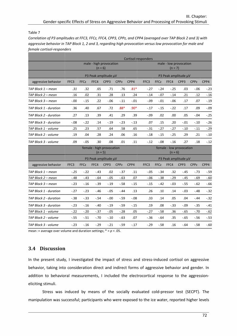
III. Chapter: Gender-specific Effects of Stress on Aggressive Behavior and Processing of Provoking Stimuli
72
Table 7
Correlation of P3 amplitudes at FFC3, FFCz, FFC4, CPP3, CPPz, and CPP4 (averaged over TAP Block 2 and 3) with
aggressive behavior in TAP Block 1, 2 and 3, regarding high provocation versus low provocation for male and
female cortisol-responders
Cortisol-responders
male - high provocation (n = 6)
male - low provocation (n = 7)
P3 Peak amplitude µV P3 Peak amplitude µV
aggressive behavior FFC3 FFCz FFC4 CPP3 CPPz CPP4 FFC3 FFCz FFC4 CPP3 CPPz CPP4
TAP Block 1 – mean .31 .32 .65 .71 .76 .81* -.27 -.24 -.25 .03 -.06 -.23
TAP Block 2 – mean .16 .02 .31 .28 .13 .24 -.14 -.07 -.14 .21 .12 -.16
TAP Block 3 – mean -.00 -.15 .22 -.06 -.11 -.01 -.09 -.01 -.06 .17 .07 -.19
TAP Block 1 - duration .36 .40 .67 .72 .88* .90* -.17 -.15 -.22 .17 .09 -.09
TAP Block 2 - duration .27 .13 .39 .41 .29 .39 -.09 .02 .00 .05 -.04 -.25
TAP Block 3 - duration -.08 -.22 .14 -.19 -.23 -.13 .07 .15 .20 -.01 -.10 -.26
TAP Block 1 - volume .25 .23 .57 .64 .58 .65 -.31 -.27 -.27 -.10 -.11 -.29
TAP Block 2 - volume .19 .04 .28 .24 .06 .16 -.18 -.15 -.25 .29 .21 -.10
TAP Block 3 - volume .09 -.05 .30 .08 .01 .11 -.12 -.08 -.16 .27 .18 -.12
female - high provocation (n = 5)
female - low provocation (n = 6)
P3 Peak amplitude µV P3 Peak amplitude µV
aggressive behavior FFC3 FFCz FFC4 CPP3 CPPz CPP4 FFC3 FFCz FFC4 CPP3 CPPz CPP4
TAP Block 1 – mean -.25 -.22 -.43 .02 -.37 .11 -.05 -.34 .32 -.45 -.73 -.59
TAP Block 2 – mean -.48 -.43 -.64 -.05 -.63 .07 -.06 -.38 -.29 -.45 -.69 -.60
TAP Block 3 – mean -.23 -.16 -.39 -.19 -.58 -.15 -.15 -.42 -.03 -.55 -.62 -.66
TAP Block 1 - duration -.27 -.23 -.46 -.05 -.44 .13 .26 .10 .14 -.03 -.48 -.32
TAP Block 2 - duration -.38 -.33 -.54 -.00 -.59 -.08 .33 .14 .05 .04 -.44 -.32
TAP Block 3 - duration -.23 -.16 -.40 -.19 -.59 -.15 .19 .08 -.33 -.09 -.35 -.41
TAP Block 1 - volume -.22 -.20 -.37 -.05 -.28 .05 -.27 -.58 .36 -.65 -.70 -.62
TAP Block 2 - volume -.55 -.51 -.70 -.10 -.63 .07 -.36 -.64 .35 -.65 -.56 -.53
TAP Block 3 - volume -.23 -.16 -.29 -.21 -.59 -.17 -.29 -.58 .16 -.64 -.58 -.60
mean := average over volume and duration settings; * = p < .05.
3.4 Discussion
In the present study, I investigated the impact of stress and stress-induced cortisol on aggressive
behavior, taking into consideration direct and indirect forms of aggressive behavior and gender. In
addition to behavioral measurements, I included the electrocortical response to the aggression-
eliciting stimuli.
Stress was induced by means of the socially evaluated cold-pressor test (SECPT). The
manipulation was successful; participants who were exposed to the ice water, reported higher levels

III. Chapter: Gender-specific Effects of Stress on Aggressive Behavior and Processing of Provoking Stimuli
73
of self-reported stress and pain compared to the warm water control group. More importantly, male
and female cortisol-responders showed a considerable increase of free cortisol in response to the
stress procedure, similar to which was found in other studies using this stressor (e.g., Lass-Hennemann
et al., 2011; Schwabe & Wolf, 2009; Smeets, 2011).
3.4.1 Aggressive Behavior - Impact of Stress and Provocation
Aggressive behavior was experimentally induced and measured with the Taylor Aggression Paradigm
(TAP). As intended, high provocation led to enhanced aggressive behavior, reduced good temper, and
higher levels of reported anger. Regarding different types of aggression, influence of gender became
apparent. When being exposed to high provocation, both male and female participants, reacted with
similar enhanced overt aggressive behavior, i.e., chose higher values for volume settings of the punitive
noise. Mildly provoked female participants showed merely descriptively less overt aggressive behavior
compared to mildly provoked men. In contrast, covert aggressive behavior in response to low
provocation was significantly reduced in female participants relative to male participants. However,
high provocation led to a distinct increase in this form of aggressive behavior for female participants,
which even exceeded the duration settings of highly provoked male participants. Thus, the present
results correspond only in parts with assumptions about gender differences in aggressive behavior and
the mediating influence of provocation (Bettencourt & Miller, 1996; Eagly, 2013; Frodi et al., 1977).
Nevertheless, other studies using retaliation paradigms with or without additional provocation
(Böhnke, Bertsch, Kruk, & Naumann, 2010; Denson, Capper et al., 2011; Hoaken, 2000; Krämer et al.,
2008; Terrell, Hill, & Nagoshi, 2008) did not find significant gender differences in (direct) aggressive
behavior either. In sum, it might be that punitive noises are more likely to elicit direct, as well as
indirect, aggressive behavior in highly provoked women compared to electro shocks, for instance, from
which women might shrink, as it actually constitutes more severe physical harm (e.g., Zeichner,
Parrott, & Frey, 2003). Consequently, this paradigm does not emphasize gender differences in the
fashion that females show more indirect and less direct aggressive behavior without provocation.
The acute psychophysiological stressor and the thereby induced increase in cortisol altered
aggressive behavior as a function of gender, provocation, TAP Block, and type of aggressive behavior.
Partly in accordance with my hypothesis, cortisol-responders showed enhanced aggressive behavior
in response to provocation relative to cortisol-nonresponders. However, only highly provoked female
cortisol-responders reacted statistically significantly more aggressively relative to highly provoked
female cortisol-nonresponders. Both female and male cortisol-responders did not differ from the
warm water control group in either the case of high or low provocation, except that mildly provoked
male cortisol-responders showed significantly less covert aggressive behavior in TAP Block 3 compared
to the warm water control group. Additionally, in contrast to the hypothesis, female cortisol-
III. Chapter: Gender-specific Effects of Stress on Aggressive Behavior and Processing of Provoking Stimuli
74
responders showed no particularly enhanced indirect aggressive behavior, nor did male cortisol-
responders show pronounced direct aggressive behavior. Quite the opposite, the latter reacted to
intermediate (TAP Block 2) and high provocation (TAP Block 3) with significantly higher indirect
aggressive behavior compared to low provocation. In male cortisol-responders, neither type of
aggressive behavior significantly exceeded the results for cortisol-nonresponders or the warm water
group, either in response to high or to low provocation. Mildly provoked male cortisol-responders even
showed, most descriptively, the lowest direct and indirect aggressive behavior compared to both other
groups. A similar pattern was found in mildly provoked female cortisol-responders. In the case of
intermediate or high provocation, however, this group showed enhanced direct and indirect aggressive
behavior compared to cortisol-nonresponders. In short, the results suggest a reinforcing impact of
stress-induced cortisol on aggressive behavior in case of high provocation, especially in females and
more pronounced for indirect forms, albeit without exceeding the amount of aggressive behavior
shown by participants without any stress.
These findings stand in contrast to preliminary findings of enhanced aggressive behavior due
to stress or cortisol. As already mentioned in the introduction, in a series of experiments Verona et al.
investigated the influence of a physical stressor on aggressive behavior in a modified teacher/learner
paradigm in healthy men and women (Verona & Curtin, 2006; Verona et al., 2006; Verona & Kilmer,
2007). Participants were told that the study aimed to investigate the influence of distraction on
supervisor and employee performance. Participants, assigned to the role of the supervisor, were
instructed to punish an alleged employee of the same sex with electric shocks in case of incorrect
responses in a digit memory task. Chosen shock settings served as measurement for aggressive
behavior. During the procedure, half of the participants were exposed to an acute physical stressor,
namely, compressed air blasts administered to their throat. They found increased aggressive behavior
in stressed men, while women responded to stress with a decrease of aggressive behavior (Verona
& Kilmer, 2007). Besides, men carrying short alleles for the serotonin transporter gene seemed to be
particularly vulnerable to enhanced stress-caused aggressive behavior (Verona et al., 2006). Moreover,
they were able to confirm their findings concerning men in parts when the stressor was realized prior
to the aggression paradigm, similar to the present experimental design, and not concurrently (Verona
& Kilmer, 2007). Böhnke, Bertsch, Kruk, and Richter et al. (2010) focused on the role of the stress
hormone cortisol in the relationship between stress and aggression. They administered either an oral
dose of 20 mg hydrocortisone or a placebo to healthy male and female adults, who were subsequently
exposed to high or low provocation in the same version of the TAP used in the present study. They
found increasing aggressive behavior over the course of the paradigm in the cortisol group, which was
independent of the amount of provocation. Unlike Verona et al., Böhnke, Bertsch, Kruk, and Richter et
al. (2010) could show that females who received exogenous cortisol reacted significantly more

III. Chapter: Gender-specific Effects of Stress on Aggressive Behavior and Processing of Provoking Stimuli
75
aggressively relative to females in the placebo group, obtaining the same level of aggressive behavior
as male participants, who did not differ in their aggressive behavior. Interestingly, the amount of
provocation again did not influence the impact of cortisol on female aggressive behavior. Thus, the
present results confirm their findings only in parts, as in the present study women with a stress-
induced increase in cortisol reacted more aggressively, but only in case of high provocation and their
behavior did not exceed the one of the warm water control group.
Several reasons can be adduced to explain these divergent findings. First, the type of stress
manipulation used by Verona et al. differed from the stress test applied in the present study (Verona
& Curtin, 2006; Verona et al., 2006; Verona & Kilmer, 2007; Verona et al., 2007). Dickerson and Kemeny
(2004) characterize uncontrollability and social-evaluative threat as necessary elements of acute
psychological stressors to induce HPA axis activation. The air blast used by Verona and colleagues,
constituted a mere physical stressor; though uncontrollable, it lacked the social-evaluative component.
Consequently, it is not clear whether the air blast actually activated the HPA axis and led to an increase
of cortisol levels, as the authors did not include cortisol measurements in their studies. Even though
physical stressors are supposed to elicit a cortisol response, Dickerson and Kemeny (2004) found no
support for this in their meta-analysis. Scarpa and Luscher (2002), for instance, reported no or only an
extremely slight increase in saliva cortisol in response to white noises (100 dB). Similarly, McRae et al.
(2006), comparing the cold-pressor test (CPT) without social evaluation with the Trier Social Stress Test
(TSST), found distinctly lower cortisol responses to the CPT relative to the TSST. Besides, the cortisol
peak in response to a stressor occurs 10 to 20 min after stressor onset (Dickerson & Kemeny, 2004;
Furlan, DeMartinis, Schweizer, Rickels, & Lucki, 2001; McRae et al., 2006; Testa et al., 1994).
Accordingly, cortisol was quite unlikely to affect aggressive behavior in the studies of Verona and
colleagues. Even if the air blast activated the HPA axis to some extent, the necessary time lag for the
cortisol peak to emerge would have fallen outside the measurement of aggressive behavior, as both
occurred simultaneously. The relevance of timing of the stressor becomes apparent in the study of
Verona and Curtin (2006), in which participants were exposed to the stressor prior to the
supervisor/employee paradigm. While stressed men again showed an increase in aggressive behavior
over the course of the aggression paradigm, the former observed significant decrease in stressed
women relative to not stressed women could not be confirmed.
Böhnke, Bertsch, Kruk, and Richter et al. (2010) did not expose participants to a stressor, but
administered cortisol orally. While a psychological or physiological stressor leads to activation not only
of the HPA axis, but of the adrenergic system as well, hydrocortisone artificially raises cortisol levels,
lacking the quality of a real-life stressor. The absence of the activation of the adrenergic system might
be crucial for the impact of cortisol on aggressive behavior, as physiological reactivity is more positively
related to aggression in men compared to women (Burns, 1995; Burns & Katkin, 1993). Hence, the
III. Chapter: Gender-specific Effects of Stress on Aggressive Behavior and Processing of Provoking Stimuli
76
missing effect of cortisol on aggressive behavior in men in the study of Böhnke, Bertsch, Kruk, and
Richter et al. (2010) might be due to the lack of autonomic arousal. Besides these qualitative aspects,
exogenous cortisol administration generally leads to substantially higher levels of cortisol compared
to a stressor, causing an allocation of all high affinity receptors. Böhnke, Bertsch, Kruk, and Richter et
al. (2010), for example, achieved cortisol concentrations above 90 nmol/l by means of 20 mg
hydrocortisone administration9. Hence, the pronounced effect of exogenous cortisol on the aggressive
behavior in females, which was independent of provocation, might be caused by an abundance of
cortisol. In other words, the achieved levels of cortisol in female cortisol-responders by means of the
SECPT in the present study might have been too low, for what reasons high provocation was necessary
to elicit aggressive behavior and which did not exceed the amounts of aggressive behavior in the
control group.
Second, the laboratory aggression paradigm of Verona and colleagues varies in several aspects
from the TAP. Even though in either case the aggressive behavior was directed towards a spatially
divided confederate of the same sex who was not responsible for the preceding stress exposure
(Verona & Curtin, 2006; Verona et al., 2006; Verona & Kilmer, 2007; Verona et al., 2007), the social
interaction was different. On the one hand, the Taylor Aggression Paradigm creates a competitive
interactive “tit for tat”-situation with an opponent of an equal status. In contrast, participants in the
supervisor/employee paradigm were always assigned the role of the supervisor, whereby they
achieved a superior role on a higher level in the hierarchy. Consequently, the physical punishment was
directed towards a passive subordinate. Indeed, there is evidence that men and women differ in their
behavior as supervisor towards subordinate (Eagly & Johnson, 1990). Moskowitz, Suh, and Desaulniers
(1994), for example, investigated agency and communion behavior in male and female supervisors and
supervisees. They found that “women with women were more communal than men with men. In
particular, women with women were less quarrelsome than men with men.” (Moskowitz et al., 1994,
p. 759). Moreover, provocation is known to diminish gender differences in aggression (Bettencourt
& Miller, 1996). In contrast to the supervisor/employee paradigm, the TAP always includes some
(albeit low) form of provocation. Thus, especially in case of high provocation, the generated
environment of retaliation stimulates the occurrence and escalation of aggressive behavior. Apart
from the fact that women use in general less direct and physical aggression than men (Archer, 2004),
these reasons may also contribute to the reduced aggressive behavior in females found by Verona et
al.
Third, as already outlined in the introduction, different forms of aggressive behavior may
account for gender-dependent differences. The present study revealed effects of stress and stress-
9 Stated log transformed data was retransformed.

III. Chapter: Gender-specific Effects of Stress on Aggressive Behavior and Processing of Provoking Stimuli
77
induced cortisol with respect to overt and covert aggressive behavior. The above reviewed studies of
Verona and colleagues (Verona & Curtin, 2006; Verona et al., 2006; Verona & Kilmer, 2007) and
Böhnke, Bertsch, Kruk, and Richter et al. (2010) did not differentiate between these forms of aggressive
behavior. Notwithstanding, in a recent study, Verona et al. (2007) gathered in addition to delivered
electric shock intensity as a measure for an overt form for aggression, shock duration representing
covert aggression. Similar to the other studies, half of the male and female participants were exposed
to air blasts while performing the supervisor/employee paradigm. They found that men acted under
low and high stress more aggressively than women, who reduced shock intensity under high stress,
whereas shock duration was by trend enhanced under high stress. Despite the above explained
restrictions in comparability, this study is in line with the findings of the present study insofar as stress
enhancing effects on aggressive behavior in females seem to be distinct depending on the type of
aggression.
3.4.2 Electrophysiological Data – Impact of Stress and Provocation on Processing of
Provoking Stimuli
The results of the P3 for the provoking stimuli fit into the complex pattern of the behavioral findings.
Again, results differed in males and females as a function of stress, stress-induced cortisol increase,
and provocation. In general, high provocation caused more positive P3 amplitudes for provoking
stimuli at left and central electrode positions with a maximum at frontocentral to central sites. In male
participants of the warm water control group high provocation led to enhanced P3 amplitudes relative
to low provocation, too. This positivity was found both at frontocentral and centroparietal sites,
apparently with a maximum at frontocentral to central leads. A very similar pattern was found in
female cortisol-responders. Here too, high provocation led to enhanced P3 amplitudes at both
frontocentral to central, as well as to a lesser extent at centroparietal to parietal leads. And yet,
especially at frontocentral electrodes, the P3 amplitudes were considerably reduced in the case of high
and low provocation compared to male participants of the control group. In contrast, male cortisol-
responders showed the reversed pattern with relatively less positive P3 amplitudes for the provoking
stimuli in the case of high relative to low provocation at both frontocentral to central and
centroparietal to parietal electrode sites. Here, the maximum of the P3 component was at
centroparietal leads. A somewhat similar pattern was found in females of the warm water control
group. This group also showed a reduced P3 amplitude when being exposed to high provocation, albeit
only at centroparietal to parietal leads. Cortisol-nonresponders reacted to high provocation with
enhanced P3 amplitudes compared to low provocation, though at different leads for males and
females. The maximum of the P3 amplitude was found at centroparietal to parietal electrode sites in
III. Chapter: Gender-specific Effects of Stress on Aggressive Behavior and Processing of Provoking Stimuli
78
females, while it was shifted to more frontocentral and central leads in males. These effects seem to
be rather specific for the time domain of the P3, although descriptively succeeding intervals show still
differences between these groups.
Previous research on electrocortical responses associated with aggressive or aggressive-
related behavior reported fairly consistently reduced P3 amplitudes at centroparietal to parietal leads
for various stimuli (for a review see also Patrick, 2008; Patrick et al., 2007). For instance, as outlined in
the introduction, Bartholow et al. (2006) found reduced parietal P3 amplitudes for violent images
among habitual violent video game players, which predicted aggressive behavior in the subsequent
Taylor Aggression Paradigm. Additionally, Engelhardt, Bartholow, Kerr, and Bushman (2011) were able
to extend these findings, showing that immediately preceding playing of violent video games led to
the same results. In line with Bartholow et al. (2006), they concluded that the reduction in the parietal
distributed P3 component for violent images indicated violence desensitization. Similar results were
obtained by Barratt, Stanford, Kent, and Felthous (1997) who examined inmates in a standard two-
stimulus oddball task and found reduced P3 amplitudes to the target stimuli in impulsively aggressive
individuals. Furthermore, Bernat et al. (2007) reported an association of the P3 amplitude reduction
with the frequency of violent offending in male prisoners. Both assume that the reduction of the P3
amplitude might reflect deficits in information processing of salient stimuli. Following these
interpretations, the present results suggest that high provocation led to desensitization for provoking
stimuli in male cortisol-responders and female participants of the warm water control group, who
showed both reduced parietal P3 amplitudes to high provocation. Additionally, in both groups low
negative correlations were found between parietal P3 amplitudes and aggressive behavior in case of
high provocation. Accordingly, the enhanced parietal P3 amplitudes found in highly provoked stressed
females without a stress-induced rise in cortisol suggest an increased sensitization for provoking
stimuli.
However, beside the parietal distributed P3 in the context of aggression, alterations at anterior
regions of the scalp are of particular interest not only for aggression, but also with regard to stress
research. Regarding aggression, lesions of the prefrontal cortex (PFC) have been repeatedly linked to
impulsive aggressive behavior (e.g., Anderson et al., 1999; Giancola, 1995; Pardini et al., 2011), and
neuroimaging studies revealed increased activity in the ventral and dorsal medial PFC during
(imagined) reactive aggression (e.g., Lotze et al., 2007; Pietrini, Guazzelli, Basso, Jaffe, & Grafman,
2000). Integrating neurobiological and neurophysiological results of research on impulsivity and
emotional reactivity, it was supposed that dysfunction in neural circuitry, including the PFC, leads to
impairments or failure of emotional regulation (Davidson et al., 2000; Lewis, Granic, & Lamm, 2006),
impulsive control (Struber et al., 2008), and “top-down” control systems (Siever, 2008), which in turn
increase the risk for impulsive or reactive aggression. In respect to stress, the PFC has been shown to
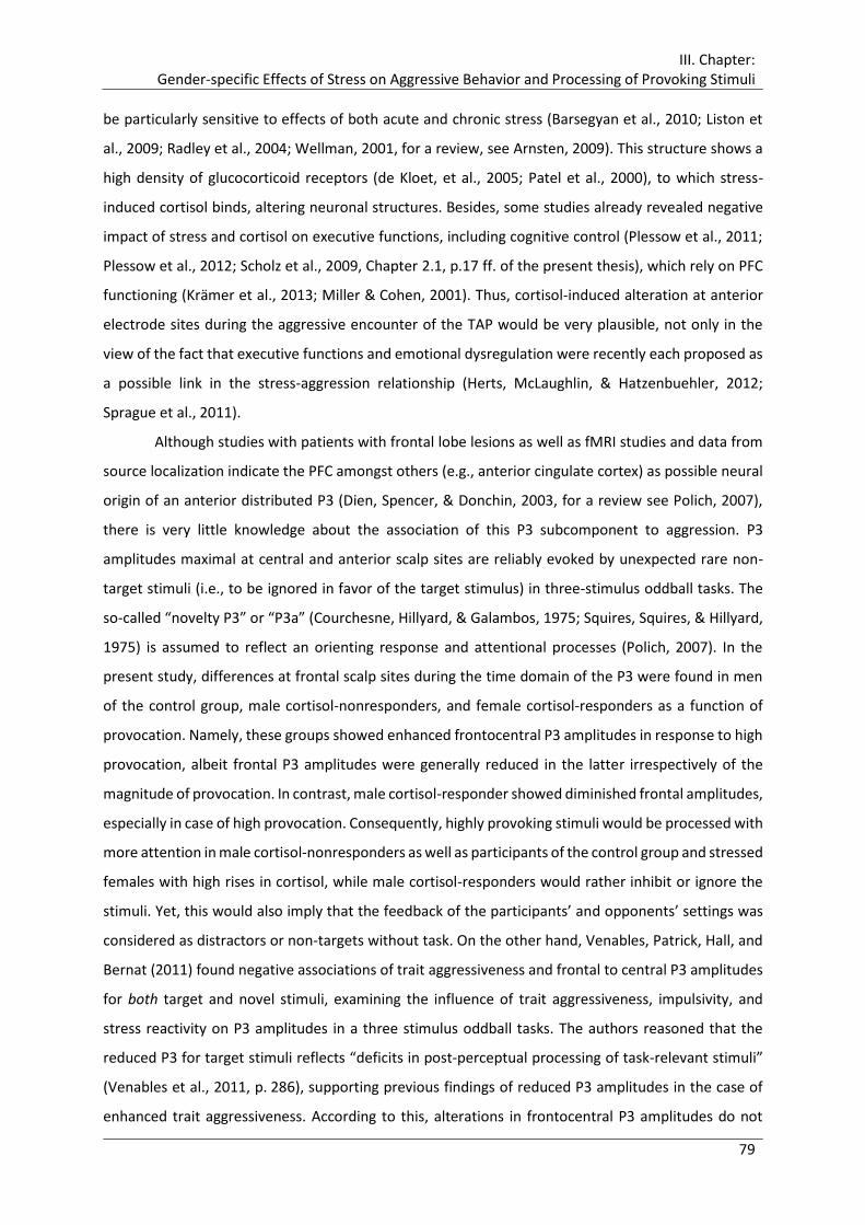
III. Chapter: Gender-specific Effects of Stress on Aggressive Behavior and Processing of Provoking Stimuli
79
be particularly sensitive to effects of both acute and chronic stress (Barsegyan et al., 2010; Liston et
al., 2009; Radley et al., 2004; Wellman, 2001, for a review, see Arnsten, 2009). This structure shows a
high density of glucocorticoid receptors (de Kloet, et al., 2005; Patel et al., 2000), to which stress-
induced cortisol binds, altering neuronal structures. Besides, some studies already revealed negative
impact of stress and cortisol on executive functions, including cognitive control (Plessow et al., 2011;
Plessow et al., 2012; Scholz et al., 2009, Chapter 2.1, p.17 ff. of the present thesis), which rely on PFC
functioning (Krämer et al., 2013; Miller & Cohen, 2001). Thus, cortisol-induced alteration at anterior
electrode sites during the aggressive encounter of the TAP would be very plausible, not only in the
view of the fact that executive functions and emotional dysregulation were recently each proposed as
a possible link in the stress-aggression relationship (Herts, McLaughlin, & Hatzenbuehler, 2012;
Sprague et al., 2011).
Although studies with patients with frontal lobe lesions as well as fMRI studies and data from
source localization indicate the PFC amongst others (e.g., anterior cingulate cortex) as possible neural
origin of an anterior distributed P3 (Dien, Spencer, & Donchin, 2003, for a review see Polich, 2007),
there is very little knowledge about the association of this P3 subcomponent to aggression. P3
amplitudes maximal at central and anterior scalp sites are reliably evoked by unexpected rare non-
target stimuli (i.e., to be ignored in favor of the target stimulus) in three-stimulus oddball tasks. The
so-called “novelty P3” or “P3a” (Courchesne, Hillyard, & Galambos, 1975; Squires, Squires, & Hillyard,
1975) is assumed to reflect an orienting response and attentional processes (Polich, 2007). In the
present study, differences at frontal scalp sites during the time domain of the P3 were found in men
of the control group, male cortisol-nonresponders, and female cortisol-responders as a function of
provocation. Namely, these groups showed enhanced frontocentral P3 amplitudes in response to high
provocation, albeit frontal P3 amplitudes were generally reduced in the latter irrespectively of the
magnitude of provocation. In contrast, male cortisol-responder showed diminished frontal amplitudes,
especially in case of high provocation. Consequently, highly provoking stimuli would be processed with
more attention in male cortisol-nonresponders as well as participants of the control group and stressed
females with high rises in cortisol, while male cortisol-responders would rather inhibit or ignore the
stimuli. Yet, this would also imply that the feedback of the participants’ and opponents’ settings was
considered as distractors or non-targets without task. On the other hand, Venables, Patrick, Hall, and
Bernat (2011) found negative associations of trait aggressiveness and frontal to central P3 amplitudes
for both target and novel stimuli, examining the influence of trait aggressiveness, impulsivity, and
stress reactivity on P3 amplitudes in a three stimulus oddball tasks. The authors reasoned that the
reduced P3 for target stimuli reflects “deficits in post-perceptual processing of task-relevant stimuli”
(Venables et al., 2011, p. 286), supporting previous findings of reduced P3 amplitudes in the case of
enhanced trait aggressiveness. According to this, alterations in frontocentral P3 amplitudes do not

III. Chapter: Gender-specific Effects of Stress on Aggressive Behavior and Processing of Provoking Stimuli
80
imply distractor characteristics of the stimuli. Hence, it can be concluded that male cortisol-responders
showed reduced attention to provoking stimuli. Together with desensitization reflected by diminished
parietal P3 amplitudes, this may contribute for the increased open and covert aggressive behavior in
response to high relative to low provocation. In turn, enhanced frontocentral P3 amplitudes, found in
highly provoked male cortisol-nonresponders, males of the control group, and female cortisol-
responders, indicate that provoking stimuli were processed with enhanced neural resources and
attention. However, these three groups showed strong distinctions in aggressive behavior, especially
with regard to their response to high provocation, and their correlations of frontal P3 amplitudes with
aggressive behavior were low and diverging. In summary, these interpretations do not offer a
compelling explanation with regard to the behavioral outcome. Nevertheless, the reduced frontal
amplitudes found in male cortisol-responders, pronounced in the case of high provocation, and in
female cortisol-responders, especially in the case of low provocation, are in line with the assumption,
that cortisol inhibits the processing of threat-related information (Putman, Hermans, Koppeschaar et
al., 2007; Putman, Hermans, & van Honk, 2010; Taylor, V. A. et al., 2011; van Peer, Spinhoven, &
Roelofs, 2010, but see Bertsch, Böhnke, Kruk, Richter et al., 2011).
Alternatively to the assumptions of (Venables et al., 2011), Godleski and colleagues found
hostile attribution biases to predict enhanced P3 amplitudes at frontal scalp sites. The authors
assessed self-reported relational and physical aggression as well as hostile intent attributions for
relational and instrumental situations in 112 male and female college students. P3 amplitudes were
evoked by terms of an auditory perseveration task (Godleski, Ostrov, Houston, & Schlienz, 2010). They
conclude “that individuals high in hostile attribution biases for relational provocation situations are
overly sensitive to potentially salient stimuli.” (Godleski et al., 2010, p. 30), as enhanced P3 amplitudes
reflect the intensified processing of the stimuli. This seems to be an intriguing explanation for the
enhanced frontocentral P3 amplitudes in highly provoked female cortisol-responders against the
background of their reported high levels of anger and hopelessness in the course of the experimental
manipulation. Thus, their attribution of the SECPT and the provocation as hostility might have led to
increased aggressive behavior. Furthermore, this effect became apparent only in the case of stress-
induced cortisol, causing a general inhibition of processing of the stimuli on the basis of which high
provocation was processed with enhanced attention. Hence, in females, inhibition tendencies induced
by cortisol might have been abolished through high provocation.
Nevertheless, the results and interpretation of the association between the P3 amplitude and
aggression gathered through oddball tasks, affective pictures, or related tasks are not applicable
without further ado to the present findings. First, electrocortical responses were not obtained while
participants were actually engaged in aggressive behavior in the above reviewed studies (e.g., Barratt
et al., 1997; Bartholow et al., 2006; Godleski et al., 2010; Venables et al., 2011). These situational
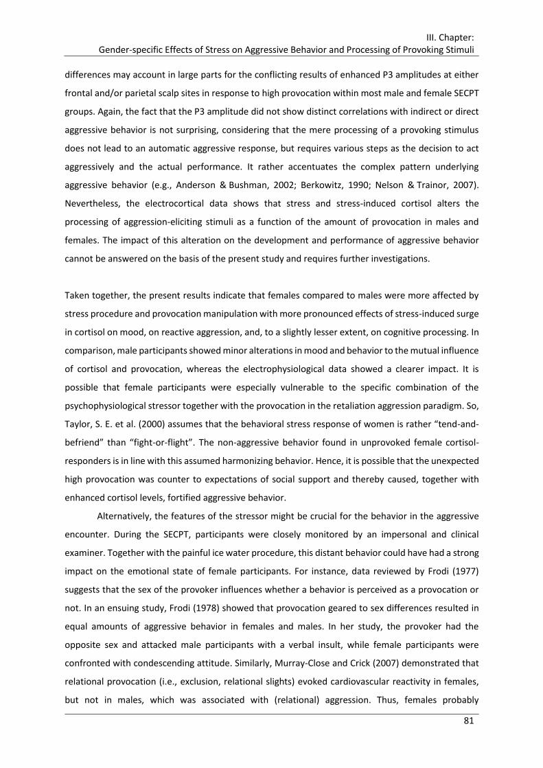
III. Chapter: Gender-specific Effects of Stress on Aggressive Behavior and Processing of Provoking Stimuli
81
differences may account in large parts for the conflicting results of enhanced P3 amplitudes at either
frontal and/or parietal scalp sites in response to high provocation within most male and female SECPT
groups. Again, the fact that the P3 amplitude did not show distinct correlations with indirect or direct
aggressive behavior is not surprising, considering that the mere processing of a provoking stimulus
does not lead to an automatic aggressive response, but requires various steps as the decision to act
aggressively and the actual performance. It rather accentuates the complex pattern underlying
aggressive behavior (e.g., Anderson & Bushman, 2002; Berkowitz, 1990; Nelson & Trainor, 2007).
Nevertheless, the electrocortical data shows that stress and stress-induced cortisol alters the
processing of aggression-eliciting stimuli as a function of the amount of provocation in males and
females. The impact of this alteration on the development and performance of aggressive behavior
cannot be answered on the basis of the present study and requires further investigations.
Taken together, the present results indicate that females compared to males were more affected by
stress procedure and provocation manipulation with more pronounced effects of stress-induced surge
in cortisol on mood, on reactive aggression, and, to a slightly lesser extent, on cognitive processing. In
comparison, male participants showed minor alterations in mood and behavior to the mutual influence
of cortisol and provocation, whereas the electrophysiological data showed a clearer impact. It is
possible that female participants were especially vulnerable to the specific combination of the
psychophysiological stressor together with the provocation in the retaliation aggression paradigm. So,
Taylor, S. E. et al. (2000) assumes that the behavioral stress response of women is rather “tend-and-
befriend” than “fight-or-flight”. The non-aggressive behavior found in unprovoked female cortisol-
responders is in line with this assumed harmonizing behavior. Hence, it is possible that the unexpected
high provocation was counter to expectations of social support and thereby caused, together with
enhanced cortisol levels, fortified aggressive behavior.
Alternatively, the features of the stressor might be crucial for the behavior in the aggressive
encounter. During the SECPT, participants were closely monitored by an impersonal and clinical
examiner. Together with the painful ice water procedure, this distant behavior could have had a strong
impact on the emotional state of female participants. For instance, data reviewed by Frodi (1977)
suggests that the sex of the provoker influences whether a behavior is perceived as a provocation or
not. In an ensuing study, Frodi (1978) showed that provocation geared to sex differences resulted in
equal amounts of aggressive behavior in females and males. In her study, the provoker had the
opposite sex and attacked male participants with a verbal insult, while female participants were
confronted with condescending attitude. Similarly, Murray-Close and Crick (2007) demonstrated that
relational provocation (i.e., exclusion, relational slights) evoked cardiovascular reactivity in females,
but not in males, which was associated with (relational) aggression. Thus, females probably
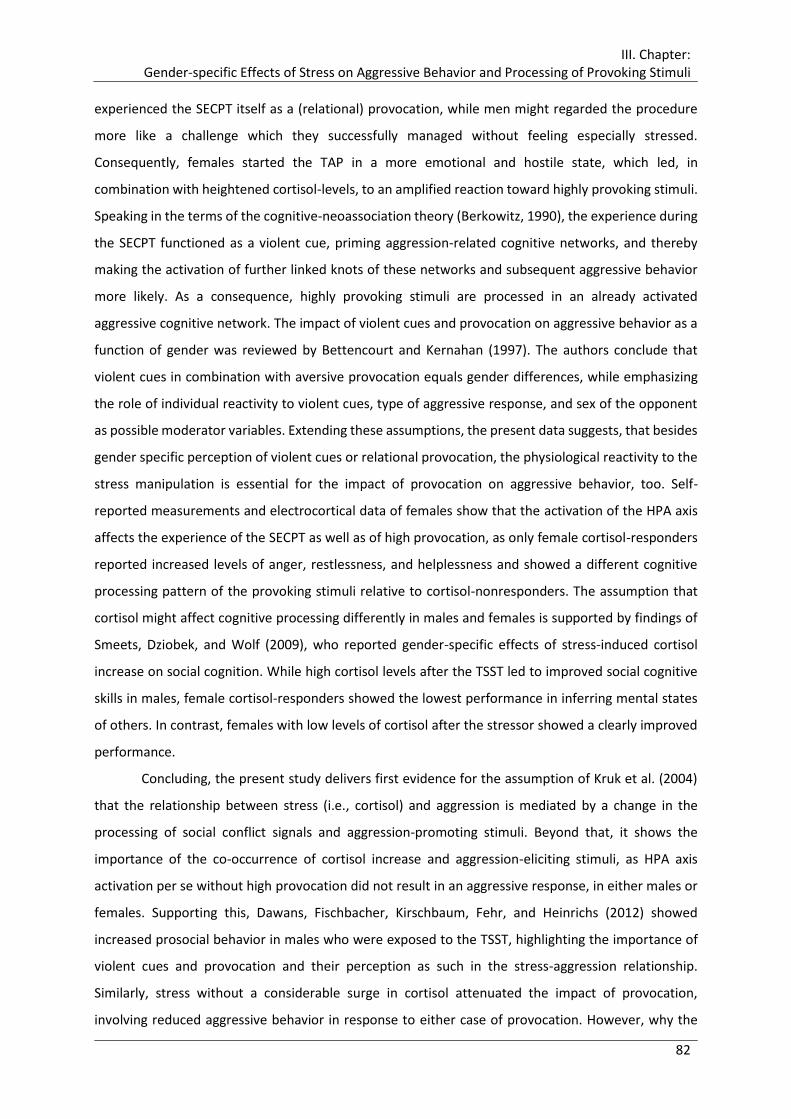
III. Chapter: Gender-specific Effects of Stress on Aggressive Behavior and Processing of Provoking Stimuli
82
experienced the SECPT itself as a (relational) provocation, while men might regarded the procedure
more like a challenge which they successfully managed without feeling especially stressed.
Consequently, females started the TAP in a more emotional and hostile state, which led, in
combination with heightened cortisol-levels, to an amplified reaction toward highly provoking stimuli.
Speaking in the terms of the cognitive-neoassociation theory (Berkowitz, 1990), the experience during
the SECPT functioned as a violent cue, priming aggression-related cognitive networks, and thereby
making the activation of further linked knots of these networks and subsequent aggressive behavior
more likely. As a consequence, highly provoking stimuli are processed in an already activated
aggressive cognitive network. The impact of violent cues and provocation on aggressive behavior as a
function of gender was reviewed by Bettencourt and Kernahan (1997). The authors conclude that
violent cues in combination with aversive provocation equals gender differences, while emphasizing
the role of individual reactivity to violent cues, type of aggressive response, and sex of the opponent
as possible moderator variables. Extending these assumptions, the present data suggests, that besides
gender specific perception of violent cues or relational provocation, the physiological reactivity to the
stress manipulation is essential for the impact of provocation on aggressive behavior, too. Self-
reported measurements and electrocortical data of females show that the activation of the HPA axis
affects the experience of the SECPT as well as of high provocation, as only female cortisol-responders
reported increased levels of anger, restlessness, and helplessness and showed a different cognitive
processing pattern of the provoking stimuli relative to cortisol-nonresponders. The assumption that
cortisol might affect cognitive processing differently in males and females is supported by findings of
Smeets, Dziobek, and Wolf (2009), who reported gender-specific effects of stress-induced cortisol
increase on social cognition. While high cortisol levels after the TSST led to improved social cognitive
skills in males, female cortisol-responders showed the lowest performance in inferring mental states
of others. In contrast, females with low levels of cortisol after the stressor showed a clearly improved
performance.
Concluding, the present study delivers first evidence for the assumption of Kruk et al. (2004)
that the relationship between stress (i.e., cortisol) and aggression is mediated by a change in the
processing of social conflict signals and aggression-promoting stimuli. Beyond that, it shows the
importance of the co-occurrence of cortisol increase and aggression-eliciting stimuli, as HPA axis
activation per se without high provocation did not result in an aggressive response, in either males or
females. Supporting this, Dawans, Fischbacher, Kirschbaum, Fehr, and Heinrichs (2012) showed
increased prosocial behavior in males who were exposed to the TSST, highlighting the importance of
violent cues and provocation and their perception as such in the stress-aggression relationship.
Similarly, stress without a considerable surge in cortisol attenuated the impact of provocation,
involving reduced aggressive behavior in response to either case of provocation. However, why the

III. Chapter: Gender-specific Effects of Stress on Aggressive Behavior and Processing of Provoking Stimuli
83
warm water control group reacted as aggressive as cortisol-responders remains unclear so far. The
electrophysiological data suggest another underlying process resulting in equal amounts of aggressive
behavior, for what reason the quality of aggression might be different, i.e., less hostile.
3.4.3 Limitations and Future Directions
Despite the strengths of the present study as including hormone measurements, gender, and
electrophysiological correlates while inducing and measuring aggression under controlled laboratory
conditions, several important limitations have to be considered.
First, regarding aggressive behavior, the used aggression paradigm indeed comprised overt
and covert types of aggressive behavior, but both resulted in a physical harm of the opponent.
Consequently, future studies should include more typical forms of indirect or relational aggression like
ostracism, gossiping, and bullying in comparison to clear direct, physical forms of aggressive behavior
to further elucidate the impact of gender typical forms of aggression in their relation to HPA axis
activation. Besides, the application of different laboratory aggression paradigms as the “Point
Subtraction Aggression paradigm” or the “Ultimatum Game” (cf. Carre, McCormick, & Hariri, 2011)
would offer a generalizability of the present findings.
Second, measurements of the autonomic stress response were not included and therefore I
cannot rule out or specify confounding or mutual effects of the adrenergic system in the stress –
aggression relation. Especially, cardiovascular reactivity seems to constitute an important variable in
aggressive conduct (e.g., Murray-Close & Crick, 2007), and should be particularly considered with
regard to the distinction of cortisol-responders and –nonresponders. Likewise, other hormones should
be included in future studies, as recent research emphasizes the importance of testosterone in
interaction with cortisol in the context of aggression-related behavior (cf. Carre & Mehta, 2011;
Montoya et al., 2012; Popma et al., 2007). Additionally, oxytocin might help to untangle gender
differences (Dawans et al., 2012; Smeets et al., 2009; Taylor, S. E. et al., 2000).
Third, the applied stress test led to an intermediate rise in cortisol. To further clarify the role
of the amount of free cortisol on the extent of aggressive behavior, a dose-response study or the usage
of stress tests resulting in a higher surge of cortisol, like the TSST (Kirschbaum et al., 1993), could
provide evidence to integrate the results of the present study and those of Böhnke, Bertsch, Kruk, and
Richter et al. (2010) with special regard to the role of provocation to elicit aggressive behavior after
stress exposure. Additionally, the results of the present study suggest that the relationship between
aggressive behavior and stress-induced cortisol rise in men and women is not only a function of the
type of aggressive behavior, but also depends on the kind of provocation. Thus, it might be worthwhile
to further investigate whether relational provocation as somehow mediated through the SECPT in
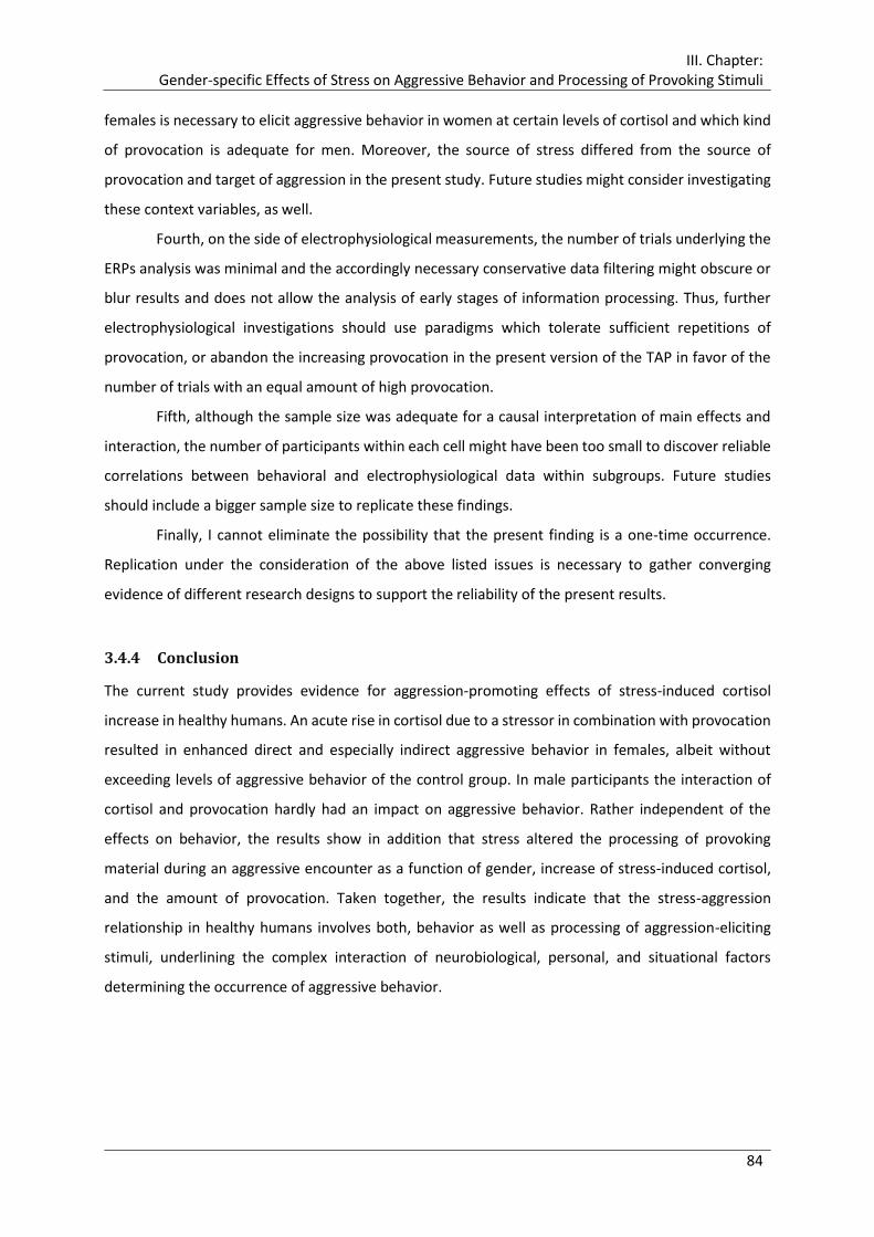
III. Chapter: Gender-specific Effects of Stress on Aggressive Behavior and Processing of Provoking Stimuli
84
females is necessary to elicit aggressive behavior in women at certain levels of cortisol and which kind
of provocation is adequate for men. Moreover, the source of stress differed from the source of
provocation and target of aggression in the present study. Future studies might consider investigating
these context variables, as well.
Fourth, on the side of electrophysiological measurements, the number of trials underlying the
ERPs analysis was minimal and the accordingly necessary conservative data filtering might obscure or
blur results and does not allow the analysis of early stages of information processing. Thus, further
electrophysiological investigations should use paradigms which tolerate sufficient repetitions of
provocation, or abandon the increasing provocation in the present version of the TAP in favor of the
number of trials with an equal amount of high provocation.
Fifth, although the sample size was adequate for a causal interpretation of main effects and
interaction, the number of participants within each cell might have been too small to discover reliable
correlations between behavioral and electrophysiological data within subgroups. Future studies
should include a bigger sample size to replicate these findings.
Finally, I cannot eliminate the possibility that the present finding is a one-time occurrence.
Replication under the consideration of the above listed issues is necessary to gather converging
evidence of different research designs to support the reliability of the present results.
3.4.4 Conclusion
The current study provides evidence for aggression-promoting effects of stress-induced cortisol
increase in healthy humans. An acute rise in cortisol due to a stressor in combination with provocation
resulted in enhanced direct and especially indirect aggressive behavior in females, albeit without
exceeding levels of aggressive behavior of the control group. In male participants the interaction of
cortisol and provocation hardly had an impact on aggressive behavior. Rather independent of the
effects on behavior, the results show in addition that stress altered the processing of provoking
material during an aggressive encounter as a function of gender, increase of stress-induced cortisol,
and the amount of provocation. Taken together, the results indicate that the stress-aggression
relationship in healthy humans involves both, behavior as well as processing of aggression-eliciting
stimuli, underlining the complex interaction of neurobiological, personal, and situational factors
determining the occurrence of aggressive behavior.
85
IV. Chapter:
Stress-induced Cortisol and Aggression
Alter Subsequent Social Information Processing

IV. Chapter: Stress-induced Cortisol and Aggression Alter Subsequent Social Information Processing
86
4.1 Introduction
Aggression and violence in their different forms and shades are common and ubiquitous in our
everyday life. Though natural and adaptive in principle, they constitute a substantial social problem
claiming the life of more than 1.6 million people annually and entailing enormous economic costs as
reports of the World Health Organization show (Krug, 2002; Waters et al., 2004). The occurrence and
the extent of aggressive behavior is not only influenced by personal (e.g., impulsivity) and situational
factors (i.e., provocation) as for example suggested by Anderson and colleagues (general aggression
model, Anderson & Bushman, 2002), but recent studies emphasized the role of (neuro-)biological
mechanisms in the development and expression of aggressive behavior (for reviews see Anholt &
Mackay, 2012; Bertsch, 2012; Montoya et al., 2012; Nelson & Trainor, 2007). Stress as a
psychoneuroendocrinological mechanism has been identified as a central factor in precipitating and
promoting aggressive behavior (cf. Barnett et al., 1991; Craig, 2007). In particular, animal and human
research revealed that the hypothalamic-pituitary-adrenal (HPA) axis, the so-called stress axis, and the
stress hormone cortisol or corticosterone10, respectively, are causally involved in the genesis, the
elicitation, and reinforcement of aggressive behavior (Böhnke, Bertsch, Kruk, Richter et al., 2010;
Hayden-Hixson & Ferris, 1991; Kruk et al., 2004; Lopez-Duran et al., 2009; Wommack & Delville, 2007).
Accordingly, Kruk et al. (2004) found that an acute administration of corticosterone reduced the
threshold for attack behavior in rats, facilitating the release of aggressive behavior. Beyond that, a
further experiment revealed that stimulation of the hypothalamic attack area itself led to a surge of
corticosterone (Kruk et al., 2004, study 1). Hence, a fast positive feedback loop between glucocorticoid
stress response and brain structures engaged in aggressive behavior was identified. Based on these
findings, the authors concluded that “such mutual facilitation could constitute a vicious circle, which
would explain why aggressive behavior escalates so easily, and why it is so difficult to stop once it has
started” (Kruk et al., 2004, p. 1068). Moreover, the authors proposed that the causal relationship
between stress and aggression is mediated by a change in the processing of social conflict signals and
aggression-promoting stimuli. Accordingly, processing of relevant social information in the context of
an aggressive encounter seems to play a key role in the escalation of aggression and its persistence
once it has begun.
On the side of stress, there is profound evidence that preceding stress or administration of
cortisol, respectively, alter processing of social relevant information across various tasks (e.g.,
Buchanan & Lovallo, 2001; Ellenbogen et al., 2002; Oei et al., 2012; Putman & Roelofs, 2011; Roelofs
10 Corticosterone is the primary glucocorticoid within rodents, whereas cortisol is the most important glucocorticoid hormone in humans.

IV. Chapter: Stress-induced Cortisol and Aggression Alter Subsequent Social Information Processing
87
et al., 2005; van Marle et al., 2009). Threat-related stimuli especially are processed differently under
increased levels of cortisol, albeit with inconsistent results concerning the direction of this impact. For
the most part, studies revealed a preferential processing of threat-related stimuli. Putman, Hermans,
and van Honk (2007), for instance, found a bias for angry faces in immediate memory performance
after administration of 40 mg cortisol. Similar results were reported after stress exposure: A high
increase in cortisol levels in response to a psychological stressor led to relatively enhanced selective
attention for angry faces in a masked pictorial emotional Stroop task (Roelofs et al., 2007). Akinola and
Mendes (2012) investigated the influence of acute stress on threat-related decision making in police
officers and found improved performance in participants with a large cortisol increase to the stressor.
Regarding electrocortical measurements, Weymar et al. (2012) reported enhanced late positive
potentials (LPP) for unpleasant pictures in a passive viewing paradigm after an acute stressor in healthy
subjects, indicating a more effortful and elaborate processing of significant information. Likewise, van
Peer and colleagues explored the influence of cortisol on threat processing in a series of experiments.
In highly avoidant and highly anxious individuals exogenous cortisol led to increased attentional
processing of angry faces in an approach avoidance paradigm, reflected in enhanced early positive
amplitudes (van Peer et al., 2007; van Peer et al., 2009). On the other hand, after administration of 10
mg hydrocortisone Taylor, V. A. et al. (2011) found increased inhibition for angry faces but not for
happy and sad faces in a negative priming task. Similarly, a high dose of orally administered cortisol
reduced preconscious attention for fearful faces in a masked emotional Stroop task (Putman, Hermans,
Koppeschaar et al., 2007) and to a reduced interference by task-irrelevant negative pictures used as
distractors in a Sternberg Working memory task (Oei et al., 2009).
Concerning aggression, idiomatic expressions as “seeing red” or “blind with rage” illustrate
vividly how anger and aggression can alter our perception and experience of our surroundings. The
processing of social information has been frequently assumed to be a crucial factor in the
development, occurrence, and escalation of aggressive behavior (e.g., Anderson & Bushman, 2002;
Calvete & Orue, 2011; Crick & Dodge, 1994; Dodge & Crick, 1990; Huesmann, 1988; Potegal, 2012).
However, previous research on the consequences of aggression on social information processing has
concentrated so far mainly on maltreated children, trait aspects of anger and aggression, self-reported
aggressive experience, and samples with pathological aggression-related behavior (e.g., Anderson
& Stanford, 2012; Calvete & Orue, 2011; Coccaro et al., 2007; Crick & Dodge, 1996; Houston
& Stanford, 2001; Verona et al., 2012; Zelli et al., 1999). Beyond that, investigating processing during
an aggressive encounter, a few studies found altered cortical activity during retaliation (Lotze et al.,
2007) or the decision to react aggressively as a function of the amount of provocation (Krämer et al.,
2008; Krämer et al., 2007; Wiswede et al., 2011). Yet these studies do not offer a direct explanation
for the escalation of aggression or its persistence. As stated by (Potegal) (2012), aggressive behavior

IV. Chapter: Stress-induced Cortisol and Aggression Alter Subsequent Social Information Processing
88
and aggressive motivation may last beyond the actual encounter (Potegal, 2012, p. 388) and is
frequently transferred into another context (Bertsch, Böhnke, Kruk, & Naumann, 2009, p. 1). In the
terms of the cognitive-neoassociation theory (Berkowitz, 1990), an aggressive encounter should
activate aggression-related cognitive networks. Consequently, subsequent situations should be
processed in an already activated aggressive cognitive network. To elucidate this, studies are necessary
which explore the processing of social information after aggressive behavior took place.
Pioneering work in this regard was carried out by Bertsch and colleagues (2009). In an event-
related potential (ERP) study, healthy male and female participants were either highly or mildly
provoked by means of a retaliation paradigm, namely, the Taylor Aggression Paradigm (TAP, Taylor,
1967). Subsequently, participants performed an emotional Stroop task with colored happy, angry,
fearful, and neutral facial expressions, in which they were required to name the color of the stimuli.
Results revealed by tendency slower reaction times naming the color of emotional facial expressions
in highly provoked participants. Moreover, electrocortical data of this group showed enhanced early
(P2) and late positive amplitudes for all facial expressions, with the greatest effect for threat-related
faces (i.e., angry and fearful) for the P2 component, indicating greater relevance and salience to the
respective facial expressions. Thus, preceding provocation and aggressive behavior led to altered social
information processing beyond the aggressive encounter.
Taking into account the aggression-promoting effects of stress and cortisol and their impact of
processing of threat-related social information, as a next step, Bertsch, Böhnke, Kruk, and Richter et
al. (2011) investigated the combined effect of cortisol and aggressive behavior on the subsequent
processing of emotional stimuli. Healthy participants received an oral dose of 20 mg hydrocortisone or
a placebo and were afterwards again exposed to either high or low provocation in the TAP. Next,
participants performed the emotional Stroop task, described above. Cortisol in combination with high
provocation and thus enhanced aggressive behavior led to faster reaction times, independently of the
facial expression. Concerning the electrocortical correlates, this preferential processing of social cues
was not confirmed. On the contrary, exogenous cortisol relative to the placebo diminished the
attentional bias for angry faces, reflected in reduced early positive frontocentral ERPs. However,
replicating the previous result, provocation on the other hand led to enhanced early and late positive
posterior ERPs, suggesting enhanced relevance of all facial expression, irrespectively of the depicted
emotion.
In summary, there is preliminary evidence that stress hormone cortisol in combination with
experimentally provoked aggressive behavior alters the processing of social relevant information in
healthy adults beyond the actual aggressive encounter. However, this needs further investigations for
several reasons. First, even though the cortisol manipulation chosen by Bertsch, Böhnke, Kruk, and
Richter et al. (2011) provides the opportunity to explore the specific impact of the stress hormone

IV. Chapter: Stress-induced Cortisol and Aggression Alter Subsequent Social Information Processing
89
itself, hydrocortisone artificially raises cortisol levels, lacking the quality of a real-life stressor and thus
the ecological validity of a stressful experience. Besides, autonomic arousal has been shown to
influence occurrence of aggressive behavior (Zillmann, Johnson, & Day, 1974) and might also alter
processing of affective stimuli (cf. Reisenzein, 1983). Moreover, studies applying an acute stressor in
contrast to exogenous cortisol manipulation consistently found a preferential processing of threat-
related stimuli, as reported above. Hence, a replication with an acute stressor is standing to reason.
Second, the emotional material used by Bertsch et al. (2009, 2011) is limited to facial expressions.
Although emotional facial expressions are considered as most significant and immediate universal
social cues in personal interaction (e.g., Ekman, 1993; Frith, 2009), in the context of aggression other
stimuli should be considered, too. In particular, pictures depicting violence or an assault as well as
weapons are of specific interest, as they represent more natural or relevant signals in the context of
aggression than the mere fearful or angry facial expression (cf. Anderson et al., 1998; Berkowitz
& LePage, 1967; Engelhardt et al., 2011; Huesmann, 2007). Third, there is evidence that the
performance requirements regarding the emotional stimuli are crucial for the impact of cortisol on the
processing of those. For instance, in two studies, van Peer and colleagues (2007, 2009) found enhanced
attentional processing of angry faces in an Approach Avoidance task wherein the processing of the
facial expression was task-relevant. If the emotional face served as task-irrelevant distractors,
however, the impact of cortisol was reversed (van Peer et al., 2010). Besides, laboratory surroundings
are very likely to constitute a stimulative nature to perform well, whereby behavioral effects and
electrocortical measurements might be obscured by regulating control processes. Hence, an ERP study
of a passive viewing paradigm might be more adequate.
The electrocortical response to affective pictures is well known, comprising ERP component
pattern from short (P1, N1) to middle (P2, N2) to long (P3, LPP11) latencies, reflecting different steps of
the information processing stream (e.g., Anderson & Stanford, 2012; Dietrich, Naumann, Maier, &
Becker, 1997; Keil et al., 2002, for a review see Olofsson, Nordin, Sequeira, & Polich, 2008). Early ERP
components reflect mainly sensory processing, whereas the P2 is considered as an index of attention-
related process (Carretie, Mercado, Tapia, & Hinojosa, 2001). In contrast, less is known about the N2
within this context. Similar to the P2, this component is assumed to be related to rather automatic
attention driven by stimulus characteristics (Carretie, Hinojosa, Martin-Loeches, Mercado, & Tapia,
2004). P3 and LPP are involved in memory formation, mental resource allocation, and elaborate
evaluation for motivationally significant stimuli (Olofsson et al., 2008; Schupp, Flaisch, Stockburger, &
Junghöfer, 2006). These early and late components have been shown to be modulated by valence,
most often with (threat-related) negative pictures eliciting larger positive and reduced negative (N2)
11 The term LPP for the positive modulation of the ERP in this time range is adopted from Hajcak, MacNamara, and Olvet (2010) and refers to the definition therein.

IV. Chapter: Stress-induced Cortisol and Aggression Alter Subsequent Social Information Processing
90
amplitudes relative to positive pictures (e.g., Carretie et al., 2004; Carretie et al., 2001; Huang & Luo,
2006; Ito, Larsen, Smith, & Cacioppo, 1998; Smith, Cacioppo, Larsen, & Chartrand, 2003, but see
Cuthbert, Schupp, Bradley, Birbaumer, & Lang, 2000; Ito et al., 1998; Olofsson et al., 2008). Alike, highly
arousing pictures cause larger amplitudes compared to low arousing ones, albeit this effect occurs at
longer latencies and less consistently (Leite et al., 2012; Sabatinelli, Lang, Keil, & Bradley, 2007; Schupp
et al., 2000).
Taking these issues into account, the present study sought to explore the impact of stress and
aggression on social information processing further. In common with Bertsch, Böhnke, Kruk, and
Naumann (2009) and Bertsch, Böhnke, Kruk, and Richter et al. (2011), a modified version of the TAP
was chosen to induce and measure aggressive behavior. Half of the participants were highly provoked,
whereas the other half, as a control group, were only mildly provoked. Prior to this retaliation
paradigm, participants were either exposed to an acute stressor, the socially evaluated cold-pressor
test (SECPT, Schwabe et al., 2008) or to a control procedure with warm water. Afterwards, a passive
viewing paradigm was realized, comprising affective pictures depicting positive, negative, or aggressive
scenes with either humans or objects. Besides positive pictures, negative pictures were included to
distinguish specific effects for aggressive stimuli from a general bias for all kind of negative contents.
Gender was included as a controlling factor, since females and males differ with respect to the stress-
aggression relationship as well as to processing of affective and threat-related stimuli (Bettencourt
& Kernahan, 1997; Böhnke, Bertsch, Kruk, Richter et al., 2010; Bradley, Codispoti, Sabatinelli, & Lang,
2001; Verona et al., 2007). With respect to previous findings, it was expected that stress and aggression
would jointly influence the processing of affective pictures. In particular, participants who reacted to
the stressor with an increase of cortisol levels and were subsequently highly provoked should show
preferential processing of pictures depicting aggressive scenes with humans as well as with objects
(i.e., weapons). This bias was explored at different stages of information processing, including
attention-related components (P2, N2) as well as higher level stages (P3, LPP).
4.2 Materials and Methods
4.2.1 Participants
Participants were recruited from the University of Trier, Germany. Out of 75 participants, who
completed the experiment, four individuals had to be excluded due to abnormal high salivary cortisol
values (one female) or incorrect experimental procedure (three individuals), leaving 71 participants
(36 males, 35 females) with a mean age of 23.96 years (SD= 2.27, range 20-31 years) and a mean BMI
of 22.77 kg/m² (SD=2.54). Criteria for exclusion were (1) acute or chronic physical disease, (2) a mental
disorder or a history of such, (3) use of medication, (4) smoking, as it is known to influence HPA axis

IV. Chapter: Stress-induced Cortisol and Aggression Alter Subsequent Social Information Processing
91
activity (Granger et al., 2007), and (5) being not a native German speaker. Only right-handed students
were included, as handedness affects hemispheric specialization, thus altering EEG measurements
(Galin et al., 1982). To ensure no problems with the experimental manipulations, individuals who
reported to suffer from dyschromatopsia or stated to be sensitive to loud noises or to cold, were
excluded. Additionally, students taking classes in psychology were excluded to guarantee an unbiased
behavior during the experiment. In order to control for hormonal status, only non-pregnant women
who used hormonal contraceptives12 were included in the study. The experiment was conducted in
accordance with the Declaration of Helsinki. The Research Ethics Committee of the University of Trier
approved all parts of the study, and all participants gave written informed consent. Participation was
compensated with 45 € (approximately US $57).
4.2.2 Socially Evaluated Cold-Pressor Test- SECPT
Participants who were assigned to the stress condition, were exposed to the socially evaluated cold-
pressor test (SECPT, Schwabe et al., 2008), an economic and efficient stress induction causing
significant activation of the HPA axis, thereby a rise in cortisol levels, as well as an activation of the
adrenergic system and an increase in subjective stress experience (e.g., Schwabe & Wolf, 2009;
Weymar et al., 2012). Namely, an unfamiliar experimenter of the opposite sex, who acted neutrally
and distanced, asked them to immerse their left hand up to the wrist into ice water (0-3 °C) and to look
at a camera throughout the whole procedure as they would be videotaped and their facial expressions
would be analyzed. Meanwhile, the experimenter watched them closely, took notes, and stopped the
time. At the end of three minutes, they were asked to remove their hand. No further communication
between experimenter and participants was permitted and participants were uninformed about the
elapsed time. Participants in the non-stressful control condition underwent the same procedure with
warm water (37-39 °C) instead of ice water. Two participants, one female and one male, removed their
hand from the ice water before the expiration of the term, because the cold hurt them too much. Since
they were obviously strongly stressed and their data did not constitute outliers, they were included in
all analyses.
4.2.3 Taylor Aggression Paradigm- TAP
Aggression was elicited and assessed with a modified version of the Taylor Aggression Paradigm (TAP,
Taylor, 1967). Participants were led to believe that they were playing a competitive reaction time task
12 Except for the contraceptives pills containing Drospirenone, which is an antagonist for the mineralocorticoid
receptor, and therefore might have skewed the cortisol measurements (Genazzani et al., 2007); namely Yasmine,
Yasminelle, Petibelle, Aida, Angeliq or Yaz.
IV. Chapter: Stress-induced Cortisol and Aggression Alter Subsequent Social Information Processing
92
against another participant of the same sex, whom they met prior to the experiment. Participants were
instructed to react as fast as possible to a green square by pressing a key in order to win a trial. The
slower player would receive a blast of noise by the competitor. The game consisted of 3 blocks of 10
trials each. Each trial started with setting of the punitive noise, to which the competitor would be
exposed in case the competitor would lose the trial. Participants were asked first to specify the
duration and then the volume of the noise on two separate scales. Each scale was subdivided into 11
increments, reaching from level 0 to 10, with noise duration ranging from 0.5 s (level 1) to 5 s (level
10) and noise volume ranging 60 dB (level 1) and 105 dB (level 10), both in equidistant increments of
0.5 s or 5 dB, respectively. Level 0 corresponded to 0s on the scale for duration and 0dB on the scale
for volume. After each trial, feedback whether the participant won or lost the given trial was presented
on the screen, followed by the settings of duration and volume selected by each for the other player.
If the participant had lost the trial, the noise was presented.
Unknown to the participants, there was no actual competitor. The outcome of the trials was
held constant for all participants: each participant won and lost half of the trials. Additionally, noise
volume and duration were selected by the experimenter and varied by trial block to realize high
provocation or low provocation, respectively. During the first block, all participants received short and
gentle noises when they lost a trial (volume: M = 62.5 dB, range 0–70 dB; duration: M = 0.75 s, range
0–1.5 s). Participants in the mildly provoked control group received the same noises during the second
and third blocks. Participants in the highly provoked group received noises of intermediate intensity
and duration in the second block (volume: M = 82.5 dB, range 75– 90 dB; duration: M = 2.75 s, range
2–3.5 s) and high intensity and duration in the third block (volume: M = 99 dB, range 90–105 dB;
duration: M = 4.4 s, range 3.5–5 s). The duration and volume settings of the participants were recorded
in each trial on the scales from 0 to 10. An average was computed for each participant and each trial
of the volume and duration setting. Finally, the ten trials belonging to one block of the TAP were
averaged for each participant. These values were later used as the dependent variable “aggressive
behavior” in each of the three blocks.
4.2.4 Presentation of Affective Pictures
A total of 144 colored stimuli were chosen from the International Affective Picture System (IAPS: Lang,
Bradley, & Cuthbert, 2008), the Emotional Picture System (EmoPics: Wessa, Kanske, Neumeister, Bode,
& Schönefelder, 2010), and a private picture pool, depicting positive, negative, and aggressive scenes
with either human or nonhuman objects:13 Positive human pictures depicted for example smiling
13 Slide numbers (three-place numbers refer to EmoPics, four-place numbers refer to IAPS) were as follows:
positive human pictures, 001, 021, 035, 037, 041, 076, 173, 2091, 2158, 2550, 7325, 005, 013, 033, 034, 042, 073,
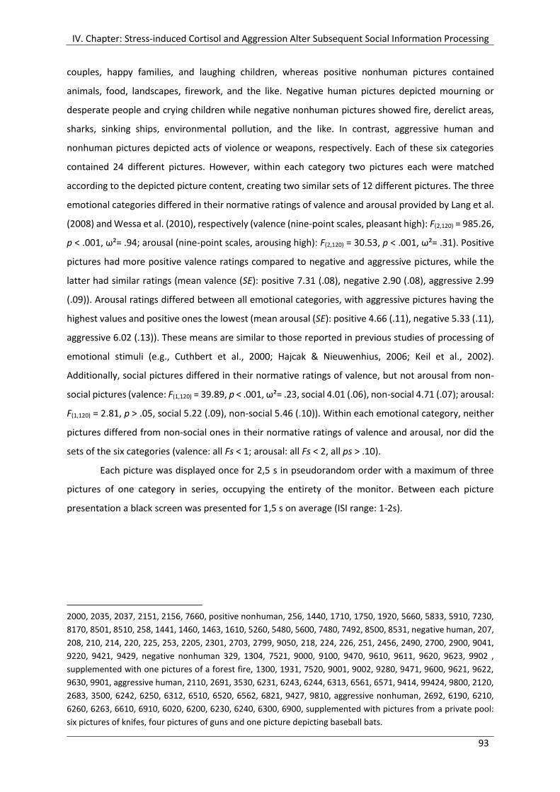
IV. Chapter: Stress-induced Cortisol and Aggression Alter Subsequent Social Information Processing
93
couples, happy families, and laughing children, whereas positive nonhuman pictures contained
animals, food, landscapes, firework, and the like. Negative human pictures depicted mourning or
desperate people and crying children while negative nonhuman pictures showed fire, derelict areas,
sharks, sinking ships, environmental pollution, and the like. In contrast, aggressive human and
nonhuman pictures depicted acts of violence or weapons, respectively. Each of these six categories
contained 24 different pictures. However, within each category two pictures each were matched
according to the depicted picture content, creating two similar sets of 12 different pictures. The three
emotional categories differed in their normative ratings of valence and arousal provided by Lang et al.
(2008) and Wessa et al. (2010), respectively (valence (nine-point scales, pleasant high): F(2,120) = 985.26,
p < .001, ω²= .94; arousal (nine-point scales, arousing high): F(2,120) = 30.53, p < .001, ω²= .31). Positive
pictures had more positive valence ratings compared to negative and aggressive pictures, while the
latter had similar ratings (mean valence (SE): positive 7.31 (.08), negative 2.90 (.08), aggressive 2.99
(.09)). Arousal ratings differed between all emotional categories, with aggressive pictures having the
highest values and positive ones the lowest (mean arousal (SE): positive 4.66 (.11), negative 5.33 (.11),
aggressive 6.02 (.13)). These means are similar to those reported in previous studies of processing of
emotional stimuli (e.g., Cuthbert et al., 2000; Hajcak & Nieuwenhius, 2006; Keil et al., 2002).
Additionally, social pictures differed in their normative ratings of valence, but not arousal from non-
social pictures (valence: F(1,120) = 39.89, p < .001, ω²= .23, social 4.01 (.06), non-social 4.71 (.07); arousal:
F(1,120) = 2.81, p > .05, social 5.22 (.09), non-social 5.46 (.10)). Within each emotional category, neither
pictures differed from non-social ones in their normative ratings of valence and arousal, nor did the
sets of the six categories (valence: all Fs < 1; arousal: all Fs < 2, all ps > .10).
Each picture was displayed once for 2,5 s in pseudorandom order with a maximum of three
pictures of one category in series, occupying the entirety of the monitor. Between each picture
presentation a black screen was presented for 1,5 s on average (ISI range: 1-2s).
2000, 2035, 2037, 2151, 2156, 7660, positive nonhuman, 256, 1440, 1710, 1750, 1920, 5660, 5833, 5910, 7230,
8170, 8501, 8510, 258, 1441, 1460, 1463, 1610, 5260, 5480, 5600, 7480, 7492, 8500, 8531, negative human, 207,
208, 210, 214, 220, 225, 253, 2205, 2301, 2703, 2799, 9050, 218, 224, 226, 251, 2456, 2490, 2700, 2900, 9041,
9220, 9421, 9429, negative nonhuman 329, 1304, 7521, 9000, 9100, 9470, 9610, 9611, 9620, 9623, 9902 ,
supplemented with one pictures of a forest fire, 1300, 1931, 7520, 9001, 9002, 9280, 9471, 9600, 9621, 9622,
9630, 9901, aggressive human, 2110, 2691, 3530, 6231, 6243, 6244, 6313, 6561, 6571, 9414, 99424, 9800, 2120,
2683, 3500, 6242, 6250, 6312, 6510, 6520, 6562, 6821, 9427, 9810, aggressive nonhuman, 2692, 6190, 6210,
6260, 6263, 6610, 6910, 6020, 6200, 6230, 6240, 6300, 6900, supplemented with pictures from a private pool:
six pictures of knifes, four pictures of guns and one picture depicting baseball bats.

IV. Chapter: Stress-induced Cortisol and Aggression Alter Subsequent Social Information Processing
94
4.2.5 Salivary Cortisol Measurement
Saliva samples for cortisol analysis were obtained using Salivette® collection devices (Sarstedt,
Nürnbrecht, Germany). Samples were collected at seven assessment points over the course of the
experiment: before the start of the experiment (C0, -15 min, with reference to the beginning of the
SECPT), before the SECPT (C1, -2 min), after the SECPT (C2, +5 min), after the TAP (C3, +20 min), before
the emotional picture task (C4, +30 min), after the emotional picture task (C5, +40 min), and before
debriefing (C6, +60 min). Sampling instructions were given via computer and Salivettes® were
positioned on the table in front of the participants. Immediately after the experiment, samples were
frozen for biochemical analysis. Salivary cortisol was analyzed with a time-resolved immunoassay with
fluorescence detection as described in detail elsewhere (Dressendörfer et al., 1992). Intra- and
interassay variability was less than 10 and 12%, respectively.
4.2.6 Procedure
Prior to the experimental session, participants were invited individually to an informational interview
to check exclusion criteria and to inform about the aim and procedure of the study, i.e., to assess the
relationship between stress, reaction time, and cognitive functions. They were informed at full length
that they might be exposed to a stress procedure involving cold water, videotaping, and observation
as well as loud noises. Additionally, the electroencephalogram (EEG) and the sampling of cortisol were
described. Moreover, participants were required to refrain from physical exercise on the day prior as
well as alcohol, caffeinated drinks, and meals within 1 h prior to the date fixed for experimental
session. All participants gave their written informed consent being aware that participation was
voluntary and that they may withdraw at any time without any consequences and without having to
give reasons.
The actual experiment was conducted between 01:30 p.m. and approximately 07:30 p.m.,
starting at 01:30 p.m., 03:30 p.m., and 05:30 p.m., where endogenous cortisol levels are low (Schreiber
et al., 2006). All participants were examined individually. Participants were randomly assigned to the
stress or control procedure as well as to the highly provoked or mildly provoked control group, while
sex was balanced across conditions. On arrival, the experimenter acquainted the participant with
another participant of the same sex, who was in fact a confederate of the investigator, with whom he
or she was to play a computer game during the experiment. Next, the participant was led in the EEG
laboratory, where he or she was seated in a dimly lit sound-attenuated room, 1 m from the monitor
(20 in. Eizo FlexScan S2031W). To further increase credibility of the cover story, the experimenter left
the laboratory to ostensibly lead the confederate into another EEG laboratory. After preparation of
EEG, electrooculogram (EOG), and electrocardiogram (ECG) recording devices, the actual experiment
started. The participants received all instructions for the different tasks, saliva samples, and

IV. Chapter: Stress-induced Cortisol and Aggression Alter Subsequent Social Information Processing
95
questionnaires via the computer screen. First, they worked on an Approach Avoidance Task
(description and results reported in Fechtner, 2012), then they were exposed to SECPT or the warm
water control condition. Afterwards, they played the Taylor Aggression Paradigm, followed by a second
block of the Approach Avoidance task. Finally, about 25 min after the SECPT was completed,
participants passively viewed pictures with different emotional and social content for about 10 min.
During the course of the experiment, participants filled out short state questionnaires (for details see
Chapter 3.2.4, p.50 ff.) several times and provided seven saliva samples for cortisol (C). After removal
of the physiological recording devices, participants were extensively debriefed. We thanked and
compensated them for their participation.
E-Prime presentation software (Eprime 2.0, Psychological Software Tools, Pittsburgh, PA) was
used to present the stimuli. The experiment, from arrival to debriefing had a duration of about 100
min. Figure 17 presents the timeline of the experimental procedure.
Figure 17. Timeline of the experimental session of study 2. SECPT:= Socially evaluated cold-pressor test. C := saliva
samples for cortisol analyses. Time information refers to the beginning of the SECPT. Experimental parts printed
in bold are topic of the present chapter.
4.2.7 EEG Recording and Quantification
The EEG was recorded from 32 electrode sites including the mastoids according to the 10–10 electrode
reference system (Chatrian et al., 1988) with the Easy-Cap electrode system (Falk Minow Services,
Munich). All sites were referenced to FCz. A bipolar horizontal EOG was recorded from the epicanthus
of each eye and a bipolar vertical EOG was recorded from supra- and infra-orbital positions of the left
eye. Ag/AgCl electrodes were used for EEG and EOG recording. Prior to the electrode placement, the
electrode sites on the participant's scalp and face were cleaned with alcohol and gently abraded. The
conduction was facilitated using Abralyt-light (FMS, Munich) electrode gel or EC2® Genuine Grass
Electrode Cream (Grass Products, Natus Neurology) for the EOG, respectively. A BrainAmp amplifier
(input impedance: 10 MΩ; Brain Products, GmbH) in AC mode was used to record the EEG and EOG at
1000 Hz using a pass-band set to 0.016 to 499 Hz (−12 dB/octave roll-off). All impedances of the EEG
Approach Avoidance
Task
SECPT
Taylor Aggression Paradigm
Approach Avoidance
Task
emotional pictures
C0
-15 min
C1
-2 min
C2
+5 min
C3
+20 min
C4
+30 min
C5
+40 min
C6
+60 min
EEG, EOG, ECG
preparation

IV. Chapter: Stress-induced Cortisol and Aggression Alter Subsequent Social Information Processing
96
electrodes were maintained below 10 kΩ. Data was stored to hard disk for later analysis using
BrainVision Analyzer 2 (Brain Products, Munich, Germany).
The EEG was re-referenced offline to linked mastoids. The data was resampled at 200 Hz and
low pass filtered using a digital filter with high cutoff of 12 Hz, 24 dB/oct. Artifacts due to eye
movements were corrected semiautomatically via the algorithm developed by Gratton et al. (1983). If
necessary, blinks were detected and marked using Ocular Correction with Independent Component
Analysis (ICA) before. Trials with non-physiological artifacts were excluded from analysis via
semiautomatic artifact rejection. EEG and EOG were epoched off-line into 1200-ms periods, starting
200 ms prior to picture onset and ending 1000 ms after picture onset. A baseline correction was
performed using the first 200-ms as a reference. Separate averages were computed for each electrode
and individual for the 6 different picture categories: positive human, positive nonhuman, negative
human, negative nonhuman, aggressive human and aggressive nonhuman. Using the grand average
across participants to guide window selection, ERP maximum peak amplitude (µV) for the stimulus-
locked P2, N2, and P3 component were detected semiautomatically for Fz, FCz, Cz, CPz, and Pz. ERP
values were assessed by measuring the component amplitude relative to the pre-stimulus baseline
and latency of local maximum peak amplitude from stimulus onset within windows of 180 - 250 ms
post stimulus for the P2, 250 - 300 ms for the N2, and 300 - 400 ms for the P3. Additionally, the late
positive potential (LPP) was identified based on visual inspection of grand average ERPs averaged
across all participants and quantified by computing the average amplitude from 500 to 1000 ms
relative to baseline14.
4.2.8 Statistical Analyses
The data was edited with Microsoft Excel 2003 and analyzed with SPSS 17.0 and IBM SPSS Statistics 20.
Non-normality of sampling distribution and violation of homogeneity of variance were checked using
Q-Q plots and Shapiro-Wilk tests of normality or Levene test, respectively. These analyses revealed
that the cortisol data was skewed and showed slight heterogeneity of variance. However, as the
analysis of variance is known to be robust against these violations if degrees of freedom for error are
greater than 20 and if sample sizes are large and fairly equal (Eid et al., 2010; Tabachnick & Fidell,
2007), I refrained from transformation of these data.
Stress Manipulation. Based on their cortisol reaction in response to the SECPT, participants of
the stress condition were post-hoc allocated to a cortisol-responder group or a cortisol-nonresponder
group: The stress-induced cortisol-response of each individual was computed by calculating the
14 As previous analyses of successive stimulus-locked intervals of 100 ms within the window of 500 – 1000 ms showed the same pattern of results in every interval, analysis and results of the average of the entire timeframe are presented.

IV. Chapter: Stress-induced Cortisol and Aggression Alter Subsequent Social Information Processing
97
difference of the cortisol levels C3 and C2, which reflected the HPA axis activation right before and
after the stressor. A median split (.79 nmol/l) of this cortisol change divided the participants of the
stress condition (male: n=24; female: n=24) into cortisol-responders (male: n=13; female: n=11) and
cortisol-nonresponders (male: n=11; female: n=13). The warm water control group consisted of 12
male and 11 female participants. Sample sizes within each condition are listed in Table 8. A 2 x 3 x 7
analysis of variance with the between-subjects factors gender (male, female), SECPT groups (cortisol-
responders, cortisol-nonresponders, warm water control group) and the within-subjects factor time of
cortisol measurement (C0-C6) was conducted to check whether the stress induction was successful,
how long the cortisol increase lasted and whether cortisol levels were influenced by gender.
Furthermore, a one-way analysis of variance with the factors gender (male, female) and SECPT groups
(cortisol-responders, cortisol-nonresponders, warm water control group) and difference of cortisol
level at time point C4 and C3 as the dependent variable was used to test significance of the stress
groups’ categorization and possible differences in male and female participants.
Table 8
Sample sizes in each condition of study 2
Men
(n = 36)
Women
(n= 35)
SECPT groups
high provocation
(n = 18)
low provocation
(n = 18)
high provocation
(n = 17)
low provocation
(n = 18)
Warm water control group (n=23) n = 6 n = 6 n = 5 n = 6
Cortisol-nonresponders (n=24) n = 6 n = 5 n = 7 n = 6
Cortisol-responders (n=24) n = 6 n = 7 n = 5 n = 6
Aggressive behavior in the TAP. In order to analyze the effects of increasing provocation on
aggressive behavior, a 2 x 2 x 3 mixed-design analysis of variance was conducted, including between-
subjects factors gender (male, female) and provocation (high provocation, low provocation) and the
within-subjects factor TAP Block (1, 2, 3).
Electrophysiological data. To investigate the effects of the acute cortisol rise in response to the
SECPT and provocation on the peak or mean amplitude of the four ERPs (P2, N2, P3, LPP) during
processing of emotional pictures, separated 3 x 2 x 2 x 3 x 2 x 5 mixed-design analyses of variance were
performed, including the between-subjects factors gender (male, female), SECPT groups (cortisol-
responders, cortisol-nonresponders, warm water control group), and provocation (high provocation,
low provocation) and the within-subject factors depicted valence (positive, negative, aggressive),
object (human vs. nonhuman), and electrode position (Fz, FCz, Cz, CPz, Pz).

IV. Chapter: Stress-induced Cortisol and Aggression Alter Subsequent Social Information Processing
98
The calculation of the sample size prior to the experiment showed that with sample size of
N=72 and a power of 1-ß=.80 an effect Ω² of at least .01 for highest order interactions of the ERP data
can be revealed. However, only effects greater than or equal to .05 were deemed relevant and are
reported. Hays’ ω² (Hays, 1974) was calculated as an effect size measure, with .01 considered as a
small effect, .05 considered as medium, and .14 considered as a large effect (Cohen, 1988). For main
effects of within-subject factors or interaction with those, ω² was corrected for mean correlation r̄ of
the respective levels or combination of those. In case the assumption of sphericity was violated, the
degrees of freedom for all ANOVAs were Huynh-Feldt corrected (Huynh & Feldt, 1976). The statistical
significance level was set to alpha = .05 (two-tailed). Where appropriate, Dunn's Multiple Comparison
Tests were used as post hoc tests. Since the focus of the present chapter is not on gender differences,
significant effects which comprise gender without interaction with either SECPT group or provocation
are not reported here.
4.3 Results
4.3.1 Stress Induction
Analysis of HPA- axis activation after the stressor or the control condition, respectively, revealed that
cortisol-responders showed a clear increase in cortisol (C3-C2) in response to the SECPT (M = 4.08
nmol/l, SE = .50) compared to cortisol-nonresponders (M = -.48 nmol/l, SE = .49) and participants of
the warm water control group (M = -.47 nmol/l, SE = .50), which both showed even a slight decrease
in cortisol levels (F(1,65) = 28.44, p < .001, ω² = .44). Subsequent post-hoc tests confirmed this pattern.
No further effects reached significance (all Fs < 1.16, all ps > .10).
Mean levels of free salivary cortisol over the course of the experiment of the three SECPT
groups are depicted in Figure 18. The analysis of variance showed a significant effect of time of cortisol
measurement (F(6,390) = 9.70, p < .001, ω² = .33, r̄ = .79), which was qualified by a significant interaction
of time of cortisol measurement and SECPT groups (F(12,390) = 7.37, p < .001, ω² = .57, r̄ = .88). Post-hoc
tests revealed that cortisol-responders had higher cortisol levels after the SECPT between points of
time C3 and C5 compared to cortisol-nonresponders and to the warm water control group. No
differences were found from points of time C0 to C2.
Male participants had in general higher cortisol levels (M = 5.39 nmol/ml, SE = .51) compared
to female participants (M = 3.58 nmol/ml, SE = .52) (F(1,65) = 6.31, p < .05, ω² = .07). This main effect
was qualified by an interaction with time of cortisol measurement (F(6,390) = 4.18, p < .05, ω² = .16, r̄ =
.80). Male participants had higher cortisol levels than female participants from points of time C0 to C4,
independently of stress manipulation or provocation.

IV. Chapter: Stress-induced Cortisol and Aggression Alter Subsequent Social Information Processing
99
Figure 18. Mean levels of free salivary cortisol during the experimental session of study 2 for cortisol-responders,
cortisol-nonresponders, and the warm water control group. Error bars indicate standard errors of the mean.
:= p < .05
4.3.2 Aggressive Behavior in the Taylor Aggression Paradigm
Effects of provocation. As intended, participants responded to high provocation with enhanced
aggressive behavior (M = 2.80, SE = .19) compared to those of the low provocation group (M = 1.79,
SE = .18) (F(1,67) = 14.90, p < .001, ω² = .16). Moreover, the analysis revealed a main effect of Tap Block
(F(2,134) = 18.03, p < .001, ω² = .33, r̄ = .68), which was qualified by an interaction of Tap Block and
provocation (F(2,134) = 30.20, p < .001, ω² = .52, r̄ = .75). As depicted in Figure 19, participants who were
subjected to increasing provocation in TAP Block 2 and 3 showed enhanced aggressive behavior, while
their behavior did not differ from mildly provoked participants in TAP Block 1. Post-hoc tests showed
that high provocation resulted in more aggressive behavior in TAP Block 2 and 3 compared to low
provocation. Likewise, participants respond to high provocation with increasing aggressive behavior
over all TAP Blocks.
2
3
4
5
6
7
8
9
10
C0 -15 C1 -5 C2 +5 C3 +20 C4 +30 C5 +40 C6 +60 min
salivary
cort
isol[n
mol/
l]warmwater control group
cortisol-nonresponder
cortisol-responder
SECPT TAPemotional pictures
Approach Avoidance
Approach Avoidance
C0 -15 C1 -2 C2 +5 C3 +20 C4 +30 C5 +40 C6 +60 min

IV. Chapter: Stress-induced Cortisol and Aggression Alter Subsequent Social Information Processing
100
Figure 19. Mean aggressive behavior in block 1, 2, and 3 of the Taylor Aggression Paradigm (TAP) for high versus
low provocation. Error bars indicate standard errors of the mean. Brackets indicate significant differences. p <
.05. Note: range of possible settings was 0 to 10.
4.3.3 Electrophysiological Data of Affective Picture Processing
Figure 20 shows grand average ERP responses to the six different categories of picture content
(positive human, positive nonhuman, negative human, negative nonhuman, aggressive human,
aggressive nonhuman) averaged over gender, SECPT groups, and provocation at Fz, Cz and Pz. The
general morphology of the waveform included an early positive peak at about 200 ms (P2), followed
by a negative peak at approximately 280 ms (N2), another positive peak at approximately 370 (P3),
and a late positive potential (LPP) starting about 500 ms and lasting till 1000 ms after stimulus onset.
The P2 peak amplitude (mean latency: 208.56 ms; SE = 1.63) showed a positive parietal
distribution (see Figure 21). Analysis of variance revealed a main effect of electrode position (F(4,236) =
126.62, p < .001, ω² = .89, r̄ = .82). Subsequent post hoc tests indicated that all electrodes differed
from each other, with negative values at frontal to central sides and increasing positive values at
centroparietal and parietal leads. The N2 peak amplitude (mean latency: 282.99 ms; SE = 1.65) was
most negative at frontal to frontocentral leads, as depicted in Figure 21 (electrode position: F(4,236) =
243.57, p < .001, ω² = .95, r̄ = .86). Post-hoc tests revealed that all electrodes differed from each other
significantly, except Fz and FCz. The P3 peak amplitude was distributed at parietal leads (see Figure 21,
third from left map). Statistical analysis showed that all electrodes differed from each other with the
exception of Fz and FCz (electrode position: F(4,236) = 254.06, p < .001, ω² = .95, r̄ = .84).
0
1
2
3
4
5
6
7
1 2 3
Tap Block
me
anag
gre
ssiv
e b
eh
avio
r
high provocation
low provocation

IV. Chapter: Stress-induced Cortisol and Aggression Alter Subsequent Social Information Processing
101
Figure 20. Grand average ERP waveforms at Fz, FCz, and Pz for the six different categories of picture content
(positive human, positive nonhuman, negative human, negative nonhuman, aggressive human, aggressive
nonhuman) averaged over gender, SEPCT groups, and provocation.
Figure 21. Grand grand mean topographic maps of the stimulus-locked P2, N2, P3, and LPP averaged over
valence, object, gender, SEPCT groups, and provocation. Respective time domain and scale division are specified
for every ERP below each map.

IV. Chapter: Stress-induced Cortisol and Aggression Alter Subsequent Social Information Processing
102
The LPP showed a parietal positivity, while frontal, frontocentral, and central leads showed negative
values (see Figure 21, fourth from left map) (F(4,236) = 243.76, p < .001, ω² = .94, r̄ = .84). Post-hoc tests
revealed that all electrodes differed from each other significantly.
4.3.3.1 Effect of Picture Content- Valence and Object
P2 peak amplitude. The statistical analysis showed that valence of picture content as well as
depicted object influenced the P2 amplitude independently (valence F(2,118) = 3.46, p < .05, ω² = .16, r̄
= .87; electrode position x valence (F(8,472) = 3.66, p < .01, ω² = .08, r̄ = .75), object (F(1,59) = 37.19, p <
.001, ω² = .66, r̄ = .87; electrode position x object (F(4,236) = 6.80, p < .01, ω² = .12, r̄ = .76)). The P2
amplitude for negative pictures was significantly more negative compared to positive (Fz, FCz, Cz, CPz)
and aggressive pictures (Fz, FCz, Cz), as depicted in Figure 22. Regarding depicted object, pictures of
humans elicited a more positive P2 amplitude compared to nonhuman objects at all electrode sites
(see Figure 23).
N2 peak amplitude. Positive, negative, and aggressive pictures elicited different N2 peak
amplitudes (valence: F(2,118) = 4.72, p < .05, ω² = .25, r̄ = .90; electrode position x valence: F(8,472) = 4.34,
p < .01, ω² = .11, r̄ = .80). Namely, the N2 for negative pictures was more negative compared to
aggressive pictures (Fz, FCz, Cz, CPz, Pz) and positive pictures (Fz, FCz). Aggressive pictures elicited a
less negative N2 peak amplitude than positive pictures (FCz, Cz, CPz, Pz) (see Figure 22). Besides, the
analysis of variance revealed an interaction between valence and object (F(2,118) = 3.55, p < .05, ω² =
.07, r̄ = .85), which was further qualified by a three-way interaction with electrode position (electrode
position x valence x object: F(8,472) = 4.45, p < .001, ω² = .07, r̄ = .82). The N2 peak amplitude for
aggressive human pictures was more negative compared to aggressive nonhuman pictures at frontal
to centroparietal sites. Similarly, positive human pictures led to a more negative N2 than positive
nonhuman picture content at centroparietal and parietal electrodes (cf. Figure 20).
P3 peak amplitude. Similar to the P2 and N2 component, picture content affected the P3 peak
amplitude. Analysis of variance revealed a main factor of valence (F(2,118) = 52.31, p < .001, ω² = .81, r̄
= .89) which was further qualified by an interaction of valence and electrode position (F(8,472) = 13.57, p
< .001, ω² = .30, r̄ = .78). As shown in Figure 22, aggressive pictures elicited a relatively more positive
P3 amplitude at all electrode sites compared to positive and negative pictures. Moreover, P3 amplitude
for positive pictures was relatively more positive compared to negative pictures at frontal to central
electrode sites. Additionally, analysis of variance revealed a main effect of object (F(1,59) = 6.91, p < .05,
ω² = .30, r̄ = .90) and a two-way interaction of object and valence (F(2,118) = 16.55, p < .001, ω² = .29, r̄
= .82). While positive (M = -2.56 µV, SE = .67) and negative human (M = -2.48 µV, SE = .66) pictures led
to relatively more positive P3 amplitudes compared to positive (M = -3.55 µV, SE = .62) and negative
nonhuman (M = -4.57 µV, SE = .60) pictures, respectively.
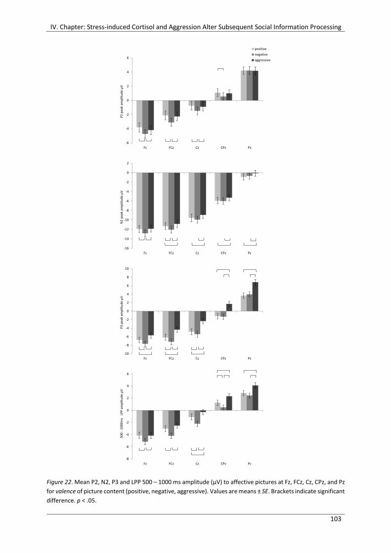
IV. Chapter: Stress-induced Cortisol and Aggression Alter Subsequent Social Information Processing
103
Figure 22. Mean P2, N2, P3 and LPP 500 – 1000 ms amplitude (µV) to affective pictures at Fz, FCz, Cz, CPz, and Pz
for valence of picture content (positive, negative, aggressive). Values are means ± SE. Brackets indicate significant
difference. p < .05.
-6
-4
-2
0
2
4
6
Fz FCz Cz CPz Pz
P2
pea
k am
plit
ud
e µ
V
positive
negative
aggressive
-16
-14
-12
-10
-8
-6
-4
-2
0
2
Fz FCz Cz CPz Pz
N2
pea
k am
plit
ud
e µ
V
-10
-8
-6
-4
-2
0
2
4
6
8
10
Fz FCz Cz CPz Pz
P3
pea
k am
plit
ud
e µ
V
-8
-6
-4
-2
0
2
4
6
Fz FCz Cz CPz Pz
50
0 -
10
00
ms
LP
P a
mp
litu
de
µV

IV. Chapter: Stress-induced Cortisol and Aggression Alter Subsequent Social Information Processing
104
This pattern was reversed for aggressive pictures. Aggressive human pictures (M = -1.23 µV, SE = .68)
elicited a reduced P3 amplitude compared to aggressive nonhuman depicted objects (M = -.30 µV, SE
= .58) (cf. Figure 20).
LPP 500-100 ms. In line with the other ERP components, the LPP amplitude differed with regard
to valence of picture content and depicted object (valence: F(2,118) = 15.71, p < .001, ω² = .38, r̄ = .79;
electrode position x valence: F(8,472) = 4.98, p < .001, ω² = .10, r̄ = .72; object: F(1,59) = 31.98, p < .001, ω²
= .64, r̄ = .88; electrode position x object: F(4,236) = 3.52, p < .05, ω² = .10, r̄ = .88)). Again, aggressive
pictures elicited a relatively more positive LPP amplitude compared to negative pictures (Fz through
Pz) and positive pictures (Cz through Pz). Besides, the LPP amplitude for positive pictures was relatively
more positive compared to negative pictures at frontal to centroparietal sites (see Figure 22).
Regarding depicted object, similar to the P2 amplitude, pictures of human elicited an enhanced LPP
amplitude compared to nonhuman objects at all electrode sites (see Figure 23).
Figure 23. Mean P2 and LPP 500 – 1000 ms amplitude (µV) to affective pictures at Fz, FCz, Cz, CPz and Pz for
depicted object (human, nonhuman). Values are means ± SE. Brackets indicate significant difference. p < .05.
-8
-6
-4
-2
0
2
4
6
Fz FCz Cz CPz Pz
P2
pe
ak a
mp
litu
de
µV
human objects
nonhuman objects
-8
-6
-4
-2
0
2
4
6
Fz FCz Cz CPz Pz
50
0 -
10
00
ms
LP
P a
mp
litu
de
µV
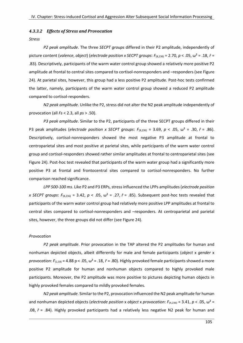
IV. Chapter: Stress-induced Cortisol and Aggression Alter Subsequent Social Information Processing
105
4.3.3.2 Effects of Stress and Provocation
Stress
P2 peak amplitude. The three SECPT groups differed in their P2 amplitude, independently of
picture content (valence, object) (electrode position x SECPT groups: F(8,236) = 2.70, p < .05, ω² = .18, r̄ =
.83). Descriptively, participants of the warm water control group showed a relatively more positive P2
amplitude at frontal to central sites compared to cortisol-nonresponders and –responders (see Figure
24). At parietal sites, however, this group had a less positive P2 amplitude. Post-hoc tests confirmed
the latter, namely, participants of the warm water control group showed a reduced P2 amplitude
compared to cortisol-responders.
N2 peak amplitude. Unlike the P2, stress did not alter the N2 peak amplitude independently of
provocation (all Fs < 2.3, all ps > .50).
P3 peak amplitude. Similar to the P2, participants of the three SECPT groups differed in their
P3 peak amplitudes (electrode position x SECPT groups: F(8,236) = 3.69, p < .05, ω² = .30, r̄ = .86).
Descriptively, cortisol-nonresponders showed the most negative P3 amplitude at frontal to
centroparietal sites and most positive at parietal sites, while participants of the warm water control
group and cortisol-responders showed rather similar amplitudes at frontal to centroparietal sites (see
Figure 24). Post-hoc test revealed that participants of the warm water group had a significantly more
positive P3 at frontal and frontocentral sites compared to cortisol-nonresponders. No further
comparison reached significance.
LPP 500-100 ms. Like P2 and P3 ERPs, stress influenced the LPPs amplitudes (electrode position
x SECPT groups: F(8,236) = 3.42, p < .05, ω² = .27, r̄ = .85). Subsequent post-hoc tests revealed that
participants of the warm water control group had relatively more positive LPP amplitudes at frontal to
central sites compared to cortisol-nonresponders and –responders. At centroparietal and parietal
sites, however, the three groups did not differ (see Figure 24).
Provocation
P2 peak amplitude. Prior provocation in the TAP altered the P2 amplitudes for human and
nonhuman depicted objects, albeit differently for male and female participants (object x gender x
provocation: F(1,59) = 4.88 p < .05, ω² = .18, r̄ = .80). Highly provoked female participants showed a more
positive P2 amplitude for human and nonhuman objects compared to highly provoked male
participants. Moreover, the P2 amplitude was more positive to pictures depicting human objects in
highly provoked females compared to mildly provoked females.
N2 peak amplitude. Similar to the P2, provocation influenced the N2 peak amplitude for human
and nonhuman depicted objects (electrode position x object x provocation: F(4,236) = 3.41, p < .05, ω² =
.08, r̄ = .84). Highly provoked participants had a relatively less negative N2 peak for human and
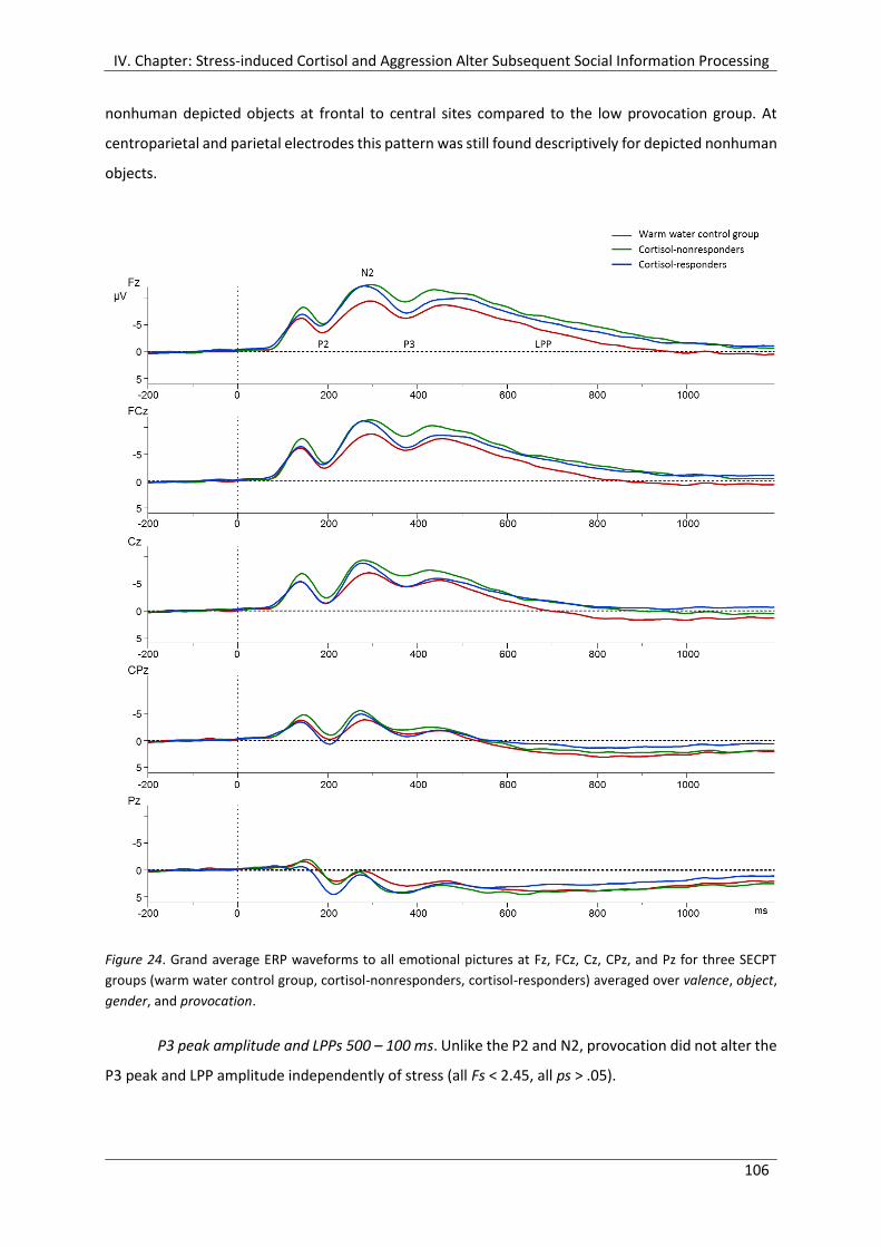
IV. Chapter: Stress-induced Cortisol and Aggression Alter Subsequent Social Information Processing
106
nonhuman depicted objects at frontal to central sites compared to the low provocation group. At
centroparietal and parietal electrodes this pattern was still found descriptively for depicted nonhuman
objects.
Figure 24. Grand average ERP waveforms to all emotional pictures at Fz, FCz, Cz, CPz, and Pz for three SECPT
groups (warm water control group, cortisol-nonresponders, cortisol-responders) averaged over valence, object,
gender, and provocation.
P3 peak amplitude and LPPs 500 – 100 ms. Unlike the P2 and N2, provocation did not alter the
P3 peak and LPP amplitude independently of stress (all Fs < 2.45, all ps > .05).
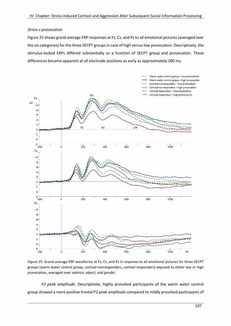
IV. Chapter: Stress-induced Cortisol and Aggression Alter Subsequent Social Information Processing
107
Stress x provocation
Figure 25 shows grand average ERP responses at Fz, Cz, and Pz to all emotional pictures (averaged over
the six categories) for the three SECPT groups in case of high versus low provocation. Descriptively, the
stimulus-locked ERPs differed substantially as a function of SECPT group and provocation. These
differences became apparent at all electrode positions as early as approximately 200 ms.
Figure 25. Grand average ERP waveforms at Fz, Cz, and Pz in response to all emotional pictures for three SECPT
groups (warm water control group, cortisol-nonresponders, cortisol-responders) exposed to either low or high
provocation, averaged over valence, object, and gender.
P2 peak amplitude. Descriptively, highly provoked participants of the warm water control
group showed a more positive frontal P2 peak amplitude compared to mildly provoked participants of
IV. Chapter: Stress-induced Cortisol and Aggression Alter Subsequent Social Information Processing
108
this group, as well as to cortisol-nonresponders and -responders. Particularly at parietal sites, it
became apparent that cortisol-responders did not differ in their P2 amplitude comparing high versus
low provocation. However, statistical analysis revealed no significant influence of the interaction
between stress and provocation (interactions containing SECPT groups x provocation: all Fs < 1.29; all
ps > .10).
N2 peak amplitude. The N2 peak amplitude was significantly influenced by stress and
provocation as a function of valence, object, and gender (valence x object x gender x SECPT groups x
provocation: F(4,118) = 2.63, p < .05, ω² = .12, r̄ = .89). As shown in Figure 26, subsequent post-hoc tests
revealed that highly provoked female participants of the warm water control group showed distinctly
reduced negative N2 amplitude for all human and nonhuman emotional pictures in comparison to
mildly provoked females of this group. Male participants of the control group, on the other hand, did
not differ in their N2 amplitude when subjected to high or low provocation. Descriptively, cortisol-
nonresponders showed a prominently different pattern. High provocation led to more negative N2
amplitudes in female participants for positive human and negative nonhuman pictures as well as
aggressive human and nonhuman pictures. In contrast, high provocation in male participants only led
to more negative N2 amplitudes in aggressive human pictures. However, this did not reach
significance. Besides, male cortisol-responders reacted to high provocation with more negative N2
amplitudes for all emotional human and nonhuman pictures. Post-hoc tests confirmed significance for
this pattern for positive nonhuman pictures. On the contrary, female cortisol-responders showed
descriptively diminished N2 amplitude in response to high provocation versus low provocation; this
reached significance regarding positive nonhuman pictures.
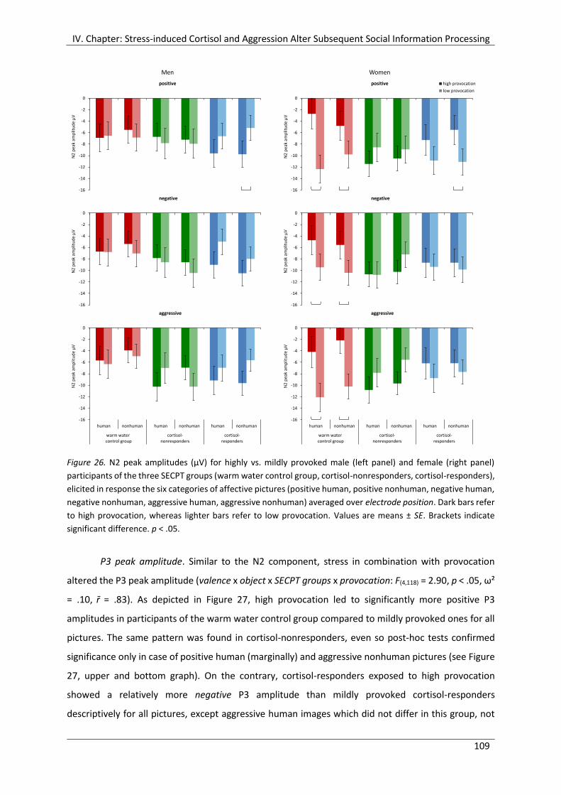
IV. Chapter: Stress-induced Cortisol and Aggression Alter Subsequent Social Information Processing
109
Figure 26. N2 peak amplitudes (µV) for highly vs. mildly provoked male (left panel) and female (right panel)
participants of the three SECPT groups (warm water control group, cortisol-nonresponders, cortisol-responders),
elicited in response the six categories of affective pictures (positive human, positive nonhuman, negative human,
negative nonhuman, aggressive human, aggressive nonhuman) averaged over electrode position. Dark bars refer
to high provocation, whereas lighter bars refer to low provocation. Values are means ± SE. Brackets indicate
significant difference. p < .05.
P3 peak amplitude. Similar to the N2 component, stress in combination with provocation
altered the P3 peak amplitude (valence x object x SECPT groups x provocation: F(4,118) = 2.90, p < .05, ω²
= .10, r̄ = .83). As depicted in Figure 27, high provocation led to significantly more positive P3
amplitudes in participants of the warm water control group compared to mildly provoked ones for all
pictures. The same pattern was found in cortisol-nonresponders, even so post-hoc tests confirmed
significance only in case of positive human (marginally) and aggressive nonhuman pictures (see Figure
27, upper and bottom graph). On the contrary, cortisol-responders exposed to high provocation
showed a relatively more negative P3 amplitude than mildly provoked cortisol-responders
descriptively for all pictures, except aggressive human images which did not differ in this group, not
-16
-14
-12
-10
-8
-6
-4
-2
0
N2
pe
ak a
mp
litu
de
µV
positive
-16
-14
-12
-10
-8
-6
-4
-2
0
N2
pe
ak a
mp
litu
de
µV
positive
-16
-14
-12
-10
-8
-6
-4
-2
0
N2
pe
ak a
mp
litu
de
µV
negative
-16
-14
-12
-10
-8
-6
-4
-2
0
N2
pe
ak a
mp
litu
de
µV
negative
-16
-14
-12
-10
-8
-6
-4
-2
0
human nonhuman human nonhuman human nonhuman
warm water control group
cortisol-nonresponders
cortisol-responders
N2
pe
ak a
mp
litu
de
µV
aggressive
-8
-6
-4
-2
0
2
4
6
warm water control group
cortisol-nonresponders
cortisol-responders
50
0 -
10
00
ms
LP
P a
mp
litu
de
µV
high provocation
low provocation
Men Women
-16
-14
-12
-10
-8
-6
-4
-2
0
human nonhuman human nonhuman human nonhuman
warm water control group
cortisol-nonresponders
cortisol-responders
N2
pe
ak a
mp
litu
de
µV
aggressive
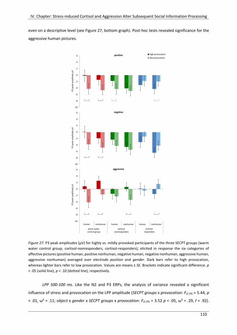
IV. Chapter: Stress-induced Cortisol and Aggression Alter Subsequent Social Information Processing
110
even on a descriptive level (see Figure 27, bottom graph). Post-hoc tests revealed significance for the
aggressive human pictures.
Figure 27. P3 peak amplitudes (µV) for highly vs. mildly provoked participants of the three SECPT groups (warm
water control group, cortisol-nonresponders, cortisol-responders), elicited in response the six categories of
affective pictures (positive human, positive nonhuman, negative human, negative nonhuman, aggressive human,
aggressive nonhuman) averaged over electrode position and gender. Dark bars refer to high provocation,
whereas lighter bars refer to low provocation. Values are means ± SE. Brackets indicate significant difference. p
< .05 (solid line), p < .10 (dotted line), respectively.
LPP 500-100 ms. Like the N2 and P3 ERPs, the analysis of variance revealed a significant
influence of stress and provocation on the LPP amplitude (SECPT groups x provocation: F(2,59) = 5.44, p
< .01, ω² = .11; object x gender x SECPT groups x provocation: F(2,59) = 3.52 p < .05, ω² = .29, r̄ = .92).
-10
-8
-6
-4
-2
0
2
4
6
human nonhuman human nonhuman human nonhuman
warm water control group
cortisol-nonresponders
cortisol-responders
P3
pe
ak a
mp
litu
de
µV
positive
-10
-8
-6
-4
-2
0
2
4
6
human nonhuman human nonhuman human nonhuman
warm water control group
cortisol-nonresponders
cortisol-responders
P3
pe
ak a
mp
litu
de
µV
negative
-10
-8
-6
-4
-2
0
2
4
6
human nonhuman human nonhuman human nonhuman
warm water control group
cortisol-nonresponders
cortisol-responders
P3
pe
ak a
mp
litu
de
µV
aggressive
-8
-6
-4
-2
0
2
4
6
warm water control group
cortisol-nonresponders
cortisol-responders
50
0 -
10
00
ms
LP
P a
mp
litu
de
µV
high provocation
low provocation
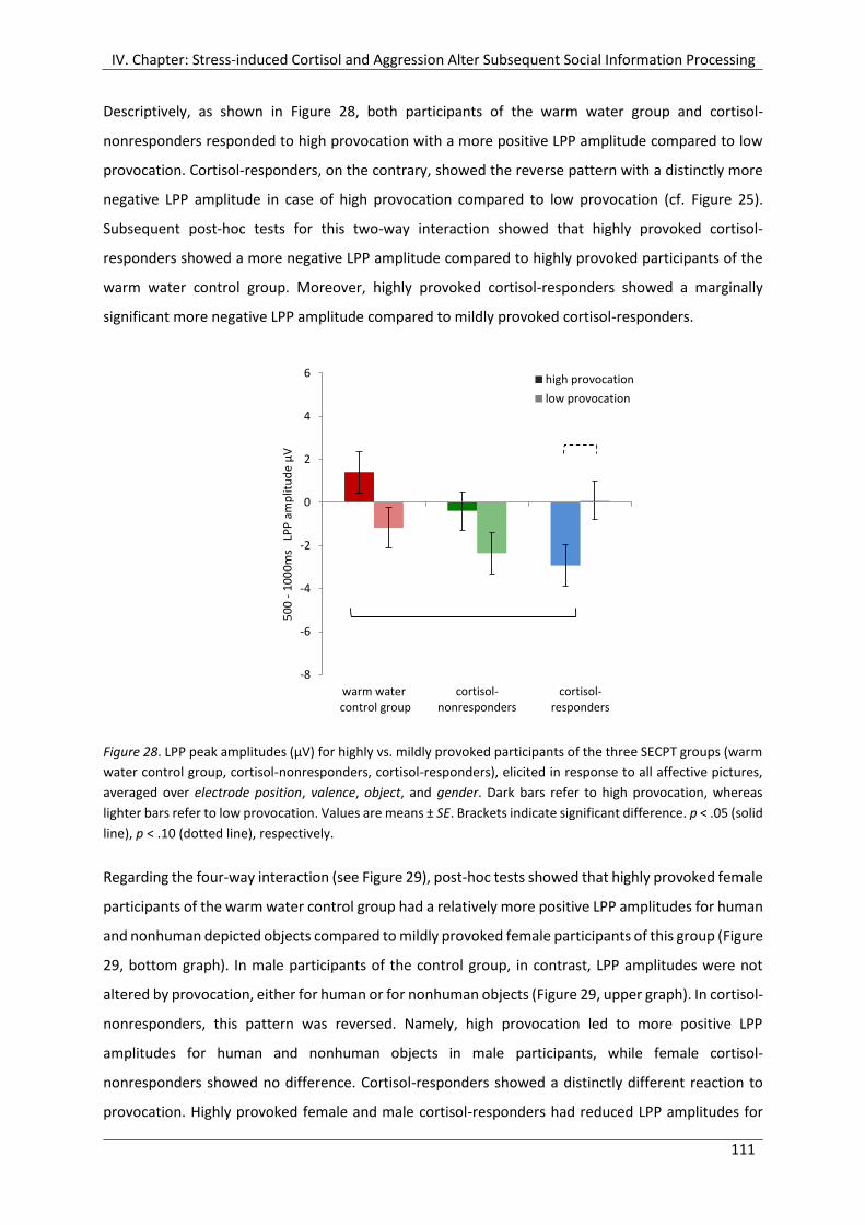
IV. Chapter: Stress-induced Cortisol and Aggression Alter Subsequent Social Information Processing
111
Descriptively, as shown in Figure 28, both participants of the warm water group and cortisol-
nonresponders responded to high provocation with a more positive LPP amplitude compared to low
provocation. Cortisol-responders, on the contrary, showed the reverse pattern with a distinctly more
negative LPP amplitude in case of high provocation compared to low provocation (cf. Figure 25).
Subsequent post-hoc tests for this two-way interaction showed that highly provoked cortisol-
responders showed a more negative LPP amplitude compared to highly provoked participants of the
warm water control group. Moreover, highly provoked cortisol-responders showed a marginally
significant more negative LPP amplitude compared to mildly provoked cortisol-responders.
Figure 28. LPP peak amplitudes (µV) for highly vs. mildly provoked participants of the three SECPT groups (warm
water control group, cortisol-nonresponders, cortisol-responders), elicited in response to all affective pictures,
averaged over electrode position, valence, object, and gender. Dark bars refer to high provocation, whereas
lighter bars refer to low provocation. Values are means ± SE. Brackets indicate significant difference. p < .05 (solid
line), p < .10 (dotted line), respectively.
Regarding the four-way interaction (see Figure 29), post-hoc tests showed that highly provoked female
participants of the warm water control group had a relatively more positive LPP amplitudes for human
and nonhuman depicted objects compared to mildly provoked female participants of this group (Figure
29, bottom graph). In male participants of the control group, in contrast, LPP amplitudes were not
altered by provocation, either for human or for nonhuman objects (Figure 29, upper graph). In cortisol-
nonresponders, this pattern was reversed. Namely, high provocation led to more positive LPP
amplitudes for human and nonhuman objects in male participants, while female cortisol-
nonresponders showed no difference. Cortisol-responders showed a distinctly different reaction to
provocation. Highly provoked female and male cortisol-responders had reduced LPP amplitudes for
-8
-6
-4
-2
0
2
4
6
warm water control group
cortisol-nonresponders
cortisol-responders
50
0 -
10
00
ms
LP
P a
mp
litu
de
µV
high provocation
low provocation
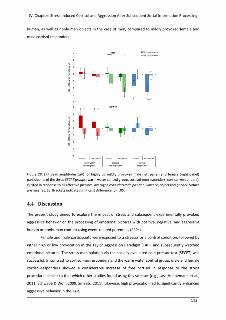
IV. Chapter: Stress-induced Cortisol and Aggression Alter Subsequent Social Information Processing
112
human, as well as nonhuman objects in the case of men, compared to mildly provoked female and
male cortisol-responders.
Figure 29. LPP peak amplitudes (µV) for highly vs. mildly provoked male (left panel) and female (right panel)
participants of the three SECPT groups (warm water control group, cortisol-nonresponders, cortisol-responders),
elicited in response to all affective pictures, averaged over electrode position, valence, object and gender. Values
are means ± SE. Brackets indicate significant difference. p < .05.
4.4 Discussion
The present study aimed to explore the impact of stress and subsequent experimentally provoked
aggressive behavior on the processing of emotional pictures with positive, negative, and aggressive
human or nonhuman content using event-related potentials (ERPs).
Female and male participants were exposed to a stressor or a control condition, followed by
either high or low provocation in the Taylor Aggression Paradigm (TAP), and subsequently watched
emotional pictures. The stress manipulation via the socially evaluated cold-pressor test (SECPT) was
successful. In contrast to cortisol-nonresponders and the warm water control group, male and female
cortisol-responders showed a considerable increase of free cortisol in response to the stress
procedure, similar to that which other studies found using this stressor (e.g., Lass-Hennemann et al.,
2011; Schwabe & Wolf, 2009; Smeets, 2011). Likewise, high provocation led to significantly enhanced
aggressive behavior in the TAP.
-8
-6
-4
-2
0
2
4
6
50
0 -
10
00
ms
LP
P a
mp
litu
de
µV
Men
-8
-6
-4
-2
0
2
4
6
human nonhuman human nonhuman human nonhuman
warm water control group
cortisol-nonresponders
cortisol-responders
50
0 -
10
00
ms
LP
P a
mp
litu
de
µV
Women
-8
-6
-4
-2
0
2
4
6
warm water control group
cortisol-nonresponders
cortisol-responders
50
0 -
10
00
ms
LP
P a
mp
litu
de
µV
high provocation
low provocation

IV. Chapter: Stress-induced Cortisol and Aggression Alter Subsequent Social Information Processing
113
4.4.1 Effects of Picture Content on Processing of Affective Pictures
Established effects within the event-related potentials (ERPs) of affective picture processing could be
replicated: picture valence had a distinct effect on early to later stages of information processing.
Negative pictures elicited relatively more negative frontal to central P2, N2, P3, and LPP amplitudes
compared to positive pictures, in line with other studies (e.g., Carretie et al., 2004; Cuthbert et al.,
2000; Olofsson et al., 2008). Confirming previous findings, pictures with aggressive content led to
relatively more positive N2, P3, and central to parietal LPP amplitudes compared to positive pictures,
confirming the so-called negativity bias (Ito et al., 1998; Schupp et al., 2000; Thomas, Johnstone, &
Gonsalvez, 2007; Tso, Chiu, King-Casas, & Deldin, 201115). Interestingly, aggressive pictures also elicited
more positive P2, N2, P3, and LPP amplitudes relative to negative pictures as well. To my knowledge,
there is sparse knowledge about the distinction in processing of the different subcategories of negative
stimuli. Hot and Sequeira (2013), analyzing the neural correlates of positive and two different negative
picture categories, i.e., disgusting and sad pictures, reported differences at early ERP components for
each of the three emotions and concluded that each emotion might cause a specific response pattern
in brain activation. The present findings confirm this assumption, suggesting a more elaborate
processing of aggressive pictures at both early and later stages of the information processing stream
relative to other negative pictures. This is in line with the so-called threat-superiority effect, which was
found for both phylogenetically (i.e., ancient threats as snakes) and ontogenetically (i.e., modern
threats as guns) threatening stimuli, albeit only in comparison to neutral stimuli (cf. Blanchette, 2006;
Brown, El-Deredy, & Blanchette, 2010; Fox, Griggs, & Mouchlianitis, 2007). Besides, other studies
revealed enhanced early and later ERP amplitudes for highly arousing pictures, comprised of aggressive
content among others, relative to low arousing ones (e.g., Amrhein, Mühlberger, Pauli, & Wiedemann,
2004; Hajcak, MacNamara, & Olvet, 2010; Junghöfer, Bradley, Elbert, & Lang, 2001; Keil et al., 2002;
Schupp et al., 2004). Regarding the depicted object, pictures of humans elicited relatively enhanced
early (P2, N2) as well as later (P3, LPP) amplitudes, confirming results of previous studies, (Groen,
Wijers, Tucha, & Althaus, 2013; Proverbio, Adorni, Zani, & Trestianu, 2009; Proverbio, Zani, & Adorni,
2008). Moreover, this pattern was altered by valence of the pictures. Most importantly, however,
stress and provocation both jointly and independently influenced the information processing stream.
4.4.2 Combined Effects of Stress and Provocation on Processing of Affective Pictures
In accordance with the hypothesis, stress-induced elevated cortisol levels and provocation jointly
altered social information processing. High provocation in the TAP led to a distinctly different
15 Note: Ito, Larsen, Smith, and Cacioppo (1998) used IAPS pictures no. 3030 and 6230, depicting a mutilated face and a handgun; hence these stimuli are comparable rather with the aggressive human pictures, used in the present study.
IV. Chapter: Stress-induced Cortisol and Aggression Alter Subsequent Social Information Processing
114
electrocortical response to affective pictures in cortisol-responders relative to cortisol-nonresponders
and participants of the warm water control group. This impact was equally apparent in early as well as
later stages of information processing, albeit - contrary to the hypothesis - not specific for threat-
related stimuli.
While the mutual impact of stress and provocation was found in the time domain of the P2 on
a mere descriptive level, it reached significance for the N2 regarding the depicted object (i.e., human
vs. nonhuman) with different directions for male and female participants. High provocation led to
reduced N2 amplitudes in females of the warm water control group, whereas N2 amplitudes of men
in this group remained unaffected. Stress-induced increase in cortisol in combination with high
provocation, however, led to more negative N2 amplitudes for all picture categories in men, reaching
significance for positive nonhuman pictures. On the contrary, highly provoked female cortisol-
responders showed diminished N2 amplitudes relative to those mildly provoked, similar to the pattern
of the warm water control group. However, this effect was less pronounced, reaching significance only
in case of positive nonhuman picture content. Thus, an endogenous surge of cortisol together with
high provocation had the opposite effect in male participants than in females.
Gender differences in the processing of emotional visual stimuli have been frequently reported
(e.g., Bradley et al., 2001; Kim et al., 2013; Stevens & Hamann, 2012; Wrase et al., 2003). Wrase et al.
(2003), for instance, found greater cortical activation for negative pictures in the anterior and medial
frontal gyrus in female compared to male participants. Similarly, Bradley et al. (2001), investigating
peripheral physiological responses to emotional and neutral pictures, conclude that “women showed
a broad disposition to respond with greater defensive reactivity to aversive pictures” (p. 300).
Regarding event-related potentials, the present results are in line with previous studies finding the
anterior N2 sensitive for gender differences in processing of social affective stimuli (Groen et al., 2013;
Proverbio et al., 2009; Proverbio et al., 2008). Female participants in those studies showed enhanced
N2 amplitudes to (positive) pictures depicting humans compared to unanimated scenes (Groen et al.,
2013; Proverbio et al., 2009; Proverbio et al., 2008) and negative pictures independent of human or
nonhuman content (Groen et al., 2013). Hence, the enhanced N2 amplitudes for mildly provoked
female participants in the warm water control group support the assumption that women
preferentially process social and affective, especially aversive, information relative to men.
High provocation reduced N2 amplitudes in females of the control group, equalizing them to
the level of men of the control group. The impact of violent cues and provocation on aggressive
behavior as a function of gender was reviewed by Bettencourt and Kernahan (1997). They concluded
that violent cues in combination with aversive provocation caused enhanced aggression in females,
equalizing gender differences in aggressive behavior. Extending these assumptions, the present data
suggests that high provocation alters already the processing of affective information. And yet, not only

IV. Chapter: Stress-induced Cortisol and Aggression Alter Subsequent Social Information Processing
115
threat-related stimuli were processed differently in females after an aggressive encounter, but also of
positive and non-violent negative material. Moreover, stress exposure and the physiological reactivity
in response to it altered this impact gender-specifically, too. While stress-induced HPA-axis activation
reduced the influence of provocation in females, especially on negative and aggressive picture
categories, male participants under high provocation and increased cortisol levels showed a
preferential processing of all types of pictures, reflected by (descriptively) enhanced N2 amplitudes
compared to those mildly provoked.
So far no consistent concept of the functionality and psychological interpretation of the N2
component in the context of the processing of emotional stimuli exist (c.f. Kovalenko, Pavlenko, &
Chernyi, 2010; Tso et al., 2011). Based on paradigms such as distraction paradigms and oddball
paradigms, the anterior N2 has been shown to be modulated by novelty and mismatch (Carretie et al.,
2004; Folstein & van Petten, 2008; Kropotov et al., 2011; Tarbi, Sun, Holcomb, & Daffner, 2011). Thus,
larger anterior N2 amplitudes have been found for novel and emotional stimuli, even if they served as
distractors or deviants (Carretie et al., 2004; Tarbi et al., 2011). López-Martín, Albert, Fernández-Jaén,
and Carretié (2013) concluded that “this N2 component is related to stimulus-driven attentional
processes.” (p. 18). Likewise, increased N2 amplitudes have been assumed to reflect enhanced
automatic processing (Schupp et al., 2006). Furthermore, Gardener, Carr, MacGregor, Felmingham,
and Gray (2013) found enhanced N2 amplitude for negative images, especially in women and
construed this as enhanced emotional reactivity in accordance with a female negativity bias. Following
these interpretations, increased cortisol levels together with high provocation led to enhanced
emotional reactivity in men for all pictures, capturing more automatic attention allocation. The same
applies to highly provoked female participants who showed no HPA axis activation in response to the
stressor, albeit only by tendency. While high provocation caused deficits in the processing of emotional
stimuli in females of the warm water control group, an endogenous surge of cortisol attenuated this
effect, albeit without abolishing it, indicating minor enhanced emotional reactivity by tendency as well.
Yet, this was still minor relative to low provocation.
Unlike the N2, the mutual impact of stress and provocation on the P3 was independent of
gender. High provocation led to more positive P3 amplitudes for all pictures in the warm water control
group. A similar pattern was found in cortisol-nonresponders, albeit less pronounced, reaching
significance only in case of nonhuman aggressive pictures. The opposite was found in cortisol-
responders. More precisely, preceding aggressive behavior due to high provocation resulted in
reduced P3 amplitudes, especially for nonhuman aggressive pictures.
The P3 amplitude is primarily determined by task-relevance, stimulus probability, motivational
significance, and stimulus salience (e.g., Kok, 2001; Olofsson et al., 2008; Polich, 2007; Polich & Kok,
1995 and cited therein). As probability of and instructions for the pictures did not differ between the
IV. Chapter: Stress-induced Cortisol and Aggression Alter Subsequent Social Information Processing
116
categories, the last two are more important in the current context of passive emotional information
processing. In this respect, previous research revealed the P3 to be sensitive to arousal and valence,
with more positive P3 amplitudes for high arousing and positive (and occasionally negative images)
relative to low arousing or neutral ones, respectively (Amrhein et al., 2004; Keil et al., 2002; Olofsson
et al., 2008; Rozenkrants, Olofsson, & Polich, 2007; Yuan et al., 2007). Accordingly, Hajcak et al. (2010)
concluded that “emotional stimuli might be automatically processed as task-relevant, and because of
their intrinsic motivational significance, emotional stimuli might be considered natural targets.”
(p. 133). Hence, high provocation and subsequent aggressive behavior led to enhanced “motivated
attention” (Bradley et al., 2003) towards all kind of emotional stimuli in the warm water control group.
This contradicts previous results in the context of aggression, reporting fairly consistently reduced P3
amplitudes in participants showing antisocial and aggressive behavior (e.g., Bernat et al., 2007; Gao
& Raine, 2009; Gerstle et al., 1998 for a review see Patrick, 2008). These studies, however, did not
investigate the immediate consequences of an aggressive encounter or actual provoked aggressive
behavior on subsequent information processing, but rather concentrate on trait aggressiveness, self-
reported measures of aggression, or (habitual) exposure to media violence. In contrast, Bertsch,
Böhnke, Kruk, and Naumann (2009) reported greater P3 amplitudes in an emotional Stroop task after
provoked aggressive behavior in the TAP. In accordance with the present findings, P3 amplitudes were
enhanced for all facial expression (i.e., neutral and emotional) in highly provoked participants (Bertsch,
Böhnke, Kruk, & Naumann, 2009). Hence, emotional stimuli in the present study gained in importance
after experimentally provoked aggressive behavior, irrespectively of valence and (un-)animated
content. Nonetheless, whether and how this unspecific altered processing would influence subsequent
situation in respect of aggressive behavior is unclear. To my knowledge so far no study reported
enhanced P3 amplitudes to be positively associated with aggressive or antisocial behavior (see below).
Prior exposure to the stressor abolished or even inverted this impact as a function of acute
HPA axis activation. Stress without an HPA axis activation resulted solely in a preferential processing
of weapons (i.e., nonhuman aggressive pictures) in case of high provocation, while all other emotional
stimuli were processed without enhanced motivational attention relative to low provocation. Under
an acute surge of cortisol and high provocation, however, emotional stimuli received reduced
motivated attention, specifically applying for images of weapons, contradicting the hypothesis. Yet,
the selective attention bias to threat-related objects (i.e., weapons) found in mildly provoked cortisol-
responders, reflected by enhanced P3 amplitudes these aggressive pictures, is in line with previous
studies reporting preferential processing for threat due to cortisol (e.g., Akinola & Mendes, 2012;
Putman, Hermans, & van Honk, 2007; Roelofs et al., 2007). From an evolutionary perspective, this
hypervigilant attention towards and assessment of threat signals from the environment is highly
adaptive as it promotes the response that most likely guarantee the survival of the individual, for

IV. Chapter: Stress-induced Cortisol and Aggression Alter Subsequent Social Information Processing
117
instance fight or flight responses (Cannon, 1929; de Kloet, et al., 2005; van Marle et al., 2009). High
provocation seems to abolish this bias, reflected by reduced P3 amplitudes.
Reduced P3 amplitudes for stimuli depicting violence were assumed to reflect a desensitization
which in turns lowers the threshold for aggressive behavior to occur (Bartholow et al., 2006; Engelhardt
et al., 2011). Similarly, Felmingham, Bryant, Kendall, and Gordon (2002) found the P3 amplitude in an
auditory oddball paradigm to be negatively correlated to symptoms of numbness in patients diagnosed
with posttraumatic stress disorder (PTSD). Hence, high glucocorticoid stress responsiveness in
combination with high provocation resulted in a more or less general desensitization or numbness,
respectively, for all emotional stimuli, albeit most pronounced for weapons. In the sense of escalation
of aggression or its persistence beyond the actual encounter, this callousness might imply a facilitation
of further aggressive and violent behavior. Indeed, reduced P3 amplitudes predicted enhanced
subsequent aggressive behavior (Bartholow et al., 2006; Engelhardt et al., 2011) and callousness is
associated with severe antisocial behavior (Pardini, 2006).
However, an alternative interpretation might be that those participants actively suppress the
processing of emotional stimuli to protect their emotional state from further influences. Krompinger,
Moser, and Simons (2008) and Moser, Hajcak, Bukay, and Simons (2006) found reduced amplitudes
within the time domain of the P3 for positive and neutral or negative stimuli, respectively, when
participants were instructed to suppress their emotional response. Hence, highly provoked cortisol-
responders may have been engaged in coping with the aversive experience of the stressor and the
aggressive encounter, trying to recover their physiological and psychological homeostasis.
Nevertheless, at first glance, it seems odd that participants should suppress with intent their reaction
towards positive stimuli as well, as a processing of those would improve their emotional state. Possibly,
the higher number of unpleasant pictures relative to positive ones may have resulted in a general
suppression towards all stimuli.
Scenes of assault and abuse, i.e., human aggressive pictures, formed an exception, as those
were processed under an acute increase of cortisol with by tendency enhanced cognitive resources
unaffected by the amount of provocation. Two different reasons might account for this result. On the
one hand, violent scenes might be processed as particularly salient or relevant, as they correspond
with an aggressively motivational tendency caused predominately by the increase of cortisol level. As
already elucidated in the introduction, stress and cortisol are considered as a major factor in promoting
aggressive behavior (Bertsch, 2012; Böhnke, Bertsch, Kruk, & Naumann, 2010; Böhnke, Bertsch, Kruk,
Richter et al., 2010; Craig, 2007; Kruk et al., 2004). Böhnke, Bertsch, Kruk, and Richter et al. (2010), for
instance, could show that an oral dose of cortisol led to more aggressive behavior in the TAP in females.
Moreover, this enhancement was independent of the amount of provocation, similar to the present
findings of the P3 amplitude. Thus, the enhanced motivational attention towards violent animated
IV. Chapter: Stress-induced Cortisol and Aggression Alter Subsequent Social Information Processing
118
scenes indicates that stress-induced cortisol has an impact beyond an aggressive encounter, thereby
possibly contributing to an escalation of aggressive behavior. Speaking in the terms of the cognitive-
neoassociation theory (Berkowitz, 1990), pictures of assault and abuse are preferentially processed in
an already active aggression-related cognitive network, whereby further aggression-related thoughts
are primed and the threshold for aggressive behavior might be lowered. However, this assumption
stands in contraction with the possible desensitization for all other affective contents, including
weapons, as a promoting factor for future aggressive behavior. Likewise, it is surprising that the
amount of provocation should have no impact on a more elaborate processing of human violence, as
it was found in the warm water control group. On the other hand, it is possible, that aggressive human
pictures gained relevance for both highly and mildly provoked cortisol-responders as they identified
themselves rather with the victim than with the offender, probably empathizing with the depicted
attacked humans. Hence, cortisol-responders might react in the sense of a flight- rather than a fight-
response to further violent animated stimuli after an aggressive encounter. Indeed, there is evidence,
that cortisol can affect empathic responses (Buchanan, Bagley, Stansfield, & Preston, 2012; Shirtcliff
et al., 2009). Buchanan et al. (2012), for instance, reported enhanced cortisol-levels of observers of
participants who were exposed to a psychological stressor and this cortisol response of the observer
was positively related to trait empathy. However, based on the present findings one cannot draw a
conclusion which explanation is true, as participants were not asked to report their emotional response
or motivational tendencies towards the applied pictures.
Likewise, LPP amplitudes were altered by stress and provocation, although this did not interact
with valence of the pictures, but with depicted object and was different for men and women, similar
to the N2. Female participants of the warm water control group responded to high provocation with
enhanced LPP amplitudes for both human and nonhuman pictures, while men of this group showed
similar LPP amplitudes in case of both high and low provocation. Stress without an HPA axis activation
led to greater LPP amplitudes for highly provoked male participants relative to those mildly provoked,
whereas female cortisol-nonresponders showed equally reduced amplitudes irrespectively of the
amount of provocation. Stress-induced increase of cortisol, however, led to reduced LPP amplitudes
in both highly provoked men and women for pictures depicting humans and in the case of men also
for pictures depicting nonhuman objects.
The LPP is assumed to reflect a sustained enhanced allocation of attentional resources towards
and processing of intrinsically motivationally relevant stimuli (Hajcak et al., 2010). It has not only been
shown to be sensitive towards the emotional and arousing nature of the stimuli (e.g., Cuthbert et al.,
2000; Hajcak, Dunning, & Foti, 2009; Keil et al., 2002; Schupp et al., 2000; Schupp et al., 2004), but to
be modulated by emotional regulation strategies as for instance reappraisal or emotion suppression
(Gardener et al., 2013; Hajcak & Nieuwenhius, 2006; Moser, Krompinger, Dietz, & Simons, 2009;

IV. Chapter: Stress-induced Cortisol and Aggression Alter Subsequent Social Information Processing
119
Murata, Moser, & Kitayama, 2013, for a review see Hajcak et al., 2010). In fact, the LPP was suggested
to present a neurophysiological marker for emotion regulation (Dennis & Hajcak, 2009; Hajcak et al.,
2010; Moran, Jendrusina, & Moser, 2013). Similarly to the N2 amplitude, gender differences in
processing of affective stimuli have been reported for the LPP as well (Gardener et al., 2013; Groen et
al., 2013; Proverbio et al., 2009). Groen et al. (2013), for instance, found enhanced LPP amplitudes for
human and negative scenes in females compared to male participants. Alike, the study by Proverbio
et al. (2009) revealed greater LPP in women for pictures depicting aversive human relative to men.
These results stand in contrast to the present findings in the warm water control group. Namely, mildly
provoked females in this group showed rather decreased LPP amplitudes to human and nonhuman
pictures compared to male participants. However, the above mentioned studies focused on the
parietal distributed LPP, while the present effect was independent of electrode position and the
negative values of this component suggest that the effect was mainly borne by frontal to central leads,
where the LPP was still in negative codomain.
In the warm water control group, high provocation led to enhanced LPP amplitudes solely in
females, mirroring the findings of the N2 amplitudes regarding the greater sensitivity to the
manipulation in women relative to men. Alike, high provocation resulted in greater LPP amplitudes in
stressed men without a glucocorticoid stress response. Enhanced LPP to affective pictures were
reported by Gardener et al. (2013), manipulating the emotional response to these stimuli. In this study
participants were asked to increase, maintain, or decrease their emotional response towards
unpleasant, highly arousing IAPS pictures. The instruction to increase the emotional response, more
precisely, to view the images as if the depicted scenario was happening to them or to significant others,
led to increased LPP amplitudes compared to the “maintain” instruction. The authors concluded that
this reflects greater emotional appraisal and up-regulation of emotional responses to negative stimuli.
Following this interpretation, high provocation resulted in an enhanced emotional response to both
human and nonhuman stimuli in male cortisol-nonresponders and females of the warm water control
group.
Stress-induced cortisol increase in combination with subsequent high provocation, however,
caused a distinct attenuation of LPP amplitudes, especially in men. Reduced LPP amplitudes were
rather consistently associated with effective down-regulation of the emotional response (Hajcak
& Nieuwenhius, 2006; Moran et al., 2013; Moser, Krompinger et al., 2009; Moser, Most, & Simons,
2010). Hajcak and Nieuwenhius (2006), for instance, instructed participants to generate a less negative
interpretation of unpleasant pictures. This reappraisal led to reduced LPP amplitudes and the extent
of this reduction was positively associated with reductions in the self-reported emotional intensity
after the emotion regulation. Likewise, Moser, and Krompinger et al. (2009) and Moser, and Most et
al. (2010) found diminished LPP amplitudes for unpleasant pictures under the instruction to decrease

IV. Chapter: Stress-induced Cortisol and Aggression Alter Subsequent Social Information Processing
120
the emotional response by viewing the picture either from a detached perspective or by imagining an
improved scenario. Hence, the present findings suggest that both female and particular highly
provoked male cortisol-responders engaged in emotion down-regulation during affective picture
viewing, possibly via reinterpretation. Besides this form of reappraisal, it is possible that participants
directed their attention towards more neutral image portions, which would result in a similar LPP
reduction (cf. Dunning & Hajcak, 2009; Hajcak et al., 2009). However, similar to the finding concerning
the P3 amplitudes, it hardly seems plausible that participants should regulate actively through shifting
attention or reinterpreting their emotional response towards positive pictures as well, as those should
prompt positive emotions. As suggested above, they might rather seek to generally suppress their
emotional experience. Corresponding, Moser, and Hajcak et al. (2006) and Murata et al. (2013)
reported reduced LPP amplitudes in (Asian) participants, after instructing them to suppress their
feelings, i.e., to decrease the intensity of the elicited emotion. Furthermore, Moser, and Krompinger
et al. (2009) found in the above mentioned study that participants decreased their emotional response
by detaching from the depicted content and only scarcely imagined an improved scenario. Moreover,
LPP amplitudes were found to be related to affective empathy (Groen et al., 2013).
Accordingly, whilst taking into account the desensitization for all but aggressive human
pictures reflected by reduced P3 amplitudes in this group, the present findings indicate that an acute
surge of cortisol together with provoked aggressive behavior led to emotional regulation through
suppression or emotional self-distancing from the depicted content. However, the assumptions about
the particular applied emotion regulation strategy remain speculative so far, as the present study did
not involve corresponding informative data as self-reports. Interestingly, human aggressive pictures
constituted no exception anymore in the time domain of the LPP.
4.4.3 Distinct Effects of Stress and Provocation on Processing of Affective Pictures
The origin of the present study began with the investigations by Bertsch and colleagues (2009, 2011).
Contrary to the mutual influence of stress and provocation on several stages of the processing of
socially relevant stimuli found in the present study, Bertsch, Böhnke, Kruk, and Richter et al. (2011)
reported disjunctive effects for exogenous cortisol and provocation in an emotional Stroop task with
respect to the electrocortical response. An oral dose of 20 mg hydrocortisone led to reduced frontal
P2 amplitudes for all facial expressions, but especially for angry faces, reflecting a diminished bias for
threatening stimuli. Besides, provocation altered the information processing, too, albeit temporally
and spatially distinct from the impact of cortisol. More precisely, the authors found enhanced early
(P1) and later posterior ERPs for all facial expressions (i.e., happy, angry, fearful, and neutral) in highly
provoked participants than in mildly provoked ones. The present findings confirm both of these
patterns insofar as beyond the combined effects of stress and provocation, both exerted influence on
IV. Chapter: Stress-induced Cortisol and Aggression Alter Subsequent Social Information Processing
121
processing of the affective pictures separately. In the present study, stress led to reduced frontal
positive ERPs irrespectively of provocation, even so all positive waveforms were affected in contrast
to Bertsch, Böhnke, Kruk, and Richter et al. (2011).
Descriptively, cortisol-responders and cortisol-nonresponders compared to the warm water
control group showed an enhanced negativity at frontal leads at beginning around 200 ms after picture
onset and lasting for the time of the LPP. Additionally, independent of the stress manipulation, high
provocation especially enhanced early positive ERPs (P2) for human pictures, irrespectively of the
stimulus valence, indicating enhanced discrimination and categorization of human pictures at early
stages of attention (c.f. Thomas et al., 2007 and cited therein). However, opposite to Bertsch, Böhnke,
Kruk, and Richter et al. (2011), this was not restricted to posterior sites, occurred somewhat later in
the information processing stream, and affected the ensuing N2, too. Moreover, this effect was not
limited to participants with stress-induced elevated cortisol levels, but, regarding the P2 and P3
amplitude, was even more pronounced in cortisol-nonresponders. Hence, stress and stress-induced
increase of cortisol and provoked aggressive behavior had a broader impact on emotional information
processing, extending the previous results of Bertsch, Böhnke, Kruk, and Richter et al. (2011).
Comparing the experimental design of Bertsch, Böhnke, Kruk, and Richter et al. (2011) to the
one of the present study, several differences become apparent which might account for the somewhat
divergent results. First, beyond the fact that hydrocortisone in contrast to a stressor does not affect
the autonomic nervous system similarly, exogenous cortisol manipulation in generally cause
substantially higher levels of cortisol compared to a stressor causing an allocation of all high affinity
receptors. The relevance of the quantity of administered cortisol in the context of emotional
information processing was demonstrated by Taylor, V. A. et al. (2011). They could show that 10 mg
of orally administered hydrocortisone caused increased inhibition for angry faces in a negative priming
task, while 40 mg had no effect. Therefore, similar to cognitive functions, where an inverted U shape
was found rather consistently between influence and quantity of cortisol (e.g., Abercrombie et al.,
2003; Lupien et al., 1999; Mateo, 2008; Salehi et al., 2010; Schilling et al., 2013), the extent of the
impact on emotional information processing might be dose-dependent too, extending effects from
early to later stages of processing. Second, in the emotional Stroop task used by Bertsch, Böhnke, Kruk,
and Richter et al. (2011), the facial expression were task-irrelevant and, as already stated in the
introduction, the task requirements can alter the impact cortisol has on information processing
(Putman, Hermans, Koppeschaar et al., 2007). Third, Bertsch, Böhnke, Kruk, and Richter et al. (2011)
presented facial expressions as emotional stimuli, while in the presented study complex affective
scenes were used. Although both types of stimuli constitute similarly relevant social cues, there is
evidence that they activate brain structures such as the amygdala to a variable extent (e.g., Britton,
Taylor, Sudheimer, & Liberzon, 2006; Hariri, Tessitore, Mattay, Fera, & Weinberger, 2002). Hence,
IV. Chapter: Stress-induced Cortisol and Aggression Alter Subsequent Social Information Processing
122
these methodological aspects may account for the missing interaction between cortisol and
provocation in the study of Bertsch, Böhnke, Kruk, and Richter et al. (2011).
Taken together, the present results show a pronounced effect of stress-induced cortisol in combination
with provocation on early and later stages of affective processing, even after a considerable time lag
to the stressor and the aggressive encounter. While high provocation caused enhanced discrimination
of, motivational attention for, and emotional response to affective pictures, cortisol abolished or even
reversed this effect. This led to a prominent general negativity, affecting ERPs from about 300 ms after
picture onset. This cortical response it considered as a neural correlate of emotion regulation (Hajcak
et al., 2010; Hajcak & Nieuwenhius, 2006; Moser, Hajcak et al., 2006; Moser, Krompinger et al., 2009;
Moser, Most et al., 2010), which can be defined as the attempts to maintain, reduce, restrain, or
enhance the intensity and time course of emotional experience and expressions (Dennis & Hajcak,
2009; Gross, 2002; Roberton, Daffern, & Bucks, 2012). As mentioned above, this can result in up- or
down-regulation of emotional response towards affective stimuli by strategies as reappraisal or
suppression. The finding that the down-regulation became apparent already around 300 ms, indicates
that this modulation contributes to several early as well as later ERP components. Similarly, Hajcak and
Nieuwenhius (2006) and Moser, and Hajcak et al. (2006) reported modulation of ERPs due to regulation
instruction beginning at 200 ms or 250 ms, respectively, after stimulus onset lasting until offset. The
greater emotional appraisal or reactivity found in the present study, reflected by enhanced N2
amplitudes, falls into line with the assumed emotional down-regulation insofar as Moser, and Hajcak
et al. (2006) as well as Gardener et al. (2013), reporting a similar pattern, concluded that enhanced
emotional reactivity at early stages of the information processing stream proceeds or can even
enhance subsequent regulation. Moreover, taking into account the specific impact of cortisol, there is
evidence that stress has different consequences on the different processing stages. In particular, stress
is assumed to “shift the balance of attention away from a task-directed mode, governed by prefrontal
cortex (PFC), to a sensory-vigilance mode, governed by the amygdala and other threat-sensitive
regions” (Shackman, Maxwell, McMenamin, Greischar, & Davidson, 2011, p. 1156). Accordingly and in
line with the present findings, Shackman et al. (2011) found enhanced early ERPs together with
diminished later ERPs after stress manipulation via threat of shocks. Furthermore, this enhanced
vigilance is at the expense of specificity of amygdala reactivity (van Marle et al., 2009), which accounts
for lack of specificity regarding the stress-provocation impact on the different affective categories.
The present findings do not allow drawing a conclusion about the underlying strategies used
to regulate the emotional experience, more precisely, whether cortisol in combination with provoked
aggressive behavior led to suppression, attention shifting, or reappraisal. Likewise, the motivations
and reasons underlying this regulation remain open. Taking into account the different interpretations
IV. Chapter: Stress-induced Cortisol and Aggression Alter Subsequent Social Information Processing
123
of outstanding processing of human aggressive scenes in the time domain of the P3, at least two
possible explanations can be drawn with different implications for cognitive processing, experience,
and behavior of subsequent situations.
On the one hand, emotion regulation in the terms of suppression may aim to negotiate
aggressive motivational tendencies elicited through cortisol and provocation. On the other hand,
highly provoked cortisol-responders might seek to cope with the negative emotional state due to the
manipulation to restore their emotional well-being. Both the stressor and the aggressive encounter
might be experienced as aversive, threatening, and thus in a sense as uncontrollable, for what reasons
participants might protect themselves from further (aversive) influences and engage in self-distancing
strategies. Whereas the first explanation suggests a possible escalation of aggressive behavior in case
of further provocation or otherwise subjectively aggression-promoting conditions, the second
explanation indicates a flight response or avoidance tendencies. However, in this respect the employed
strategies of emotion regulation are of particular importance, as different types require different
amount of physiological or cognitive costs (Gross, 2002; Gross & John, 2003; Hajcak & Nieuwenhius,
2006; Richards & Gross, 2000; Roberton et al., 2012). Accordingly, reappraisal is considered as an
effective and economic strategy in terms of required effort and cognitive resources (Dennis & Hajcak,
2009; Richards & Gross, 2000). In contrast, suppression and emotional avoidance are counted among
maladaptive over-regulating strategies, which decrease the resources to manage further internal or
external stressors (Roberton et al., 2012). Roberton et al. (2012) further points out that over-regulation
might enhance aggression by several mechanisms, as for instance by intensifying negative affect and
physical arousal, hampering decision making processes as well as inhibitory mechanisms against
aggression, and impairing social networks. Consequently, despite distinct emotion (over-)regulation,
maladaptive suboptimal strategies might be precisely the reason why individuals may be more
vulnerable for further (misattributed) aggression-provoking events and stimuli. Thereby aggression
might be facilitated and escalation as well as transfer into another context becomes more likely.
Finally, one aspect needs further notice. The lag of time between the stressor, and thereby the
peak of cortisol, and the passive viewing constituted nearly half an hour. About 10 min passed after
the provocation until affective pictures were presented. On that account, the present findings have to
be considered with respect of temporal dynamics of acute cortisol, arousal, and anger or aggressive
state, respectively. While (intense) anger can last over half an hour (Potegal, 2010), the temporal
progress of different aspects of the stress response including cortisol increase and subsequent
decrease are shorter (e.g., Koolhaas, Meerlo, De Boer, Strubbe, & Bohus, 1997). In the present study,
though still significantly enhanced, the level of cortisol due to the stressor had decreased by half to
the point when the affective pictures were presented. Furthermore, physiological counter-regulatory
processes in response to the HPA axis activation and sympathetic arousal are presumably already

IV. Chapter: Stress-induced Cortisol and Aggression Alter Subsequent Social Information Processing
124
proceeding. Thus, the direct impact immediately after the aggressive encounter might be different to
the current findings. Nevertheless, the substantial impact of acute endogenous cortisol increase and
provocation in the context of an aggressive encounter on affective social information processing,
despite elapsed time, underline its relevance in subsequent discrete social interaction.
4.4.4 Limitations and Future Directions
The present study is limited in some respects. First, regarding the presented pictures, at least two
confounding effects have to be considered. To begin with, aggressive pictures generated higher levels
of arousal compared to negative and positive pictures. Although this is a natural, ecologically valid, and
unavoidable characteristic of this type of stimuli, it cannot be ruled out that the observed effects were
due solely to arousal and not to the affective category. Moreover, the nonhuman aggressive picture
category consisted exclusively of images of weapons, while the other two nonhuman categories
included, besides unanimated man-made objects, landscapes and animals as well. Additionally, the
lack of neutral images does not allow drawing conclusion whether the reported effects are limited to
emotional stimuli. Furthermore, concerning human aggressive scenarios, future studies should
enquire whether individuals viewed these images from the perspective of the offender or the victim.
Second, no measurements of the autonomic response to the stressor and the aggressive
encounter were included in the present study. Therefore I cannot rule out or specify confounding or
mutual effects of the adrenergic system in the stress – aggression relation on the information
processing. Cardiovascular reactivity especially seems to constitute an important variable in aggressive
conduct (e.g., Herrero, Gadea, Rodriguez-Alarcon, Espert, & Salvador, 2010; Murray-Close & Crick,
2007; Scarpa & Raine, 1997), and should be considered with regard to the distinction of cortisol-
responders and –nonresponders. Besides, there is some evidence that arousal of the autonomic
nervous system has an impact on processing of emotional material as well (Berntson, Sarter, &
Cacioppo, 2003; Chamberlain, Müller, Blackwell, Robbins, & Sahakian, 2006). Moreover, measures as
the emotion-modulated startle or eye tracking techniques would elucidate the role of appetitive versus
aversive motivational tendencies and directed attention and thereby help clarifying the processes
underlying the LPP reduction.
Third, the applied stress test led to an intermediate rise in cortisol. To further clarify the role
of the amount of free cortisol on emotional information processing, a dose-response study or the
usage of stress tests resulting in a higher surge of cortisol, like the TSST (Kirschbaum et al., 1993), could
provide evidence to integrate the results of the present study and those of Bertsch, Böhnke, Kruk, and
Richter et al. (2011).
Fourth, from a methodological perspective, the singular measurement of the passive viewing
task is suboptimal, especially with regard to the post-hoc classification of the stress group in cortisol-

IV. Chapter: Stress-induced Cortisol and Aggression Alter Subsequent Social Information Processing
125
responders and –nonresponders. A pre-post design would, amongst others, clarify whether these
groups differ in their emotional information processing independently of the acute HPA axis, as well.
Fifth, the validity of the present results is limited to the given time lag between stressor,
aggressive encounter, and emotional information. Hence, further investigation of the temporal
dynamics of the caused emotional regulation is necessary. Moreover, the extension by another
subsequent distinct measure of aggressive behavior might be worthwhile to elucidate possible
escalation or transfer of violence.
Finally, the present study is also restricted with regard to its ecological validity. Despite the
passive viewing paradigm, the laboratory surroundings still require some sort of self-control to
complete the experimental session, for what reason emotion regulation might be prompted. In this
regard the usage of transcranial magnetic stimulation (TMS) might be interesting to inhibit neural
circuits of emotion regulation in the context of stress and provocation.
4.4.5 Conclusion
The present study provides evidence for a mutual impact of stress-induced cortisol and provocation
on emotional information processing after an aggressive encounter. While provocation led to a more
elaborate and effortful processing, cortisol abolished or even reversed this influence, resulting in
pronounced emotional regulation. This was apparent for all emotional stimuli, positive as well as
threat–related and -unrelated negative ones. Since emotional regulation depletes limited cognitive
resources at costs of further processing, these results indicate that stress might promote aggression
beyond a current aggressive encounter, and in this way contributing to escalation and transfer of
aggressive behavior into ensuing situations.

126
V. Chapter:
General Discussion
V. Chapter: General Discussion
127
5.1 Summary of the Findings
The present thesis aimed to further elucidate the relationship between stress and aggression and
possible influencing factors, more precisely inhibitory control and the processing of aggression-eliciting
cues and affective information, with special regard of neurophysiological correlates of information
processing. For this purpose, two event-related potential studies with healthy participants were
conducted, both successfully inducing stress and HPA axis activation by means of the socially
evaluated-cold pressor test (Schwabe et al., 2008).
Study 1, as outlined in Chapter II, covered the influence of a psychophysiological stressor and
the thereby caused increase of cortisol on inhibitory control, i.e., response inhibition, measured via a
Go Nogo task in healthy male participants. Though neither stress itself nor the stress-induced cortisol
increase had an effect upon behavioral performance, electrophysiological correlates were altered by
stress as a function of stress-induced HPA axis activation. These differences became apparent at early
positive and negative event-related potentials (ERPs) and were more pronounced in cortisol-
nonresponders. More precisely, stressed individuals without an HPA axis activation exhibited a
reduced P2 amplitude to the Go stimuli, resulting in diminished Go>Nogo pattern16, and showed more
negative N2 amplitudes to both Go and Nogo stimuli. In contrast, in individuals with a stress-induced
increase of cortisol P2 amplitudes to both types of stimuli, as well as the N2 amplitude to Nogo stimuli,
remained unchanged and only the N2 amplitude to Go stimuli was enhanced after the stressor. In
short, the absence of a notable HPA axis response to the stressor caused interferences with cognitive
processing of response activation and inhibition, while stress-induced HPA axis activation maintained
neural processes of response inhibition and augmented those for response performance, suggesting
overcorrection and caution regarding the latter in favor of successful inhibitory control.
Chapter III comprised the part of study 2, which dealt with the relationship of stress and
aggression in the proper sense, examining the impact of a psychophysiological stressor on subsequent
covert and overt aggressive behavior to different levels of provocation and the processing of the latter
in males and females. While subjective experience and aggressive behavior was more affected in
women, men showed predominantly altered neural correlates of information processing. An
endogenous increase of cortisol enhanced aggressive behavior in response to high, but not low
provocation in females and more pronounced for covert aggressive behavior, albeit without exceeding
the level of the unstressed control group. Stress without HPA axis activation, however, resulted in
hardly any aggressive behavior to low and high provocation in men and women. Regarding
electrophysiological correlates, stress and provocation complexly altered the processing of the
16 i.e., P2 amplitude was more positive in Go trials compared to Nogo trials.

V. Chapter: General Discussion
128
aggression-provoking stimulus. Depending of the stress-induced HPA axis activation, high provocation
resulted either in an enhanced or a reduced positivity in the time window of the P3. Moreover, this
pattern was differently pronounced at frontal and parietal leads and was rather opposite in men and
women. High provocation resulted in enhanced frontal and parietal P3 amplitudes, respectively, in
men of the control group and those without acute HPA axis activation to the stressor. A stress-induced
increase of cortisol, however, reversed this pattern with a reduced P3 amplitude in response to high
provocation. On the other hand, stressed women with stress-induced HPA axis activation showed an
enhanced frontal P3 amplitude to high provocation, while highly provoked female cortisol-
nonresponders exhibited an enlarged parietal distributed P3 amplitude. These findings may indicate
that stress-induced increase in cortisol caused rather a desensitization in males and an impaired
processing of aggressive-eliciting stimuli, whereas females with increased endogenous cortisol showed
more of a preferential, more elaborate processing of these. Moreover, as the ERPs were not closely
linked to aggressive behavior, these findings rather suggest different underlying processes resulting in
similar performance.
The influence of stress and aggressive behavior on subsequent affective information
processing, part of study 2, are reported in Chapter IV. Stress-induced elevated cortisol levels and
provocation jointly altered subsequent affective information processing at early as well as later stages
of the information processing stream. High provocation led to reduced N2 and enhanced P3 and LPP
amplitudes for all affective pictures in the warm water control group, especially in females. Stress
without HPA axis activation led to a more or less similar pattern in response to provocation. In contrast,
stress-induced acute HPA axis activation abolished or even reversed this pattern, resulting in reduced
P3 and LPP amplitudes, the latter particularly pronounced in male cortisol-responders. In line with this
reversion, male cortisol-responders showed enlarged N2 amplitudes comparing high versus low
provocation, whereas highly provoked female cortisol-responders still exhibit reduced N2 amplitudes,
albeit less pronounced compared to the control group. Overall, these findings were not specific for
threat-related stimuli; even so pictures with assault and abuse were processed with descriptively
enhanced motivational attention under increased levels of cortisol irrespectively of provocation, as
slightly enhanced P3 amplitudes suggest. In summary, these findings indicate that increased levels of
cortisol together with high provocation led to initially enhanced emotional reactivity in males, but
ensuing reduced motivational attention and enhanced emotional regulation in both men and women.
V. Chapter: General Discussion
129
5.2 Discussion and Integration
5.2.1 Stress, Aggression, and Inhibitory Control
The origins of the present thesis are the findings by Kruk et al. (2004), who demonstrated that
a surge of glucocorticoids facilitate hypothalamic aggressive behavior in rodents. Previous research on
the aggression-promoting impact of stress and cortisol in humans was primarily of correlative nature
(e.g., McBurnett et al., 2000; Poustka et al., 2010; Rudolph et al., 2010; Victoroff et al., 2011). Verona
and colleagues, however, investigated the causal relationship of acute stress on concurrent or
subsequent aggressive behavior, respectively, albeit without consideration of cortisol (Verona
& Curtin, 2006; Verona et al., 2006; Verona & Kilmer, 2007; Verona et al., 2007). Böhnke, Bertsch, Kruk,
and Richter et al. (2010) were the first to show aggression-promoting effects of cortisol in humans,
manipulating levels of the stress-hormone pharmacologically. Carrying this on, study 2 of the present
thesis explored the impact of acute stress-induced increase in cortisol on aggressive behavior.
Aggressive behavior was altered by stress as a function of HPA axis activation, but in a more complex
way than the results of Kruk et al. (2004) suggest, confirming previous findings only in parts. In line
with Böhnke, Bertsch, Kruk, and Richter et al. (2010), women were more affected by rise of cortisol
relative to men. However, aggressive behavior was only enhanced by cortisol in case of high
provocation and only up to the level of highly provoked females of the control group. Thus, the stress
hormone did not automatically lead to an actual outburst of violence, not even under high provocation,
highlighting the importance of the co-occurrence of cortisol increase and aggression-eliciting stimuli.
Still, relative to stress without a notably HPA axis activation, stress-induced rise in cortisol enhanced
aggressive behavior in females, particularly in the case of covert aggression. The fact that women with
stress-induced HPA axis activation reacted more aggressively to high provocation relative to men,
might be ascribed to the features of the stress-induction in combination with high provocation. So,
Knight et al. (2002), discussing emotional arousal in the context of gender differences in aggression,
suggest the circumstances in which aggression is elicited may be crucial, also with regard to the evoked
arousal, as men and women react aggressively to different situations and elicitors (cf. Chapter III).
In contrast to Verona and colleagues (Verona & Curtin, 2006; Verona et al., 2006; Verona
& Kilmer, 2007), men did not behave more aggressively after stress, either with or without induced
HPA axis activation. This insusceptibility of men toward stress and induced endogenous rise in cortisol
is found as well in study 1, which was carried out to investigate the impact of stress-induced cortisol
on inhibitory control. Contrary to expectations, again neither stress itself nor cortisol had an impact on
performance in the response inhibition task. This joins previous studies on stress and inhibitory control
yielded contradicting results, which so far revealed either improved (Oei et al., 2009; Schlosser et al.,
2013) or impaired (Scholz et al., 2009), or unaffected behavioral performance (Wolf et al., 2001;

V. Chapter: General Discussion
130
Zwissler et al., 2011). Incorporating these different results, the hypothesized assumption that stress
interferes with cognitive control processes does not hold true offhand for inhibitory control. Impacts
of stress and cortisol on response inhibition rather seem to depend on different aspects within the
situation or person, for instance workload, task characteristics, or quality and quantity of the cortisol
manipulation (see Chapters 2.4.1, p.33 ff. and 2.4.2, p.36 ff.). Unfortunately, study 1 did not include
women, leaving the question unanswered as to whether women would show rather impaired
performance in the cognitive control task or not.
Taken together, the behavioral results of both studies show no or less prominent impact of
cortisol than expected, particularly regarding male participants. Instead, men, and to a lesser extent
women as well, are able to counter the situation following the activation of the HPA axis in a more or
less adaptive manner.
5.2.2 Stress and Aggression – Impact on Information processing
Stress-induced elevated cortisol levels affected neural correlates of information processing throughout
both studies and different tasks, albeit differently than originally expected.
Contrary to the assumed impairment of prefrontal-based cognitive control processes, cortisol
maintained neural correlates of inhibitory control relative to stress without HPA axis activation, rather
leading to an overcorrection of response activation. So far, to my knowledge no study on this topic
included EEG measurements. The results revealed the frontal P2 and N2 amplitudes to be affected by
stress or stress-induced cortisol increase. These early components are assumed to reflect attentional-
and discrimination-related processes: the P2 has been associated with improved processing of stimuli
requiring a response (e.g., Benikos et al., 2013; Gajewski & Falkenstein, 2013; Potts, 2004) and the N2
has been linked among others to response conflict (Donkers & van Boxtel, 2004; Enriquez-Geppert et
al., 2010; Nieuwenhuis et al., 2003). Accordingly, stress with and without HPA axis activation altered
early sensory-driven and encoding processes with cortisol supporting a successful response inhibition.
In comparison, the ensuing component, the P3, which is linked to motor response inhibition and the
completion of the inhibitory process (e.g., Band & van Boxtel, 1999; Bruin et al., 2001; Smith et al.,
2008); remained unaffected by the stress manipulation. Accordingly, the results of the
neurophysiological basis of response inhibition mirror the behavioral findings of near-faultless
performance.
For a start, Chapter III revealed that high provocation resulted in more elaborate processing of
aggression-eliciting stimuli during the aggressive encounter, as enhanced left and central P3
amplitudes suggest. This is in line with other studies, showing altered information processing in
participants engaged in an aggressive behavior (Krämer et al., 2008; Krämer et al., 2007; Lotze et al.,
2007; Wiswede et al., 2011). Furthermore, the stress procedure had a complex impact on the

V. Chapter: General Discussion
131
processing of the aggression-eliciting stimuli, depending on the HPA axis activation and gender, albeit
without correlating with aggressive behavior. While stress-induced cortisol resulted in diminished
processing in highly provoked men, as a reduced P3 amplitude suggests, it had the reversed impact in
highly provoked females who showed an enhanced P3 amplitude, indicating enhanced sensitivity and
attention allocation for the provoking stimuli. Highly provoked females of the control group, however,
showed a reduced partial P3 amplitude. To put it in a nutshell, high provocation had the opposite
directed influence on information processing under increased cortisol levels compared to the control
group, in both men and women.
The similar pattern was found in subsequent processing of all affective pictures (Chapter IV).
Again, under stress-induced increase of cortisol high provocation had opposite effect on early as well
as later stages on the information processing stream relative to the control group and stress without
HPA axis activation. The N2, the P3, and especially the LPP amplitudes showed pronounced effect of
the stress manipulation, with enlarged N2 (in men) and reduced P3 and LPP amplitudes under
increased cortisol levels and high provocation, indicating initially increased emotional reactivity in
males, but ensuing diminished motivational attention and enhanced emotional regulation in both men
and women. To date, to my knowledge, no studies on the influence of cortisol or stress on information
processing during an aggressive encounter exist (c.f. Chapter III). However, the findings regarding the
subsequent affective information processing are in part supporting the results of Bertsch, Böhnke,
Kruk, and Richter et al. (2011) who reported enhanced P1 and LPP amplitudes for emotional facial
expressions in the case of high provocation, but diminished P2 amplitudes in the case of exogenous
cortisol administration. Nevertheless, in contrast to Bertsch, Böhnke, Kruk, and Richter et al. (2011),
the present findings revealed a mutual impact of endogenous cortisol and high provocation, affecting
almost the complete stream of processing.
In summary, the present findings support the assumption that stress-induced cortisol influences
information processing of cognitive control as well as affective information, and confirms that this
impact is more distinct regarding emotional material (Abercrombie et al., 2006; Buchanan & Lovallo,
2001; Luethi, 2008; Roozendaal, 2000; van Honk et al., 1998). More importantly study 2 highlights its
relevance in the context of aggression, endorsing the hypothesis of Kruk et al. (2004) that the
relationship of stress and aggression might be mediated by a change in the processing of aggression-
promoting stimuli. Moreover, its possible key role in escalation of aggression became apparent as the
impact of cortisol and provocation affected later affective information processing within another
context. Accordingly, these findings reach beyond the postulation of the GAM (Anderson & Bushman,
2002), as beside situational factors (i.e., provocation or stress), endocrinological processes are
essential in the elicitation and the persistence of aggressive behavior.

V. Chapter: General Discussion
132
However, similar to the results of the behavioral data, the impact of cortisol was opposite to
the expected direction. Instead of a simple reinforcement of the provocation-effect, cortisol acted
contrary to this. Alike, processes of response inhibition were supported by stress-induced HPA axis
activation. Together with the limited fortification of aggressive behavior and the successful response
inhibition performance, these results indicate a regulation-promoting effect of cortisol.
De Kloet, Oitzl, and Joëls (1999) assume that the circumstance in which the glucocorticoids
bind to the different receptor types, i.e., glucocorticoid (GRs) and the mineralocorticoid receptors
(MRs), is deciding for determining steroid-mediated effects. These glucocorticoid effects “generally
favour adaptive behaviour that is most relevant to the situation.” (de Kloet et al., 1999, p. 422). Hence,
even if the experimental manipulation, i.e., the stress procedure, induced “fight-flight” tendencies, it
took place within a superordinate setting which requires a different more controlled behavior. Quite
likely, participants supposed that they were expected to complete the tasks and the session as well as
possible. A flight in a physical sense was not an actual option, as amongst others the EEG device linked
them literally to the experimental situation, and it is uncertain whether the participants experienced
a possibility to fight in study 1 and to effectively defeat their opponent during the aggression paradigm
in study 2, as they had no control to end the encounter. Seen from this angle, the most appropriate
behavior under these conditions would have been any in favor of a proper completion of the
experimental session. This would have required regulatory mechanisms to cope with the stressor and
manage induced motivational tendencies as well as protecting the organism from further negative
influences. These processes might be reflected in the electrophysiological data.
This raises the question whether the pharmacological and animal-based findings of Böhnke, Bertsch,
Kruk, and Richter et al. (2010) and Kruk et al. (2004), respectively, as well as the assumed impairment
of cognitive control through stress cannot be readily transferred to a moderate psychophysiological
stressor. Notwithstanding the successful coping and regulation with and of the stressor and its
consequences, the present findings reveal the need for adaptive and regulatory mechanisms in these
situations, which show at least a different pattern compared to possible mechanisms of the warm
water group or cortisol-nonresponders. Besides, the activation of the HPA axis with its end products is
energetically costly. Consequently, it is questionable whether the regulatory processes are actually
more adaptive relative to the strategies of cortisol-nonresponders who showed similar successful
response inhibition and generally low levels of aggressive behavior.
According to the cognitive-energetical framework by Hockey (1997), regulatory processes
required for coping with stress allocate resources at the expense of performance. Hence, it can be
assumed that under a higher workload or greater stress the available resources would not be sufficient
to cover both stress-coping and accurate performance. Moreover, Roberton et al. (2012) reviewing the

V. Chapter: General Discussion
133
impact of emotion regulation on aggression outlines that the over-regulation of emotion can actually
enhance aggressive behavior via several routes, for instance, by increasing negative affect, lowering
inhibition of aggression through cognitive deconstruction or, increasing physical arousal. Therefore,
besides the often discussed aggression-promoting effect of impaired emotion regulation (as reviewed
for instance in Davidson et al., 2000; Roberton et al., 2012), a suppression of aggressive tendencies
through over-regulation may eventually even increase the likelihood of aggressive behavior. Similarly,
Stucke and Baumeister (2006) and DeWall et al. (2007) could both show in a series of studies that self-
regulation is a finite resource, making use of it increases aggressive behavior.
Taken together, pronounced enhanced aggressive behavior and impairment of cognitive
control through stress, i.e., cortisol, might be entirely possible under the higher workload or
circumstances requiring either too much emotion regulation or preventing regulatory mechanisms.
5.3 Strengths and Limitations
The present thesis is the first to investigate the impact of acute stress on subsequent aggressive
behavior and closely connected information processing, using a multi methodology approach. Beside
self-report and behavioral data, endocrine and physiological measures were included to gain a
comprehensive picture. Stress, aggression, inhibitory control, and affective information processing
were induced and/or measured, respectively, applying well-established and validated methods (for
further information see respective paragraphs in Chapters II – IV).
Beyond subjective stress reports, salivary cortisol measurements allowed the determination of
HPA axis reactivity to the stressor and thereby a classification of cortisol-responders and -
nonresponders. Moreover, the stress-induction via the socially evaluated-cold pressor test constitutes
a more ecologically valid method to induce stress relative to pharmacological manipulation and its
reapplication in both studies allowed a more systematic examination of its effect on different
subsequent paradigms.
In contrast to many previous studies relying on self- or other-reports of aggression and anger,
aggressive behavior was elicited through provocation and measured under experimental conditions.
Beyond that, taking up the distinction between covert and overt aggressive behavior of Verona et al.
(2007), study 2 considered the role of different, more gender-specific forms of aggression with respect
to the cortisol-aggression relationship. Moreover, the usage of the same version of Taylor Aggression
Paradigm (TAP), used by Böhnke, Bertsch, Kruk, and Richter et al. (2010) and Bertsch, Böhnke, Kruk,
and Richter et al. (2011) laid the foundations for a comparison of effects of these previous studies on
cortisol, aggression, and information processing with the present findings.

V. Chapter: General Discussion
134
With respect to social information processing, the affective viewing paradigm described in Chapter
IV includes images of different affective categories, aggressive scenes and weapons being of particular
interest, whereby the previous findings regarding social information processing in the context of stress
and aggression could be extended by new insight about its specificity. Besides, the relevance of
performance requirements vs. passive intake concerning this matter became apparent. Additionally,
the lag of time between stressor, aggressive behavior, and emotional stimuli offered further insight in
the temporal dynamics of its consequences.
Finally, applying event-related potentials, which present the key advantage of very high temporal
resolution of neural processing, in both studies for several tasks provided the opportunity to examine
the underlying mechanisms within the different stages of the stream of information processing.
Despite the above mentioned strength of the present thesis, some general limitations should be
considered.
First, the achieved HPA axis activation or more precisely, the achieved increase of cortisol due to
the stress-induction in cortisol-responders was on average relatively low compared to others
laboratory stressors as the Trier Social Stress Test (Kirschbaum et al., 1993). As a consequence, the
present findings should only carefully be universalized to other stress contexts with different HPA axis
activation.
Second, regarding study 2, the experimental induction and measurement of aggression resulted in
intermediate aggressive behavior on average, even after provocation and stress. The TAP has been
extensively validated, showing good external, construct, and discriminant validity (Anderson
& Bushman, 1997; Bernstein et al., 1987; Giancola & Parrott, 2008; Giancola & Zeichner, 1995; Phillips,
2011). Nevertheless, the applied paradigm shows the same weaknesses as most of the classical
laboratory aggression paradigms (Ritter & Eslea, 2005) and the manipulation and procedure in
laboratory settings might be considered as artificial, especially as the provocation settings were fixed
and not adaptively changing with the reactions of the participants. Besides, the stepwise increasing
amount of provocation allow time for adjusting oneself to the situation, for what reason the impact of
cortisol might be reduced (see below, fifth article of limitations) and control mechanisms are effective.
Moreover, the aggressive behavior, covert or overt, both caused physical harm of the alleged
opponent, whereby participants of a presumably rather unaggressive population are reminded of the
Milgram experiment (Milgram, 1963).
Third, both studies concentrated exclusively on cortisol and event-related potentials, although
measurements of the adrenergic system and further hormone levels may offer further insight in the
relationship of stress and aggression as well as further information to characterize cortisol-responders
and –nonresponders. In particular, cardiovascular reactivity and the hormones serotonin and

V. Chapter: General Discussion
135
testosterone seem to constitute an important variable in aggressive conduct as well as in the
processing of threat-related material (e.g., Carre & Mehta, 2011; Klinesmith, Kasser, & McAndrew,
2006; Montoya et al., 2012; Murray-Close & Crick, 2007; Popma et al., 2007).
Fourth, another aspect needs further consideration. As outlined in the introduction,
glucocorticoids exert their effect via both genomic and non-genomic pathways (de Kloet, et al., 2005;
Makara & Haller, 2001; Sutter-Dub, 2002; Tasker et al., 2006). Crucial in this regard is the temporal
delay in which the different mechanisms take place. The time frame for genomic-based effects amount
to 15 minutes at least after the stressor, whereas non-genomic effects become apparent within
seconds to minutes (Makara & Haller, 2001). Thus, the glucocorticoid effects on aggressive behavior
in all likelihood happened within the time window of non-genomic effects, whereas the processing of
affective information 30 min after the stressor should be accordingly influenced by cortisol through
genomic pathway. However, the response inhibition paradigm was carried out in alternating order
with a task switch paradigm, i.e., few minutes after the SECPT or approximately 15 min later, for what
reasons both non-genomic and genomic exerted their impacts on inhibitory control. This is of particular
interest, as rapid non-genomic effect may not inevitably have the same direction as genomic effects
(Makara & Haller, 2001, p. 379). For instance, Henckens, van Wingen, Joels, and Fernandez (2012) and
Pabst, Brand, and Wolf (2013) both reported time-dependent differences regarding the impact of
exogenously or stress-induced elevated cortisol levels on selective attention and decision making,
respectively. Opposite directed influences may have additionally contributed to the comparably lower
effects, in terms of effect sizes, of stress-induced cortisol on cognitive processing during the Go Nogo
task. Alike, these different temporal-dependent mechanisms may add to the above reviewed
differences regarding the present findings of cortisol and aggression and those of Böhnke, Bertsch,
Kruk, and Richter et al. (2010), as the latter were caused by genomic processes with certainty.
5.4 Future Research - Suggestions
In accordance with a fruitful research, the present work points out several further research questions
which should be addressed in future studies. Two overall objectives can be drafted: On the one hand,
what are the conditions in which stress leads to impaired cognitive control and distinctly enhanced
aggressive behavior and, on the other hand, by which means are we able to successful cope with the
situation?
First, to further clarify the role of the amount of free cortisol on the extent of aggressive
behavior or cognitive control, a pharmacologic dose-response study as well as the comparison of
several stress tests resulting in different levels of endogenous cortisol, like the TSST (Kirschbaum et al.,

V. Chapter: General Discussion
136
1993) in comparison to the SECPT, could provide evidence for a much-needed integration of previous
results.
Second, concerning the aggression paradigm, further research is needed in respect to several
aspects. For one thing, a paradigm with a prompt provocation instead of an increasing design would
elucidate the temporal characteristics between stressor and provoked aggression regarding genomic
and non-genomic time lags as well as whether immediate regulatory mechanisms exist. In addition,
the different forms of aggressive behavior should be considered, in particular with regard to gender-
specific differences and their naturalistic occurrence. Closely related to this, it might be promising to
examine the impact of the offender’s sex as well as if the person linked to the origin of the stressor
and to the provocation is identical or different.
Third, the present study indicated the relevance of situational factors besides the mere
endocrine stress-response in determine aggressive behavior and information processing. Thus, it might
be worthwhile to take into consideration how the different experimental manipulations throughout
the session are evaluated by male and female participants. For instance, whether a stress procedure
itself is considered a provocation or a defeature, might be determining for subsequent behavior. The
impact of the outcome of a previous fight on subsequent behavior and strategies could even be shown
in Drosophila melanogaster- the fruit fly, as a model organism for studying aggressive behavior
(Yurkovic, Wang, Basu, & Kravitz, 2006).
Fourth, further research is needed to elucidate which coping strategies and regulatory
mechanisms are used by men and women as well as subjective evaluation of the situation depending
on HPA axis activation, while taking into account their efficiency and adaptive features. In particular,
the use of Transcranial magnetic stimulation (TMS) to inhibit prefrontal-based function might clarify
cognitive control processes which may regulate the influence of stress and cortisol.
Finally, to fulfil the complexity of this topic, in particular the relationship between stress and
aggression, future studies should include both adrenergic and central nervous systems as well different
endocrine measurements. This would provide the opportunity to characterize the physiological stress-
response and the physiological reactions associated with aggressive behavior as well as conditional
features of their interaction, for example the testosterone-cortisol ratio (cf. Carre & Mehta, 2011) in
more detail. Furthermore, classifying participants according to trait endocrine measurements, as for
instance the cortisol awakening response (CAR) prior to the experimental session, allows the random
assignment of participants to the different manipulations. Additionally, as the EEG has only a rather
low spatial resolution, a combination of EEG with fMRI would give insight in the involved cortical and
subcortical structures.
V. Chapter: General Discussion
137
5.5 Conclusion
The present thesis set out to investigate the influence of stress on inhibitory control and aggressive
behavior as well as on processing of aggression-eliciting and affective information in healthy adults,
with special regard of neurophysiological correlates of information processing. For this purpose two
ERP studies using the socially evaluated-cold pressor test as a stress-manipulation were carried out. In
short, the behavioral results revealed that (1) despite a stress-induced HPA axis activation, male
participants are able to complete their tasks without noticeable impairment, and (2) in women, but
not in men, a stress-induced rise of cortisol levels causes enhanced aggressive behavior to high
provocation, highlighting the significance of aggression-eliciting cues and gender differences in the
relationship of stress and aggression. Unlike this rather moderate impact on behavior, cortisol showed
a distinct impact on neural correlates of information processing throughout inhibitory control,
aggression-eliciting stimuli, and emotional material for both men and women. At this, stress-induced
increase of cortisol (1) entailed a preservation of inhibitory processes with concurrent overcorrection
of response processes; (2) caused gender-specific counteracting alterations of motivational processing
of aggression-eliciting stimuli under high provocation; and (3) in combination with high provocation
enhanced emotional regulation of all kinds of affective material, even in a considerable time lag to the
experimental stress- and aggression induction. Accordingly, the present results not only confirm the
relevance of HPA axis activity in the elicitation and persistence of human aggressive behavior, but also
reveals the importance of compensatory and emotion regulatory strategies and mechanisms in
response to stress and provocation, confirming the relevance of social information processing in this
context. Consequently, more research is needed, in particular to disentangle the conditions under
which aggression is fortified by stress and by which strategies an outburst of violence or an escalation
can be prevented. The present work demonstrated the excellent suitability of a combination of
psychophysiological, endocrine, and behavioral methods and measures to investigate a complex topic
as the relationship of stress and aggression.

References
138
References
Abercrombie, H. C., Kalin, N. H., Thurow, M. E., Rosenkranz, M. A., & Davidson, R. J. (2003). Cortisol
variation in humans affects memory for emotionally laden and neutral information. Behavioral
Neuroscience, 117(3), 505–516. doi:10.1037/0735-7044.117.3.505
Abercrombie, H. C., Speck, N. S., & Monticelli, R. M. (2006). Endogenous cortisol elevations are
related to memory facilitation only in individuals who are emotionally aroused.
Psychoneuroendocrinology, 31(2), 187–196. doi:10.1016/j.psyneuen.2005.06.008
Aiken, L. S., West, S. G., & Reno, R. R. (1991). Multiple regression: Testing and interpreting
interactions. Newbury Park, Calif: Sage Publications.
Akinola, M., & Mendes, W. B. (2012). Stress-induced cortisol facilitates threat-related decision
making among police officers. Behavioral neuroscience, 126(1), 167–174. doi:10.1037/a0026657
Alekseyenko, O. V., Lee, C., Kravitz, E. A., & McCabe, B. D. (2010). Targeted Manipulation of
Serotonergic Neurotransmission Affects the Escalation of Aggression in Adult Male Drosophila
melanogaster. PLoS ONE, 5(5), e10806. doi:10.1371/journal.pone.0010806
Alexander, J. K., Hillier, A., Smith, R. M., Tivarus, M. E., & Beversdorf, D. Q. (2007). Beta-adrenergic
modulation of cognitive flexibility during stress. Journal of cognitive neuroscience, 19(3), 468–478.
doi:10.1162/jocn.2007.19.3.468
Amrhein, C., Mühlberger, A., Pauli, P., & Wiedemann, G. (2004). Modulation of event-related brain
potentials during affective picture processing: a complement to startle reflex and skin
conductance response? International journal of psychophysiology : official journal of the
International Organization of Psychophysiology, 54(3), 231–240.
doi:10.1016/j.ijpsycho.2004.05.009
Anderson, C. A., Benjamin, A. J., & Bartholow, B. D. (1998). Does the Gun Pull the Trigger? Automatic
Priming Effects of Weapon Pictures and Weapon Names. Psychological Science, 9(4), 308–314.
doi:10.1111/1467-9280.00061
Anderson, C. A., & Bushman, B. J. (1997). External validity of \"trivial\" experiments: The case of
laboratory aggression. Review of General Psychology, 1(1), 19–41. doi:10.1037/1089-2680.1.1.19
Anderson, C. A., & Bushman, B. J. (2001). Effects of violent video games on aggressive behavior,
aggressive cognition, aggressive affect, physiological arousal, and prosocial behavior: a meta-
analytic review of the scientific literature. Psychological science, 12(5), 353–359.
Anderson, C. A., & Bushman, B. J. (2002). Human aggression. Annual Review of Psychology, 53(1), 27–
51. doi:10.1146/annurev.psych.53.100901.135231

References
139
Anderson, N. E., & Stanford, M. S. (2012). Demonstrating emotional processing differences in
psychopathy using affective ERP modulation. Psychophysiology, 49(6), 792–806.
doi:10.1111/j.1469-8986.2012.01369.x
Anderson, S. W., Bechara, A., Damasio, H., Tranel, D., & Damasio, A. R. (1999). Impairment of social
and moral behavior related to early damage in human prefrontal cortex. Nature Neuroscience,
2(11), 1032–1037. doi:10.1038/14833
Anholt, R. R., & Mackay, T. F. (2012). Genetics of Aggression. Annual Review of Genetics, 46(1), 145–
164. doi:10.1146/annurev-genet-110711-155514
Archer, J. (1995). Testosterone and Aggression. Journal of Offender Rehabilitation, 21(3), 3–25.
doi:10.1300/J076v21n03_02
Archer, J. (2004). Sex Differences in Aggression in Real-World Settings: A Meta-Analytic Review.
Review of General Psychology, 8(4), 291–322. doi:10.1037/1089-2680.8.4.291
Archer, J. (2006). Testosterone and human aggression: an evaluation of the challenge hypothesis.
Neuroscience & Biobehavioral Reviews, 30(3), 319–345. doi:10.1016/j.neubiorev.2004.12.007
Arnold, A. P. (1975). The effects of castration and androgen replacement on song, courtship, and
aggression in zebra finches (Poephila guttata). The Journal of experimental zoology, 191(3), 309–
326. doi:10.1002/jez.1401910302
Arnsten, A. F. T. (2009). Stress signaling pathways that impair prefrontal cortex structure and
function. Nature reviews. Neuroscience, 10(6), 410–422. doi:10.1038/nrn2648
Aron, A. R., Behrens, T. E., Smith, S., Frank, M. J., & Poldrack, R. A. (2007). Triangulating a Cognitive
Control Network Using Diffusion-Weighted Magnetic Resonance Imaging (MRI) and Functional
MRI. Journal of Neuroscience, 27(14), 3743–3752. doi:10.1523/JNEUROSCI.0519-07.2007
Aron, A. R., Robbins, T. W., & Poldrack, R. A. (2004). Inhibition and the right inferior frontal cortex.
Trends in cognitive sciences, 8(4), 170–177. doi:10.1016/j.tics.2004.02.010
Band, G., & van Boxtel, G. (1999). Inhibitory motor control in stop paradigms: review and
reinterpretation of neural mechanisms. Acta Psychologica, 101(2-3), 179–211.
doi:10.1016/S0001-6918(99)00005-0
Bandura, A. (1978). Social Learning Theory of Aggression. Journal of Communication, 28(3), 12–29.
doi:10.1111/j.1460-2466.1978.tb01621.x
Barkley, R. A. (2001). The executive functions and self-regulation: an evolutionary neuropsychological
perspective. Neuropsychology review, 11(1), 1–29.
Barnett, O. W., Fagan, R. W., & Booker, J. M. (1991). Hostility and stress as mediators of aggression in
violent men. Journal of Family Violence, 6(3), 217–241. doi:10.1007/BF00980530

References
140
Baron, R., & Richardson, D. (2004). Human Aggression: Springer. Retrieved from
http://books.google.de/books?id=MOljg8xXI1oC
Barratt, E. S., Stanford, M. S., Kent, T. A., & Felthous, A. (1997). Neuropsychological and cognitive
psychophysiological substrates of impulsive aggression. Biological psychiatry, 41(10), 1045–1061.
Barry, R. J., Johnstone, S. J., Clarke, A. R., Rushby, J. A., Brown, C. R., & McKenzie, D. N. (2007).
Caffeine effects on ERPs and performance in an auditory Go/NoGo task. Clinical neurophysiology :
official journal of the International Federation of Clinical Neurophysiology, 118(12), 2692–2699.
doi:10.1016/j.clinph.2007.08.023
Barsegyan, A., Mackenzie, S. M., Kurose, B. D., McGaugh, J. L., & Roozendaal, B. (2010).
Glucocorticoids in the prefrontal cortex enhance memory consolidation and impair working
memory by a common neural mechanism. Proceedings of the National Academy of Sciences,
107(38), 16655–16660. doi:10.1073/pnas.1011975107
Bartholow, B. D., Anderson, C. A., Carnagey, N. L., & Benjamin, A. J. (2005). Interactive effects of life
experience and situational cues on aggression: The weapons priming effect in hunters and
nonhunters. Journal of Experimental Social Psychology, 41(1), 48–60.
doi:10.1016/j.jesp.2004.05.005
Bartholow, B. D., Bushman, B. J., & Sestir, M. A. (2006). Chronic violent video game exposure and
desensitization to violence: Behavioral and event-related brain potential data. Journal of
Experimental Social Psychology, 42(4), 532–539. doi:10.1016/j.jesp.2005.08.006
Baumeister, R. F., & Heatherton, T. F. (1996). Self-Regulation Failure: An Overview. Psychological
Inquiry, 7(1), 1–15. doi:10.1207/s15327965pli0701_1
Benikos, N., & Johnstone, S. J. (2009). Arousal-state modulation in children with AD/HD. Clinical
neurophysiology : official journal of the International Federation of Clinical Neurophysiology,
120(1), 30–40. doi:10.1016/j.clinph.2008.09.026
Benikos, N., Johnstone, S. J., & Roodenrys, S. J. (2013). Varying task difficulty in the Go/Nogo task: the
effects of inhibitory control, arousal, and perceived effort on ERP components. International
journal of psychophysiology : official journal of the International Organization of
Psychophysiology, 87(3), 262–272. doi:10.1016/j.ijpsycho.2012.08.005
Berkowitz, L. (1989). Frustration - aggression hypothesis: Examination and reformulation.
Psychological Bulletin, 106(1), 59–73. doi:10.1037/0033-2909.106.1.59
Berkowitz, L. (1990). On the formation and regulation of anger and aggression. A cognitive-
neoassociationistic analysis. The American psychologist, 45(4), 494–503.

References
141
Berkowitz, L. (1993). Pain and aggression: Some findings and implications. Motivation and emotion,
17(3), 277–293. doi:10.1007/BF00992223
Berkowitz, L., & LePage, A. (1967). Weapons as aggression-eliciting stimuli. Journal of Personality and
Social Psychology, 7(2, Pt.1), 202–207. doi:10.1037/h0025008
Bernat, E. M., Hall, J. R., Steffen, B. V., & Patrick, C. J. (2007). Violent offending predicts P300
amplitude. International Journal of Psychophysiology, 66(2), 161–167.
doi:10.1016/j.ijpsycho.2007.03.021
Bernstein, S., Richardson, D., & Hammock, G. (1987). Convergent and discriminant validity of the
Taylor and Buss measures of physical aggression. Aggressive Behavior, 13(1), 15–24.
Berntson, G. G., Sarter, M., & Cacioppo, J. T. (2003). Ascending visceral regulation of cortical affective
information processing. European Journal of Neuroscience, 18(8), 2103–2109. doi:10.1046/j.1460-
9568.2003.02967.x
Bertsch, K. (2012). Neurobiologie der Aggression. In F. Schneider (Ed.), Positionen der Psychiatrie
(pp. 89–94). Berlin, Heidelberg: Springer Berlin Heidelberg.
Bertsch, K., Böhnke, R., Kruk, M. R., & Naumann, E. (2009). Influence of aggression on information
processing in the emotional Stroop task - an event-related potential study. Frontiers in Behavioral
Neuroscience, 3. doi:10.3389/neuro.08.028.2009
Bertsch, K., Böhnke, R., Kruk, M. R., Richter, S., & Naumann, E. (2011). Exogenous cortisol facilitates
responses to social threat under high provocation. Hormones and Behavior, 59(4), 428–434.
doi:10.1016/j.yhbeh.2010.12.010
Best, M., Williams, J. M., & Coccaro, E. F. (2002). Evidence for a dysfunctional prefrontal circuit in
patients with an impulsive aggressive disorder. Proceedings of the National Academy of Sciences
of the United States of America, 99(12), 8448–8453. doi:10.1073/pnas.112604099
Beste, C., Saft, C., Andrich, J., Gold, R., & Falkenstein, M. (2008). Response inhibition in Huntington's
disease-a study using ERPs and sLORETA. Neuropsychologia, 46(5), 1290–1297.
doi:10.1016/j.neuropsychologia.2007.12.008
Beste, C., Willemssen, R., Saft, C., & Falkenstein, M. (2010). Response inhibition subprocesses and
dopaminergic pathways: basal ganglia disease effects. Neuropsychologia, 48(2), 366–373.
doi:10.1016/j.neuropsychologia.2009.09.023
Bettencourt, B. A., & Kernahan, C. (1997). A meta-analysis of aggression in the presence of violent
cues: Effects of gender differences and aversive provocation. Aggressive behavior, 23(6), 447–456.
Bettencourt, B. A., & Miller, N. (1996). Gender differences in aggression as a function of provocation:
a meta-analysis. Psychological bulletin, 119(3), 422–447.

References
142
Björkqvist, K. (1994). Sex differences in physical, verbal, and indirect aggression: A review of recent
research. Sex Roles, 30(3-4), 177–188. doi:10.1007/BF01420988
Blair, R. J. R. (2010). Neuroimaging of Psychopathy and Antisocial Behavior: A Targeted Review.
Current Psychiatry Reports, 12(1), 76–82. doi:10.1007/s11920-009-0086-x
Blanchette, I. (2006). Snakes, spiders, guns, and syringes: how specific are evolutionary constraints
on the detection of threatening stimuli? Quarterly journal of experimental psychology (2006),
59(8), 1484–1504. doi:10.1080/02724980543000204
Böhnke, R., Bertsch, K., Kruk, M. R., & Naumann, E. (2010). The relationship between basal and acute
HPA axis activity and aggressive behavior in adults. Journal of Neural Transmission, 117(5), 629–
637. doi:10.1007/s00702-010-0391-x
Böhnke, R., Bertsch, K., Kruk, M. R., Richter, S., & Naumann, E. (2010). Exogenous cortisol enhances
aggressive behavior in females, but not in males. Psychoneuroendocrinology, 35(7), 1034–1044.
doi:10.1016/j.psyneuen.2010.01.004
Bokura, H., Yamaguchi, S., & Kobayashi, S. (2001). Electrophysiological correlates for response
inhibition in a Go/NoGo task. Clinical neurophysiology : official journal of the International
Federation of Clinical Neurophysiology, 112(12), 2224–2232.
Book, A. S., Starzyk, K. B., & Quinsey, V. L. (2001). The relationship between testosterone and
aggression: a meta-analysis. Aggression and Violent Behavior, 6(6), 579–599. doi:10.1016/S1359-
1789(00)00032-X
Bradley, M. M., Codispoti, M., Sabatinelli, D., & Lang, P. J. (2001). Emotion and motivation II: Sex
differences in picture processing. Emotion, 1(3), 300–319. doi:10.1037/1528-3542.1.3.300
Bradley, M. M., Sabatinelli, D., Lang, P. J., Fitzsimmons, J. R., King, W., & Desai, P. (2003). Activation
of the visual cortex in motivated attention. Behavioral Neuroscience, 117(2), 369–380.
doi:10.1037/0735-7044.117.2.369
Brandstätter, V., Lengfelder, A., & Gollwitzer, P. M. (2001). Implementation intentions and efficient
action initiation. Journal of Personality and Social Psychology, 81(5), 946–960. doi:10.1037/0022-
3514.81.5.946
Breckler, S., Olson, J., & Wiggins, E. (2005). Aggression and Violence. In S. Breckler, J. Olson, & E.
Wiggins (Eds.), Social Psychology Alive (pp. 439–476). Cengage Learning.
Brendgen, M., Vitaro, F., Tremblay, R. E., & Lavoie, F. (2001). Reactive and Proactive Aggression:
Predictions to Physical Violence in Different Contexts and Moderating Effects of Parental
Monitoring and Caregiving Behavior. Journal of Abnormal Child Psychology, 29(4), 293–304.
doi:10.1023/A:1010305828208

References
143
Britton, J. C., Taylor, S. F., Sudheimer, K. D., & Liberzon, I. (2006). Facial expressions and complex IAPS
pictures: Common and differential networks. NeuroImage, 31(2), 906–919.
doi:10.1016/j.neuroimage.2005.12.050
Brown, C., El-Deredy, W., & Blanchette, I. (2010). Attentional modulation of visual-evoked potentials
by threat: investigating the effect of evolutionary relevance. Brain and cognition, 74(3), 281–287.
doi:10.1016/j.bandc.2010.08.008
Bruin, K., Wijers, A., & van Staveren, A. (2001). Response priming in a go/nogo task: do we have to
explain the go/nogo N2 effect in terms of response activation instead of inhibition? Clinical
Neurophysiology, 112(9), 1660–1671. doi:10.1016/S1388-2457(01)00601-0
Bruin, K. J., & Wijers, A. A. (2002). Inhibition, response mode, and stimulus probability: a comparative
event-related potential study. Clinical neurophysiology : official journal of the International
Federation of Clinical Neurophysiology, 113(7), 1172–1182.
Buchanan, T. W., Bagley, S. L., Stansfield, R. B., & Preston, S. D. (2012). The empathic, physiological
resonance of stress. Social neuroscience, 7(2), 191–201. doi:10.1080/17470919.2011.588723
Buchanan, T. W., & Lovallo, W. R. (2001). Enhanced memory for emotional material following stress-
level cortisol treatment in humans. Psychoneuroendocrinology, 26(3), 307–317.
doi:10.1016/S0306-4530(00)00058-5
Burns, J. W. (1995). Interactive effects of traits, states, and gender on cardiovascular reactivity during
different situations. Journal of Behavioral Medicine, 18(3), 279–303.
Burns, J. W., & Katkin, E. S. (1993). Psychological, situational, and gender predictors of cardiovascular
reactivity to stress: a multivariate approach. Journal of Behavioral Medicine, 16(5), 445–465.
Bushman, B. J., & Anderson, C. A. (2001). Is it time to pull the plug on hostile versus instrumental
aggression dichotomy? Psychological Review, 108(1), 273–279. doi:10.1037/0033-295X.108.1.273
Calvete, E., & Orue, I. (2011). The impact of violence exposure on aggressive behavior through social
information processing in adolescents. The American journal of orthopsychiatry, 81(1), 38–50.
doi:10.1111/j.1939-0025.2010.01070.x
Calvete, E., & Orue, I. (2012). Social information processing as a mediator between cognitive schemas
and aggressive behavior in adolescents. Journal of abnormal child psychology, 40(1), 105–117.
doi:10.1007/s10802-011-9546-y
Campbell, J., & Ehlert, U. (2012). Acute psychosocial stress: Does the emotional stress response
correspond with physiological responses? Psychoneuroendocrinology, 37(8), 1111–1134.
doi:10.1016/j.psyneuen.2011.12.010

References
144
Cannon, W. B. (1929). Bodily changes in pain, hunger, fear and rage: an account of recent researches
into the function of emotional excitement (2nd ed.). New York, NY: Appleton-Century-Crofts.
Carre, J. M., & McCormick, C. M. (2008). Aggressive behavior and change in salivary testosterone
concentrations predict willingness to engage in a competitive task. Hormones and behavior, 54(3),
403–409. doi:10.1016/j.yhbeh.2008.04.008
Carre, J. M., McCormick, C. M., & Hariri, A. R. (2011). The social neuroendocrinology of human
aggression. Psychoneuroendocrinology, 36(7), 935–944. doi:10.1016/j.psyneuen.2011.02.001
Carre, J. M., & Mehta, P. H. (2011). Importance of considering testosterone-cortisol interactions in
predicting human aggression and dominance. Aggressive behavior, 37(6), 489–491.
doi:10.1002/ab.20407
Carretie, L., Hinojosa, J. A., Martin-Loeches, M., Mercado, F., & Tapia, M. (2004). Automatic attention
to emotional stimuli: neural correlates. Human brain mapping, 22(4), 290–299.
doi:10.1002/hbm.20037
Carretie, L., Mercado, F., Tapia, M., & Hinojosa, J. A. (2001). Emotion, attention, and the 'negativity
bias', studied through event-related potentials. International journal of psychophysiology : official
journal of the International Organization of Psychophysiology, 41(1), 75–85.
Cerqueira, J. J., Mailliet, F., Almeida, O. F. X., Jay, T. M., & Sousa, N. (2007). The prefrontal cortex as a
key target of the maladaptive response to stress. The Journal of neuroscience : the official journal
of the Society for Neuroscience, 27(11), 2781–2787. doi:10.1523/JNEUROSCI.4372-06.2007
Chamberlain, S. R., Müller, U., Blackwell, A. D., Robbins, T. W., & Sahakian, B. J. (2006). Noradrenergic
modulation of working memory and emotional memory in humans. Psychopharmacology, 188(4),
397–407. doi:10.1007/s00213-006-0391-6
Chatrian, G. E., Lettich, E., & Nelson, P. L. (1988). Modified nomenclature for the "10%" electrode
system. Journal of clinical neurophysiology : official publication of the American
Electroencephalographic Society, 5(2), 183–186.
Chen, C.-Y., Tien, Y.-M., Juan, C.-H., Tzeng, O. J. L., & Hung, D. L. (2005). Neural correlates of
impulsive-violent behavior: an event-related potential study. Neuroreport, 16(11), 1213–1216.
Chida, Y., & Steptoe, A. (2009). Cortisol awakening response and psychosocial factors: A systematic
review and meta-analysis. Biological Psychology, 80(3), 265–278.
doi:10.1016/j.biopsycho.2008.10.004
Clow, A., Thorn, L., Evans, P., & Hucklebridge, F. (2004). The awakening cortisol response:
methodological issues and significance. Stress (Amsterdam, Netherlands), 7(1), 29–37.
doi:10.1080/10253890410001667205
References
145
Coccaro, E. F. (1989). Central serotonin and impulsive aggression. The British journal of psychiatry,
(8), 52–62.
Coccaro, E. F., McCloskey, M. S., Fitzgerald, D. A., & Phan, K. L. (2007). Amygdala and orbitofrontal
reactivity to social threat in individuals with impulsive aggression. Biological psychiatry, 62(2),
168–178. doi:10.1016/j.biopsych.2006.08.024
Cohen, J. (1988). Statistical power analysis for the behavioral sciences (2nd ed). Hillsdale, N.J: L.
Erlbaum Associates.
Conrad, C. D. (Ed.). (2011). The Handbook of Stress. Oxford, UK: Wiley-Blackwell.
Cote, K. A., McCormick, C. M., Geniole, S. N., Renn, R. P., & MacAulay, S. D. (2013). Sleep deprivation
lowers reactive aggression and testosterone in men. Biological psychology, 92(2), 249–256.
doi:10.1016/j.biopsycho.2012.09.011
Courchesne, E., Hillyard, S. A., & Galambos, R. (1975). Stimulus novelty, task relevance and the visual
evoked potential in man. Electroencephalography and clinical neurophysiology, 39(2), 131–143.
Craig, I. W. (2007). The importance of stress and genetic variation in human aggression. BioEssays :
news and reviews in molecular, cellular and developmental biology, 29(3), 227–236.
doi:10.1002/bies.20538
Crick, N. R., & Dodge, K. A. (1994). A review and reformulation of social information-processing
mechanisms in children's social adjustment. Psychological Bulletin, 115(1), 74–101.
doi:10.1037/0033-2909.115.1.74
Crick, N. R., & Dodge, K. A. (1996). Social information-processing mechanisms in reactive and
proactive aggression. Child development, 67(3), 993–1002.
Cross, C. P., & Campbell, A. (2011). Women's aggression. Aggression and Violent Behavior, 16(5),
390–398. doi:10.1016/j.avb.2011.02.012
Cuthbert, B. N., Schupp, H. T., Bradley, M. M., Birbaumer, N., & Lang, P. J. (2000). Brain potentials in
affective picture processing: covariation with autonomic arousal and affective report. Biological
Psychology, 52, 95–111.
Dalbert, C. (1992). Subjektives Wohlbefinden junger Erwachsener: Theoretische und empirische
Analysen der Struktur und Stabilität. Zeitschrift für Differentielle und Diagnostische Psychologie,
13, 207–220.
Damasio, H., Grabowski, T., Frank, R., Galaburda, A. M., & Damasio, A. R. (1994). The return of
Phineas Gage: clues about the brain from the skull of a famous patient. Science (New York, N.Y.),
264(5162), 1102–1105.
References
146
Davidson, R. J., Putnam, K. M., & Larson, C. L. (2000). Dysfunction in the neural circuitry of emotion
regulation--a possible prelude to violence. Science, 289(5479), 591–594.
Dawans, B. von, Fischbacher, U., Kirschbaum, C., Fehr, E., & Heinrichs, M. (2012). The social
dimension of stress reactivity: acute stress increases prosocial behavior in humans. Psychological
science, 23(6), 651–660. doi:10.1177/0956797611431576
de Kloet, E. R., Oitzl, M. S., & Joëls, M. (1999). Stress and cognition: are corticosteroids good or bad
guys? Trends in neurosciences, 22(10), 422–426.
de Kloet, E. R., Holsboer, F., & Joëls, M. (2005). Stress and the brain: from adaptation to disease.
Nature reviews. Neuroscience, 6(6), 463–475. doi:10.1038/nrn1683
Dennis, T. A., & Hajcak, G. (2009). The late positive potential: a neurophysiological marker for
emotion regulation in children. Journal of child psychology and psychiatry, and allied disciplines,
50(11), 1373–1383. doi:10.1111/j.1469-7610.2009.02168.x
Denson, T. F., Capper, M. M., Oaten, M., Friese, M., & Schofield, T. P. (2011). Self-control training
decreases aggression in response to provocation in aggressive individuals. Journal of Research in
Personality, 45(2), 252–256. doi:10.1016/j.jrp.2011.02.001
Denson, T. F., Pedersen, W. C., Friese, M., Hahm, A., & Roberts, L. (2011). Understanding Impulsive
Aggression: Angry Rumination and Reduced Self-Control Capacity Are Mechanisms Underlying the
Provocation-Aggression Relationship. Personality and Social Psychology Bulletin, 37(6), 850–862.
doi:10.1177/0146167211401420
DeWall, C. N., Baumeister, R. F., Stillman, T. F., & Gailliot, M. T. (2007). Violence restrained: Effects of
self-regulation and its depletion on aggression. Journal of Experimental Social Psychology, 43(1),
62–76. doi:10.1016/j.jesp.2005.12.005
DeWall, C. N., Finkel, E. J., & Denson, T. F. (2011). Self-Control Inhibits Aggression. Social and
Personality Psychology Compass, 5(7), 458–472. doi:10.1111/j.1751-9004.2011.00363.x
Dickerson, S. S., & Kemeny, M. E. (2004). Acute stressors and cortisol responses: a theoretical
integration and synthesis of laboratory research. Psychological bulletin, 130(3), 355–391.
doi:10.1037/0033-2909.130.3.355
Dien, J., Spencer, K. M., & Donchin, E. (2003). Localization of the event-related potential novelty
response as defined by principal components analysis. Brain research, 17(3), 637–650.
Dietrich, O., Naumann, E., Maier, S., & Becker, G. (1997). A frontal positive slow wave in the ERP
associated with emotional slides. Journal of Psychophysiology, 11(1), 71–84.
Dodge, K. A., & Crick, N. R. (1990). Social Information-Processing Bases of Aggressive Behavior in
Children. Personality and Social Psychology Bulletin, 16(1), 8–22. doi:10.1177/0146167290161002
References
147
Donkers, F. C. L., & van Boxtel, G. J. M. (2004). The N2 in go/no-go tasks reflects conflict monitoring
not response inhibition. Brain and cognition, 56(2), 165–176. doi:10.1016/j.bandc.2004.04.005
Dressendörfer, R., Kirschbaum, C., Rohde, W., Stahl, F., & Strasburger, C. (1992). Synthesis of a
cortisol-biotin conjugate and evaluation as a tracer in an immunoassay for salivary cortisol
measurement. The Journal of Steroid Biochemistry and Molecular Biology, 43(7), 683–692.
doi:10.1016/0960-0760(92)90294-S
Dunning, J. P., & Hajcak, G. (2009). See no evil: directing visual attention within unpleasant images
modulates the electrocortical response. Psychophysiology, 46(1), 28–33. doi:10.1111/j.1469-
8986.2008.00723.x
Eagly, A. (2013). Sex Differences in Social Behavior: A Social-role interpretation: Taylor & Francis.
Retrieved from http://books.google.de/books?id=gMFiWLB3LkQC
Eagly, A. H., & Johnson, B. T. (1990). Gender and leadership style: A meta-analysis. Psychological
Bulletin, 108(2), 233–256. doi:10.1037/0033-2909.108.2.233
Eagly, A. H., & Steffen, V. J. (1986). Gender and aggressive behavior: A meta-analytic review of the
social psychological literature. Psychological Bulletin, 100(3), 309–330. doi:10.1037/0033-
2909.100.3.309
Eid, M., Gollwitzer, M., & Schmitt, M. (2010). Statistik und Forschungsmethoden: Lehrbuch ; mit
Online-Materialien (1st ed.). Weinheim [u.a.]: Beltz. Retrieved from
http://www.worldcat.org/oclc/657730351
Eimer, M. (1993). Effects of attention and stimulus probability on ERPs in a Go/Nogo task. Biological
psychology, 35(2), 123–138.
Ekman, P. (1993). Facial expression and emotion. American Psychologist, 48(4), 384–392.
doi:10.1037/0003-066X.48.4.384
Ellenbogen, M. A., Schwartzman, A. E., Stewart, J., & Walker, C.-D. (2002). Stress and selective
attention: The interplay of mood, cortisol levels, and emotional information processing.
Psychophysiology, 39(6), 723–732. doi:10.1111/1469-8986.3960723
Ellis, M. L., Weiss, B., & Lochman, J. E. (2009). Executive functions in children: associations with
aggressive behavior and appraisal processing. Journal of abnormal child psychology, 37(7), 945–
956. doi:10.1007/s10802-009-9321-5
Elzinga, B. M., & Roelofs, K. (2005). Cortisol-Induced Impairments of Working Memory Require Acute
Sympathetic Activation. Behavioral Neuroscience, 119(1), 98–103. doi:10.1037/0735-
7044.119.1.98

References
148
Engelhardt, C. R., Bartholow, B. D., Kerr, G. T., & Bushman, B. J. (2011). This is your brain on violent
video games: Neural desensitization to violence predicts increased aggression following violent
video game exposure. Journal of Experimental Social Psychology, 47(5), 1033–1036.
doi:10.1016/j.jesp.2011.03.027
Enriquez-Geppert, S., Konrad, C., Pantev, C., & Huster, R. J. (2010). Conflict and inhibition
differentially affect the N200/P300 complex in a combined go/nogo and stop-signal task.
NeuroImage, 51(2), 877–887. doi:10.1016/j.neuroimage.2010.02.043
Euser, A. S., & Franken, I.H. (2012). Alcohol affects the emotional modulation of cognitive control: an
event-related brain potential study. Psychopharmacology, 222(3), 459–476. doi:10.1007/s00213-
012-2664-6
Falkenstein, M., Hoormann, J., & Hohnsbein, J. (1999). ERP components in Go/Nogo tasks and their
relation to inhibition. Acta psychologica, 101(2-3), 267–291.
Fallgatter, A. J., Ehlis, A.-C., Seifert, J., Strik, W. K., Scheuerpflug, P., Zillessen, K. E., . . . Warnke, A.
(2004). Altered response control and anterior cingulate function in attention-deficit/hyperactivity
disorder boys. Clinical neurophysiology : official journal of the International Federation of Clinical
Neurophysiology, 115(4), 973–981. doi:10.1016/j.clinph.2003.11.036
Fallgatter, A. J., & Strik, W. K. (1999). The NoGo-anteriorization as a neurophysiological standard-
index for cognitive response control. International journal of psychophysiology : official journal of
the International Organization of Psychophysiology, 32(3), 233–238.
Fechtner, J. (2012). The Role of Cognitive Control and Approach-Avoidance Motivation in the
Relationship between Stress and Aggression- A Psychophysiological Investigation.
Federenko, I., Wüst, S., Hellhammer, D. H., Dechoux, R., Kumsta, R., & Kirschbaum, C. (2004). Free
cortisol awakening responses are influenced by awakening time. Psychoneuroendocrinology,
29(2), 174–184.
Felmingham, K. L., Bryant, R. A., Kendall, C., & Gordon, E. (2002). Event-related potential dysfunction
in posttraumatic stress disorder: the role of numbing. Psychiatry Research, 109(2), 171–179.
doi:10.1016/S0165-1781(02)00003-3
Fink, G. (2010). Stress consequences: Mental, neuropsychological and socioeconomic. San Diego, C.A,
London: Academic.
Fletcher, E., Melle, M., & Robinson, B. (1982). The Message. On The Message [LP]. US, Sugar Hill
Records:

References
149
Folstein, J. R., & van Petten, C. (2008). Influence of cognitive control and mismatch on the N2
component of the ERP: a review. Psychophysiology, 45(1), 152–170. doi:10.1111/j.1469-
8986.2007.00602.x
Fox, E., Griggs, L., & Mouchlianitis, E. (2007). The detection of fear-relevant stimuli: are guns noticed
as quickly as snakes? Emotion (Washington, D.C.), 7(4), 691–696. doi:10.1037/1528-3542.7.4.691
Fries, E., Dettenborn, L., & Kirschbaum, C. (2009). The cortisol awakening response (CAR): Facts and
future directions. International Journal of Psychophysiology, 72(1), 67–73.
doi:10.1016/j.ijpsycho.2008.03.014
Frith, C. (2009). Role of facial expressions in social interactions. Philosophical Transactions of the
Royal Society B: Biological Sciences, 364(1535), 3453–3458. doi:10.1098/rstb.2009.0142
Frodi, A. (1977a). Sex differences in perception of a provocation, a survey. Perceptual and motor
skills, 44(1), 113–114.
Frodi, A. (1978b). Experiential and physiological responses associated with anger and aggression in
women and men. Journal of Research in Personality, 12(3), 335–349. doi:10.1016/0092-
6566(78)90060-0
Frodi, A., Macaulay, J., & Thome, P. R. (1977). Are women always less aggressive than men? A review
of the experimental literature. Psychological Bulletin, 84(4), 634–660. doi:10.1037/0033-
2909.84.4.634
Furlan, P. M., DeMartinis, N., Schweizer, E., Rickels, K., & Lucki, I. (2001). Abnormal salivary cortisol
levels in social phobic patients in response to acute psychological but not physical stress.
Biological Psychiatry, 50(4), 254–259. doi:10.1016/S0006-3223(00)01126-4
Gajewski, P. D., & Falkenstein, M. (2013). Effects of task complexity on ERP components in Go/Nogo
tasks. International Journal of Psychophysiology, 87(3), 273–278.
doi:10.1016/j.ijpsycho.2012.08.007
Gajewski, P. D., Stoerig, P., & Falkenstein, M. (2008). ERP--correlates of response selection in a
response conflict paradigm. Brain research, 1189, 127–134. doi:10.1016/j.brainres.2007.10.076
Galin, D., Ornstein, R., Herron, J., & Johnstone, J. (1982). Sex and handedness differences in EEG
measures of hemispheric specialization. Brain and Language, 16(1), 19–55. doi:10.1016/0093-
934X(82)90070-0
Gao, Y., & Raine, A. (2009). P3 event-related potential impairments in antisocial and psychopathic
individuals: a meta-analysis. Biological psychology, 82(3), 199–210.
doi:10.1016/j.biopsycho.2009.06.006

References
150
Garavan, H., Ross, T. J., & Stein, E. A. (1999). Right hemispheric dominance of inhibitory control: an
event-related functional MRI study. Proceedings of the National Academy of Sciences of the
United States of America, 96(14), 8301–8306.
Gardener, E. K. T., Carr, A. R., MacGregor, A., Felmingham, K. L., & Gray, M. (2013). Sex Differences
and Emotion Regulation: An Event-Related Potential Study. PLoS ONE, 8(10), e73475.
doi:10.1371/journal.pone.0073475
Geen, R., & Donnerstein, E. (Eds.). (1998). Human Aggression: Theories, Research, and Implications
for Social Policy: Elsevier Science. Retrieved from
http://books.google.de/books?id=M47vCHp1mUAC
Genazzani, A. R., Mannella, P., & Simoncini, T. (2007). Drospirenone and its antialdosterone
properties. Climacteric : the journal of the International Menopause Society, 10 Suppl 1, 11–18.
doi:10.1080/13697130601114891
Geniole, S. N., Carre, J. M., & McCormick, C. M. (2011). State, not trait, neuroendocrine function
predicts costly reactive aggression in men after social exclusion and inclusion. Biological
psychology, 87(1), 137–145. doi:10.1016/j.biopsycho.2011.02.020
George, D. T., Rawlings, R. R., Williams, W. A., Phillips, M. J., Fong, G., Kerich, M., . . . Hommer, D.
(2004). A select group of perpetrators of domestic violence: evidence of decreased metabolism in
the right hypothalamus and reduced relationships between cortical/subcortical brain structures in
position emission tomography. Psychiatry Research: Neuroimaging, 130(1), 11–25.
doi:10.1016/S0925-4927(03)00105-7
Gerra, G., Zaimovic, A., Giucastro, G., Folli, F., Maestri, D., Tessoni, A., . . . Brambilla, F. (1998).
Neurotransmitter-hormonal responses to psychological stress in peripubertal subjects:
relationship to aggressive behavior. Life sciences, 62(7), 617–625.
Gerra, G., Zaimovic, A., Raggi, M. A., Moi, G., Branchi, B., Moroni, M., & Brambilla, F. (2007).
Experimentally induced aggressiveness in heroin-dependent patients treated with buprenorphine:
comparison of patients receiving methadone and healthy subjects. Psychiatry research, 149(1-3),
201–213. doi:10.1016/j.psychres.2006.02.013
Gerstle, J. E., Mathias, C. W., & Stanford, M. S. (1998). Auditory P300 and self-reported impulsive
aggression. Progress in neuro-psychopharmacology & biological psychiatry, 22(4), 575–583.
GFZ - Deutschen GeoForschungsZentrums Potsdam. (2013, March 13). Auch Wasser wird unter Druck
aggressiv (Press release). Retrieved from http://www.pro-
physik.de/details/news/4480471/Auch_Wasser_wird_unter_Druck_aggressiv.html
References
151
Giancola, P. R. (1995). Evidence for dorsolateral and orbital prefrontal cortical involvement in the
expression of aggressive behavior. Aggressive Behavior, 21(6), 431–450. doi:10.1002/1098-
2337(1995)21:6<431::AID-AB2480210604>3.0.CO;2-Q
Giancola, P. R., & Parrott, D. J. (2008). Further evidence for the validity of the Taylor Aggression
Paradigm. Aggressive behavior, 34(2), 214–229. doi:10.1002/ab.20235
Giancola, P. R., & Zeichner, A. (1995). Construct validity of a competitive reaction-time aggression
paradigm. Aggressive Behavior, 21(3), 199–204.
Godleski, S. A., Ostrov, J. M., Houston, R. J., & Schlienz, N. J. (2010). Hostile attribution biases for
relationally provocative situations and event-related potentials. International Journal of
Psychophysiology, 76(1), 25–33. doi:10.1016/j.ijpsycho.2010.01.010
Gollwitzer, P. M. (1999). Implementation intentions: Strong effects of simple plans. American
Psychologist, 54(7), 493–503. doi:10.1037/0003-066X.54.7.493
Gordis, E. B., Granger, D. A., Susman, E. J., & Trickett, P. K. (2006). Asymmetry between salivary
cortisol and α-amylase reactivity to stress: Relation to aggressive behavior in adolescents.
Psychoneuroendocrinology, 31(8), 976–987. doi:10.1016/j.psyneuen.2006.05.010
Granger, D. A., Blair, C., Willoughby, M., Kivlighan, K. T., Hibel, L. C., Fortunato, C. K., & Wiegand, L. E.
(2007). Individual differences in salivary cortisol and alpha-amylase in mothers and their infants:
Relation to tobacco smoke exposure. Developmental Psychobiology, 49(7), 692–701.
doi:10.1002/dev.20247
Gratton, G., Coles, M. G., & Donchin, E. (1983). A new method for off-line removal of ocular artifact.
Electroencephalography and clinical neurophysiology, 55(4), 468–484.
Greenberg, N. (2002). Causes and Consequences of Stress. Integrative and Comparative Biology,
42(3), 508–516. doi:10.1093/icb/42.3.508
Gregg, T. R. (2003). Cortical and Limbic Neural Circuits Mediating Aggressive Behavior. In M. P.
Mattson (Ed.), Contemporary Neuroscience. Neurobiology of Aggression. Understanding and
Preventing Violence (pp. 1–20). Totowa, N.J: Humana Press.
Groen, Y., Wijers, A. A., Tucha, O., & Althaus, M. (2013). Are there sex differences in ERPs related to
processing empathy-evoking pictures? Neuropsychologia, 51(1), 142–155.
doi:10.1016/j.neuropsychologia.2012.11.012
Gross, J. J. (2002). Emotion regulation: affective, cognitive, and social consequences.
Psychophysiology, 39(3), 281–291.

References
152
Gross, J. J., & John, O. P. (2003). Individual differences in two emotion regulation processes:
implications for affect, relationships, and well-being. Journal of personality and social psychology,
85(2), 348–362.
Hajcak, G., Dunning, J. P., & Foti, D. (2009). Motivated and controlled attention to emotion: time-
course of the late positive potential. Clinical neurophysiology : official journal of the International
Federation of Clinical Neurophysiology, 120(3), 505–510. doi:10.1016/j.clinph.2008.11.028
Hajcak, G., MacNamara, A., & Olvet, D. M. (2010). Event-related potentials, emotion, and emotion
regulation: an integrative review. Developmental neuropsychology, 35(2), 129–155.
doi:10.1080/87565640903526504
Hajcak, G., & Nieuwenhius, S. (2006). Reappraisal modulates the electrocortical response to
unpleasant pictures. Cognitive, Affective, & Behavioral Neuroscience, 6(4), 291–297.
doi:10.3758/CABN.6.4.291
Halasz, J., Liposits, Z., Kruk, M. R., & Haller, J. (2002). Neural background of glucocorticoid
dysfunction-induced abnormal aggression in rats: involvement of fear- and stress-related
structures. European Journal of Neuroscience, 15(3), 561–569. doi:10.1046/j.0953-
816x.2001.01883.x
Haller, J., Do, T. K., & Makara, G. B. (1996). The physiology of social conflict in rats: What is
particularly stressful? Behavioral Neuroscience, 110(2), 353–359. doi:10.1037/0735-
7044.110.2.353
Haller, J., Halasz, J., Mikics, E., & Kruk, M. R. (2004). Chronic Glucocorticoid Deficiency-Induced
Abnormal Aggression, Autonomic Hypoarousal, and Social Deficit in Rats. Journal of
neuroendocrinology, 16(6), 550–557. doi:10.1111/j.1365-2826.2004.01201.x
Haller, J., & Kruk, M. R. (2006). Normal and abnormal aggression: human disorders and novel
laboratory models. Neuroscience & Biobehavioral Reviews, 30(3), 292–303.
doi:10.1016/j.neubiorev.2005.01.005
Haller, J., Mikics, É., & Makara, G. B. (2008). The effects of non-genomic glucocorticoid mechanisms
on bodily functions and the central neural system. A critical evaluation of findings. Frontiers in
Neuroendocrinology, 29(2), 273–291. doi:10.1016/j.yfrne.2007.10.004
Haller, J., van de Schraaf, J., & Kruk, M. R. (2001). Deviant forms of aggression in glucocorticoid
hyporeactive rats: a model for 'pathological' aggression? Journal of neuroendocrinology, 13(1),
102–107.

References
153
Hariri, A. R., Tessitore, A., Mattay, V. S., Fera, F., & Weinberger, D. R. (2002). The Amygdala Response
to Emotional Stimuli: A Comparison of Faces and Scenes. NeuroImage, 17(1), 317–323.
doi:10.1006/nimg.2002.1179
Hayden-Hixson, D. M., & Ferris, C. F. (1991). Cortisol exerts site-, context- and dose-dependent
effects on agonistic responding in hamsters. Journal of neuroendocrinology, 3(6), 613–622.
doi:10.1111/j.1365-2826.1991.tb00326.x
Hays, W. L. (1974). Statistics for the social sciences (2nd ed). London: Holt, Rinehart and Winston.
Hazlett, E. A., New, A. S., Newmark, R., Haznedar, M. M., Lo, J. N., Speiser, L. J., . . . Buchsbaum, M. S.
(2005). Reduced anterior and posterior cingulate gray matter in borderline personality disorder.
Biological psychiatry, 58(8), 614–623. doi:10.1016/j.biopsych.2005.04.029
Hegerl, U., Gallinat, J., & Mrowinski, D. (1994). Intensity dependence of auditory evoked dipole
source activity. International journal of psychophysiology : official journal of the International
Organization of Psychophysiology, 17(1), 1–13.
Hellhammer, J., Fries, E., Schweisthal, O. W., Schlotz, W., Stone, A. A., & Hagemann, D. (2007).
Several daily measurements are necessary to reliably assess the cortisol rise after awakening:
state- and trait components. Psychoneuroendocrinology, 32(1), 80–86.
doi:10.1016/j.psyneuen.2006.10.005
Henckens, M. J. A. G., van Wingen, G. A., Joels, M., & Fernandez, G. (2012). Time-dependent effects
of cortisol on selective attention and emotional interference: a functional MRI study. Frontiers in
Integrative Neuroscience, 6. doi:10.3389/fnint.2012.00066
Herman, J. P. (2011). Central Nervous System Regulation of the Hypothalamic-Pituitary-Adrenal Axis
Stress Response. In C. D. Conrad (Ed.), The Handbook of Stress (pp. 29–46). Oxford, UK: Wiley-
Blackwell.
Herrero, N., Gadea, M., Rodriguez-Alarcon, G., Espert, R., & Salvador, A. (2010). What happens when
we get angry? Hormonal, cardiovascular and asymmetrical brain responses. Hormones and
behavior, 57(3), 276–283. doi:10.1016/j.yhbeh.2009.12.008
Herts, K. L., McLaughlin, K. A., & Hatzenbuehler, M. L. (2012). Emotion Dysregulation as a Mechanism
Linking Stress Exposure to Adolescent Aggressive Behavior. Journal of abnormal child psychology,
40(7), 1111–1122. doi:10.1007/s10802-012-9629-4
Het, S., Ramlow, G., & Wolf, O. T. (2005). A meta-analytic review of the effects of acute cortisol
administration on human memory. Psychoneuroendocrinology, 30(8), 771–784.
doi:10.1016/j.psyneuen.2005.03.005
References
154
Hillyard, S. A., & Kutas, M. (1983). Electrophysiology of Cognitive Processing. Annual Review of
Psychology, 34(1), 33–61. doi:10.1146/annurev.ps.34.020183.000341
Hirvikoski, T., Lindholm, T., Nordenstrom, A., Nordstrom, A.-L., & Lajic, S. (2009). High self-perceived
stress and many stressors, but normal diurnal cortisol rhythm, in adults with ADHD (attention-
deficit/hyperactivity disorder). Hormones and behavior, 55(3), 418–424.
doi:10.1016/j.yhbeh.2008.12.004
Hoaken, P. N. S. (2000). The effects of alcohol intoxication on aggressive responses in men and
women. Alcohol and Alcoholism, 35(5), 471–477. doi:10.1093/alcalc/35.5.471
Hockey, G. R. J. (1997). Compensatory control in the regulation of human performance under stress
and high workload: A cognitive-energetical framework. Biological Psychology, 45(1-3), 73–93.
doi:10.1016/S0301-0511(96)05223-4
Hofmann, W., Schmeichel, B. J., & Baddeley, A. D. (2012). Executive functions and self-regulation.
Trends in cognitive sciences, 16(3), 174–180. doi:10.1016/j.tics.2012.01.006
Holmes, A., Murphy, D., & Crawley, J. (2002). Reduced aggression in mice lacking the serotonin
transporter. Psychopharmacology, 161(2), 160–167. doi:10.1007/s00213-002-1024-3
Hot, P., & Sequeira, H. (2013). Time course of brain activation elicited by basic emotions.
Neuroreport, 24(16), 898–902. doi:10.1097/WNR.0000000000000016
Houston, R. J., & Stanford, M. S. (2001). Mid-latency evoked potentials in self-reported impulsive
aggression. International Journal of Psychophysiology, 40(1), 1–15. doi:10.1016/S0167-
8760(00)00120-3
Huang, Y.-X., & Luo, Y.-J. (2006). Temporal course of emotional negativity bias: an ERP study.
Neuroscience letters, 398(1-2), 91–96. doi:10.1016/j.neulet.2005.12.074
Huber, T. J., Issa, K., Schik, G., & Wolf, O. T. (2006). The cortisol awakening response is blunted in
psychotherapy inpatients suffering from depression. Psychoneuroendocrinology, 31(7), 900–904.
doi:10.1016/j.psyneuen.2006.03.005
Huesmann, L. R. (1988). An information processing model for the development of aggression.
Aggressive Behavior, 14(1), 13–24.
Huesmann, L. R. (1998). The role of social information processing and cognitive schema in the
acquisition and maintenance of habitual aggressive behavior. In R. Geen & E. Donnerstein (Eds.),
Human Aggression: Theories, Research, and Implications for Social Policy . Elsevier Science.
Huesmann, L. R. (2007). The Impact of Electronic Media Violence: Scientific Theory and Research.
Journal of Adolescent Health, 41(6), S6. doi:10.1016/j.jadohealth.2007.09.005

References
155
Huster, R. J., Enriquez-Geppert, S., Lavallee, C. F., Falkenstein, M., & Herrmann, C. S. (2013).
Electroencephalography of response inhibition tasks: Functional networks and cognitive
contributions. International Journal of Psychophysiology, 87(3), 217–233.
doi:10.1016/j.ijpsycho.2012.08.001
Huster, R. J., Westerhausen, R., Pantev, C., & Konrad, C. (2010). The role of the cingulate cortex as
neural generator of the N200 and P300 in a tactile response inhibition task. Human brain
mapping, 31(8), 1260–1271. doi:10.1002/hbm.20933
Huynh, H., & Feldt, L. S. (1976). Estimation of the Box Correction for Degrees of Freedom from
Sample Data in Randomized Block and Split-Plot Designs. Journal of Educational and Behavioral
Statistics, 1(1), 69–82. doi:10.3102/10769986001001069
Hyde, J. S. (1984). How large are gender differences in aggression? A developmental meta-analysis.
Developmental Psychology, 20(4), 722–736. doi:10.1037/0012-1649.20.4.722
Ito, T. A., Larsen, J. T., Smith, N. K., & Cacioppo, J. T. (1998). Negative information weighs more
heavily on the brain: the negativity bias in evaluative categorizations. Journal of personality and
social psychology, 75(4), 887–900.
Jodo, E., & Kayama, Y. (1992). Relation of a negative ERP component to response inhibition in a
Go/No-go task. Electroencephalography and clinical neurophysiology, 82(6), 477–482.
Junghöfer, M., Bradley, M. M., Elbert, T. R., & Lang, P. J. (2001). Fleeting images: a new look at early
emotion discrimination. Psychophysiology, 38(2), 175–178.
Kaiser, S., Unger, J., Kiefer, M., Markela, J., Mundt, C., & Weisbrod, M. (2003). Executive control
deficit in depression: event-related potentials in a Go/Nogo task. Psychiatry research, 122(3),
169–184.
Kajantie, E., & Phillips, D. I. W. (2006). The effects of sex and hormonal status on the physiological
response to acute psychosocial stress. Psychoneuroendocrinology, 31(2), 151–178.
doi:10.1016/j.psyneuen.2005.07.002
Keil, A., Bradley, M. M., Hauk, O., Rockstroh, B., Elbert, T., & Lang, P. J. (2002). Large-scale neural
correlates of affective picture processing. Psychophysiology, 39(5), 641–649.
Kempes, M., Vries, H. de, Matthys, W., van Engeland, H., & van Hooff, J. (2008). Differences in
cortisol response affect the distinction of observed reactive and proactive aggression in children
with aggressive behaviour disorders. Journal of neural transmission (Vienna, Austria : 1996),
115(1), 139–147. doi:10.1007/s00702-007-0810-9
References
156
Kiefer, M., Marzinzik, F., Weisbrod, M., Scherg, M., & Spitzer, M. (1998). The time course of brain
activations during response inhibition: evidence from event-related potentials in a go/no go task.
Neuroreport, 9(4), 765–770.
Kim, E. Y., Lee, S.-H., Park, G., Kim, S., Kim, I., Chae, J.-H., & Kim, H. T. (2013). Gender difference in
event related potentials to masked emotional stimuli in the oddball task. Psychiatry investigation,
10(2), 164–172. doi:10.4306/pi.2013.10.2.164
Kirschbaum, C., & Hellhammer, D. H. (1994). Salivary cortisol in psychoneuroendocrine research:
recent developments and applications. Psychoneuroendocrinology, 19(4), 313–333.
Kirschbaum, C., Pirke, K. M., & Hellhammer, D. H. (1993). The 'Trier Social Stress Test'--a tool for
investigating psychobiological stress responses in a laboratory setting. Neuropsychobiology, 28(1-
2), 76–81.
Klinesmith, J., Kasser, T., & McAndrew, F. T. (2006). Guns, testosterone, and aggression: an
experimental test of a mediational hypothesis. Psychological science, 17(7), 568–571.
doi:10.1111/j.1467-9280.2006.01745.x
Knight, G. P., Guthrie, I. K., Page, M. C., & Fabes, R. A. (2002). Emotional arousal and gender
differences in aggression: A meta-analysis. Aggressive Behavior, 28(5), 366–393.
doi:10.1002/ab.80011
Kok, A. (2001). On the utility of P3 amplitude as a measure of processing capacity. Psychophysiology,
38(3), 557–577. doi:10.1017/S0048577201990559
Koolhaas, J. M., Meerlo, P., De Boer, S. F., Strubbe, J. H., & Bohus, B. (1997). The temporal dynamics
of the stress response. Neuroscience and biobehavioral reviews, 21(6), 775–782.
Kopp, B., Mattler, U., Goertz, R., & Rist, F. (1996). N2, P3 and the lateralized readiness potential in a
nogo task involving selective response priming. Electroencephalography and clinical
neurophysiology, 99(1), 19–27.
Kovalenko, A. A., Pavlenko, V. B., & Chernyi, S. V. (2010). Reflection of the Emotional Significance of
Visual Stimuli in the Characteristics of Evoked EEG Potentials. Neurophysiology, 42(1), 70–79.
doi:10.1007/s11062-010-9133-9
Krämer, U. M., Büttner, S., Roth, G., & Münte, T. F. (2008). Trait Aggressiveness Modulates
Neurophysiological Correlates of Laboratory-induced Reactive Aggression in Humans. Journal of
Cognitive Neuroscience, 20(8), 1464–1477. doi:10.1162/jocn.2008.20103
Krämer, U. M., Jansma, H., Tempelmann, C., & Münte, T. F. (2007). Tit-for-tat: the neural basis of
reactive aggression. NeuroImage, 38(1), 203–211. doi:10.1016/j.neuroimage.2007.07.029
References
157
Krämer, U. M., Solbakk, A.-K., Funderud, I., Løvstad, M., Endestad, T., & Knight, R. T. (2013). The role
of the lateral prefrontal cortex in inhibitory motor control. Cortex; a journal devoted to the study
of the nervous system and behavior, 49(3), 837–849. doi:10.1016/j.cortex.2012.05.003
Krompinger, J. W., Moser, J. S., & Simons, R. F. (2008). Modulations of the electrophysiological
response to pleasant stimuli by cognitive reappraisal. Emotion, 8(1), 132–137. doi:10.1037/1528-
3542.8.1.132
Kropotov, J. D., Ponomarev, V. A., Hollup, S., & Mueller, A. (2011). Dissociating action inhibition,
conflict monitoring and sensory mismatch into independent components of event related
potentials in GO/NOGO task. NeuroImage, 57(2), 565–575.
doi:10.1016/j.neuroimage.2011.04.060
Krug, E. G. (2002). World report on violence and health. Geneva: World Health Organization.
Kruk, M. R., Halász, J., Meelis, W., & Haller, J. (2004). Fast Positive Feedback Between the
Adrenocortical Stress Response and a Brain Mechanism Involved in Aggressive Behavior.
Behavioral Neuroscience, 118(5), 1062–1070. doi:10.1037/0735-7044.118.5.1062
Lang, P., Bradley, M., & Cuthbert, B. (2008). International affective picture system (IAPS): Affective
ratings of pictures and instruction manual. Technical Report A-8. University of Florida, Gainesville,
FL.
Lass-Hennemann, J., Kuehl, L. K., Schulz, A., Oitzl, M. S., Schachinger, H., & Aleman, A. (2011). Stress
Strengthens Memory of First Impressions of Others' Positive Personality Traits. PLoS ONE, 6(1),
e16389. doi:10.1371/journal.pone.0016389
Lavric, A., Pizzagalli, D. A., & Forstmeier, S. (2004). When ‘go’ and ‘nogo’ are equally frequent: ERP
components and cortical tomography. European Journal of Neuroscience, 20(9), 2483–2488.
doi:10.1111/j.1460-9568.2004.03683.x
Lee, R., Coccaro, E. F., Flannery, D. J., Vazsonyi, A. T., & Waldman, I. D. (2007). Neurobiology of
Impulsive Aggression: Focus on Serotonin and the Orbitofrontal Cortex. In D. J. Flannery, A. T.
Vazsonyi, & I. D. Waldman (Eds.), The Cambridge Handbook of Violent Behavior and Aggression
(pp. 170–186). Cambridge: Cambridge University Press.
Leite, J., Carvalho, S., Galdo-Alvarez, S., Alves, J., Sampaio, A., & Goncalves, O. F. (2012). Affective
picture modulation: valence, arousal, attention allocation and motivational significance.
International journal of psychophysiology : official journal of the International Organization of
Psychophysiology, 83(3), 375–381. doi:10.1016/j.ijpsycho.2011.12.005
Lesch, K. P., & Merschdorf, U. (2000). Impulsivity, aggression, and serotonin: a molecular
psychobiological perspective. Behavioral sciences & the law, 18(5), 581–604.

References
158
Lewis, M. D., Granic, I., & Lamm, C. (2006). Behavioral differences in aggressive children linked with
neural mechanisms of emotion regulation. Annals of the New York Academy of Sciences, 1094,
164–177. doi:10.1196/annals.1376.017
Liddle, P. F., Kiehl, K. A., & Smith, A. M. (2001). Event-related fMRI study of response inhibition.
Human brain mapping, 12(2), 100–109.
Liston, C. (2006). Stress-Induced Alterations in Prefrontal Cortical Dendritic Morphology Predict
Selective Impairments in Perceptual Attentional Set-Shifting. Journal of Neuroscience, 26(30),
7870–7874. doi:10.1523/JNEUROSCI.1184-06.2006
Liston, C., McEwen, B. S., & Casey, B. J. (2009). Psychosocial stress reversibly disrupts prefrontal
processing and attentional control. Proceedings of the National Academy of Sciences of the United
States of America, 106(3), 912–917. doi:10.1073/pnas.0807041106
Logan, G. D., Schachar, R. J., & Tannock, R. (1997). Impulsivity and Inhibitory Control. Psychological
Science, 8(1), 60–64. doi:10.1111/j.1467-9280.1997.tb00545.x
Lopez-Duran, N. L., Olson, S. L., Hajal, N. J., Felt, B. T., & Vazquez, D. M. (2009). Hypothalamic
pituitary adrenal axis functioning in reactive and proactive aggression in children. Journal of
abnormal child psychology, 37(2), 169–182. doi:10.1007/s10802-008-9263-3
López-Martín, S., Albert, J., Fernández-Jaén, A., & Carretié, L. (2013). Emotional distraction in boys
with ADHD: Neural and behavioral correlates. Brain and Cognition, 83(1), 10–20.
doi:10.1016/j.bandc.2013.06.004
Lotze, M., Veit, R., Anders, S., & Birbaumer, N. (2007). Evidence for a different role of the ventral and
dorsal medial prefrontal cortex for social reactive aggression: An interactive fMRI study.
NeuroImage, 34(1), 470–478. doi:10.1016/j.neuroimage.2006.09.028
Luethi, M. (2008). Stress effects on working memory, explicit memory, and implicit memory for
neutral and emotional stimuli in healthy men. Frontiers in Behavioral Neuroscience, 2.
doi:10.3389/neuro.08.005.2008
Lupien, S. J., Gillin, C. J., & Hauger, R. L. (1999). Working memory is more sensitive than declarative
memory to the acute effects of corticosteroids: a dose-response study in humans. Behavioral
neuroscience, 113(3), 420–430.
Lupien, S. J., & Lepage, M. (2001). Stress, memory, and the hippocampus: can't live with it, can't live
without it. Behavioural Brain Research, 127(1-2), 137–158. doi:10.1016/S0166-4328(01)00361-8
Lupien, S. J., Maheu, F., Tu, M., Fiocco, A., & Schramek, T. E. (2007). The effects of stress and stress
hormones on human cognition: Implications for the field of brain and cognition. Brain and
cognition, 65(3), 209–237. doi:10.1016/j.bandc.2007.02.007

References
159
Makara, G. B., & Haller, J. (2001). Non-genomic effects of glucocorticoids in the neural system.
Progress in Neurobiology, 65(4), 367–390. doi:10.1016/S0301-0082(01)00012-0
Mateo, J. M. (2008). Inverted-U shape relationship between cortisol and learning in ground squirrels.
Neurobiology of Learning and Memory, 89(4), 582–590. doi:10.1016/j.nlm.2007.11.002
McBurnett, K., Lahey, B. B., Rathouz, P. J., & Loeber, R. (2000). Low salivary cortisol and persistent
aggression in boys referred for disruptive behavior. Archives of general psychiatry, 57(1), 38–43.
McEwen, B. S. (2000). Allostasis and allostatic load: implications for neuropsychopharmacology.
Neuropsychopharmacology : official publication of the American College of
Neuropsychopharmacology, 22(2), 108–124. doi:10.1016/S0893-133X(99)00129-3
McEwen, B. S. (2004). Protection and Damage from Acute and Chronic Stress: Allostasis and Allostatic
Overload and Relevance to the Pathophysiology of Psychiatric Disorders. Annals of the New York
Academy of Sciences, 1032(1), 1–7. doi:10.1196/annals.1314.001
McRae, A. L., Saladin, M. E., Brady, K. T., Upadhyaya, H., Back, S. E., & Timmerman, M. A. (2006).
Stress reactivity: biological and subjective responses to the cold pressor and Trier Social stressors.
Human Psychopharmacology: Clinical and Experimental, 21(6), 377–385. doi:10.1002/hup.778
Miczek, K. A., de Almeida, R. M. M., Kravitz, E. A., Rissman, E. F., de Boer, S. F., & Raine, A. (2007).
Neurobiology of Escalated Aggression and Violence. Journal of Neuroscience, 27(44), 11803–
11806. doi:10.1523/JNEUROSCI.3500-07.2007
Miczek, K. A., Fish, E. W., & Bold, J. F. de. (2003). Neurosteroids, GABAA receptors, and escalated
aggressive behavior. Hormones and Behavior, 44(3), 242–257. doi:10.1016/j.yhbeh.2003.04.002
Mikics, E., Kruk, M. R., & Haller, J. (2004). Genomic and non-genomic effects of glucocorticoids on
aggressive behavior in male rats. Psychoneuroendocrinology, 29(5), 618–635. doi:10.1016/S0306-
4530(03)00090-8
Milgram, S. (1963). Behavioral Study of obedience. Behavioral Study of obedience. The Journal of
Abnormal and Social Psychology, 67(4), 371–378.
Miller, D. B., & O'Callaghan, J. P. (2002). Neuroendocrine aspects of the response to stress.
Metabolism, 51(6), 5–10. doi:10.1053/meta.2002.33184
Miller, E. K., & Cohen, J. D. (2001). An integrative theory of prefrontal cortex function. Annual review
of neuroscience, 24, 167–202. doi:10.1146/annurev.neuro.24.1.167
Montoya, E. R., Terburg, D., Bos, P. A., & van Honk, J. (2012). Testosterone, cortisol, and serotonin as
key regulators of social aggression: A review and theoretical perspective. Motivation and emotion,
36(1), 65–73. doi:10.1007/s11031-011-9264-3

References
160
Moran, T. P., Jendrusina, A. A., & Moser, J. S. (2013). The psychometric properties of the late positive
potential during emotion processing and regulation. Brain Research, 1516, 66–75.
doi:10.1016/j.brainres.2013.04.018
Morgan, A. B., & Lilienfeld, S. O. (2000). A meta-analytic review of the relation between antisocial
behavior and neuropsychological measures of executive function. Clinical psychology review,
20(1), 113–136.
Moser, J. S., Hajcak, G., Bukay, E., & Simons, R. F. (2006). Intentional modulation of emotional
responding to unpleasant pictures: an ERP study. Psychophysiology, 43(3), 292–296.
doi:10.1111/j.1469-8986.2006.00402.x
Moser, J. S., Krompinger, J. W., Dietz, J., & Simons, R. F. (2009). Electrophysiological correlates of
decreasing and increasing emotional responses to unpleasant pictures. Psychophysiology, 46(1),
17–27. doi:10.1111/j.1469-8986.2008.00721.x
Moser, J. S., Most, S. B., & Simons, R. F. (2010). Increasing negative emotions by reappraisal enhances
subsequent cognitive control: A combined behavioral and electrophysiological study. Cognitive,
Affective, & Behavioral Neuroscience, 10(2), 195–207. doi:10.3758/CABN.10.2.195
Moskowitz, D. S., Suh, E. J., & Desaulniers, J. (1994). Situational influences on gender differences in
agency and communion. Journal of personality and social psychology, 66(4), 753–761.
Murata, A., Moser, J. S., & Kitayama, S. (2013). Culture shapes electrocortical responses during
emotion suppression. Social cognitive and affective neuroscience, 8(5), 595–601.
doi:10.1093/scan/nss036
Murray-Close, D., & Crick, N. R. (2007). Gender differences in the association between cardiovascular
reactivity and aggressive conduct. International Journal of Psychophysiology, 65(2), 103–113.
doi:10.1016/j.ijpsycho.2007.03.011
Nakata, H., Sakamoto, K., & Kakigi, R. (2012). The relationship between reaction time and response
variability and somatosensory No-go potentials. European journal of applied physiology, 112(1),
207–214. doi:10.1007/s00421-011-1973-5
Narayan, V. M., Narr, K. L., Kumari, V., Woods, R. P., Thompson, P. M., Toga, A. W., & Sharma, T.
(2007). Regional cortical thinning in subjects with violent antisocial personality disorder or
schizophrenia. The American journal of psychiatry, 164(9), 1418–1427.
doi:10.1176/appi.ajp.2007.06101631
Natvig, G. K., Albrektsen, G., & Qvarnstrøm, U. (2001). School-Related Stress Experience as a Risk
Factor for Bullying Behavior. Journal of Youth and Adolescence, 30(5), 561–575.
doi:10.1023/A:1010448604838
References
161
Nelson, D. A., & Crick, N. R. (1999). Rose-Colored Glasses: Examining the Social Information-
Processing of Prosocial Young Adolescents. The Journal of Early Adolescence, 19(1), 17–38.
doi:10.1177/0272431699019001002
Nelson, R. J., & Trainor, B. C. (2007). Neural mechanisms of aggression. Nature reviews, 8(7), 536–
546. doi:10.1038/nrn2174
New, A. S., Hazlett, E. A., Buchsbaum, M. S., Goodman, M., Reynolds, D., Mitropoulou, V., . . . Siever,
L. J. (2002). Blunted prefrontal cortical 18fluorodeoxyglucose positron emission tomography
response to meta-chlorophenylpiperazine in impulsive aggression. Archives of general psychiatry,
59(7), 621–629.
Nieuwenhuis, S., Yeung, N., van den Wildenberg, Wery, & Ridderinkhof, K. R. (2003).
Electrophysiological correlates of anterior cingulate function in a go/no-go task: effects of
response conflict and trial type frequency. Cognitive, affective & behavioral neuroscience, 3(1),
17–26.
Oades, R. D. (1998). Frontal, temporal and lateralized brain function in children with attention-deficit
hyperactivity disorder: a psychophysiological and neuropsychological viewpoint on development.
Behavioural brain research, 94(1), 83–95.
Ochsner, K. N., & Gross, J. J. (2005). The cognitive control of emotion. Trends in cognitive sciences,
9(5), 242–249. doi:10.1016/j.tics.2005.03.010
Oddy, B. W., & Barry, R. J. (2009). The relationship of N2 and P3 to inhibitory processing of social
drinkers in a Go/NoGo task. International journal of psychophysiology : official journal of the
International Organization of Psychophysiology, 72(3), 323–330.
doi:10.1016/j.ijpsycho.2009.02.002
Oei, N. Y., Everaerd, W. T., Elzinga, B. M., van Well, S., & Bermond, B. (2006). Psychosocial stress
impairs working memory at high loads: an association with cortisol levels and memory retrieval.
Stress (Amsterdam, Netherlands), 9(3), 133–141. doi:10.1080/10253890600965773
Oei, N. Y. L., Tollenaar, M. S., Spinhoven, P., & Elzinga, B. M. (2009). Hydrocortisone reduces
emotional distracter interference in working memory. Psychoneuroendocrinology, 34(9), 1284–
1293. doi:10.1016/j.psyneuen.2009.03.015
Oei, N. Y. L., Veer, I. M., Wolf, O. T., Spinhoven, P., Rombouts, S. A. R. B., & Elzinga, B. M. (2012).
Stress shifts brain activation towards ventral 'affective' areas during emotional distraction. Social
Cognitive and Affective Neuroscience, 7(4), 403–412. doi:10.1093/scan/nsr024

References
162
Olofsson, J. K., Nordin, S., Sequeira, H., & Polich, J. (2008). Affective picture processing: An integrative
review of ERP findings. Biological Psychology, 77(3), 247–265.
doi:10.1016/j.biopsycho.2007.11.006
Pabst, S., Brand, M., & Wolf, O. T. (2013). Stress and decision making: a few minutes make all the
difference. Behavioural brain research, 250, 39–45. doi:10.1016/j.bbr.2013.04.046
Pardini, D. A. (2006). The callousness pathway to severe violent delinquency. Aggressive Behavior,
32(6), 590–598. doi:10.1002/ab.20158
Pardini, M., Krueger, F., Hodgkinson, C., Raymont, V., Ferrier, C., Goldman, D., . . . Grafman, J. (2011).
Prefrontal cortex lesions and MAO-A modulate aggression in penetrating traumatic brain injury.
Neurology, 76(12), 1038–1045. doi:10.1212/WNL.0b013e318211c33e
Parrott, D. J., & Giancola, P. R. (2007). Addressing “The criterion problem” in the assessment of
aggressive behavior: Development of a new taxonomic system. Aggression and Violent Behavior,
12(3), 280–299. doi:10.1016/j.avb.2006.08.002
Patel, P. D., Lopez, J. F., Lyons, D. M., Burke, S., Wallace, M., & Schatzberg, A. F. (2000).
Glucocorticoid and mineralocorticoid receptor mRNA expression in squirrel monkey brain. Journal
of psychiatric research, 34(6), 383–392.
Patrick, C. J. (2008). Psychophysiological correlates of aggression and violence: an integrative review.
Philosophical transactions of the Royal Society of London, 363(1503), 2543–2555.
doi:10.1098/rstb.2008.0028
Patrick, C. J., Verona, E., Flannery, D. J., Vazsonyi, A. T., & Waldman, I. D. (2007). The
Psychophysiology of Aggression: Autonomic, Electrocortical, and Neuro-Imaging Findings. In D. J.
Flannery, A. T. Vazsonyi, & I. D. Waldman (Eds.), The Cambridge Handbook of Violent Behavior and
Aggression (pp. 111–150). Cambridge: Cambridge University Press.
Pawliczek, C. M., Derntl, B., Kellermann, T., Kohn, N., Gur, R. C., & Habel, U. (2013). Inhibitory control
and trait aggression: neural and behavioral insights using the emotional stop signal task.
NeuroImage, 79, 264–274. doi:10.1016/j.neuroimage.2013.04.104
Pfefferbaum, A., & Ford, J. M. (1988). ERPs to stimuli requiring response production and inhibition:
effects of age, probability and visual noise. Electroencephalography and clinical neurophysiology,
71(1), 55–63.
Pfefferbaum, A., Ford, J. M., Weller, B. J., & Kopell, B. S. (1985). ERPs to response production and
inhibition. Electroencephalography and clinical neurophysiology, 60(5), 423–434.
References
163
Phillips, J. (2011). Construct Validity of a Laboratory Aggression Paradigm: A Multitrait - Multimethod
Approach. University of Kentucky Doctoral Dissertations. Retrieved from
http://uknowledge.uky.edu/gradschool_diss/131
Pietrini, P., Guazzelli, M., Basso, G., Jaffe, K., & Grafman, J. (2000). Neural correlates of imaginal
aggressive behavior assessed by positron emission tomography in healthy subjects. The American
journal of psychiatry, 157(11), 1772–1781.
Plessow, F., Fischer, R., Kirschbaum, C., & Goschke, T. (2011). Inflexibly focused under stress: acute
psychosocial stress increases shielding of action goals at the expense of reduced cognitive
flexibility with increasing time lag to the stressor. Journal of cognitive neuroscience, 23(11), 3218–
3227. doi:10.1162/jocn_a_00024
Plessow, F., Kiesel, A., & Kirschbaum, C. (2012). The stressed prefrontal cortex and goal-directed
behaviour: acute psychosocial stress impairs the flexible implementation of task goals.
Experimental brain research. Experimentelle Hirnforschung. Expérimentation cérébrale, 216(3),
397–408. doi:10.1007/s00221-011-2943-1
Polich, J. (2007). Updating P300: An integrative theory of P3a and P3b. Clinical Neurophysiology,
118(10), 2128–2148. doi:10.1016/j.clinph.2007.04.019
Polich, J., & Kok, A. (1995). Cognitive and biological determinants of P300: an integrative review.
Biological Psychology, 41(2), 103–146. doi:10.1016/0301-0511(95)05130-9
Popma, A., Vermeiren, R., Geluk, C. A. M. L., Rinne, T., van den Brink, W., Knol, D. L., . . . Doreleijers,
T. A. H. (2007). Cortisol moderates the relationship between testosterone and aggression in
delinquent male adolescents. Biological psychiatry, 61(3), 405–411.
doi:10.1016/j.biopsych.2006.06.006
Porcelli, A. J., Cruz, D., Wenberg, K., Patterson, M. D., Biswal, B. B., & Rypma, B. (2008). The effects of
acute stress on human prefrontal working memory systems. Physiology & behavior, 95(3), 282–
289. doi:10.1016/j.physbeh.2008.04.027
Potegal, M. (2010). The Temporal Dynamics of Anger: Phenomena, Processes, and Perplexities. In M.
Potegal, G. Stemmler, & C. Spielberger (Eds.), International Handbook of Anger (pp. 385–401).
New York, NY: Springer New York.
Potegal, M. (2012). Temporal and frontal lobe initiation and regulation of the top-down escalation of
anger and aggression. Behavioural Brain Research, 231(2), 386–395.
doi:10.1016/j.bbr.2011.10.049
Potts, G. F. (2004). An ERP index of task relevance evaluation of visual stimuli. Brain and cognition,
56(1), 5–13. doi:10.1016/j.bandc.2004.03.006

References
164
Poustka, L., Maras, A., Hohm, E., Fellinger, J., Holtmann, M., Banaschewski, T., . . . Laucht, M. (2010).
Negative association between plasma cortisol levels and aggression in a high-risk community
sample of adolescents. Journal of neural transmission (Vienna, Austria : 1996), 117(5), 621–627.
doi:10.1007/s00702-010-0386-7
Proverbio, A. M., Adorni, R., Zani, A., & Trestianu, L. (2009). Sex differences in the brain response to
affective scenes with or without humans. Neuropsychologia, 47(12), 2374–2388.
doi:10.1016/j.neuropsychologia.2008.10.030
Proverbio, A. M., Zani, A., & Adorni, R. (2008). Neural markers of a greater female responsiveness to
social stimuli. BMC Neuroscience, 9(1), 56. doi:10.1186/1471-2202-9-56
Pruessner, J. C., Hellhammer, D. H., & Kirschbaum, C. (1999). Burnout, perceived stress, and cortisol
responses to awakening. Psychosomatic medicine, 61(2), 197–204.
Pruessner, J. C., Kirschbaum, C., Meinlschmid, G., & Hellhammer, D. H. (2003). Two formulas for
computation of the area under the curve represent measures of total hormone concentration
versus time-dependent change. Psychoneuroendocrinology, 28(7), 916–931.
Putman, P., Hermans, E. J., Koppeschaar, H., van Schijndel, A., & van Honk, J. (2007). A single
administration of cortisol acutely reduces preconscious attention for fear in anxious young men.
Psychoneuroendocrinology, 32(7), 793–802. doi:10.1016/j.psyneuen.2007.05.009
Putman, P., Hermans, E. J., & van Honk, J. (2007). Exogenous cortisol shifts a motivated bias from fear
to anger in spatial working memory for facial expressions. Psychoneuroendocrinology, 32(1), 14–
21. doi:10.1016/j.psyneuen.2006.09.010
Putman, P., Hermans, E. J., & van Honk, J. (2010). Cortisol administration acutely reduces threat-
selective spatial attention in healthy young men. Physiology & behavior, 99(3), 294–300.
doi:10.1016/j.physbeh.2009.11.006
Putman, P., & Roelofs, K. (2011). Effects of single cortisol administrations on human affect reviewed:
Coping with stress through adaptive regulation of automatic cognitive processing.
Psychoneuroendocrinology, 36(4), 439–448. doi:10.1016/j.psyneuen.2010.12.001
Raaijmakers, M. A. J., Smidts, D. P., Sergeant, J. A., Maassen, G. H., Posthumus, J. A., Engeland, H., &
Matthys, W. (2008). Executive Functions in Preschool Children with Aggressive Behavior:
Impairments in Inhibitory Control. Journal of abnormal child psychology, 36(7), 1097–1107.
doi:10.1007/s10802-008-9235-7
Radley, J., Sisti, H., Hao, J., Rocher, A., McCall, T., Hof, P., . . . Morrison, J. (2004). Chronic behavioral
stress induces apical dendritic reorganization in pyramidal neurons of the medial prefrontal
cortex. Neuroscience, 125(1), 1–6. doi:10.1016/j.neuroscience.2004.01.006
References
165
Raine, A., Lencz, T., Bihrle, S., LaCasse, L., & Colletti, P. (2000). Reduced prefrontal gray matter
volume and reduced autonomic activity in antisocial personality disorder. Archives of general
psychiatry, 57(2), 119-27; discussion 128-9.
Reisenzein, R. (1983). The Schachter theory of emotion: Two decades later. Psychological Bulletin,
94(2), 239–264. doi:10.1037/0033-2909.94.2.239
Richards, J. M., & Gross, J. J. (2000). Emotion regulation and memory: the cognitive costs of keeping
one's cool. Journal of personality and social psychology, 79(3), 410–424.
Ritter, D., & Eslea, M. (2005). Hot Sauce, Toy Guns, and Graffiti: A Critical Account of Current
Laboratory Aggression Paradigms. Aggressive behavior, 31(5), 407–419.
Roberton, T., Daffern, M., & Bucks, R. S. (2012). Emotion regulation and aggression. Aggression and
Violent Behavior, 17(1), 72–82. doi:10.1016/j.avb.2011.09.006
Roelofs, K., Bakvis, P., Hermans, E. J., van Pelt, J., & van Honk, J. (2007). The effects of social stress
and cortisol responses on the preconscious selective attention to social threat. Biological
psychology, 75(1), 1–7. doi:10.1016/j.biopsycho.2006.09.002
Roelofs, K., Elzinga, B. M., & Rotteveel, M. (2005). The effects of stress-induced cortisol responses on
approach-avoidance behavior. Psychoneuroendocrinology, 30(7), 665–677.
doi:10.1016/j.psyneuen.2005.02.008
Roozendaal, B. (2000). Glucocorticoids and the regulation of memory consolidation.
Psychoneuroendocrinology, 25(3), 213–238. doi:10.1016/S0306-4530(99)00058-X
Rozenkrants, B., Olofsson, J., & Polich, J. (2007). Affective visual event-related potentials: Arousal,
valence, and repetition effects for normal and distorted pictures. International Journal of
Psychophysiology. doi:10.1016/j.ijpsycho.2007.10.010
Ruchsow, M., Groen, G., Kiefer, M., Beschoner, P., Hermle, L., Ebert, D., & Falkenstein, M. (2008).
Electrophysiological evidence for reduced inhibitory control in depressed patients in partial
remission: a Go/Nogo study. International journal of psychophysiology : official journal of the
International Organization of Psychophysiology, 68(3), 209–218.
doi:10.1016/j.ijpsycho.2008.01.010
Rudolph, K. D., Troop-Gordon, W., & Granger, D. A. (2010). Peer victimization and aggression:
moderation by individual differences in salivary cortisol and alpha-amylase. Journal of abnormal
child psychology, 38(6), 843–856. doi:10.1007/s10802-010-9412-3
Sabatinelli, D., Lang, P. J., Keil, A., & Bradley, M. M. (2007). Emotional perception: correlation of
functional MRI and event-related potentials. Cerebral cortex, 17(5), 1085–1091.
doi:10.1093/cercor/bhl017
References
166
Salehi, B., Cordero, M. I., & Sandi, C. (2010). Learning under stress: The inverted-U-shape function
revisited. Learning & Memory, 17(10), 522–530. doi:10.1101/lm.1914110
Sapolsky, R. M., Romero, L. M., & Munck, A. U. (2000). How do glucocorticoids influence stress
responses? Integrating permissive, suppressive, stimulatory, and preparative actions. Endocrine
reviews, 21(1), 55–89. doi:10.1210/edrv.21.1.0389
Scarpa, A., & Luscher, K. A. (2002). Self-esteem, cortisol reactivity, and depressed mood mediated by
perceptions of control. Biological Psychology, 59(2), 93–103. doi:10.1016/S0301-0511(01)00130-2
Scarpa, A., & Raine, A. (1997). Psychophysiology of anger and violent behavior. The Psychiatric clinics
of North America, 20(2), 375–394.
Schilling, T. M., Kölsch, M., Larra, M. F., Zech, C. M., Blumenthal, T. D., Frings, C., & Schächinger, H.
(2013). For whom the bell (curve) tolls: Cortisol rapidly affects memory retrieval by an inverted U-
shaped dose–response relationship. Psychoneuroendocrinology.
doi:10.1016/j.psyneuen.2013.01.001
Schlosser, N., Wolf, O. T., Fernando, S. C., Terfehr, K., Otte, C., Spitzer, C., . . . Wingenfeld, K. (2013).
Effects of acute cortisol administration on response inhibition in patients with major depression
and healthy controls. Psychiatry Research. doi:10.1016/j.psychres.2012.12.019
Scholz, U., La Marca, R., Nater, U. M., Aberle, I., Ehlert, U., Hornung, R., . . . Kliegel, M. (2009). Go no-
go performance under psychosocial stress: Beneficial effects of implementation intentions.
Neurobiology of Learning and Memory, 91(1), 89–92. doi:10.1016/j.nlm.2008.09.002
Schoofs, D., Preuss, D., & Wolf, O. T. (2008). Psychosocial stress induces working memory
impairments in an n-back paradigm. Psychoneuroendocrinology, 33(5), 643–653.
doi:10.1016/j.psyneuen.2008.02.004
Schreiber, J. E., Shirtcliff, E., van Hulle, C., Lemery-Chalfant, K., Klein, M. H., Kalin, N. H., . . .
Goldsmith, H. H. (2006). Environmental influences on family similarity in afternoon cortisol levels:
twin and parent-offspring designs. Psychoneuroendocrinology, 31(9), 1131–1137.
doi:10.1016/j.psyneuen.2006.07.005
Schupp, H. T., Cuthbert, B. N., Bradley, M. M., Cacioppo, J. T., Ito, T., & Lang, P. J. (2000). Affective
picture processing: the late positive potential is modulated by motivational relevance.
Psychophysiology, 37(2), 257–261.
Schupp, H. T., Flaisch, T., Stockburger, J., & Junghöfer, M. (2006). Emotion and attention: event-
related brain potential studies. In S. Anders, G. Ende, M. Junghofer, & J. Kissler (Eds.), Progress in
Brain Research. Understanding Emotions (pp. 31–51). Elsevier Science Ltd.
References
167
Schupp, H. T., Ohman, A., Junghöfer, M., Weike, A. I., Stockburger, J., & Hamm, A. O. (2004). The
facilitated processing of threatening faces: an ERP analysis. Emotion (Washington, D.C.), 4(2),
189–200. doi:10.1037/1528-3542.4.2.189
Schwabe, L., Haddad, L., & Schachinger, H. (2008). HPA axis activation by a socially evaluated cold-
pressor test. Psychoneuroendocrinology, 33(6), 890–895. doi:10.1016/j.psyneuen.2008.03.001
Schwabe, L., & Wolf, O. T. (2009). Stress Prompts Habit Behavior in Humans. Journal of Neuroscience,
29(22), 7191–7198. doi:10.1523/JNEUROSCI.0979-09.2009
Seo, D., Patrick, C. J., & Kennealy, P. J. (2008). Role of serotonin and dopamine system interactions in
the neurobiology of impulsive aggression and its comorbidity with other clinical disorders.
Aggression and Violent Behavior, 13(5), 383–395. doi:10.1016/j.avb.2008.06.003
Shackman, A. J., Maxwell, J. S., McMenamin, B. W., Greischar, L. L., & Davidson, R. J. (2011). Stress
Potentiates Early and Attenuates Late Stages of Visual Processing. Journal of Neuroscience, 31(3),
1156–1161. doi:10.1523/JNEUROSCI.3384-10.2011
Shirtcliff, E. A., Vitacco, M. J., Graf, A. R., Gostisha, A. J., Merz, J. L., & Zahn-Waxler, C. (2009).
Neurobiology of empathy and callousness: Implications for the development of antisocial
behavior. Behavioral Sciences & the Law, 27(2), 137–171. doi:10.1002/bsl.862
Siegel, A., Roeling, T. A., Gregg, T. R., & Kruk, M. R. (1999). Neuropharmacology of brain-stimulation-
evoked aggression. Neuroscience & Biobehavioral Reviews, 23(3), 359–389. doi:10.1016/S0149-
7634(98)00040-2
Siever, L. J. (2008). Neurobiology of Aggression and Violence. American Journal of Psychiatry, 165(4),
429–442. doi:10.1176/appi.ajp.2008.07111774
Simson, R., Vaughan, H. G., & Ritter, W. (1977). The scalp topography of potentials in auditory and
visual discrimination tasks. Electroencephalography and Clinical Neurophysiology, 42(4), 528–535.
doi:10.1016/0013-4694(77)90216-4
Smeets, T. (2011). Acute stress impairs memory retrieval independent of time of day.
Psychoneuroendocrinology, 36(4), 495–501. doi:10.1016/j.psyneuen.2010.08.001
Smeets, T., Dziobek, I., & Wolf, O. T. (2009). Social cognition under stress: differential effects of
stress-induced cortisol elevations in healthy young men and women. Hormones and behavior,
55(4), 507–513. doi:10.1016/j.yhbeh.2009.01.011
Smith, J. L., Johnstone, S. J., & Barry, R. J. (2008). Movement-related potentials in the Go/NoGo task:
the P3 reflects both cognitive and motor inhibition. Clinical neurophysiology : official journal of
the International Federation of Clinical Neurophysiology, 119(3), 704–714.
doi:10.1016/j.clinph.2007.11.042
References
168
Smith, N. K., Cacioppo, J. T., Larsen, J. T., & Chartrand, T. L. (2003). May I have your attention, please:
electrocortical responses to positive and negative stimuli. Neuropsychologia, 41(2), 171–183.
Sprague, J., Verona, E., Kalkhoff, W., & Kilmer, A. (2011). Moderators and mediators of the stress-
aggression relationship: Executive function and state anger. Emotion, 11(1), 61–73.
doi:10.1037/a0021788
Squires, N. K., Squires, K. C., & Hillyard, S. A. (1975). Two varieties of long-latency positive waves
evoked by unpredictable auditory stimuli in man. Electroencephalography and clinical
neurophysiology, 38(4), 387–401.
Staniloiu, A., & Markowitsch, H. (2012). Gender differences in violence and aggression – a
neurobiological perspective. Procedia - Social and Behavioral Sciences, 33, 1032–1036.
doi:10.1016/j.sbspro.2012.01.279
Steinhauser, M., Maier, M., & Hübner, R. (2007). Cognitive control under stress: how stress affects
strategies of task-set reconfiguration. Psychological science, 18(6), 540–545. doi:10.1111/j.1467-
9280.2007.01935.x
Stevens, J. S., & Hamann, S. (2012). Sex differences in brain activation to emotional stimuli: a meta-
analysis of neuroimaging studies. Neuropsychologia, 50(7), 1578–1593.
doi:10.1016/j.neuropsychologia.2012.03.011
Steyer, R., Schwenkmezger, P., Notz, P., & Eid, M. (1994). Testtheoretische Analysen des
Mehrdimensionalen Befindlichkeitsfragebogen (MDBF). Diagnostica, 40(4), 320–328.
Stroud, L. R., Salovey, P., & Epel, E. S. (2002). Sex differences in stress responses: social rejection
versus achievement stress. Biological Psychiatry, 52(4), 318–327. doi:10.1016/S0006-
3223(02)01333-1
Struber, D., Luck, M., & Roth, G. (2008). Sex, aggression and impulse control: an integrative account.
Neurocase, 14(1), 93–121. doi:10.1080/13554790801992743
Stucke, T. S., & Baumeister, R. F. (2006). Ego depletion and aggressive behavior: Is the inhibition of
aggression a limited resource? European Journal of Social Psychology, 36(1), 1–13.
Sutter-Dub, M.-T. (2002). Rapid non-genomic and genomic responses to progestogens, estrogens,
and glucocorticoids in the endocrine pancreatic B cell, the adipocyte and other cell types. Steroids,
67(2), 77–93. doi:10.1016/S0039-128X(01)00142-8
Tabachnick, B. G., & Fidell, L. S. (2007). Experimental designs using ANOVA. Australia, Belmont, CA:
Thomson/Brooks/Cole. Retrieved from http://www.worldcat.org/oclc/71664023

References
169
Takahashi, A., Quadros, I. M., Almeida, R. M. M., & Miczek, K. A. (2011). Brain serotonin receptors
and transporters: initiation vs. termination of escalated aggression. Psychopharmacology, 213(2-
3), 183–212. doi:10.1007/s00213-010-2000-y
Tarbi, E. C., Sun, X., Holcomb, P. J., & Daffner, K. R. (2011). Surprise? Early visual novelty processing is
not modulated by attention. Psychophysiology, 48(5), 624–632. doi:10.1111/j.1469-
8986.2010.01129.x
Tasker, J. G., Di, S., & Malcher-Lopes, R. (2006). Rapid Glucocorticoid Signaling via Membrane-
Associated Receptors. Endocrinology, 147(12), 5549–5556. doi:10.1210/en.2006-0981
Taylor, S. E. (2006). Tend and Befriend: Biobehavioral Bases of Affiliation Under Stress. Current
Directions in Psychological Science, 15(6), 273–277. doi:10.1111/j.1467-8721.2006.00451.x
Taylor, S. E., Klein, L. C., Lewis, B. P., Gruenewald, T. L., Gurung, R. A. R., & Updegraff, J. A. (2000).
Biobehavioral responses to stress in females: Tend-and-befriend, not fight-or-flight. Psychological
Review, 107(3), 411–429. doi:10.1037/0033-295X.107.3.411
Taylor, S. P. (1967). Aggressive behavior and physiological arousal as a function of provocation and
the tendency to inhibit aggression. Journal of Personality, 35(2), 297–310. doi:10.1111/j.1467-
6494.1967.tb01430.x
Taylor, V. A., Ellenbogen, M. A., Washburn, D., & Joober, R. (2011). The effects of glucocorticoids on
the inhibition of emotional information: A dose–response study. Biological Psychology, 86(1), 17–
25. doi:10.1016/j.biopsycho.2010.10.001
Terrell, H. K., Hill, E. D., & Nagoshi, C. T. (2008). Gender Differences in Aggression: The Role of Status
and Personality in Competitive Interactions. Sex Roles, 59(11-12), 814–826. doi:10.1007/s11199-
008-9486-3
Testa, R., Basso, A., Piantanelli, L., Coppa, G., Recchioni, A., Sio, G. de, . . . Di Paolo, P. (1994). Blood
catecholamine levels and lymphocyte beta-adrenoceptors following acute noise stress. Bollettino
della Societa italiana di biologia sperimentale, 70(8-9), 193–198.
Thiel, K. J., & Dretsch, M. N. (2011). The Basics of the Stress Response: A Historical Context and
Introduction. In C. D. Conrad (Ed.), The Handbook of Stress (pp. 1–28). Oxford, UK: Wiley-
Blackwell.
Thomas, S. J., Johnstone, S. J., & Gonsalvez, C. J. (2007). Event-related potentials during an emotional
Stroop task. International journal of psychophysiology : official journal of the International
Organization of Psychophysiology, 63(3), 221–231. doi:10.1016/j.ijpsycho.2006.10.002
References
170
Tonelli, L. H., Hoshino, A., Katz, M., & Postolache, T. T. (2008). Acute Stress Promotes Aggressive-Like
Behavior in Rats Made Allergic to Tree Pollen. International journal of child health and human
development : IJCHD, 1(3), 305–312.
Tops, M., Wijers, A. A., van Staveren, A. S. J., Bruin, K. J., Den Boer, J. A., Meijman, T. F., & Korf, J.
(2005). Acute cortisol administration modulates EEG alpha asymmetry in volunteers: relevance to
depression. Biological psychology, 69(2), 181–193. doi:10.1016/j.biopsycho.2004.07.005
Trainor, B. C., & Nelson, R. J. (2012). Neuroendocrinology of Aggression. In Handbook of
Neuroendocrinology (pp. 509–520). Elsevier.
Tso, I. F., Chiu, P. H., King-Casas, B. R., & Deldin, P. J. (2011). Alterations in affective processing of
attack images following September 11, 2001. Journal of Traumatic Stress, 24(5), 538–545.
doi:10.1002/jts.20678
van Cauter, E., Sturis, J., Byrne, M. M., Blackman, J. D., Leproult, R., Ofek, G., . . . van Reeth, O. (1994).
Demonstration of rapid light-induced advances and delays of the human circadian clock using
hormonal phase markers. The American journal of physiology, 266(6 Pt 1), E953-63.
van Goozen, S. H., & Fairchild, G. (2006). Neuroendocrine and neurotransmitter correlates in children
with antisocial behavior. Hormones and Behavior, 50(4), 647–654.
doi:10.1016/j.yhbeh.2006.06.021
van Goozen, S. H. M., Fairchild, G., Snoek, H., & Harold, G. T. (2007). The evidence for a
neurobiological model of childhood antisocial behavior. Psychological Bulletin, 133(1), 149–182.
doi:10.1037/0033-2909.133.1.149
van Honk, J., Tuiten, A., van den Hout, M., Koppeschaar, H., Thijssen, J., Haan, E. de, & Verbaten, R.
(1998). Baseline salivary cortisol levels and preconscious selective attention for threat. A pilot
study. Psychoneuroendocrinology, 23(7), 741–747.
van Marle, H. J. F., Hermans, E. J., Qin, S., & Fernandez, G. (2009). From specificity to sensitivity: how
acute stress affects amygdala processing of biologically salient stimuli. Biological psychiatry, 66(7),
649–655. doi:10.1016/j.biopsych.2009.05.014
van Peer, J. M., Roelofs, K., Rotteveel, M., van Dijk, J. G., Spinhoven, P., & Ridderinkhof, K. R. (2007).
The effects of cortisol administration on approach-avoidance behavior: an event-related potential
study. Biological psychology, 76(3), 135–146. doi:10.1016/j.biopsycho.2007.07.003
van Peer, J. M., Spinhoven, P., & Roelofs, K. (2010). Psychophysiological evidence for cortisol-induced
reduction in early bias for implicit social threat in social phobia. Psychoneuroendocrinology, 35(1),
21–32. doi:10.1016/j.psyneuen.2009.09.012
References
171
van Peer, J. M., Spinhoven, P., van Dijk, J. G., & Roelofs, K. (2009). Cortisol-induced enhancement of
emotional face processing in social phobia depends on symptom severity and motivational
context. Biological psychology, 81(2), 123–130. doi:10.1016/j.biopsycho.2009.03.006
van Santen, A., Vreeburg, S. A., Van der Does, A. J. W., Spinhoven, P., Zitman, F. G., & Penninx, B. W.
J. H. (2011). Psychological traits and the cortisol awakening response: results from the
Netherlands Study of Depression and Anxiety. Psychoneuroendocrinology, 36(2), 240–248.
doi:10.1016/j.psyneuen.2010.07.014
Venables, N. C., Patrick, C. J., Hall, J. R., & Bernat, E. M. (2011). Clarifying relations between
dispositional aggression and brain potential response: Overlapping and distinct contributions of
impulsivity and stress reactivity. Biological Psychology, 86(3), 279–288.
doi:10.1016/j.biopsycho.2010.12.009
Verona, E., & Curtin, J. J. (2006). Gender differences in the negative affective priming of aggressive
behavior. Emotion, 6(1), 115–124. doi:10.1037/1528-3542.6.1.115
Verona, E., Joiner, T. E., Johnson, F., & Bender, T. W. (2006). Gender specific gene-environment
interactions on laboratory-assessed aggression. Biological psychology, 71(1), 33–41.
doi:10.1016/j.biopsycho.2005.02.001
Verona, E., & Kilmer, A. (2007). Stress exposure and affective modulation of aggressive behavior in
men and women. Journal of abnormal psychology, 116(2), 410–421. doi:10.1037/0021-
843X.116.2.410
Verona, E., Reed, A., Curtin, J. J., & Pole, M. (2007). Gender differences in emotional and overt/covert
aggressive responses to stress. Aggressive behavior, 33(3), 261–271. doi:10.1002/ab.20186
Verona, E., Sadeh, N., & Curtin, J. J. (2009). Stress-induced asymmetric frontal brain activity and
aggression risk. Journal of abnormal psychology, 118(1), 131–145. doi:10.1037/a0014376
Verona, E., Sprague, J., & Sadeh, N. (2012). Inhibitory control and negative emotional processing in
psychopathy and antisocial personality disorder. Journal of abnormal psychology, 121(2), 498–
510. doi:10.1037/a0025308
Victoroff, J., Quota, S., Adelman, J. R., Celinska, B., Stern, N., Wilcox, R., & Sapolsky, R. M. (2011).
Support for religio-political aggression among teenaged boys in Gaza: Part II:
Neuroendocrinological findings. Aggressive Behavior, 37(2), 121–132. doi:10.1002/ab.20376
Volkow, N. D., Tancredi, L. R., Grant, C., Gillespie, H., Valentine, A., Mullani, N., . . . Hollister, L. (1995).
Brain glucose metabolism in violent psychiatric patients: a preliminary study. Psychiatry research,
61(4), 243–253.

References
172
Walker, J. J., Terry, J. R., & Lightman, S. L. (2010). Origin of ultradian pulsatility in the hypothalamic-
pituitary-adrenal axis. Proceedings of the Royal Society B: Biological Sciences, 277(1688), 1627–
1633. doi:10.1098/rspb.2009.2148
Waters, H., Hyder, A., Rajkotia, Y., Basu, S., Rehwinkel, J. A., & Butchard, A. (2004). The economic
dimensions of interpersonal violence. Geneva: Department of Injuries and Violence Prevention,
World Health Organization.
Weiss, S. L., & Moore, M. C. (2004). Activation of aggressive behavior by progesterone and
testosterone in male tree lizards, Urosaurus ornatus. General and comparative endocrinology,
136(2), 282–288. doi:10.1016/j.ygcen.2004.01.001
Wellman, C. L. (2001). Dendritic reorganization in pyramidal neurons in medial prefrontal cortex after
chronic corticosterone administration. Journal of neurobiology, 49(3), 245–253.
Wessa, M., Kanske, P., Neumeister, P., Bode, K. H. J., & Schönefelder, S. (2010). EmoPics: Subjektive
und psychophysiologische Evaluation emotionalen Bildmaterials zur klinischen und
biopsychologischen Forschung. Zeitschrift für Klinische Psychologie und Psychotherapie,
Supplementum, 1/11, 77. doi:10.1026/1616-3443/a000036
Wessa, M., Rohleder, N., Kirschbaum, C., & Flor, H. (2006). Altered cortisol awakening response in
posttraumatic stress disorder. Psychoneuroendocrinology, 31(2), 209–215.
doi:10.1016/j.psyneuen.2005.06.010
Weymar, M., Schwabe, L., Low, A., & Hamm, A. O. (2012). Stress sensitizes the brain: increased
processing of unpleasant pictures after exposure to acute stress. Journal of cognitive
neuroscience, 24(7), 1511–1518. doi:10.1162/jocn_a_00174
Wilhelm, I., Born, J., Kudielka, B. M., Schlotz, W., & Wüst, S. (2007). Is the cortisol awakening rise a
response to awakening? Psychoneuroendocrinology, 32(4), 358–366.
doi:10.1016/j.psyneuen.2007.01.008
Wilkowski, B. M., Robinson, M. D., & Troop-Gordon, W. (2010). How does cognitive control reduce
anger and aggression? The role of conflict monitoring and forgiveness processes. Journal of
personality and social psychology, 98(5), 830–840. doi:10.1037/a0018962
Wingfield, J. C. (1994). Regulation of territorial behavior in the sedentary song sparrow, Melospiza
melodia morphna. Hormones and behavior, 28(1), 1–15. doi:10.1006/hbeh.1994.1001
Wiswede, D., Taubner, S., Münte, T. F., Roth, G., Struber, D., Wahl, K., & Kramer, U. M. (2011).
Neurophysiological correlates of laboratory-induced aggression in young men with and without a
history of violence. PloS one, 6(7), e22599. doi:10.1371/journal.pone.0022599

References
173
Wit, H. de, Enggasser, J. L., & Richards, J. B. (2002). Acute administration of d-amphetamine
decreases impulsivity in healthy volunteers. Neuropsychopharmacology : official publication of the
American College of Neuropsychopharmacology, 27(5), 813–825. doi:10.1016/S0893-
133X(02)00343-3
Wolf, O. T., Convit, A., McHugh, P. F., Kandil, E., Thorn, E. L., Santi, S. de, . . . de Leon, M. J. (2001).
Cortisol differentially affects memory in young and elderly men. Behavioral neuroscience, 115(5),
1002–1011.
Wommack, J. C., & Delville, Y. (2007). Cortisol controls the pubertal development of agonistic
behavior in male golden hamsters via type II corticosteroid receptors. Hormones and behavior,
51(3), 306–312. doi:10.1016/j.yhbeh.2006.11.007
Wrase, J., Klein, S., Gruesser, S. M., Hermann, D., Flor, H., Mann, K., . . . Heinz, A. (2003). Gender
differences in the processing of standardized emotional visual stimuli in humans: a functional
magnetic resonance imaging study. Neuroscience letters, 348(1), 41–45.
Yang, B., Yang, S., Zhao, L., Yin, L., Liu, X., & An, S. (2009). Event-related potentials in a Go/Nogo task
of abnormal response inhibition in heroin addicts. Science in China, 52(8), 780–788.
doi:10.1007/s11427-009-0106-4
Yuan, J., Zhang, Q., Chen, A., Li, H., Wang, Q., Zhuang, Z., & Jia, S. (2007). Are we sensitive to valence
differences in emotionally negative stimuli? Electrophysiological evidence from an ERP study.
Neuropsychologia, 45(12), 2764–2771. doi:10.1016/j.neuropsychologia.2007.04.018
Yurkovic, A., Wang, O., Basu, A. C., & Kravitz, E. A. (2006). Learning and memory associated with
aggression in Drosophila melanogaster. Proceedings of the National Academy of Sciences, 103(46),
17519–17524. doi:10.1073/pnas.0608211103
Zeichner, A., Parrott, D. J., & Frey, F. C. (2003). Gender differences in laboratory aggression under
response choice conditions. Aggressive Behavior, 29(2), 95–106. doi:10.1002/ab.10030
Zelli, A., Dodge, K. A., Lochman, J. E., & Laird, R. D. (1999). The distinction between beliefs
legitimizing aggression and deviant processing of social cues: Testing measurement validity and
the hypothesis that biased processing mediates the effects of beliefs on aggression. Journal of
Personality and Social Psychology, 77(1), 150–166. doi:10.1037/0022-3514.77.1.150
Zetzsche, T., Preuss, U. W., Frodl, T., Schmitt, G., Seifert, D., Münchhausen, E., . . . Meisenzahl, E. M.
(2007). Hippocampal volume reduction and history of aggressive behaviour in patients with
borderline personality disorder. Psychiatry Research: Neuroimaging, 154(2), 157–170.
doi:10.1016/j.pscychresns.2006.05.010

References
174
Zillmann, D., Johnson, R. C., & Day, K. D. (1974). Attribution of apparent arousal and proficiency of
recovery from sympathetic activation affecting excitation transfer to aggressive behavior. Journal
of Experimental Social Psychology, 10(6), 503–515. doi:10.1016/0022-1031(74)90075-4
Zillmann, D., Katcher, A. H., & Milavsky, B. (1972). Excitation transfer from physical exercise to
subsequent aggressive behavior. Journal of Experimental Social Psychology, 8(3), 247–259.
doi:10.1016/S0022-1031(72)80005-2
Zwissler, B., Koessler, S., Engler, H., Schedlowski, M., & Kissler, J. (2011). Acute psycho-social stress
does not disrupt item-method directed forgetting, emotional stimulus content does. Neurobiology
of learning and memory, 95(3), 346–354. doi:10.1016/j.nlm.2011.01.007
175
Erklärung
Hiermit erkläre ich, dass ich diese vorliegende Dissertation selbstständig verfasst und keine anderen
als die angegebenen Quellen als Hilfsmittel verwendet habe. Zudem wurde die Arbeit an keiner
anderen Universität zur Erlangung eines akademischen Grades eingereicht.
Trier,
Angelika M. Dierolf