Embed Size (px)
Citation preview
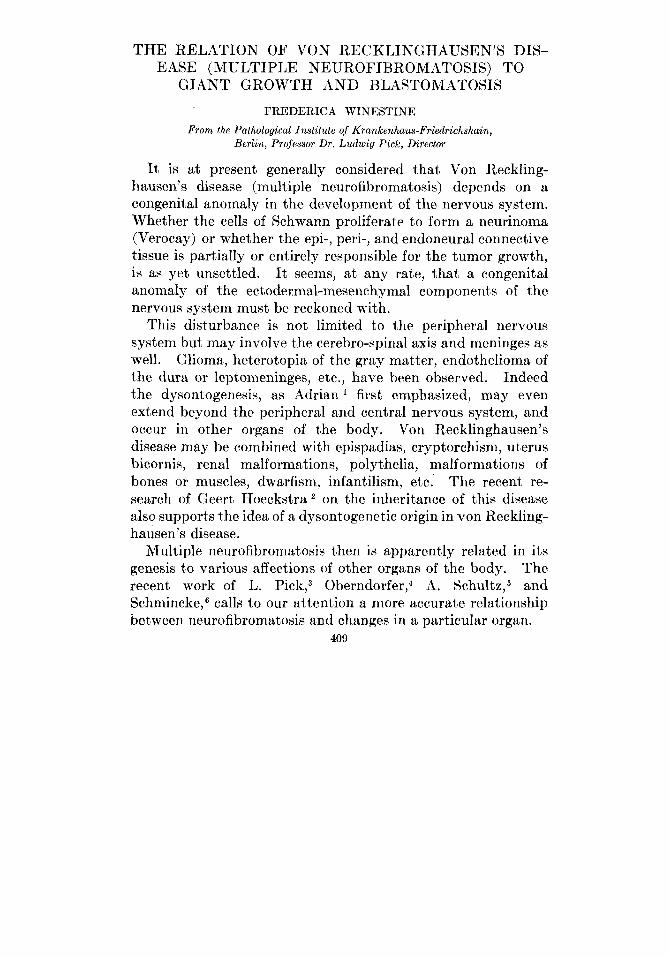
THE RELATION OF VON RECKLINGHAUSEN’S DIS- EASE (MULTIPLE NEUROFIBROMATOSIS) TO
GIANT GROWTH AND BLASTOMATOSIS
FREDERICA WINESTINE From thP Pathological Institute of Krankenhaus-Friedrichshain,
Berlin, Projessor Dr. Ludwig Pick, Director
It is a t present generally considered that Von Reckling- hausen’s disease (multiple neurofibromatosis) depends on a congenital anomaly in the development of the nervous system. Whether the cells of Schwann proliferate to form a neurinonia (Verocay) or whether the epi-, peri-, and endoneural connective tissue is partially or entirely responsible for the tumor growth, is as yet unsettled. It seems, a t any rate, that a congenital anomaly of the ectodermal-mesenchymal components of the nervous system must be reckoned with.
This dist,urbance is not limited to the peripheral nervous system but may involve the cerebro-spinal axis and meninges as well. Glioma, heterotopia of the gray matter, endothelioma of the dura or leptomeninges, etc., have been observed. Indeed the dysontogenesis, as Adrian first emphasized, may even extend beyond the peripheral and central nervous system, and occur in otjher organs of the body. Von Recklinghausen’s disease may be combined with epispadias, cryptorchism, uterus bicornis, renal malformations, polythelia, malformations of bones or muscles, dwarfism, infantilism, etc. The recent re- search of Geert Hoeckstra2 on the inheritance of this disease also supports the idea of a dysontogenetic origin in von Reckling- hausen’s disease.
Multiple neurofibromatosis then is apparently related in its genesis to various affections of other organs of the body. The recent work of L. Pick,3 Obernd~rfer ,~ A. Schultz,” and Schmincke,6 calls to our attention a more accurate relationship between neurofibromatosia and changes in a particular organ.
409
410 F. WINESTINE
L. Pick was the first to show that there occurs in the small intestine of the horse a partial giant growth which bears a very close relation to a neurofibromatosis of the corresponding mesenteric sector. The cases of Oberndorfer, A. Schultz, and Schmincke involved the human appendix. Oberndorfer’s case of a giant appendix with neurofibromatosis of its mesentery resembles in most details the findings in the following case described by Pick who has recently studied the combination of giant growth of the intestine and mesenteric neurofibromatosis. A sharply delineated segment of gut 50 cm. in length presented enormous increase in thickness. Leading up to this segment there is a moderate widening of the lumen and some hypertrophy of the muscle coats resulting from stenosis of the lumen in the giant part. The histological examination and measurement show that all layers of the intestine participate in the giant growth in proportion to their normal width. It, therefore, appears as if an intestinal segment from some giant animal had been incorporated in the horse’s intestine. This is, therefore, an actual partial giant growth of the intestine perhaps the most perfect form of true giant growth of a soft part of the body that has as yet been described.
This growth is circumscribed and in exact correlation with a neurofibromatosis of the innervating splanchnic nerve-branches. The latter form a fan shaped neurofibroma which definitely limits the giant growth to the adjacent intestinal wall. This extends from a point in the mesentery towards the lumen.
Further microscopic examination shows that there are present around the convoluted neurofibroma a nuniber of branches of the mesenteric nerves not involved in the tumor formation but showing instead a true nervous hyperplasia. Within t,he giant intestine itself, the sympathetic intestinal plexuses--the my- enteric as well as the submucosal (Meissner’s)-present a giant development.
Finally there is some blastomatosis of the cells of Schwann (i.e., neurinomatous changes) not only involving the finer nerve- branches of the intestinal serosa, but also the hypertrophic branches of the rnesenteric plexus and the branches of the
VON RECKLINGHAUSEN’S DISEASE 41 1
submucosal giant plexus. Thus, in the entire sector of intestine and mesentery there is a mixture of giant growth and neurofibro- matosis. In the mesentery the neuroblastomatosis predomi- nates and in the intest,ine the giant growth,
As mentioned above, there is a definite agreement between these findings and those observed by Oberndorfer in the giant human appendix. A. Schultz describes a giant growth and neurinomatosis of the nerve plexus of the appendix without giant growth of the intestine itself. In Schmincke’s case the marked thickening of the appendix, which projected into the cecum, no doubt resulted from a neurinomatosis of the nerves in the wall and mesentery of the appendix but here also there was no giant growth of nerves or intestine.
L. Pick leaves it an open question as to whether the changes observed by A. Schultz and Schmincke are not changes in the nervous apparatus accompanying chronic inflammation of the appendix.
If the observations of A. Schultz and Schmincke belong to the class under consideration, we have a graduated scale in the intensity and completeness of the change, as follows:
1. Giant growt,h and neurofibromatosis (neurinomatosis) of the intestinal nerves, giant growth of the intestine itself; giant growth and neurofibromatosis of the nerves of the corresponding mesentery (case of L. Pick).
2. Giant growth and neurofibromatosis (neurinomatosis) of the intestine combined with giant growth of the nerves of the adjacent mesentery (case of Oberndorfer).
3. Giant growth and neurinomatosis of the nerve plexus of the intestine without giant growth of the intestine itself, apparently also without changes in the mesentery (case of A. Schultz). 4. Pure neurinomatosis of the nerves of the intestine and
adjacent mesentery without giant growth of the nerves or intestine (case of Schmincke).
It is evident t,hat the individual cases in this series show suc- cessively less changes; hence it is logical to suppose that cases with even slighter changes do occur.

412 F. WINESTINE
As L. Pick has shown, the individual forms of giant growth are sometimes combined with true neoplasms-angioma, lipoma, etc.-especially in the region of the growth itself. If we could prove that these new growths as well as the giant growth were congenital, then the blastoma could be considered a partial giant growth of a single tissue associated with the partial giant growth of an organ. Theoretical considerations would make a sort of scale here also plausible, for it is possible that giant growth and neurinomatosis of the splanchnic region or a pure rieurinomatosis could occur without a corresponding giant growth or a neurinomatosis of the related intestinal segment. Indeed, instead of this, neoplasms of another sort might occur here without a simultaneous giant growth of the intestinal segment itself. Such tumors would be in the topographical union with the changes in the innervating intestinal nerves. It, would be especially notewort,hy if the blastoma in its own special way also indicated an “anlage” defect as its origin. That this theoretical deduction has an actual basis will be shown in the following observations made on the autopsy material of Prof. I,. Pick in the Pathological Institute of the Friedriclishain Hospital in Berlin, with whose kind assistance I have studied this material.
The rase is that of a inan sixty years old, admitted to the surgical service of Fricdrichshain Hospital February 13th, 1922. His family and past history was negative, except for one attack of rheu- matic fever. A ycar prior to admission, symptoms of the presrnt disease began with variable periods of sharp abdominal pain. The symptoms became marked six wceks prior to admission, when hc brgan to have diarrhoea, rectal incontincncc, tenesmus, and periods of obstipation. Treatment at home was ineflcctual and he was referred to the hospital. The patient had persistently lost weight in the past, ycar.
Physical examination. Examination on admission revealed a poorly nourished, pale, elderly man.
The abdomen was distended and rigid, and palpation elicited marked tenderness in both lower quadrants, especially the right, where a definite resistant lumpy inass was felt. There was also resistance in the rest of the lower abdomen. The livcr and splcrn
History.
He had bcrn alcoholic.
VON RECKLINGHAUSEN'S DISEASE 413
were normal in size. surrounding it infiltrated, oedematous, and lumpy. the anus was a pencil-sized perforation.
The anus was puffy and swollen, and the tissue On either side of
The nervous system showed no pathological findings. The rectal wall bled easily, was thickly infiltrated, and the ampulla
filled out with a soft tumor mass. Most resistance was felt in the right upper segment.
The symptoms were a continuation of those described-diarrhoea with thin watery blood-tinged stools, continuous incontinence and tenesmus, abdominal pain.
Laboratory &dings.-Urine : acid, albumin and sugar not present. No urobilin, urobilinogen, bilirubin, or diacetic acid were found. Indican was slightly increased. Blood examination: HB 56 per cent; R. R. C. 3,200,000; color index 0.8 Indican positive in the urine. Later Bac.-coli communis was found in the urine and considerable amounts of albumin and many leucocytes.
Thirteen days after admission the patient developed a temperature of 100.6" F., and the following week he began to show signs of pul- monary involvement. I n the left interscapular space the percussion note was impaired; higher up dullness and bronchial breathing; in the right lung posteriorly diminished resonance as high as the superior angle of the scapula. At the same time a bed sore began to form over the sacrum.
Six weeks after admission the patient developed oedcma of the legs and back, and two months after admission succumbed to his illness.
Autopsy findings (Prof. Dr. L. Pick): Body measures 176 cm., poorly nourished, Musculature and fat are scant. Decubitus sore 12 cni. in diameter over the sarcum, dark greyish green discoloration and bone exposed. Extensive phlegmonous infiltration of both gluteal muscles, and oederna of both legs and scrotum.
Both lungs contain air and are oedematous, the lower lobes very hyperaemic. In one of thc main branches of the left pulmonary artery there is a reddish-white thrombus 2 cm. long.
Heart : Size average; muscle lax. Both ventricles greatly dilated. Fresh verrucou~ vegetations on the mitral valve and to a lesser extent on the aortic cusps. Heart muscle yellowish. Coronary arteries negative. On the intima of the aorta slight fatty deposits.
Course o j illness.
Later a gangrenous area developed.
Lungs:
Spleen soft, pulpy, bluish-red on section. Other unimportant lesions are summed up in the anatomical
diagnosis. 27
414 F. WINESTINE
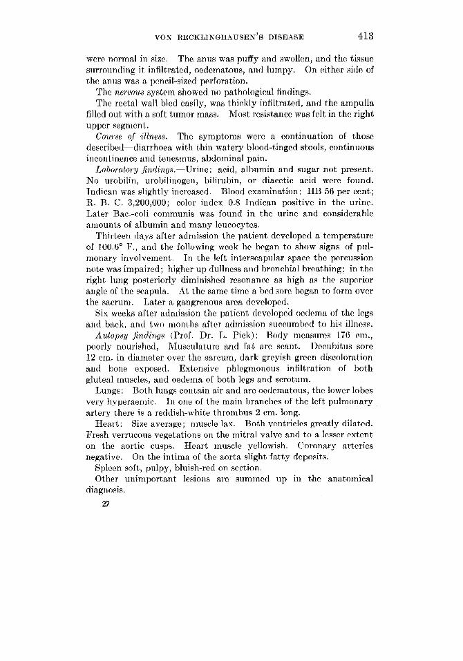
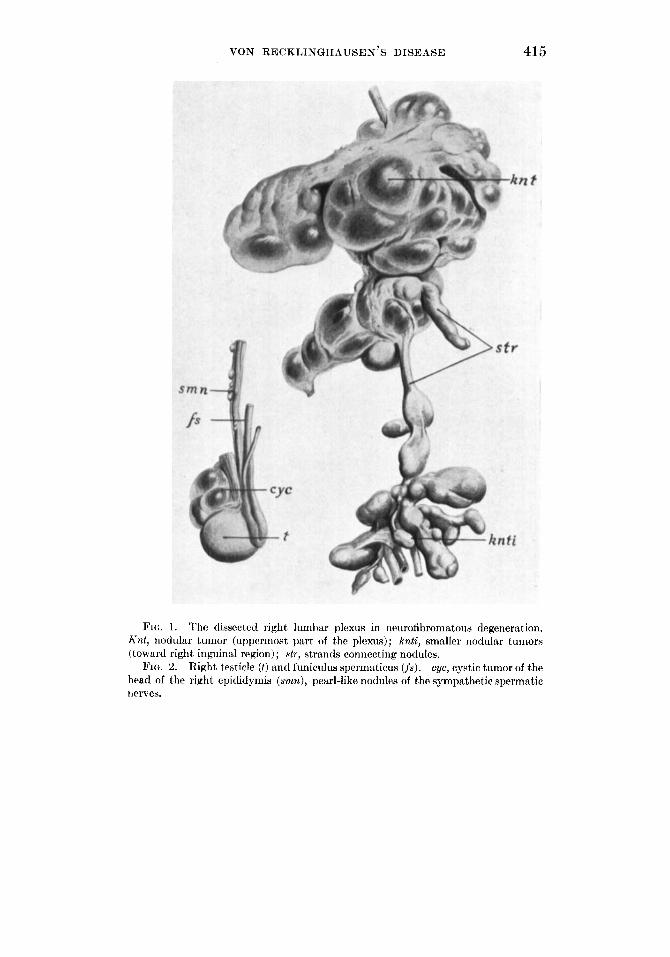
Thc caecum, as well as the appendix and ascending colon, is elevated by a convoluted retropcritoneal mass, which is double the size of a man’s fist. This mass (see Fig. 1, knt) is tough, clastic, greyish white and conncctcd with other similar retroperitoneal masses visible through the peritoneum. Other tumors of smaller size extend downward to the pelvic minor. Detailed dissection shows:
1. All the tumors are connected by strands (str) the size of the little finger or somewhat smaller. These strands are of similar color and consistency.
2. The entire group of tumors and strands-in all its aspects- represents the completely changed right lumbar plexus. Below Poupart,’s ligament, and connected with the above masses, are found nodular tumors (knt i ) , approaching the size of a pigeon’s egg. These represent the right femoral nerve and its branchcs in this region.
The entire mass of tumors and strands as dissected out is shown in the picturc in Fig. 1.
Adherent to the ileo-inguinal nerve, which is unchanged in its course, is a walnut-sized, ellipsoid, grayish white, isolated tumor. The 1uml)ar plexus on the left side is unchanged; likewise both sciatic nerves. The right femoral nerve, below the tumor described, along with all the remaining large nerves of both legs, shows nothing patho- logical; likewise the nerves of thc ccrvical and brachial plexuses. The right lumbar plexus, carefully dissected, is in all its parts so entirely changed by strands and tumors that it is impossible to identify with certainty the particular roots or nerves.
The largest of thcse measures 18 x 12 x 7 cm. (i.e. twice the size of a man’s fist). Relow follows a turnor measuring 14 x 5% x 4% cm. (see Fig. 1). On cross section the tissue is whitish, in part fascicular, elsewhere oedema- tous and even gelatinous.
The right femoral vein is thrombosed to 15 cm. below the saphenous opening-partly by reddish and partly by lighter firmly adherent masses.
The left femoral vein is in its middle third almost fully occluded by firmly adherent dark red thrombi.
On the hcad of the right epididyrnis (Fig. 2) there is a group of srnall transparent cysts (cyc) approximately the size of a walnut. Smaller transparent tumor nodules on the fine nerve branches (smn) accompanying the vas deferens (fs) present themselves in pearl-like rows. They range in size to that of a lentil, and larger, and are greyish
The most voluminous tumors are situated proximally.
VON BECKLINGHAUSEN’S DISEASE 415
PIC:. 1. The dissccted right lumbar plexus in neurofibromatous degeneration. A-nt, nodular tumor (uppermost part of thc plexus); knti, smaller nodular tumors (toward right inguinal region); str, strands connecting nodules.
cyc, cystic tumor of the head of the right epididymis (srnr~), pearl-like nodules of the sympathetic spermatic nerves.
FIG. 2. Right testicle ( t ) and funiculus sperinaticus (fs).

416 F. WINESTINE
whitc in color. That they are softened tumors is evident from the presence of solid whitish particles.
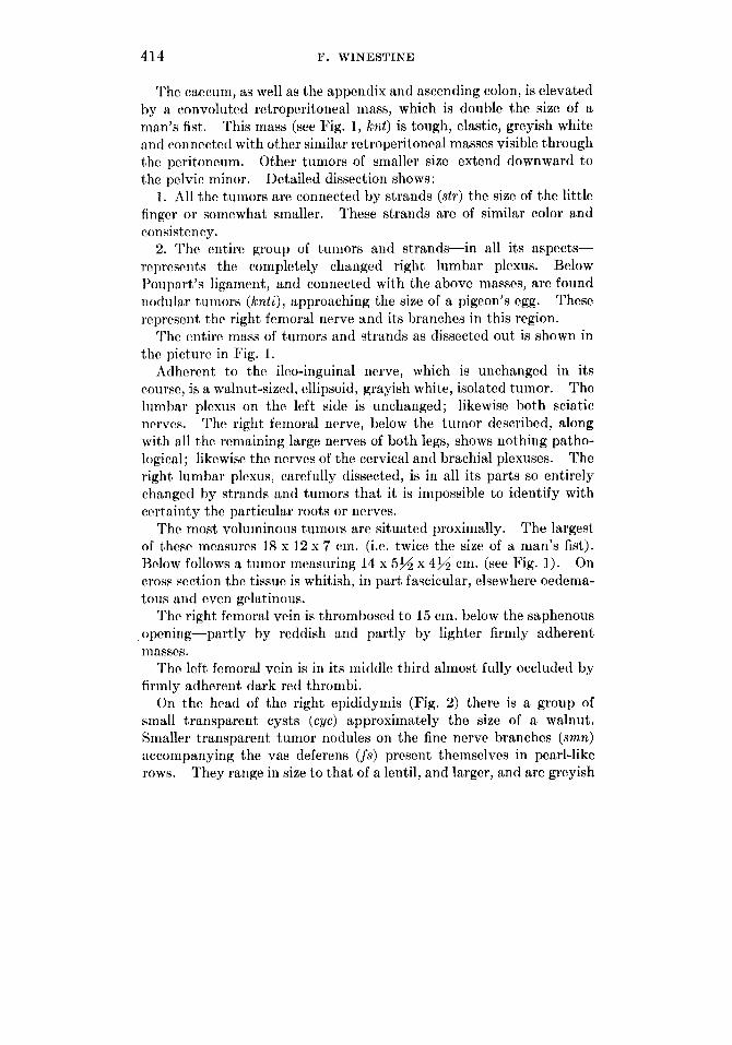
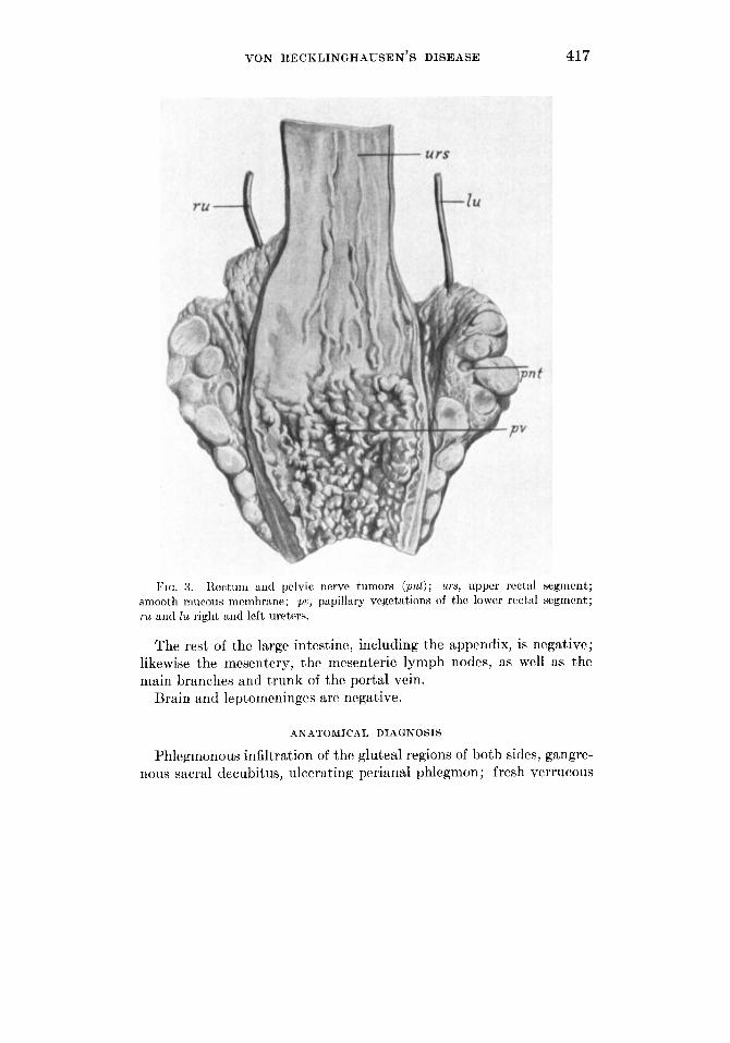
The bladder and rectum are surrounded on both sides by very tough convoluted masses (Fig. 3, p a t ) ; the largest of these are the size of a pigeon’s egg. The tumors are adherent to the bladder and rectum on the one hand and to the prblvic wall on the other. Finer dissection shows that these tumor masses represent the pclvic nerve plexus, i.e., the branches supplying the bladder and rectum. These nerves are entirely lost in the tumor mass. The consistence, color, and cross section of thesc tumors is similar to t>hat of the lumbar plexus tumors previously described. The ureters (ru, Zu) which pass through the tumor mass, are not dilated; thc blatlder and ureteral orifices are normal; likewise the pouch of Douglas, the prostate, thc scminal vesicles, the sacral plcxus and its main branches, and the sciatic and pudendal plexuses.
Around the most distal rect a1 segmcnt is a circumscribed phlrgmon- 011s infiltration corrrspontling to the pcrforations on rach side of the anus; the infiltration does not reach any higher.
Thc niucous mcmbrane ( ~ r i i n ) is palc and sniooth arid lics in longitudinal folds. Eight cni. abovr the anus a very marked changc bcgins in the mucous membrane. This change involves the wholc arca as far down as the anus. The I~ginning of this change, encircling the intestinal lumen, corresponds to the exact placr where the tuinor masses firmly impinge on the rectal wall and unite with it (see Fig. 3). From here to the anus the entire mucous membrane is transforriled into a coarse papillary, rather pale tumor-like niass which is, in parts, gelatinous ( p u ) . The coarse papillary prominences often carry with them snialler villi, variable in form and number, so that a form of branching results. The vegeta- tions float in running water. On section, the tissue of the plump papillary rnasses is everywherr derriarcatet-l from the rest of the intestinal wall.
The stlparation from the upper rectum is sharp, except that the longitiidinal folds of th r rriiicous merribranc gradually run into the papillary clevations (Fig. 3).
The wall of th r uppcr rrctal scgrncrit is of normal thickness and all its laycrs arc casily distinguishable. The wall of the lower rectal segment is four times tht. normal thickness, devoid of definite layers, and instiparable from the neighbouring tumor nodes and strands.
The pclvic organs are difficult to reiiiove.
Thc rrctum contains no unusual products.
VON RECKLINGHAUSEN’S DISEASE 417
tt
FIG. 3. ltectuin a i d prlvic nerve tumors (pn t ) ; urs, upper rcctal segment; smooth mucous rncmt)mne; T J ~ J , papillary vegetations of the lower r rc td segment; ru and Zu right nnd left uretm.
The rest of the large intestine, including the appendix, is negative; likewise the mesentery, the mesenteric lymph nodes, as wcll as the main branches and trunk of the portal win .
Brain and leptomeninges are negative.
ANATOMICAL DIAGNOSIS
l’hlegmonous infiltration of the gluteal regions of both sides, gangre- nous sacral dccubitus, ulcerating perianal phlegmon; fresh verrucous
418 F. WINESTINE
endocarditis on rxiitral and aortic valves, marked dilatation of both vcntricles, fa t ty nietainorphosis of the hcart muscle, acute splenic tumor, thrombosis in a branch of the lcft pulmonary artery; recent thrombosis of the lcft femoral vein, carlier thrombosis of the right.
Multiple voluminous neurofibromata of the entire right lumbar plexus and its main branches as far as Poupart’s ligament; neuro- fibromatosis of the entire sympathctic pelvic plexus, especially around the bladder and rcct,um; ncurofibromatosis of the plexus of the vas dcf(wns.
Voluminous papillary growth of the rnucoiis mcmbranc of thc lower rectal segment with thickcning of the cntire wall.
Left hydrothorax, ocdcma of the scrotum and of both legs. Slight arterio-sclerotic ncyhrocirrhosis, small cysts and adenoma in the cortcx of the right kidney, slight cirrhosis of thc liver and sinall cavernoma, small calcium deposit in the left lobe of the thyroid, unthracosis of the trachco-bronchial lymph nodes.
Microscopical observations: After hardening the material in 10 per cent formalin, frozen and parrafin sections werc stained with hematoxylin, cwsin, with Van Gieson, and with Biclchowsky’s method for axis cylindcrs.
The cxaminat ion shows : 1. Nodular rnasses of thc ncurofibromatosis of the right lumbar
plexus and of thc hemorrhoidal plexus. C~orrcsponding to the macroscopic impression the tumors arc mainly
rnyxofibrornatous. In large areas thcw are delicate collagen fihrils pressed apart by transparcnt fluid (octlcma). Bctwecn the fibrils and in some places connected with thcni are small stellate and spindle shaped cells, and also many round ones. In gradual transition from this area are interlaced fibrillar conncctivc tissue bundlcs, containing fairly numerous long nuclei. All these fibrils take a rcd stain with Van Gieson. Herr and there within this tissue therc appear areas wpccially rich in nuclei and with well-developed finely fibrillar intercel- lular substance. The nuclei have a pronounced spindle shape and arc pardlel t o the direction of the fibrillar bundles. Besides the spindle forms of niiclei there are voluminous round ones excccdingly rich in chromatin. Sometimes four or five nuclei lie close together in a sort of ( ~ ~ 1 1 nest.
2. Thcsc rclations, esprcially the red staining of the delicate fibrils with Van Cieson become still more pronounced in the sections from thc trimors of the hemorrhoidal plexus. Thc largcr nodulcs show a
VON RECKLINGHAUSEN’S DISEASE 419
myxofibromatous structure like that of the large tumors from the lumbar plexus. On the other hand the fine macroscopically invisible branches of the sympathetic, without exception blastomatous, give an entirely different picture. Here one sees enclosed in purely fibromatoue fasiculi, bundles of the finest fibrils, which in contrast to the surround- ing red collagen fibrils, stain exquisitely yellow. They are accompanied by very many spindle-shaped and also round nuclei. Not infrequently we see, on transverse section of these bundles, a concentric arrange- ment of the cells. At times, too, we meet with some resemblance to the well-known palisade or “parade formation” of the nuclei seen in neurinoma.
3. The nerve bundles accompanying the vas deferens have in some places an enormous volume, and show nearly everywhere the changes of neurofibroniatosis and neurinomatosis. Here also are found fibrillar fasiculi, which, on transverse section, show a concentric arrangement of their elements. The nuclei here show some poly- morphism; some are spindle shaped, some large, notched, and rich in chromatin.
4. The wall of the rectum above the distal affected segment shows no special changes microscopically.
5. Sections through the entire thickness of the wall of the lowest rectal segment (Fig. 4). Corresponding with the macroscopic findings, the muscle sheaths show extraordinary general hyperplasia. Between the hundles there is often an inflammatory infiltration of small round cells; this is partly diffuse, partly circumscribed-and in other places takes the forni of a real granulation tissue.
The papillary membrane vegetations show everywhere the same picture. They arc purely adenomatous (mn) ; the glands approximate each other. The stroma is finely fibrillar and contains numerous spindle-shaped and small round cells. It is, however, to a large extent, reduced to extremely fine septa. The epithelium lining the glands is without exception of one layer. In most places it consists of fairly high cylindrical cells, practically filled with rather long dark staining nuclei. Nowhere is there histological evidence of mucus production, or of any other secretion. Lower epithelial cell forms occur especially where thc glands are cystically dilated. This cystic widen- ing is very frequent but nowhere reaches a marked degree. The cysts are filled, partly with globular or finely granular masses, partly with desquamated epithelial cells-round, swollen, and in various combi- nations. The gcneral form of the glands is everywhere simple tubular, nowhrre branched to a greater extent, and never labyrinthine.
420 F. WINESTINE
In contrast to the macroscopic impression, it is evident from the microscopic examination that the boundary line between the mucous membrane and musculature is not a sharp one (Fig. 4). The glands are intermingled with the inner layers of the muscle (mu) in broad projec- tions along their entire strorna (gs), not only in single form but “en masse. ”
The submucosa is entirely missing everywhere; the intermixture is, however, everywhere limited to the innermost muscle layers. The main part of the intestinal wall is frcc from glandular projections.
The nervous apparatus of the rectal wall is partly destroyed by the inflammatory process, and wherc it is preserved it is nowhere hyper- trophic or blastomatous.
According to the microhcopic examination, the tumors of the right lumbar plexus and of the cellular pelvic tissue are neurofi- bromatous, in certain parts myxofibromatous, in other parts neurinomatous. Finally, special attention is called to the delicate fibrils staining yellow with Van Gieson, and their accompanying spindle cells; such pictures are rnet with in both the nerve tumors, especially in the pelvic connective tissue. The blastomatosis is entirely limited to the lumbar and pelvic sympathetic plexuses. Nowhere in other nerve sterns are tumors present.
Noticeably unusual in its outward manifestations is the affection of the lower rectal segment. It is striking on account of its coarse papillary surface, uninterrupted by ulceration. The microscope shows adenomatous structure together with in- significant cysts. The epithelium of the glands and cysts is without exception of one layer. Forms suggesting carcinoma or aderiocarcinoma are entirely lacking. The sharp macroscopic demarcation of these papillary inasses from the rest of the intestinal wall, is not confirmed by microscopic examination. The submucosa is entirely lacking (Fig. 4), and not only do single glands occasionally penetrate into the innerrnost muscle layer, but, also the adenomatow tissue, glands, and stroma intermingle with the musculature in broad projections and strands. ,4s shown above, this intermixture affects only the innermost layer of the muscular coat,. For this reason, and also because of the lack of any metastases, we do not consider it a
VON RECKLINGHAUSEN’S DISEASE 42 1
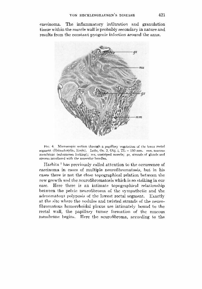
carcinoma. The inflammatory infiltration and granulation tissue within the muscle wall is probably secondary in nature and results from the constant pyogenic infection around the anus.
FIG. 4. Microscopic section through a papillary vegetation of t,ho lowcr rectal segment (Heirnatoxylin, Eosin). Leitz, Oc. 2, Obj. i, TI, = 150 mm. ~ I L I I L , mucous membrane (subniucosa lacking) ; ?nu, unstriped muscle; gs, strands of glands and stromn int,erlacctl with the muscular hundles.
Harbitz ’ has previously called attention to the occurrence of carcinoma in cases of multiple neurofibromatosis, but in his cases there is not the close topographical relation between the new growth and t,he neurofibromatosis which is so striking in our case. Here there is an intimate topographical relationship betwecn the pelvic neurofibroma of the sympathetic and the adenomatous polyposis of the lowest rectal segment. Exactly at the site where the nodules and twisted strands of the neuro- fibromatous hemorrhoidal plexus are intimately bound to the rectal wall, the papillary tumor formation of the mucous membrane begins. Here the neurofibroma, according to the
422 F. WINESTINE
current view, is explained on the basis of a congenital defect and this is suggested by the peculiar mixture of mucous membrane derivatives with the inner muscle layers, and the absence of a submucosa. Hence we have apparently a combined defect of the nerves and their connected intestinal segment. Of course, in the region of the pelvic nerves and the lumbar plexus this reaches far beyond the affected intestinal segment. On the basis of this congcnital defect, the blastomatosis develops on these nerves as well as in the intestine; it does not, however, involve the nerve branches in the intestinal wall.
The cause of the marked t)hickening of the muscle wall of the affected rectal segment is not entirely clear. An “anlage defect ” or a hyperplasia from the influence of the inflammation around the anus are possible explanations.
No actual giant growth is present; there is instead a syntropic combination of neurofibromatosis with pure blastornatosis, a parallel to the combination of neurofibromatosis and true giant growth; hence this case may be classified in the above-mentioned category. Of course, here also the neurofibromatosis and blastomatosis are in genetic coordination.
There is a close topographical relation and genetic coordination between true giant growth and blasto- matosis of an intestinal segment and its corresponding nerves. Our case shows that this relation can occur in the form of a pure tdastornatosis of nerves arid intestinal segment without giant growth. It presents neurofibromatosis of the pelvic and synipathetic plexuses combined with a very unusual blastorna- tosis of the lowest rectal segment, namely, papillary adenoma- tosis of the mucous rnembranc penctrating into the inner layer of the thickened muscular wall.
Conclusions: