Embed Size (px)
Citation preview
Alternative Medicine Review Volume 14, Number 4 2009
Page 373
Review Article
AbstractIn recent years, Alzheimer’s disease (AD) has been considered to be, in part, a neuroendocrine disorder, even referred to by some as type 3 diabetes. Insulin functions by controlling neurotransmitter release processes at the synapses and activating signaling pathways associated with learning and long-term memory. Novel research demonstrates that impaired insulin signaling may be implicated in AD. Post-mortem brain studies show that insulin expression is inversely proportional to the Braak stage of AD progression. It was also demonstrated that neurotoxins, coined amyloid beta-derived diffusible ligands (ADDLs), disrupt signal transduction at synapses, making the cell insulin resistant. ADDLs reduce plasticity of the synapse, potentiate synapse loss, contribute to oxidative damage, and cause AD-type tau hyperphosphorylation.Diabetes and AD have signs of increased oxidative stress in common, including advanced glycation end products (AGEs), when compared to normal subjects. Diabetic patients appear to have an increased risk for AD because AGEs accumulate in neurofibrillary tangles and amyloid plaques in AD brains. This research should encourage a more proactive approach to early diagnosis of diabetes and nutritional counseling for AD patients.(Altern Med Rev 2009;14(4):373-379)
IntroductionThe epidemic of insulin resistance/prediabetes
and type 2 diabetes may be associated with the emer-gence of higher rates of Alzheimer’s disease (AD). New research delineates a direct correlation between sugar imbalance and AD.1 AD is associated with consistent pathological findings, including neurofibrillary tangles, amyloid-beta deposits, and signs of oxidative stress. No
The Relationship between Alzheimer’s Disease and Diabetes:
Type 3 Diabetes?Zina Kroner, DO
common link among the proposed pathological pro-cesses has been identified. Novel evidence demonstrates that impaired insulin signaling may significantly con-tribute to the pathogenesis of AD, contributing to the idea that it is actually a neuroendocrine disease.2 Neu-rotoxins called amyloid beta-derived diffusible ligands (ADDLs) have been implicated as a cause of impaired insulin signaling.3 Advanced glycation end products (AGEs) are found in higher concentration in both hy-perglycemia and AD, contributing to oxidative stress and cell damage. These AGEs are known to be further modified to reactive advanced glycation end products, (RAGEs), which can generate oxidative injury.
Understanding the mechanism of action of this neuroendocrine disorder, termed type 3 diabetes by some, may shed light on new tools for diagnosing and treating AD and for the need for early intervention in obese patients with insulin resistance.
The Clinical Link: Diabetes and ADThe research linking diabetes and AD has its
roots in the groundbreaking Rotterdam study. Of 6,370 elderly subjects studied for 2.1 years, 126 developed dementia; 89 of these were specifically diagnosed with AD. Type 2 diabetes doubled the risk of a patient hav-ing dementia and patients on insulin had four times the risk.4 As rates of insulin resistance and diabetes in the senior population are both increasing, this landmark study, conducted almost a decade ago, has been getting more attention in recent years since further studies have solidified the connection between diabetes and AD.
Zina Kroner, DO – Diplomate, American Board of Internal Medicine; completed residency program at North Shore University Hospital, an NYU affiliate; medical director of Advanced Medicine of New York, PLLC, New York, NY. Correspondence address: 121 East 60th St, Suite 3C, New York, NY 10022 www.advanced-medicine.comEmail: [email protected]

Copyright © 2009 Thorne Research, Inc. All Rights Reserved. No Reprint Without Written Permission.
Alternative Medicine Review Volume 14, Number 4 2009
Review Article
Page 374
Since type 2 diabetes is reaching epidemic proportions and is under-diagnosed, and AD may be associated with hyperglycemia, more attention should be drawn to early diagnosis of diabetes. The Gertner Institute for Epidemiology and Health Policy Research in Israel, in a recently published 25-year, cross-sectional study of 623 adults, demonstrated that approximately 13 percent of the studied population had undiagnosed type 2 diabetes. This study reinforces the importance of early diagnosis of type 2 diabetes by identifying patients with risk factors, including hypertension, hypertriglyc-eridemia, and a large waist circumference (males: ≥40 inches [102 cm], females: ≥35 inches [88 cm]) – factors seen in metabolic syndrome. These results encourage early detection via screening methods targeting those with traits of metabolic syndrome in otherwise healthy adults.5
Another study demonstrating the high preva-lence of diabetes showed almost one-third of elderly patients in a sample of 7,267 subjects had diabetes, and three-fourths had impaired fasting glucose (glucose lev-els >99 but <126) or diabetes.6
Elevated body mass index (BMI), adiposity, impaired fasting glucose, and diabetes increase the risk of AD substantially. The latest study, utilizing data on 2,322 participants in the Baltimore Longitudinal Study of Aging, shows the incidence of AD increased in men who gained weight between the ages of 30 and 45 and in women with a BMI >30 at ages 30, 40, and 45.7 This suggests more emphasis should be placed on early weight-loss strategies for preventing AD.
A 2008 Swedish study showed a statistically significant increase in the risk of developing AD in men who develop type 2 diabetes in midlife.1 The researchers followed 2,269 men for 32 years and found that those with low insulin production at age 50 were 150-per-cent more likely to develop AD than those with ad-equate insulin production. This association was great-est in patients who did not have the apolipoprotein E4 (ApoE4) genetic predisposition to AD (which renders individuals less efficient at breaking down beta-amyloid plaques), thereby making diabetes a possible indepen-dent risk factor for AD. This study illustrates the im-portance of maintaining healthy blood glucose control in middle-aged men as a possible means of preventing AD later in life.
A recent investigation suggests that AD is as-sociated with metabolic syndrome. After studying 50 patients diagnosed with AD and comparing them to 75 cognitively normal controls, the AD patients had a greater waist circumference, higher triglyceride and glu-cose levels, and lower high-density lipoprotein choles-terol.8 Patients with metabolic syndrome are diagnosed with AD at a younger age than AD patients without metabolic syndrome.9
Type 3 Diabetes: Is It Actually a Unique Condition?
The term type 3 diabetes was coined in 2005 by Suzanne de la Monte, MD, MPH, Associate Profes-sor of Pathology and Medicine and neuropathologist at Brown Medical School. Her team, examining postmor-tem brain tissue of AD patients, found that AD may be a neuroendocrine disease associated with insulin sig-naling. The team termed it type 3 diabetes because it harbors elements of both types 1 and 2 diabetes, since there is both a decrease in the production of insulin and a resistance to insulin receptors.2,10
The team analyzed 45 postmortem brains of patients of varying Braak stages of AD neurodegen-eration and found that insulin expression was inversely proportional to the Braak stage, with an 80-percent de-crease in the number of insulin receptors in AD patients compared to normal subjects. In addition, the ability of insulin to bind to the receptors was compromised. There was a reduced level of mRNA corresponding to insulin, insulin-like growth factor-1 (IGF-1) and -2 polypep-tides, and their receptors. The research team also noted a reduction in the tau protein, which is regulated by in-sulin and IGF-1.10,11 This phenomenon ultimately could lead to neuronal cell death and AD exacerbation.
The postmortem studies inspired a rat study in which intracerebral injection of streptozotocin resulted in a chemical depletion of insulin and an alteration of IGF-signaling mechanisms together with oxidative in-jury. The combination of alterations resulted in neu-rodegeneration, including reduction in brain size and other neurological changes seen in AD.12
AD is characterized by a reduction in the uti-lization of glucose, and treatment with insulin has been associated with improved memory. Insulin, important in memory processing, crosses the blood-brain barrier and is even produced in brain tissue itself. AD patients

Copyright © 2009 Thorne Research, Inc. All Rights Reserved. No Reprint Without Written Permission.
Alternative Medicine Review Volume 14, Number 4 2009
Alzheimer’s/Diabetes
Page 375
have less insulin and fewer insulin receptors than non-AD patients, and correction of insulin levels improves cognition. Insulin binds to insulin receptors in the brain, most of which are located in the cerebral cortex, olfacto-ry bulb, hippocampus, cerebellum, and hypothalamus. Since there are more insulin receptors in the cognition-pertinent areas of the brain, it is logical to consider the association between insulin and cognition.13
Several studies utilizing intranasal, intrave-nous, and intracerebral administration of insulin dem-onstrate improved cognition. A study utilizing intra-nasal insulin showed that its administration enhanced verbal recall in normoglycemic adults with early AD or cognitive impairment.14 In the study, 25 partici-pants were randomly assigned to receive either placebo (n=12) or 20 IU intranasal insulin (n=13) twice daily. After 21 days of treatment, changes in cognition were measured. The fasting plasma glucose and insulin lev-els were unchanged with treatment. However, when compared with the placebo treated subjects, the insulin-treated subjects retained more verbal information and displayed superior attention and functional status.
A study utilizing intravenous (IV) insulin as-sessed cognitive performance in 22 adults with AD and 15 normal adults receiving five consecutively higher IV doses of insulin resulting in five plasma insulin levels (10, 25, 35, 85, and 135 microU/mL), while plasma glucose levels of ~100 mg/dL were maintained. Cog-nitive performance was measured after 120 minutes of infusion. AD patients who were ApoE4-positive were found to have improved memory at lower insulin levels of 25 microU/mL, compared to their ApoE4-negative counterparts, who required a higher blood insulin level of 35 and 85 microU/mL before an improvement in memory was noted. Interestingly, normal adults also showed improved memory at insulin levels of 25 and 85 microU/mL. This shows that AD patients who are ApoE4-negative may not be as sensitive to insulin. 15
A study utilizing intracerebroventricular insu-lin showed that its administration enhanced memory formation in rodents undergoing a step-through passive avoidance task.16 These studies suggest that insulin may have a role in enhancement of cognition and memory. The other implication is that patients with the ApoE4 genetic predisposition to AD may not reap the benefits of improvement in AD by glycemic control.
Based on a recent epidemiological study, indi-viduals who are ApoE4-positive are not more likely to be insulin resistant than those who are ApoE4-negative. Therefore, insulin resistance and being positive for the ApoE4 allele are independent risk factors for AD; hav-ing both may pose an additive risk.17
Pathophysiological Connections between Insulin and AD
AD is characterized by both low insulin levels and insulin resistance within the central nervous system (CNS), as opposed to type 2 diabetes, which is char-acterized by high insulin levels and insulin resistance outside of the CNS. Insulin resistance and hyperin-sulinemia cause a reduction in brain insulin.18 Several mechanisms might explain why insulin mediates mem-ory facilitation. As noted, insulin receptors are found in areas of the brain responsible for cognition. Insulin ac-tivates signaling pathways associated with learning and long-term memory.19 According to de la Monte, insulin helps to regulate processes such as neuronal survival, energy metabolism, and plasticity. These processes are required for learning and memory. Peripheral insulin resistance, therefore, affects cognition.20
In addition to regulating blood sugar levels, insulin functions as a growth factor for all cells, includ-ing neurons in the brain. Thus, insulin resistance or lack of insulin, in addition to adversely affecting blood sugar levels, contributes to degenerative processes in the brain.21
When insulin levels reach an exceedingly high level, the beta-amyloid peptide, the hallmark of AD that accumulates in senile plaques, is modulated.20,22 Exaggerated elevation of plasma insulin levels causes amyloid peptide levels in the cerebrospinal fluid to in-crease, resulting in memory insult.21
Amyloid beta-Derived Diffusible LigandsA group of researchers at Northwestern Uni-
versity studied why brains of AD patients are both low in, and resistant to, insulin. According to William Klein, PhD, who led the research, amyloid beta-derived dif-fusible ligands may be responsible for the phenomenon. ADDLs are oligomers similar in morphology and size to prions that have been linked to neurodegenerative disease. ADDLs may contribute to lowered insulin
Copyright © 2009 Thorne Research, Inc. All Rights Reserved. No Reprint Without Written Permission.
Alternative Medicine Review Volume 14, Number 4 2009
Review Article
Page 376
levels and insulin resistance in AD brains.3 Because the ADDLs are so small, they are more diffusible and there-fore more harmful than amyloid.
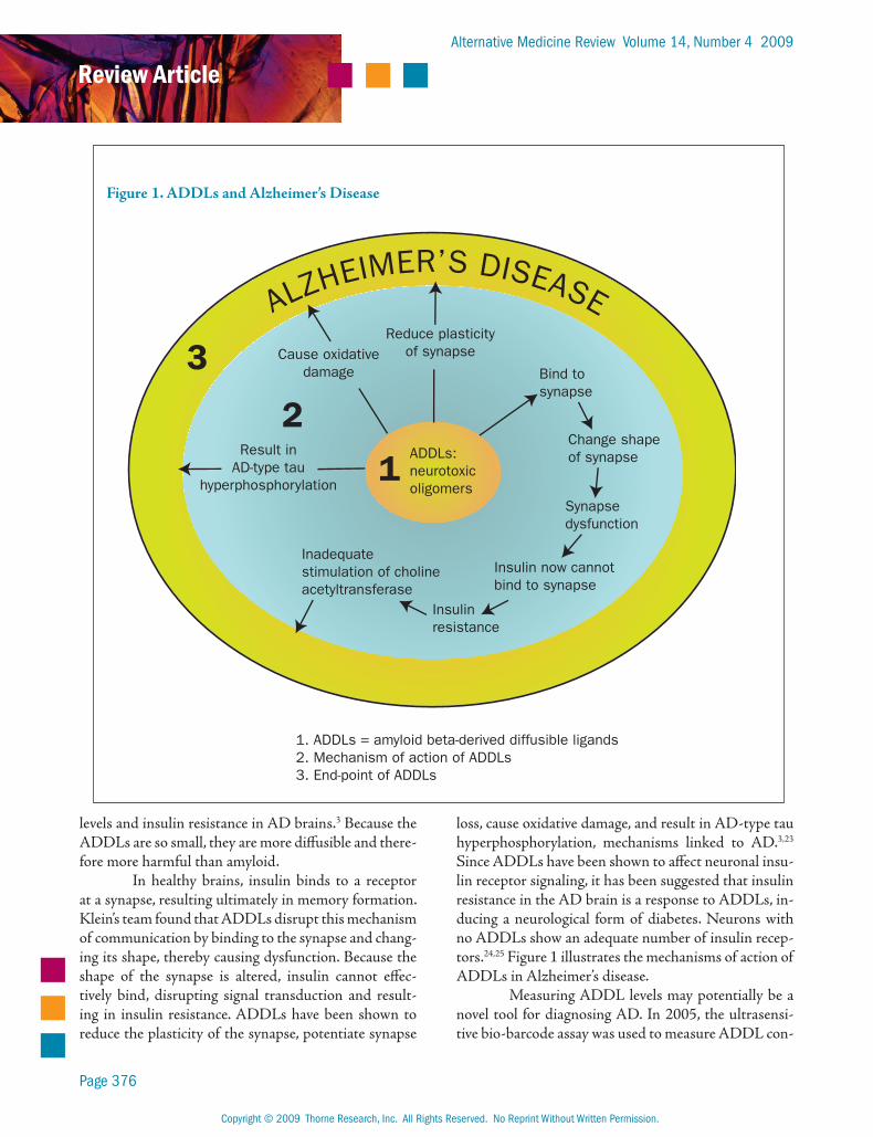
In healthy brains, insulin binds to a receptor at a synapse, resulting ultimately in memory formation. Klein’s team found that ADDLs disrupt this mechanism of communication by binding to the synapse and chang-ing its shape, thereby causing dysfunction. Because the shape of the synapse is altered, insulin cannot effec-tively bind, disrupting signal transduction and result-ing in insulin resistance. ADDLs have been shown to reduce the plasticity of the synapse, potentiate synapse
loss, cause oxidative damage, and result in AD-type tau hyperphosphorylation, mechanisms linked to AD.3,23 Since ADDLs have been shown to affect neuronal insu-lin receptor signaling, it has been suggested that insulin resistance in the AD brain is a response to ADDLs, in-ducing a neurological form of diabetes. Neurons with no ADDLs show an adequate number of insulin recep-tors.24,25 Figure 1 illustrates the mechanisms of action of ADDLs in Alzheimer’s disease.
Measuring ADDL levels may potentially be a novel tool for diagnosing AD. In 2005, the ultrasensi-tive bio-barcode assay was used to measure ADDL con-
Figure 1. ADDLs and Alzheimer’s Disease
Cause oxidativedamage
Reduce plasticityof synapse
Bind tosynapse
Change shapeof synapse
Synapsedysfunction
Insulin now cannotbind to synapse
Insulinresistance
Inadequatestimulation of cholineacetyltransferase
Result inAD-type tau
hyperphosphorylation
ADDLs:neurotoxicoligomers
1
2
3
ALZHEIMER’S DISEASE
1. ADDLs = amyloid beta-derived diffusible ligands2. Mechanism of action of ADDLs3. End-point of ADDLs

Copyright © 2009 Thorne Research, Inc. All Rights Reserved. No Reprint Without Written Permission.
Alternative Medicine Review Volume 14, Number 4 2009
Alzheimer’s/Diabetes
Page 377
centration in cerebrospinal fluid. Of 30 subjects, ADDL concentra-tions were found to be higher in those diagnosed with AD com-pared to non-AD patients. This test is not readily available and less invasive testing is underway.26 An ADDL vaccine is being studied and ADDL-blocking drugs are being considered by Klein et al.27
Insulin and the Cholinergic Hypothesis
The cholinergic hypo-thesis that suggests AD is caused by an inadequate production of acetylcholine may also have links to blood sugar abnormalities and insulin resistance. The researchers at Brown point out that insulin also participates significantly in neurological function by stimu-lating the expression of choline acetyltransferase (ChAT), the en-zyme responsible for acetylcholine synthesis (Figure 2). Therefore, suboptimal insulin levels as well as poor insulin recep-tor sensitivity can ultimately contribute to a decrease in acetylcholine, which further elucidates a possible bio-chemical link between diabetes and AD.28
AGEs and Oxidation – Common Thread between Diabetes and AD
Another mechanism linking diabetes with AD is that both diseases, as mentioned previously, are as-sociated with increased oxidative stress and production of AGEs. Although the association between vascular dementia and AGEs is well established, new research points to a link between AGEs and AD. AGEs are formed by a sequence of events originally identified in 1912 as the end-products of the Maillard reaction,29
during which reducing sugars can react with the amino groups of proteins to produce cross-linked complexes and unstable compounds.
AGEs have been found in retinal vessels, pe-ripheral nerves, kidneys, and the CNS of diabetics. AGEs couple with free radicals and create oxidative damage, which in turn leads to cellular injury.30,31 Dia-betic patients could have an increased risk of AD via AGE production.32 Oxidative stress on its own also causes AGEs, creating a vicious cycle.31
AGEs are also known to modify plaques and neurofibrillary tangles, both implicated in AD.33 AGEs have been identified in neurofibrillary tangles (consist-ing of tau protein) and senile plaques (consisting of beta-amyloid protein). Since type 2 diabetes accelerates the production of AGEs, it may be another causative factor in the development of AD.34 It has been pro-posed that a potential biomarker for early detection of AD may be measurement of toxic AGEs in the serum or cerebrospinal fluid.35
Figure 2. Insulin’s Role in the Cholinergic Hypothesis: The Importance of Insulin in Memory Formation
Nerve Ending
Acetyl-CoA
Choline
Acetylcholine
Cholineacetyltransferase
(enhanced by insulin)
Copyright © 2009 Thorne Research, Inc. All Rights Reserved. No Reprint Without Written Permission.
Alternative Medicine Review Volume 14, Number 4 2009
Review Article
Page 378
ConclusionUnderstanding that AD has its foundation in
neuroendocrinology is persuasive evidence that there should be greater emphasis on early diagnosis of meta-bolic syndrome, insulin resistance, and type 2 diabetes. Referring to AD as type 3 diabetes has its foundation in the fact that the CNS in AD is characterized by a paucity of insulin and resistance of the insulin recep-tors. This results in cognitive dysfunction, since insulin is crucial for neurological signaling processes to occur. Insulin also participates in neurological function by stimulating the expression of ChAT, the enzyme re-sponsible for acetylcholine synthesis; acetylcholine is in turn a necessary neurotransmitter for cognition. AGEs, found in greater amounts in diabetic patients compared to controls with normal glucose regulation, have also been found in high concentration in AD brains.
The links between hyperglycemic states and AD can allow for better future diagnostic strategies. Since ADDLs may contribute to lowered insulin levels and insulin resistance in AD brains, the future of di-agnosis may entail the measurement of ADDLs. Mea-surement of AGEs has also been proposed.
Treatment strategies utilizing this informa-tion require more research. The knowledge that there is a reduction of the sensitivity to insulin in AD patients who are not ApoE4-positive suggests that optimization of blood sugar levels may have therapeutic benefits. In-sulin-sensitizing agents may potentially be used in the setting of early AD.
References1. Rönnemaa E, Zethelius B, Sundelöf J, et al. Impaired
insulin secretion increases the risk of Alzheimer disease. Neurology 2008;71:1065-1071.
2. Steen E, Terry BM, Rivera EJ, et al. Impaired insulin and insulin-like growth factor expression and signaling mechanisms in Alzheimer’s disease – is this type 3 diabetes? J Alzheimers Dis 2005;7:63-80.
3. Viola KL, Velasco PT, Klein WL. Why Alzheimer’s is a disease of memory: the attack on synapses by A beta oligomers (ADDLs). J Nutr Health Aging 2008;12:51S-57S.
4. Ott A, Stolk RP, van Harskamp F, et al. Diabetes mellitus and the risk of dementia: The Rotterdam Study. Neurology 1999;53:1937-1942.
5. Dankner R, Geulayov G, Olmer L, Kaplan G. Undetected type 2 diabetes in older adults. Age Ageing 2009;38:56-62.
6. Cowie CC, Rust KF, Ford ES, et al. Full accounting of diabetes and pre-diabetes in the U.S. population in 1988-1994 and 2005-2006. Diabetes Care 2009;32:287-294.
7. Beydoun MA, Lhotsky A, Wang Y, et al. Association of adiposity status and changes in early to mid-adulthood with incidence of Alzheimer’s disease. Am J Epidemiol 2008;168:1179-1189.
8. Razay G, Vreugdenhil A, Wilcock G. The metabolic syndrome and Alzheimer disease. Arch Neurol 2007;64:93-96.
9. Vilalta-Franch J, López-Pousa S, Garre-Olmo J, et al. Metabolic syndrome in Alzheimer’s disease: clinical and developmental influences. Rev Neurol 2008;46:13-17.
10. Rivera EJ, Goldin A, Fulmer N, et al. Insulin and insulin-like growth factor expression and function deteriorate with progression of Alzheimer’s disease: link to brain reductions in acetylcholine. J Alzheimers Dis 2005;8:247-268.
11. de la Monte SM, Tong M, Lester-Coll N, et al. Therapeutic rescue of neurodegeneration in experimental type 3 diabetes: relevance to Alzheimer’s disease. J Alzheimers Dis 2006;10:89-109.
12. Lester-Coll N, Rivera EJ, Soscia SJ, et al. Intracerebral streptozotocin model of type 3 diabetes: relevance to sporadic Alzheimer’s disease. J Alzheimers Dis 2006;9:13-33.
13. Craft S, Watson GS. Insulin and neurodegenerative disease: shared and specific mechanisms. Lancet Neurol 2004;3:169-178.
14. Reger MA, Watson GS, Green PS, et al. Intranasal insulin improves cognition and modulates beta-amyloid in early AD. Neurology 2008;70:440-448.
15. Craft S, Asthana S, Cook DG, et al. Insulin dose-response effects on memory and plasma amyloid precursor protein in Alzheimer’s disease: interactions with apolipoprotein E genotype. Psychoneuroendocrinology 2003;28:809-822.
16. Park CR, Seeley RJ, Craft S, Woods SC. Intracerebroventricular insulin enhances memory in a passive-avoidance task. Physiol Behav 2000;68:509-514.
17. Peila R, Rodriguez BL, Launer LJ, et al. Type 2 diabetes, APOE gene, and the risk for dementia and related pathologies: The Honolulu-Asia Aging Study. Diabetes 2002;51:1256-1262.
18. Zhao WQ, Alkon DL. Role of insulin and insulin receptor in learning and memory. Mol Cell Endocrinol 2001;177:125-134.
19. Bingham EM, Hopkins D, Smith D, et al. The role of insulin in human brain glucose metabolism: an 18fluoro-deoxyglucose positron emission tomography study. Diabetes 2002;51:3384-3390.
20. de la Monte SM. Insulin resistance and Alzheimers’s disease. BMB Rep 2009;42:475-481.
21. Li L, Holscher C. Common pathological processes in Alzheimer disease and type 2 diabetes: a review. Brain Res Rev 2007;56:384-402.

Copyright © 2009 Thorne Research, Inc. All Rights Reserved. No Reprint Without Written Permission.
Alternative Medicine Review Volume 14, Number 4 2009
Alzheimer’s/Diabetes
Page 379
22. Westerman MA, Cooper-Blacketer D, Mariash A, et al. The relationship between Abeta and memory in the Tg2576 mouse model of Alzheimer’s disease. J Neurosci 2002;22:1858-1867.
23. De Felice FG, Wu D, Lambert MP, et al. Alzheimer’s disease-type neuronal tau hyperphosphorylation induced by A beta oligomers. Neurobiol Aging 2008;29:1334-1347.
24. Zhao WQ, De Felice FG, Fernandez S, et al. Amyloid beta oligomers induce impairment of neuronal insulin receptors. FASEB J 2008;22:246-260.
25. Gong Y, Chang L, Viola KL, et al. Alzheimer’s disease-affected brain: presence of oligomeric A beta ligands (ADDLs) suggests a molecular basis for reversible memory loss. Proc Natl Acad Sci U S A 2003;100:10417-10422.
26. Georganopoulou DG, Chang L, Nam JM, et al. Nanoparticle-based detection in cerebral spinal fluid of a soluble pathogenic biomarker for Alzheimer’s disease. Proc Natl Acad Sci U S A 2005;102:2273-2276.
27. http://www.ibis.northwestern.edu/faculty/klein.html [Accessed October 19, 2009]
28. Rivera EJ, Goldin A, Fulmer N, et al. Insulin and insulin-like growth factor expression and function deteriorate with progression of Alzheimer’s disease: link to brain reductions in acetylcholine. J Alzheimers Dis 2005;8:247-268.
29. Yamagishi S, Ueda S, Okuda S. Food-derived advanced glycation end products (AGEs): a novel therapeutic target for various disorders. Curr Pharm Des 2007;13:2832-2836.
30. Pasquier F, Boulogne A, Leys D, Fontaine P. Diabetes mellitus and dementia. Diabetes Metab 2006;32:403-414.
31. Sato T, Shimogaito N, Wu X, et al. Toxic advanced glycation end products (TAGE) theory in Alzheimer’s disease. Am J Alzheimers Dis Other Demen 2006;21:197-208.
32. Valente T, Gella A, Fernàndez-Busquets X, et al. Immunohistochemical analysis of human brain suggests pathological synergism of Alzheimer’s disease and diabetes mellitus. Neurobiol Dis 2009;Sep 22 [Epub ahead of print]
33. Zhu X, Su B, Wang X, et al. Causes of oxidative stress in Alzheimer disease. Cell Mol Life Sci 2007;64:2202-2210.
34. Takeuchi M, Yamagishi S. Possible involvement of advanced glycation end-products (AGEs) in the pathogenesis of Alzheimer’s disease. Curr Pharm Des 2008;14:973-978.
35. Takeuchi M, Sato T, Takino J, et al. Diagnostic utility of serum or cerebrospinal fluid levels of toxic advanced glycation end-products (TAGE) in early detection of Alzheimer’s disease. Med Hypotheses 2007;69:1358-1366.
Physician Training in Environmental MedicineCertification Course Series for Healthcare Professionals
Course Highlights:Distance Learning with DVD Lectures, Notes & ArticlesThree Weekend Seminars for In-Depth Case Study, Guest Lecturesand Question & AnswerBegin & End Coursework at AnytimeMonthly Phone Conferences
This material is well done, easy to follow and the best environmental
presentation I’ve ever seen done. It should be required for all docs.
Sponsored by SpiritMed and the Southwest College of Naturopathic MedicineTuition for the full course: $3450; $2300 for residents and first-year graduates (verification required)For more information and registration details, please email Dr. Kelly Crinnion at [email protected] Weekends: 2/7-8, 5/16-17, 11/7-8 (All held in Scottsdale, AZ except May seminar will be held in Kirkland, WA)
“”– Dorothy “Dot” Merritt, MD