Embed Size (px)
Citation preview

THE RELATIONSHIP OF DIETARY FIBER
AND HUMAN HEALTH
by
ROGER ALLEN SHEWMAKE, B.A., M.A.T,
A DISSERTATION
IN
HOME ECONOMICS
Submitted to the Graduate Faculty of Texas Tech University in
Partial Fulfillment of the Requirements for
the Degree of
DOCTOR OF PHILOSOPHY
Approved
May, 1980

f :
r •., ,:t^ ACKNOWLEDGMENTS
I would like to express my deep appreciation to Dr. S,P. Yang
and Dr, Charles V. Morr for their encouragement to pursue my
interest in dietary fiber. They both possess the somewhat rare
humanistic philosophy toward me and other students. Special thanks
are extended to Dr. Leon Hopkins, Dr. John Martin, and Dr. Shamus
Mehaffie for their advice and criticism while serving as members of
the dissertation committee.
Special thanks are extended to Dr. M.G. Crews for his unending
support, assistance and advice while serving as a member of the
dissertation committee. I would not have completed the project
without him.
I would also like to thank Dr. Leo Juarez for suggesting that
I "look into" the field of nutrition.
Special recognition should be given to my wife, Jean, her
parents, Mr. and Mrs. D.W. Cooper, and my parents, Mr. and Mrs,
J,W. Shewmake, for their help and encouragement.
n
TABLE OF CONTENTS
Page
ACKNOWLEDGMENTS i i
LIST OF FIGURES ,..,. v
CHAPTER
I. INTRODUCTION 1
II. EPIDEMIOLOGICAL STUDIES 4
III. FIBER RELATIONSHIPS TO GASTROINTESTINAL AND
RELATED DISORDERS 6
Diverticular Disease of the Colon 6
Appendicitis 11
Hiatus Hernia 13
Varicose Veins 13
Hemorrhoi ds 16
Cancer 17
Case Control Studies 19
IV. ENERGY RELATIONSHI,?S 28
Caloric Density 28
Nutrient Absorption 30
Obes i ty 32
Diabetes 35
V. CURRENT CONCEPTS OF THE INFLUENCE OF DIETARY FIBER UPON TRANSn TIME 5!
VT FErAL NUTRIENTS/TRACE MINERALS: ALTERATIONS IN RELATION TO FIBER CONSUMPTION 56
VII. TOXICITY PROTECTION 39
ni
VIII. THE INFLUENCE OF FIBER ON INTESTINAL FLORA..,,,..,. 63
IX. FIBER'S RELATIONSHIP TO LIPID METABOLISM..., 56
Fiber and Coronary Heart Disease ,..,,. 66
Gal 1 stones and Choi ecysti ti s ,. 77
X. PHYSICAL ASPECTS OF DIETARY FIBER 79
Fiber as a Food Additive or Ingredient 79
Fiber Analysis 79
Components of Dietary Fiber 84
Industrial Applications of Food Fiber 86
XI. SUMMARY 92
XII. CONCLUSIONS AND RECOMMENDATIONS 97
LIST OF REFERENCES 104
APPENDIX A: FIBER ANALYSIS METHODOLOGY 133
APPENDIX 3: DIETARY FIBER IN SELECTED FOODS 142
IV
LIST OF FIGURES
Figure Page
1. Possible relationships between decreased fiber intake and certain disease states 5
2. Perceived risk levels of possible influencing
factors on coronary heart disease 69
3. Commercially available gums 80
4. Classification and nomenclature of dietary fiber 87
CHAPTER I
INTRODUCTION
Recent epidemiological observations have led to the suggestion
that fiber plays an essential role in the gut and in maintaining
man's health.(1-8) However, Cleave over twenty years ago paved
the way for the current surge of fiber interest with his "Master
Disease" in "The Neglect of Natural Principles in Current Medical
Practice," in which he discusses the problems of natural roughage
removal from the diet.(9) Cleave is considered responsible for
illustrating that fiber depletion through refinement of carbo
hydrates not only slows transit time, but also greatly increases
caloric density along with resultant overconsumption of energy.(10)
Some investigators see an effective relationship between
insufficient dietary fiber and a group of diseases related to
increased transit time, reduced fecal mass, and increased intra
luminal pressures. The major diseases resulting from these
characteristics are believed to be diverticula^ disease,
appendicitis, hiatus hernia, varicose veins, hemorrhoids, and
cancer of the colon and rectum.{!!-17)
Other research indicates possible direct ar.d/or indirect
relationships of fiber deficiency and cholelithiasis, (17,18) blood
lipid levels, (18-27) increased fecal bile lipids, (28-30) fecal
steroids and lipid excretion, (31) constipation, (17,32-34)
coronary heart disease, (35) diabetes, (36-38) obesity, (10,39-42,
1
43-45) intestinal flora and fauna, (46-49) and protection from
toxic substances in the diet.(50-56)
The literature on the implications of food fiber and health
is extensive. The ramifications of food fiber's importance are
diverse. In view of the importance of the topic and the numerous
sources of pertinent information it would be helpful to compile the
major concepts of the relationships and through such a synthesis
produce a professional reference work for the worker in
nutritional research, education, and allied health fields.
Modern nutritional science has advanced to its present state
by the designed and/or accidental demonstration that certain
constituents of foodstuffs eaten by man and animals are required
for maintenance of health. Soon recognition of disease resultant
from deficiency or excess of nutrients led to knowledge of safe
levels. The idea that something eaten in addition to these
nutrients is needed to assure optimal health is relatively new,
based largely on observed differences in the global distribution
of chronic disorders.(57) Epidemiological investigations are
studies of the distribution and dynamics of diseases or conditions
affecting population groups.(58) These studies have raised
questions that may be tested in controlled experiments. However,
one must be cautious in that to prove on the basis of epidemio
logical studies alone that separate observations are causally
related is often difficult if not impossible. Resultant hypotheses
may be very useful in guiding future research, but they must not
be mistaken for facts.
Fiber and health interrelationships developed from
epidemiological studies have resulted in an hypothesis,(59) Most
of the studies compared Western nations with lesser developed
countries of Africa. Perhaps the most publicised of these studies
were by Burkitt, et a!., who pointed out that ischemic heart
disease, appendicitis, diverticular disease, gallstones, varicose
veins, hiatus hernia, hemorrhoids, and colon cancer were very
rarely seen in rural areas of Africa.(60) Consumption of a
traditional diet high in fiber was the basis for this "fiber
theory." Other investigations have produced data supportive of
this theory, including Cleave, (9,19,61) Trowel!, (62-66) Walker,
(67-73) and Eastwood, (74-76) whose combined work has provided a
focal point for the expanding interest in fiber.

CHAPTER II
EPIDEMIOLOGICAL STUDIES
The epidemiological evidence now available upon those diseases
listed in the introduction indicates that they are either directly
or indirectly related to environmental factors and directly or
indirectly related to the degree of economic development. There
are striking contrasts between the high prevalences of all these
diseases in black and white Americans and the low prevalence of
the same medical disorders in rural Africans.(77) Prevalences of
intermediary levels were also found.(61,78)
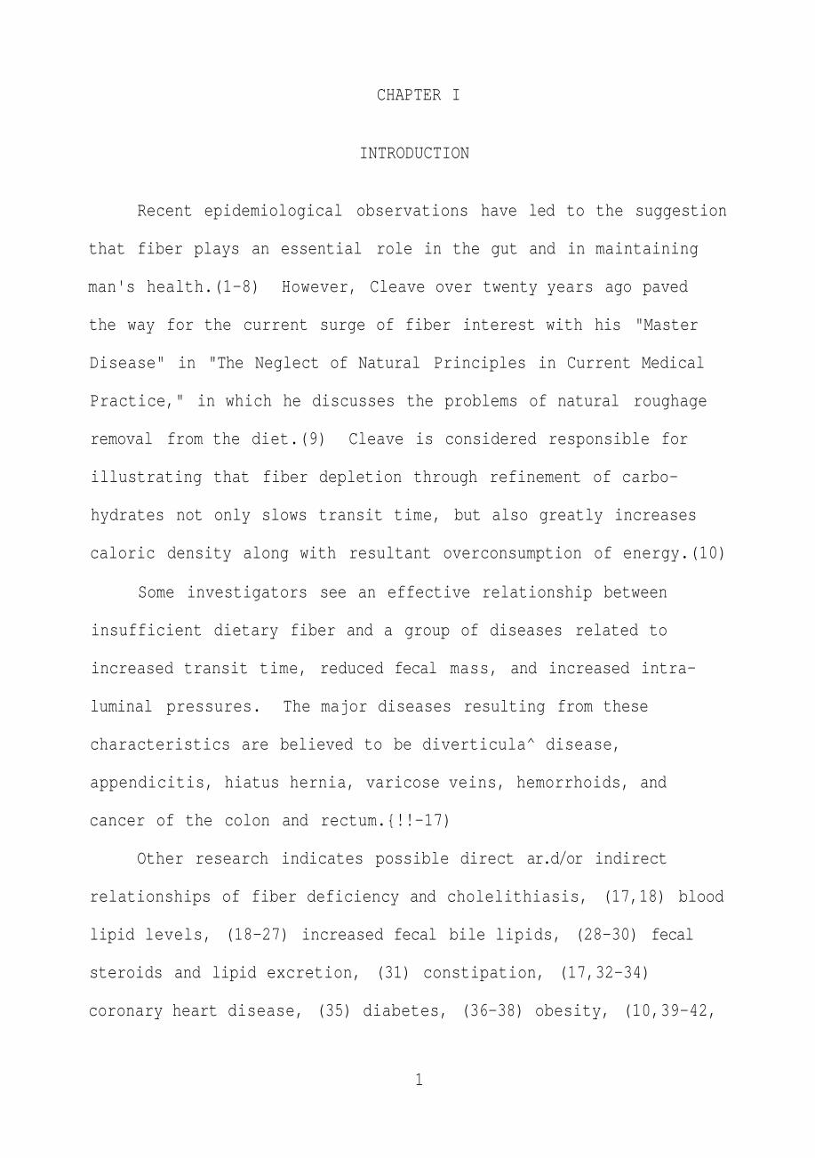
According to Denis Burkitt, all the different effects of a
common cause will tend to be associated with one another.(79)
On the other hand associated effects (i.e. diseases) suggest a
causative factor which is common to each but not necessarily the
only factor in any one disorder (Figure 1). Certain diseases have
been shown to be related not only in geographical distribution and
historical emergence, but also in those who emigrate from less
developed areas to more westernized societies as well as in those
who make the change from a rural to urban environment.
Some of the diseases associated with economic development, thus
having their maximum prevalence in the more affluent western nations,
are rare in some nations and virtually unknown in other areas sti^l
living largely in a traditional manner.(80) A hundredfold dif
ference is seen in prevalence between areas where the disease is
most and least common. The average being m.ore than tenfold. (80)

DECREASED DIETARY FIBER IN FOODS
Decreased Fiber Intake
Reduced fecal bulk
and increased firmness
increased intraluminal
and defecation pressures
and stasis of
feces
constipation diverticular
disease appendicitis colonic cancer hemorrhoids
varicose veins hiatus hernia
Altered cholesterol
and bile salt
metabolism
Decreased Satiety/Energy Ratio
increased energy intake
and decreased
digestibility
gallstones ischaemic heart disease
diabetes obesity
Figure 1.--Possible relationships between decreased fiber intake and certain disease states.(12)

CHAPTER III
FIBER RELATIONSHIPS TO GASTROINTESTINAL
AND RELATED DISORDERS
Diverticular Disease of the Colon
Burkitt states that the most common of all gastrointestinal
disorders in the western world today, diverticulosis, probably
affects a third of the population over fifty years of age.(80)
Yet it is either rare or unknown in indiginous Africans and in all
developing countries.(81) Looking closer at rural Africa, Goulston
in Ethiopia stated that diverticulosis is infrequent and
diverticulitis unknown.(82) In Ghana (Accra) Dadoe saw only one
case of diverticulitis in sixteen years in the medical school
hospital.(83) Diggs in Liberia (Monrovia) reported two patients
with diverticular disease in approximately 300 barium enema
examinations, both in the highest socio-economic group.(83) Williams
reported seven patients in Sierra Leone (Freetown) with the
disorder over a period of twenty years, all from upper socio
economic groups,(83) In Nigeria (Lagos) Kyle, et al., recorded
two cases of diverticulosis in the University Hospital over a
period of three years,(84) Wapnick and Levin reported what they
believed to be the only recorded case in a black Rhodesian
(Salisbury). (85) In reports from South A-'rica (Johannesburg),
Keeley found no diverticula in a series of 2367 autopsies between
1954 and 1956.(86) At the same hospital Solomon found six cases
in approximately 1000 barium enemas in Bantu patients over a tnree-

year period while Levy at the non-European hospital saw no cases
in thirteen years,(83) Higgins and Simson in 2000 consecutive
autopsies found one case (87) and Bremmer and Ackermann stated
the disorder practically never developed in the Bantu,(88) In
South Africa (Pretoria), Simpson found only five cases in 3000
Bantu autopsies while Chapman in Durban reported only one African
case in fourteen years at the King Edward teaching hospital.(83)
In Kenya (Nairobi) Miller saw only one case in an African in
eleven years at the Kenyatta Hospital.(83) Davies reported from
Uganda (Kampala) two cases in 4000 autopsies (89) and Templeton
in 300 autopsies of subjects over thirty years of age specifically
looking for diverticulosis found only one case of the disorder
(in a female over eighty years of age).(83) Jain observed one
case in nine years in a medical school hospital in Zaire (Kinshasa)
(83)
If we shift emphasis from Africa to India and the Middle East,
we again find low incidence of the disorder, even among the urban
areas. Bhardwaj in India (Delhi) found nine cases with 9000
barium enema examinations and Bhargava using the same technique
reported twelve cases, all of which were in the more "westernized"
Indians.(83) In Calcutta, Bannerjee, and Ahmed in Assam reported
a similar wery low incidence.(83)
Zarabi and Farpour in Iran (Shiraz) saw only five cases in
eight years and Abu-Tabikh in Iraq (Bahgdad) reported not more
than three cases of diverticulosis in 1000 barium enema
examinations,(83)

8
Looking at other areas of the world, diverticular disease
has, until recently, been wery rare in Japan.(90) Kyle, et al,
found a distinct difference in incidence among Europeans (three
cases per 15,000) and Chinese, Indian and Malaysians (ten cases
per 1,500,000) and native Fijians (one case per 137,000),(84) In
Malaysia, Kutty found no cases upon autopsy in three years (83)
and Kim in 500 barium enemas found no diverticula in Koreans.(91)
Diverticular disease was rare in Great Britain and in North
America as late as the 1920's, but the prevalence now is in the
range of five to ten percent whereas studies in Africa and India
indicate a prevalence of less than one percent.(80)
Gross in 1845, (92) Cruveilhier in 1849, (93) Rokitansky in
1849, (94) Haberschon in 1857, (95) and Klebs in 1869 (96) all
believed that diverticula of the colon were acquired and thought
to be induced by constipation. This rare disease became
relatively common in two decades.(83) In 1899 Dr. Telling first
reported the disease and none of his colleagues were familiar with
it but by 1908, he was describing the complications and in 1917
his classic paper on diverticulosis and diverticulitis was
published.(97-99) By 1920 Sir John Bland-Sutton remarked that
"in the last ten years, acute diverticulosis is recognized with
the same certainty as appendicitis and is a newly discovered
bane of elders."(83)
Available evidence indicates that divertitular disease of the
colon was a rarity at the beginning of this century, but has
risen in incidence dramatically to become the com.monest disease of
the colon in westernized countries.(100-105) In striking contrast
the disease is almost unknown in those communities only recently
affected by customs and habits characteristic of modern western
civilization.(83)
Diverticular disease among North Americans and Europeans
always affects the sigmoid colon.(106) Alterations, of the
longitudinal muscles, in the form of thickening and shortening,
is believed to be secondary to increased intraluminal pressures.
(77,107-109) It has been suggested by Edwards that the changes
responsible for diverticulosis of the intestine are due to forces
from the bowel wall.(110) Burkitt postulated a hypothetical
pathogenesis dependent upon diets containing too little fiber
with resultant firm stools of diminished bulk.(77) It is believed
the increased transit time and additional mechanical force needed
for propulsion and expulsion of the feces through the colon and
from the rectum raises pressures not only within the lumen of the
bowel but also within the whole abdominal area.(4,16,57,77,83,110-
113) If muscular weakness occurs between areas of hypersegmentation
as is believed occurs with a long history of a diet poor in fiber,
the lumenal pressure may cause a herniation of the mucosa with
the resultant production of small cul-de-sacs referred to as
diverticula. (77,114-116) Roentgenograms taken after barium m,eals
reveal these diverticula as sacculations along the intestinal
wal1.(114) It is believed that fecal material may become
entrapped in these sacculations and with time may produce the
condition known as diverticulitis. Experimental evidence supporting
10
these views is not abundant but Carlson and Hoelsel in an early
study reported that rats maintained on low fiber diets developed
diverticulosis of the colon but not on diets providing appreciable
roughage.(117) The clear relationship between diet and colonic
diverticulosis as proposed by Painter and Burkitt would be
difficult to substantiate or refute in that a possible development
period of forty years is suggested.(12,16)
Other researchers have suggested an alternative theory based
upon observations of Africans eating high-fiber diets who rarely
exhibited diverticulosis in direct contrast to whites in Europe
and North America eating diets deficient in fiber who have a much
higher incidence of the disease.(3,76,118,119) This association
is strengthened by the relief of symptoms by ingestion of cereal
bran.(75) Many patients suffering from the disease have been
shown to have increased rectal pressures and upon treatment with
cereal bran the pressure in the bowel is reduced.(2) It was such
work as this that lead to the postulations in regard to diver
ticular development, high intraluminal pressures and low fiber
diets. Eastwood, et a 1. do point out that therapeutic value is
not necessarily an indicator of a previous true deficiency.(74)
A wery positive aspect of these considerations is the
possibility that diverticular disease is not inevitable and they
point out that numerous considerations may assist in the eradica
tion of the disorder.(13) Future research needs to expand the
basic knowledge of rectosigmoid pressures among healthy as well
as diseased individuals, among various age and racial groups.

11
1.
Further studies as those relating disease prevalence and difference
in fruit and vegetable intake need to be undertaken.(75)
Appendicitis
Appendicitis is the most common indication for emergency
abdominal surgery (4) and occurs most frequently among the young.
(120,121) The prevalence of this disorder is wery small among
Africans.(122) Burkitt believes it is found only in those Africans
who have adopted a westernized life style,(118) Walker gathered
information on appendectomy prevalence from 15,317 sixteen to
twenty year old pupils among four ethnic groups.(73) Data revealed
that among students eighteen to twenty years, appendectomy was
wery rare in rural Negroes (0,5%), slightly more common in urban
Negroes (l,4/o) but much more common in whites (16.5'0- The same
research revealed that defecation frequency increases and transit
time decreases among rural Negroes with large fiber intakes.
Similar results, indicating appendectomy incidence was only 23
percent of the appropriate control group, were found on 1325
white pupils in institutional homes serving less refined diets as
compared to the general population. Short found similar results.
(123) In considering the validity of such findings, appendicitis
data from the United States Navy (124) and comparable probability
data from New Zealand (125) should be looked at. When such data
are used to predict appendicitis among 100,000 Negro laborers
living in mining camps fed diets of highly refined foods one
would expect to find approximately 850 appendectomies, yet in
1970 the data revealed only twenty-two surgical procedures
12
performed.(126)
South African whites reveal an incidence of 17 percent, whites
in England 14 percent, (120) and New Zealand about 16 percent (125)
as compared to much lower ranges reported earlier among Negroes.
It is of interest to look at "found dietary changes" and
their relationship to appendicitis incidence. Conditions were such
after World War I that Russian diets affected by severe privations
became more coarse at the same time appendicitis almost disappeared.
(123) Similar findings were observed during World War II among
the Swiss, (127) the residents of the Channel Islands during German
occupation, and the Dutch.(128)
Earlier studies of asylums, prisons and similar institutions
reveal a very low incidence of appendicitis.(73,129,130) Institu
tional diets consisted of simpler and higher bulk-forming
capacity foods. Miller in Nairobi, Kenya, (131) Wilkie in
Rumania, (132) Clark in China, (131) Harrison in Southern Arabia,
(131) all reported upward trends, sometimes quite dramatic, in
appendicitis with introduction of "roller" mills and finely
ground flours.
According to Burkitt, clinical and pathological evidence
suggests that a possible causative mechanism of the development
of appendicitis results from pressure changes leading to
devitilization of the mucosa with secondary bacterial invasion.(131)
The structure of the appendix does not allow for ready drainage,
its blood supply is limited and its circulation is easily
interferred with because the vessels anastomose to a very limited
13
extent.(133) Considering the blood supply and small luminal
diameter one might suggest that obstruction, plus increased
pressures lowering circulation lead to the inflamatory change in
the mucosa.(134-136) Barium meal studies have shown that addition
of bran to the diet increases the exchange of materials in and out
of the appendix lumen, (134) while high fiber meals with resultant
soft feces rarely obstruct the lumen.(131)
Hiatus Hernia
The protrusion of the stomach upward into the mediastinal
cavity through the esophageal hiatus of the diaphragm results in
the disorder known as hiatus hernia. This condition is usually
believed to be the result of congenital weakness. Repeated
increases in intra-abdominal pressures due to low fecal bulk might
tend to force the gastroesophageal junction upward through the
hiatus either aggravating or initiating the disorder.(77)
It is suggested by Painter (115) and Burkitt (49,137) that
fecal arrest (which is associated with fiber depleted diets) and
increased oressures during defecation are largely responsible
for hiatus hernia. These pressures often exceed 100 torr and can
exceed 200 torr.(131)
Varicose Veins
The valves of the veins are arranged so that blood flows only
in a heartward direction.(138,139) When skeletal muscle is
constricted it exerts pressure in the nearby veins and propel Is
blood toward the heart. This pumping mechanism is known as the
14
"venous pump" or "muscle pump." When no movement occurs the
venous pump does not operate and venous pressure can rise to the
full hydrostatic pressure of 90 torr. Capillary pressure also
increases dramatically and fluid begins to leak from the systemic
circulation into the interstitial spaces. This loss may account
for as much as fifteen to twenty percent of the blood volume. This
loss accompanied with the pooling of blood in the leg veins
reduces venous return with resultant lowering of cardiac output
and possible fainting.
The valves of the veins are sometimes destroyed or become
incompetent. Destruction of the valves occurs particularly after
prolonged stretching due to high venous pressure. The veins have
stretched without a compensating increase in valve size, resulting
in an inability to restrict reverse blood flow. As more blood
pools, stretching or increase cross-sectional diameter occurs with
further increase in blood pooling and final destruction of valve
function. Venous and capillary pressures become wery high with
resultant constant edema. The clinical picture is worsened by
lowered diffusion of nutritional requirements due to the edema.
Muscle and skin weakness soon appears and the skin often atrophies
and finally ulcerates.
Varicose veins are among the most common medical problems in
the western world, affecting ten to seventeen percent of adults
in England and North America.(140) Varicose veins have their
lowest incidence in those areas of the world that have deviated
least from their traditional way of life.
15
Many ideas have been suggested as the cause of varicose
veins. Early suggestions included that this problem reflected a
failure in man to adapt to erect posture. Such supposition is
now held untenable in that in those areas where women stand the
most erect and carrying heavy loads on their heads, we find the
least incidence of the problem.(5) Studies on hereditary factors
seem to refute genetic makeup as a primary causative factor
although it may be contributory.(141) Prolonged standing and
pregnancy have both been taken into consideration as well as
constrictive clothing, but differences in occurrence were not
significant,(142,143) It is now suggested that in those areas of
the world where lower fiber diets are ingested that there is a
higher incidence of the problem,040) It is believed that abdominal
straining associated with constipation and evacuation of small
compact feces causes raised intra-abdominal pressures that are
transmitted to the vena cava and its associated veins of the
lower limb. Straining has been found to raise these pressures to
over 200 torr.
Present knowledge of venous pressures would make one suspect
that veins repeatedly subjected to these abnormal pressures may
become partially or completely ineffective, followed by the blood
pooling and its complicating problems discussed earlier.(49)
Apparently it is not a histological change in the valves, but the
fundamental problem is the dilatation of the lumen of the vein with
resultant separation and incompetence of the valves.(144)
16
Incompetence is not always an "all or none" proposition in that
visible varicosities are not always accompanied by clinically
detectable cough impulses.(140)
Burkitt summarizes the relationship of varicose veins and
factors raising intraluminal pressures (i.e. low dietary fiber).
(140) He states that varicose veins implies reverse blood flow
and in turn,valve incompetence. No histological changes in valve
cusps seem to occur but rather vascular stretching and separation
of cusD edges. He therefore believes that the problem is a result
of raised intraluminal pressures.
Hemorrhoids
Hemorrhoids seem to be extremely rare in the undeveloped
areas of the world and much more prevalent in those peoples
associated with western civilization.(89,145) As with varicose
veins it is believed that the vessels of the rectal column become
enlarged over a period of time due to abdominal straining and
result in the condition known as hemorrhoids. Straining in the
defecation process is inevitably associated with constipation or
low bulk feces.(5,141,146) Increased straining via abdominal
muscles and diaphragm accompanies attempted evacuation of firm
feces with resultant increase in transmitted pressure to vessels
in the anal canal. Whatever the precise mechanism a prominent
role is likely played by constipation, (140) with a mechanism
similar to hemorrhoid production observed in childbirth.
17
Cancer
In the United States cancer is the second major cause of
death; one third of these cancers arise in the digestive organs,
mostly in the large intestine.(147-149) In recent years over
350,000 people died annually from various forms of this disease.
Preliminary data indicate that cancer will account for more than
650,000 new patients each year.
Three major influences which are believed to influence
carcinogenesis receiving increasing attention are environmental,
(including dietary factors) genetics, and hormonal influences.
Epidemiological research suggests that environmental factors are
considered to have a much greater influence on cancer incidence
than do genetic traits (150) and contributes directly or indirectly
to as much as seventy-five percent of human cancer.(151) Two
recent symposia bringing together world wide data relative to
nutrition and cancer should adequately illustrate the importance
of nutrition in modifying susceptibility to neoplasm.(152-153)
The genetics of carcinogenesis in man is a relatively new
discipline which has made significant progress in recent years.
(154) Genetic studies in man are seriously hampered by the nature
of the subject, evidence for advances in the important area of
research is supported by the large number of cancer and precancer
ous disorders in which familial or genetic etiology has been
determined.(151) Clearly, progress in the search for causes of
cancer and its preventions will be greatly augmented as genetics
18
of cancer assumes an increasingly important role along with the
more complete elucidation of th.e influence of environment.
A considerable body of evidence has also accumulated
indicating hormonal factors significantly influence some forms of
cancer. This is particularly true for liver neoplasia (155);
hypophysectomy, thyroidectomy or adrenalectomy, completely inhibit
the induction of liver tumors by the azo dyes and by amino
fluorenes.(156-158) Breast cancer has been convincingly
associated with hormonal and nutritional imbalances in women (159)
and in experimental animals.(160)
A possible involvement of diet and cancer was first directly
reported at the turn of the century.(161) Although both clinical
and experimental investigations were abundant during the past
forty years, the subject has received special impetus during the
past ten to fifteen years.
Three types of data are used to substantiate current evidence
of the involvement of diet in cancer etiology: a) indirect
relationships between the consumption of selected food constituents
and cancer incidence or mortality, b) case control studies in
humans, and c) experimental data,(162) Indirect relationships
consist of observed relationships between the consumption of
selected nutritional constituents and cancer incidence or mortality,
in different countries, regions, or religious groups include the
following: 1) Quantified correlations, e,g., fat consumption and
breast or colon cancer, (163) and, to a lesser extent, certain
19
starchy foods and gastric cancer,(164,165) 2) Nonquantified
reported associations between specific nutritional patterns or
availability of specific food ingredients and cancer in high or
low risk areas, e.g., high intake of salty food (166) and low
intake of fat (167-168) in Japan in relation to gastric and
colorectal cancer, respectively; vegetarian diet among Seventh Day
Adventist; (167) or aflatoxin contamination of staple food in
Africa.(169,170) 3) Time trends in disease incidence, e.g., the
overall decrease of gastic cancer paralleled by major changes in
nutritional patterns.(171 ,172) 4) Comparison of food patterns
among samples of healthy individuals belonging to high and low
risk populations (173,174) and observed changes in disease incidence
among migrants,(175-179)
Each of the above mentioned types of study are hindered by
the fact that human diet does not consist of isolated food
components. A high intake of animal protein is usually associated
with a high intake of fat and a relatively low intake of carbo
hydrates and fiber. Therefore attempts at isolating any one factor
as a factor in carcinogenesis may be useless unless confirmed by
experimental data or case control studies. Implications drawn
from such studies should be handled cautiously.
Case Control Studies
Dietary case control studies are often criticized for
producing inaccurate information due to poor dietary recall,
quantification inaccuracies, relationships of diet at onset of the
20
disease as compared to present diet, and problems in selecting
adequate control groups,(180)
If recent dietary habits are not representative of an
individual's long term nutritional patterns, and there is no
guarantee of continuing those habits, then long term orospective
studies do not provide a good avenue of research. Thus obtaining
dietary information at one point in time may not reveal the dietary
pattern involved in the carcinogenic process. This is not to say
that retrospective studies will not provide leads to subsequent
more comprehensive studies.
One recent study reviewing thirty such case control studies
focused primarily on gastrointestinal cancer resulted in vague,
scattered, and inconsistent conclusions.(8) Nevertheless, some
consistency does occur suggesting an association, in this case,
of lowered dietary fiber and colon cancer.(181-183)
Laboratory animal experimentation orobably offers the most
definite data, but caution must be used in relating these findings
to humans. Important considerations are threshold levels, dosage,
tissue response and immune mechanisms, which all may vary from
species to species.
Three areas are of prime importance in animal experimental
studies: 1) identification of carcinogenic agents, 2) identifi
cation of metabolic pathways which modify or activate the known
carcinogens, and 3) identification of protective mechanisms or
states.

21
Experimental work has revealed that carcinogenic agents include
food additives, (184) plant toxicants, (185-188) aflatoxins,
(189-190) polycyclic hydrocarbons, (191,192) nitrosamines or their
precursors, (193) as well as certain normal major food constituents.
(194-199)
Recent studies have enhanced the knowledge of carcinogenic
and dietary relationships in that it has been revealed that dietary
constituents play a direct or indirect role in determining
intestinal flora and the state of bile acid metabolism.(200-205)
Most efforts have been related to colonic carcinogenesis, but
expanding research is looking into associations with other forms
of cancer such as breast (206) and gastric,(207)
Recent work indicates that carcinogenesis may need both an
initiator as well as promoter substance,(208) Environmental
contact with the initiator or promoter alone produces no cancer
whereas initiator contact followed by promoter contact immediately
or after a time interval of even twenty to thirty years promotes
the carcinogenesis. Reversing the sequence seems to not promote
cancer development.
Cancer of the colon and rectum is one of the most common forms
of malignancies.(76,209,210) Adenocarcinoma is the most common
tumor,(106) The highest incident rate for colon cancer is in
English-speaking countries (211) and is relatively more common
among whites and blacks in the United States than am.ong indigenous
Africans who rarely exhibit the disease.(212-214) Among European
22
Dorn Jews there is a higher incidence than those born in Asia or
Africa.(76) It is also more common among Japanese living in the
United States as compared to those living in Japan.(14,215,216)
World incidence of carcinoma of the colon is illustrated as follows
black people in the United States 70/100,000; Caucasians living
in Hawaii, 68/100,000; Japanese living in Hawaii, 66/100,000;
Japanese living in Japan 12/100,000; black Rhodesians 18/100,000;
black South Africans 11/100,000; Nigerians 6/100,000.(76) Racial
characters seem to be outweighed by environmental influences.(209)
Black Americans have a carcinoma rate five times greater than
Ugandans, because black Americans generally live longer.(217)
Black Africans do have a shorter life expectancy, (218) but the
disease occurs largely in the young.(214,219)
The highest correlation in developing colonic cancer is
between where the individual resides rather than where he was
born,(220,221) The high incidence of the disease in Europeans
and North Americans, the wery low incidence among black Africans,
and the dramatic incidence changes associated with migration,
urged many researchers to look at dietary variables.
In discussing such dietary and nutritional variables, one
must remember that different types of cancer are not necessarily
affected in similar ways by similar dietary components.(222)
Tumors develop from living cells and grow by assimilating
nutrients from the host, therefore the nutritional status of the
host might be critical to neoplastic growth.(150) Chronic caloric
23
restrictions have been shown to inhibit development of many types
of tumors and lower the incidence of neoplasm.(184,222-227) An
association between body weight and tumor incidence seems to
exist.(222,224) Neoplasm development studies in rats have shown
that the incidence of tumors were consistently higher in heavy
rats than lean rats.(227) Epidemiological studies indicate that
a correlation exists between obesity and cancers of the intestinal
tract, genitourinary tract and liver, (222) uterus, (224,228) gall
bladder, (159,222) breast, (159,228) and large bowel in men.(13,14,
224,228-232) Dietary fat seems to have a direct correlation with
breast neoplasia.(233-234) Other researchers have noted an
association among certain cancers of the large bowel, gall bladder,
pancreas, breast, ovary, endometrium and prostate with increased
dietary fat consumption in the "westernization" of the diet.(14,228)
It should be noted that studies indicate that it is not only the
amount of fat but the type.(235-244) Dietary protein ef*ects on
tumor development is not clear.(150,245) A wide variety of
responses to differing levels of dietary protein have been observed.
(243) Extremely low levels predisposing rats to an early occurence
and high morbidity of adrenal and lymphoid tumors, while high
protein intake increased susceptibility to urinary bladder papil
lomas and adequate diets, showed the highest incidence of carcinomas
occurring in the thyroid, pancreas, and pituitary. Various amino
acid deficiencies seem to depress some tumors (222,246) and has
been attributed to the differences in eel 1-mediated immunity,
24
believed to be a major defense against cancer, and humoral immunity
which may enhance tumor development.(247-249) Vitamin deficiencies
and excesses can enhance or suppress tumor development,(150) A
deficiency of vitamin A has been related to certain cancers and
precancerous lesions, (184,224,239,250,251) susceptibility to
chemical carcinogens, (168,208,252) increased carcinogenic potency.
(253) On the other hand, high intakes of vitamin A have been
reported to increase respiratory tract tumors,(254,255) Studies
have shown that riboflavin deficiency retards growth of certain
tumors, (184,256,257) and a deficiency of lipotropic agents --
vitamin B12, folic acid, choline and the amino acid methionine --
enhances chemically induced tumorigenesis,(258) Ascorbic acid is
believed to be important in protection from the harmful effects of
nitrosamines and nitrosamides,(259-262) It is also postulated by
others that vitamin C may suppress tumorigenesis by maintaining a
hyaluronidase inhibitor.(263-265)
Many inorganic substances have been shown to increase tumor
incidence. Among these are arsenic, beryllium, chromates, radium,
lead, nickel and cadmium.(150,184,222,266,267) Deficiencies of
some trace minerals have increased tumor incidence (222,224) while
animal studies have shown tumor inhibition with copper.(268)
Disagreement exists in the influence of zinc, sodium, potassium,
calcium, magnesium and selenium in whether they act as promoters
or inhibitors of carcinogenesis.(150,184,269,270) Relative
hardness of water and carcinogenesis seems to have no correlation.
(271)
25
One aspect of the diet, fiber, is receiving increasing
attention. This interest originated with demographic findings
correlating large bowel cancer and low intake of dietary fiber,(150,
156,167,168,182,210,220,272-274) Later research supported this
hypothesis and led to fairly general acceptance that the high
incidence of colon and rectal cancers in certain areas of the world
are the result of dietary changes,(150,162,182,205,254,275-277)
Low dietary fiber, high fat and high protein intakes coexist with
increased incidence of colonic cancer.(57,76,212,278) Research into
the low fiber-colonic cancer relationship is centering around:
1) fecal microflora and 2) alterations in transit time. It appears
likely that fecal bacteria play a role in the origination of large
bowel tumors.(279) Chemicals that normally produce bowel tumors
in animals living normally did not evoke the same response in rats
raised in sterile environments.(14) Similar findings were reported
in other germ-free animals.(280) The feces of the latter lacked
the enzyme to split the precursor to form the ultimate carcinogens.
Disagreement exists as to whether microflora is altered greatly
by diet or an alteration in adaptive enzymes occurs. Some
researchers have indicated a difference in bacterial flora among
low and high prevalence bowel cancer population,(281) An increased
amount of anaerobes capable of degrading bile acids to potential
carcinogens existed in the stools from high-incidence areas.
Others believe that bacterial flora of the colon is very resistant
to dietary manipulation, but propose that the differences arise in
26
adaptive enzyme levels from bacteria whose species classification
remains unchanged.(57,282-284) Whether or not either of these
postulated mechanisms of bowel carcinogenesis is correct, it is
significant that even though the mucosal surface area of the
small intestine is over one hundred times greater than the colon,
malignant tumors occur with an incidence more than one hundred times
higher in the latter than in the former.(285) Even considering
histological differences among the two mucosa, the observations
suggest that formation or activation of carcinogenic factors is
most likely to occur in the large bowel.
In support of altered microflora and subsequent changes in
produced metabolites, Beher states that intestinal microflora
metabolize primary bile salts to a number of products and it is
therefore reasonable to expect properties of the fecal bile pool
to vary with alterations in bacterial population of the intestines,
(.286) He continues that microorganisms might play a notable role
in the rate of bile salt metabolism. Bile salt excretion rates
were shown to be higher in conventional rats as compared to germ-
free rats (287) and bile salt pools contents varied as well,(288)
Drasar and Jenkins state that bacteria in the bowel could convert
these bile salts or steroids in the diet into carcinogens,(282)
They continue that dietary components (i.e.both nutritive and
non-nutritive), and thus substrates for bacterial metabolism must
be wery different in the largely agricultural, non-industrialized
areas as compared to the high industrialized, high incidence areas.
27
The second major aspect being looked at in the relationship
of low fiber and high colonic cancer incidence is the alteration
of transit time. Studies have shown that increased fiber content
in the diet was associated with bulkier stools (1,3,8,32,74,289-291)
which contained appreciably larger amounts of sterols and bile acids
(292,293) In regard to transit time many researchers have reported
a relationship between increased dietary fiber and decreased transit
time.(2,3,33,61,76,77,131 ,279,294-296) Burkitt states that the
feces associated with populations with high incidence of bowel
cancer is small, hard and slowly-passing (285) while Walker suggests
that feces associated with populations of low incidence are more
likely to be unformed, voluminous, soft and passed with ease.(291)
In conclusion it seems that a relationship between dietary
fiber and cancer is indicated but not yet proven. Colonic-rectal
cancer seems the most closely associated form of cancer.(297)
Factors appear to support the dietary fiber-cancer relationship
hypothesis but do not negate the fat-cancer hypothesis. It has
been suggested that as a possible prophylactic measure against
colon cancer that 1) dietary fat intake should be lowered, or 2)
intake of dietary fiber be increased.(291) It is also indicated
that fiber alters bacterial abilities to metabolize various
chemicals (283) especially bile salts or steroids in the diet to
possible carcinogens,(282,298) This alteration of fecal contents,
in combination with dietary fiber's ability to mechanically dilute
feces thus decreasing exposure to gut mucosa, is the basis for
much of the interest in dietary fiber.
CHAPTER IV
ENERGY RELATIONSHIPS
Caloric Density
Fiber depleted food is calorically more concentrated than
fiber-intact food.(299) If one looks for instance at a 100 gram
serving of fresh apple with approximately 58 kilocalories (kcal)
and 100 grams of caramels at 415 kcal, we see a striking difference
in caloric content of two carbohydrate sources,(300) Even white
bread at 275 kcal___as compared to whole wheat at 240 kcal is
significantly different in caloric content.
One of the major nutritional problems of developed countries
is over nutrition, i,e, the amount of energy required to maintain
human life (that amount needed to satisfy requirements of basal
metabolism, specific dynamic action of food, growth, repair and
physical activity) is exceeded,(300) Obesity has become a public
health problem of great magnitude. In the United States 35 percent
of all adults over forty years of age, and 20 percent of all
adults, are overweight to a degree that may interfere with optimal
health and longevity. Increasing life span is also placing more
people into an age when fat is more easily acquired, harder to
lose, and increases susceptibility to chronic degenerative diseases.
Obesity aggravates cardiovascular disease, osteoarthritis,
increases incidence of hypertension, atherosclerious, hernia and
gall bladder disease.(300) Adult onset diabetes is commonly associ-
28
29
ated with overweight conditions. Actuarial statistics reveal that
those overweight will not meet the life expectancies of the lean
person. The overweight person will be considered to be ten
percent or more above the desirable body weight (based on height
and build) and the term obesity refers to an excess of twenty
percent or more over the desirable body weight.
How might fiber content affect control of desirable body
weight? It has already been stated that food fiber reduces caloric
density; therefore we have already reduced the available kcal per
serving. Higher amounts of fiber are also found in those foods
which naturally require more chewing, thereby increasing the effort
required to eat and at the same time retarding the rate of food
ingestion. How quickly can you eat 100 grams of baked potato as
compared to 100 grams of caramel? It is believed by some
researchers that chewing is part of the satiety factor. The
slower eating rate gives control mechanisms (satiety) more time to
respond and prevent over consumption of nutrients.(299) Dietary
fiber also tends to reduce the efficiency of intestinal
absorption of certain nutrients, notably fat and protein, therefore
reducing caloric intake. Foods high in fiber also are tradtionally
considered "bulk" foods which produce a high degree of satiety
relative to their caloric density.(41) A sense of "fullness"
results much earlier with the higher fiber foods.
Obesity is rare among groups consuming traditional diets,
normally high in fiber, even when abundant food supplies are
30
present.(80) Even considering their life style, which one might
say requires much higher energy expenditures than the relatively
inactive western life style, one can get some strong indicators
of needed changes to improved health conditions. If greater
energy is exerted to gather or even prepare foods the total energy
utilization and storage tends toward a negative energy balance.
Our present western life way makes energy so readily available, we
are not often required to exert much energy to acquire large
amounts of energy. It has been shown that certain animals that
normally eat high-fiber diets become obese on diets reduced in
fiber content,(299) Thus, the fiber content of foods as
illustrated in the preceding information suggests some wery
valuable protective measures related to dietary fiber.
Nutrient Absorption
The indigestibility of fiber, nutritionally, provides a space
occupying material in foods and in predigested gastrointestinal
contents. It is therefore a source of fullness and satiety but
not of calories.(301) During periods of food scarcity, dietary
fiber is an unwanted item, but during times of plenty it provides
a level of satiety without the disadvantage of high caloric
density. It takes a determined eater to ingest calorically equal
amounts of certain nutrients in low and high fiber foods.
Increase in fiber intake increases the fecal output of fatty
acids and a significant reduction in digestibility of dietary fat.
(299,302) Fat digestibility approximates 96 to 97 percent in
31
normal subjects, whereas those on controlled high-fiber diets
had fat digestibility reduced to 93 to 95 percent depending upon
amount of fiber added to the diet. Energy utilization was also
reported to be as low as 94 percent on diets with 21.5 grams of
fiber per day as compared to 97 percent when the diet consisted
of nine grams of fiber per day.
Earlier studies in England in which subjects were fed low-
fiber diets containing five grams per day of crude fiber revealed
that the low-fiber diet subjects were able to absorb 93 percent of
the total energy intake (kcal) while subjects on a high-fiber diet
(crude fiber approximately doubled) energy absorption decreased to
91 percent.(303) Other research showed that dietary fiber of
wholemeal bread, fruit and vegetables apparently decreased the
absorption of energy two percent, while energy absorption from
fat was reduced similarly with energy absorption from protein
being reduced at a level of three percent.
Foods having a high E/F ratio (i.e. energy/fiber ratio) are
considered to be fattening.(304) Examples of high E/F foods
are sugar, refined starchy carbohydrate foods, fats, milk and
alcohol- Foods exhibiting low E/F ratios would be leafy
vegetables, fruits and whole cereal breads.
Many factors influence body weight, but studies of diets
consumed ad libitum suggest that the E/F ratio of any diet is an
Important factor in man and in animals.(305-307)
32
Obesity
Hypothalamic regulation of appetite for food depends
primarily upon the interaction of two areas of the hypothalamus:
a lateral "feeding center" and a medial "satiety center."(139)
Feeding center stimulation evokes an eating behavior in conscious
animals whereas destruction of that area results in a fatal
anorexia. On the other hand stimulation of the medial "satiety
center" causes a cessation of eating, whereas lesions of the same
area results in hyperphagia. In the latter case, if food is
abundant, the resulting syndrome is known as hypothalamic obesity.
It is not certain that the feeding center and the satiety
center simply control the desire for food. It has been suggested
that it is the setpoint for body weight rather than food intake
per se which is regulated by the hypothalamic centers.
In addition to the hypothalamic centers the cerebral cortex
and the amygeloid nuclei also play a part in the regulation of
food intake. These areas are probably not as important in total
food intake as much as specific choice of foods. Memories of
prior food experiences are stored in these areas and such memories
play a part in the adjustment of appetite.(308)
Regulation of food intake depends upon two different types of
mechanisms. Long term regulation which indicates regulation of
food intake in regard to nutritive stores in the body is believed
to be influenced by blood levels of certain nutrients. The
precise mechanism by which these nutrient stores affect satiety
33
is not known. Some investigators believe that the arteriovenous
glucose difference affects the hypothalamus while others feel
that too much importance has been placed upon the glucostatic
explanation and feel that blood levels of amino acids and fats are
probably equally important.(300,309)
Short term regulation on the other hand means regulation of
dietary intake in relation to the amount of food that can be
processed by the gastrointestinal system in a given period.(308)
It is believed that in the process of ingesting food, two principal
mechanisms come into play. The first involves the "metering" of
food as it passes through the mouth, and secondly, reflexes
caused by increased distention of the upper gastrointestinal tract.
Thus the detection of the amount of chewing, salivation, swallow
ing, and tasting quantitates the amount of food passing through
the mouth. This sensation in some yet to be discovered mechanism
inhibits the hypothalamic feeding center for up to an hour.
Similarly the filling and resultant distention of the stomach and
upper gastrointestinal tract via visceral sensory impulses signals
the feeding center and inhibits it. In such fashion over filling
of the gastrointestinal tract is avoided until food previously
eaten has been digested.
A multiplicity of additional factors is probably involved in
the regulation of food intake. These factors no doubt revolve
around one's environment, habits, and social customs as well as
conscious and unconscious emotional drives.(300)
34
Appetite and hunger are often confused. We often continue to
eat after the hunger sensation is suppressed. Our early training
seems to be wery important in the amounts and types of foods we
consume as adults. A problem often cited by researchers is the
separation of physical hunger for food and an emotional hunger for
love and affection.(300) Food often becomes a substitution
mechanism for love and acceptance. Many individuals who are
striving for acceptance and recognition often fall into a
compensatory eating habit in which eating of good food entails
sensory pleasure through which tension is diminished and anxiety
levels are lowered. Such habits are critical to a weight reduction
program.
Overingestion of food is usually blamed on endocrine factors,
body fluid, or heredity rather than food intake. Endocrine
dysfunction resulting in obesity is exceptionally rare as is true
"familial" obesity which is most often an environmental factor
rather than a truly hereditary problem.(300)
Obesity results when the intake of energy has exceeded the
expenditure, and the excess remaining in the body has been
deposited as fat in the subcutaneous tissues and around the
internal organs.(310)
It is this deposition of excess calories as fat that has
become a major concern in the United States where an abundant
food supply in conjunction with a multitude of energy-conserving
devices has made obesity a major health problem.(311)

35
Much research has been done on the obesity problem in
relation to cell types, numbers, and age of development. It
appears that the identification of an adipose cell depends upon
a minimum amount of lipid and may eliminate potential adipose
cells or preadipocytes and mature adipose cells depleted of lipid.
Calculations of total adipose cells may be innaccurate due to
assumptions of mean cell diameter being the same for all portions
of the body.(312) Another concern in obesity studies is the
accurate determination of total body fat.
Research of adipose tissue development in early life may
reveal important information in regard to obesity development.
Recent work has often centered around the idea that adult obesity
could be prevented through dietary regulation during infancy.(310)
It has been suggested that the first year after birth is a
sensitive period for adipocyte replication, and, that once these
cells are produced they are permanent.(313) If this is correct,
an adult who was obese as an infant would have more difficulty
maintaining an optimum weight. On the other hand, other research
indicates that only one-third of the obese adults in one study
were obese as children.(31 4)
It seems that successful maintenance of optimum weight
after obesity is low without regard to numbers of adipocytes or
onset time of obesity. Prevention is indicated as the best
measure and should be employed throughout childhood and more
specifically during those physiological stages of development
when fat increases occur.(310)
36
It is now believed that the number of adipose cells in adults
is not fixed. In vitro experiments indicate that preadipocytes
from obese adults show an increased potential of multiplication
than do those from lean individuals.(315)
In summary, prevention of obesity is indicated as a critical
factor in maintenance of health. Historically, it has been noted
that those populations who consume the majority of their dietary
calories in the form of traditional diets consisting of vegetables,
whole grains and other non-fiber depleted foods do not commonly
exhibit obesity.(316,317) Points considered in the hypothesis
of low-fiber diets and obesity relationships are: 1) food
depleted of fiber is calorically more dense than fiber containing
foods, 2) fiber promotes the mechanical chewing of food thus
reducing the rate of consumption, 3) intestinal absorption is
reduced in regard to certain nutrients, and 4) the higher the
fiber content, the higher the satiety factor of bulk.(299)
Diabetes
There are about six million diagnosed diabetics in the United
States and another four million who do not realize that they are
diabetic.(318) At the current rate of increase, the number of
Americans with diabetes will double ewery fifteen years. The
National Commission on Diabetes has stated that when diabetes and
its complications are considered together, it emerges as the third
leading cause of death in the United States.(319) The increase
of diabetes is partly due to an increase in life expectancy,
37
better diagnosis opportunities and techniques. The incidence of
diabetes is related to heredity, viruses, histocompatibility
antigens, and autoimmunity factors.(320)
Two hormones, insulin and glucogon, among others, are
secreted by the pancreas. Both hormones are involved in glucose,
lipid and protein metabolism. The pancreas consists of two major
types of tissues of which the acini secrete digestive juices into
the duodenum and the islets of Langerhans which produce insulin
and glucogon. Both hormones are emptied directly into the blood.
The islets of Langerhans are composed of two different types of
cells, the alpha and beta cells. The islets are approximately
seventyfive percent insulin secreting B cells and approximately
twenty percent glucogon secreting A cells.(139)
Control of glucose metabolism is the basic function of
insulin. The single basic effect of insulin is enhanced
diffusion of glucose through cellular membranes of most cells of
the body.(308) In the absence of insulin the entrance of glucose
into the cells is greatly reduced, and there is an increased
liberation of glucose into the circulation from the liver via
hepatic gluconeogenesis. There is thus an excess of extracellular
glucose and a deficiency of intracellular glucose. A resultant
"starvation in the midst of plenty. "(139) The presence of insulin
increases the transfer rate of glucose into the tissue as much as
three to five fold with even higher glucose clearance (fifteen to
twenty fold) with large amounts of insulin.
38
The acceleration of glucose transfer from the extracellular
fluids to the interiors of cells corresponds with a decrease in
blood glucose levels. The converse is true with lowered or absent
insulin. The fasting blood glucose level in the early morning,
at least eight hours after any previous meal, is normally 80 to
90 milligram percent, and 120 milligram percent is generally
considered to be the upper limit of normal. A fasting blood
glucose level above this value usually is indicative of diabetes
millitus. Complete lack of insulin activity usually produces a
rise in blood glucose concentration up to about 350 milligram
percent.
The kidney also plays a part in glucose metabolism in that
glucose is removed from urine by active transport. In the normal
state reabsorption occurs in the proximal tubule and no more than
a few milligrams appear in the urine per 24 hours. The renal
threshold for glucose is the plasma level at which glucose first
appears in the urine in more than the normal minute amounts.
The actual threshold is approximately 180 milligram percent
glucose in the blood.
Glucose metabolism is also affected by liver activity under
the control of the hormone glucagon. Glucagon from the alpha
cells opposes insulin in some functions and complements it in
other ways. Glucagon tends to raise blood glucose levels while
insulin tends to reduce it. Both pancreatic hormones increase
the availability of glucose to the cells. Glucagon accomplishes
39
this by way of accelerating liver glycogen to glucose conversion
while insulin increases transport of glucose into the cells.
The breakdown of liver glycogen to glucose is the first mode of
action in glucagon's attempt to elevate blood glucose. Activation
of the enzyme adenylcyclase by glucagon promotes the increases of
cyclic AMP in liver cells. Cyclic AMP causes glycogenolysis.
Glucagon's second mode of action is the increase of gluconeo
genesis in the liver. This conversion of proteins to glucose is
a result of protein mobilization from body tissues and a
corresponding increase in liver uptake of amino acids for this
conversion. Glucagon can produce a dramatic blood glucose
elevation in a matter of minutes.
The control of glucagon secretion is almost exactly the
opposite of insulin control. When blood glucose concentration
falls below normal, the pancreatic secretion of glucagon increases.
Starvation and severe exercise will activate the glucagon mechanis"
to insure adequate blood glucose levels.
In addition to glucose metabolism the pancreatic hormone,
insulin has profound effects upon fat metabolism. When glucose
is readily available insulin increases the transport of this
glucose into adipose tissue. Acetyl coenzyme A and alpha-
glycerophosphate, both products of glucose metabolism, promote fat
storage. The polymerization of acetyl coenzyme A to a fatty acid,
which then reacts with glycerophosphate to form a neutral fat,
results in the synthesizing of fat. The absence of insulin lowers
the orovision of appropriate products for fat synthesis. The
40
balance between synthesis and breakdown of triglycerides depends
in part on whether the supply of glycerophosphate (from glucose)
is sufficient to pick up the fatty acids release by the spontaneous
breakdown. Insulin normally inhibits release of free fatty acids
directly. Epinephrine, adrenocorticotrophin, and somatotrophin
stimulate fat mobilization directly. Thus, in the presence of in
sulin, carbohydrates are utilized preferentially and excess
carbohydrate is stored as fat, whereas in the absence of insulin
fatty acids are mobilized and utilized in the place of carbohydrates,
(308)
In regard to protein deposition in cells, insulin is almost
as potent an influence as somatotrophin. Insulin increases the
rate at which amino acids are transported through cellular
membranes, thus increasing their availability for protein
synthesis, as well as increasing protein formation by the ribosomes.
Less directly insulin influences protein metabolism through its
promotion of glucose utilization by the cells. This increased
utilization is said to be the "protein-sparing" effect in that
carbohydrates are used in preference to proteins for energy
production. The opposite occurs in the absence of insulin
resulting in large quantities of protein and fat rather than
carbohydrates being converted to energy.
Insulin's overall effect on growth is a powerful one. Its
promotion of protein anabolism, as well as making large quantities
of carbohydrate available for energy, makes it critical in the
41
normal growth processes.
A major disorder of carbohydrate metabolism, diabetes
mellitus, has probably afflicted man for thousands of years. It
was first clearly described in the first century A.D. by Aretaeus
who described a "melting down of the flesh and limbs to urine,"
and named the disease "diabetes" from the Greek word for "siphon,"
due to the polyuria and polydipsia associated with it.(321)
Susruta recognized the sweetness of the urine in the fifth century
A.D., and the presence of sugar was recognized by Dodson in the
eighteenth century.(321) Dogs, pancreatictomized by Von Mering and
Minkowski in 1889, developed the disease.(321) Banting and Best,
in 1921, produced a purified pancreatic extract capable of
supporting life in pancreatectomized dogs and in humans.(321)
Later modifications of the explanation of diabetes mellitus
syndrome did not only include information in regard to insulin but
other endocrine, immunologic and chemical interactions combining
to regulate the blood glucose concentration and that diabetic
individuals do not necessarily lack insulin. Even with all these
interrelated factors diabetes most likely results from irregulari
ties of insulin production.
In diabetes, glucose accumulates in the blood stream,
especially after meals. If a glucose load is given to a diabetic,
the rise in blood glucose is higher and returns to the baseline
more slowly than it normally does. This response is utilized in
the standard oral alucose tolerance test as the chemical diagnosis
42
of diabetes. The impaired glucose tolerance is due in part to
decreased peripheral utilization of glucose. In the absence of
insulin, glucose entry into skeletal muscle, cardiac and smooth
muscle and other tissues is inadequate to sustain life. Intestinal
absorption and renal tubule reabsorption of glucose is unaffected.
Brain and red blood cell uptake of glucose is also normal.
A second and perhaps the major cause of hyperglycemia in
diabetes is the alteration of liver glucostatic function. The
liver normally removes glucose from the blood and stores it in
the form of glycogen, but due to the presence of glucose-6-
phosphase in the liver, it also discharges glucose into the blood.
Insulin facilitates the synthesis of glycogen and in turn inhibits
hepatic glucose output. During periods of high blood glucose,
insulin secretion is normally elevated while hepatic glucogenesis
is decreased. Glucose output remains elevated in the diabetic.
It is this continued elevated level of glucose that indicates
the primary abnormality of diabetes in that the affected person
fails to utilize adequate quantities of glucose for energy. This
continual high level of glucose results in urinary loss of glucose
with resultant high osmotic pressure in the renal tubules, thus
diminishing the reabsorption of water. As a result the diabetic
loses copious amounts of water along with the glucose. In the
extreme case extracellular dehydration can be wery damaging.
This failure to utilize glucose deprives the diabetic of a
major portion of the normal dietary energy supply. As a result
43
of nutrient deficiency the diabetic person usually becomes very
hungry, and even though he eats in large quantities, the carbo
hydrate portion contributes little to his energy demands.
The shift to fat metabolism in the diabetic often increases
the quantity of keto acids in the extracellular fluid to as high
as 25 to 50 times normal levels. This occasionally shifts the
pH of body fluids from its normal value of 7.4 to as low as 7.0,
Acidosis of this nature is incompatible with life for more than
a few hours.
One of the major problems resulting from prolonged diabetes
is that carbohydrate metabolism, even with the best possible
treatment, cannot be maintained at a sufficiently high level
to prevent some excess fat metabolism. Cholesterol deposition in
the walls of the blood vessels is normally an accompaniment of
rapid fat metabolism. Although diabetic acidosis can be rapidly
fatal and therefore a fearful complication of the disease, it
is now less important than these changes in blood vessels.
Diabetics are prone to develop atherosclerosis prematurely, an
effect that probably reflects the elevated levels of cholesterol
and plasma triglycerides when the disease is poorly controlled.
In the diabetic plasma cholesterol is usually elevated, although
a controversial point, it has been shown that in severe diabetics
cholesterol synthesis is decreased. The rise in levels being
due to an increase in cholesterol containing wery low density
and low-density B-1apoproteins secondary to the great increase in
44
circulating triglycerides.(139) Another factor may be a decline
in hepatic degradation of cholesterol which contributes to the
rise when greater than the rate of synthesis, thus resulting in a
pathogenicity essentially no different from other forms of
hypertriglyceridemia. Unique, though, are the small vessel changes
in the diabetic. Basement membrane thickening is seen as a result
of excessive deposition of collagen and mucoproteins.(321) Many
areas of the body are affected, but expecially important clinical
effects are seen in the retina, kidneys, nervous system and skin.
The etiology of diabetes suggests variations in the disease.
The spontaneous diabetic has a defect in secretion of insulin by
the beta cells. In the obese patient who may not be overtly
diabetic, and the individual treated with corticoids, the presence
of high insulin levels when glucose levels are normal may indicate
reduced insulin effectiveness. Thus, it becomes a question of
relative or absolute deficiency of insulin.
It is known that certain hereditary patterns, chemicals and
degenerative diseases alter the pancreatic ability to produce
insulin. It is also known that many middle-aged or elderly
persons develop a type of immunity to insulin. Apparently the
immune system produces antibodies that destroy insulin before it
can produce appropriate effect.(308) Another important factor
relates to work done in the late fifties when a new dietary agent,
glucose tolerance factor, was postulated, (322) and it was
described as an enhancer of insulin's association with receptor
45
sites of sensitive tissue.(323) Work in the mid-sixties revealed
demonstrated improvements of glucose tolerance in humans upon
chromium supplementation.(324,325) Chromium in its biological
form resembles a hormone. It is released in response to the
physiological stimulus of insulin and is carried to peripheral
tissues where it exerts a facilitating effect upon a biological
action which in its absence would occur at a reduced rate. (326)
Presently, since cure of the disease is impossible, the
objectives of control must be to minimize ketoacidosis and other
symptoms resulting from hyperglycemia. Although such efforts
are often effective, the brittle diabetic may have difficulty in
achieving even these limited objectives. Avoidance of patient
damage by treatment is also of prime concern. Treatment takes
several forms including diet, insulin, exercise and the oral
hypoglycemic drugs. Clinical assessments of glycosuria, blood
glucose, the progress of complications and nutritional status are
needed to adjust and monitor progress of treatment.
Early dietary control was suggested in 1675 by Thomas Willis,
who recommended a high carbohydrate diet. In 1797, John Rollo
suggested complete avoidance of dietary carbohydrate. A starvation
diet was encouraged in the late 1800's.(327) In 1914, F.M. Allen
encouraged rigorous caloric control.(327) Geyelin, in 1923,
administered high carbohydrate diets to insulin-treated patients.
(327) Progress in dietary considerations has revealed that in
insulin-independent diabetics, the main objective is caloric
46
reduction, whereas in insulin-dependent diabetics (juvenile-onset)
diet therapy should allow for normal growth and attainment of
desirable weight.(328-331) In both types of diabetes, there is
decreasing emphasis on the priority of carbohydrate restriction.
(327,328-330,332,333) Although some research indicates a
relationship between high carbohydrate diets and elevated
triglyceride levels, (334) the former American Dietetic Association
diet seemed to favor fat intake and is therefore potentially
dangerous to the vascular system.(333) The new ADA diet has
reduced fat and increased carbohydrates allowances. Research now
encourages the ingestion of starches high in fiber.(64,335,336)
Three main diet philosophies now include the weighed diet, (337)
the constant carbohydrate diet, (338) and the measured diet.(339)
It has been suggested that increased consumption of fiber-
depleted carbohydrate diets has played a part in the etiology of
the increasing incidence of diabetes.(40,335,336) Cleave et al.
based their conclusions upon the relationship of sugar consumption
and diabetes.(5) They reviewed the work of Himsworth (340) and
suggested the reduction of sugar consumption in the years 1941-
1947 was largely responsible for the reduction in female diabetic
mortality in the same period. Work by Trowel 1 examined the
relationships of dietary fiber, especially the effects of the
1942-1953 period of compulsory National flour (a high fiber flour)
and the return to high extraction white flour after 1953.(40,69)
It was noted that during the years of the comipulsory National flour.
47
female diabetes mortality decreased by more than 25 percent. It
was during this period that the average crude fiber content of the
wheat was 5.0 g/kg. In the two periods reported it was noted that
fat intake was similar and sugar intake was 5kg/person/year lower
in 1942 to 1946 than in the period 1939-1941.
Kiehm et al. conducted a study similar to the study of Stone
and Connor (341) in which patients were switched from a 2200 kcal
diet that contained 234 gm. of carbohydrate (43 calorie percent)
and containing starch to simple sugar ratio of 1.15 to a diet
containing 419 gm. carbohydrate (75 calorie percent) and a starch
to simple sugar ratio of 2.63.(36) Average cholesterol levels
fell from 198 to 151 mg/dl.(p 0.01), triglyceride levels fell from
165 to 140 mg/dl and blood glucose fell from 183 to 136 mg/dl
(p 0.05). No significant weight changes occurred and those
individuals on low insulin maintenance or on oral hypoglycemic
therapy were able to suspend therapy.
In a summary of twelve similar studies in which starch was
substituted for sucrose on an isocaloric basis it was found that
in ewery case there was a reduction in serum cholesterol levels
(average 13 mg/dl).(20) Substituted foods consisted of bread,
legumes, rice, potatoes and fruit and they clearly had more fiber
components but no exact data on content was available.
Another study in which diabetics were fed a fat free, high
carbohydrate diet with 85 percent of its calories derived from
carbohydrates, it was found by Brunzell et al. that in individuals
48
with mild diabetes (i.e. those with normal fasting blood glucose
but elevated levels two hours after a meal), an improvement in
glucose tolerance was observed after ten days on the test diet.
(342) When Brunzell et al. fed the same diet to moderate and severe
diabetics (those with fasting hyperglycemia) a reduced glucose
tolerance and increased urinary glucose loss was seen in patients
controlled solely by diet. Some improvement was noted in drug
sensitivity by the diet, (343)
Crapo et al, using whole foods as carbohydrate sources found
test meals of potato, bread, rice, or corn produced lower plasma
insulin peaks than glucose in non-diabetics.(344) Corn and rice
gave lowest results while bread and potato were higher in insulin
response. Digestable carbohydrates were maintained at the same
level in all meals. Results such as these indicate that not only
the amount of carbohydrate but also the source can be influential
in tolerance of carbohydrates. Although these researchers attributed
the changes to the type of starch, the results of other workers
indicate that the type and amount of indigestable components may
make the difference.(345,346)
Such responses with diabetic patients should encourage further
investigations to identify what alterations or components of diet
are responsible for improved glucose tolerance. In one such
investigation Haber et al. using raw apples, raw apple puree or
depectinized apple juice looked at the effects of dietary fiber on
glucose tolerance.(347,348) The subjects ingested in one phase of
49
the experiment, one of the three sources of sugars over a
specified time span to minimize the effects of the ability to
consume the product (juice faster than puree, puree faster than
whole apple). In another phase of the experiment an ingestion time
span was not specified, blood glucose and insulin levels were
recorded for three hours following the meals. Blood glucose levels
were similar in both timing and amplitude for all the test meals.
An interesting difference was seen in that by one and one half
hours after "fast and slow" test meals of juice and puree, blood
glucose had fallen significantly lower than fasting levels,
although less of a drop was seen with the puree than with the juice,
Raw apple on the otherhand, produced no hypoglycemia, but rather
produced a curve with maintenance of fasting levels after one and
one-half hours. Amplitude of the insulin response was greatest
with juice and lowest for raw apples. Correspondingly the lowest
insulin rise accompanied the most favorable blood glucose picture.
Thus in addition to the known correlation between obesity
and diabetes, high-fiber, high-carbohydrate diets have been shown
to have beneficial effects on glucose tolerance and insulin
dependency of diabetics. (299) In individuals with mild diabetes
there was an improvement in glucose tolerance explained on the
basis of increased sensitivity to insulin.(36) Moderate and
severe diabetics (with fasting hyperglycemia) were also helped by
the diet. Of the thirteen patients in the experiment, three who
required forty or more units of insulin per day, showed no response
50
Nine others, using insulin or sulfonylurease were able to
discontinue use of the medication while one other was able to
reduce daily medication by nearly fifty percent. Perhaps another
positive response to the diet was indicated in that all patients
reduced their serum cholesterol while some also had reduced
levels of serum triglycerides.
Other studies suggest that dietary fiber may reduce post
prandial hyperglycemia by reducing the rate of food emptying
from the stomach into the intestine, (349,350) while some workers
report that animals on high fat diets with the complex carbohydrate
starch as the carbohydrate source had significantly lower insulin
and glucose levels after glucose load than those fed the less
complex carbohydrate sucrose. The effects of dietary fiber or
the more complex carbohydrates upon insulin response and serum
glucose levels is far from clear, but it is suggested that the
effect may be from increasing the time required for ingestion (345)
or limiting energy intake.(8,25,26,112,351)
CHAPTER V
CURRENT CONCEPTS OF THE INFLUENCE OF DIETARY
FIBER ON TRANSIT TIME
The time taken for the passage of material from the mouth to
anus is known as transit time and varies from person to person, and
even in an individual, transit time is variable.(13) Various
techniques have been devised to more accurately measure and
hopefully standardize transit time measurements. These techniques
have ranged from the ingestion of colored glass beads (352) to
sophisticated radioisotope methods.(113)
It has long been known that certain unabsorbable portions of
food have laxative effects. The effects of fiber on transit time
was known by Hippocrates (353) as well as others. In the present
century, Cowgill and Anderson (353) and Fantus et al. (134) brought
attention to the effects of bran on bowel function and content.
Dimock stated a relation between cellulose fiber and prevention
of constipation.(354) During the same period, Williams and
Olmstead studied laxative properties of other foods.(355) Many
workers have since reported decreased transit time and increased
fecal bulk in relation to cell wall contents of various foods.(70,
72,113,356-359) Connell stated, ". . . only one point is
definitely known about fiber and colonic activity, namely that
fiber produces a rather larger stool," and it is proposed that
". . . stool weight may be related exponentially, to the transit
51
52
time . . ."(356)
Not all subjects decrease transit time when given bran. When
some individuals possessing rapid transit times (<24 hr) are given
bran, there is an increase in transit time,(351) Mitchell and
Eastwood state that such findings suggest that there is some
regulating mechanism or that there is an ideal transit time
varying with the source of dietary plant fiber,(13) The mechanism
by which fiber alters the transit time is not adequately elucidated
but it has been suggested that the filling of the colon with a
more bulky, moisture laden stool results in a more easily passed
stool requiring less straining.
Kirwin and Smith concluded that specific gravity is quite
important in transit time measurement. Using pellets of various
specific gravity, they found that those with specific gravity
similar to that of gut contents were propelled at the same rate
as gut contents while those heavier and lighter pellets moved to
the periphery of the gut contents and were expelled more rapidly,
(360) These findings paralleled similar results obtained by
Hoelzel in a much earlier experiment.(361) Such work raises the
possibility of specific gravity alterations due to fiber contents
of the feces.(13) It is also suggested that small intestinal
transit time is relatively constant in normal persons or those
with constipation, or those with diarrhea whereas the differences
often seen are due to colonic transit changes.(360,362) It is
known that the small intestine contents are high in water relative

53
to colonic contents. The functions of the colon are (1) absorption
of water and electrolytes from chyme and (2) storage of fecal matter
until it can be expelled. Ordinarily 500 to 800 ml. of chyme is
emptied into the colon each day, and of this, most of the water and
electrolytes are reabsorbed, leaving an average volume of feces of
100 to 200 ml. each day.(308) It is thus suggested that with this
normal physiological withdrawal of water the presence of solid
dietary fiber will alter the specific gravity of the colonic contents
and therefore the colonic transit time.(13) Burkitt (13,212) did
find rapid transit time (25-40 hrs.) in native African school
children eating high fiber diets, but found much slower transit
time (over 70 hrs.) in English boarding school boys consuming low
fiber diets. Decreased transit time and increased fecal bulk
relative to fiber in the diet has been reported by other workers.
(70,72,356,363-366) Eastwood et al. reported a doubling in fecal
weight with the addition of sixteen grams of bran per day to human
diets for three weeks. Both wet and dry weights increased.(33)
Wyman et al. found that raw bran increased wet and dry weight of
feces but they did not find a similar value with cooked bran.(367)
Harvey et al. reported a "normalization" or convergence of transit
times in individuals given thirty grams of bran per day over a
four week period.(368)
It seems appropriate in a discussion of transit time to include
pertinent points involved in the pharmacology of laxatives. Many
individuals exist who cannot produce a bowel movement without taking
54
some form of purgative or colon stimulant and it is estimated that
one person in three over the age of sixty takes a laxative at least
once a week.(34) Effective laxative action results in an increase
in fecal water excretion, and increased fecal water excretion is
usually secondary to altered intestinal fluid and electrolyte
movement.(32) The exact role of motility in the production of
alterations of fluid and electrolyte movement is ill defined and
requires further study. There is at present no direct evidence
that changes in motility, motor function, or transit time alter
intestinal electrolyte absorption, and a decrease in transit time
alone should never result in fluid and electrolyte accumulation.(32)
Pharmacological agents, such as codeine, increase transit time and
may decrease fecal water excretion, but probably do so by increasing
the time that the absorbing surface is exposed to luminal contents.
Traditionally laxatives have been classified into groupings
that neither reflect pathophysiological considerations nor possess
any logical basis to explain laxative action.(369) Those classed
as stimulants (irritants) were believed to act by "stimulation"
of peristalsis by "irritation" of colonic mucosa. Recent works
indicate that these "stimulants" probably act by altering fluid and
electrolyte absorption. Another major class is the "stool
softener," a classification which could apply to all laxatives
for, if effective, all laxatives should result in "stool softening."
The saline or osmotic laxatives make up another major category.
Their laxative effect is attributed to their poor absorbability
55
with resulting hyperosmolarity. Magnesium salts as a representa
tive of the group may act in a way mediated by cholecystokinin,
and recent works show that magnesium salts decrease both circular
smooth muscle contractility of the ileum and decrease transit
time.(370,371) The final major class of laxatives is that which
is grouped under the heading "bulk." Several different materials
are classified as bulk laxatives, and the recent interest in
dietary fiber has brought increased attention to this group.
These agents are the brans, the semi-synthetic cellulose esters
(methylcellulose), mucilaginous seeds and seed coats (e.g.
"metamucil"), or the mucilaginous gums (e,g, "normacol"). Data
does not suggest that bulk products directly affect mucosal
electrolyte transport, but the general assumption is that they
provide laxative action by virtue of their ability to absorb fluid
resulting in less fluid available for absorption. Other possible
explanations may be suggested in that certain "bulky" agents alter
fecal bile acid composition,(50)
Laxative action in general should be considered in terms of
increases in fecal water excretion in spite of prior emphasis on
mobility abnormalities.(32) Most studies indicate that laxative
action is a result of altered intestinal fluid and electrolyte
movement often associated with net fluid and electrolyte
accumulation. It is this alteration of fluid and electrolyte
movement that is central to effective laxative action.
CHAPTER VI
FECAL NUTRIENTS/TRACE MINERALS: ALTERATIONS
IN RELATION TO FIBER CONSUMPTION
Fiber with its ion-exchange and water-holding capacity can
interact with other nutrients in the diet and alter their
utilization,(299,372-375) Some researchers have reported that
fiber can cause a decrease in the capacity of the body to utilize
several nutrients while others report no adverse effects (376,377)
or even a beneficial role,(378) Evaluation of any such work must
consider (1) quantitative and qualitative dietary fiber and (2)
test period duration.
It has been shown that subjects on various high whole wheat
diets show evidence of negative calcium balance.(379-381) This
effect of whole grain products on calcium absorption has been
ascribed to the phytic acid content of these foods.(76,382) One
of the advantages of white flour is that the removal of the bran
means a great reduction in the phytate (inositol hexophosphate).
A possible harmful aspect of phytate is that it plays a role in
the forming of insoluble calcium compounds and, hence, leading
to a possibly reduced absorption of calcium. There are indications
that these changes in calcium balance are only short term, and
that over longer periods man has shown an ability to adapt to the
higher fiber intake and return to normal calcium balance, while
others believe these adaptations are too selective and incomplete.
56
57
(383) Perhaps as important is the observation that yeast
leavened whole wheat bread does not produce a decrease in mineral
balance due to fermentation of phytate.(301)
Studies involving other trace minerals are also in conflict.
(380) Zinc, iron and copper studies have also shown lessened
availability in those diets high in fiber.(299,373,384-389)
Binding of a portion of dietary trace minerals, especially zinc,
iron and calcium by wheat fiber may help explain why deficiencies
of these metals are prevalent in rural areas of the middle East
where high fiber, high phytate diets exist. It is now believed
that a viscious circle begins in the diets that limit the
availability of zinc, in that the enzyme responsible for the
destruction of phytic acid in the gut appears to be a zinc-enzyme
whose activity depends upon dietary zinc availability.(390)
From a nutritional perspective, it would be advisable to
monitor the nutritional status of subjects altering fiber intake
to any great extent, especially those considered to be in a
marginal state of balance in regard to these minerals.(299)
In addition to the classically recognized diseases
associated with inadequate intakes of iron and iodine and the
beneficial effects of fluorine in preventing tooth decay, several
recent studies have brought considerable attention to trace
elements deficiencies and imbalances in a number of clinical
disorders. Protein-calorie malnutrition studies in infants whose
diets were based on dairy products revealed hypocupraemia with a
58
range of copper-responsive symptoms including demineralization of
bone, defective erythrocyte production and poor iron absorption.
(391) It has also been suggested that a high zn:cu ratio in the
diet predisposes one to coronary heart disease.(392) Zinc
deficiency is suggested to be at least partially responsible for
growth retardation and arrested sexual development in adolescent
boys,(393) Clinical abnormalities of taste and smell have been
reversed by zinc supplementation (394) Zinc supplementation of
patients has also been shown to improve wound healing after surgery
(395) although, possibly, only when the patients are initially
zinc-depleted,(394) Some workers have reported marked improvement
in glucose tolerance in patients suffering from maturity-onset
diabetes after chromium treatment.(323,324) It has also been
postulated that silicon may be involved in the evolvement of
arthritic conditions and atherosclerosis.(396,397) In light of the
growing body of knowledge on trace elements, it would seem clear
that one should not ignore these nutrients when considering the
adequacy of diets in the future. If trace elements play such an
important and widespread role in human health, it would also seem
important that any aspect of the diet (fiber, e t c ) should be
thoroughly researched to more completely elucidate any possible
interactions.
CHAPTER VII
TOXICITY PROTECTION
The antitoxic properties of fiber in rat and mouse rations
were reviewed by Ershoff in 1974.(50) Fiber has been shown to
reduce the toxic effects of Tween 60 and Tween 20, glucoascorbic
acid, sodium cyclamate, and Red Dye #2, cyclamate, yellow #5 and
yellow #6.(51-56,76,299,330,398) It seems that the protective
effect of the plant fiber-containing materials are greater than
could be accounted for on the basis of their cellulose content per
se,(51) Studies related to fiber component antitoxic effects
were initiated by Wooley and Krampitz in 1943.(399) Their work
indicated that immature mice fed a purified ration containing
5 - 1 0 percent glucoascorbic acid failed to grow and eventually
died. These symptoms did not occur in mice fed similar doses of
glucoascorbic acid in conjunction with a natural food stock ration,
nor did they in mice fed purified diet supplemented with dried
grass. Later studies indicated that alfalfa meal at a ten percent
level provided similar protection as did dehydrated rye, orchard,
wheat, fescue, and oat grasses, (51) Cellulose per se when incor
porated into the purified diet had a moderate effect in mice and
rats, but its protective effect was considerably less than that
obtained from the plant-fiber containing materials indicated above.
(51,400,401)
The concurrent administration of bulk-forming substances
59
60
counteracted adverse effects of such nonionic surface-active
agents as polyoxyethylene sorbitan monostearate (Tween 60) in
weanling rats.(402) These investigators suggested that the toxic
effects of Tween 60, when fed with the purified, low-fiber diet
were due to the lack of sufficient residues in the diet to absorb
the surface active agent. It was also observed that the addition
of celluflour or agar to the diet prevented intestinal irritation
but did note that marked differences exist in the toxicity protection
among varying bulk-forming substances.
The protective action of alfalfa meal has been demonstrated
in rats fed a purified, low-fiber diet containing toxic levels of
chlorazanil hydrochloride.(398) Findings indicated that the levels
of chlorazanil hydrochloride used in the experiments caused a
highly significant retardation in weight increment and an increase
in serum nonprotein nitrogen, urea nitrogen, and creatinine.
These effects were largely alleviated by the inclusion of 20
percent alfalfa meal level in the diet. Purified cellulose at the
ten percent level or supplements of the known nutrients provided
no protection.
Sodium cyclamate, when incorporated at the five percent level
in a purified, low-fiber diet for immature rats produced a marked
retardation in growth, hair alterations and diarrhea.(53) Various
levels of alfalfa meal incorporated into the diet had a protective
influence. A distinct growth-promoting effect was seen at the 15
percent and 20 percent levels. The animals receiving alfalfa
61
supplementation appeared healthy and exhibited normal hair but had
a mild diarrhea with soft but well-formed stools. Dessicated kelp
and wheat bran fed at ten percent level also had a protective
effect. Purified cellulose at a five or ten percent level was
effective in counteracting the sodium cyclamate diet, but was less
effective than the other supplements already mentioned. Blond
psyllium seed powder, blond psyllium husk powder, guar gum,
watercress powder, carrot root powder, sugar cane bagasse, rice
straw and pectin incorporated at the ten percent level all showed
significant protective activity against cyclamate toxicity
exceeding that of a ten percent cellulose supplement.(51)
Immature rats fed amaranth (FD&C Red No. 2) at a five percent
level in a purified, low-fiber diet exhibited a greatly decreased
growth rate, an unthrifty appearance and resultant death within
two weeks of first feeding. (54) Known nutrient supplementation,
either alone or in combination, produced little if any protective
effect. Ten percent levels of alfalfa meal, alfalfa residue,
watercress powder or parsley powder incorporated into the above
diet counteracted the toxic effects. In tnis case purified
cellulose when incorporated at the ten percent level produced
similar effects, although no or little protection was exhibited
when the level was reduced to the two percent level which corre
sponded to a crude fiber content of ten percent alfalfa meal
supplementation. Pectin fed at the ten percent level in the ration
was also active but less so than the ten percent cellulose
supplement.

62
Thus, it can be seen that a number of chemicals when
incorporated in a purified, low-fiber diet induce toxic effects
in rats and mice at dosages which have little if any adverse effect
when fed with a natural food-stock ration.(299) Supplementation
of the purified diets with known nutrients, either alone or in
combination, was without protective effect. In most cases the
amounts of these various chemical additives were given in massive
doses, but the toxic effects were largely counteracted by
supplementation with various types of fiber.
The postulated mechanisms for such protective effects by
dietary fiber include:
1. The bulk in the intestines is increased by the addition
of fiber resulting in an effective dilution of toxicant
concentration.
2. The toxicant and fiber bind preventing (a) its absorption
or direct effect on the animal tissue or (b) its modification
(activation) by the intestinal environment.
3. Increases in fiber content of the diet decreases transit
time, thus lowering the time for incubation of non-absorbed toxins
or intestinal carcinogens.(299)
CHAPTER VIII
THE INFLUENCE OF FIBER ON INTESTINAL FLORA
Anaerobic culture methods have revealed that well-known
intestinal species such as Escherichia coli,, Lactobacillus
acidophilus, or Streptococcus faecal is are greatly outnumbered by
anaerobic bacteria including many species not previously
characterized or described,(403) It is estimated that between 400
and 500 species, with a wide range of metabolic activities, occur
in numbers of 200 to 400 billion per gram of colonic contents and
approximates half the fecal mass of the contents of the colon.
Intestinal flora probably represents one of the most complex
interrelationships of organisms and environment found in the natural
world. Research has yielded preliminary analysis of this ecosystem,
but much work needs to be done in order to more accurately describe
the interplay among bacterial species and their effects upon host
physiology.
The effects of diet per se, in altering intestinal flora
remain unclear because of conflicting evidence. Some researchers
have reported increased numbers of certain anaerobes, notably
bacteroides, and lower counts of streptococci and other aerobic
microorganisms in intestinal contents of individuals consuming high
fat, high meat, low fiber diets.(284,404) Another report indicates
that certain aerobic organisms were significantly higher among
subjects on a Japanese diet than those on an American diet.(405)
63
64
Others have found no significant differences in intestinal
bacterial flora in those subjects consuming different diets, i.e.
high meat versus no meat diet, vegetarians versus omnivores, etc.
(49,57) One study indicates that a liquid diet containing no
fiber and virtually no fat caused an increase in fecal enterobac-
teria and a reduction in Enterococci and other lactic acid bacteria.
(406) Others have reported that bacterial flora of the colon are
wery resistant to dietary manipulation but suggest that changes in
adaptive enzymes occur in bacteria whose species classification
does not appear to be altered.(57) Studies with gnotobiotic rats
in which diets were altered produced no practical guide to
relationships of diet, numbers of bacteria or bacterial
interaction,(407) In one study where the diet was supplemented
with fiber the total anaerobic counts increased significantly
while the subjects were on the high fiber diet.(46) Another study
in which foods relatively rich in dietary fiber were added to the
normal diets of the subjects produced no alteration in the fecal
flora.(282)
Although findings have often been inconsistent in demonstrating
changes in bacterial species and genera with dietary changes it has
been shown that dietary components do effect changes in the
metabolic properties of the flora.(408-410)
Several difficulties arise when one attempts to determine
exact host-gut microflora interactions. The nature of dietary
fiber will vary from plant to plant; with the age, tyoe and variety
of plant and also the anatomical source of the fiber.(411)
65
Substantial amounts of pectins and hemicelluloses can be found in
rapidly growing plants whereas the more mature plant will contain
a preponderance of lignin and cellulose, Lignins seem not to be
modified by bacteria whereas it has been reported that polysac
charides are subject to bacterial degradation in the colon,(1)
The alignment of bacteria on dietary fiber in the gut has not
been clearly elucidated. We do not know whether absorption to
the fiber occurs or if a random distribution in the heterogenous
phase is present. The production of bacterial extracellular
enzymes which could permeate the gut contents may provide
extracellular as well as intracellular metabolism.(411)
In summary, fiber, bacteria, digestive secretions, and
nutrients have a wery complex interrelationship. The physical
characteristics of fiber as discussed earlier indicate that a
combination of wettability, ion exchange capabilities, and bile
acid absorption exist. Both fiber and bile are metabolizable
by bacteria and bile and its resultant metabolites influence
bacterial metabolism. To add to the complexity, one must also
consider fiber's influence on water distribution and amounts,
surface configuration, spaces, time, redox potentials,
concentration and pH alterations.
CHAPTER IX
FIBER'S RELATIONSHIP TO LIPID METABOLISM
Fiber and Coronary Heart Disease
In the United States and other technologically developed
countries coronary heart disease accounts for a large proportion
of disabilities and death,(412) United States mortality
attributed to atherosclerosis is estimated to be 165,000 persons
under 65 years of age and 625,000 persons of all ages annually.
It is the largest component of cardiovascular diseases that are
reported to account for 50 percent of United States mortality. It
is expected that a middle aged male of the United States has
about a 25 percent chance of developing some form of coronary
heart disease during the fourth decade to seventh decade of life.
(412)
One aspect of coronary heart disease (CHD) is coronary
occlusion. In this disease process a coronary vessel becomes
totally or partially blocked. If this occurs, all area beyond
the stenosis or occlusion is not adequately supplied with nutrients
and stops contraction, with the resultant pathological condition
commonly referred to as a "heart attack,"(308) If a major
portion of coronary muscle is affected, stroke volume and rate
are greatly depressed resulting in an ineffective pump which will
no longer sustain life. Occlusion may occur rapidly or slowly,
and in the majority of cases it results from atherosclerosis.(412)
66
67
Atherosclerosis is a disease of fatty deposition in the
walls of the arteries. The deposition products include a high
cholesterol content, with smaller amounts of phospholipids and
neutral fat.(413) Fibrous tissue gradually grows around and
infiltrates the fatty deposits. As the condition progresses
there is often calcium incorporated into the deposits with
resultant solid calcium compounds. Eventually, in many instances
the vessels become extremely fibrotic, constricted and even wery
hard in consistency, a condition referred to as arteriosclerosis
or "hardening of the arteries."
Acute occlusion of the coronary vessels may occur as a result
of atherosclerosis in one of two ways: (1) clotting of the blood
may occur due to fatty deposits breaking through the inside surface
of the vessel, or (2) the fatty deposits may actually break free
of their point of origin and be carried to an area where its
passage may be physically impeded due to a decrease in the lumen
size.(308) Either situation may result in diminished coronary
function. Unfortunately, in about twenty-five percent of the
cases death occurs within the first few hours. About one-third of
those that survive the initial symptoms die within a year.(414)
In those cases where only small vessels are involved the heart is
often only temporarily weakened for one to three months while
collateral circulation attempts to replace lost or lessened
blood flow.(308)
Coronary occlusion is believed to occur in everyone if they
6S
live long enough.(308,415)
The lipid hypothesis of coronary heart disease proposes
that plasma lipids, particularly cholesterol, are involved in the
development of atherosclerosis.(416-419) It has been proposed
that the accumulation of lipid in the arterial wall involves at
least three processes: (1) the transfer of plasma lipids or
lipoproteins from blood to the artery (2) the binding and
sequestration of lipids in the arterial wall, and (3) the
metabolism and removal of lipids or lipoproteins from the artery.
(413,420) Genetic factors alone, or in conjunction with,
hyperlipidemia may participate in the development of atherosclerosis.
(418) Genetic factors may include hypertension, abnormality in
the structure and function of platelets, endothelial cells, and
arterial smooth muscle cells.(421,422). Almost everything
imaginable has been implicated. The various factors that have been
related to arteriosclerosis and heart disease are illustrated in
Figure 2.
Although exact mechanisms of hyperlipidemia's effects on the
rate of atherosclerosis are not known, a considerable amount of
epidemiological data suggests that hyperlipidemia is associated
with the premature development of atherosclerosis.(423-425)
Familial hyperlipidemia studies indicate that those members with
genetically determined hyperlipidemia have a higher correlation
with atherosclerosis which lends support to the lipid hypothesis.
(426-423) Lipoprotein importance in the lipid hypothesis :̂s o^
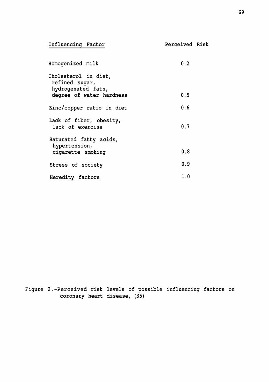
69
Influencing Factor Perceived Risk
Homogenized milk
Cholesterol in diet, refined sugar, hydrogenated fats, degree of water hardness
Zinc/copper ratio in diet
Lack of fiber, obesity, lack of exercise
Saturated fatty acids, hypertension, cigarette smoking
Stress of society
Heredity factors
0.2
0.5
0.6
0.7
0.8
0.9
1.0
Figure 2.—Perceived risk levels of possible influencing factors on coronary heart disease, (35)
70
current interest. It has been suggested that increased amounts of
plasma high-density lipoproteins (HDL) have a "protective" role
in lipid metabolism.(377,429-431)
Plasma lipoproteins are necessary for the transport of
cholesterol.(432) These complex particles consist of a membranous
coat of specific apoproteins, phospholipids, free cholesterol, and
a non-protein core containing lipids.(433) Ultracentrifugation
and electrophoresis have provided major classes of lipoproteins.
Listed in order of decreasing density, protein and phospholipid
concentration, and increasing triglyceride concentration, these
include: (1) high density lipoproteins (HDL), (2) low density
lipoproteins (LDL), (3) intermediate density lipoproteins (IDL),
(4) wery low density lipoproteins, (VLDL), and (5) chylomicrons.
Dietary and recycled cholestrol are transported initially after
absorption in chylomicrons and VLDL fractions and eventually
secreted via the thoracic duct into the bloodstream.(432) VLDL's
contain about 15 percent of blood cholesterol with LDL's
accounting for about 65 percent.(434) The ready uptake of LDL's
by many tissues appears to be mediated by cell surface LDL
binding receptors.(435-437)
Following release of LDL cholesterol in tissues, any excess
not utilized by the tissue must be released into the extra
cellular fluid for return to the liver where it may be incorporated
into bile acid synthesis or may be excreted into the plasma. HDL
may incorporate unesterified membrane cholesterol which is either
71
transferred to VLDL through the action of lecithin-cholesterol
acyltransferase (LCAT) or directly returned to the liver.(434-436,
437) It is also suggested that HDL may directly block LDL
cholesterol uptake by cell receptors and thus prevent cholesterol
accumulation.(438) Approximately twenty percent of the plasma
cholesterol is carried by the HDL and it has been suggested that
the relative concentrations of plasma LDL and HDL could vary the
balance of tissue cholesterol uptake and removal. Diet has been
reported to have little effect on HDL as compared to LDL. HDL
concentration is higher after puberty in females than males, is
increased by physical activity, moderate alcohol ingestion and
estrogens.(434)
Cholesterol's degradation and excretion from the body is
achieved mainly through the conversion of cholesterol to bile
acids, but is also converted to steroid hormones and by losses in
feces and from the skin surface.(432,437)
The lowering of cholesterol levels in the plasma has been
attributed to several factors.(433,439) Among various dietary
components, fiber has been suggested as having an effect upon
plasma lipids and cholesterol.(440) One reviewer found apparent
disagreement in regard to the effects of dietary saturated fat on
lipid metabolism. Hyperlipidemic or atherogenic effects were
not seen with any of the fats when fed to rats with commercial
laboratory rations.(440) However, when saturated fat was fed as
part of a semipurified diet, it was definitely atherogenic and
cholesteremic.(441 ,442) It was concluded that since the diets
72
had only saturated fats in common some other dietary component,
most likely fiber, was accountable for the differences.(440)
Fiber's possible implication in influencing lipid metabolism
has led to extensive research that has provided relevant data in
the problems of coronary heart disease.(443)
It is believed that fiber, by binding bile salts, prevents
the absorption of cholesterol and reabsorption of bile acids.
Bile is secreted by the liver and is made up of a large quantity
of bile salts, a moderate quantity of cholesterol, a small amount
of the green pigment biliverdin and a number of other less
important substances.(139) The bile salts are not enzymes, but
act as a powerful detergent.(308) The detergent action aids in
the intestine in mixing and breaking the large fat globules of the
food into small globules. The smaller globules with their larger
surface area allow the lipases of the intestinal tract to act
more effectively. Without this action of bile wery little dietary
fat would be absorbed. Bile secretion is a continuous process
but the flow of bile into the intestinal tract is not continuous.
A circular muscle, the sphincter of Oddi , around the outlet of the
common bile duct prevents flow into the duodenum causing the bile
to flow into the gallbladder. The gallbladder mucosa reabsorbs
much of the fluid and electrolytes of the bile.(139,308)
Concentration of bile may be as great as twelve-fold allowing the
gallbladder with a maximum volume of approximately 50 ml, to
accomodate the active components of the approximate 600 ml. daily
73
production by the liver.(308) Two simultaneously occurring
mechanisms are believed to cause the gallbladder to empty into the
small intestine. First, the presence of food (particularly fats)
causes the release of cholecystokinin from the tissues of the
duodenum, the hormone passes via the blood to the smooth muscle of
the gallbladder and causes muscular contraction thus a release of
the bile into the intestinal tract. Second, the presence of food
in the duodenum increases peristalsis which causes inhibitory
nervous stimuli to the sphincter of Oddi.(139,308)
Ninety to 95 percent of the bile salts that are ejected into
the lumen of the duodenum are absorbed from the terminal ileum by
an extremely efficient active transport system.(139) The resorbed
bile salts are transported back to the liver via the portal vein
and are eventually reexcreted in the bile. This system of
excretion, reabsorption and excretion, is known as the enterohepatic
circulation. The total bile salt pool of approximately 3.5g is
repeatedly recycled in this manner. It has been estimated that
the bile salt pool may recycle as many as six to eight times per
day.(139) The bile salts not reabsorbed are eventually converted
to the salts of deoxycholic acid and lithocholic acid,(286)
It has been proposed that fiber interferes with this
enterohepatic circulation, thus increasing the rate of catabolism
of cholesterol to bile acids.(299,444) This interruption in
conjunction with the possibility that fiber could prevent micellar
formation essential for cholesterol absorption could lower
74
cholesterol pools in the body.(443)
The effects of various forms of dietary bulk on serum lipids
are not clearly understood but it is known that individuals on
high vegetable content diets tend to have lower serum cholesterol
levels than individuals on lower fiber diets.(68,445) True
vegetarians whose cholesterol levels are about twenty-eight percent
lower than those of the standard population ingest 125 percent
more fiber. Lacto-ovo vegetarians ingest fifty percent more fiber
and have cholesterol levels that are eleven percent lower.(299)
These reports have been supported by others indicating that certain
foods high in fiber influence blood lipid levels and atheroma
formation.(446,447) Trowel 1 extensively reviewed the relationships
and concluded that natural dietary fiber, which is a structural
heterogenous mixture of celluloses, lignins and pectic substances
could differ in their effects upon cholesterol.(81) Several
investigators began to evaluate various fiber components effects
on blood lipid values.
Bran and cellulose have little effect on lowering serum
cholesterol in humans and other animals although they might
increase fecal bile acids excretion.(19,29,446,448-454) Earlier
animal studies indicated that certain dietary fiber components
did affect serum cholesterol.(455,456) In one such study New
Zealand white rabbits were fed a purified ration of 30 percent
casein, 15 percent beef tallow, 15 percent ground cellulose, six
percent salt mixture and other essential vitamins and minerals.
75
During the first five weeks of the experiment the purified
rations were fed without added cholesterol, and during the second
five weeks, cholesterol was added to the ration at the rate of 0.5
percent. The experimental data revealed that serum cholesterol
concentrations in rabbits fed purified ration containing fifteen
percent beef tallow were lowered with the addition of either five
percent pectin or 0.5 percent cholestyramine to the ration during
the five week period. The effects of the addition of both pectin
and cholestyramine were additive in lowering the serum cholesterol
values. During the second five week period of feeding, the same
ration but with 0.5 percent cholesterol added, the pectin and
cholestyramine, singly or in combination, slowed the increase in
serum cholesterol.(457) Similar research in rabbits and other
animals have shown a hypocholesterolemic action of pectin.(458-474)
Experimentation on human subjects have shown that pectin,
guar gum, lignin, and mixed complex carbohydrates reduce serum
cholesterol levels and increase fecal acidic and neutral steroids
excretion.(19,21,299,475-478)
One report indicated that 15 grams of pectin per day resulted
in a 15 percent drop in serum cholesterol, a 16 percent increase
in fecal excretion of neutral steroids and a 40 percent increase
in excretion of bile acids.(479) Another report showed no
hypocholesterolemic effect by pectin.(480) Subsequently it was
reported that the cholesterol-lowering effect of pectin was dose-
related. (481 ) A plasma cholesterol decrease by two to six percent

76
was seen in 16 subjects when two to ten grams of pectin were
administered. Another investigator found a twelve percent reduction
in plasma cholesterol in seven men after two weeks of administration
of 36 grams of pectin per day.(25)
Although no effect was seen on serum triglycerides the
administration of 40 to 50 grams per day of pectin over a two week
period to nine normolipidemic and hyperlipidemic patients showed
a significant decrease in serum total and unesterified cholesterol.
(482)
In addition to pectin, guar gum and psyllium seed celloid
have been reported to yield an hypocholesteremic effect in man.
(21,25,29,480) The substances are chemically dissimilar and it is
suggested that their physical state in the intestine may determine
their action. The presence of a gel in the intestine could cause
interference with the equilibrium between the micellar phase and
the molecular phase which- passes into the layer on the brush
border and might reduce lipid absorption.(479)
The interrelationship of diet and lipemia is neither simple
nor direct. One would hope that a simple nutrient ingestion and
dose-response situation would exist, but unfortunately this is not
so. The complexity of the modern diet and the interactions among
dietary components are far from being completely clarified.
One dietary component, complex carbohydrates are generally
found to reduce serum lipid levels, although it may be some time
before assessment of what component and what quantity is needed

77
to produce the desired response.(21,483)
Gallstones and Cholecystitis
Disease of the gallbladder is secondary to gallstones in
nearly all cases.(484) It has been reported that 98 percent of
cholecystectomies in England and America are the result of stone
formation.(485,486) In western societies 85 percent of the stones
are cholesterol stones.(139) Minor components are made up of
calcium carbonate, calcium palmitate and calcium phosphate.(487)
Therefore, gallstones are caused mainly by the precipitation of
the fatty product cholesterol which is excreted in the bile.(308,
484)
Formation of stones occurs in the gallbladder or bile ducts,
either when an atypical material appears in the bile or the relative
composition of the bile is altered in such a way that a normal
constituent precipitates. Calcium billirubinate stones form if
certain bacteria containing a glucuronidase deconjugate bilirubin
in the bile. Calcium combines with the free bilirubin to form
the highly insoluble calcium bilirubinate.(139) The ratio of
cholesterol and bile acids normally is such that cholesterol
remains in solution and stones do not form. If that ratio is
altered with resultant super-saturated bile, precipitation and
formation of stones occurs.(488-490)
Epidemiological studies have indicated gallstones are rare
among primitive peoples, even though their diets are greatly
varied.(50,449,491) Gallstone incidence is reported to increase
78
with urbanization and westernization.(423,484,492) Diseases
associated with cholesterol-rich gallstones include obesity and
diabetes.(493-498)
Gallstone patients have been reported to have bile salt
pools reduced by as much as 40 to 50 percent of normal size.(499-
501) In animals it has been shown repeatedly that liver synthesis
of bile salts is inhibited when refined carbohydrates are added
to the diet.(484) It has also been shown that in experiments with
bile fistula, dogs in which there is a constant drainage of bile
salts, the liver is stimulated to synthesize new bile salt at a
maximal rate. In fasting dogs, synthesis could be increased when
protein was fed, but this result could be reduced by pre-feeding
high calorie semi-synthetic diets and completely abolished with
the addition of sucrose,(502) Other animal studies indicate
that bile salt synthesis is reduced on diets high in refined
carbohydrates and little or no fiber,(293,434,503) Further
studies have confirmed these findings, and have shown that dietary
fiber markedly increases the intestinal bile salt pool,(286)
CHAPTER X
PHYSICAL ASPECTS OF DIETARY FIBER
Fiber as a Food Additive or Ingredient
Fiber materials such as polysaccharides have been used as
functional ingredients in the food industry for many years. Among
the primary fiber materials used are the "gums" which are used as
gelling agents or stabilizers. Gums in this sense are considered
to be polysaccharides that are dispersible in either hot or cold
water to produce viscous mixtures or solutions.(299)
The structural and textural properties of plants are largely
determined by the gum constituents present in almost all natural
foods.(504) Gums are often used as food additives in prepared
foods to impart desirable texture and functional properties to
finished products.(505) Commercially gums are normally used as
water soluble or water-dispersible hydrocolloids. Their properties
usually consist of suspending, dispersion, and stabilization in
aqueous dispersions. Gums may act as emulsifiers, adhesives,
coagulants, binders, lubricants or film formers.(506)
Commonly used commercially available gums are shown in
Figure 3.
Fiber Analysis
The quantitative and qualitative analysis of dietary fiber is
a complex process. A mixture of various substances arising from
the cell wall of plants and an array of polysaccharides occurring
79

80
Seaweed Extracts
Agar Alginates Carrageenan Furcellaran
Plant Exudates
Gum Acacia (Arabic) Gum Tragacanth Gum Ghatti Gum Karaya
Seed Gums
Locust Bean Gums Guar Gum
Plant Extracts
Pectin
Biosynthetic Gums
Xanthan
Cellulose Derivatives
Carboxymethyleel 1ulose Methylcellulose and
Hydroxypropylmethylcellulose
Figure 3.--Commercially available gums.(506)
81
naturally or derived from food additives make up dietary fiber.
In order to facilitate research it would be desirable to
fractionate plant-based foods into their various chemical
constituents and accurately ascertain the quantity of each
component. Current technology does not provide this approach,
but a number of alternatives have been developed.
Historically, fiber analysis began in early ruminant
nutrition studies.(507,508) These studies soon revealed that a
significant portion of many forages were not digestable, and the
amount of this fraction in a ration had a lowering effect on the
availability of other components in the diet.
Crude fiber determination methods were developed in the
early eighteenth century and were standardized by the Association
of Official Analytical Chemists in 1887.(507) The method is
still widely used even though its shortcomings are well
documented.
New methods of analysis have been developed which have
proven far more useful than crude fiber determinations. The
increased interest in human nutrition and dietary fiber have
produced a need for even better methods.
Crude fiber is defined as the resultant residue after
treating a weighed food sample with solvents to extract lipids,
then extracting with acid, dilute base and, finally weighing the
sample.(299) This procedure is highly empirical, and strict
adherence to procedural detail is required for reproductibility.
82
The resultant crude fiber is not of constant composition and does
not accurately reflect hemicellulose and lignin amounts in the
sample.(509-511) (see Appendix A)
The normal-Acid Fiber (NAF) method was developed as an
alternative to the crude fiber method. An important difference is
that the alkali stage was eliminated with a resultant minimization
of variability encountered in the crude fiber method.
The results of this method are not greatly affected by
variations in conditions, time of hydrolysis, sample size, and rate
of heating (Appendix A). The crude fiber method is still more
widely used than this method.
The Acid Detergent Fiber (ADF) work of Van Soest's is now
regarded as classic in this area of research. His aim was to
modify the NAF method and produce an acid fiber with a low
nitrogen content.(509) The results of this work produced a method
to measure a fraction that was closely correlated with the
nutritive value of a forage. The ADF method produced a fraction
composed mostly of cellulose and lignin.
The results of the ADF method correlate well with indigest
ible matter in ruminant nutrition and give accurate estimates of
the cellulose and lignin content of forages. In regard to human
nutrition problems arise in that recovery of one-two percent
cellulose and lignin is difficult in small sample size and
increasing the sample size results in problems with foaming and
final filtration. Any samples with high lipid content must be
83
defatted. The major drawback to ADF is the inadequate measure
of all dietary fiber in a sample (Appendix A).
The Neutral Detergent Fiber (NDF) method was also developed
by Van Soest and is believed to yield accurate measurements of
cell wall components in plant food stuffs. Apparently this method
fractionates the dry matter of feeds into those components
nutritionally available by normal digestive processes and those in
which microbial fermentation is required.
In this method sodium lauryl sulfate, the detergent,
solubilizes lipids and proteins, EDTA removes minerals, and heat
gelatinizes and solubilizes starch (Appendix A).
The NDF method has proven useful for the estimation of
total dietary fiber. Again problems have arisen when this method
has been utilized for human foods. Precision is difficult due to
small sample size and human foods are normally higher in starch
and lipid contents thus requiring lipid and starch removal.
Detergent soluble components may be lost in the process thus
underestimating total dietary fiber in some samples. The sum of
cellulose, lignin and hemicellulose are determined by the NDF
procedure. A comparison of total dietary fiber and NDF fiber for
selected foods is given in Appendix A,
The Unavailable Carbohydrate Method functions as a part of
a more-or-less complete food carbohydrate analysis where
unavailable carbohydrate is equated with total dietary fiber.
The unavailable carbohydrate method is a quantitative index of
84
dietary fiber, but not a qualitative measurement of the dietary
fiber components (Appendix A ) .
This method was elaborated by Southgate to give an index of
total dietary fiber as the sum of its various components.
Although this method does not isolate or characterize soluble
polymers, it does yield values expressed in terms of sugar
components. Problems arise with isolation of soluble proteins of
foods high in sunflower or soy proteins. Amounts of pectins and
gums are not quantitatively determined in this lengthy procedure.
The unavailable carbohydrate method was further modified by
Englyst and Southgate.(513)
For studies of the polysaccharide composition of the plant
cell wall Siege! developed a method that attempts to characterize
components in specific fashion.(514) Polysaccharide degradation
may occur in the delignification stage and alkali extraction.
This method is not used frequently in human food analysis. A
hypothetical complete analysis scheme proposed by Southgate is
arduous and unrealistic for periodic genera! analysis.(512)
(Appendix A)
Components of Dietary Fiber
The term fiber has caused much confusion and controversy.
(10,45,518) Fiber illicits different responses from the botanist,
cereal chemist, animal nutritionist and human nutritionist.
There is yet no concise definition of fiber that conveys fully the
85
concept of fiber in human nutrition.(519) A major difficulty
inherent in studies of human nutrition and food fiber has been a
failure of agreement in what fiber actually is.(520) Until
recently several terms have been used interchangeably indicating
various fiber sources, thus producing experimental results often
difficult to interpret and compare.(521)
Dietary fiber is a complex chemical substance of plant origin
and the term generally refers to cell wall biopolymers. It is
often defined as those components of foods that human enzymes do
not break down in the digestive tract.
Terms often used to indicate fiber in human nutrition are:
CRUDE FIBER (CF)
DIETARY FIBER (DF)
PLANT FIBER (PF)
FIBER
NONPURIFIED PLANT FIBER (NPPF)
PURIFIED PLANT FIBER (PPF)
NONNUTRITIVE NATURAL FIBER
NONNUTRITIVE SYNTHETIC FIBER
UNDIGESTIBLE CARBOHYDRATE (UC)
UNAVAILABLE CARBOHYDRATE (UC)
PLANTIX (PX)
COMPLANTIX (CPX)
PARTIALLY DIGESTIBLE PLANT POLYMERS (PDPP)
PARTIALLY DIGESTIBLE BIOPOLYMERS (PDB).
86
The relationships of th.e components of dietary fiber and the
proposed nomenclatures are shown in Figure 4.(76,515,517)
Industrial Applications of Food Fiber
Interestingly the role of dietary fiber in human nutrition
has attracted the attention of the food industry. Breakfast
cereal manufactures were the first to respond to the increased
consumer awareness of dietary fiber.(522) Many commercially
available products were already using bran as a component, thus
little product development was required. Alterations in advertising
emphasis have focused on the fiber component. Soon after breakfast
cereal fiber emphasis got underway, the baking industry responded
with "high fiber" products.(523) Promotional schemes emphasized
the reduced caloric density and alluded to other possible benefits
of fiber.(524)
Dietary fiber is now available in a variety of forms for
manufacturers.(525)
Powdered cellulose
Microcrystalline cellulose
Wheat bran
Corn bran
Soy bran
Oat hulls
Linseed meal (from cotton)
Rice bran
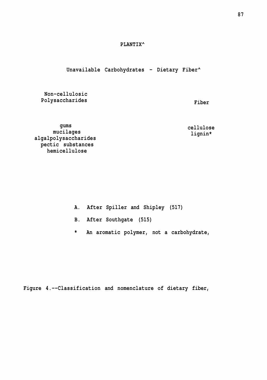
87
PLANTIX^
Unavailable Carbohydrates — Dietary Fiber^
Non-cellulosic Polysaccharides Fiber
gums mucilages
algalpolysaccharides pectic substances
hemicellulose
cellulose lignin*
A. After Spiller and Shipley (517)
B. After Southgate (515)
* An aromatic polymer, not a carbohydrate,
Figure 4.--Classification and nomenclature of dietary fiber,
88
Corn germ meal
Wheat germ flower
Brewers spent grains
Wheat shorts
Wheat middlings
Nuts (fibrous skins)
Soybean hulls
Peanut hulls
Coconut kernal residue
Citrus pectin.
The first two materials are essentially purified cellulose
products. The remaining products are normal by-products from
various processing methods.
Powdered cellulose (alpha-cellulose) is usually derived from
maple, birch and beech wood pulp. Bark is removed from the wood
which is chipped, "cooked" in pressure digesters with appropriate
solvents to dissolve lignin. The softened pulp is washed,
bleached, then ground to appropriate size and texture.
Microcrystalline cellulose differs from powdered cellulose.
Cellulose pulp is treated in a manner that hydrolyzes the fiber,
breaking down the bundle-like structure of cellulose producing
crystals. Drying processes cause the recombination of these
crystals into an aggregate. Microcrystalline cellulose is
commercially available in sizes ranging from two to two hundred
microns.(299)
89
Purified cellulose is used in many foods because it achieves
a lowered calorie objective and has specific functional properties
including binding, consistency control, and absorption.(526) It
is presently being used in flour based products, snack products,
processed meats, sauces, salad dressings, sausage casings, and
breedings for meat and fish.(299)
Wheat bran is probably the most widely used of any of the
many by-products of commodity processing that are available as
commercial sources of dietary fiber. Extensive studies of wheat
fractions have produced considerable information in regard to
particle size, density, hydration capacity, and ion exchange
capacity.(522,527)
The development and introduction of high fiber breads by
the baking industry has been a considerable undertaking. Many
dietary fiber materials were discovered to be nonacceptable in
bread formulas due to raw ingredient supply levels, color, flavor,
and texture.(522,528,529) The most widely used fiber components
are wheat bran and powdered cellulose followed to a lesser extent
by corn and soy brans. Cellulose has been the most successful
in producing a high fiber "white type bread." Citrus products
have had problems with producing a bitter taste.(299)
Critics of the purified cellulose type breads claim that they
are made with "sawdust." Such criticism reveals a lack of
understanding to the wide spread nature of cellulose in foods
consumed by humans. The use of this purified cellulose has permitted
90
the production of a bread with characteristics more similar to
conventional white bread but with a much higher fiber content than
even whole grain breads. An additional claim can be made for
decreased caloric density.
One obstacle facing the food industry is that health claims
in relation to dietary fiber have not all been substantiated.
Thus in marketing techniques one is faced with limited ability to
make health related claims. At present most labeling claims are
related to "fewer calories," "increased dietary fiber," "less
carbohydrate," and "get the roughage you know you need."
Current research by food processors is placing a degree of
emphasis upon the consumers' heightened interest in dietary fiber
and realize the existence of a market for high fiber products.
The awareness of such a market has led to valuable basic research
into fiber characteristics. Practical applications of this
knowledge has led to many high fiber food products currently on
the market.(530) Current research is also being aimed at more
efficient use of fiber obtained from processing by-products.(299)
If the nutritionist accepts the idea of need for more fiber
in American diets, the food technologist must be ready to answer
some formidable questions in regard to fiber evaluation and what
foods are the best vehicles. Work is now being carried on in
order to produce standardization of methods in the evaluation of
various fiber sources,(530)
If fiber is found to be a disease preventive measure in human

91
health, the vehicle selection becomes a complex situation. The
material must be introduced into food products in such a way as to
do the greatest good for the greatest number of individuals
without detrimental effects to any sub-population.
CHAPTER XI
SUMMARY
The fiber hypothesis has brought forth much controversy.
Although epidemiological evidence outweighs experimental data, we
are seeing an emergence of facts about dietary fiber.
In regard to fiber's effect upon diverticulosis we see that
colonic motor activity is affected. The exaggerated colonic
intraluminal pressures of diverticulosis correlate with its
pathology, i.e. hypertrophy of muscle. A hypertrophy believed to
result from the propulsion of hard, small, low-volume stools.
Lowered colonic pressure changes have been seen in diverticular
disease patients given bran.(363) Bran supplementation has also
been seen to improve patients symptomatica!ly.(296) The
therapeutic value of bran in prevention of the disorder is not
proven. Post-operative studies however indicate a possible
prophylactic effect in those suffering from diverticular disease.
(531)
Relationships between cancer and dietary fiber have been
demonstrated by epidemiological data and studies with laboratory
animals. Colonic cancer being the most often indicated in fiber-
depleted diets. Research is being carried out as to whether these
associations are causative and to determine possible meahanisms
of action. Fecal bacteria determinations have often shown
differences among populations with high and low rates of large
92
93
bowel cancer. These populations showed differences in fiber
consumption but no great differences were seen in fat consumption
or fecal steroids. The fiber hypothesis of colonic-rectal cancer
is supported by such findings but does not refute the fat
hypothesis. Fiber may exert its protective effects through
binding to ingested carcinogens, diluting their concentration or
decreasing their incubation time.
In regard to fiber's effect on fecal weight there seems to be
agreement. The precise mechanism by which fiber increases fecal
weight has not been determined but several theories are proposed.
CD
Transit time alterations by dietary fiber is a complex picture.
Generally, investigators agree that transit time decreases as stool
weight increases, but there are accounts of nonsignificant changes
with increased fecal weight. It has also been reported that in
those individuals with fast transit times prior to fiber
supplementation there has been a slowing down of transit time.
Heaton has suggested that the fiber has "normalized" colonic
activity.(498) The concept of transit time is often over
simplified, and should be considered in light of the mixing and
streaming of the gastrointestinal contents. The passing of a single
marker or a dye may not be indicative of actual processes. The
data being gathered do suggest a significant influence of dietary
fiber on food residue's passage through the gastro intestinal
tract.
94
Fiber's relationship to coronary heart disease may be indirect
A diet high in fiber is probably a diet already low in cholesterol.
Caloric density is also generally lower in the higher fiber foods.
If one accepts the idea of decreased transit time with increased
dietary fiber, it is suggested that if food travels through the
gastrointestinal tract more rapidly there is less opportunity for
food to be broken down and absorbed. It has already been proposed
that fiber in some way binds bile salts and alters both its
metabolism and reabsorption. This decreased efficiency of
reabsorption has been seen as a means of lowering body cholesterol
pools via bile synthesis. Those that place reliance on this
hypothesis indicate that this would yield a slowing of hardening
of the arteries, as well as decreased incidence of gallstones.
If dietary fiber has a relationship to coronary disease its
most important role may be in combating obesity. High fiber diets
not only are normally lower in calories, but have been shown to
generally produce satiety more rapidly than highly refined foods.
A correlation exists between obesity and diabetes.(532)
High-fiber, high-carbohydrate diets have been shown to have
beneficial effects on glucose tolerance and the insulin dependency
of many diabetics. More and more consideration is being given
to fiber's effects upon hypoglycemic rebound.
Another protective influence of fiber is its suggested role
in lowering or alleviating certain toxic effects of various drugs,
chemicals and food additives.(533) It should be noted that
95
indications are that different plant fibers vary significantly
in their ability to counteract toxic effects.
The widespread interest in dietary fiber is shared by those
working in nutrition and medicine as well as by the lay person.
The latter often seeing it as a "health" food which will cause
phenomenal changes in his well being. This perception of dietary
fiber's role in nutrition is a logical offshoot to the trends in
the "back to nature" movement of late. It may just be this
movement that has spurred much of the fiber research being done by
the food industry. An obvious market is ready and waiting to
consume high fiber "health foods,"
These popular concepts about food fiber need to be tempered
with some of its possible harmful side effects. One of the
advantages of white flour is that the major source of phytates is
removed, Phytate's effect on calcium absorption may have been
overstated, but with the recognition of the importance of the
trace minerals, one must consider fiber's effect on them. If the
intake of such divalent metal ions as zinc, chromium, magnesium,
and copper is marginal at best in some populations a deficiency
state is a recognized possibility in high phytate content diets.
Anatomically over consumption of fiber may produce
pathological states involving the gastrointestinal tract.
In those cases where caloric adequacy is a problem, one
would be wise to attempt to reduce fiber amounts thus increasing
caloric density. It has also been reported that certain food fiber

96
may contain trypsin and chymotrypsin inhibitors, thus adding to
the problems of those already existing on a marginal diet.
CHAPTER XII
CONCLUSIONS AND RECOMMENDATIONS
Any dietary component when considered alone produces debatable
information. Dietary fiber is no exception especially in light of
all that has been attributed to it. Fiber may not have all these
"health" promoting characteristics, but the controversy over fiber
has greatly stimulated its research. That research has already
proven beneficial in several health problems and may provide more.
Dietary fiber's correlation with leading diseases is
currently a favorite basis for many hypotheses. Despite the
controversy surrounding dietary fiber there appears to be potential
for fiber in preventative medicine through good nutrition that
uses food as the mode of conveyence.
In general it is recommended that people increase the overall
consumption of dietary fiber in light of its relationship to
intraluminal pressures. Increased dietary fiber has a high
correlation with larger and softer stools. Resultant reduced
intraluminal pressures in the lower gastrointestinal tract relieves
symptoms of diverticular disease and related gastrointestinal
disorders. Reduction in such pressures may play a role in the
prevention of muscle hypertrophy and lessen the danger of high
pressure damage to weakened areas in the bowel wall.
These same alterations brought about by fecal matter density
and volume may act as a preventative measure in constipation thus
97
98
aiding in reducing defecation straining and lessening the
intraabdominal pressures that may lead to retrograde pressures in
the venous system therefore reducing the likelihood of vericose
veins and hemorrhoids.
Another important aspect of increased fiber consumption is
its relation to caloric density and energy consumption. Higher
fiber foods tend to be lower in calories per unit than fiber
depleted or low fiber foods. Overnutrition is a major health
problem in developed countries of the world. It is therefore
recommended that we encourage increased consumption of high fiber
foods in order to take advantage of lowered caloric density, and
satiety factors related to less refined foods.
There is increasing evidence that coronary heart disease is
essentially the result of habitual overnutrition. Epidemiological
studies indicate that prevalence of the coronary heart disease in
various countries correlates with the per capita caloric
consumption. This correlation appears to exist in all four
primary coronary risk factors: (1) systolic blood pressure, (2)
serum cholesterol, (3) fasting blood sugar levels and (4) serum
uric acid levels. The major emphasis in any discussion of dietary
fiber and heart disease should center not on the "curative" effects
of a single food component, fiber, but on the effects of reduction
of fiber in the diet in total. The factor which most strongly
relates fiber depleted diets with increased incidence of heart
disease is not simply fiber but the increased caloric density and
99
concurrent overnutrition (malnutrition) that attends the
"westernization of the diet." The modern diet makes the ingestion
of excess calories, i.e. physiological excesses of nutrients,
an easy possibility. Thus a derangement of diet exists, so easily
obtainable, rather than a disorder of the individual. Many of our
familiar carbohydrate foods (sugars, flours, and breakfast cereals)
are processed in such a way as to remove a high portion of the
dietary fiber materials with resultant foods not only more easily
ingested but more calorically dense than their less processed
counterparts. One of the more dramatic examples is the use of
refined sugars in the increasingly popular sweetened beverages.
The shortened ingestion time coupled with lessened bulk produced
a secondary lessening of satiety level thus enhancing the likelihood
of over consum.ption especially in regard to calories (and secondly
a lack of other nutrients that accompany the calories in less
processed or refined foods).
If this view is correct, the prevention of the disease may be
closely correlated with the maintenance of a thin physiological
physique. The content and type of fat in the diet may become of
little or no importance if the total diet makeup and energy intake
is correct for body size, age, sex and metabolic state.
Dietary fiber's relationship to cancer is less clear, but it
is believed that the fiber depleted diet promotes fecal stasis.
The resultant increase in transit time allows for increased
bacterial proliferation, increased time for conversion of potential
100
carcinogens and contact with intestinal mucosa. I suggest that the
increased consumption of dietary fiber may provide protection via
one of the following effects attributed to dietary fiber: (1)
decreased transit time, thus reduced exposure to carcinogens or
potentially toxic materials, (2) increased peristalsis as a result
of increased water-binding properties of various fiber components,
(3) increased volatile fatty acid production with a cathartic effect,
(4) bile acid metabolism alterations, (5) dilution of intestinal
contents and (6) binding capacities which may reduce the absorb
ability and reactivity of other intestinal contents.
Development of a recommended intake of dietary fiber should
be considered as a distinct possibility. At present there is no
consensus of what constitutes a suitable level, hence parameters
need to be established to assess recommended levels in average
adult humans. The following criteria might be used as a starting
point: (1) establish fiber intake which produce 140-150 g/day
of feces (a level which normally decreases transit time but does
not interfere with predicted motility), (2) adjust fiber intake to
levels which produce transit times of 24-48 hours, and (3) use of
fecal weight and transit time correlations to arrive at dietary
needs of fiber which produce the desired fecal weight.
Consideration must be given in regard to body weight, age,
sex and the various sources or types of fiber ingested. The level
will probably approximate .3-.5 g bran or equivalent per kg body
weight as a minimum.
101
In regard to diabetes there is no one "diabetic diet"
appropriate for all cases. Individual variations, disease
severity, the type and extent of insulin therapy administered and
the level of physical activity will alter the dietary needs.
Caloric content of the diet must be established in regard to the
patient's desirable weight as compared to actual weight. The
overweight diabetic requires a dietary regimen aimed at achieving
a "diabetic" ideal weight of about ten percent lower than the
statistically desirable weight. Once this level is reached the
diet must be altered to maintain this reduced weight. Insulin
treatment without a carefully coordinated dietary regimen risks
the possibility of insulin-induced hypoglycemic shock and the
dire consequences of poorly controlled metabolism.
The single most important objective in adult onset diabetes
should be weight reduction. Responsiveness of cells to insulin
largely depends upon their size.
In light of the relationship of weight to diabetes and the
experimental evidence indicating lessened plasma glucose, more
normalized insulin and glucagon responses after ingestion of high
fiber meals, we need to stress this relationship and encourage
continued investigations into these responses.
Implementation of increased awareness and use of high fiber
foods has already begun in that the public has been exposed to
"natural" and/or return to "nature" concepts (perhaps not always
supported with sound reasoning or evidence) as well as an ever
102
widening selection of commercially available "high fiber" foods.
Perhaps the better vehicle for such increased usage of dietary
fiber will be through encouragement of the use of a varied diet
with emphasis on increased use of vegetables, fruits, and legumes.
In this manner one would remain within the scope of traditional
nutritional education without the incorporation of new "high
fiber" food products. The advantages of such encouragement
include using an existing system, thus lessening learning time,
and expense, using readily available food vehicles and perhaps
incorporate what is referred to as "food specific satiety."
The increased utilization of dietary fiber will probably do
no harm, except in a few specific instances. Current evidence
indicates that a general increase in use along with proper intake
of other nutrients will prove beneficial to man.
When considering the effects of dietary fiber one must
remember all the variables involved, i.e. components of the fiber,
stage of plant development upon extraction and the extraction
process itself. These variables as well as the physiological and
hereditary aspects of the test animal are crucial in determining
fiber's influence. All test animal results cannot necessarily be
applied to humans. I do encourage the use of the appropriate
test animals and increased utilization of human subjects.
This author would like to caution against relying upon
indiscriminate associations with various fibers and their effects.
One must remember that the "type" of fiber varies with the maturity
103
of the plant as well as the extraction process utilized for
obtaining "pure" fiber. It is most likely that the influences of
individual fiber components may greatly vary as compared to the
"whole" plant with its mixture of fibers and other nutrients.
A natural matrix effect of these fibers and nutrients may produce
a completely different effect than a similar amount of purified
component.
A current trend in the consumer awareness of "health foods"
is the use of individual nutrients sometimes in pharmacological
rather than physiological levels. This practice could have dire
consequences in some individuals, especially those in marginal
nutritional status in regard to those nutrients most affected
by dietary fiber (i.e. fats, fat soluble compounds, and minerals).
I would like to reiterate that the best way to increase intake of
dietary fiber is through a good mixed diet. Such a diet enhances
our ability to gain the essential nutrients as well.

LIST OF REFERENCES
1. Cummings, J.H., Gut. T4, 69 (1973).
2. Painter, N.S., Burkitt, D.P., Clin, Gastroent. 4., 3 (1975).
3. Burkitt, D.P., Walker, A.R.P., and Painter, N.S., J. Am. Med. Asso, 229, 1068 (1974).
4. Burkitt, D.P., Cereal Foods World 22_ (1), 6 (1977).
5. Cleave, T.L., Campbell, G.D. and Painter, N.S., "Diabetes, Coronary Thrombosis and the Saccharine Disease," 2nd ed., 89, John Wright, Bristol (1969).
5. Hutt, M.S.R. and Burkitt, D,, Brit, Med. J. 2_, 719 (1965).
7, Mann, G,V,, Spoerry, A., Gray, M. and Jarsakow, D., Am. J. Epidemic. 95_, 26 (1972),
8, Editorial, New Zealand Med. J,, Jan, 12, 17 (1977).
9, Cleave, T.L., J. Roy. Nav. Med. Serv. 4£, 116 (1956).
10, Kimura, K.K., Cereal Foods World 22̂ (1), 16 (1977).
11, Burkitt, D.P., J. Am. Med. Asso. 231, 517 (1975).
12, Burkitt, D,P,, Trowell, H,C., (eds.), "Refined Carbohydrate Foods and Disease," p, 333, Academic Press, New York (1975).
13, Mitchell, W.D, , Eastwood, M,A,, in "Fiber in Human Nutrition," (G.A. Spiller, R.J. Amen, eds.) p, 185, Plenum Press, New York (1976).
14, Eastwood, M,A., Eastwood, J. and Ward, M., in "Fiber in Human Nutrition," (G.A. Spiller and R.J. Amen, eds.) p. 207, Plenum Med. Book Co., New York (1976).
15, Walker, A.R.P., in "Fiber in Human Nutrition," (G.A. Spiller and R.J, Amen, eds,) p, 241, Plenum Med. Book Co., New York (1976).
16, Painter, N,S, , in "Fiber Deficiency and Colonic Disorders," (R.W. Reilly and J.B. Kirsner, eds.) p. 109, Plenum Med. Book Co., New York (1975).
104

105
17. Almy, T.P., Hospital Practice. ]]_ (3), 11, March (1976).
18. Miettinen, T.A., in "Hypolipidemic Agents," (David Kritchevsky ed.) p. 125, Springer-Verlag, New York (1975).
19. Keys A., Grande, F., Anderson, J.T., Proc Soc Exp. Biol. 106, 555 (1961).
20. Grande, F., in "Sugars in Nutrition," (H.L. Sipple, K.W. McNutt, eds.) p. 401, Academic Press, Inc., New York 0974).
21. Kritchevsky, David, Arch. Surg. 1J3̂ , 52 (1978),
22. Editorial, Lancet 2_, 353 (1975).
23. Ellis, F.R. and Montegritto, V.M.E,, Am. J. Clin. Nutr, 23_, 249 (1970),
24. Hayes, T.M., Munn, J., Jones, A. and Mottran, R., Diabetologia 11, 52 (1977).
25. Jenkins, D.J.A., Leeds, A.R. , Newton, C. and Cummings, J.H., Lancet 1, 1116 (1975).
26. Jenkins, D.T.A., Leeds, A.R., Gassul, M.A., Wolver, T,M,S., Goff, D.V., Alberti, K.G,M.M,, Hockaday, T,D.R., Lancet 2_, 172 (1976).
27. Hayes, T,M., J. Human Nutr. 31, 337 (1977).
28. Forman, D.T., Garvin, J.E., Forestner, J.E., Taylor, C.B., Proc. Soc. Exp. Biol. 127_, 1060 (1968).
29. Stanley, M. , Paul, D., Gacke, D., Murphy, J., Gast. 62_, 816 (1972).
30. Pomare, E.W., Heaton, K.W. , Lowbeer, T.S., White, C, Gut 15_, 824 (1974).
31. Walters, R.L., Baird, I.M,, Davies, P,S., Hill, M,J,, et al., Brit, Med. J, i, 536 (1975).
32. Binder, H.J., Ann. Rev. Pharmacol. Toxicol. U_, 355 (1977).
33. Eastwood, M.A., Kirkpatrick, J.R., Mitchell, W.E., Bone, A. and Hamilton, T. , Brit, Med. J. 4, 392 (1973),
106
34. Sarner, Martin, The Practitioner 216, 661, June (1976).
35. Labuza, Theodore P., "Food and Your Wellbeing," p, 175, West Pub, Co., San Francisco (1977).
36. Kiehm, T.G., Anderson, J.W. and Ward, K., Am. J. Clin. Nutr. 2i, 895 (1976).
37. Heaton, K.W. and Pomare, E.W., Lancet 1, 49 (1974).
38. Leeds, A.R., Hockaday, T.D.R., Lancet 2, 1086 (1976).
39. Southgate, D.A.T., Proceedings West. Hemisphere Nutr. Cong., 4, 51 (1975).
40. Trowell, H., Plant Foods for Man 1, 11 (1973).
41. Shearer, Robin S., Current Therapeutic Research 19 (4) April (1976). —
42. Bleehen, S.S., Edwards, I.R. and Clark, R.G., Brit. J. Derm. 95, 219 (1976).
43. Blaton, J.R., Can. J. Pub. Health 58̂ , 479 (1967).
44. Mayer, J., in "Modern Nutrition in Health and Disease Dietotherapy," 5th Ed., (R.S. Goodhart, M.E. Shills, eds.) p. 477, Lea and Febiger, Philadelphia (1973).
45. Pomare, E.W. , Drugs 1^, 213 (1977).
46. FucKs, H., Dorfman, S., Floch, M., Am. J. Clin. Nutr. ^, 1443 (1976).
47. Drasar, B.S., and Hill, M.J., Am. J. Clin. Nutr. 25_, 1399 (1972).
48. Reddy, B.S., Weisburger, J.H, and Wynder, E.L., J. Nutr. 105, 878 0975).
49. Hill, M.J,, Cancer Res, 3^, 338 (1975).
50. Ershoff, B,H., Am, J, Clin, Nutr. 27_, 139 (1974).
51. Ershoff, B.H. and Marshall, W.E., J. Food Sci. 40̂ , 357 (1975).
52 Ershoff, B.H., Proc. Soc. Exp. Biol, and Med. ] ^ , 65 (1977).

107
53. Ershoff, B.H., Proc. Soc. Exp. Biol, and Med. 141, 857 (1972).
54. Ershoff, B.H. and Thurston, E.W., J. Nutr. 104, 937 0974).
55. Ershoff, B.H. and Bernandez, H.J., J. Nutr. 69_, 172 0959).
56. Ershoff, B.H., J. Nutr. 7^, 484 O960).
57. Mendeloff, A.I,, New England J. Med, 297_ (15), 811 (1977).
58. McNutt, K.W., McNutt, D.R,, "Nutrition and Food Choices," p, 381, Science Research Assoc, Inc., Chicago (1978).
59. Connell, A.M., J, Am. Diet. Assoc. 71, 235, Sept. (1977).
60. Burkitt, D.P., Walker, A.R.P, and Painter, N,S,, J, Amer, Med, Assoc, 229, 1068 (1974).
61. Cleave, T,L., "The Saccharine Disease," p. 1, John Wright and Sons, Ltd., Bristol (1974).
62. Trowell, H. Atherosclerosis 15, 138 (1972).
63. Trowell, H., Amer. J. Clin. Nutr. 25_, 926 (1972),
64. Trowell, H., Proc. Nutr. Soc. 32_, 151 (1973).
65. Trowell, H., Amer, J. Clin. Nutr. 28, 798 (1975).
66. Trowell, H,, Plant Foods for Man 1, 157 (1975),
67. Burkitt, D,P, and Walker, A,R,P,, S. Afr. Med. J, 5^, 2136 (1976),
68. Walker, A.R.P. and Arvidson, U.B,, J, Clin, Invest. 33_, 1366 (1954).
69. Walker, A.R.P., Mortimer, K.L., Kloppers, P.J., and Settel, H.C., Am. J. Clin. Nutr. 9., 643 (1961),
70. Walker, A.R.P., So. Afr. Med. J, 35., 114 (1951),
71. Walker, A.R.P. and Walker, B.F., Brit. Med. J. 1, 238 (1969).
72 Walker, A.R.P., Walker, B.F. and Richardson, B.D., Brit. Med. J. 3, 48 (1970).

108
73. Walker, A.R.P., in "Fiber in Human Health," (G.A. Spiller and R.J. Amen, eds.) p. 241, Plenum Press, New York 0976).
74. Eastwood, M,A., Fisher, N. , Greenwood, C.T. and Hutcheson, Lancet 1, 1029 (1974).
75. Eastwood, M.A., Mitchell, W.D., Brit, J, Hosp. Med. 12, 123 (1974). —
76. Eastwood, M,A., Eastwood, J,, and Ward, M, in "Human Nutrition," (G.A, Spiller and R,J. Amen, eds.) p. 207, Plenum Press, New York (1976).
77. Burkitt, D.P., Dig. Diseases 21 (2), 104 (1976).
78. Burkitt, D,P., Brit, Med, J, 1, 274 (1973),
79. Burkitt, D.P., Lancet^, 1237 (1970).
80. Burkitt, D,P., Nutrition Today IL (1), 6 (1976).
81. Trowell, H.C., Painter, N.S. and Burkitt, D.P,, Am, J, Dig. Dis. 19, 864 (1974).
82. Goulston, E., Brit. Med J. 2_, 378 (1967).
83. Painter, N., and Burkitt, D. , in "Refined Carbohydrate Foods and Disease," (D.P. Burkitt and H.C. Trowell, eds.) p. 99, Academic Press, New York (1975).
84. Kyle, J., Adesola, A.O., Tinkler, L.F., and DeBeaux, J., Scandinavian J. of Gastro. 2_, 77 (1967).
85. Wapnick, S. and Levin, L. , Brit. Med. J. 1, 115 (1971).
86. Keeley, K.J., Med. Proc. i, 281 (1958),
87. Higginson, J. and Simson, I., Schw. Zeit, fur Allge, Path. and Dact. 21, 577 (1958),
88. Bremmer, C,G. and Ackerman, L.V., Cancer 26_, 991 (1970).
89. Trowell, H.C, "Non-infective Disease in Africa," p. 218, Edward Arnold, London (1960).
90. Sato, T., Matuzaki, S., Fujiwara, Y., Takahashi, J. and Suguro, T., Naika 25, 563 (1970).

109
91. Kim, E.H., New Eng. J. Med. 271, 764 (1964).
92. Gross, S., "Elements of Pathological Anatomy," p. 554, Blanchard and Lee, Philadelphia (1815).
93. Cruveilhier, J., "Traite d'Anatomie Pathologigue Generale," vol. 1, p. 59, Bailliere, Paris (1849).
94. Rokitansky, C, "A Manual of Pathological Anatomy," vol. 2, p. 48, The Sydenham Society, London (1849).
95. Haberschon, S.O., "Observations on the Alimentary Canal," Churchill, London (1857).
96. Klebs, E., "Handbuch der Pathologischen Anatomie," p. 271, Hirschwald, Berlin (1869).
97. Telling, W.H.M., Lancet 1, 843 (1908).
98. Telling, W.H.M., Gruner, O.C. Brit. J. Surg. 4, 468 (1917).
99. Telling, W.H.M., Proc Roy. Soc. Med. Sect. Surg. 13, 5 (1920). ~
100. Mayo, W.J., Annals of Surg. 9^, 739 (1930).
101. Morton, J.J., Annals of Surg. 124, 725 (1946).
102. Edwards, H.C, Postgrad. Med. J. 29_, 20 (1953).
103. Parks, T.G,, Proc. Roy. Soc. Med. 61, 932 (1968).
104. Hughes, L.E,, Gut 10, 336 (1969).
105. Cleland, J.B., Brit. Med. J. 1, 579 (1968).
106. Morson, B.C. and Dawson, I.M.P., "Gastrointestinal Pathology, Blackwell Scientific Publication, Oxford (1972).
107. David, V.C, Surg, Gynae. Obstet. 56_, 375 (1933).
108. Morson, B.C, Proc Roy. Soc. Med, ^, 798 (1963).
109. Rodkey, G,V,, Welch, C E , , Clin. Nutr. Am, 45_, 1231 0965).
110. Edwards, H.C, "Diverticula and Diverticulitis of the Intestine," J. Wright and Sons, Bristol (1939).
111. Editorial, Lancet 1, 337 (1977).

no
112. VanOuwerkerk, L.W., Archivum Chirurgicum Neerlandilum 3, 164 (1951).
113. Kirwan, W.O., Smith, A.N,, McConnell, A.A., Mitchell, W.D,, and Eastwood, M.A., Brit. Med. J. _4, 187 (1974).
114. Bing, F.C, J. Am. Diet, Assoc. 69̂ , 498, (1976).
115. Painter, N.S., Ann, R, Coll. Surg. Eng. 34, 98 0964).
116. Painter, N.S. Nutrition 26_, 95 (1962).
117. Carlson, A.J., and Hoelzel, F., Gastroent. 12, 108 0949).
118. Burkitt, D.P., Rend. Gastroenterol, 5_, 33 (1973),
119. Burkitt, D.P., Proc Nutr. Soc. 32̂ , 145 (1973).
120. Ashley, D.J.B., Gut 8_, 533 (1967).
121. Taiwo, 0,, Itayemi, S.O., and Seriki, 0., Trop. Geogr. Med. i9, 35 (1977).
122. Carayon, A., Soc. Med. Afr. Noire ]3_, 696 (1968),
123. Short, A.R., "The Causation of Appendicitis," Wright, Bristol (1946).
124. Natl. Med. Bull. 49_, 1180 (1949).
125. Ludbrook, J,, and Speari, G,F.S,, Brit, J. Surg. 52_, 856 (1965)
126. Walker, A.R,P., Richardson, B,D,, Walker, B.F,, and Woolford, Postgrad, Med. J. 49̂ , 243 (1973).
127. Fleisch, A., Schweiz. Med. Wschr. 76_, 889 (1946).
128. Banks, A.L., Magee, H.E., Mon. Bull. Men. Hlth. Lond. 1, 184 (1945).
129. Briscoe, J.F., Lancet 2_, 175 (1912).
130. Spencer, A.M., Brit, Med. J. 1, 227 (1938).
131. Burkitt, D., "Appendicitis in Refined Carbohydrate Foods and Disease, Some Implications of Dietary Fibre," (D,P. Burkitt and H.C. Trowell, eds.) p. 87, Academic Press, New York (1975),

Ill
132. Wilkie, D.P.D,, Brit. Med, J. 2_, 959 0914).
133. Miller, M.A., and Leavell , L.C, "Kimber - Gray - Stackpole's Anatomy and Physiology," 16th ed., p. 413, MacMillan Co., New York (1972).
134. Fantus, B., Kopstein, G., and Schmidt, H.R., J. Am. Med. Assoc, m, 404 (1940).
135. Wahgensteen, O.H., and Bowers, W.F., Archives of Surg, 34, 496 (1937). —
136. Burkitt, D.P., Brit. J. Surg. 58, 695 (1971).
137. Burkitt, D.P., and James, P.A., Lancet 2_, 128 (1973).
138. Guyton, A . C , "Textbook of Medical Physiology," o. 227, W.B. Saunders, Co., Philadelphia (1971).
139. Ganong, W.F., "Review of Medical Physiology," 8th ed., Lange Med. Pub., Los Altos, Calif. (1977),
140. Burkitt, D.P,, in "Refined Carbohydrate Foods and Disease," (D.P. Burkitt, and H.C, Trowell, eds,) p. 146, Academic Press, New York (1975),
141. Cleave, T.L., "On the Causation of Varicose Veins," John Wright, Bristol (1960).
142. Smith, J.W., Acta Orthopedic Scaninavia 21, 159 (1953).
143. Guberan, E. , Widmer, L.K., Glaus, L., Muller, R., Rougemont, A., DaSilva, A., and Gendre, F., J. Vascular Dis. 2_, 115 (1973),
144. Edwards, J.E., and Edwards, E,A,, Am, Heart J. 19, 338 (1940),
145. Dodd, H., Lancet 2_, 809 (1964).
146. Avery Jones, F., in "Management of Constipation," (F. Avery Jones and E.W. Dodoing, eds.) ch, 4, Blackwell Scientific Pub,, Oxford (1972),
147. Peterson, M,L., in "Textbook of Medicine," (P.B. Beeson, and W. McDermott, eds.) p. 1358, W.B. Saunders, Co., Philadelphia (1971).
148. Gasner, D., Fam. Hlth. 7, 27 (1975).
112
149. Higginson, J., Can. Cancer Conf. 8̂ , 40 (1969).
150. Alcantara, E.N., Speckmann, E.W., Am. J. Clin. Nutr. 29, 1035 (1976). —
151. Newberne, P.M., and Gross, R.L., in Onco-Developmental Gene Expression," (W.H. Fishman and S. Sell, eds.) p. 7, Academic Press, New York (1976).
152. Cancer Research ̂ 5. (part II), 3231 (1975).
153. Fed. Proc. 35, 1307 (1976).
154. Lynch, H.T., Guirgis, H.A., and Lynch, J.F., Cancer Detection and Prevention 1 ( 1 ) , (1976).
155. Goodal, CM., New Zealand Med. J. 67_, 32 (1967).
156. Bielschowsky, F., and Hall, W.H., Brit. J. Cancer 7_, 358 (1953).
157. Bielschowsky, F., Bielschowsky, M,, and Fletcher, E,K., Brit. J. Cancer 16, 267 (1962),
158. Perry, D.J., Brit. J. Cancer l^, 284 (1960).
159. deWaard, F, , Cancer Res, 35_, 3351 (1975),
160. Carroll, K.K. , Cancer Res. 35_, 3374 (1975).
161. Van Alstyne, E.V., and Beebe, S.P., J. Med. Res. 29_, 217 (1913-14).
162. Modan, B., Cancer 4p_, 1887 (1977).
163. Drasar, B.S., and Irving, D., Brit. J. Cancer 27_, 167 (1973).
164. Hakama, M. , and Saxen, E.A., Int. J. Cancer 2_, 265 (1967).
155. Howell, M.A., Brit. J. Cancer 29_, 328 (1974).
166. Sato, T., Fukuyama, T., Suzuki, T., et al., Bull. Inst. Public Health Tokyo 8, 187 (1959).
167. Wynder, E.L., Cancer Research 35_, 3388 (1975).
168. Wynder, E.L., Kajitani, T. , Ishikawa, S., et al., Cancer 23_, 1210 0969).
113
169. Korobkin, M. and Williams, E.H., J. Biol. Med. H, 69 0968]
170. Shank, R.C., Gordon, J.E., Wogan, G.N,, et al,. Food Cosmet. Toxicol. ]0_, 71 0972).
171. Crumb, C K., Willetts, P.F,, Jr., and Stephenson, H,E., Jr., Surgery 68, 277 (1970),
172. Terris, M. and Hall, C E . , J. Natl. Cancer Inst, 31, 155 (1963). —
173. Hormozdiari, H., Day, N.E., Aramesh, B., et al., Cancer Res. 35_, 3493 (1975).
174. MacDonald, W . C , Can. Cancer Conf. 6̂, 451 (1961).
175. Buell, P., J. Natl, Cancer Inst, 51, 1479 (1973).
176. Dunn, J.E., Jr., Cancer Res. 35_, 3240 (1975).
177. Kmet, J,, J. Chron. Dis. 23_, 305 (1970).
178. Staszewsdk, J., Recent Results Cancer Res. 34-, 85 (1972).
179. Correa, P., Sasano, N., Stemmermenn, G,N., et al,, J. Natl, Cancer Inst. 5]_, 1449 (1973).
180. Lilienfeld, D,E., Garagliano, C F . and Lilienfeld, A.M., J. Natl. Cancer Inst. 57_, 9 (1976).
181. Bjelke, E., Scand. J. Gastroent. 9_(suppl. 31), 1 (1974).
182. Modan, B,, Barell, V., Lubin, F., et al. , J. Natl. Cancer Inst, 55_, 15 (1975),
183. Watanabe, K,, Reddy, B.S,, and Kritchevsky, D,, Fed, Am, Soc Exp. Biol., Fed. Proceedings 37_ (3), March 1, (1978).
184. Kraybill, H.F., Clin. Pharmacol. Ther. _4, 73 (1963).
185. Kawaji, K., Fukunish, R., Terashi, S., et al,, Gann 59̂ , 361 0968).
186. Weisburger, J.H., Grantham, P.H., Horton, R.E., et al., Biochem. Pharmacol. 19, 151 (1970).
187. Pamukoo, A.M., Erturk, E., Price, J.M., et al., Cancer Res. 32, 1442 (1972).
114
188. Schoental, R., Head, M.A., and Peacock, P.R., Brit. J. Cancer 8, 458 0954).
189. Adamson, R.H., Correa, P., Sieber, S.M., et al., J. Natl. Cancer Inst. 57_, 67 (1976).
^^°* I'oQS^r^^^^*^" ''̂ '̂ ^̂*"̂* •̂̂ •' "̂̂ Philip, J.M., Nature 192,
191. Dungal, N., Can. Cancer Conf. 6_, 441 (1966).
192. Masuda, Y. and Kuratsune, M., Gann, 62_, 27 (1971).
193. Mirvish, S.S., J. Natl. Cancer Inst. 4^, 1183 (1971).
194. Visscher, M.B., Ball, Z.B., Barnes, R.H., et al., Surgery 11, 48 (1942). y ^ _'
195. Tannenbaum, A. and Silverstone, H., Adv. Cancer Res. 1, 451 (1953).
196. Wogan, G.N., Prog. Exptl. Tumor Res. 21, 134 0969).
197. Roe, F.J.C, Proc Roy. Soc. Med. 66_, 23 (1973).
198. Grasso, P., Proc Roy. Soc. Med. 66̂ , 26 (1973).
199. Epstein, S.S. Cancer Res. 31, 2425 (1974).
200. Burkitt, D.P., Walker, A.R.P., and Painter, N.S., Lancet 2, 1408 (1972).
201. Pomare, E.W. and Heaton, K.W., Brit. Med. J. 4-, 262 (1973).
202. Hill, M.J., Am. J. Clin. Nutr. 27̂ , 1475 (1974).
203. Hill, M.J., Cancer li, 2387 (1975).
204. Finegold, S.M., Flora, D.J., Attebery, H.R., et al., Cancer Res. 35, 3407 (1975).
205. Kritchevsky, D., Cancer Res. 35_, 3450 (1975).
206. Hill, M.J., Goddard, P., and Williams, R.E.O., Lancet
207. Hill, M.J., Hawksworth, G. and Tattersall, G., Brit. J. Cancer 28, 562 (1973).

115
208. Prager, M.D., Tex. Jr. Col. Teachers Assoc. Proceedings, p. 9, Feb. 24 (1979).
209. Doll, R., Brit. J. Cancer 23_, 1 (1969).
210. Wynder, E,L. and Shigematsu, T., Cancer 2£, 1520 0967).
211. Steward, H.L., Cancer 28̂ , 25 (1971).
212. Burkitt, D.P., Cancer 28_, 3 (1971).
213. Davies, J.N.P., Knowelden, J. and Wilson, B.A., J. Natl, Cancer Inst. 35_, 789 (1965).
214. Kolade, S.O., Chung, E,B,, White, J,E, and Leffall, L.D,, Jr., J. Natl. Med. Assoc. 65_, 142 (1973).
215. Buell, P. and Dunn, J.E,, Jr,, Cancer 18, 656, (1965),
216. Haenszel, W, , Berg, J.W,, Segi, M,, Kurihara, M,, and Locke, F,B., J. Natl. Cancer Inst. 51, 1765 (1973).
217. Adetayo Grille, I., Bond, L.F. and Bong, W.W.E., J. Natl, Med. Assoc, 63_, 357 (1971),
218. Mitchell, H,F,, in "Tumours of the Alimentary Tract in Africans," (J,F, Murray, ed,) Monograph No, 25, Natl. Cancer Inst., Washington (1967).
219. Murray, J.F., ed., "Tumours of the Alimentary Tract in Africans," Monograph No, 25, Natl, Cancer Inst,, Washington (1971).
220. Haenszel, W., and Correa, P., Cancer 28_, 14 (1971).
221. Haenszel, W. and Dawson, E.A,, Cancer 18, 265 (1965),
222. Tannenbaum, A., "Nutrition and Cancer," p, 517, Hoeber-Harper, New York (1959).
223. Tannenbaum, A. and Silverstone, H., "Nutrition and the Genesis of Tumours," p. 306, Butterworth and Co., London (1957).
224. Shils, M.E., "Nutrition and Neoplasm," p. 981, Lea and Febiger, Philadelphia (1973).

116
225. Homburger, F., "Modifiers of Carcinogenesis," p. 110, S. Karger, Basel 0974).
226. Goldstein, S., New Eng. J. Med. 285_, 1120 0971).
^̂ *̂ nQ7^^*"* "̂"̂ '̂'̂ '̂ ^" '̂ ^^^^' ^^^^^^ ^"St. 47_, 1095 11971).
228. Wynder, E,L. and Mabuchi, K., Prevent. Med. 1, 300 0972),
229. Berg, J.W., Howell, M.A. and Silverman, S.J. , Health Serv. Rep. 83, 915 0973).
230. Wynder, E,L., Prew. Med, 1, 322 (1975),
231. Wynder, E.L. and Reddy, B.S., J. Natl. Cancer Inst, 54, 7 (1975). —
232. Howell, M.A., J. Chronic Diseases 28, 67_ (1975).
233. Carroll, K.K., Gamma1, E.B. and Plunkett, E.R., Canadian Med. Assoc J. 98_, 590 (1968).
234. Lea, A.J., Lancet 1, 332 (1966).
235. Miller, J.A. Kline, B.E., Rusch, H.P, and Baumann, CA,, Cancer Res. 1, 153 (1944).
236. Gamma1, E.A., Carroll, K.K, and Plunkett, E.R., Cancer Res. 21, 1737 (1967).
237. Harman, D., Intern. Cong. Gerontology 5_, 259 (1966).
238. Harman, D., J. Gerontology 26_, 451 (1971).
239. Reddy, B.S., Narisawa, T,, Maronpot, R,, Weisburger, J,H,, and Wynder, E.L., Cancer Res. 3^, 3421 (1975).
240. Kavnitz, H., and Johnson, R.E., J, Japan Oil Chemists' Soc,
22, 411 (1973).
241. Pearce, M,L. and Dayton, S., Lancet 1, 464 (1971).
242. Mackie, B.S., Med. J. Australia 1, 810 (1974).
243. Ross, M.H. and Bras, G., J. Nutr. ]03_, 944 (1973). 244. Wilson, R.B., Hutcheson, D.P. and Wideman, L., Am. J. Clin.
Nutr. 30, 176 (1977).
117
245. Bone, E., Tamm, A., and Hill, M., Am. J. Clin. Nutr. 29, 1448 (1976). ~
246. Daly, J.M., Reynolds, H.M., Rowland, B.J., Copeland, E.M. and Dudrick, S.J., Fed. Am. Soc Exp. Biol., Fed. Proceedings 38, (3), 3350 (1979).
247. Editorial, Lancet 1, 303 (1973).
248. Good, R.A. Proc. Natl. Acad. Sci. 69, 1026 (1972).
249. Worthington, B.S., J. Am. Diet, Assoc. 65_, 123 (1974).
250. Rogers, A.E. and Newberne, P.M., Cancer Res. 35_, 3427 0975).
251. Chu, E.W. and Malmgren, R.A., Cancer Res. 25_, 884 (1965).
252. Sporn, M.B., Dunlop, N.M., Newton, D.L. and Smith, J.M., Fed. Am. Soc. Exp. Biol., Fed. Proceedings 35_, (3) 1332 (1976),
253. Maugh, T,H., Science 186_, 1198 (1974).
254. Smith, D.M., Rogers, A.E., Herndon, B.J. and Newberne, P.M., Cancer Res. 35_, 11 (1975).
255. Smith, D.M., Rogers, A.E. and Newberne, P.M., Cancer Res. 15_, 1485 (1975).
256. Nutr. Rev. 32, 308 (1974).
257. Rivlin, R.S., Cancer Res. 33_, 1977 (1973).
258. Rogers, A.E., Cancer Res. 35_, 2469 (1975).
259. Clayson, D.B., Cancer Res. 3^, 3292 (1975).
260. Raineri, R. and Weisburger, J.H., Ann. N.Y. Acad. Sci. 258, 181 (1975).
261. Mirvish, S.S., Ann. N.Y. Acad. Sci. 258,, 175 (1975).
262. Kamm, J.J., Dashman, T., Conney, A.H. and Burns, J.J., Ann. N.Y, Acad. Sci. 258, 169 (1975),
263. Cameron, E. and Pauling, L., Oncology 2^, 181 0973).
264. Pauling, L., Am. J. Clin. Nutr. 3^, 61 (1977).
265 Alcantara, E.N. and Speckman, E.W., Am. J. Clin. Nutr. 30_, 662 (1977).

118
266. Woolrich, P.F., Am. Ind, Hyg. Assoc. J. 3£, 217 0973),
267. Berg, J.L.W. and Burband, F., Ann. N.Y. Acad. Sci. 199, 249 (1972).
268. Schwartz, M.K., Cancer Res. 35_, 3481 (1975).
269. Shapiro, J.R., Ann. N.Y. Acad. Sci. 192_, 215 0972).
270. Food Cosmet. Toxicol. 10, 867 (1972).
271. Anspaugh, L.R. and Robison, W.L., Prog. Atomic Med, 3, 63 0971).
272. Walker, A.R.P., S. Afr. Med. J. 46, 1127 (1972).
273. Thompson, T.F., J. Am. Med. Assoc. 235_, 2815 (1976).
274. Dorfman, S.G., Madad, A. and Flach, M.H., Am. J. Clin. Nutr. 29, 87 (1976).
275. Ward, J.M., Yamamoto, R.S,, and Weisburger, J.H., J. Natl, Cancer Inst. 51, 713 (1973),
276. Weininger, J. and Briggs, CM., J. Nutr. Educ 8 (4), Oct.-Dec (1976).
277. Modan, B., Barell, V,, Lubin, F,, and Modan, M., Cancer Res, 35_, 3503 (1975),
278. Doll, R., Brit. J. Cancer 21, 1 (1969).
279. Burkitt, D., in "Refined Carbohydrate Foods and Disease," (D.P. Burkitt and H.C. Trowell, eds.) p. 117, Academic Press, New York (1975).
280. Lagueur, G.L., Fed. Am. Soc. Exp. Biol., Fed, Proceedings 23_, (3), 1386 (1964).
281. Hill, M.J., Crowther, J.S., Drasar, B.S., Hawksworth, G., Aries, V. and Williams, R.E.O., Lancet 1, 95 (1971),
282. Drasar, B.S, and Jenkins, D,J.A., Am, J. Clin. Nutr, 29_, 1410 (1976).
283. Scheline, R.R., Pharmacol. Rev. 25_, 45 (1973).
284. Reddy, B.S., Mastromarino, A. and Wynder, E.L., Cancer Res. 35_, 3403 (1975),

119
286
285, Burkitt, D., in "Fiber Deficiency and Colonic Disorders," (R.W, Reilly and J.B. Kirsner, eds.) p, 139, Plenum Med. Book Co., New York (1975),
Beher, W.T,, "Bile Acids: Chemistry and Physiology of Bile Acids and Their Influence on Atherosclerosis," p. 133, S, Karger, New York (1976),
Gustatsson, B.E., Bergstrom, S., Lindstedt, S. and Norman, A., i-ed. Am. Soc. Exp. Biol., Fed. Proceedings 94, 467 (1957).
^̂ *̂ ngfiq^' '̂ *̂ ' "̂̂ Wostmann, B.S., J. Lipid. Res. ]0_, 495
289. Cummings, J.H., Jenkins, D.J.A., and Wiggins, H.S., Gut 17, 210 (1976). yy » , _,
290. Leeds, A.R., J. Hum. Nutr. 31, 95 (1977).
287
291
\
Walker, A.R.P., Am. J. Clin. Nutr. ^, 1417 (1976).
292. Antonis, A. and Bersohn, I., Am. J. Clin. Nutr. 11, 142 (1962). —
293. Portman, O.W. and Murphy, P., Archs. Biochem. Biophys. 76, 367 (1958). —
294. Gustafsson, B.E. and Norman, A., Brit. J. Nutr. 23_, 429 (1969)
295. Hodgson, J., Brit. Med. J. 3_, 729 (1972).
296. Painter, N.S. Almeida, A.Z. and Colebourne, K.W., Brit. Med. J. 2, 137 (1972).
297. Burkitt, D.P., Am. J. Clin. Nutr. 31, 5213 (1978).
298. Mastromarina, A., Bandarv, S., and Wynder, E.L., Am. J. Clin. Nutr. 29_, 1455 (1976).
^299. Atallah, M.T. and Ponte, J.C, Jr,, "Dietary Fiber," Food and Nutrition Press, Inc., Westport, Conn, (1979).
^300. Burton, B.T., "Human Nutrition," 3rd ed., p. 490, McGraw-Hill Co., New York (1976).
301. Heaton, K.W., Trans. Med. Soc. 91, 55 0975).
302. Schaeffer, M.C and Bennink, M.R., Fed. Am. Soc Exp. Biol., Fed. Proceedings 38, (3) 2851 (1979).

120
303. Durnin, J.V.G.A. Proceedings of the Nutrition S o c , 10, 2 (1961). —
304. Trowell, H. in "Refined Carbohydrate Foods and Disease," (D.P. Burkitt and H.C Trowell, eds.) p. 227 Academic Press, New York (1975).
305. Brown, J., Bourke, G.J., Gearty, G.F., et al.. World Rev. Nutr. and Diet, l^, 1 (1970).
306. Lubbe, A.M., S. Afr. Med. J. 45_, 1289 (1971).
307. Loots, J.M. and Lambrecht, D.V,, S. Afr. Med. J. 45, 1284 (1971). —
308. Guyton, A . C , "Physiology of the Human Body," 5th ed., p. 456, W.B. Saunders, Co., Philadelphia (1979).
309. Van Stratum, P. Lussenbur, R.N., Van Vezel, L.A., Vergroesen, A.J., and Cremer, H.D., Am. J. Clin. Nutr. 31' 206 (1978).
310. Widdowson, E.M. and Dauncey, M.J., in "Present Knowledge in Nutrition," 4th ed., p. 17, The Nutrition Foundation, Inc., New York (1976).
311. Pike, R.L. and Brown, M.L., "Nutrition: An Integrated Approach," 2nd ed., p. 806, John Wiley and Sons, Inc., New York (1975).
312. Dauncey, M.J, and Gairdner, D,, Arch, Dis, Child, 5^, 286 (1975),
313. Brook, D.G.D., Lancet 2_, 624 (1972).
314. Lloyd, J.K., in "Obesity in Children," (I.M. Baird and A.N. Howard, eds.) p, 25, E, & S. Livingston, Ltd., Edinburgh, Eng. (1969).
315. Ng, C.W., Poznanski, W.J., Borowecki, M., Reimer, G., Nature 231, 445 (1971).
316. Van Itallie, T.B., Am. J. Clin. Nutr. 31, s252, (1978).
317. Trowell, H. , Am. J. Clin. Nutr. 29_, 417 (1976).
318. Laufer, I.J. and Kadison, H., "Diabetes Explained: A Layman's Guide," E.P. Dutton & Co., Inc., New York (1976).

121
319. Diabetes 24, 2 (1975).
320. Felig, P. and Davidson, J.K., Postgrad. Med. 59, 0) 114 (1976). _' ^ /
321. Bondy, P.K., in "Cecil-Loeb Textbook of Medicine," p. 1639, W.B. Saunders, Co., Philadelphia (1971).
322. Schwarz, K. and Mertz, W., Arch. Biochem. Biophys. 72, 515 (1957). ^ —
323. Mertz, W., Physiol. Rev. 4g_, 163 (1969).
324. Hopkins, L.L., Jr., and Price, M . C , W. Hemis, Nutr, Congress, vol. II, p. 40, Puerto Rico (1968).
325. Hopkins, L.L., Jr., Ransome, K. and Majaji, A.S., Am. J. Clin. Nutr. 21, 203 (1968).
326. Mertz, W, , in "Present Knowledge in Nutrition," 4th ed,, p. 365 Nutrition Found., Inc., New York (1976).
327. Wood, F.C, and Bierman, E.L., Nutr, Today 1 (3), 4 (1972).
328. Diabetes ^ (9), 633 (1971),
329. Weinsier, R. , Seeman, A., et al., Diabetes 2^, 699 0974).
330. Letter, Brit. Med, J. 2_, 780 (1976).
331. The Diabetes Educ. 2_ (1), 5 (1976).
332. West, K.M., Nutr. Rev. 33, 193 (1975).
333. Dorchy, H. and Loeb, H., Arch. Dis. Child. 52_ (3), 252 0977).
334. Congress of the International Diabetes Fed., Excerpta Medica, Amsterdam (1976).
335. Trowell, H. , Lancet 2_, 998 (1974).
336. Cleave, T.L, and Campbell, CD., "Diabetes, Coronary Thrombosis and the Saccharine Disease," John Wright and Sons, Ltd., Bristol (1966).
337. West, K.M., Ann. Int. Med. 79_, 425 (1973).
338. Schmitt, B.D., Clin. Pediatrics ]± (1), 68 (1975).
339. Letter, Med. J. Aust. 1, 22 (1975).
122
340. Himsworth, H.P., Proceedings Roy. Soc. Med. 42̂ , 323 (1949).
341. Stone, D.B. and Connor, W.E., Diabetes 12, 27 0963).
342. Brunzell, J.D., Lerner, R.L., Hazzard, W.R., Porte, D. and Bierman, E.L., New Eng. J. Med. 284, 521 (1971).
343. Brunzell, J.D., Lerner, R.L., Porte, D. and Bierman, E.L., Diabetes ̂ 3, 138 0974).
344. Crapo, P.A., Raeven, G., and Olefsley, J., Diabetes 26, 1178 (1977). ~
345. Briggs, S. and Spiller, G.A., Food Prod. Devel., p, 81, April 0978).
346. Munoz, J.M., Sandstea, H.H., Jacob, R.A., Logan, C M . and Klevay, L.M., Fed. Am. Soc. Exp. Biol., Fed. Proceedings 31 (3), (1978).
347. Haber, G.B., Heaton, K.W., Murphy, D. and Burroughs, L.F., Lancet 2_, 679 (1977).
348. Heaton, K.W. , Haber, G.B., Burroughs, L. and Murphy, D., Am. J. Clin. Nutr. 31, 5280 (1978).
349. Peng, B. and Tsai, A . C , Fed. Am, Soc Exp. Biol,, Fed, Proceedings H (3), (1978),
350. Seelig, R,A., Nutrition Notes _78., United Fresh Fruit and Veg. Assoc., Winter (1979).
351. Heaton, K,W,, Lancet 2_, 1418 (1973),
352. Alvarez, W . C , in "Introduction to Gastroenterology," 4th ed., (W.C Alvarez, ed.) p. 617 Heineman, London (1975).
353. McCance, R.A. and Widdowson, E.M., Lancet 2_, 205 (1955).
354. Dimock, E., M.D. Thesis, Univ. Cambridge (1936).
355. Williams, R.D. and OlmStead, W.H., Ann. Int. Med. 10, 717 0936).
356. Connell, A.M. in "Fiber Deficiency and Colonic Disorders," (R.W. Reilly and J.B. Kirsner, eds.) p. 81, Plenum Med. Book Co., New York (1975).

123
357. Wong, M.A. and Oace, S.M., Fed. Am. Soc. Exp. Biol., Fed. Proceedings 38 C3), 852 0979).
358. Lo, G.S., Settle, S.L., Steinke, F.H. and Hopkins, D.T., Fed. Am. Soc. Exp. Biol., Fed. Proceedings 38 (3), 1684 (1979).
359. Slavin, J.L. and Marlett, J.A,, Fed, Am. Soc Exp. Biol., Fed Proceedings 31 (3), 2846 0978).
360. Kirwin, W.C and Smith, A.N., Scand. J. Gastroent. 9, 763 0974).
361. Hoelzel, F., Am. J. Physiol. 92_, 466 (1930).
362. Waller, S., Gut 16, 372 (1975),
363. Finlay, J.M., Smity, A.N., Mitchell, W.D., Anderson, A.J.B., and Eastwood, M.A. , Lancet 1, 146 (1974).
364. Parks, T.G., in "Proceedings of the Fourth International Symposium on Gastrointestinal Motility," p. 369, Mitchell Press, Vancouver (1974).
365. Payler, D.K., Pomare, E.W., Heaton, K.W. and Harvey, R.F., Gut 16, 209 (1975).
366. Weinreich, J., Pedersen, 0., and Dinesen, K., Acta Med. Scand 202, 125 (1977).
367. Wyman, J.B., Heaton, K.W., Manning, A.P. and Wicks, A.C.B., Am. J. Clin. Nutr. 29_, 1474 (1976).
368. Harvey, R.F., Pomare, E.W., and Heaton, K.W., Lancet 1, 1278 (1973).
369. Goulston, K., Drugs H, 128 (1977).
370. Stewart, J.J., Gaginella, T.S. and Bass, P., J. Phann. Exp. Ther. 19^, 347 (1975).
371. Wanitschke, R. and Ammon, H.V., Gastroent. 70_, 949 (1976).
372. Tsai, C Y . and Lei, K.Y., Fed. Am. Soc. Exp. Biol., Fed. Proceedings H (3), 732 (1978).
373. Harland, B.F., O'Dell, R.C, Stone, C L . and Prosky, L., Fed. Am. Soc. Exp. Biol., Fed. Proceedings H (3), 2844 (1978).

124
374. Rahmanifer, A., and Chenoweth, W.L., Fed. Am. Soc Exp. Biol. Fed, Proceedings 31 (3), 2845 (1978).
375. Flynn, M.A. , Geheke, C, Maier, B,R,, Tsutakawa, R,K., and Hentges, D.J., J. Am. Diet. Assoc, H, 521 0977),
376. Sandstead, H., Klevay, L., Munoz, J., Jacor, G., Lagan, J., Dintzis, F., Inglett, G. and Shvey, W., Fed. Am. Soc. Exp. Biol., Fed, Proceedings rj_ (3), 218 (1978).
377. Dairy Council Digest 48_, (5), 11 (1977).
378. Roe, D.A., Wrick, K., McLain, D., and Van Soest, P., Fed. Am. Soc. Exp. Biol., Fed. Proceedings 31 (3), 3841 (1978),
379. Reinhold, John G., Lancet 21, 1132 (1976).
380. Walker, A.R.P. and Walker, B.F., Brit. Med. J, 2_, 771 (1977),
381. Kelsay, J.L., Behall, K.M. and Prather, E.S., Fed. Am, Soc, Exp. Biol., Fed. Proceedings 31 (3), 2843 0978),
382. Spiller, G.A. and Gates, J.A,, Fed. Am. Soc Exp. Biol., 38_ (3), 1688 (1979).
383. Guthrie, B.E. and Robinson, M.F., Fed. Am, Soc Exp. Biol., 11, (3), 219 (1978).
384. Eastwood, M.A. and Mitchell, W.D., Proc. Nutr. Soc. 35_, 78 (1976).
385. Ismail-Beigi, F., Faraji, B., and Reinhold, J.G., Am. J, Clin. Nutr. 30, 1721 (1977),
386. Mendeloff, A.I,, Am. J. Clin, Nutr, 31, sl54 (1978).
387. Harland, B.F., Stringfellow, D.E,, Connor, D.H., Foster, W.D. and Heggie, CM., Fed. Am. Soc. Exp. Biol., 38 (3) 1625 (1979).
388. Plant, A., Kies, C, and Fox, H.M., Fed. Am. Soc, Exp, Biol., 38_ (3), 1693 0979).
389. Papakyrikos, H., Kies, C and Fox, H.M., Fed. Am. Exp. Biol,, 38 (3), 1699 0979).
390. Chesters, J.K., Proc. Nutr. Soc. 15, 15 (1976).
391. Graham, G.C and Cordano, A., Johns Hopkins Med. J. ]2A_, 139 (1969).

125
392
398
Klevay, L.M., Nutr. Rep. Int. 1, 237 (1975).
393. Burch, R.E., Hahn, H.K.J, and Sullivan, J.F., Clin. Chem. 21, 501 (1975).
394. Henkin, R.I., New Eng. J. Med. 291, 675 (1971).
395. Pories, W.J., Henzel, J.H., Rob, C G . and Strain, W.H., Lancet 1, 121 (1967).
396. Carlisle, E.M., Fed. Am. Soc. Exp. Biol., Fed. Proceedings 33, 1758 (1974). —
397. Schwarz, K., in "Trace Element Metabolism in Animals," (W.C Hoekstra, J.W., Suttie, H.E., Ganther, and W. Mertz, eds.) p. 406, Univ. Park Press, Baltimore, Md. (1974).
Ershoff, B.H., Expt. Med. Surg. 21, 204 (1959).
399. Wooley, D.W. and Krampitz, L.C, J. Expt. Med, 78, 33, 0943).
400. Ershoff, B.H., Fed. Am. Soc. Exp. Biol., Fed, Proceedings 87, 134 (1954), "~
401. Ershoff, B,H., Fed. Am. Soc. Exp. Biol., Fed. Proceedings 95, 656 (1957). "~
402. Chow, B,F., Burnett, J,M., Ling, C,T. and Barrows, L., J. Nutr. 49_, 563 (1953).
403. Moore, W.E.C, Am. J. Clin. Nutr. H, sill (1978).
404. Naier, B.R. , Flynn, M.A., Burton, B.C., Tsutakaga, R,K., and Hentges, D.J., Am. J, Clin, Nutr. 21, 1470 (1974),
405. Finegold, S.M,, Atterbery, H,R. and Sutter, V,L,, Am, J. Clin. Nutr. 21, 1456 (1974).
406. Crowther, J,S,, Drasar, B,S., Goddard, P., Hill, M.J., and Johnson, K. , Gut 21, 790 0973),
407. Raibaud, P, , Ducluzeau, R., Muller, M . C , et al., Am. J. Clin. Nutr, 25_, 1467 (1972).
408. Drasar, B.S., Renwick, A . C , and Williams, R.T., Biochem. J. 129, 881 (1972).
409. Renwick, A.G. and Williams, R.T., Biochem. J. 119, 869 0972).

126
410. Chang, G.W., Fukumoto, H.E., Gyory, C.P., Block, A.P., Kretsch, M.J., and Calloway, D.H., Fed. Am. Soc. Exp. Biol., Fed. Proceedings 31 (3), 2846 (1979).
411. Eastwood, M.A., in "Fiber Deficiency and Colonic Disorders," (R.W. Reilly and J.B. Kirsner, eds.) p. 17, Plenum Pub. Corp., New York (1975).
412. McGill, H.C. and Mott, G.E., in "Present Knowledge in Nutrition," (D.M, Hegsted, et al., eds.) p. 376, Nutrition Found. Inc., Washington (1976).
413. Davignon, J., Arch. Surg. 221, 28 (1978).
414. Blackburn, H. , Prog. Card. 3_, 1 (1974).
415. Tejada, C, Strong, J.P., Montenegro, M.R,, Restrepo, C, and Solberg, L.A. , Lab. Invest, 21, 509 (1968).
416. Connor, W,E,, Adv, Exp, Med, Biol, 81, 630 (1977).
417. Krumdieck, C L . and Ho, K., Am. J. Clin. Nutr. 31, 255 (1977).
418. Glueck, C.J., and Kwiterovich, P.O., Arch, Surg, ]U_, 35 (1978),
419. Stamler, J., Arch. Surg. 211, 21 (1978).
420. Zilversmit, D.B., Am. J. Card. 15, 559 (1975).
421. Murphy, E.A., Sandorama 1, 4 (1975).
422. Ross, R. and Glamset, J., N, Eng. J. Med. 291, 369 (1976).
423. Kannel , W.B., Castelli, W.P., Gordon, T., et al,, Ann, Intern. Med. 74, 1 (1971).
424. Carlson, L.A., Bottiger, L.E., Lancet 1, 865 (1972).
425. Goldstein, J.L., Hazzard, W,R., Schrott, J.C, et al., J, Clin, Invest. 51, 1523 0973),
426. Goldstein, J.L., Schrott, H.C, Hazzard, W.R., et al. , J. Clin. Invest. 52, 1544 (1973).
427. Glueck, C.J. and Fallat, R.W,, in "Lipids, Lipoproteins and Drugs," (D, Kritchevsky, R, Paolettie and W.C Holmes, eds,) p. 305, Plenum Press, Inc., New York, (1974).
428. Slack, J., Postgrad. Med. J. 8, 27 (1975).

127
429. Miller, G.J., Miller, N.E., Lancet 1, 16 (1975).
430. Rhoads, G.G., Gulbrandsen, C L . and Kagan, A., New Eng. J. Med. 294, 293 0976).
431. Glueck, C.J., Gartside, P.L. Fallat, R.W., et al., J. Lab. Clin. Med, 88, 941 (1976).
432. Sabine, J.R., "Cholesterol," Marcel Dekker, Inc., New York (1977).
433. Dairy Council Dig. 51, (6) (1979).
434. Thompson, P. and Bortz, W.M., J. Am. Geriatr. Soc 26, 440 (1978). —
435. Brown, M.S. and Goldstein, J.L., Science l^L* 150 0976).
436. Glamset, J.A. and Verdery, R.B., Expos. Ann. Biochem. Med. 33, 137 (1977). ~~
437. Grundy, S.M. , West J. Med. 211, 13 (1978),
438. Levy, R.I., Lipids H, 911 (1978),
439. Reid, V,, Mulcahy, R., Hickey, N,, Graham, I,, and McAirt, J,, J. Human Nutr. 1[, 11 (1977),
440. Kritchevsky, D. , J. Atheroscher. Res. 1, 103 (1964).
441. Lambert, G.F., Miller, J.P., Olsen, R.T. and Frost, D.V., Proc. Soc Exp. Biol, Med, _97, 544 (1958).
442. Malmros, H. and Wigland, C, Lancet 1, 749 (1959),
443. Story, J.A. and Kritchevsky, D., in "Fiber in Human Nutrition," (G.A. Spiller and R.J, Amen, eds,) p, 171, Plenum Press, New York (1976).
444. Kritchevsky, D., Am. J, Clin. Nutr, 3^, 979 (1977).
445. Hardinge, M.G. and Stake, F.J., Am. J. Clin. Nutr. 2_, 83 0959).
446. Morgan, B., et al., Brit, J. Nutr. 32_, 447 0974).
447. Balmer, J. and Zilversmit, D,B,, J. Nutr. ]0±, 1319 0974).
448 Eastwood, M.A. and Hamilton, D., Biochem Biophys. Acta 152, 165 (1968),

128
449. Falaige, J.M. , Lancet 1, 7864 (1964).
450. Truswell, A.S. and Kay, R.M., Lancet 1, 367 0976).
451. Owen, D.E., Munday, K.A., Taylor, T.G., and Turner, M.R., Proc. Nutr. Soc. 15, 38a 0976),
452. Brooks, P.M., Bremmer, W.F. and Third, J.H.L.C, The Med. J. Aust. 21, 753 0976).
453. Kay, R.M. and Truswell, A.S., Brit. J. Nutr. 31, 227 0977).
454. Arvanitakis, C, Stamnes, C.L., Folscroft, J., and Beyer, P., Fed. Am. Soc. Exp. Biol., Fed. Proceedings 151, 550 0977).
455. Wooley, J.G., J. Nutr. H, 39 (1954).
456. Kritchevsky, D., Tepper, S.A., Williams, D.E., and Story, J.A., Atherosclerosis 26_, 397 (1977).
457. Borgmen, R.F. and Wardlow, F.B., Am. J. Vet. Res. 35, (11), 1445 (1974). "~
458. Lin, T.M. , Kim, K.S., Karvinen, E. and Ivy, A.C, Am.J. Phys. 188, 66 (1957).
459. Wells, A.F. and Ershoff, B.H., J. Nutr. H, 87 (1961).
460. Ershoff, B.H. and Wells, A.F., Fed. Soc, Exp. Biol. Med. Proc 110, 580 (1962).
461. Kritchevsky, D. and Tepper, S.A., Life Sci. 1, 1477 0965).
462. Leveille, C A . and Sauberlich, H.E., J, Nutr. 88, 209 (1966).
463. Fisher, H. and Griminger, P., Proc. Soc Exp, Biol, Med, 126, 108 (1967).
464. Riccardi, B.A. and Fahrenbach, M.J. , Proc, Soc. Exp. Biol. Med. 124, 749 (1967).
465. Kritchevsky, D. and Tepper, S.A., J. Atheroscler. Res. Q_, 357 0968).
466. Karvinen, E. and Miettinen, M., Acta Physiol. Scand. 72_, 62 (1969).
467. Anderson, T.A. and Bowman, R.D., Proc. Soc. Exp. Biol. Med. 130, 665 (1969).

129
468. Berenson, L.N., Bhandarv, R.R., Radhakrishnamruthy, B., Sriniyasan, S.R., and Berensen, G.S., Life Sci. 16, 1533 (1975). —
^^^' n g y ^ T " ' ^'^'^' "̂̂ Carroll, K.K., Atherosclerosis 24, 47
470. Storey, J.A., Czarnecki, S.K., Baldina, A. and Kritchevsky, D., Proc. Soc. Exp. Biol. Med. 36_, 1134 (1977),
471. Johnson, M.A. and Chang, L,W,, Fed, Am. Soc. Exp. Biol., Fed. Proceedings 17 (3), 1736 (1978),
472. Elliott, J., Kritchevsky, D., Mulvihill, E,, Duncan, C and Forsythe, R., Red. Am. Soc Exp. Biol., Fed. Proceedings 37 (3), 2196 (1978). —
473. Vahouny, G.V,, Roy, T., Cassicy, M., Gullo, L.L., Kritchevsky, D., Story, J., and Treadwell, C.R., Fed, Am, Soc. Exp. Biol., Fed. Proceedings H (3), 2840 (1978).
474. Chen, W.L. and Anderson, J.W., Fed. Am. Soc. Exp. Biol., Fed. Proceedings 31 (3) 1676 (1979),
475. Kay, R.M., Lancet 1, 922 (1976).
476. Hellendoorn, E.W., J. Am. Diet. Assoc. 69_, 248 (1976).
477. Ahuja, M.M.S., J. Indian Med. Assoc 5_, 214 (1977).
478. Jenkins, D.G.A., Leeds, A.R., Slavin, B. and Jepson, E.M., Lancet 1, 1351 (1976).
479. Kay, R.M. and Truswell, A.S,, Am, J, Clin. Nutr. 10, 171 (1971).
480. Fahrenbach, M.J., Riccardi, B.A,, Sanders, J.C, Lourie, I.N. and Heider, J.C, Circulation 31, 11 (1965).
481. Palmer, G.H. and Dixon, D.G. , Am. J. Clin. Nutr. 18, 437 (1966).
482. Miettinen, T.A. and Tarpila, S., Clinica Chi mica Acta H, 471 (1977).
483. Davidson, S., Passmore, R., Brock, J,F, and Truswell, A,S,, "Human Nutrition and Dietetics," p. 82, 7th ed., Churchill Livingstone, New York (1979),
484. Heaton, K, , in "Refined Carbohydrate Foods and Disease," (D,P, Burkitt and H.C Trowell, eds.) p. 173, Academic Press, New York 0975).
130
485. Munster, A.M. and Brown, J.R., Am, J. Surg. 211, ^̂ ^ 0967).
486. Holland, C and Heaton, K.W., Brit. Med. J. 1, 672 0972).
487. Sutor, D.J, and Wooley, S,E,, Gut 21, 55 (1971),
488. Nutr. Rev. 34 (1), 20 (1976).
489. Batey, R.C, Drugs 21, 116 (1977).
490. Redinger, R.M. and Grace, D.M., Gastroent, 74, 201 0978).
491. Edington, G.M., Trans. Roy. Soc. Trop. Med. and Hyg. S]_, 48 (1957),
492. Nakayama, F. and Miyake, H., Am, J, Surg, 211, 794 0970).
493. Newman, W.F. and Northup, J.D., Internatl. Abst. Surg. 251, 1 (1959).
494. Watkinson, C, "Proceedings of Third World Congress of Gastroenterology," vol. 4, p. 125, Darger, Basel 0967).
495. Sarles, H., Hauton, J,, Lafont, H., Teissier, N,, Planche, N.E. and Gerolami, A., Clinica Chimica Acta ] ! , 147 0968).
496. Sarles, H., Hauton, J., Planche, N., Lafont and Gerolami, A., Am. J. Dig. Dis. 21, 251 (1970).
497. Grundy, S.M,, Metzger, A,L. and Adler, R.D., J. Clin. Invest, 1I_, 3026 (1972),
498. Heaton, K.W., Clinics Gastroent. 1, 67 (1973).
499 Vlahcevic, Z.R., Bell, CO., Buhac, I., Farrar, J.T. and Swell, L., Gastroent. H, 165 (1970),
500. Vlahcevic, Z.R., Bell, CO., Gregory, D.H., Buker, G Juttijudata, P. and Swell, L., Gastroent. 61, 73 (1972).
501. Pomare, E.W. and Heaton, K.W., Gut 21, 885 0973).
502. Coburn, F.F. and Annegers, J., Am. J. Phys. M, 48 (1950),
503 Hellstrom, K., Sjovall , J, and Wigand, C, J. Lipid Res. 1, 405 0962),
504. Anonymous, Food Product Devel. 21' (2) 21 (1979).

131
505. Glicksman, M., "Gum Technology in the Food Industry," p. 102, Academic Press, New York (.1969).
506 Whistler, R.L., "Industrial Gums," p. 447, Academic Press, New York (1973).
507. Mangold, D.E., Nutr. Abstr. Rev. 1647 (1934).
508. Horwitz, W. , in "Official Methods of Analysis of the Association of Official Analytical Chemist, U.S.A.," 11th ed., p. 129, Assoc. Official Analytica. Chem., Washington 0970).
509. VanSoest, P.J., J. Assoc Off. Agric Chem. 4^, 825 0963).
510. Southgate, D.A.T., J. Sci. Food Agric. 21, 331 (1969).
511. VanSoest, P.J. and McQueen, R.W. , Proc. Nutr. Soc. 32, 123 (1973). ~
512. Southgate, D.A.T., "Determination of Food Carbohydrates," Applied Science, London (1976).
513. Southgate, D.A.T., Hudson, G.J. and Englyst, H., J. Sci. Ind. Agric 21, 979 (1978).
514. Siegel, S.M. in "Comprehensive Biochemistry," vol. 26A, p. 1, Elsevier, Amsterdam (1968).
515. Southgate, D.A.T., Nutr. Rev. 31, (3), 31 (1977).
516. VanSoest, P.J. and Wine, R.H., J. Anim. Sci. 21, 940 (1967).
517. Spiller, G.A., Shipley, E.A. and Blake, J.A., CRC Crit. Rev. Food Tech. , Sept. (1978).
518. Fasset-Cornelius, G. and Spiller, G.A., Am. J. Clin. Nutr. H, p. 200 (1978).
519. Cummings, J.H., in "Fiber in Human Nutrition," (G.A. Spiller and R.J. Amen eds.) p, 1, Plenum Press, New York 0976).
520. VanSoest, P,J., Am. J. Clin. Nutr, 3]_, s75 (1978),
521. Spiller, G.A. and Shipley, E.A., "World Review of Nutrition Dietetics," vol. 27, p. 105, Karger, Basel (1977).
522. Rasper, V.F., Food Tech. Jan. (1979).
523. Dubois, D.K., Bakes Dig., Feb. (1978).

132
524. Volpe, T. and Lehman, T., Bakers Dig., Apr. (2977).
525. Zabik, M.E., et al. , J. Food Sci. 41 (6), 1428 0977).
526. Morse, E., Cer. Food World H, (11), 655 0978).
527. Rajchel, C.L., Zabik, M.E. and Everson, E., Bakers Dig. 41 (3), 27 (1975).
528. Brockmole, C L . and Zabik, M.E., J. Food Sci. 41' 357 0976).
529. Pomeranz, Y., Bakers Dig., Oct. (1977).
530. Scala, J., Cer. Foods Wld. 2]_ (8), 356 (1976).
531. Smith, A.N., in "Fiber Deficiency and Colonic Disorders," (R.W. Reilly and J.B, Kirsner, eds.) p. 127 Plenum Pub., New York (1975).
532. Moriyama, I.M., Krueger, D.E., Stamler, J., "Cardiovascular Diseases in the United States," Harvard Univ. Press, Cambridge (1971).
533. Ershoff, B.H., Am. J. Clin. Nutr. 31 (4), 463 (1977).

APPENDIX A
FIBER ANALYSIS METHODOLOGY
133
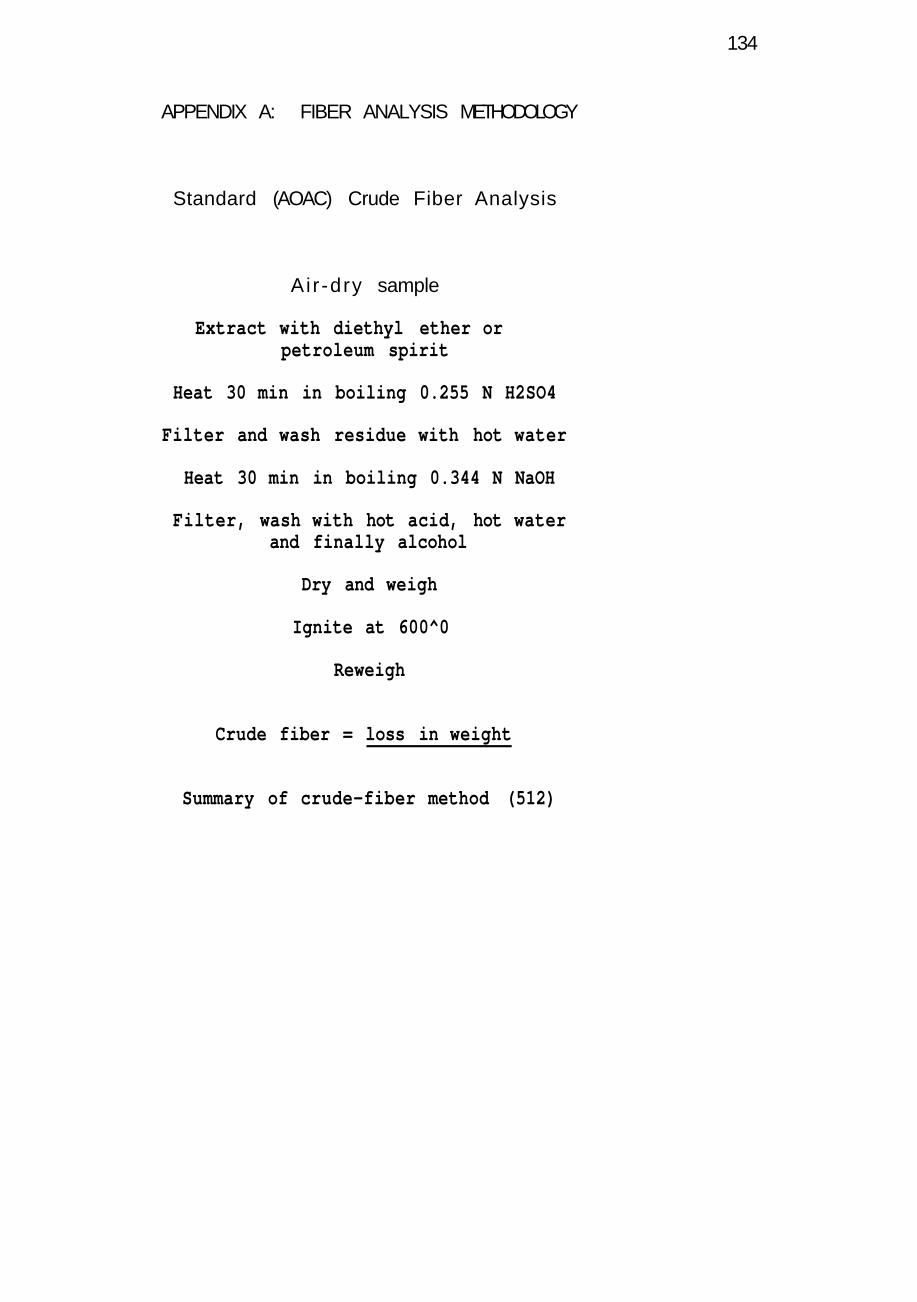
134
APPENDIX A: FIBER ANALYSIS METHODOLOGY
Standard (AOAC) Crude Fiber Analysis
Ai r -dry sample
Extract with diethyl ether or petroleum spirit
Heat 30 min in boiling 0.255 N H2SO4
Filter and wash residue with hot water
Heat 30 min in boiling 0.344 N NaOH
Filter, wash with hot acid, hot water and finally alcohol
Dry and weigh
Ignite at 600^0
Reweigh
Crude fiber = loss in weight
Summary of crude-fiber method (512)

135
Air-dry sample (finely ground, 0.3 -v 1 mm sieve)
Extract with ethanol:benzene 8 h
Boil under reflux in IN H0SO4 (1 g/200 ml acid)
60 min
Filter, wash with hot water alcohol and diethyl ether
Dry and weigh
On a replicate
Measure total nitrogen Ash at 500^0
Normal-acid fiber = loss in weight - (total N x 6.25)
Summary of normal-acid-fiber method (512)
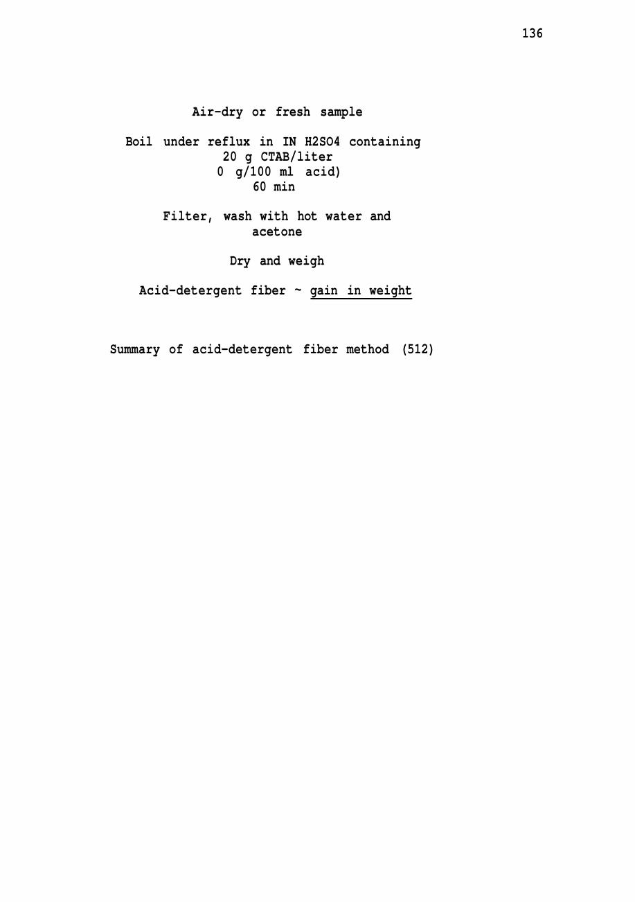
136
Air-dry or fresh sample
Boil under reflux in IN H2SO4 containing 20 g CTAB/liter 0 g/100 ml acid)
60 min
Filter, wash with hot water and acetone
Dry and weigh
Acid-detergent fiber ~ gain in weight
Summary of acid-detergent fiber method (512)

137
Air-dry or fresh sample
Boil under reflux with buffered SLS (30 g/liter) solution
containing borate, phosphate, EDTA, and 2-ethoxy ethanol
(0.5 '^^ 1 g/lQO ml
Filter, wash with hot water and acetone
Dry and weigh
Ignite
Reweigh
Neutral-detergent fiber = loss in weight
Summary of neutral-detergent fiber method (512)
138
Food Sample
Extract with 90% v/v ethanol
Residue dried and weighed
Starch measured in residue
Total nitrogen measured
Unavailable carbohydrate = residue insoluble in alcohol - (starch + total N X 6.25)
Total unavailable carbohydrate (512)
139
Food Sample
Extraction of Starches lipids, pigments, etc.
Extraction of water-soluble components
Fractionation
Acidic Neutral
Pectin Alginates
Components Components
Starch -glucans
Arabino-xylans Arabino-galactans Galacto monnans Cellulose ethers,
etc. Fractionation of water-insoluble
components
Non-cellulose Cellulose Lignin
Complete f ract ionat ion of a l l polysaccharides (512)
140
Food^
Bread, white Flour, white Flour, whole meal All bran Cornflakes Bran^ Applet Orange Lettuce Carrot Pea Potato
(g per 100
NDF
4.2 2.9 10.3 27.9 5,9
53.9 14.7 10.5 25.4 10.8 51.0 14.1
g dry matter)
Total Dietary Fiber
4.6 3.5 11.0 29.7 11.7 48.0 9.2 13.7 37.2 38.6 47.6 18.8
A. Analysis carried out in different samples therefore comparison not exact.
B. Endosperm levels vary in brans.
C Different varieties analyzed - refers to apple, fresh.
Comparison of total NDF and total dietary fiber in selected foods (513)
141
A comparison of crude fiber and dietary fiber in foods commonly
considered good sources of fiber.
Food Crude Fiber* g/100 g
Dietary Fiber"^ g/100 g
Breads and cereals
All bran cereal Puffed wheat Cornflakes Whole wheat bread Puffed wheat, sugar :oated
7.8 2.0 0.7 1.6 0.9
26.70 15.41 11.00 8.50 6.08
Vegetables
Peas, canned Sweetcorn, cooked Broccoli tops, boiled Carrots, boiled Lettuce, raw
2.3 0,7 1.5 1,0 0,6
6.28 4,74 4.10 3.70 1.53
Fruits
Peaches, with skin Strawberries, raw Apples, without skin
0.6 1.3 0.6
2.28 2.12 1.42
Nuts
Peanuts Brazil Peanut butter
1.9 3.1 1.9
9.30 7.73 7.55
* Crude fiber data from U.S.D.A, Handbook 8, Composition of Foods, ••• Southgate et al, (513)

APPENDIX B
DIETARY FIBER IN SELECTED FOODS
142
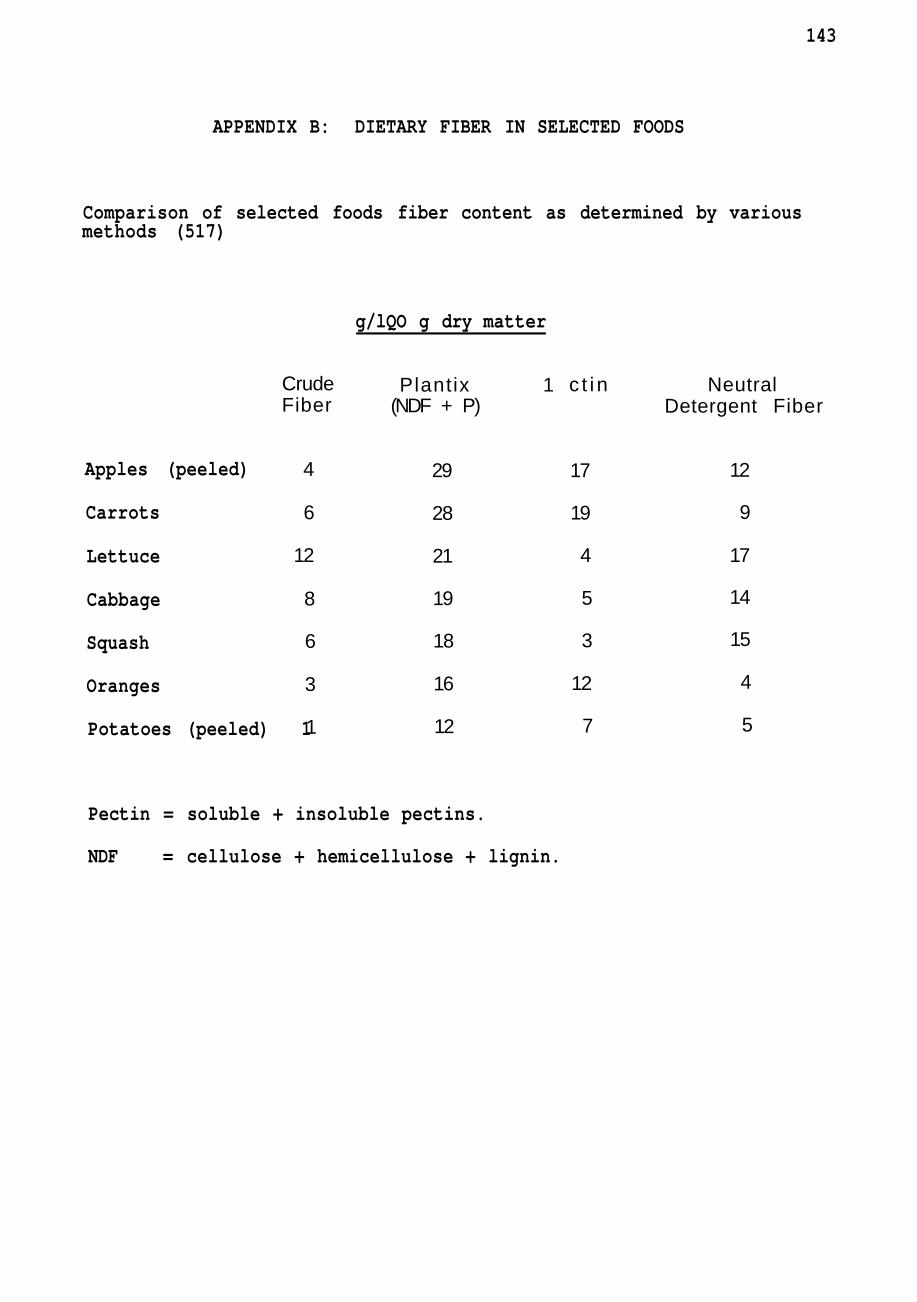
143
APPENDIX B: DIETARY FIBER IN SELECTED FOODS
Comparison of selected foods fiber content as determined by various methods (517)
g/lQO g dry matter
Crude Fiber
4
6
12
8
6
3
1
Plantix 1 (NDF + P)
29
28
21
19
18
16
12
Apples (peeled)
Carrots
Lettuce
Cabbage
Squash
Oranges
Potatoes (peeled) 1
Pectin = soluble + insoluble pectins.
NDF = cellulose + hemicellulose + lignin.
c t i n
17
19
4
5
3
12
7
Neutral Detergent Fiber
12
9
17
14
15
4
5
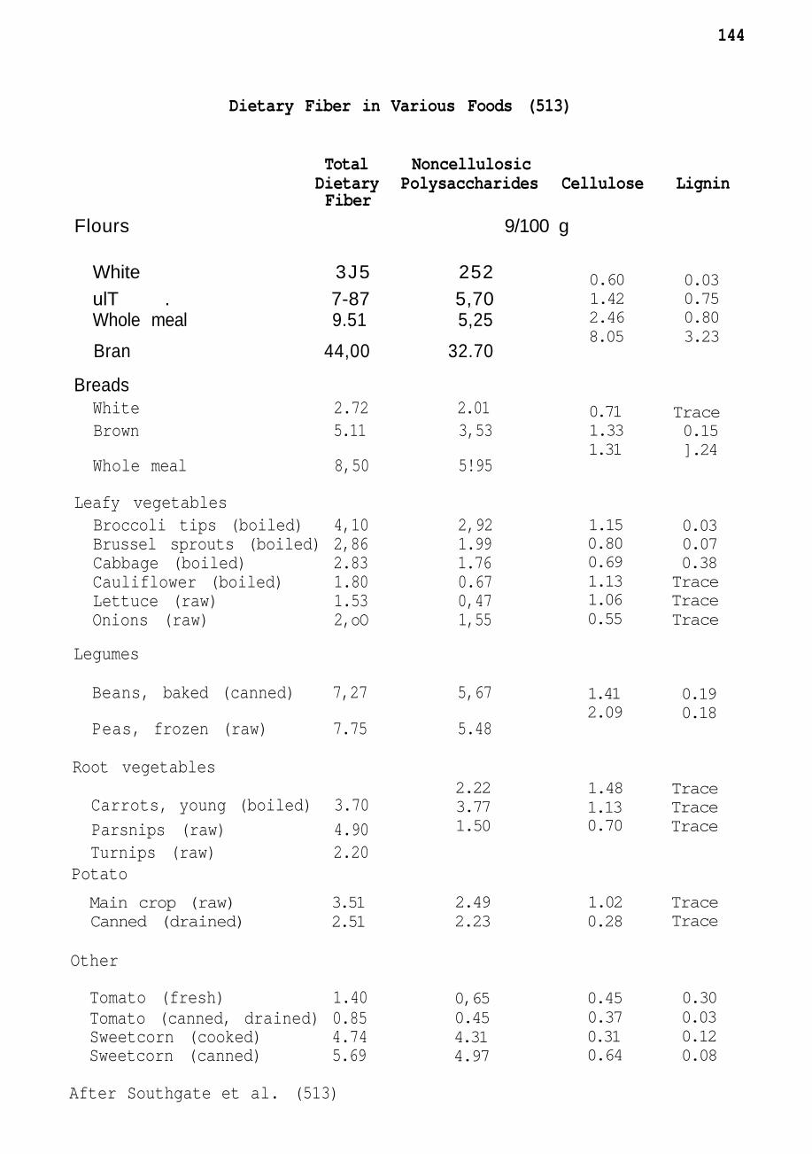
144
Dietary Fiber in Various Foods (513)
Total Noncellulosic Dietary Polysaccharides Cellulose Lignin Fiber
Flours 9/100 g
White 3J5 252 ulT . 7-87 5,70 Whole meal 9.51 5,25
Bran 44,00 32.70
Breads White 2.72 2.01 Brown 5.11 3,53
Whole meal 8,50 5!95
Leafy vegetables Broccoli tips (boiled) 4,10 2,92 Brussel sprouts (boiled) 2,86 1.99 Cabbage (boiled) 2.83 1.76 Cauliflower (boiled) 1.80 0.67 Lettuce (raw) 1.53 0,47 Onions (raw) 2,oO 1,55
Legumes
Beans, baked (canned) 7,27 5,67
Peas, frozen (raw) 7.75 5.48
Root vegetables
Carrots, young (boiled) 3.70 Parsnips (raw) 4.90 Turnips (raw) 2.20
Potato
Other
Tomato (fresh) 1.40 Tomato (canned, drained) 0.85 Sweetcorn (cooked) 4.74 Sweetcorn (canned) 5.69
After Southgate et al. (513)
0.60 1.42 2.46 8.05
0.03 0.75 0.80 3.23
0.71 1.33 1.31
Trace 0.15 ].24
1.15 0.80 0.69 1.13 1.06 0.55
1.41 2.09
0.03 0.07 0.38 Trace Trace Trace
0.19 0.18
2.22 3.77 1.50
1.48 1.13 0.70
Trace Trace Trace
Main crop (raw) Canned (drained)
3.51 2.51
2.49 2.23
1.02 0.28
Trace Trace
0,65 0.45 4.31 4.97
0.45 0.37 0.31 0.64
0.30 0.03 0.12 0.08


![Mechanisms linking dietary fiber, gut microbiota and colon … · 2017-05-01 · dietary fiber intake and a low incidence of colon cancer, and that dietary fiber has anticancer properties[6-8]](https://img.pdfslide.net/doc/110x75/5fb890e799e2b30fe92d2665/mechanisms-linking-dietary-fiber-gut-microbiota-and-colon-2017-05-01-dietary.jpg)