Embed Size (px)
Citation preview
The Relative Localizing Value of Interictal andImmediate Postictal SPECT in Seizures ofTemporal Lobe OriginKalarickal J. Oommen, MD1; Sadia Saba, MD1; Joseph A. Oommen, MD1; Paul C. Francel, MD, PhD2;Charles D. Arnold, MD3; and Don A. Wilson, MD3
1Department of Neurology, University of Oklahoma Health Sciences Center, Oklahoma City, Oklahoma; 2Department ofNeurosurgery, University of Oklahoma Health Sciences Center, Oklahoma City, Oklahoma; and 3Department of Radiology,University of Oklahoma Health Sciences Center, Oklahoma City, Oklahoma
Although interictal hypoperfusion and ictal hyperperfusion areestablished localizing findings in partial epilepsy, their relativevalue is disputed. After a meta analysis of several publishedarticles on SPECT brain imaging in patients with epilepsy (withextractable data on at least 6 patients per article), institutionsusing SPECT for evaluation of epilepsy have been encouragedto perform ictal scanning or interictal and postictal SPECT stud-ies. Methods: We compared the relative localizing values ofhypoperfusion in video-electroencephalographically (EEG) moni-tored interictal SPECT (IISPECT) and hyperperfusion in immedi-ate postictal or periictal SPECT (PISPECT) in nonlesional pa-tients who underwent temporal lobectomies in our epilepsycenter from 1995 to 1998. We also evaluated the usefulness ofcombined interpretation of IISPECT and PISPECT when avail-able. Results: Our experience with continuous cerebral blood-flow monitoring, published elsewhere, and SPECT results indi-cate that these recommendations are valid, but obtaining ictalSPECT is often serendipitous. We found that (a) interictal hypo-perfusion was easier to demonstrate by SPECT but was lessoften concordant with the EEG focus than hyperperfusion inPISPECT, but not significantly (P � 0.11) so; (b) the lowerincidence of hyperperfusion in PISPECT in our series was due tothe occurrence of hypoperfusion in PISPECT, which was seen in34.5% of our patients; and (c) hypoperfusion in PISPECT didhave localizing value when it occurred on the same side as thehypoperfusion noted in IISPECT. Conclusion: On the basis ofour findings, we recommend the use of 3 distinct perfusionpatterns that emerge from the combined interpretation ofIISPECT and PISPECT we proposed earlier (patterns 1–3), forlocalization purposes when possible, rather than ictal SPECT,IISPECT, or PISPECT by itself.
Key Words: epilepsy; hypoperfusion; hyperperfusion; SPECT
J Nucl Med 2004; 45:2021–2025
The site of onset of ictal events provides the best meansof localization of the seizure focus (1,2). Since propagationof the ictal electrical discharge from the epileptogenic zoneto the symptomatic zone may not be appreciable in the scalprecording, intracranial elecrtrocorticography (ECoG) anddepth recordings are presumably the most accurate methodsof localization. Even such recordings have been shown to beassociated with lack of improvement in seizure control (3).This led to the search for additional methods for the local-ization of the surgical focus in the last quarter of the 20thCentury (4). MRI can detect almost 100% of the structurallesions that are associated with epilepsy and the structuralcorrelates of mesial temporal sclerosis (5). Other widelyused techniques include PET and SPECT, which can bepositive even when MRI is negative. PET claims highersensitivity and specificity but is more expensive and is notavailable except in selected epilepsy centers. SPECT, on theother hand, is less expensive and is more widely availablethan PET.
However, SPECT is also the most controversial and,according to some, possibly the least specific (6). Theprinciple behind the technology is the perfusional abnor-malities that accompany the metabolic changes in epilepto-genic brain tissue in the interictal phase and the changes inthe blood flow in the periictal phase. In the 1970s, regionalcerebral blood flow measurements using the radioactiveisotope of xenon (133Xe) documented the cerebral bloodflow changes during seizures. Using this technique, inves-tigators found increased blood flow in the ictal phase (7) anddecreased blood flow in the interictal phase at the site of theelectroencephalographically (EEG) demonstrated focus (8).Several studies that followed, using PET (9) and SPECT(10), confirmed these early findings of interictal hypoper-fusion and ictal hyperperfusion in the epileptogenic region,thus adding another dimension to the localization of theepileptogenic focus in partial seizures. However, reports ofinterictal increase in perfusion by some authors (11,12) aswell as ictal (13) and late ictal (14) hypoperfusion reported
Received Mar. 17, 2004; revision accepted Jul. 15, 2004.For correspondence or reprints contact: Kalarickal J. Oommen, MD, De-
partment of Neurology, University of Oklahoma Health Sciences Center, 711Stanton L. Young Blvd., Suite 215, Oklahoma City, Oklahoma, 73104.
E-mail: [email protected]
WHEN TO DO SPECT TO LOCALIZE EPILEPSY? • Oommen et al. 2021
by on April 14, 2019. For personal use only. jnm.snmjournals.org Downloaded from
by others added to the controversy surrounding the useful-ness of SPECT for surgical localization in epilepsy.
As a result of subsequent technologic advances in CT andmore and more stable isotopes, SPECT, with its ability todemonstrate localized changes in cerebral blood flow (CBF)interictally, during ictus, and postictally became a valuabletool for the evaluation of epilepsy (10,15). Its major disad-vantage has been the controversy surrounding its sensitivityand specificity. Although most reports favored interictalhypoperfusion and ictal hyperperfusion, departures fromthis principle were noted early on (11–14). Some claimedinterictal studies to be as sensitive as PET (15,16), othersconsidered ictal studies to be more reliable (17,18). and yetothers doubted their usefulness (19,20). The reasons for thecontroversy are that the earlier perfusional studies—somedone with 133Xe, others with 123I-isopropyliodoamphet-amine, 99mTc-hexamethylpropyleneamine oxime (HMPAO),yet others with 18F-FDG PET, and a small number of centersusing the more stable 99mTc-ethylcysteinate dimer (99mTc-ECD), using differing technologies, performed on varied co-horts—had little in common to justify comparisons. At-tempts at making sense of the enormous data accumulatedworldwide, under such circumstances, only added to thecontroversy.
Interictal hypometabolism and ictal hypermetabolism arethe most common PET findings in the epileptogenic cortexin partial epilepsies (9,21,22). We also know that blood flowis coupled to epileptiform activity and metabolism. Penfieldin 1933 (23), Penfield et al. in 1939 (24), and Plum et al.(25) had already demonstrated that there is increased CBFin the region of the epileptogenic focus during and imme-diately after partial seizures. PET studies in a few patientsduring focal status epilepticus (26) and during or after asingle complex partial seizure have confirmed the presenceof periictal hyperperfusion and hypermetabolism.
More recent evidence from long-term surface corticalCBF monitoring in temporal lobe epilepsy also has con-firmed that the interictal epileptic foci are significantlyhypoperfused relative to the nonepileptic cortex (27,28).
The range of positive interictal SPECT (IISPECT) studiesvaries from 57% to 95% (12,29). From a meta analysis of 30published studies from a literature search with at least 6patients in each study, Devous et al. (30) concluded thatIISPECT, has a sensitivity of 0.75, which is similar to theresults obtained with interictal PET, which shows interictalfocal abnormalities in 70% of patients with complex partialseizures (9). But the relative validity of IISPECT and ictalSPECT has been the subject of a long controversy (6,31).Our own experience with SPECT has been that it is avaluable addition to MRI and video-EEG in reducing theneed for invasive monitoring, with its consequent morbidityand expense, similar to the experience of Rowe et al. (32)and, hence, in reducing the cost of presurgical evaluation ofseizures of temporal lobe origin. SPECT has thus become aroutine procedure at epilepsy centers, and the Society ofNuclear Medicine has included presurgical localization of
epilepsy as a standard indication for brain SPECT (33) intheir Procedure Guidelines. However, not enough studieshave compared the value of IISPECT, ictal SPECT, andimmediate postictal SPECT (PISPECT) (34), and their rel-ative merit is thus yet to be established.
In this study we compared the concordance of perfusionalchanges in video-EEG monitored, semiquantitative IISPECTand PISPECT studies in nonlesional patients with seizures oftemporal lobe onset who underwent temporal lobectomies atthe Comprehensive Oklahoma Program for Epilepsy from1995 to 1998. We also attempted correlation of the previouslydescribed perfusional patterns we proposed earlier (patterns1–3), resulting from combined analysis and interpretation ofIISPECT and PISPECT studies.
MATERIALS AND METHODS
Forty-two patients underwent temporal lobectomies during thestudy period. Twenty-one patients were male and 21 were female(mean age � SD, 32.14 � 10.95 y old). All patients underwentcontinuous video-EEG monitoring, MRI, and scalp EEG for lo-calization of the seizure focus. Seizure foci were localized byreviewing the edited video-EEG, of a minimum of 3 each, of eachthe patient’s typical seizures. In cases in which the scalp EEG wasnonlocalizing or was in conflict with the MRI findings, video-EEGmonitoring was repeated after implantation of subdural strip elec-trodes. IIPSPECT was performed after the patients had been sei-zure free, under video-EEG surveillance for at least 24 h, to reducethe effect of prior seizures (35,36) over the temporal lobe bloodflow and, thereby, the SPECT results. A safety window of 24 h wasset after the initial injection of the radioisotope before the secondinjection to allow for adequate decay of radioactivity produced bythe first injection and to enhance radiation safety.
The radioisotope used was 99mTc-ECD. PISPECT was accom-plished by injection of 99mTc-ECD with either the clinical orelectrical onset of seizures in the epilepsy-monitoring unit undercombined closed-circuit video and EEG surveillance of the patientby specially trained EEG-monitoring technologists. The doseswere restocked twice a day at the bedside for easy availability andthe patients had heparin-locks for immediate intravenous accessduring the seizure. The injection was performed by speciallytrained nurses. SPECT image acquisition was done as soon as itwas practically feasible after the ictal injection, in most caseswithin 3 h of injection. Imaging data were acquired using aSiemens triple-head scanner with a high-intensity fanbeam colli-mator in a 128 � 128 matrix with a zoom factor of 1.23 (2.89mm/pixel) with 120 stops or projections at 60,000 counts per stop,for both the IISPECT and PISPECT scans. This required that somepatients had to stay longer in the scanner than others but ensureda measure of consistency in counts that was as standardized and asquantitative as possible.
We were able to obtain images of excellent quality because thebrain distribution of 99mTc-ECD is stable over time (37) and thebrain-to-background ratio is 17:1 for 5 h as opposed to 2:1 forHMPAO (38). The images were interpreted by a nuclear medicinephysician who was unaware of the MRI and EEG results, and thestudies were later reviewed and correlated with MRI and EEG, inconference with the electroencephalographer and the neuroradi-ologist. Studies were reported as normal or as showing evidence ofhypoperfusion or hyperperfusion in the various brain regions in the
2022 THE JOURNAL OF NUCLEAR MEDICINE • Vol. 45 • No. 12 • December 2004
by on April 14, 2019. For personal use only. jnm.snmjournals.org Downloaded from
interictal and the immediate postictal periods. The IISPECT andPISPECT were then compared to evaluate the preictal and imme-diate postictal perfusion patterns.
RESULTS
Of the 42 patients, who were included in the study, 40had IISPECT scans. Two patients did not have IISPECT dueto technical reasons. The relative concordance of the 2studies to the EEG is shown in Table 1. Of the 40 patientswho had IISPECT, 36 (90%) showed evidence of focaltemporal hypoperfusion. Four (10%) were nonlocalizing, 2because they were normal and 2 because they had evidenceof bilateral temporal hypoperfusion. PISPECT was avail-able for 29 of the 42 patients. Of the 29 who had PISPECT,19 (65.5%) showed focal hyperperfusion. In 10 (34.5%)patients, there was accentuation of the focal hypoperfusionin PISPECT, on the same side as the interictal hypoperfu-sion. None had interictal hyperperfusion.
The mean injection time was 1.86 � 0.88 min from onsetof the seizure. In our experience, it took 15–20 s for thebolus of 99mTc-ECD to be administered. Since it takes 20 sfor 95% brain extraction of the isotope (37), only thepatients in whom the injection was started within 40 s ofseizure onset would have had 95% extraction of the isotopewithin 60 s. The timing of the injection is extremely impor-tant if one is to obtain true ictal SPECT since the electricalactivity, to which the initial rise in blood flow is coupled,lasts 60–140 s (39) only. Because our mean injection timewas about 114 s in most cases, the brain extraction of theisotope occurred after the rhythmic electrical activity asso-ciated with the seizure had peaked or was on the declinewhen the injection was finished. Therefore, we consideredthat the term immediate postictal or periictal SPECT(PISPECT) was more appropriate than the term ictalSPECT, even though the injections in our patients wereaccomplished in a very short time to justify our use of theterm “ictal SPECT” as it has been used in the literature (13)by other investigators.
In our series of 42 patients, we observed that IISPECTwas easier to obtain, with the study being done in 40 of 42,or 95.2%, of patients. PISPECT was more difficult to ob-
tain, with the study available in only 29 of the 42 patients(69%). This was because the other patients had all therequired seizures before the next batch of isotope wasstocked, because the second and third seizures occurredwithin the 24-h window when another injection was notpermitted and the patient was discharged, or because theseizures occurred over the weekend when scanning was notavailable. In 2 cases, only PISPECT was available. This wasdone before IISPECT because the patients had the firstseizure before an interictal study could be performed andthey had to be discharged before IISPECT could be done.Interictal hypoperfusion was the most common finding seenin 36 of 40 (90%) patients, with 32 of the 36 (88.9%) beingconcordant with the EEG focus. None had interictal hyper-perfusion. PISPECT was available in 29 patients. Immedi-ate postictal hyperperfusion was seen in 19 of 29 (65.5%)patients and was less frequent than the occurrence of inter-ictal hypoperfusion but 18 of 19 (94.7%) were concordantwith the EEG focus. This was slightly better than the rate ofconcordance (88.9%) for interictal hypoperfusion but wasnot significantly better (P � 0.5). Keeping this in mind, ourdata would indicate that PISPECT has a higher concordancerate (94.7%) with the EEG focus than IISPECT (88.9%).The lower incidence of hyperperfusion in the immediatepostictal studies, (65.5%) as opposed to hypoperfusion ininterictal studies (90%), was due to the occurrence of thephenomenon of accentuation of hypoperfusion in PISPECT,compared with their own interictal studies, in 10 of 29(34.5%) patients. In 9 (90%) of these 10 patients who hadipsilateral accentuation of hypoperfusion in PISPECT, itwas concordant with the EEG focus, whereas in 1 (10%) theaccentuation of hypoperfusion was contralateral.
DISCUSSION
In 1993, Oommen et al., using long-term ECoG andcortical CBF monitoring with specially designed subduralstrip electrodes capable of measuring CBF and ECoG at thesame time, showed that the ictal hyperperfusion was cou-pled to the ictal spike train. It was also shown that that therhythmic electrical discharge in the temporal lobe during theseizure is short-lived and that the CBF after the cessation ofthe rhythmic electrical activity or spike train may show 3different phenomena: The blood flow may immediatelyrevert to the preictal level, change to a hyperperfusionalstate, or change to a hypoperfusional state. These latterphenomena lasted up to 3 h in that particular study (39).Thus, the dynamic nature of blood flow in the periictalperiod systematically studied by Penfield (23) and Penfieldet al. (24) during surgery, and later demonstrated by Plum etal. (25) experimentally, was confirmed by Oommen et al. byin vivo CBF monitoring in the human brain using this noveltechnology. This observation became the basis of theircombined interpretation of IISPECT and immediate post-
TABLE 1SPECT Findings
SPECT findingTotal
(n)Ipsilateral toEEG focus*
Contralateralto EEG focus*
IISPECTHypoperfusion 36 32 (88.9) 4 (11.1)Hyperperfusion 0 0 0
PISPECTHyperperfusion 19 18 (94.7) 1 (5.3)Hypoperfusion 10 9 (90) 1 (10)
*Data are presented as number (%).
WHEN TO DO SPECT TO LOCALIZE EPILEPSY? • Oommen et al. 2023
by on April 14, 2019. For personal use only. jnm.snmjournals.org Downloaded from
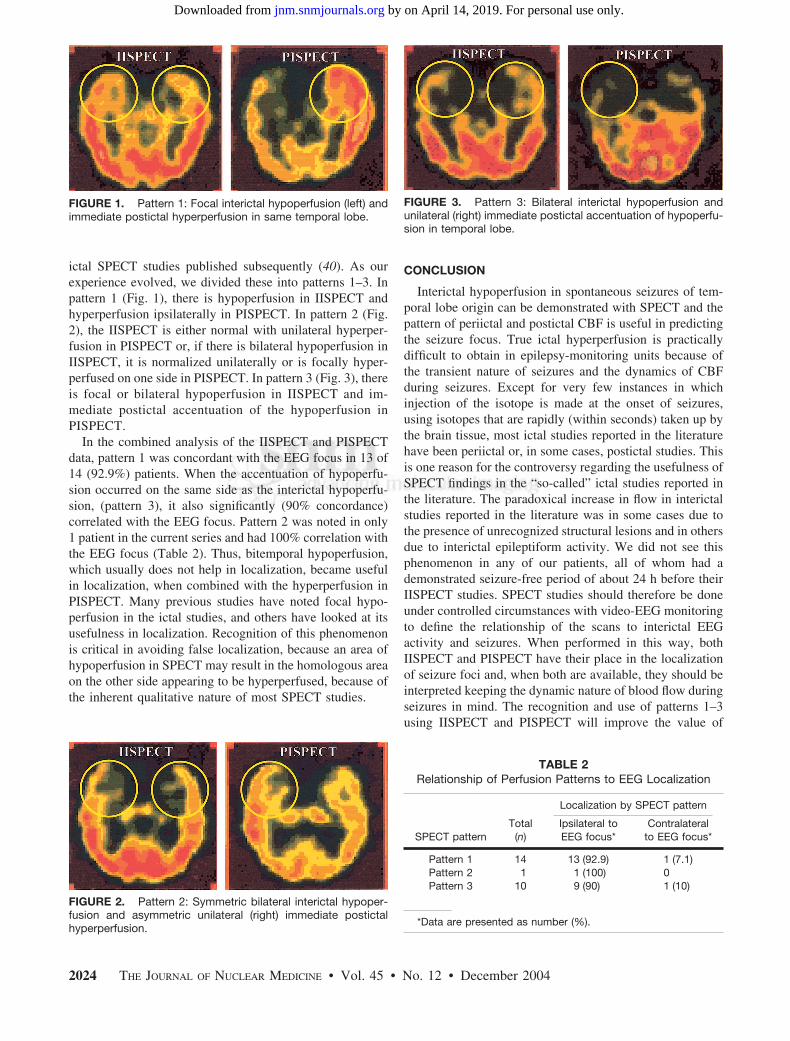
ictal SPECT studies published subsequently (40). As ourexperience evolved, we divided these into patterns 1–3. Inpattern 1 (Fig. 1), there is hypoperfusion in IISPECT andhyperperfusion ipsilaterally in PISPECT. In pattern 2 (Fig.2), the IISPECT is either normal with unilateral hyperper-fusion in PISPECT or, if there is bilateral hypoperfusion inIISPECT, it is normalized unilaterally or is focally hyper-perfused on one side in PISPECT. In pattern 3 (Fig. 3), thereis focal or bilateral hypoperfusion in IISPECT and im-mediate postictal accentuation of the hypoperfusion inPISPECT.
In the combined analysis of the IISPECT and PISPECTdata, pattern 1 was concordant with the EEG focus in 13 of14 (92.9%) patients. When the accentuation of hypoperfu-sion occurred on the same side as the interictal hypoperfu-sion, (pattern 3), it also significantly (90% concordance)correlated with the EEG focus. Pattern 2 was noted in only1 patient in the current series and had 100% correlation withthe EEG focus (Table 2). Thus, bitemporal hypoperfusion,which usually does not help in localization, became usefulin localization, when combined with the hyperperfusion inPISPECT. Many previous studies have noted focal hypo-perfusion in the ictal studies, and others have looked at itsusefulness in localization. Recognition of this phenomenonis critical in avoiding false localization, because an area ofhypoperfusion in SPECT may result in the homologous areaon the other side appearing to be hyperperfused, because ofthe inherent qualitative nature of most SPECT studies.
CONCLUSION
Interictal hypoperfusion in spontaneous seizures of tem-poral lobe origin can be demonstrated with SPECT and thepattern of periictal and postictal CBF is useful in predictingthe seizure focus. True ictal hyperperfusion is practicallydifficult to obtain in epilepsy-monitoring units because ofthe transient nature of seizures and the dynamics of CBFduring seizures. Except for very few instances in whichinjection of the isotope is made at the onset of seizures,using isotopes that are rapidly (within seconds) taken up bythe brain tissue, most ictal studies reported in the literaturehave been periictal or, in some cases, postictal studies. Thisis one reason for the controversy regarding the usefulness ofSPECT findings in the “so-called” ictal studies reported inthe literature. The paradoxical increase in flow in interictalstudies reported in the literature was in some cases due tothe presence of unrecognized structural lesions and in othersdue to interictal epileptiform activity. We did not see thisphenomenon in any of our patients, all of whom had ademonstrated seizure-free period of about 24 h before theirIISPECT studies. SPECT studies should therefore be doneunder controlled circumstances with video-EEG monitoringto define the relationship of the scans to interictal EEGactivity and seizures. When performed in this way, bothIISPECT and PISPECT have their place in the localizationof seizure foci and, when both are available, they should beinterpreted keeping the dynamic nature of blood flow duringseizures in mind. The recognition and use of patterns 1–3using IISPECT and PISPECT will improve the value of
FIGURE 2. Pattern 2: Symmetric bilateral interictal hypoper-fusion and asymmetric unilateral (right) immediate postictalhyperperfusion.
FIGURE 3. Pattern 3: Bilateral interictal hypoperfusion andunilateral (right) immediate postictal accentuation of hypoperfu-sion in temporal lobe.
TABLE 2Relationship of Perfusion Patterns to EEG Localization
SPECT patternTotal
(n)
Localization by SPECT pattern
Ipsilateral toEEG focus*
Contralateralto EEG focus*
Pattern 1 14 13 (92.9) 1 (7.1)Pattern 2 1 1 (100) 0Pattern 3 10 9 (90) 1 (10)
*Data are presented as number (%).
FIGURE 1. Pattern 1: Focal interictal hypoperfusion (left) andimmediate postictal hyperperfusion in same temporal lobe.
2024 THE JOURNAL OF NUCLEAR MEDICINE • Vol. 45 • No. 12 • December 2004
by on April 14, 2019. For personal use only. jnm.snmjournals.org Downloaded from

SPECT in localization of epileptogenic foci, and the com-bined interpretation can improve the usefulness of the pro-cedure, particularly in patients in whom the IISPECT isnormal or shows bilateral hypoperfusion.
REFERENCES
1. Gloor P. Contributions of electroencephalography and electrocortico-graphy tothe neurosurgical treatment of the epilepsies. Adv Neurol. 1975;8:59–106.
2. Walter RD. Principles of clinical investigation of surgical candidates. Adv Neu-rol. 1975;8:49–58.
3. Crandall PH. Prospective management and criteria for evaluation. Adv Neurol.1975;8:265–280.
4. Jack CR Jr, Sharbrough FW, Twomey CK, et al. Temporal lobe seizures:lateralization with MR volume measurements of hippocampal formation. Radi-ology. 1990;175:423–429.
5. Bronen RA, Fulbright RK, Spencer DD, Spencer SS, Kim JH, Lance RC. MRcharacteristics of neoplasms and vascular malformations associated with epi-lepsy. Magn Reson Imaging. 1995;13:1153–1162.
6. Spencer SS. The relative contributions of MRI, SPECT and PET imaging inepilepsy. Epilepsia. 1994;35(suppl 6):S72–S89.
7. Ingvar DH. Regional cerebral blood flow in focal cortical epilepsy. Stroke.1973;4:359–360.
8. Lavy S, Melamed E, Portnoy Z, et al. Interictal regional cerebral blood flow inpatients with partial seizures. Neurology. 1976;26:418–422.
9. Kuhl DE, Engel J Jr, Phelps ME, Selin C. Epileptic patterns of local cerebralmetabolism and perfusion in humans determined by emission computed tomog-raphy of 18 FDG and 13 NH3. Ann Neurol. 1980;8:348–360.
10. Magistretti P, Uren R, Blume H, Schomer D, Royal H. Delineation of epilepticfocus by single photon emission tomography. Eur J Nucl Med. 1982;7:484–485.
11. Hougaard K, Oikawa T, Sveindottir E, et al. Regional cerebral blood flow in focalcortical epilepsy. Arch Neurol. 1976;33:527–535.
12. Sakai F, Meyer JS, Naritomi H, et al. Regional cerebral blood flow and EEG inpatients with epilepsy. Arch Neurol. 1978;35:648–657.
13. Weis M, Feistel H, Stefan H. Utility of ictal SPECT: peri-ictal, postictal. ActaNeurol Scand. 1994;152(suppl):145–147.
14. Kuikka JT, Mervaala E, Vanninen E, Kalviainen E. Does technetium-99m bici-sate image local brain metabolism in late ictal temporal lobe epilepsy? Eur J NuclMed. 1994;21:1247–1251.
15. Devous M, Leroy R, Homan R. Single photon emission computed tomography inepilepsy. Semin Nucl Med. 1990;20:325–341.
16. Alavi A, Hirsch L. Studies of central nervous system disorders with single photonemission computed tomography: evolution over the past two decades. Semin NuclMed. 1991;21:58–81.
17. Rowe C, Berkovic S, Austin M, Saling M, Kalnins R. Visual and quantitativeanalysis of interictal SPECT with technetium-99m-HMPAO in temporal lobeepilepsy. J Nucl Med. 1991;32:1688–1694.
18. Newton M, Berkovic S, Austin M, Rowe C, McKay W, Bladin P. Ictal postictaland interictal single-photon emission tomography in the lateralization of temporallobe epilepsy. Eur J Nucl Med. 1994;21:1067–1071.
19. Berkovic S, Newton M, Chiron C, Dulac O. Single photon emission tomography.In: Engel J Jr, ed. Surgical Treatment of the Epilepsies. 2nd ed. New York, NY:Raven Press; 1993:233–243.
20. Chugani H. Functional brain imaging in pediatrics. Pediatr Clin North Am.1992;39:777–799.
21. Engel J Jr, Kuhl D, Phelps M, et al. Interictal cerebral glucose metabolism inpartial epilepsy and its relation to EEG changes. Ann Neurol. 1982;12:510–517.
22. Theodore W, Brooks R, Sato S, et al. The role of positron emission tomographyin the evaluation of seizure disorders. Ann Neurol. 1984;15:S176–S179.
23. Penfield W. The evidence for a cerebral vascular mechanism in epilepsy. AnnIntern Med. 1933;7:303–310.
24. Penfield W, von Santha K, Cipriani A. Cerebral blood flow during inducedepileptiform seizures in animals and man. J Neurophysiol. 1939;2:257–267.
25. Plum F, Posner J, Troy B. Cerebral metabolic and circulatory responses toinduced convulsion in animals. Arch Neurol. 1968;18:1–13.
26. Engel J Jr, Henry T, Risinger M, et al. Pre-surgical evaluation of partial epilepsy:relative contributions of chronic depth electrode recording vs. FDG-PET andscalp sphenoidal ictal EEG. Neurology. 1990;40:1670–1677.
27. Weinand M, Carter L, Patton D, Oommen K, Labiner D, Talwar D. Long termsurface cortical cerebral blood flow monitoring in temporal lobe epilepsy. Neu-rosurgery. 1994;35:657–664.
28. Weinand M, Carter L. Surface cortical cerebral blood flow monitoring and singlephoton emission computed tomography: prognostic factors for selecting temporallobectomy candidates. Seizure. 1994;3:55–59.
29. LaManna M, Sussman N, Harner R, et al. Initial experience with SPECT imagingof the brain using I-123 p-iodoamphetamine in focal epilepsy. Clin Nucl Med.1989;14:428–430.
30. Devous M Sr, Thisted R, Morgan G, Leroy R, Rowe C. SPECT brain imaging inepilepsy: a meta analysis. J Nucl Med. 1998;39:285–293.
31. Oliveira A, Costa J, Hilario L, Anselmi O, Palmini A. Localization of epilepto-genic zone by ictal and interictal SPECT with Tc-ethyl cysteinate dimer inpatients with medically refractory epilepsy. Epilepsia. 1999;40:693–702.
32. Rowe C, Berkovic S, Austin M, McKay W, Bladin P. Patterns of postictalcerebral blood flow in temporal lobe epilepsy: qualitative and quantitative anal-ysis. Neurology. 1991;41:1096–1103.
33. Juni JE, Waxman AD, Devous MD Sr, et al. Brain perfusion single photonemission computed tomography (SPECT) using Tc-99m radiopharmaceuticals.Procedure Guidelines. 1999:113–118. Society of Nuclear Medicine Web site.Available at: http://interactive.snm.org/index.cfm?PageID�800. Accessed Octo-ber 31, 2004.
34. Duncan R, Patterson J, Roberts R, Hadley D, Bone I. Ictal/postictal SPECT inpresurgical localization of complex partial seizures. J Neurol Neurosurg Psychi-atry. 1993;56:141–148.
35. Jibiki I, Kubota T, Fujimoto K, et al. Reexamination of I231-IMP-SPECT atinterictal stages in adult partial epilepsy. Jpn J Psychiatry Neurol. 1989;43:385–388.
36. Jibiki I, Yamaguchi N, Matsuda H, Hisada K. Fluctuations of interictal brainimaging in repeated I231-IMP-SPECT scans in an epileptic patient. J Neurol.1990;237:372–375.
37. Walovitch R, Hill T, Garrity S, et al. Characterization of technetium-99-m-1,1ECD for brain perfusion imaging. Part I. Pharmacology of technetium-99-mECD in nonhuman primates. J Nucl Med. 1989;30:1892–1901.
38. Leveille J, Demonceau G, Walovitch R. Intrasubject comparison between tech-netium 99m-ECD and technetium 99m-HMPAO in healthy human subjects.J Nucl Med. 1992;33:480–484.
39. Oommen K, Carter L, Weinand M. Cerebral blood-flow over epileptogenictemporal cortex before, during and after seizures. Epilepsia. 1993;34(suppl6):127–128.
40. Oommen K, King J, Carter LP, Chacko G, Wilson D. Utility of combinedinter-ictal and immediate post-ictal SPECT (IISPECT and PISPECT) in local-ization of epileptogenic foci by using 99mTc ECD (Neurolite) [abstract]. Epilep-sia. 1997;38(suppl 8):146.
WHEN TO DO SPECT TO LOCALIZE EPILEPSY? • Oommen et al. 2025
by on April 14, 2019. For personal use only. jnm.snmjournals.org Downloaded from
2004;45:2021-2025.J Nucl Med. Kalarickal J. Oommen, Sadia Saba, Joseph A. Oommen, Paul C. Francel, Charles D. Arnold and Don A. Wilson Seizures of Temporal Lobe OriginThe Relative Localizing Value of Interictal and Immediate Postictal SPECT in
http://jnm.snmjournals.org/content/45/12/2021This article and updated information are available at:
http://jnm.snmjournals.org/site/subscriptions/online.xhtml
Information about subscriptions to JNM can be found at:
http://jnm.snmjournals.org/site/misc/permission.xhtmlInformation about reproducing figures, tables, or other portions of this article can be found online at:
(Print ISSN: 0161-5505, Online ISSN: 2159-662X)1850 Samuel Morse Drive, Reston, VA 20190.SNMMI | Society of Nuclear Medicine and Molecular Imaging
is published monthly.The Journal of Nuclear Medicine
© Copyright 2004 SNMMI; all rights reserved.
by on April 14, 2019. For personal use only. jnm.snmjournals.org Downloaded from