Embed Size (px)
Citation preview
The Respiratory Pathogen Moraxella catarrhalis Targets Collagen forMaximal Adherence to Host Tissues
Birendra Singh,a Maria Alvarado-Kristensson,b Martin Johansson,b Oskar Hallgren,c Gunilla Westergren-Thorsson,c
Matthias Mörgelin,d Kristian Riesbecka
Clinical Microbiologya and Molecular Pathology,b Department of Translational Medicine, Lund University, Malmö, Sweden; Respiratory Medicine and Allergology,Department of Experimental Medical Sciences, Lund University, Lund, Swedenc; Section of Infectious Medicine, Department of Clinical Sciences, Lund University, Lund,Swedend
ABSTRACT Moraxella catarrhalis is a human respiratory pathogen that causes acute otitis media in children and is associatedwith exacerbations in patients suffering from chronic obstructive pulmonary disease (COPD). The first step in M. catarrhaliscolonization is adherence to the mucosa, epithelial cells, and extracellular matrix (ECM). The objective of this study was to eval-uate the role of M. catarrhalis interactions with collagens from various angles. Clinical isolates (n � 43) were tested for collagenbinding, followed by a detailed analysis of protein-protein interactions using recombinantly expressed proteins. M. catarrhalis-dependent interactions with collagen produced by human lung fibroblasts and tracheal tissues were studied by utilizing confocalimmunohistochemistry and high-resolution scanning electron microscopy. A mouse smoke-induced chronic obstructive pulmo-nary disease (COPD) model was used to estimate the adherence of M. catarrhalis in vivo. We found that all M. catarrhalis clini-cal isolates tested adhered to fibrillar collagen types I, II, and III and network-forming collagens IV and VI. The trimeric auto-transporter adhesins ubiquitous surface protein A2 (UspA2) and UspA2H were identified as major collagen-binding receptors.M. catarrhalis wild type adhered to human tracheal tissue and collagen-producing lung fibroblasts, whereas UspA2 and UspA2Hdeletion mutants did not. Moreover, in the COPD mouse model, bacteria devoid of UspA2 and UspA2H had a reduced level ofadherence to the respiratory tract compared to the adherence of wild-type bacteria. Our data therefore suggest that the M. ca-tarrhalis UspA2 and UspA2H-dependent interaction with collagens is highly critical for adherence in the host and, furthermore,may play an important role in the establishment of disease.
IMPORTANCE The respiratory tract pathogen Moraxella catarrhalis adheres to the host by interacting with several components,including the ECM. Collagen accounts for 30% of total body proteins, and therefore, bacterial adherence to abundant host colla-gens mediates bacterial persistence and colonization. In this study, we characterized previously unknown M. catarrhalis-depen-dent interactions with host collagens and found that the trimeric autotransporter adhesins ubiquitous surface protein A2(UspA2) and UspA2H are highly important. Our observations also suggested that collagen-mediated adherence of M. catarrhalisis indispensable for bacterial survival in the host, as exemplified by a mouse COPD model.
Received 14 January 2016 Accepted 23 February 2016 Published 22 March 2016
Citation Singh B, Alvarado-Kristensson M, Johansson M, Hallgren O, Westergren-Thorsson G, Mörgelin M, Riesbeck K. 2016. The respiratory pathogen Moraxella catarrhalistargets collagen for maximal adherence to host tissues. mBio 7(2):e00066-16. doi:10.1128/mBio.00066-16.
Editor Melinda M. Pettigrew, Yale School of Public Health
Copyright © 2016 Singh et al. This is an open-access article distributed under the terms of the Creative Commons Attribution 4.0 International license.
Address correspondence to Kristian Riesbeck, [email protected].
Moraxella catarrhalis is a Gram-negative diplococcus that col-onizes the human respiratory tract. The pathogen causes
acute otitis media in children and is also associated with bronchi-tis, sinusitis, laryngitis, and exacerbations in patients with chronicobstructive pulmonary disease (COPD) (1–4). It is frequentlyfound in coinfections with Haemophilus influenzae and/or Strep-tococcus pneumoniae.
The first step of a successful colonization is adhesion to the hostmucosal surface, epithelial cells, and finally, extracellular matrix(ECM). Adhesion is mediated by an array of outer membraneproteins (3), including the M. catarrhalis ubiquitous surface pro-teins (Usp). This protein family comprises well-characterized tri-meric autotransporter adhesins (TAAs), which are recognized asmultifunctional virulence factors of M. catarrhalis (5–9). Ubiqui-tous surface proteins occur as lollipoplike structures that consist
of a membrane anchor, stalk, neck, and head domain on the outermembrane (10). They are further divided into three subgroups, asfollows: (i) ubiquitous surface protein A1 (UspA1) is present in allclinical isolates; (ii) UspA2 is found in 75% of strains; and finally,(iii) approximately 25% of clinical isolates carry UspA2H insteadof UspA2. Ubiquitous surface protein A1 binds to CAECAM-1surface receptors, and hence, plays an important role in bacterialcolonization (11). Either UspA2 or both UspA1 and UspA2 inter-act with components of the complement pathway to protectM. catarrhalis from the bactericidal activity of serum, and they alsobind to ECM components to enhance adherence to the host (5–8,12–15).
Collagens are the most abundant glycoproteins of the humanbody and account for 30% of the total protein mass involved in theformation of structural scaffolds, cell adhesion, and angiogenesis
RESEARCH ARTICLE
crossmark
March/April 2016 Volume 7 Issue 2 e00066-16 ® mbio.asm.org 1
on March 1, 2020 by guest
http://mbio.asm
.org/D
ownloaded from

and the development of organs (16, 17). The basic structural unitof a collagen molecule consists of an �-chain (monomer). The�-chains associate into a trimer in a triple-helix form to build aprotomer, and these associate further, forming a supramolecularorganization. On the basis of their supramolecular arrangements,collagens have been categorized into the following five differentgroups: fibril-forming collagens, network-forming collagens,FACITs (fibril-associated collagen with interrupted triple helices),MACITs (membrane-associated collagen with interrupted triplehelices), and multiplexins (16). The triple-helix region of the�-chains consists of the recurring triplet Gly-X-Y repeated n timesin the central part of each �-chain, with X and Y often beingproline and hydroxyproline, respectively. The other domains of�-chains, which have ordinary amino acid sequences, are termednoncollagenous (NC) domains and are present at the N- orC-terminal end of �-chains. The NC domains are cleaved off insome protomers (fibril-forming collagens), while they are re-tained in the network-forming collagens (17).
An individual collagen does not represent a tissue’s specificity;rather, several types of collagens associate to form complex struc-tures by defined protein-protein interactions (17–19). Type I col-lagen is one of the most abundant, present in all human tissues,whereas type II collagen forms fibrils and associates with hyalineand elastic cartilages, including tracheal cartilage (20). Type IIIcollagen is present in connective tissues (reticulate fibers), such asthe skin, lungs, and the vascular system (21). The epithelium of allorgans anchors to the underlying tissues by interacting with thebasement membrane (BM), which is composed of laminin andcollagen IV networks (22). Collagen type VI is abundantly presentin the ECM of the respiratory system and is upregulated in COPD(23).
It has previously been shown that M. catarrhalis adheres tocollagen IV by its outer membrane IgD-binding protein (MID/Hag) (24). Here, we studied in detail the interactions of M. ca-tarrhalis with various human collagens and further investigatedtheir in vivo relevance. The abundant collagens, including fibril-forming types I, II, and III and network-forming types IV, V, VI,and VIII, were tested for binding with several clinical isolates.Bacterial surface proteins interacting with collagens were identi-fied, and protein-protein interactions were elucidated at the mo-lecular level. Moreover, the biological significance of M. catarrha-lis-dependent collagen interactions was verified by using humanfibroblasts and upper respiratory tract tissue and was finally ex-amined in an in vivo COPD mouse model. Our results indicatethat M. catarrhalis efficiently targets collagens for optimal adher-ence to host tissues.
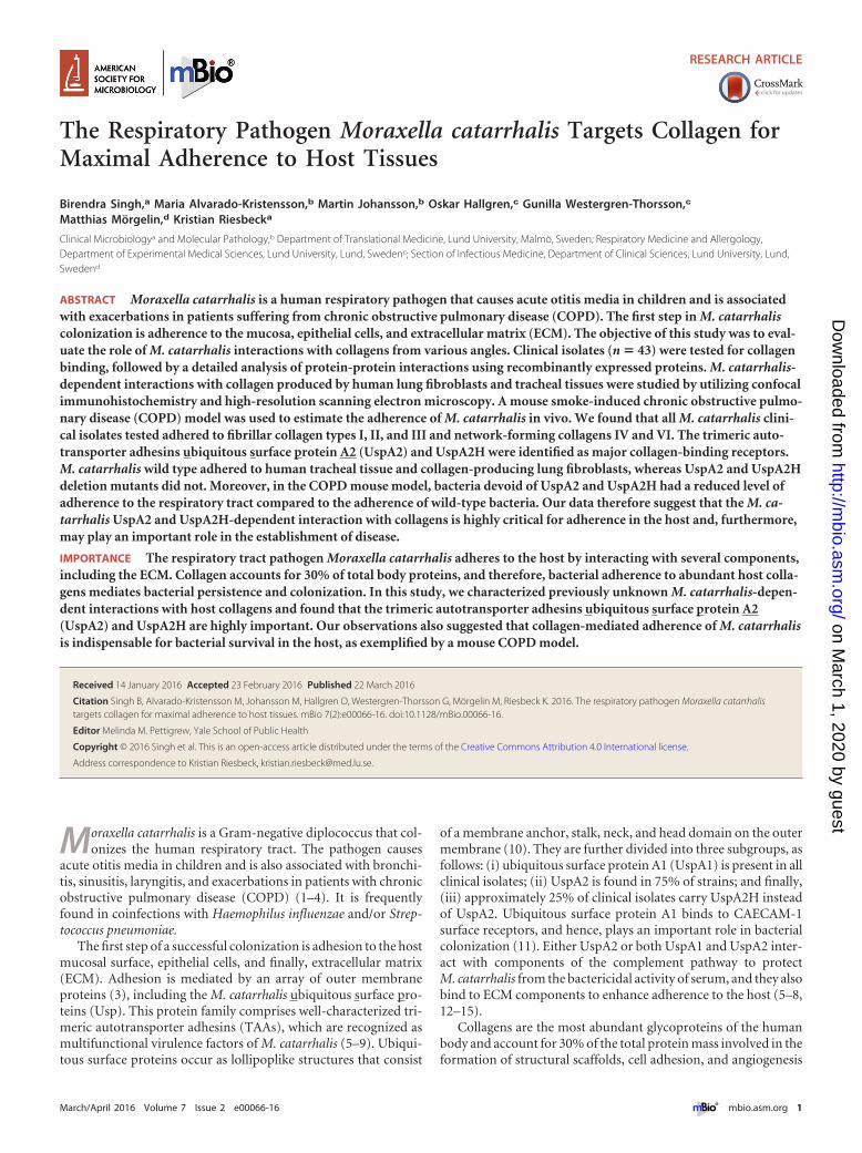
RESULTSM. catarrhalis binds to different fibril- and network-formingcollagens. M. catarrhalis Bc5 and BBH18 wild-type (WT) strainswere chosen to test interactions with various collagens. Increasingconcentrations of bacteria were added to microtiter plates thathad been precoated with collagens. Unbound bacteria were re-moved by washing, and attached bacteria were analyzed by usinganti-Moraxella polyclonal antibodies (PAbs). Our results revealedsignificant adherence of M. catarrhalis Bc5 to most collagenstested. However, M. catarrhalis Bc5 bound more to collagens IIand VI than to collagens I, III, and IV, whereas collagens V andVIII were not targeted by M. catarrhalis (Fig. 1A). In parallel withthe enzyme-linked immunosorbent assay (ELISA), we coated
glass slides with collagen and visualized the adherence of M. ca-tarrhalis by Gram staining and microscopy. Similar results wereobserved using this method (Fig. 1B). The other M. catarrhalisstrain, BBH18, also adhered to the collagens at significant levels(Fig. 1C and D). In contrast, M. catarrhalis did not adhere tohuman serum albumin (HSA), which was included as a negativecontrol (Fig. 1B and D). These results clearly indicated that M. ca-tarrhalis has a selective specificity for fibrillar collagens I, II, andIII, in addition to network-forming collagens IV and VI. Since theremaining network-forming collagens, types V and VIII, were nottargeted by M. catarrhalis, we excluded those from further down-stream analyses.
In the next set of experiments, various clinical isolates of M. ca-tarrhalis (5, 25) were selected to verify interactions with collagens.All M. catarrhalis clinical isolates tested bound collagens I, II, III,IV, and VI, but the binding capacity varied between isolates. Themean levels of binding of M. catarrhalis to collagens I, II, III, andVI were significantly higher than the mean level of binding tocollagen IV (Fig. 1E). A comparison of various clinical isolatesregarding their collagen-binding capacities is shown in Fig. S1 inthe supplemental material. Our data thus indicated that clinicalM. catarrhalis isolates recognize several human collagens that maycontribute to bacterial adherence.
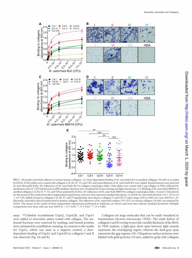
The trimeric autotransporters UspA2 and UspA2H are ma-jor collagen-binding proteins of M. catarrhalis. Trimeric auto-transporter adhesins, as exemplified by Yersinia enterocoliticaYadA (39), Aggregatibacter actinomycetemcomitans EmaA (26),and Bartonella hensalae BadA (27), bind human collagens. Simi-larly, the high-molecular-weight TAA MID of M. catarrhalis in-teracts with collagen IV (24). These previous studies prompted usto investigate whether M. catarrhalis TAAs also recognize an arrayof human collagen molecules. We performed an ELISA to screensingle and multiple mutants of M. catarrhalis TAAs. Interestingly,selective deletion of UspA2 in M. catarrhalis Bc5 (�uspA2) re-sulted in significantly reduced binding to collagens I, II, IV, and VI(Fig. 2A). In contrast to the UspA2 mutant (M. catarrhalis Bc5�uspA2), the mutant devoid of MID (�mid) had slightly de-creased binding to collagen I but not to the other collagens tested(Fig. 2A). Similar results were obtained with M. catarrhalis BBH18WT and the �uspA2 and �uspA2H mutants (Fig. 2B). Finally,mutants with deletion of UspA2 or UspA2H in combination withdeletion of UspA1 and MID were analyzed, proving that UspA2and UspA2H played the largest role in Moraxella-dependent ad-herence to collagens (Fig. 2).
To verify the specificity of UspA2- and UspA2H-dependentcollagen binding, we performed a competitive inhibition assay.Fibrillar collagens I, II, and III significantly inhibited the adher-ence of M. catarrhalis Bc5 and BBH18 to collagens I, II, IV, and VIin comparison to the inhibition by the network-forming collagensIV and VI (Fig. 3A and B). Moreover, fibrillar collagens alsoblocked the interaction of network-forming collagens (Fig. 3Aand B), confirming that both UspA2 and UspA2H have a singledomain interacting with collagen. Taken together, our results re-vealed that UspA2 and UspA2H are the major collagen-bindingproteins in M. catarrhalis and that fibrillar collagens are primetargets for optimal adherence.
M. catarrhalis UspA2 and UspA2H target gap regions offibrillar collagens. To define molecular interactions betweenUspA2 and UspA2H and the different collagens, we performed aseries of protein-protein interaction studies using a direct binding
Singh et al.
2 ® mbio.asm.org March/April 2016 Volume 7 Issue 2 e00066-16
on March 1, 2020 by guest
http://mbio.asm
.org/D
ownloaded from
assay. 125I-labeled recombinant UspA2, UspA2H, and UspA1were added to microtiter plates coated with collagen. The un-bound fractions were removed by washing, and bound proteinswere estimated by scintillation counting. In contrast to the resultsfor UspA1, which was used as a negative control, a dose-dependent binding of UspA2 and UspA2H to collagens I and IIwas observed (Fig. 4A and B).
Collagens are large molecules that can be easily visualized intransmission electron microscopy (TEM). The triple helices ofcollagens I and II overlap to provide variable thickness of the fibril.In TEM analysis, a light-gray short span between light strandsrepresents the overlapping region, whereas the dark-gray spanrepresents the gap regions (18). Ubiquitous surface proteins werelabeled with gold particles (10 nm), added to grids with collagens
FIG 1 Moraxella catarrhalis adheres to various human collagens. (A) Dose-dependent binding of M. catarrhalis Bc5 to purified collagens (50 nM) as revealedby ELISA. ELISA plates were coated with collagens I, II, III, IV, VI, and VIII, and serial dilutions of M. catarrhalis Bc5 were added. Bound bacteria were detectedby anti-Moraxella PAbs. (B) Adherence of M. catarrhalis Bc5 to collagen-coated glass slides. Glass slides were coated with 2 �g collagen or HSA, followed byincubation with 107 CFU/ml bacteria in BHI medium. Bacteria were visualized by Gram staining and light microscopy. (C) Binding of M. catarrhalis BBH18 topurified collagens I, II, III, IV, V, VI, and VIII as analyzed by ELISA. (D) Adherence of M. catarrhalis BBH18 to collagen-coated glass slides. (A and C) Data shownare the means of the results from three independent experiments, and error bars represent standard deviations. (A and B) M. catarrhalis bacteria (3.0 � 105 to 1.0� 107 CFU added) bound to collagens I, II, III, IV, and VI significantly more than to collagens V and VIII (P value range, 0.05 to 0.001; two-way ANOVA). (E)Moraxella catarrhalis clinical isolates bind to human collagens. The adherence of M. catarrhalis isolates (106 CFU) to various collagens (50 nM) was analyzed byELISA. The means of the results of three independent experiments performed in triplicates are shown and error bars indicate standard deviations. Multiplecomparisons were done with one-way ANOVA. *, P � 0.05; **, P � 0.01; ***, P � 0.001.
Moraxella catarrhalis and Collagens
March/April 2016 Volume 7 Issue 2 e00066-16 ® mbio.asm.org 3
on March 1, 2020 by guest
http://mbio.asm
.org/D
ownloaded from
I or II, and visualized by negative staining. Interestingly, UspA2and UspA2H bound directly to fibrillar collagens I and II at over-lap regions of the triple helices (Fig. 4C and D). Quantification ofgold particles in 50 randomly selected regions revealed signifi-cantly higher levels of binding of UspA2 and UspA2H than ofUspA1 to collagens I and II (see Fig. S2A in the supplementalmaterial). These data were in agreement with the results of ourdirect binding assay (Fig. 4A and B).
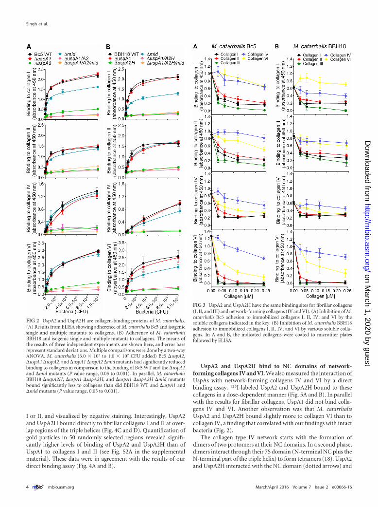
UspA2 and UspA2H bind to NC domains of network-forming collagens IV and VI. We also measured the interaction ofUspAs with network-forming collagens IV and VI by a directbinding assay. 125I-labeled UspA2 and UspA2H bound to thesecollagens in a dose-dependent manner (Fig. 5A and B). In parallelwith the results for fibrillar collagens, UspA1 did not bind colla-gens IV and VI. Another observation was that M. catarrhalisUspA2 and UspA2H bound slightly more to collagen VI than tocollagen IV, a finding that correlated with our findings with intactbacteria (Fig. 2).
The collagen type IV network starts with the formation ofdimers of two protomers at their NC domains. In a second phase,dimers interact through their 7S domain (N-terminal NC plus theN-terminal part of the triple helix) to form tetramers (18). UspA2and UspA2H interacted with the NC domain (dotted arrows) and
FIG 2 UspA2 and UspA2H are collagen-binding proteins of M. catarrhalis.(A) Results from ELISA showing adherence of M. catarrhalis Bc5 and isogenicsingle and multiple mutants to collagens. (B) Adherence of M. catarrhalisBBH18 and isogenic single and multiple mutants to collagens. The means ofthe results of three independent experiments are shown here, and error barsrepresent standard deviations. Multiple comparisons were done by a two-wayANOVA. M. catarrhalis (3.0 � 105 to 1.0 � 107 CFU added) Bc5 �uspA2,�uspA1 �uspA2, and �uspA1 �uspA2 �mid mutants had significantly reducedbinding to collagens in comparison to the binding of Bc5 WT and the �uspA1and �mid mutants (P value range, 0.05 to 0.001). In parallel, M. catarrhalisBBH18 �uspA2H, �uspA1 �uspA2H, and �uspA1 �uspA2H �mid mutantsbound significantly less to collagens than did BBH18 WT and �uspA1 and�mid mutants (P value range, 0.05 to 0.001).
FIG 3 UspA2 and UspA2H have the same binding sites for fibrillar collagens(I, II, and III) and network-forming collagens (IV and VI). (A) Inhibition of M.catarrhalis Bc5 adhesion to immobilized collagens I, II, IV, and VI by thesoluble collagens indicated in the key. (B) Inhibition of M. catarrhalis BBH18adhesion to immobilized collagens I, II, IV, and VI by various soluble colla-gens. In A and B, the indicated collagens were coated to microtiter platesfollowed by ELISA.
Singh et al.
4 ® mbio.asm.org March/April 2016 Volume 7 Issue 2 e00066-16
on March 1, 2020 by guest
http://mbio.asm
.org/D
ownloaded from
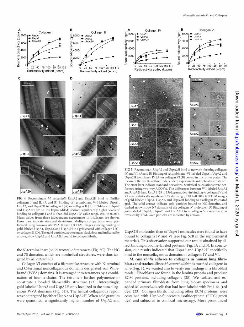
the N-terminal part (solid arrows) of tetramers (Fig. 5C). The NCand 7S domains, which are nonhelical structures, were thus tar-geted by M. catarrhalis.
Collagen VI consists of a filamentlike structure with N-terminaland C-terminal noncollagenous domains designated von Wille-brand (WVA) domains. It is arranged into tetramers by a combi-nation of four �-chains. The tetramers further polymerize toconstitute a beaded filamentlike structure (23). Interestingly,gold-labeled UspA2 and UspA2H only localized to the noncollag-enous WVA domains (Fig. 5D). The helical collagenous regionwas not targeted by either UspA2 or UspA2H. When gold granuleswere quantified, a significantly higher number of UspA2 and
UspA2H molecules than of UspA1 molecules were found to havebound to collagens IV and VI (see Fig. S2B in the supplementalmaterial). This observation supported our results obtained by di-rect binding of iodine-labeled proteins (Fig. 5A and B). In conclu-sion, our results indicated that UspA2 and UspA2H specificallybind to the noncollagenous domains of collagens IV and VI.
M. catarrhalis adheres to collagens in human lung fibro-blasts and trachea. Since M. catarrhalis binds purified collagens invitro (Fig. 1), we wanted also to verify our findings in a fibroblastmodel. Fibroblasts are found in the lamina propria and produceECM proteins, including collagens (28). We isolated and ex-panded primary fibroblasts from lung biopsy specimens andadded M. catarrhalis cells that had been labeled with Fm4-64 (reddye) (23). Collagen fibrils, including other ECM proteins, werecostained with UspA2-fluorescein isothiocyanate (FITC; greendye) and subjected to confocal microscopy. More pronounced
FIG 4 Recombinant M. catarrhalis UspA2 and UspA2H bind to fibrillarcollagens I and II. (A and B) Binding of recombinant 125I-labeled UspA1,UspA2, and UspA2H to collagen I (A) or collagen II (B). 125I-labeled UspA2and UspA2H (28 to 156 kcpm added) showed significantly higher levels ofbinding to collagens I and II than did UspA1 (P value range, 0.01 to 0.001).Mean values from three independent experiments in triplicates are shown.Error bars indicate standard deviations. Multiple comparisons were per-formed using two-way ANOVA. (C and D) TEM images showing binding ofgold-labeled UspA1, UspA2, and UspA2H to a grid coated with collagen I (C)or collagen II (D). The gold particles, appearing as black dots and indicated byarrows, show UspA2 and UspA2H bound to collagen fibrils.
FIG 5 Recombinant UspA2 and UspA2H bind to network-forming collagensIV and VI. (A and B) Binding of recombinant 125I-labeled UspA1, UspA2 andUspA2H to collagen IV (A) or collagen VI (B) coated in microtiter plates. Themeans of the results of three independent experiments in triplicates are shown.The error bars indicate standard deviations. Statistical calculations were per-formed using two-way ANOVA. The differences between 125I-labeled UspA2and UspA2H and UspA1 (28 to 156 kcpm added) in binding to collagen IV andVI were statistically significant (P value range, 0.01 to 0.001). (C) TEM imagesof gold-labeled UspA1, UspA2, and UspA2H binding to a collagen IV-coatedgrid. The solid arrows indicate gold particles bound to NC domains, anddashed arrows show N7 domains of the collagen IV molecule. (D) Binding ofgold-labeled UspA1, UspA2, and UspA2H to a collagen VI-coated grid asrevealed by TEM. Gold particles are indicated by arrows.
Moraxella catarrhalis and Collagens
March/April 2016 Volume 7 Issue 2 e00066-16 ® mbio.asm.org 5
on March 1, 2020 by guest
http://mbio.asm
.org/D
ownloaded from
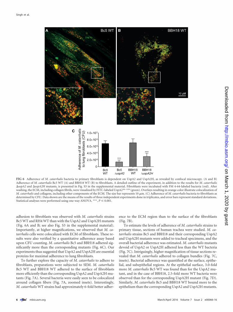
adhesion to fibroblasts was observed with M. catarrhalis strainsBc5 WT and RH4 WT than with the UspA2 and UspA2H mutants(Fig. 6A and B; see also Fig. S3 in the supplemental material).Importantly, at higher magnifications, we observed that M. ca-tarrhalis cells were colocalized with ECM of fibroblasts. These re-sults were also verified by a quantitative adherence assay basedupon CFU counting. M. catarrhalis Bc5 and BBH18 adhered sig-nificantly more than the corresponding mutants (Fig. 6C). Ourexperiments thus suggested that UspA2 and UspA2H are essentialproteins for maximal adherence to lung fibroblasts.
To further explore the capacity of M. catarrhalis to adhere tofibroblasts, preparations were subjected to SEM. M. catarrhalisBc5 WT and BBH18 WT adhered to the surface of fibroblastsmore efficiently than the corresponding UspA2 and UspA2H mu-tants (Fig. 7A). Several bacteria were easily seen to be colocalizedaround collagen fibers (Fig. 7A, zoomed insets). Interestingly,M. catarrhalis WT strains had approximately 6-fold better adher-
ence to the ECM region than to the surface of the fibroblasts(Fig. 7B).
To estimate the levels of adherence of M. catarrhalis strains toprimary tissue, sections of human trachea were studied. M. ca-tarrhalis strains Bc5 and BBH18 and their corresponding UspA2and UspA2H mutants were added to tracheal specimens, and theoverall bacterial adherence was estimated. M. catarrhalis mutantsdevoid of UspA2 or UspA2H adhered less than the WT bacteria(Fig. 7C). Intriguingly, higher magnification of tissue sections re-vealed that M. catarrhalis adhered to collagen bundles (Fig. 7C,insets). Bacterial adherence was quantified at the surface, epithe-lial, and subepithelial regions. At the epithelial surface, 3.0-foldmore M. catarrhalis Bc5 WT was found than for the UspA2 mu-tant, and in the case of BBH18, 2.5-fold more WT bacteria wereobserved than for the corresponding UspA2H mutant (Fig. 7D).Similarly, M. catarrhalis Bc5 and BBH18 WT bound more to theepithelium than the corresponding UspA2 and UspA2H mutants.
FIG 6 Adherence of M. catarrhalis bacteria to primary fibroblasts is dependent on UspA2 and UspA2H, as revealed by confocal microscopy. (A and B)Adherence of M. catarrhalis Bc5 WT (A) and BBH18 WT (B) to fibroblasts. A detailed outline of the experiment, in addition to the results for M. catarrhalis�uspA2 and �uspA2H mutants, is presented in Fig. S3 in the supplemental material. Fibroblasts were incubated with FM 4-64-labeled bacteria (red). Afterwashing, the ECM, including collagen fibrils, were visualized by FITC-labeled UspA230 –539 (green). Overlays resulting in orange color illustrate colocalization ofM. catarrhalis and collagens, including other components of the ECM. The size bar represents 10 �m. (C) Adherence of M. catarrhalis bacteria to fibroblasts asdetermined by CFU. Data shown are the means of the results of three independent experiments done in triplicates, and error bars represent standard deviations.Statistical analyses were performed using one-way ANOVA. ***, P � 0.001.
Singh et al.
6 ® mbio.asm.org March/April 2016 Volume 7 Issue 2 e00066-16
on March 1, 2020 by guest
http://mbio.asm
.org/D
ownloaded from
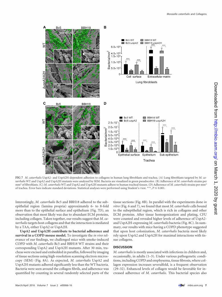
Interestingly, M. catarrhalis Bc5 and BBH18 adhered to the sub-epithelial region (lamina propria) approximately 6- to 8-foldmore than to the epithelial surface and epithelium (Fig. 7D), anobservation that most likely was due to abundant ECM proteins,including collagen. Taken together, our results suggest that M. ca-tarrhalis targets host collagens and that the interaction is mediatedby a TAA, either UspA2 or UspA2H.
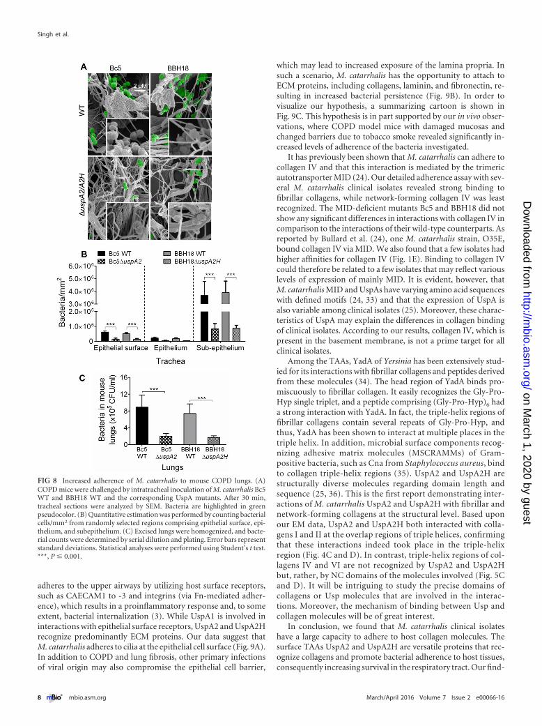
UspA2 and UspA2H contribute to bacterial adherence andsurvival in a COPD mouse model. To investigate the in vivo rel-evance of our findings, we challenged mice with smoke-inducedCOPD with M. catarrhalis Bc5 and BBH18 WT strains and theircorresponding UspA2 and UspA2H mutants. After 30 min, tra-cheas were excised and embedded in paraffin, followed by imagingof tissue sections using high-resolution scanning electron micros-copy (SEM) (Fig. 8A). As expected, M. catarrhalis UspA2 andUspA2H mutants adhered significantly less than the WT bacteria.Bacteria were seen around the collagen fibrils, and adherence wasquantified by counting in several randomly selected parts of the
tissue sections (Fig. 8B). In parallel with the experiments done invitro (Fig. 6 and 7), we found that most M. catarrhalis cells boundto the subepithelial region, which is rich in collagens and otherECM proteins. After tissue homogenization and plating, CFUwere counted and revealed higher levels of adherence of UspA2-and UspA2H-expressing M. catarrhalis bacteria (Fig. 8C). In sum-mary, our results with mice having a COPD phenotype suggestedthat upon host colonization, M. catarrhalis bacteria most likelyrely upon UspA2 and UspA2H for maximal interactions with tis-sue collagens.
DISCUSSION
M. catarrhalis is mostly associated with infections in children and,occasionally, in adults (1–3). Under various pathogenetic condi-tions, including COPD and emphysema, tissue fibrosis, where col-lagen expression increases severalfold, is a frequent observation(29–32). Enhanced levels of collagen would be favorable for in-creased adherence of M. catarrhalis. This bacterial species also
FIG 7 M. catarrhalis UspA2- and UspA2H-dependent adhesion to collagens in human lung fibroblasts and trachea. (A) Lung fibroblasts targeted by M. ca-tarrhalis WT and UspA2 and UspA2H mutants were analyzed by SEM. Bacteria are visualized in green pseudocolor. (B) Adherence of M. catarrhalis strains permm2 of fibroblasts. (C) M. catarrhalis WT and UspA2 and UspA2H mutants adhere to human tracheal tissues. (D) Adherence of M. catarrhalis strains per mm2
of trachea. Error bars indicate standard deviations. Statistical analyses were performed using Student’s t test. ***, P � 0.001.
Moraxella catarrhalis and Collagens
March/April 2016 Volume 7 Issue 2 e00066-16 ® mbio.asm.org 7
on March 1, 2020 by guest
http://mbio.asm
.org/D
ownloaded from
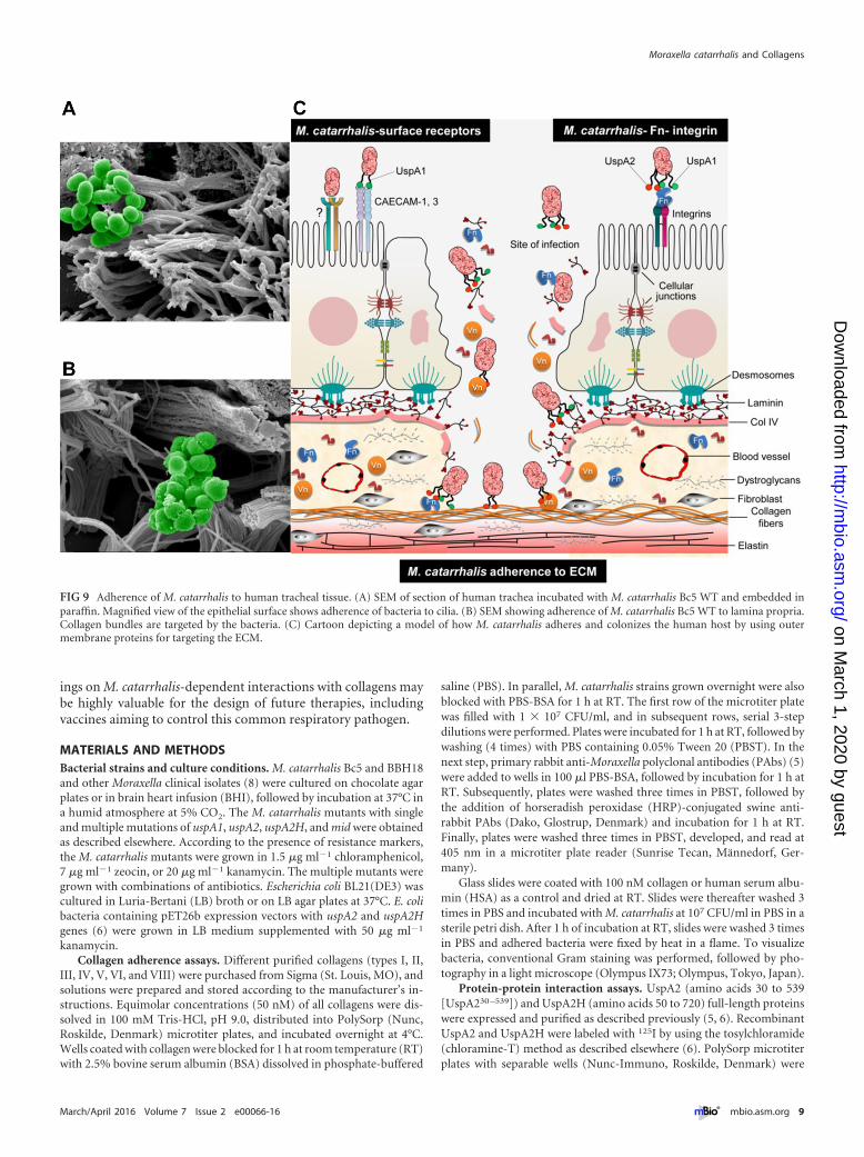
adheres to the upper airways by utilizing host surface receptors,such as CAECAM1 to -3 and integrins (via Fn-mediated adher-ence), which results in a proinflammatory response and, to someextent, bacterial internalization (3). While UspA1 is involved ininteractions with epithelial surface receptors, UspA2 and UspA2Hrecognize predominantly ECM proteins. Our data suggest thatM. catarrhalis adheres to cilia at the epithelial cell surface (Fig. 9A).In addition to COPD and lung fibrosis, other primary infectionsof viral origin may also compromise the epithelial cell barrier,
which may lead to increased exposure of the lamina propria. Insuch a scenario, M. catarrhalis has the opportunity to attach toECM proteins, including collagens, laminin, and fibronectin, re-sulting in increased bacterial persistence (Fig. 9B). In order tovisualize our hypothesis, a summarizing cartoon is shown inFig. 9C. This hypothesis is in part supported by our in vivo obser-vations, where COPD model mice with damaged mucosas andchanged barriers due to tobacco smoke revealed significantly in-creased levels of adherence of the bacteria investigated.
It has previously been shown that M. catarrhalis can adhere tocollagen IV and that this interaction is mediated by the trimericautotransporter MID (24). Our detailed adherence assay with sev-eral M. catarrhalis clinical isolates revealed strong binding tofibrillar collagens, while network-forming collagen IV was leastrecognized. The MID-deficient mutants Bc5 and BBH18 did notshow any significant differences in interactions with collagen IV incomparison to the interactions of their wild-type counterparts. Asreported by Bullard et al. (24), one M. catarrhalis strain, O35E,bound collagen IV via MID. We also found that a few isolates hadhigher affinities for collagen IV (Fig. 1E). Binding to collagen IVcould therefore be related to a few isolates that may reflect variouslevels of expression of mainly MID. It is evident, however, thatM. catarrhalis MID and UspAs have varying amino acid sequenceswith defined motifs (24, 33) and that the expression of UspA isalso variable among clinical isolates (25). Moreover, these charac-teristics of UspA may explain the differences in collagen bindingof clinical isolates. According to our results, collagen IV, which ispresent in the basement membrane, is not a prime target for allclinical isolates.
Among the TAAs, YadA of Yersinia has been extensively stud-ied for its interactions with fibrillar collagens and peptides derivedfrom these molecules (34). The head region of YadA binds pro-miscuously to fibrillar collagen. It easily recognizes the Gly-Pro-Hyp single triplet, and a peptide comprising (Gly-Pro-Hyp)6 hada strong interaction with YadA. In fact, the triple-helix regions offibrillar collagens contain several repeats of Gly-Pro-Hyp, andthus, YadA has been shown to interact at multiple places in thetriple helix. In addition, microbial surface components recog-nizing adhesive matrix molecules (MSCRAMMs) of Gram-positive bacteria, such as Cna from Staphylococcus aureus, bindto collagen triple-helix regions (35). UspA2 and UspA2H arestructurally diverse molecules regarding domain length andsequence (25, 36). This is the first report demonstrating inter-actions of M. catarrhalis UspA2 and UspA2H with fibrillar andnetwork-forming collagens at the structural level. Based uponour EM data, UspA2 and UspA2H both interacted with colla-gens I and II at the overlap regions of triple helices, confirmingthat these interactions indeed took place in the triple-helixregion (Fig. 4C and D). In contrast, triple-helix regions of col-lagens IV and VI are not recognized by UspA2 and UspA2Hbut, rather, by NC domains of the molecules involved (Fig. 5Cand D). It will be intriguing to study the precise domains ofcollagens or Usp molecules that are involved in the interac-tions. Moreover, the mechanism of binding between Usp andcollagen molecules will be of great interest.
In conclusion, we found that M. catarrhalis clinical isolateshave a large capacity to adhere to host collagen molecules. Thesurface TAAs UspA2 and UspA2H are versatile proteins that rec-ognize collagens and promote bacterial adherence to host tissues,consequently increasing survival in the respiratory tract. Our find-
FIG 8 Increased adherence of M. catarrhalis to mouse COPD lungs. (A)COPD mice were challenged by intratracheal inoculation of M. catarrhalis Bc5WT and BBH18 WT and the corresponding UspA mutants. After 30 min,tracheal sections were analyzed by SEM. Bacteria are highlighted in greenpseudocolor. (B) Quantitative estimation was performed by counting bacterialcells/mm2 from randomly selected regions comprising epithelial surface, epi-thelium, and subepithelium. (C) Excised lungs were homogenized, and bacte-rial counts were determined by serial dilution and plating. Error bars representstandard deviations. Statistical analyses were performed using Student’s t test.***, P � 0.001.
Singh et al.
8 ® mbio.asm.org March/April 2016 Volume 7 Issue 2 e00066-16
on March 1, 2020 by guest
http://mbio.asm
.org/D
ownloaded from
ings on M. catarrhalis-dependent interactions with collagens maybe highly valuable for the design of future therapies, includingvaccines aiming to control this common respiratory pathogen.
MATERIALS AND METHODSBacterial strains and culture conditions. M. catarrhalis Bc5 and BBH18and other Moraxella clinical isolates (8) were cultured on chocolate agarplates or in brain heart infusion (BHI), followed by incubation at 37°C ina humid atmosphere at 5% CO2. The M. catarrhalis mutants with singleand multiple mutations of uspA1, uspA2, uspA2H, and mid were obtainedas described elsewhere. According to the presence of resistance markers,the M. catarrhalis mutants were grown in 1.5 �g ml�1 chloramphenicol,7 �g ml�1 zeocin, or 20 �g ml�1 kanamycin. The multiple mutants weregrown with combinations of antibiotics. Escherichia coli BL21(DE3) wascultured in Luria-Bertani (LB) broth or on LB agar plates at 37°C. E. colibacteria containing pET26b expression vectors with uspA2 and uspA2Hgenes (6) were grown in LB medium supplemented with 50 �g ml�1
kanamycin.Collagen adherence assays. Different purified collagens (types I, II,
III, IV, V, VI, and VIII) were purchased from Sigma (St. Louis, MO), andsolutions were prepared and stored according to the manufacturer’s in-structions. Equimolar concentrations (50 nM) of all collagens were dis-solved in 100 mM Tris-HCl, pH 9.0, distributed into PolySorp (Nunc,Roskilde, Denmark) microtiter plates, and incubated overnight at 4°C.Wells coated with collagen were blocked for 1 h at room temperature (RT)with 2.5% bovine serum albumin (BSA) dissolved in phosphate-buffered
saline (PBS). In parallel, M. catarrhalis strains grown overnight were alsoblocked with PBS-BSA for 1 h at RT. The first row of the microtiter platewas filled with 1 � 107 CFU/ml, and in subsequent rows, serial 3-stepdilutions were performed. Plates were incubated for 1 h at RT, followed bywashing (4 times) with PBS containing 0.05% Tween 20 (PBST). In thenext step, primary rabbit anti-Moraxella polyclonal antibodies (PAbs) (5)were added to wells in 100 �l PBS-BSA, followed by incubation for 1 h atRT. Subsequently, plates were washed three times in PBST, followed bythe addition of horseradish peroxidase (HRP)-conjugated swine anti-rabbit PAbs (Dako, Glostrup, Denmark) and incubation for 1 h at RT.Finally, plates were washed three times in PBST, developed, and read at405 nm in a microtiter plate reader (Sunrise Tecan, Männedorf, Ger-many).
Glass slides were coated with 100 nM collagen or human serum albu-min (HSA) as a control and dried at RT. Slides were thereafter washed 3times in PBS and incubated with M. catarrhalis at 107 CFU/ml in PBS in asterile petri dish. After 1 h of incubation at RT, slides were washed 3 timesin PBS and adhered bacteria were fixed by heat in a flame. To visualizebacteria, conventional Gram staining was performed, followed by pho-tography in a light microscope (Olympus IX73; Olympus, Tokyo, Japan).
Protein-protein interaction assays. UspA2 (amino acids 30 to 539[UspA230 –539]) and UspA2H (amino acids 50 to 720) full-length proteinswere expressed and purified as described previously (5, 6). RecombinantUspA2 and UspA2H were labeled with 125I by using the tosylchloramide(chloramine-T) method as described elsewhere (6). PolySorp microtiterplates with separable wells (Nunc-Immuno, Roskilde, Denmark) were
FIG 9 Adherence of M. catarrhalis to human tracheal tissue. (A) SEM of section of human trachea incubated with M. catarrhalis Bc5 WT and embedded inparaffin. Magnified view of the epithelial surface shows adherence of bacteria to cilia. (B) SEM showing adherence of M. catarrhalis Bc5 WT to lamina propria.Collagen bundles are targeted by the bacteria. (C) Cartoon depicting a model of how M. catarrhalis adheres and colonizes the human host by using outermembrane proteins for targeting the ECM.
Moraxella catarrhalis and Collagens
March/April 2016 Volume 7 Issue 2 e00066-16 ® mbio.asm.org 9
on March 1, 2020 by guest
http://mbio.asm
.org/D
ownloaded from
coated with collagens (50 nM), and the binding of 125I-labeled UspA2 orUspA2H was estimated as described previously (15).
Fibroblast cultures and adherence of M. catarrhalis. Fibroblastsfrom lung tissues (23) were cultured in Dulbecco modified Eagle medium(DMEM) (Gibco; Life Technologies, Grand Island, NY) supplementedwith glucose and pyruvate in a humid atmosphere with 5% CO2 at 37°C.For infection experiments, cells were seeded into 24-well plates and grownuntil cells covered the plastic surface. Subsequently, 107 CFU/ml of bac-teria was added to each well and the plates incubated for 1 to 2 h at 37°C.Wells were washed 5 times with PBS in order to remove unbound bacteria.Trypsin (50 �l) was added to each well, and after dilution, cells were platedonto chocolate agar plates. After incubation at 37°C overnight, CFU werecounted manually.
Confocal microscopy. Fibroblasts (primary cells) were cultured on glasscover slips. After the cells reached 70 to 80% confluence, coverslips werewashed 3 times in PBS and fixed with 4% paraformaldehyde for 10 min.Bacteria were labeled with FM 4-64 (N-[3-triethylammoniumpropyl]-4-{6-[4-(diethylamino) phenyl] hexatrienyl} pyridinium dibromide); LifeTechnologies) and washed 3 times in PBS to remove excess dye. UspA230 –
539 was labeled with fluorescein isothiocyanate (FITC) according to themanufacturer’s instructions (Sigma). The unincorporated FITC from theUspA2-FITC complex was removed on a D-10 column (Sephadex G-25;GE Healthcare Life Sciences, Freiburg, Germany). To determine adher-ence, coverslips were blocked with 1% BSA in PBS for 1 h and incubatedwith 107 CFU/ml FM 4-64-labeled bacteria in 1% BSA for 1 h at RT.Unbound bacteria were removed by washing 3 times in PBS. Thereafter,5 nM UspA2-FITC solution in 1% BSA was added. After 30 min of incu-bation, the coverslips were washed 5 times with PBS and mounted influorescent mounting medium (Dako) containing 0.3 �g/ml DAPI (4=,6-diamidino-2-phenylindole dihydrochloride). Images were taken in a con-focal Zeiss Axio Observer microscope combined with a laser module forLSM 700 (Carl Zeiss, Göttingen, Germany) and processed using Zen Soft-ware.
COPD mouse model. C57BL/6 female mice (6 to 8 weeks old) wereobtained from Jackson Laboratories (Bar Harbor, ME) and housed understandard pathogen-free conditions in an animal facility at Lund Univer-sity. Mice were kept in 12-h light-dark cycles and fed with standard rodentchow and water ad libitum. All experiments were performed according tothe animal research ethics guidelines (Canadian Council on Animal Care;www.ccac.ca). For the COPD model, animals were exposed to smokefrom 12 3R4F reference cigarettes without filters (Tobacco and HealthResearch Institute, University of Kentucky, Lexington, KY) in an SIU-48whole-body cigarette smoking machine (Promech Lab, Vintrie, Sweden).Mice were exposed for 50 min twice daily for 24 weeks. Control mice wereexposed to normal room air only. Mice (n � 3 per group) were anesthe-tized to a surgical plane using isoflurane. The tracheas were exposed sur-gically, and bacteria were injected intratracheally. The wound was closedwith 5.0 silk sutures, and the animals were allowed to recover. After30 min, the animals were sacrificed by CO2 exposure, and tracheas, alongwith lungs, were excised. For estimation of contaminating normal flora,one group of mice was injected with PBS alone and treated similarly asdescribed above. Tracheal tissues were processed for SEM. In parallel, theright lung lobe was excised and homogenized by using a Multi-Gen 7homogenizer (Pro Scientific, Monroe, CT) in 1 ml chilled PBS, pH 7.4.Lung homogenates (100 �l) were mixed with 100 �l PBS containing6.86% sucrose. Thereafter, samples were serially diluted and plated onchocolate agar plates. After incubation overnight at 37°C in 5% CO2, CFUwere determined by manual counting. All experiments were performed intriplicates.
TEM and SEM. The interactions of UspA1, UspA2, and UspA2H withcollagens were analyzed by negative staining and TEM. Bacterial proteinswere labeled with colloidal gold nanoparticles (10 nm; British Biocell In-ternational, Cardiff, United Kingdom) as described elsewhere (37). Col-lagens I, II, IV, and VI were absorbed into 400-mesh carbon-coated cop-per grids and stained with 0.75% (wt/vol) uranyl formate as recently
described in detail (38). The grids were incubated with gold-labeled Us-pA(s), and after washing, the samples were visualized in a Philips/FEI CM100 TWIN transmission electron microscope (FEI, Hillsboro, OR) at60-kV accelerating voltage. Images were recorded with a side-mountedOlympus Veleta camera with a resolution of 2,048 by 2,048 pixels (2K by2K) using iTEM software. For bacterial interactions, fibroblasts or tissuesections obtained with permission (LU339-00) from the Ethical Commit-tee on Animal Experiments, the Swedish Board of Agriculture, were incu-bated with 2 � 109 CFU/ml of bacteria for 1 h at 37°C. Unbound bacteriawere washed with PBS, and specimens were fixed with 4% formaldehydeand 2.5% glutaraldehyde in PBS for 2 h at RT. Mouse tracheal specimenswere fixed overnight in 2.5% glutaraldehyde in cacodylate buffer andwashed with cacodylate buffer. Subsequently, samples were dehydrated byusing an ascending ethanol series from 50% (vol/vol) up to absolute eth-anol and then dried with carbon dioxide. Finally, tissue samples weremounted on aluminum holders, sputtered with 20-nm palladium-gold,and examined in a Philips/FEI XL-30 field emission scanning electronmicroscope using an Everhart-Tornley secondary electron detector. Im-ages were processed using Scandium software. The electron microscopicwork was performed at the Core Facility for Integrated microscopy (Pa-num Institute, University of Copenhagen). Images were enhanced withpseudocolors in Adobe Photoshop CS6 to visualize bacteria.
Statistical analysis. Data were analyzed by using GraphPad Prism 6.The statistical analysis was performed according to the data setup by usingone-way or two-way analysis of variance (ANOVA). Evaluation of the EMdata was performed by counting bacteria in at least 50 different cellularprofiles from three different experiments. Data sets were also evaluated byStudent’s t test for paired data.
SUPPLEMENTAL MATERIALSupplemental material for this article may be found at http://mbio.asm.org/lookup/suppl/doi:10.1128/mBio.00066-16/-/DCSupplemental.
Figure S1, TIF file, 0.3 MB.Figure S2, TIF file, 0.1 MB.Figure S3, TIF file, 1.9 MB.
ACKNOWLEDGMENTS
The authors report no potential conflicts of interest.
FUNDING INFORMATIONThis work was supported by grants from the Anna and Edwin BergerFoundation, Åke Wiberg Foundation, Lars Hierta Memorial Foundation,and O. E. and Edla Johansson Foundation, as well as the Swedish MedicalResearch Council (grant no. K2015-57X-03163-43-4; http://www.vr.se),the Cancer Foundation at the University Hospital in Malmö, and SkåneCounty Council’s research and development foundation (K.R.), and thePhysiographical Society (Forssman’s Foundation) (B.S.).
REFERENCES1. Murphy TF. 1996. Branhamella catarrhalis: epidemiology, surface anti-
genic structure, and immune response. Microbiol Rev 60:267–279.2. Karalus R, Campagnari A. 2000. Moraxella catarrhalis: a review of an
important human mucosal pathogen. Microbes Infect 2:547–559. http://dx.doi.org/10.1016/S1286-4579(00)00314-2.
3. Su YC, Singh B, Riesbeck K. 2012. Moraxella catarrhalis: from interac-tions with the host immune system to vaccine development. Future Mi-crobiol 7:1073–1100. http://dx.doi.org/10.2217/fmb.12.80.
4. Murphy TF, Parameswaran GI. 2009. Moraxella catarrhalis, a humanrespiratory tract pathogen. Clin Infect Dis 49:124 –131. http://dx.doi.org/10.1086/599375.
5. Singh B, Al-Jubair T, Voraganti C, Andersson T, Mukherjee O, Su YC,Zipfel P, Riesbeck K. 2015. Moraxella catarrhalis binds plasminogen toevade host innate immunity. Infect Immun 83:3458 –3469. http://dx.doi.org/10.1128/IAI.00310-15.
6. Singh B, Blom AM, Unal C, Nilson B, Mörgelin M, Riesbeck K. 2010.Vitronectin binds to the head region of Moraxella catarrhalis ubiqui-tous surface protein A2 and confers complement-inhibitory activity.
Singh et al.
10 ® mbio.asm.org March/April 2016 Volume 7 Issue 2 e00066-16
on March 1, 2020 by guest
http://mbio.asm
.org/D
ownloaded from
Mol Microbiol 75:1426 –1444. http://dx.doi.org/10.1111/j.1365-2958.2010.07066.x.
7. Tan TT, Nordström T, Forsgren A, Riesbeck K. 2005. The respiratorypathogen Moraxella catarrhalis adheres to epithelial cells by interactingwith fibronectin through ubiquitous surface proteins A1 and A2. J InfectDis 192:1029 –1038. http://dx.doi.org/10.1086/432759.
8. Tan TT, Forsgren A, Riesbeck K. 2006. The respiratory pathogenMoraxella catarrhalis binds to laminin via ubiquitous surface proteins A1and A2. J Infect Dis 194:493– 497. http://dx.doi.org/10.1086/505581.
9. Slevogt H, Zabel S, Opitz B, Hocke A, Eitel J, N’Guessan PD, Lucka L,Riesbeck K, Zimmermann W, Zweigner J, Temmesfeld-Wollbrueck B,Suttorp N, Singer BB. 2008. CEACAM1 inhibits Toll-like receptor2-triggered antibacterial responses of human pulmonary epithelial cells.Nat Immunol 9:1270 –1278. http://dx.doi.org/10.1038/ni.1661.
10. Łyskowski A, Leo JC, Goldman A. 2011. Structure and biology of trimericautotransporter adhesins. Adv Exp Med Biol 715:143–158. http://dx.doi.org/10.1007/978-94-007-0940-9_9.
11. N’Guessan PD, Vigelahn M, Bachmann S, Zabel S, Opitz B, Schmeck B,Hippenstiel S, Zweigner J, Riesbeck K, Singer BB, Suttorp N, Slevogt H.2007. The UspA1 protein of Moraxella catarrhalis induces CEACAM-1-dependent apoptosis in alveolar epithelial cells. J Infect Dis 195:1651–1660. http://dx.doi.org/10.1086/514820.
12. Nordström T, Blom AM, Forsgren A, Riesbeck K. 2004. The emergingpathogen Moraxella catarrhalis interacts with complement inhibitor C4bbinding protein through ubiquitous surface proteins A1 and A2. J Immu-nol 173:4598 – 4606. http://dx.doi.org/10.4049/jimmunol.173.7.4598.
13. Nordström T, Blom AM, Tan TT, Forsgren A, Riesbeck K. 2005. Ionicbinding of C3 to the human pathogen Moraxella catarrhalis is a uniquemechanism for combating innate immunity. J Immunol 175:3628 –3636.http://dx.doi.org/10.4049/jimmunol.175.6.3628.
14. Attia AS, Ram S, Rice PA, Hansen EJ. 2006. Binding of vitronectin by theMoraxella catarrhalis UspA2 protein interferes with late stages of the com-plement cascade. Infect Immun 74:1597–1611. http://dx.doi.org/10.1128/IAI.74.3.1597-1611.2006.
15. Liu G, Gradstedt H, Ermert D, Englund E, Singh B, Su YC, JohanssonME, Aspberg A, Agarwal V, Riesbeck K, Blom AM. 2016. Moraxellacatarrhalis evades host innate immunity via targeting cartilage oligomericmatrix protein. J Immunol 196:1249 –1258. http://dx.doi.org/10.4049/jimmunol.1502071.
16. Shoulders MD, Raines RT. 2009. Collagen structure and stability.Annu Rev Biochem 78:929 –958. http://dx.doi.org/10.1146/annurev.biochem.77.032207.120833.
17. Gelse K, Pöschl E, Aigner T. 2003. Collagens—structure, function, andbiosynthesis. Adv Drug Deliv Rev 55:1531–1546. http://dx.doi.org/10.1016/j.addr.2003.08.002.
18. Kadler KE, Baldock C, Bella J, Boot-Handford RP. 2007. Collagens at aglance. J Cell Sci 120:1955–1958. http://dx.doi.org/10.1242/jcs.03453.
19. Singh B, Fleury C, Jalalvand F, Riesbeck K. 2012. Human pathogensutilize host extracellular matrix proteins laminin and collagen for adhe-sion and invasion of the host. FEMS Microbiol Rev 36:1122–1180. http://dx.doi.org/10.1111/j.1574-6976.2012.00340.x.
20. Wachsmuth L, Söder S, Fan Z, Finger F, Aigner T. 2006. Immunolo-calization of matrix proteins in different human cartilage subtypes. HistolHistopathol 21:477– 485.
21. Liu X, Wu H, Byrne M, Krane S, Jaenisch R. 1997. Type III collagen iscrucial for collagen I fibrillogenesis and for normal cardiovascular devel-opment. Proc Natl Acad Sci U S A 94:1852–1856. http://dx.doi.org/10.1073/pnas.94.5.1852.
22. Mundel TM, Kalluri R. 2007. Type IV collagen-derived angiogenesisinhibitors. Microvasc Res 74:85– 89. http://dx.doi.org/10.1016/j.mvr.2007.05.005.
23. Abdillahi SM, Bober M, Nordin S, Hallgren O, Baumgarten M, ErjefältJ, Westergren-Thorsson G, Bjermer L, Riesbeck K, Egesten A, MörgelinM. 2015. Collagen VI is upregulated in COPD and serves both as an ad-
hesive target and a bactericidal barrier for Moraxella catarrhalis. J InnateImmun 7:506 –517. http://dx.doi.org/10.1159/000381213.
24. Bullard B, Lipski S, Lafontaine ER. 2007. Regions important for theadhesin activity of Moraxella catarrhalis Hag. BMC Microbiol 7:65. http://dx.doi.org/10.1186/1471-2180-7-65.
25. Su YC, Hallström BM, Bernhard S, Singh B, Riesbeck K. 2013. Impactof sequence diversity in the Moraxella catarrhalis UspA2/UspA2H headdomain on vitronectin binding and antigenic variation. Microbes Infect15:375–387. http://dx.doi.org/10.1016/j.micinf.2013.02.004.
26. Jiang X, Ruiz T, Mintz KP. 2011. The extended signal peptide of thetrimeric autotransporter EmaA of Aggregatibacter actinomycetemcomitansmodulates secretion. J Bacteriol 193:6983– 6994. http://dx.doi.org/10.1128/JB.05813-11.
27. Schmidgen T, Kaiser PO, Ballhorn W, Franz B, Göttig S, Linke D,Kempf VA. 2014. Heterologous expression of Bartonella adhesin A inEscherichia coli by exchange of trimeric autotransporter adhesin domainsresults in enhanced adhesion properties and a pathogenic phenotype. JBacteriol 196:2155–2165. http://dx.doi.org/10.1128/JB.01461-13.
28. Narayanan AS, Page RC, Swanson J. 1989. Collagen synthesis by humanfibroblasts. Regulation by transforming growth factor-beta in the presenceof other inflammatory mediators. Biochem J 260:463– 469. http://dx.doi.org/10.1042/bj2600463.
29. Kranenburg AR, Willems-Widyastuti A, Moori WJ, Sterk PJ, Ala-gappan VK, de Boer WI, Sharma HS. 2006. Enhanced bronchialexpression of extracellular matrix proteins in chronic obstructive pul-monary disease. Am J Clin Pathol 126:725–735. http://dx.doi.org/10.1309/JC477FAEL1YKV54W.
30. Jankowich MD, Rounds SI. 2012. Combined pulmonary fibrosis andemphysema syndrome: a review. Chest 141:222–231. http://dx.doi.org/10.1378/chest.11-1062.
31. King TE, Jr., Pardo A, Selman M. 2011. Idiopathic pulmonary fibro-s i s . Lancet 378:1949 –1961. http://dx.doi.org/10.1016/S0140-6736(11)60052-4.
32. Chilosi M, Poletti V, Rossi A. 2012. The pathogenesis of COPD and IPF:distinct horns of the same devil? Respir Res 13:3. http://dx.doi.org/10.1186/1465-9921-13-3.
33. Brooks MJ, Sedillo JL, Wagner N, Wang W, Attia AS, Wong H,Laurence CA, Hansen EJ, Gray-Owen SD. 2008. Moraxella catarrhalisbinding to host cellular receptors is mediated by sequence-specific deter-minants not conserved among all UspA1 protein variants. Infect Immun76:5322–5329. http://dx.doi.org/10.1128/IAI.00572-08.
34. Leo JC, Elovaara H, Bihan D, Pugh N, Kilpinen SK, Raynal N, SkurnikM, Farndale RW, Goldman A. 2010. First analysis of a bacterial collagen-binding protein with collagen toolkits: promiscuous binding of YadA tocollagens may explain how YadA interferes with host processes. InfectImmun 78:3226 –3236. http://dx.doi.org/10.1128/IAI.01057-09.
35. Zong Y, Xu Y, Liang X, Keene DR, Höök A, Gurusiddappa S, Höök M,Narayana SV. 2005. A “collagen hug” model for Staphylococcus aureusCNA binding to collagen. EMBO J 24:4224 – 4236. http://dx.doi.org/10.1038/sj.emboj.7600888.
36. Brooks MJ, Sedillo JL, Wagner N, Laurence CA, Wang W, Attia AS,Hansen EJ, Gray-Owen SD. 2008. Modular arrangement of allelic variantsexplains the divergence in Moraxella catarrhalis UspA protein function. InfectImmun 76:5330–5340. http://dx.doi.org/10.1128/IAI.00573-08.
37. Roth J. 1996. The silver anniversary of gold: 25 years of the colloidal goldmarker system for immunocytochemistry and histochemistry. HistochemCell Biol 106:1– 8. http://dx.doi.org/10.1007/BF02473197.
38. Oehmcke S, Mörgelin M, Herwald H. 2009. Activation of the humancontact system on neutrophil extracellular traps. J Innate Immun1:225–230. http://dx.doi.org/10.1159/000203700.
39. Nummelin H, Merckel MC, Leo JC, Lankinen H, Skurnik M, GoldmanA. 2004. The Yersinia adhesin YadA collagen-binding domain structure isa novel left-handed parallel beta-roll. EMBO J 23:701–711.
Moraxella catarrhalis and Collagens
March/April 2016 Volume 7 Issue 2 e00066-16 ® mbio.asm.org 11
on March 1, 2020 by guest
http://mbio.asm
.org/D
ownloaded from