Embed Size (px)
Citation preview

out
renal corpuscle
(lecture 1)
Proximal tubule Distal tubuleHenle's loop(lecture 4)
The rest of the nephron is divided into 3 main zones:
(each is subdivided into segments, but we don’t need to worry about that yet)
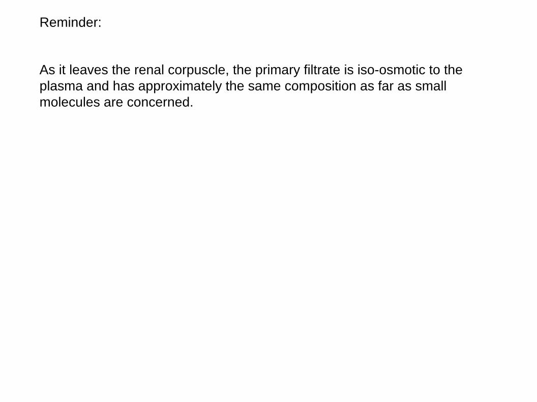
Reminder:
As it leaves the renal corpuscle, the primary filtrate is iso-osmotic to theplasma and has approximately the same composition as far as smallmolecules are concerned.
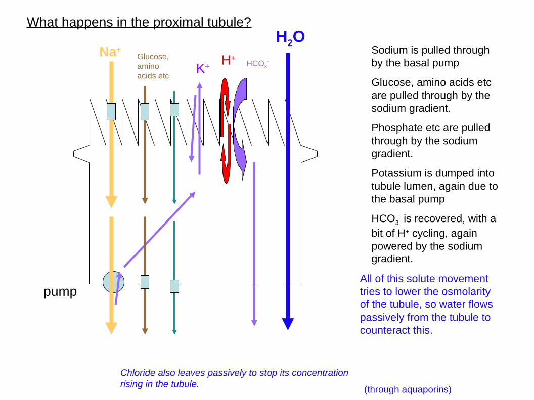
What happens in the proximal tubule?
pump
Sodium is pulled throughby the basal pump
Glucose, amino acids etcare pulled through by thesodium gradient.
Phosphate etc are pulledthrough by the sodiumgradient.
Potassium is dumped intotubule lumen, again due tothe basal pump
HCO3- is recovered, with a
bit of H+ cycling, againpowered by the sodiumgradient.
All of this solute movementtries to lower the osmolarityof the tubule, so water flowspassively from the tubule tocounteract this.
Na+Glucose,aminoacids etc
K+H+
HCO3-
H2O
(through aquaporins)
Chloride also leaves passively to stop its concentrationrising in the tubule.

We need quite a lot of surface area to do this well:
1) Microvilli
2) Pack a lot of length into a small space
Proximal convoluted tubule (PCT)
53 tubules (mouse)One tubule (mouse)
From : Zhai XY, Birn H, Jensen KB, Thomsen JS, Andreasen A, Christensen EI. (2003) Digital three-dimensional reconstruction and ultrastructure of themouse proximal tubule. J Am Soc Nephrol. 14(3):611-9

So what has the proximal tubule achieved?
Recovery of sodium, chloride, phosphate, calcium etc YES (65%)
Recovery of water YES (some)
Concentration of urine NO (still roughly iso-osmotic)
Control of acid/base NO (or only slightly)
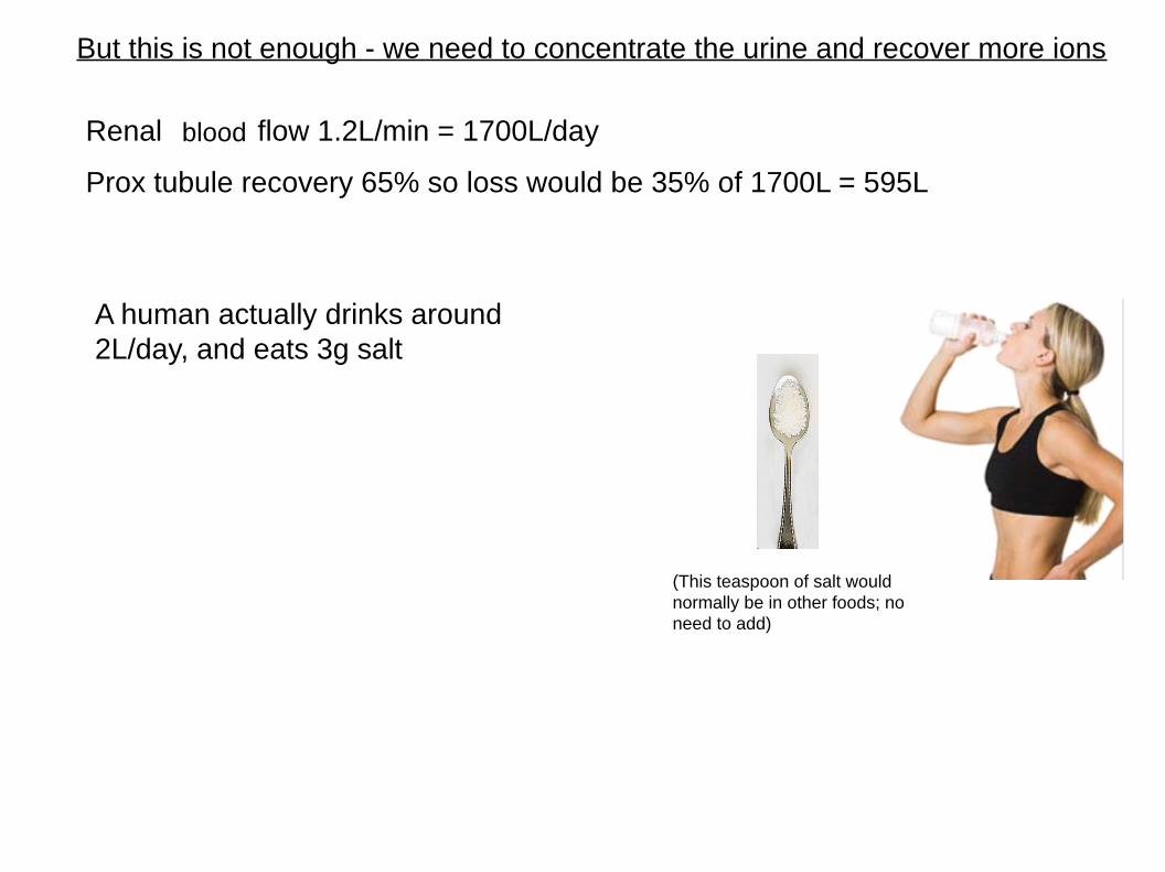
Renal filtrate flow 1.2L/min = 1700L/day
Prox tubule recovery 65% so loss would be 35% of 1700L = 595L
A human actually drinks around2L/day, and eats 3g salt
(This teaspoon of salt wouldnormally be in other foods; noneed to add)
But this is not enough - we need to concentrate the urine and recover more ions
blood


So - what tools have we got to concentrate urine?
1) We do NOT have a water pump !
2) We do have the Na+/K+ ATPase
3) We have the SLCs and ion channels that can parasitize the Na+ gradient to move ions and small molecules about.
4)We have the fact that osmosis will make water 'follow' ions
Osmosis only takes water from a dilute solution (of solute) to aconcentrated one.
…. So if we are to tempt water out of the tubule to concentrate urine, wehave to provide a destination that is more concentrated that the urine.
BASAL SIDE
ATP
ADP+Pi
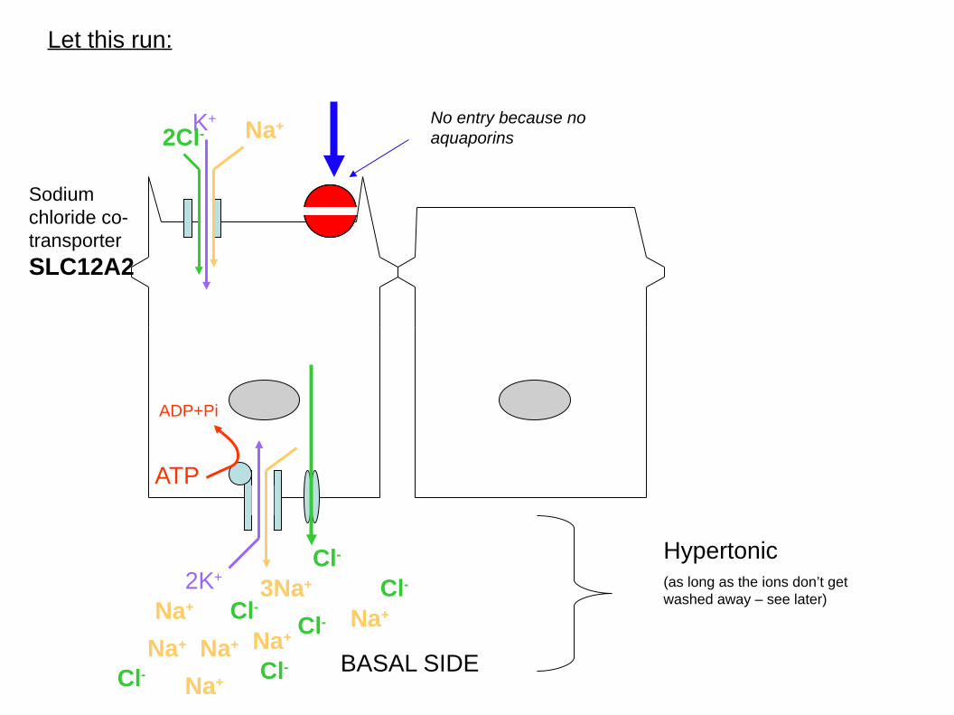
Let this run:
2K+3Na+
Sodiumchloride co-transporter
SLC12A2
Na+2Cl-
Cl-
No entry because noaquaporins
Na+ Na+Na+
Na+
Na+Na+
Cl-Cl-
Cl-Cl-
Cl-
Hypertonic(as long as the ions don’t getwashed away – see later)
K+

OK – so we can easily make a bit of tissue just outside the tubules veryhypertonic.
But we did this by stopping water moving.
… so we can’t use it to draw water across the same cells.
But what if we pull it across some othercells?
- This would take an anatomical arrangementthat has two bits of tubule running in thesame surrounding environment.

Renal corpuscle
PCT
Loop of HenleDescending thinlimb
Ascending thin limb
Thick ascending limb
The Loop of Henle
Friedrich Henle

Renal corpuscle
PCT
Descending thinlimb
Ascending thin limb
The Loop of Henle
Permeable to water
'Impermeable' to ions
'Impermeable' to urea
Impermeable towater
Permeable to ions
Permeable to urea
Thick ascending limb
Active recovery of ions(driven by Na pump)

(image has been stretched for clarity of labelling)
Gets more diluted(but that's mainlybecause so muchhas beenrecovered)
Locally veryhypertonic
Cells in the thin descending limb have lotsof aquaporins but little ion transport.
Water drawn out ofdescending limb
Tubular contentsget much moreconcentrated('countercurrentmultiplication')Concentration
numbers arein osmol/kg
0.29
0.29
1.4
0.1
THIS MECHANISMRECOVERS ABOUT10% OF FILTERED WATER AND 25% Na+, Cl-
(running total – 75%water 90% NaCl)

This recovery of water may not seem much, but view the pointof the LoH is making a hypertonic zone.
We'll use that zone again in a moment, and that's when wereally get the payback.
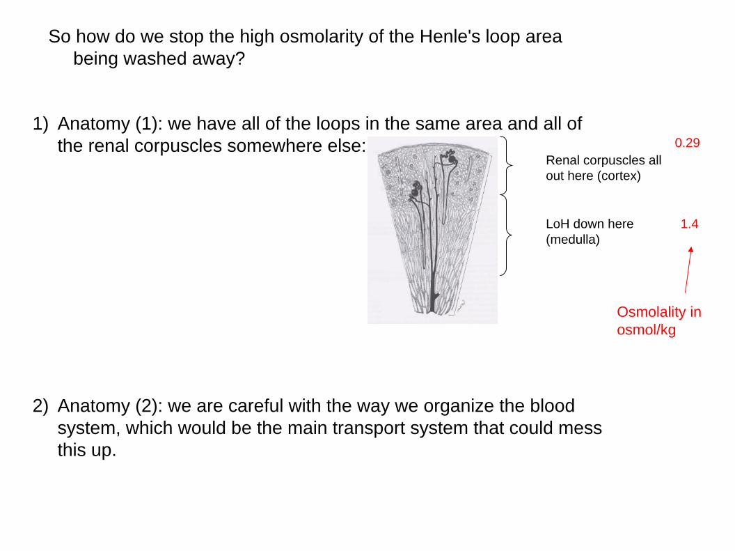
So how do we stop the high osmolarity of the Henle's loop areabeing washed away?
1) Anatomy (1): we have all of the loops in the same area and all ofthe renal corpuscles somewhere else:
2) Anatomy (2): we are careful with the way we organize the bloodsystem, which would be the main transport system that could messthis up.
Renal corpuscles allout here (cortex)
LoH down here(medulla)
0.29
1.4
Osmolality inosmol/kg

Problem: how do we stop out hypertonic region being swept awayby blood flow in the tissues?
The blood vessels emergingfrom the glomerulus go on toform a secondary capillarysystem – the vasa recta
Wikipedia commons
The important thing to see is thatthe blood comes in up the concgradient and goes out the exactopposite way.

PCT
Hypertonicregion
"Countercurrentexchange":
Water drawnout, ions in (passive) asblood descends
Blood now vconc and salty
Watertaken in asbloodmoves upout ofmedulla,salt givenup
(there are still some losses of course)
Analogy: blood vessels enteringpenguins' feet run next to theones leaving, so pass heat to thereturning blood not to the icebelow.

Renal corpuscle
PCTThe distal tubule:more recovery ofions. No watertransport.
NB: this nephron has been 'spread out' – the Distal Tubule will really come close to the corpuscle again
NaCl
By now, 95% ofNaCl recovered.
75% of water
(removal of ionsmakes contentseven more dilute)
0.29
1.4
0.1
0.08
For developmentalreasons (see later), thisis the end of thenephron. The tubulejoins into a collectingduct system.

Renal corpuscle
PCTThe collecting duct:regulated permeability to water
NB: this nephron has been 'spread out' – the Distal Tubule will really come close to the corpuscle again
0.29
1.4
0.1
0.08
More NaCl (2-5%)
H2O
Up here, it equilibrateswith plasma
0.29
Waterreabsorptiondriven byhypertoniczone
Up to1.4
Up to 24% of filteredwater removed here
Cumulative >99%
Coll duct cells can choose how muchaquaporin to put in the membrane and howmuch to store on vacuoles. Regulated byvasopressin (see lecture 4)

Renal corpuscle
PCT
The collecting duct:
The duct can choose to leak urea(again regulated by vasopressin –see lecture 4)
NB: this nephron has been 'spread out' – the Distal Tubule will really come close to the corpuscle again
0.29
1.4
0.1
0.08
H2O
Up here, it equilibrateswith plasma
0.29
Up to1.4
Up to 24% of filteredwater removed here
Cumulative >99%
Urea adds tohypertonicity
Urea now vconcentrated
SLC14A2
urea

Layton AT, Layton HE. (2011) Countercurrent multiplication may notexplain the axial osmolality gradient in the outer medulla of the ratkidney. Am J Physiol Renal Physiol. 2011 Nov;301(5):F1047-56
Please note: I have given you the accepted 'text-book' explanation, butthere are uncomfortable pieces of data that challenge it. This is goodreview:
I suspect that the story I told you will be modified in its detail, but the corewill remain.
As year 2 medical students, you do not have to engage with thisdebate – I am just flagging it in case you come across it and get
confused.

Summary so far:
● The loop of Henle makes the medulla veryhypertonic
● This draws water from tubules that passthrough there – especially collecting duct.
● The duct can help by letting some urea intothis area

This function depends on a specific anatomical arrangement
1) We need a separationbetween normal andhypertonic zones
2) We need the route to passagain through thehypertonic route
3) So it makes sense to havethe normal zone on theoutside of the organ andthe hypertonic zone nearthe place that urine willcollect:
4) And, for a big animal like ahuman, to group several ofthese units around acentral urine collectingplace:
normal
hypertonic

In more detail: (with some names)
Sectioned human kidney (clottedblood obscures finer features)
Cortex (renalcorpuscles,PCT, DCT)
Medulla (LoH,collecting duct)
Renalpyramid
Renalpelvis
Renal papilla
calyces
capsule

Given that the collecting duct system is a branched system that radiatesfrom the pelvis, if makes sense for the blood system to follow the samepattern:
To bladder
ureter
Renal vein
Renal artery
(you saw this in lecture 1)
aorta
VC
Segmentalartery

There is substantialvariation betweenindividuals:

Given that the collecting duct system is a branched system that radiatesfrom the pelvis, if makes sense for the blood system to follow the samepattern:
To bladder
ureter
Renal vein
Renal artery
(you saw this in lecture 1)
aorta
VC
Segmentalartery
Interlobararteries

Renal arteriogram;
Left renal artery showsstenosis (narrowing bydamage and scarring) -see arrow.
Right renal artery absent.
Aorta irregular andatheromatous.
{Image obtained byinjecting contrast materialand X-raying)
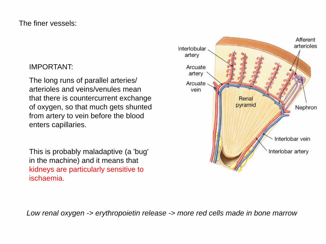
The finer vessels:
IMPORTANT:
The long runs of parallel arteries/arterioles and veins/venules meanthat there is countercurrent exchangeof oxygen, so that much gets shuntedfrom artery to vein before the bloodenters capillaries.
This is probably maladaptive (a 'bug'in the machine) and it means thatkidneys are particularly sensitive toischaemia.
Low renal oxygen -> erythropoietin release -> more red cells made in bone marrow

0.29
1.4
0.1
0.08
H2O0.29
Up to1.4
Back to function: How is this lot all controlled?
Things that can bealtered:
1) Blood flow toglomerulus:
2) Na+ recovery(SLC9A3)
3) Urea recovery byCD (SLC14A2)
4) Water permeabilityof CD (aquaporins)
5) Acid-base balancein CD

Blood flow
There are 3controls:
1) Systemicbloodpressure
2) Constriction ofafferentarterioles
3) Constriction ofefferentarterioles
Lowers glompressure
Raises glompressure
NB – diabetic changes in vessels can cause this too
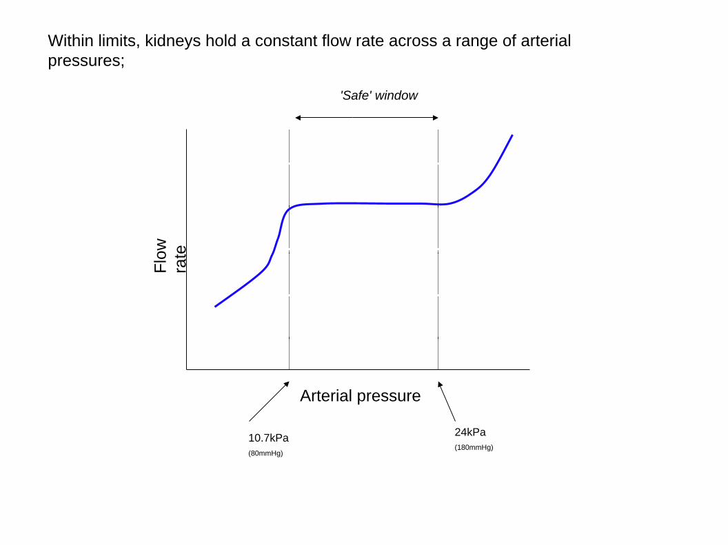
Within limits, kidneys hold a constant flow rate across a range of arterialpressures;
Flo
wra
te
Arterial pressure
10.7kPa(80mmHg)
24kPa(180mmHg)
'Safe' window
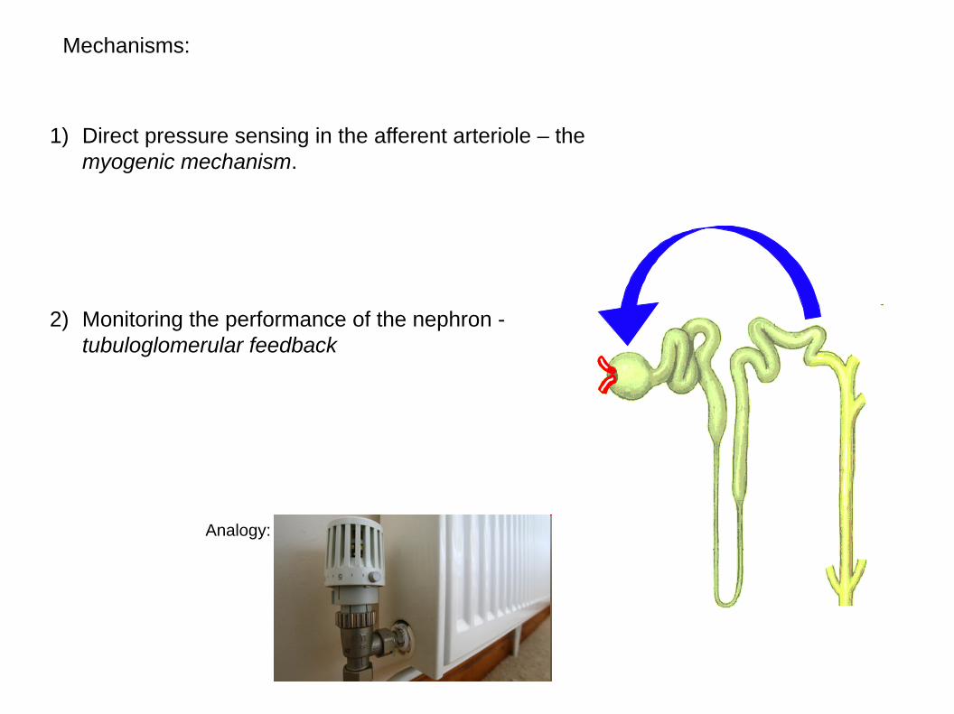
Mechanisms:
1) Direct pressure sensing in the afferent arteriole – themyogenic mechanism.
2) Monitoring the performance of the nephron -tubuloglomerular feedback
Analogy:

Problem:
We need this control to be nephron-by-nephron, not global, so we can’t have asignal diffusing long distances.
Solution:
Arrange a nephron so that the end of thedistal tubule makes 'kissing contact' with thearterioles entering the glomerulus

Source: Kaul, C.L., Ramarao, P. (2000) Renin release and thesympathetic nervous system. Drugs Today 2000, 36(10): 699
A special zone of the distal tubule, called the macula densa, forms where contact ismade:
I don’t have permission toinclude the picture I usedlive on the LEARN slide-set, but you can find ithere:
Click
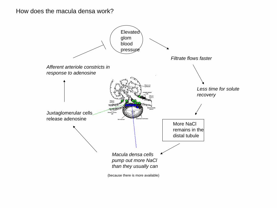
How does the macula densa work?
Elevatedglombloodpressure
Filtrate flows faster
Less time for soluterecovery
More NaClremains in thedistal tubule
Macula densa cellspump out more NaClthan they usually can
(because there is more available)
Juxtaglomerular cellsrelease adenosine
Afferent arteriole constricts inresponse to adenosine
Next lecture – the kidney working with the rest of the body.