Embed Size (px)
Citation preview
University of Central Florida University of Central Florida
STARS STARS
HIM 1990-2015
2015
The Risk of Secondary Lymphedema due to Procedures in the The Risk of Secondary Lymphedema due to Procedures in the
Affected Arm Post-Mastectomy: A Literature Review Affected Arm Post-Mastectomy: A Literature Review
Lindsay Perna University of Central Florida
Part of the Nursing Commons
Find similar works at: https://stars.library.ucf.edu/honorstheses1990-2015
University of Central Florida Libraries http://library.ucf.edu
This Open Access is brought to you for free and open access by STARS. It has been accepted for inclusion in HIM
1990-2015 by an authorized administrator of STARS. For more information, please contact [email protected].
Recommended Citation Recommended Citation Perna, Lindsay, "The Risk of Secondary Lymphedema due to Procedures in the Affected Arm Post-Mastectomy: A Literature Review" (2015). HIM 1990-2015. 618. https://stars.library.ucf.edu/honorstheses1990-2015/618
THE RISK OF SECONDARY LYMPHEDEMA DUE TO PROCEDURES IN THE AFFECTED ARM POST-MASTECTOMY: A LITERATURE REVIEW
by
LINDSAY A. PERNA
A thesis submitted in partial fulfillment of the requirements for Honors in the Major Program in Nursing
in the College of Nursing and in the Burnett Honors College at the University of Central Florida
Orlando, FL
Summer Term 2015
Thesis Chair: Dr. Leslee D’Amato-Kubiet

ii
©2015 Lindsay A. Perna
iii
ABSTRACT
The risk for upper extremity lymphedema post-mastectomy in women surgically
treated for breast cancer is a concern since it is often painful, aesthetically displeasing, and
can increase the risk of infection. However, there is a paucity of data examining if
diagnostic procedures performed in the ipsilateral arm post-mastectomy increases the risk
of lymphedema. The purpose of this research is to examine the relationship between
diagnostic procedures performed in the ipsilateral arm post-mastectomy and the
occurrence of lymphedema with or without related complications. A systematic review of
the literature was conducted from multiple, online databases available from 1992 through
2014, and included CINAHL, MedLine, PsychInfo, and ERIC. Search terms included
lymphedema, breast cancer, mastectomy, blood pressure, and infection. Exclusion criteria
comprised articles focused on male gender, primary lymphedema, metastases, survival,
quality of life studies, reoccurrence breast cancer, breast conservation, lymphedema
management, lymphedema, and lymphoma. The results of this study were inconclusive
concerning a relationship between upper extremity lymphedema and procedures
performed in the ipsilateral arm after mastectomy. This literature review outlines gaps in
the data showing a need for more focused research on the causes of secondary
lymphedema after breast cancer surgery with lymph node removal. Further research on
the impact of diagnostic and other invasive procedures on the ispilateral arm after
mastectomy should be considered.
iv
DEDICATION
For all nurses, using evidence-based practice
to provide the optimum standard of patient care,
For all women, living with or surviving breast cancer,
And especially, for my mentor, Dr. Leslee D’Amato-Kubiet, for inspiring me to achieve my goals.

v
ACKNOWLEDGEMENTS
I would like to thank all who gave me the strength and determination to compose this review of the literature. Thank you to my committee members, Dr. Angeline Bushy, Dr. Victoria Loerzel, and Dr. H. Edward Fouty. Your combined expertise and suggestions were
instrumental to this paper. Thank you to Dr. Angeline Bushy, for your proofreading and edits to make my paper stronger. Thank you to Dr. Victoria Loerzel for your expertise in
content review. I am especially grateful for your presence and encouragement at the Showcase of Undergraduate Research Excellence (SURE) presentations. To Denise Crisafi,
who provided assistance throughout the Honors in the Major process, thank you for keeping me on track with deadlines.
Above all, thank you to my thesis chair, Dr. Leslee D’Amato-Kubiet. I could never have accomplished this literature review without your support and encouragement. You
were always there to give me the power and courage to reach for the moon. To my fiancé, Dr. Bradley Stephan, thank you for your unfailing support and
understanding. Thank you to the University of Central Florida College of Nursing instructors and
staff.
vi
TABLE OF CONTENTS
INTRODUCTION ............................................................................................................................................................ 1
PROBLEM ........................................................................................................................................................................ 2
BACKGROUND ............................................................................................................................................................... 3
Lymphedema – What is it? ........................................................................................................................... 3
Measurement of lymphedema .................................................................................................................... 3
Possible factors influencing the risk of lymphedema ........................................................................ 4
Non-modifiable risk factors ..................................................................................................................... 4
Age ................................................................................................................................................................ 4
Severity of disease .................................................................................................................................. 5
Axillary node resection ......................................................................................................................... 6
Post Surgical Radiation and Chemotherapy ................................................................................. 7
Modifiable Risk factors .............................................................................................................................. 8
Hypertension ............................................................................................................................................ 8
BMI and weight ........................................................................................................................................ 8
Current prevention of lymphedema ......................................................................................................... 9
PURPOSE ...................................................................................................................................................................... 11
METHOD ....................................................................................................................................................................... 12
RESULTS ....................................................................................................................................................................... 14
vii
Invasive Procedures ..................................................................................................................................... 16
Mastectomy ..................................................................................................................................................... 17
Radiation and Chemotherapy ................................................................................................................... 18
Axillary Dissection and Lymph Node Involvement .......................................................................... 19
Advanced Diagnosis ..................................................................................................................................... 20
Body Mass Index ............................................................................................................................................ 21
Comorbidities ................................................................................................................................................. 22
Age ...................................................................................................................................................................... 23
DISCUSSION ................................................................................................................................................................. 24
LIMITATIONS .............................................................................................................................................................. 26
CONCLUSION .............................................................................................................................................................. 27
NURSING IMPLICATIONS ....................................................................................................................................... 29
Appendix A: Figures ................................................................................................................................................. 30
Appendix B: Tables ................................................................................................................................................... 32
REFERENCES .............................................................................................................................................................. 49

viii
LIST OF FIGURES
Figure 1: Consort Diagram ............................................................................................................................. 31

ix
LIST OF TABLES
Table 1: Treatment Characteristics................................................................................................................ 9
Table 2: Risk Factors for Lymphedema ..................................................................................................... 15
Table 3: Table of Evidence .............................................................................................................................. 33
1
INTRODUCTION
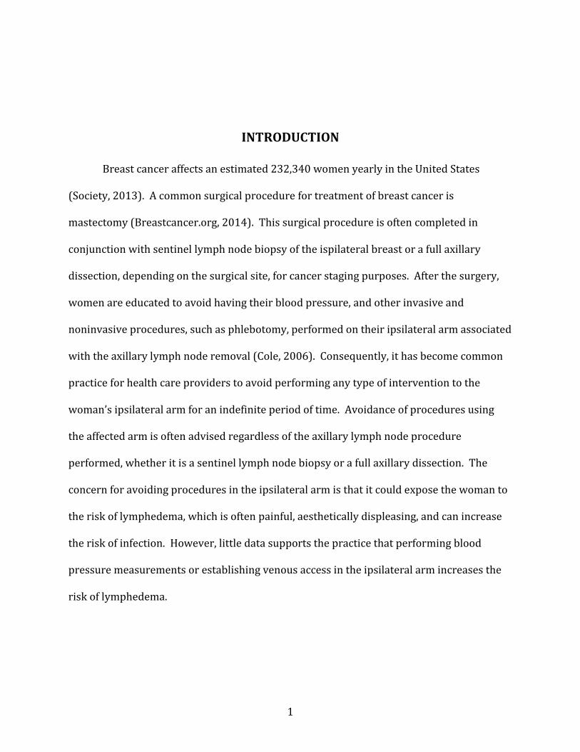
Breast cancer affects an estimated 232,340 women yearly in the United States
(Society, 2013). A common surgical procedure for treatment of breast cancer is
mastectomy (Breastcancer.org, 2014). This surgical procedure is often completed in
conjunction with sentinel lymph node biopsy of the ispilateral breast or a full axillary
dissection, depending on the surgical site, for cancer staging purposes. After the surgery,
women are educated to avoid having their blood pressure, and other invasive and
noninvasive procedures, such as phlebotomy, performed on their ipsilateral arm associated
with the axillary lymph node removal (Cole, 2006). Consequently, it has become common
practice for health care providers to avoid performing any type of intervention to the
woman’s ipsilateral arm for an indefinite period of time. Avoidance of procedures using
the affected arm is often advised regardless of the axillary lymph node procedure
performed, whether it is a sentinel lymph node biopsy or a full axillary dissection. The
concern for avoiding procedures in the ipsilateral arm is that it could expose the woman to
the risk of lymphedema, which is often painful, aesthetically displeasing, and can increase
the risk of infection. However, little data supports the practice that performing blood
pressure measurements or establishing venous access in the ipsilateral arm increases the
risk of lymphedema.
2
PROBLEM
After a diagnosis of breast cancer is confirmed, 89.2% of women have a predicted
survival of at least 5 years (National Cancer Institute, 2014). The length of survival time
provides a longer time interval for an opportunity of an increased risk for the post-
operative complication of lymphedema. In fact, there is a lifetime risk for developing
lymphedema in women who survive breast cancer (Armer & Stewart, 2010). Prevention of
the development of debilitating and painful lymphedema as a result of treatment for breast
cancer should be emphasized as much as possible. A method to decrease the risk of
secondary lymphedema is to identify specific risk factors associated with lymphedema. A
study of female breast cancer survivors showed that 77% developed lymphedema within 3
years post-operatively and linked weight gain, previous infection, or injury as a cause of the
onset (Petrek, Senie, Peters, & Rosen, 2001). Injury was further defined as a cut or bruise
to the area. This definition could be interpreted to include invasive or non-invasive
procedures.
According to the National Cancer Institute (2014), “lymphedema is one of the most
poorly understood, relatively underestimated, and least researched complications of
cancer or its treatment” (Introduction section, para. 1). Understanding risk factors
associated with the development of lymphedema post-mastectomy can assist health care
providers in educating patients about how to avoid this painful and debilitating condition.
3
BACKGROUND
Lymphedema – What is it?
Two types of lymphedema predominate in atypical conditions of the endocrine
system. Primary lymphedema, which is directly associated with a disease process, is not
within the scope of this paper since it is associated with genetic abnormalities (National
Lymphedema Network, 2015). Secondary lymphedema is defined as protein rich fluid
accumulating in the interstitial spaces of the body. Secondary lymphedema related to
women post-mastectomy is characterized by upper extremity swelling in the ipsilateral
arm of lymph removal. It is often described as a perception of tightness, fullness, or
heaviness in the affected extremity and can restrict movement and range of motion, such as
those during activities of daily living (National Cancer Institute, 2014). Lymphedema in the
affected limb can be painful and frequently requires interventions such as elevation and
compression to decrease swelling, thereby relieving pain.
Measurement of lymphedema Measurement of lymphedema in the upper extremities occurs through a variety of
techniques. There are two units of measure for lymphedema; subjective and objective.
Subjectively, women often “self-report” the symptoms of lymphedema (Geller, Vacek,
O'Brien, & Secker-Walker, 2003). Research questions serve to characterize and validate
these “self-reported” lymphedema claims (Norman, Miller, Erikson, Norman, & McCorkle,
2001).
4
Objectively, arm circumference measurements and water displacement volumetry
are clinical units of measure for lymphedema (Geller et al., 2003). Other quantitative
studies suggest that limb volume, calculated by a formula, is a more basic and easily
reproducible measure as opposed to water displacement volumetry (Bates, Levick, &
Mortimer, 1994). This study also evaluated the pressure caused by interstitial fluid in the
compartment spaces of the arm and the trends of fluid movement over a period of time
using Starling pressures (Bates et al., 1994). These objective measurements are not direct,
invasive measures. However, both are used in practice today. Although objective methods
of measuring lymphedema in the affected arm post mastectomy are available, most
research related to lymphedema is qualitative research and focuses on “self-report” by
participation in a study.
Possible factors influencing the risk of lymphedema
Non-modifiable risk factors
Age Studies focusing on factors contributing to lymphedema often compare women at
the time of lymphedema onset with their age. One study concluded that out of multiple
factors, age had the most significant and independent effect. Women under the age of 50
years have an increased risk of lymphedema post mastectomy (Geller et al., 2003). Several
studies showed no significant correlation between age and reflection on lymphedema risk
(Geller et al., 2003). Other studies found increased age (women over 50 years) to have an
increased risk for developing lymphedema as opposed to younger women (under 50 years
of age) (Geller et al., 2003). Currently, the research regarding age as a non-modifiable risk
5
factor for lymphedema does not agree on whether increased age has an impact on post-
mastectomy lymphedema or not.
Severity of disease Cancer staging is based on the tumor node metastasis (TMN) system. The severity,
or stage, of breast cancer diagnosis leads women to choose the most appropriate level of
care they receive. If the cancer is diagnosed at an earlier stage, the standard of care for
treatment may only include a lumpectomy. One study stated that breast conservation
surgery, such as lumpectomy, should be used over mastectomy to decrease the risk of
developing lymphedema (Nesvold, Dahl, Løkkevik, Marit Mengshoel, & Fosså, 2008). On
the other hand, if the woman has later stage cancer, the treatment often includes
mastectomy as well as axillary node dissection along with chemotherapy and radiation.
Studies have shown a relationship between more aggressive treatment regimens and an
increased risk of lymphedema on the affected side (Kim et al., 2013).
While the severity of the disease is a non-modifiable factor, the type of surgery is
based on the woman’s need and the surgeon’s discretion. The term mastectomy, in this
case, encompasses radical mastectomy but also modified radical mastectomy, simple
mastectomy, and partial mastectomy. The differences are the extent of the surgery. The
partial mastectomy is closely related to a lumpectomy, where only a portion of the breast
tumor is removed. The partial mastectomy, however, removes a larger portion of breast
tissue, yet not the entire breast tissue (Breastcancer.org, 2015b).
6
The simple mastectomy is the least complex surgery performed involving the
removal of the entire breast tissue (Breastcancer.org, 2015b). The simple mastectomy is a
common procedure for women with a lower staged cancer or for those seeking
prophylactic measures when there is a family history of breast cancer and the risk is high
for the woman.
A more complicated surgery approach is the modified radical mastectomy. This
procedure involves removing the entire tissue from the breast and, in addition, removing
lymph nodes. This is the most frequently used mastectomy amongst women with breast
cancer because the lymph node involvement provides information for staging purposes
(Breastcancer.org, 2015b).
The most drastic and extensive procedure, while not widely practiced, is the radical
mastectomy. This procedure can be more disfiguring to the woman due to removal of not
only the entire breast tissue and lymph nodes, but also part of the muscle within the chest
wall. As previously stated, this procedure is now rarely used due to statistics suggesting
modified radical mastectomy to have similar survival outcomes to the radical mastectomy
while sparing gross disfigurement (Breastcancer.org, 2015b).
Axillary node resection
Prior research has suggested that sentinel lymph node biopsy should decrease the
risk of lymphedema as opposed to axillary node dissection (Pillai, Sharma, Ahmed, &
Vijaykumar, 2010). According to a recent meta-analysis, risk for lymphedema was 4 times
higher when women were treated with axillary lymph node dissection compared to
7
sentinel lymph node biopsy (DiSipio, Rye, Newman, & Hayes, 2013). The difference
between axillary node dissection and sentinel lymph node biopsy is that the dissection
removes considerably more lymph nodes than the sentinel biopsy.
Another study showed a correlation between the numbers of lymph nodes removed,
a woman’s BMI, and lymphedema development (Keskin et al., 2013). Since BMI can be
considered a modifiable risk factor, women could possibly decrease the amount of lymph
nodes needing to be removed by losing weight and therefore decrease their overall risk for
lymphedema.
Post Surgical Radiation and Chemotherapy Data regarding radiation interventions or chemotherapy post mastectomy and
lymph node removal causing lymphedema is contradicting. One study claimed high dose
radiation, with some radiation directly on the axilla, increases the risk of arm swelling or
lymphedema (Geller et al., 2003). Another study found a positive relationship between
radiation and developing lymphedema suggesting that combining radiation with axillary
node dissection should be avoided (Deo et al., 2004). Conversely, there are studies that
suggest no relationship between lymphedema and treatments, such as radiation and/or
chemotherapy (Werner RS, 1991). Other studies, those of Geller et al. (2003) and Kiel and
Rademacker (1996), showed decreased risk of lymphedema if chemotherapy was
performed.
8
Modifiable Risk factors
Hypertension
Studies have shown that women with arterial hypertension have a higher risk for
post-mastectomy lymphedema (Böhler, Rhomberg, & Doringer, 1992). In one study,
women treated for hypertension had a decreased risk of lymphedema. It was suggested
that there is a need for further research comparing treated or controlled hypertension with
medication and uncontrolled hypertension (Geller et al., 2003). The authors based this
need off of their own research, as well as a German study that determined hypertension to
be a diagnosis but did not specify whether the hypertension was treated or not (Togawa et
al., 2014). If further research is done on hypertension and whether or not controlling it will
decrease the risk of lymphedema, then women may have an opportunity to have more
data-driven options to choose from and potentially decrease their risk for lymphedema.
BMI and weight
Body Mass Index or BMI is a measurement of body fat that is calculated based on a
person’s height and weight. A normal BMI ranges from 19 to 24, overweight ranges from
25 to 29, obese ranges from 30 to 39, and extreme obesity is anything over 40 (National
Institute of Health, n.d. ). A higher BMI appears to be a risk for developing lymphedema.
However, many studies have shown that obesity is a more commonly reported risk factor
than BMI (Meeske et al., 2009). While several studies showed a relationship between
increased BMI and risk of lymphedema, others did not show any significance (Geller et al.,
2003). The non-significant finding between BMI and weight may be due to the fact that
data was only collected at the time of mammogram pre-diagnosis (Geller et al., 2003).
9
More comprehensive studies should be conducted on weight and the effects of developing
lymphedema since the absence of weight gain could prevent complications.
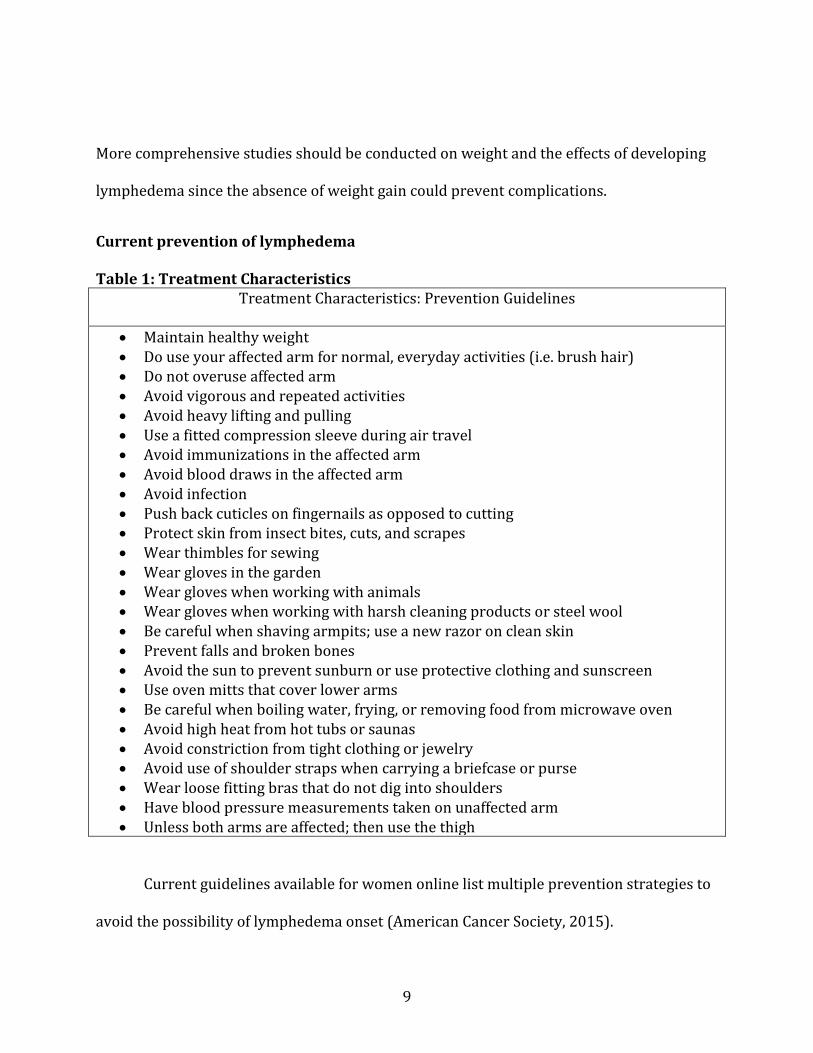
Current prevention of lymphedema Table 1: Treatment Characteristics
Treatment Characteristics: Prevention Guidelines
Maintain healthy weight Do use your affected arm for normal, everyday activities (i.e. brush hair) Do not overuse affected arm Avoid vigorous and repeated activities Avoid heavy lifting and pulling Use a fitted compression sleeve during air travel Avoid immunizations in the affected arm Avoid blood draws in the affected arm Avoid infection Push back cuticles on fingernails as opposed to cutting Protect skin from insect bites, cuts, and scrapes Wear thimbles for sewing Wear gloves in the garden Wear gloves when working with animals Wear gloves when working with harsh cleaning products or steel wool Be careful when shaving armpits; use a new razor on clean skin Prevent falls and broken bones Avoid the sun to prevent sunburn or use protective clothing and sunscreen Use oven mitts that cover lower arms Be careful when boiling water, frying, or removing food from microwave oven Avoid high heat from hot tubs or saunas Avoid constriction from tight clothing or jewelry Avoid use of shoulder straps when carrying a briefcase or purse Wear loose fitting bras that do not dig into shoulders Have blood pressure measurements taken on unaffected arm Unless both arms are affected; then use the thigh
Current guidelines available for women online list multiple prevention strategies to
avoid the possibility of lymphedema onset (American Cancer Society, 2015).
10
Providers should routinely educate women on proper precautions to prevent
lymphedema after mastectomy surgery with axillary node removal. Women are informed
that any trauma to the ispilateral arm, including burns and wounds made by the puncture
stick of a needle, can disrupt the compromised lymphatic system and increase the risk for
lymphedema (Brennan & Weitz, 1992). Although definite risk factors for lymphedema are
unclear, clinicians are encouraged to cast a wide net in order to encompass any influencing
factors from becoming a potential risk (American Cancer Society, 2015). Currently,
avoidance of performing invasive and non-invasive procedures to the affected arm is
considered prudent practice. Total avoidance of blood pressure measurements and any
procedure, including injections or venipuncture, in the ispilateral arm may attest difficult
when medical management becomes necessary.
11
PURPOSE
The primary purpose of this literature review was to examine the relationship
between blood pressure measurement and related procedures performed in the ipsilateral
arm after mastectomy as factors leading to secondary lymphedema in post-mastectomy
women. The secondary purpose was to evaluate which types of risk factors increase the
occurrence of lymphedema post mastectomy.
Understanding risk factors for developing upper extremity lymphedema post-
mastectomy is essential for assisting women with decision-making processes regarding
precautions and use or disuse of their ipsilateral arm for procedures. Research is needed
to develop evidence-based interventions to guide women about use of the ipsilateral arm
for invasive and noninvasive procedures following mastectomy with lymph node biopsy or
removal. Furthermore, evidence-based guidelines are needed for all health care providers
regarding the care of women with a risk for lymphedema following mastectomy for day-to-
day interventions requiring upper extremity access to reduce the risk of lymphedema and
for education purposes.
12
METHOD
A literature review was performed using information published from 1992 to 2015
regarding lymphedema risk in the postoperative woman. The information acquired over
the 23-year span presented a comprehensive array of literature to confirm recent
acquisitions. CINAHL, MedLine, PsychInfo, and ERIC databases were used. An initial search
using the key terms lymphedema, breast cancer, mastectomy, blood pressure, infection,
risk of lymphedema, women, post surgical, and axillary node dissection was conducted.
Exclusion criteria for this literature review included male gender, primary
lymphedema, metastases, survival, quality of life (QOL) studies, reoccurrence breast
cancer, breast conservation, lymphedema management, lymphedema only articles, and
lymphoma. The review process compared and contrasted the research studies on each
factor as it contributed to being a risk factor to secondary lymphedema. Sixteen articles
were eliminated based on the exclusion criteria.
A total of 22 articles were retrieved that examined a relationship between
secondary lymphedema and use of the ipsilateral arm for invasive and non-invasive
therapies. Sixteen studies were utilized for including relevant criteria.
Additional searches were conducted manually from the article citations, which
yielded 13 more articles of relevance, of which six were excluded due to comorbid
conditions associated with generalized lymphedema that also included the ipsilateral arm.
A total of 16 articles were analyzed for conclusive data related to the topic. Any further
information resulting from secondary lymphedema based on infection, complications, and

13
interventions performed in the ipsilateral arm was presented based on the applicability of
the obtained data (n = 51).

14
RESULTS
Of the 51 articles, no studies directly addressed the relationship between blood
pressure measurement and lymphedema development. One article discussed a significant
40% increase in lymphedema onset when procedures, like phlebotomy, were done in the
ispilateral arm (Cole, 2006). Furthermore, one article stated the invasive procedure of
repeated finger sticks by a needle directly correlated to an increased risk of lymphedema
(Brennan & Weitz, 1992).
There were 16 articles that were directly relevant to secondary lymphedema risk
factors. Six articles stated that mastectomy verses breast conservation therapy or
lumpectomy increased a woman’s risk of complications. Three articles had noticed a
correlation between chemotherapy treatment and lymphedema. The most supported
claim, with 12 articles, determined a direct link between an increased number of lymph
nodes removed during an extensive axillary lymph node dissection (ALND) and
lymphedema formation. There were 8 articles relating radiation therapy to an increased
risk of complications and also 8 articles supporting a relationship between a higher body
mass index (BMI) and lymphedema. Four studies found that an advanced diagnosis of
breast cancer led to a higher risk of lymphedema. Comorbidities played a role in relation to
lymphedema, but for the purpose of this review they have been separated into 2 main
subcategories, hypertension and diabetes mellitus. Four studies found hypertension to be
significant, while two studies mentioned diabetes as a pertinent risk factor. Age is a risk
factor, yet studies are conflicting on whether advanced age or younger age increases risk.
15
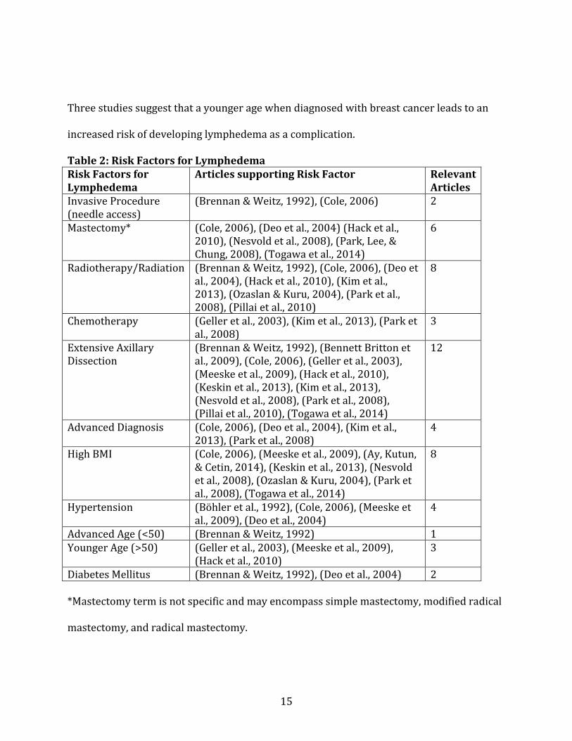
Three studies suggest that a younger age when diagnosed with breast cancer leads to an
increased risk of developing lymphedema as a complication.
Table 2: Risk Factors for Lymphedema Risk Factors for Lymphedema
Articles supporting Risk Factor Relevant Articles
Invasive Procedure (needle access)
(Brennan & Weitz, 1992), (Cole, 2006) 2
Mastectomy* (Cole, 2006), (Deo et al., 2004) (Hack et al., 2010), (Nesvold et al., 2008), (Park, Lee, & Chung, 2008), (Togawa et al., 2014)
6
Radiotherapy/Radiation (Brennan & Weitz, 1992), (Cole, 2006), (Deo et al., 2004), (Hack et al., 2010), (Kim et al., 2013), (Ozaslan & Kuru, 2004), (Park et al., 2008), (Pillai et al., 2010)
8
Chemotherapy (Geller et al., 2003), (Kim et al., 2013), (Park et al., 2008)
3
Extensive Axillary Dissection
(Brennan & Weitz, 1992), (Bennett Britton et al., 2009), (Cole, 2006), (Geller et al., 2003), (Meeske et al., 2009), (Hack et al., 2010), (Keskin et al., 2013), (Kim et al., 2013), (Nesvold et al., 2008), (Park et al., 2008), (Pillai et al., 2010), (Togawa et al., 2014)
12
Advanced Diagnosis (Cole, 2006), (Deo et al., 2004), (Kim et al., 2013), (Park et al., 2008)
4
High BMI (Cole, 2006), (Meeske et al., 2009), (Ay, Kutun, & Cetin, 2014), (Keskin et al., 2013), (Nesvold et al., 2008), (Ozaslan & Kuru, 2004), (Park et al., 2008), (Togawa et al., 2014)
8
Hypertension (Böhler et al., 1992), (Cole, 2006), (Meeske et al., 2009), (Deo et al., 2004)
4
Advanced Age (<50) (Brennan & Weitz, 1992) 1 Younger Age (>50) (Geller et al., 2003), (Meeske et al., 2009),
(Hack et al., 2010) 3
Diabetes Mellitus (Brennan & Weitz, 1992), (Deo et al., 2004) 2 *Mastectomy term is not specific and may encompass simple mastectomy, modified radical
mastectomy, and radical mastectomy.
16
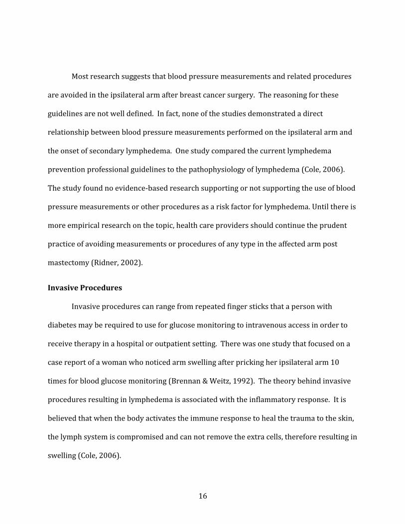
Most research suggests that blood pressure measurements and related procedures
are avoided in the ipsilateral arm after breast cancer surgery. The reasoning for these
guidelines are not well defined. In fact, none of the studies demonstrated a direct
relationship between blood pressure measurements performed on the ipsilateral arm and
the onset of secondary lymphedema. One study compared the current lymphedema
prevention professional guidelines to the pathophysiology of lymphedema (Cole, 2006).
The study found no evidence-based research supporting or not supporting the use of blood
pressure measurements or other procedures as a risk factor for lymphedema. Until there is
more empirical research on the topic, health care providers should continue the prudent
practice of avoiding measurements or procedures of any type in the affected arm post
mastectomy (Ridner, 2002).
Invasive Procedures Invasive procedures can range from repeated finger sticks that a person with
diabetes may be required to use for glucose monitoring to intravenous access in order to
receive therapy in a hospital or outpatient setting. There was one study that focused on a
case report of a woman who noticed arm swelling after pricking her ipsilateral arm 10
times for blood glucose monitoring (Brennan & Weitz, 1992). The theory behind invasive
procedures resulting in lymphedema is associated with the inflammatory response. It is
believed that when the body activates the immune response to heal the trauma to the skin,
the lymph system is compromised and can not remove the extra cells, therefore resulting in
swelling (Cole, 2006).
17
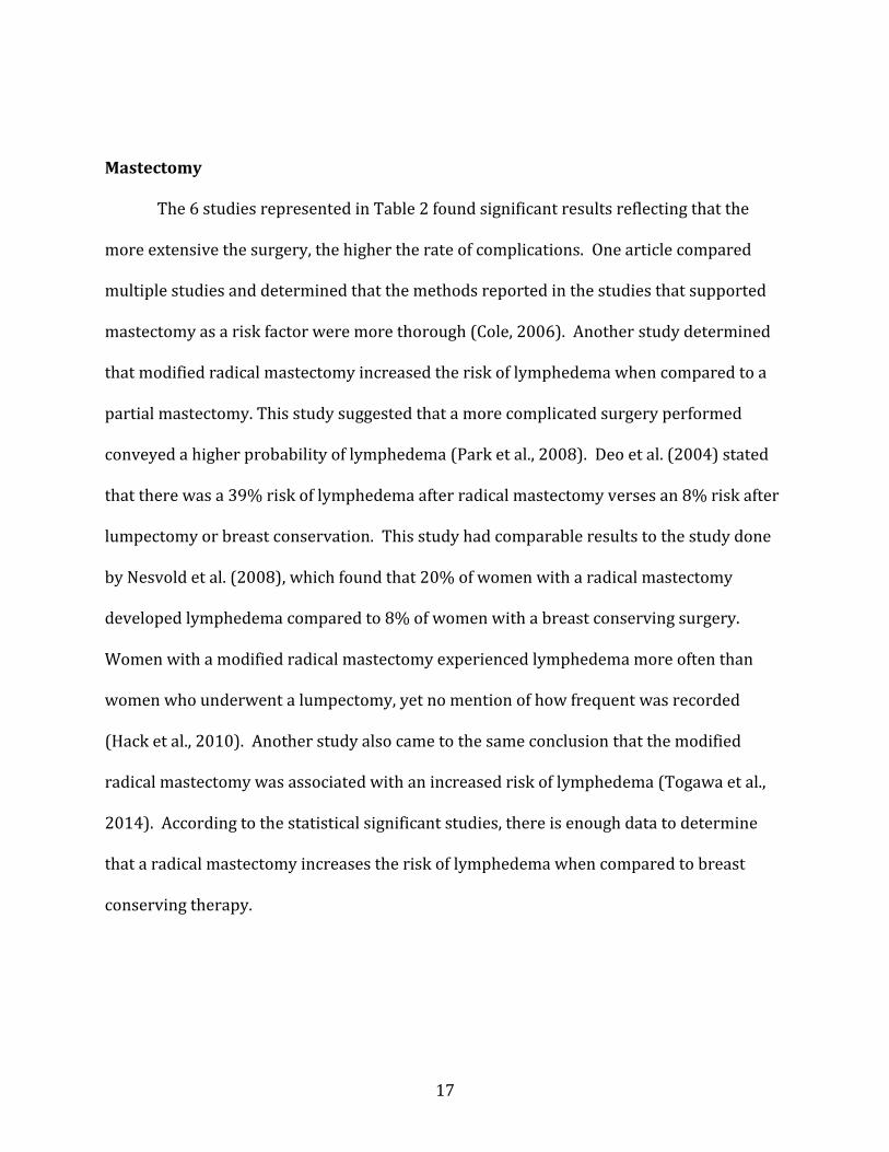
Mastectomy The 6 studies represented in Table 2 found significant results reflecting that the
more extensive the surgery, the higher the rate of complications. One article compared
multiple studies and determined that the methods reported in the studies that supported
mastectomy as a risk factor were more thorough (Cole, 2006). Another study determined
that modified radical mastectomy increased the risk of lymphedema when compared to a
partial mastectomy. This study suggested that a more complicated surgery performed
conveyed a higher probability of lymphedema (Park et al., 2008). Deo et al. (2004) stated
that there was a 39% risk of lymphedema after radical mastectomy verses an 8% risk after
lumpectomy or breast conservation. This study had comparable results to the study done
by Nesvold et al. (2008), which found that 20% of women with a radical mastectomy
developed lymphedema compared to 8% of women with a breast conserving surgery.
Women with a modified radical mastectomy experienced lymphedema more often than
women who underwent a lumpectomy, yet no mention of how frequent was recorded
(Hack et al., 2010). Another study also came to the same conclusion that the modified
radical mastectomy was associated with an increased risk of lymphedema (Togawa et al.,
2014). According to the statistical significant studies, there is enough data to determine
that a radical mastectomy increases the risk of lymphedema when compared to breast
conserving therapy.
18
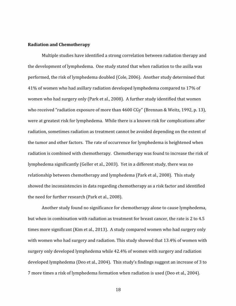
Radiation and Chemotherapy
Multiple studies have identified a strong correlation between radiation therapy and
the development of lymphedema. One study stated that when radiation to the axilla was
performed, the risk of lymphedema doubled (Cole, 2006). Another study determined that
41% of women who had axillary radiation developed lymphedema compared to 17% of
women who had surgery only (Park et al., 2008). A further study identified that women
who received “radiation exposure of more than 4600 CGy” (Brennan & Weitz, 1992, p. 13),
were at greatest risk for lymphedema. While there is a known risk for complications after
radiation, sometimes radiation as treatment cannot be avoided depending on the extent of
the tumor and other factors. The rate of occurrence for lymphedema is heightened when
radiation is combined with chemotherapy. Chemotherapy was found to increase the risk of
lymphedema significantly (Geller et al., 2003). Yet in a different study, there was no
relationship between chemotherapy and lymphedema (Park et al., 2008). This study
showed the inconsistencies in data regarding chemotherapy as a risk factor and identified
the need for further research (Park et al., 2008).
Another study found no significance for chemotherapy alone to cause lymphedema,
but when in combination with radiation as treatment for breast cancer, the rate is 2 to 4.5
times more significant (Kim et al., 2013). A study compared women who had surgery only
with women who had surgery and radiation. This study showed that 13.4% of women with
surgery only developed lymphedema while 42.4% of women with surgery and radiation
developed lymphedema (Deo et al., 2004). This study’s findings suggest an increase of 3 to
7 more times a risk of lymphedema formation when radiation is used (Deo et al., 2004).
19
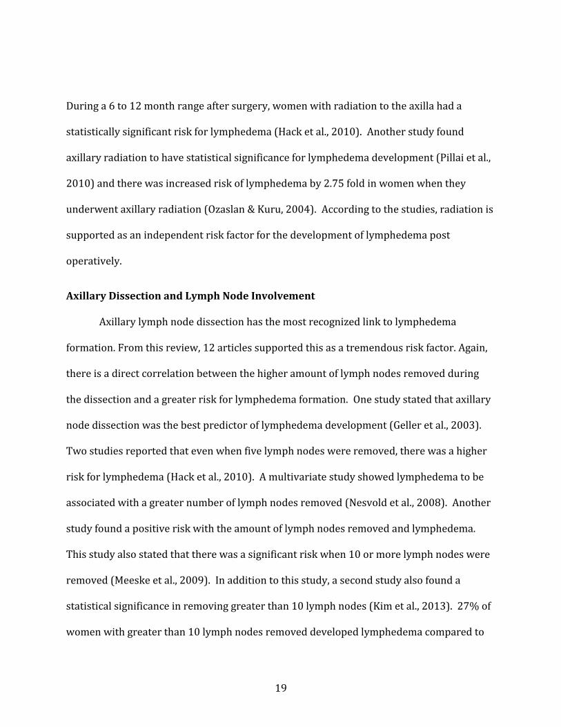
During a 6 to 12 month range after surgery, women with radiation to the axilla had a
statistically significant risk for lymphedema (Hack et al., 2010). Another study found
axillary radiation to have statistical significance for lymphedema development (Pillai et al.,
2010) and there was increased risk of lymphedema by 2.75 fold in women when they
underwent axillary radiation (Ozaslan & Kuru, 2004). According to the studies, radiation is
supported as an independent risk factor for the development of lymphedema post
operatively.
Axillary Dissection and Lymph Node Involvement Axillary lymph node dissection has the most recognized link to lymphedema
formation. From this review, 12 articles supported this as a tremendous risk factor. Again,
there is a direct correlation between the higher amount of lymph nodes removed during
the dissection and a greater risk for lymphedema formation. One study stated that axillary
node dissection was the best predictor of lymphedema development (Geller et al., 2003).
Two studies reported that even when five lymph nodes were removed, there was a higher
risk for lymphedema (Hack et al., 2010). A multivariate study showed lymphedema to be
associated with a greater number of lymph nodes removed (Nesvold et al., 2008). Another
study found a positive risk with the amount of lymph nodes removed and lymphedema.
This study also stated that there was a significant risk when 10 or more lymph nodes were
removed (Meeske et al., 2009). In addition to this study, a second study also found a
statistical significance in removing greater than 10 lymph nodes (Kim et al., 2013). 27% of
women with greater than 10 lymph nodes removed developed lymphedema compared to
20
9% of women with less than 10 lymph nodes removed (Kim et al., 2013). A multivariable
analysis determined that “the risk of lymphedema increased by 5% for each lymph node
removed” (Togawa et al., 2014, section Multivariable analyses overall, para 1).
When women received axillary lymph node dissection, this increased their risk of
lymphedema by 6.61 fold (Park et al., 2008). Another study showed a statistically
significant risk for lymphedema when women had an aggressive lymph node involvement
(pN3) (Pillai et al., 2010). When these studies are reviewed, the conclusion that the risk for
lymphedema becomes increased when there are more lymph nodes removed or there is an
extensive axillary surgery is thoroughly supported. These articles show a positive
correlation between the number of lymph nodes removed and the risk for lymphedema
development.
Advanced Diagnosis Breast cancer diagnosis is determined by the TNM staging system. This system
bases the diagnosis on the tumor size, the number of lymph nodes with cancer
involvement, and whether or not the cancer cells have spread to other parts of the body.
When the stage of the breast cancer diagnosis is high, the tumor is more aggressive and
thus women are managed with a more aggressive form of treatment to eradicate the cancer
(National Cancer Institute, 2015). An advanced diagnosis generally involves other
treatment options, such as a more complicated surgery combined with radiation and
chemotherapy (Cole, 2006). A woman with more treatments and procedures at an
advanced diagnosis would put her at a higher risk than a woman with an early diagnosis.
21
In the univariate analysis of one study, a locally advanced cancer when compared to
an early breast cancer was found to have a statistically significant risk for lymphedema
(Deo et al., 2004). Women with a higher pathologic stage (either stage II or III) had a
statistically significant increased risk of developing lymphedema (Kim et al., 2013).
Additionally, another study determined that women with stage II cancer had an increase
risk of lymphedema by 2.58 fold and women with stage III cancer had an increase risk of
lymphedema by 2.84 fold compared to women with a stage I cancer (Park et al., 2008).
From the studies in this literature review, it seems that there is a positive correlation with
lymphedema and the pathologic staging of cancer.
Body Mass Index Most studies agreed upon a high BMI correlating with an increased risk of
lymphedema (Cole, 2006), but what each study considered a high BMI varied. A few studies
determined women in the category of obese, BMI greater than 30, to have a higher risk of
lymphedema. One study determined the probability of lymphedema development to be 3
times the risk when obese (Meeske et al., 2009). This same study found overweight
women, BMI of 25-29, to have 2 times the risk of lymphedema when compared to women
with a normal BMI (Meeske et al., 2009). One study found any BMI greater than 30 to be
significant (Ay, Kutun, & Cetin, 2014). While another study observed the same significance
with having a BMI greater than 30 (Togawa et al., 2014).
Further studies defined overweight and obesity to have significance in developing
complications. This included those with a BMI of greater than 25 (Park et al., 2008). Yet
22
another study had the same significance regarding a BMI of greater than 25 (Ozaslan &
Kuru, 2004). Other multivariate studies stated that there was a significant increase in the
risk of lymphedema when BMI was considered (Nesvold et al., 2008). The relationship
between weight and lymphedema may be due to hormones located within fat tissue
(Keskin et al., 2013). Regardless of the specific BMI the studies have outlined, the end
result determines that being overweight or obese increases the risk for lymphedema.
Comorbidities
The comorbidities outlined within this literature review include hypertension
(HTN) and diabetes mellitus. The diagnosis of diabetes is a risk factor for lymphedema
secondary to breast cancer according to two studies. The first study determined diabetes
to be a risk factor due to the daily maintenance of this disease, the blood glucose
monitoring (Brennan & Weitz, 1992). The second study grouped diabetes and
hypertension together under the canopy of co-morbid conditions but there was statistical
significance found to support both as being a risk factor for complications (Deo et al.,
2004).
Cole (2006) mentioned that hypertension was a risk factor for lymphedema. One
retrospective study of 130 women with breast cancer showed a statistical significance that
women with arterial hypertension had an increased risk of developing lymphedema after
surgery (Böhler et al., 1992). Another study determined that women with hypertension
were twice as likely to develop lymphedema than women with normal blood pressure
23
(Meeske et al., 2009). Although a few studies have shown statistical significance, further
research should be conducted.
Age There were three studies that found women of a younger age (less than 50 years) to
be associated with a higher chance of lymphedema development. In one of the studies, age
was the only independent variable for lymphedema formation and it was statistically
significant, yet the reason for the high significance may have been due to the tendency for
younger women to be more apt to report symptoms (Geller et al., 2003). Another study
found that “women diagnosed before the age of 55 were nearly twice as likely to develop
lymphedema as those diagnosed at an older age” (Meeske et al., 2009, p. 386). Hack et al.
(2010), brought mention to other studies supporting that younger age played a role as a
factor for lymphedema development, but did not include this factor in their study as
significant.
On the other hand, a single study supported advanced age as a risk factor for
lymphedema. It referred to one case study in which an 85 year-old woman developed
lymphedema 30 years after her extensive surgery (Brennan & Weitz, 1992). In the article
supporting age as a significant factor, the authors have considered the reporting of the data
to be a limitation (Geller et al., 2003). Based off of these studies, age, whether advanced or
not, has not received extensive research as an independent variable in order to be
considered a significant risk factor for the formation of lymphedema.
24
DISCUSSION
As women are surviving breast cancer after surgery, they have an approximate 40%
probability of developing secondary lymphedema and its associated complications (Fu,
2014). Healthcare providers are unaware of the triggers that set off the advancement of
lymphedema and must utilize current practices based off of past research. Current prudent
practices include avoidance of procedures in the ipsilateral arm and even avoidance of hot
tubs (Breastcancer.org, 2015a)
At the time of this literature review, there are clinical trials involved in educating
women with breast cancer of the signs and symptoms of lymphedema and lymphedema
prophylaxis for women at high risk in the form of new devices (National Cancer Institute,
n.d.). These trials outline the need for further research on the cause of lymphedema for
educations purposes. Once the causes are properly identified, healthcare providers should
be better able to assist women with options regarding use or disuse of their ipsilateral arm
for procedures. In turn, women may take the necessary precautions to avoid stimulating
the process of lymphedema.
The implications of blood pressure measurements and related procedures on the
compromised arm have not been studied. Based on the articles reviewed for this thesis,
mostly retrospective studies have been conducted on this area due to the possibility of
harm to participants. In theory, blood pressure measure taken by a manual cuff may not be
as harmful as an automatic blood pressure cuff, due to the fact that an automatic cuff holds
inflation longer and at higher pressures than manual measurements (American Cancer
Society, 2015).
25
From the results of this literature review, blood pressure measurements and
procedures in the ipsilateral arm post mastectomy as a cause of lymphedema were not
supported On the other hand, having a radical mastectomy, axillary radiation, extensive
axillary dissection, having greater than 10 lymph nodes removed, and having a
pathologically staged II or III cancer are statistically significant and supported risk factors
for developing lymphedema.
26
LIMITATIONS
Several limitations were noted in this review of the literature. Initial search results
revealed numerous findings on keywords lymphedema, breast cancer, mastectomy, blood
pressure, infection, risk of lymphedema, women, post-surgical, and axillary node
dissection. However, fewer original research articles remained relevant to the purpose of
this investigation. Only 22 initial results met inclusion criteria for this review of the
literature. Search terms were expanded to include citations from initial articles in order to
provide more relevant search results. This limitation may be an indication of the relative
absence of specific and interactive causative factors for lymphedema in the ipsilateral arm
post-mastectomy; thus, an indication for future research.
Other limitations included lymphedema measured subjectively as opposed to
objectively. There was no standard determinant that women based their measurement off
of. Measurements were conducted via “self-report” and telephone interviews, not
numerical measurements or weights. Furthermore, there were no articles specifically
addressing the topic of blood pressure measurements directly causing the onset of
secondary lymphedema in the post-surgical woman. According to some, “self-report” may
act as an easier clinical tool for healthcare providers to use as opposed to another objective
measurement (Geller et al., 2003).
Further limitations may include demographics. The participants in these studies
were compliant to treatments and follow up studies. There may be women with
lymphedema who are unaccounted for due to not seeking care or reporting symptoms,
amongst other reasons.
27
CONCLUSION
The purpose of this literature review was to determine if blood pressure
measurements or invasive procedures had a direct relationship with secondary
lymphedema in the post-surgical woman. The results yielded inconclusive findings. The
cause for secondary lymphedema is not well known and appears to be multifactorial. Even
with the available technology, there is no specific diagnostic test that can determine the
cause of secondary lymphedema. The only proposed studies that could determine if blood
pressure measurements or invasive procedures had a direct correlation to secondary
lymphedema could be unethical. The reason for this would be subjecting the woman to the
irreversible harm of an incurable complication.
Secondary lymphedema in the post-surgical woman causes swelling, tightness, and
physical disfigurement of the upper extremity. There is no cure for this and symptom
management is required daily.
Specifically, research on blood pressure measurements relating to lymphedema is
lacking. A manual or an automatic blood pressure cuff might be inflated to such a high
amount that it could cause pressure to the small vessels within the ipsilateral arm. When
an axilla has been compromised with surgery and lymph node removal, there is resulting
damage to the microvasculature of the arm, which may lead to the formation of
lymphedema when pressure is applied. Finally, the compiled results of this review of the
literature showed no evidence of blood pressure measurements or other procedures
performed in the ipsilateral arm post-mastectomy; therefore, neither supporting nor
negating the cause of secondary lymphedema by blood pressure measurement.
28
Furthermore, there is insufficient evidence to suggest that procedures involving the
ipsilateral arm after axillary node dissection will have any effect on the risk of
lymphedema. A prospective, randomized double blind study has yet to be conducted to
determine if procedures on the ipsilateral arm after axillary node dissections cause
secondary lymphedema. A prototype study and further research into causes of upper
extremity lymphedema in post mastectomy women has not been conducted. Partly, this
lack of evidence is attributable to past observations, which have alluded to a potential
connection between invasive and non-invasive procedures and lymphedema (Cole, 2006).
Nonetheless, the risk of lymphedema is multi-factorial in nature even when modifiable risk
factors have been minimized.
Further research should focus on long-term and intricate retrospective studies to
determine factors associated with lymphedema. Invasive procedures should continue to be
studied. There is also a need to determine whether inflation pressures are significant when
comparing an automatic cuff verses a manual blood pressure cuff to the risk of
lymphedema. Furthermore, weight reduction and hypertension may contribute to an
increased risk of lymphedema development and further studies should be conducted to
determine any significance.
29
NURSING IMPLICATIONS
In the field of nursing, this review of the literature should serve as a reminder to ask
women if they have ever had surgery for breast cancer or if they have ever had axillary
lymph nodes removed. Some studies have shown that not all women have been educated
on the risks of lymphedema post operatively. As advocates for women, nurses have the
ability to educate and reinforce the education of preventative measures by assisting
women to find reliable resources and to ask their physicians about preventative measures.
Since causative factors of secondary lymphedema have not been perfectly identified,
nurses and other healthcare providers should follow the current preventative guidelines
determined by the American Cancer Society (2015).

30
Appendix A: Figures
31
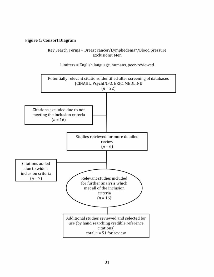
Figure 1: Consort Diagram
Key Search Terms = Breast cancer/Lymphedema*/Blood pressure Exclusions: Men
Limiters = English language, humans, peer-reviewed
Potentially relevant citations identified after screening of databases (CINAHL, PsychINFO, ERIC, MEDLINE
(n = 22)
Citations excluded due to not meeting the inclusion criteria
(n = 16)
Studies retrieved for more detailed review (n = 6)
Relevant studies included for further analysis which
met all of the inclusion criteria (n = 16)
Additional studies reviewed and selected for use (by hand searching credible reference
citations) total n = 51 for review
Citations added due to widen
inclusion criteria (n = 7)

32
Appendix B: Tables
33
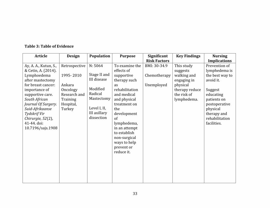
Table 3: Table of Evidence
Article Design Population Purpose Significant Risk Factors
Key Findings Nursing Implications
Ay, A. A., Kutun, S., & Cetin, A. (2014). Lymphoedema after mastectomy for breast cancer: importance of supportive care. South African Journal Of Surgery. Suid-Afrikaanse Tydskrif Vir Chirurgie, 52(2), 41-44. doi: 10.7196/sajs.1908
Retrospective 1995- 2010 Ankara Oncology Research and Training Hospital, Turkey
N: 5064
Stage II and III disease Modified Radical Mastectomy Level I, II, III axillary dissection
To examine the effects of supportive therapy such as rehabilitation and medical and physical treatment on the development of lymphedema, in an attempt to establish non-surgical ways to help prevent or reduce it.
BMI: 30-34.9 Chemotherapy Unemployed
This study suggests walking and engaging in physical therapy reduce the risk of lymphedema.
Prevention of lymphedema is the best way to avoid it. Suggest educating patients on postoperative physical therapy and rehabilitation facilities.
34
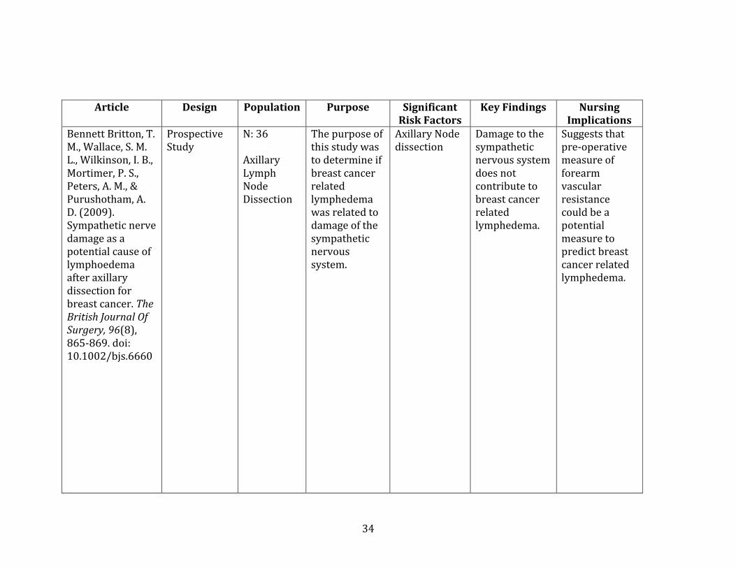
Article Design Population Purpose Significant Risk Factors
Key Findings Nursing Implications
Bennett Britton, T. M., Wallace, S. M. L., Wilkinson, I. B., Mortimer, P. S., Peters, A. M., & Purushotham, A. D. (2009). Sympathetic nerve damage as a potential cause of lymphoedema after axillary dissection for breast cancer. The British Journal Of Surgery, 96(8), 865-869. doi: 10.1002/bjs.6660
Prospective Study
N: 36 Axillary Lymph Node Dissection
The purpose of this study was to determine if breast cancer related lymphedema was related to damage of the sympathetic nervous system.
Axillary Node dissection
Damage to the sympathetic nervous system does not contribute to breast cancer related lymphedema.
Suggests that pre-operative measure of forearm vascular resistance could be a potential measure to predict breast cancer related lymphedema.
35
Article Design Population Purpose Significant Risk Factors
Key Findings Nursing Implications
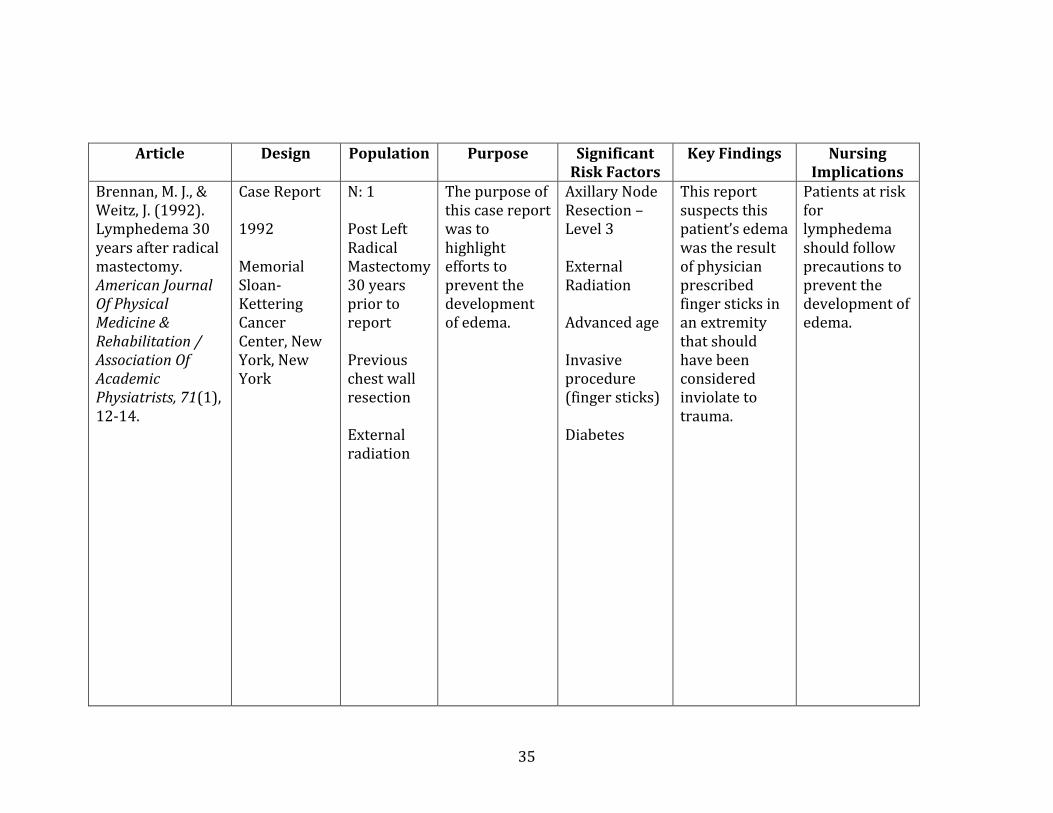
Brennan, M. J., & Weitz, J. (1992). Lymphedema 30 years after radical mastectomy. American Journal Of Physical Medicine & Rehabilitation / Association Of Academic Physiatrists, 71(1), 12-14.
Case Report 1992 Memorial Sloan-Kettering Cancer Center, New York, New York
N: 1 Post Left Radical Mastectomy 30 years prior to report Previous chest wall resection External radiation
The purpose of this case report was to highlight efforts to prevent the development of edema.
Axillary Node Resection –Level 3 External Radiation Advanced age Invasive procedure (finger sticks) Diabetes
This report suspects this patient’s edema was the result of physician prescribed finger sticks in an extremity that should have been considered inviolate to trauma.
Patients at risk for lymphedema should follow precautions to prevent the development of edema.
36
Article Design Population Purpose Significant Risk Factors
Key Findings Nursing Implications
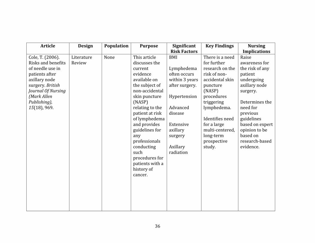
Cole, T. (2006). Risks and benefits of needle use in patients after axillary node surgery. British Journal Of Nursing (Mark Allen Publishing), 15(18), 969.
Literature Review
None This article discusses the current evidence available on the subject of non-accidental skin puncture (NASP) relating to the patient at risk of lymphedema and provides guidelines for any professionals conducting such procedures for patients with a history of cancer.
BMI Lymphedema often occurs within 3 years after surgery. Hypertension Advanced disease Extensive axillary surgery Axillary radiation
There is a need for further research on the risk of non-accidental skin puncture (NASP) procedures triggering lymphedema. Identifies need for a large multi-centered, long-term prospective study.
Raise awareness for the risk of any patient undergoing axillary node surgery. Determines the need for previous guidelines based on expert opinion to be based on research-based evidence.
37
Article Design Population Purpose Significant Risk Factors
Key Findings Nursing Implications
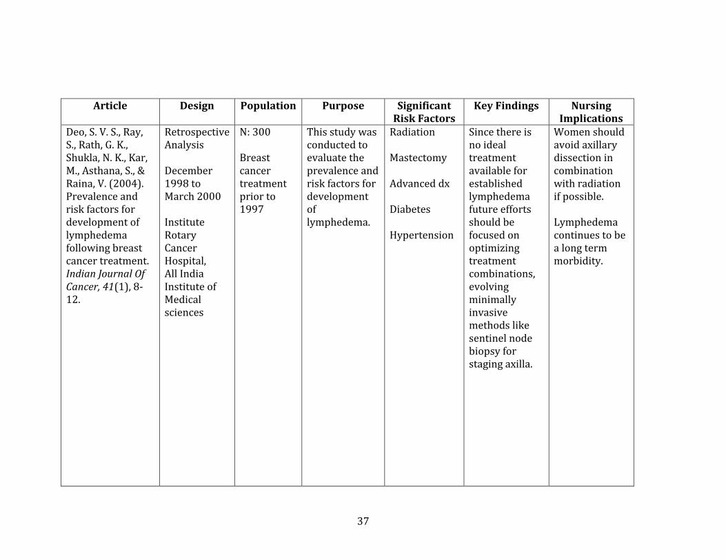
Deo, S. V. S., Ray, S., Rath, G. K., Shukla, N. K., Kar, M., Asthana, S., & Raina, V. (2004). Prevalence and risk factors for development of lymphedema following breast cancer treatment. Indian Journal Of Cancer, 41(1), 8-12.
Retrospective Analysis December 1998 to March 2000 Institute Rotary Cancer Hospital, All India Institute of Medical sciences
N: 300 Breast cancer treatment prior to 1997
This study was conducted to evaluate the prevalence and risk factors for development of lymphedema.
Radiation Mastectomy Advanced dx Diabetes Hypertension
Since there is no ideal treatment available for established lymphedema future efforts should be focused on optimizing treatment combinations, evolving minimally invasive methods like sentinel node biopsy for staging axilla.
Women should avoid axillary dissection in combination with radiation if possible. Lymphedema continues to be a long term morbidity.
38
Article Design Population Purpose Significant Risk Factors
Key Findings Nursing Implications
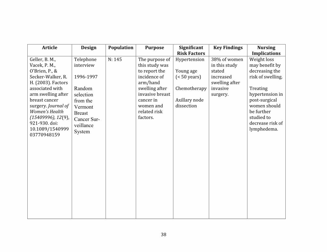
Geller, B. M., Vacek, P. M., O'Brien, P., & Secker-Walker, R. H. (2003). Factors associated with arm swelling after breast cancer surgery. Journal of Women's Health (15409996), 12(9), 921-930. doi: 10.1089/154099903770948159
Telephone interview 1996-1997 Random
selection
from the
Vermont
Breast
Cancer Sur-
veillance
System
N: 145
The purpose of this study was to report the incidence of arm/hand swelling after invasive breast cancer in women and related risk factors.
Hypertension Young age (< 50 years) Chemotherapy Axillary node dissection
38% of women in this study stated increased swelling after invasive surgery.
Weight loss may benefit by decreasing the risk of swelling. Treating hypertension in post-surgical women should be further studied to decrease risk of lymphedema.
39
Article Design Population Purpose Significant Risk Factors
Key Findings Nursing Implications
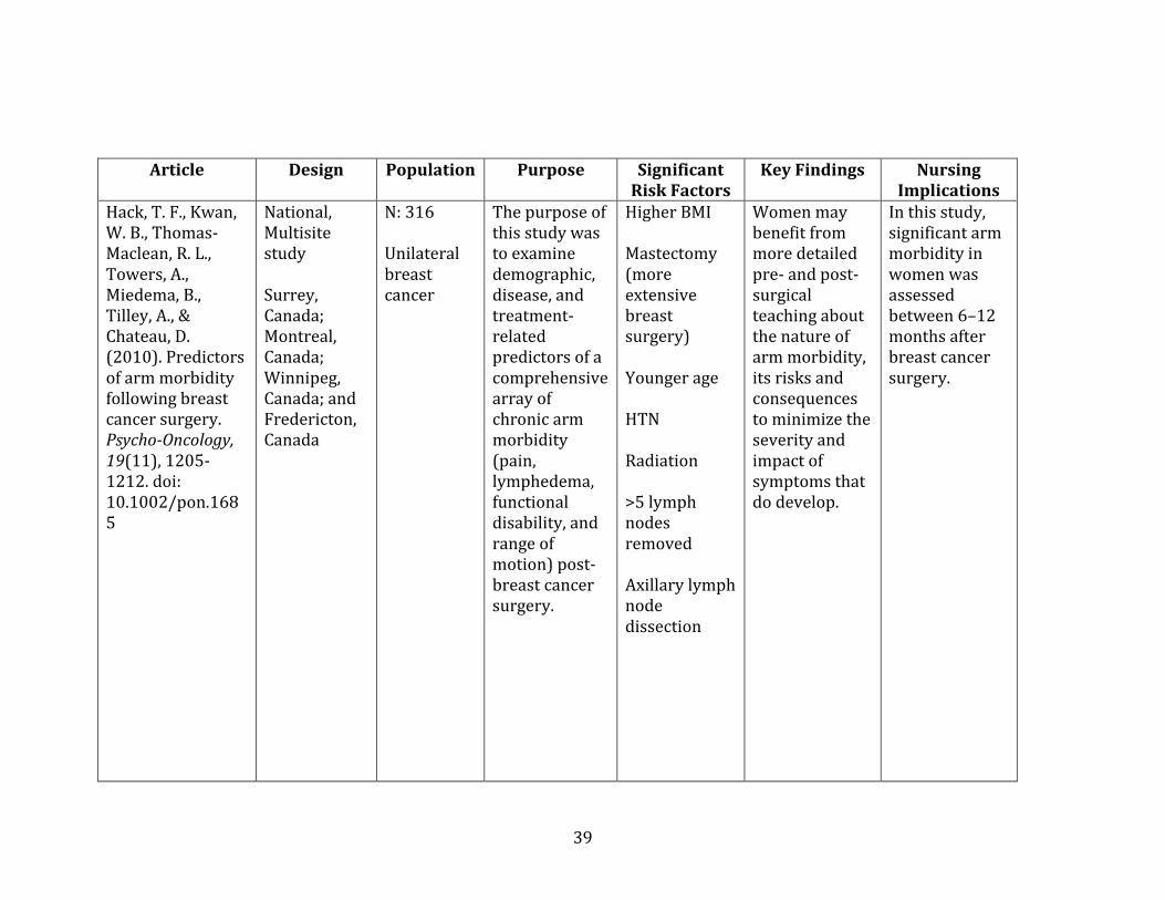
Hack, T. F., Kwan, W. B., Thomas-Maclean, R. L., Towers, A., Miedema, B., Tilley, A., & Chateau, D. (2010). Predictors of arm morbidity following breast cancer surgery. Psycho-Oncology, 19(11), 1205-1212. doi: 10.1002/pon.1685
National, Multisite study Surrey, Canada; Montreal, Canada; Winnipeg, Canada; and Fredericton, Canada
N: 316 Unilateral breast cancer
The purpose of this study was to examine demographic, disease, and treatment-related predictors of a comprehensive array of chronic arm morbidity (pain, lymphedema, functional disability, and range of motion) post-breast cancer surgery.
Higher BMI Mastectomy (more extensive breast surgery) Younger age HTN Radiation >5 lymph nodes removed Axillary lymph node dissection
Women may benefit from more detailed pre- and post-surgical teaching about the nature of arm morbidity, its risks and consequences to minimize the severity and impact of symptoms that do develop.
In this study, significant arm morbidity in women was assessed between 6–12 months after breast cancer surgery.
40
Article Design Population Purpose Significant Risk Factors
Key Findings Nursing Implications
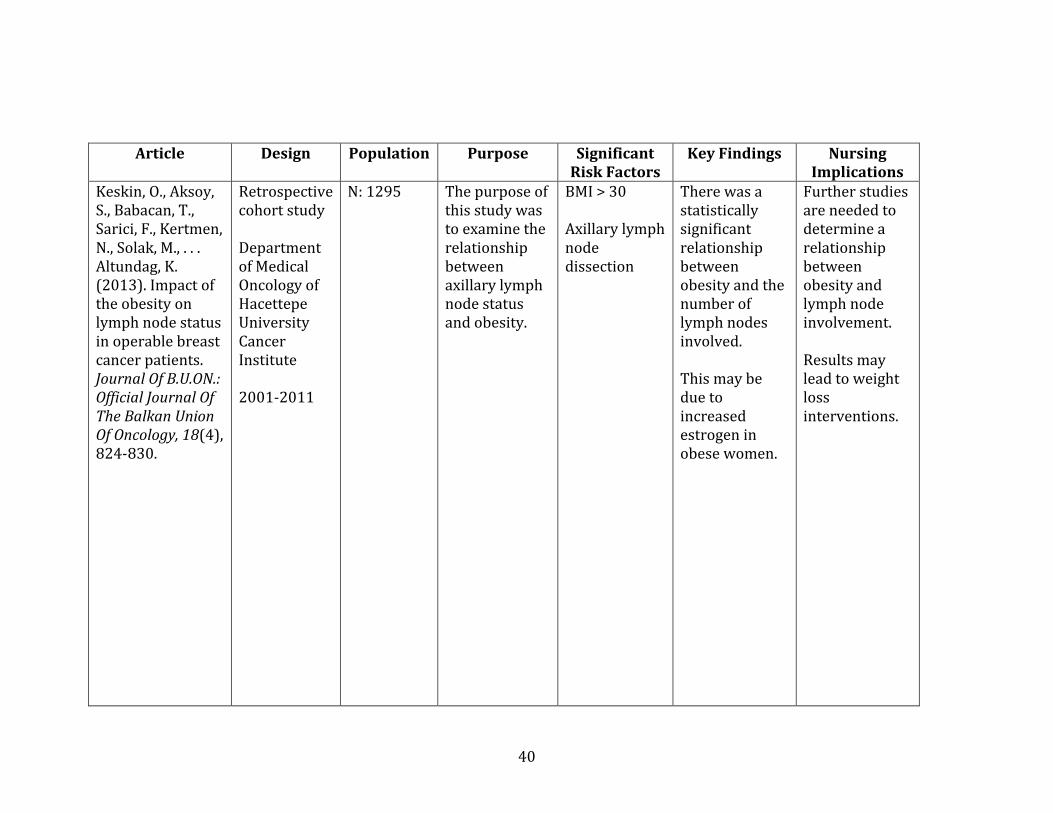
Keskin, O., Aksoy, S., Babacan, T., Sarici, F., Kertmen, N., Solak, M., . . . Altundag, K. (2013). Impact of the obesity on lymph node status in operable breast cancer patients. Journal Of B.U.ON.: Official Journal Of The Balkan Union Of Oncology, 18(4), 824-830.
Retrospective cohort study Department of Medical Oncology of Hacettepe University Cancer Institute 2001-2011
N: 1295 The purpose of this study was to examine the relationship between axillary lymph node status and obesity.
BMI > 30 Axillary lymph node dissection
There was a statistically significant relationship between obesity and the number of lymph nodes involved. This may be due to increased estrogen in obese women.
Further studies are needed to determine a relationship between obesity and lymph node involvement. Results may lead to weight loss interventions.
41
Article Design Population Purpose Significant Risk Factors
Key Findings Nursing Implications
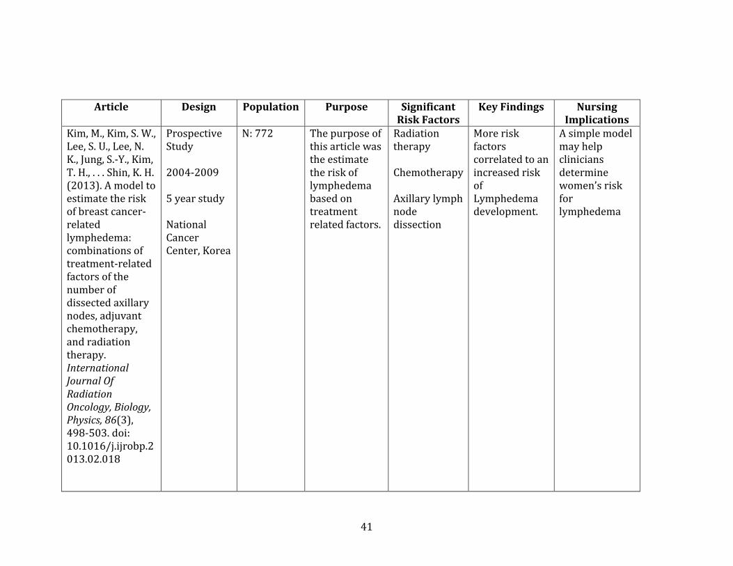
Kim, M., Kim, S. W., Lee, S. U., Lee, N. K., Jung, S.-Y., Kim, T. H., . . . Shin, K. H. (2013). A model to estimate the risk of breast cancer-related lymphedema: combinations of treatment-related factors of the number of dissected axillary nodes, adjuvant chemotherapy, and radiation therapy. International Journal Of Radiation Oncology, Biology, Physics, 86(3), 498-503. doi: 10.1016/j.ijrobp.2013.02.018
Prospective Study 2004-2009 5 year study National Cancer Center, Korea
N: 772 The purpose of this article was the estimate the risk of lymphedema based on treatment related factors.
Radiation therapy Chemotherapy Axillary lymph node dissection
More risk factors correlated to an increased risk of Lymphedema development.
A simple model may help clinicians determine women’s risk for lymphedema
42
Article Design Population Purpose Significant Risk Factors
Key Findings Nursing Implications
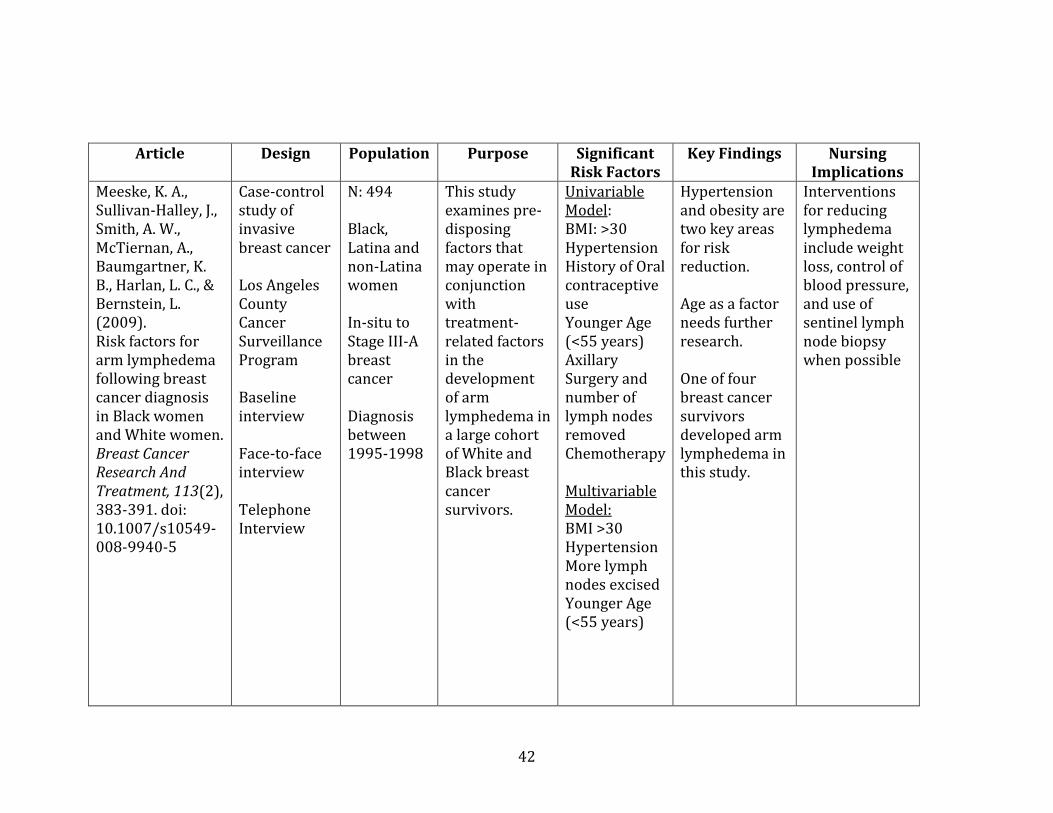
Meeske, K. A., Sullivan-Halley, J., Smith, A. W., McTiernan, A., Baumgartner, K. B., Harlan, L. C., & Bernstein, L. (2009). Risk factors for arm lymphedema following breast cancer diagnosis in Black women and White women. Breast Cancer Research And Treatment, 113(2), 383-391. doi: 10.1007/s10549-008-9940-5
Case-control study of invasive breast cancer Los Angeles County Cancer Surveillance Program Baseline interview Face-to-face interview Telephone Interview
N: 494 Black, Latina and non-Latina women In-situ to Stage III-A breast cancer Diagnosis between 1995-1998
This study examines pre-disposing factors that may operate in conjunction with treatment-related factors in the development of arm lymphedema in a large cohort of White and Black breast cancer survivors.
Univariable Model: BMI: >30 Hypertension History of Oral contraceptive use Younger Age (<55 years) Axillary Surgery and number of lymph nodes removed Chemotherapy Multivariable Model: BMI >30 Hypertension More lymph nodes excised Younger Age (<55 years)
Hypertension and obesity are two key areas for risk reduction. Age as a factor needs further research. One of four breast cancer survivors developed arm lymphedema in this study.
Interventions for reducing lymphedema include weight loss, control of blood pressure, and use of sentinel lymph node biopsy when possible
43
Article Design Population Purpose Significant Risk Factors
Key Findings Nursing Implications
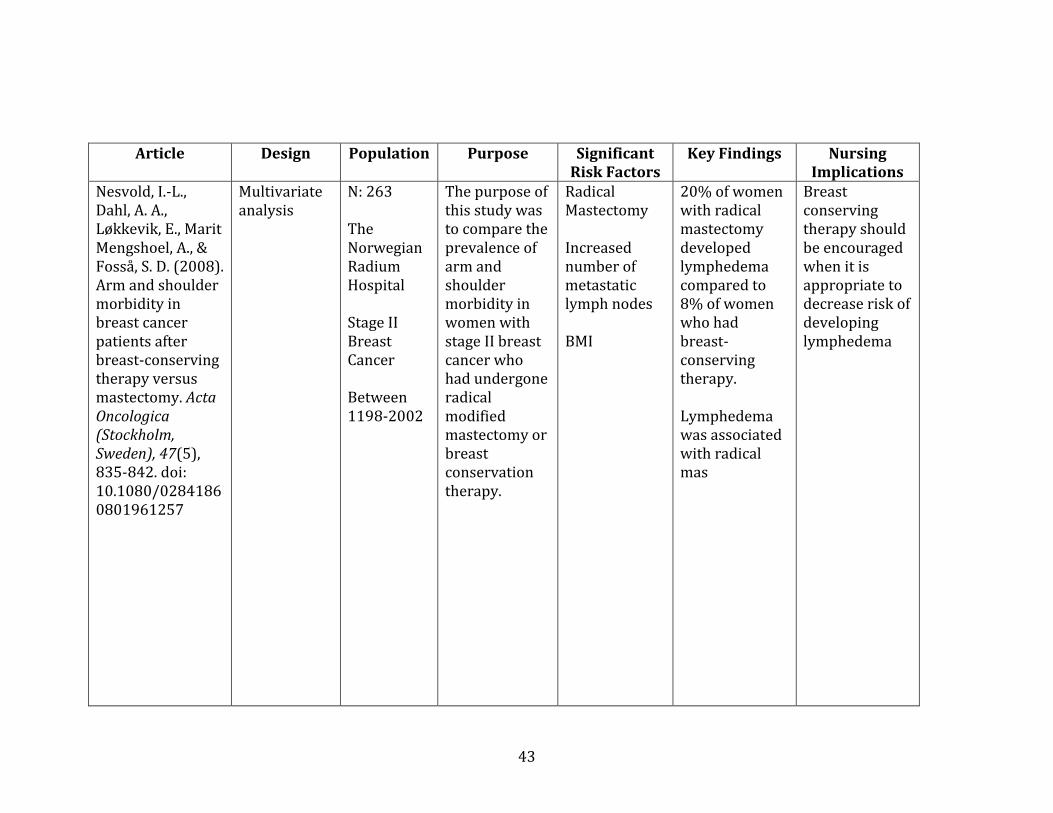
Nesvold, I.-L., Dahl, A. A., Løkkevik, E., Marit Mengshoel, A., & Fosså, S. D. (2008). Arm and shoulder morbidity in breast cancer patients after breast-conserving therapy versus mastectomy. Acta Oncologica (Stockholm, Sweden), 47(5), 835-842. doi: 10.1080/02841860801961257
Multivariate analysis
N: 263 The Norwegian Radium Hospital Stage II Breast Cancer Between 1198-2002
The purpose of this study was to compare the prevalence of arm and shoulder morbidity in women with stage II breast cancer who had undergone radical modified mastectomy or breast conservation therapy.
Radical Mastectomy Increased number of metastatic lymph nodes BMI
20% of women with radical mastectomy developed lymphedema compared to 8% of women who had breast-conserving therapy. Lymphedema was associated with radical mas
Breast conserving therapy should be encouraged when it is appropriate to decrease risk of developing lymphedema
44
Article Design Population Purpose Significant Risk Factors
Key Findings Nursing Implications
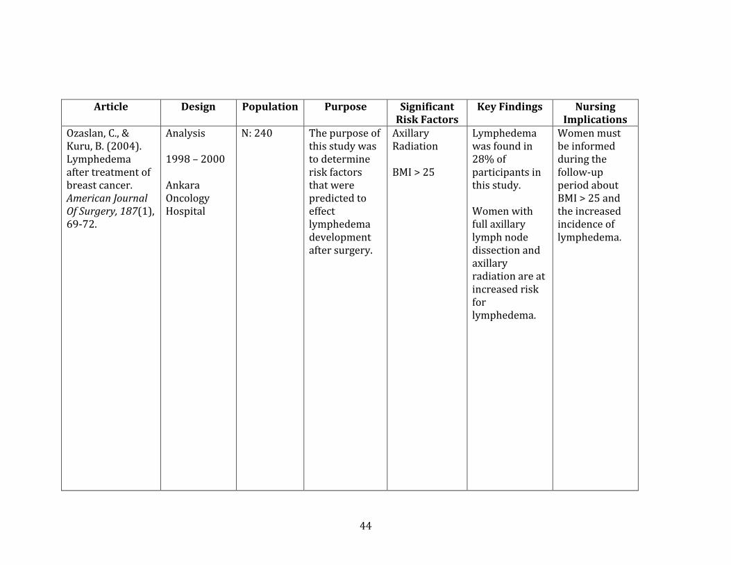
Ozaslan, C., & Kuru, B. (2004). Lymphedema after treatment of breast cancer. American Journal Of Surgery, 187(1), 69-72.
Analysis 1998 – 2000 Ankara Oncology Hospital
N: 240 The purpose of this study was to determine risk factors that were predicted to effect lymphedema development after surgery.
Axillary Radiation BMI > 25
Lymphedema was found in 28% of participants in this study. Women with full axillary lymph node dissection and axillary radiation are at increased risk for lymphedema.
Women must be informed during the follow-up period about BMI > 25 and the increased incidence of lymphedema.
45
Article Design Population Purpose Significant Risk Factors
Key Findings Nursing Implications
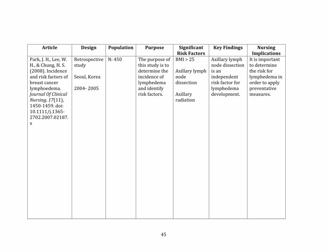
Park, J. H., Lee, W. H., & Chung, H. S. (2008). Incidence and risk factors of breast cancer lymphoedema. Journal Of Clinical Nursing, 17(11), 1450-1459. doi: 10.1111/j.1365-2702.2007.02187.x
Retrospective study Seoul, Korea 2004- 2005
N: 450 The purpose of this study is to determine the incidence of lymphedema and identify risk factors.
BMI > 25 Axillary lymph node dissection Axillary radiation
Axillary lymph node dissection is an independent risk factor for lymphedema development.
It is important to determine the risk for lymphedema in order to apply preventative measures.
46
Article Design Population Purpose Significant Risk Factors
Key Findings Nursing Implications
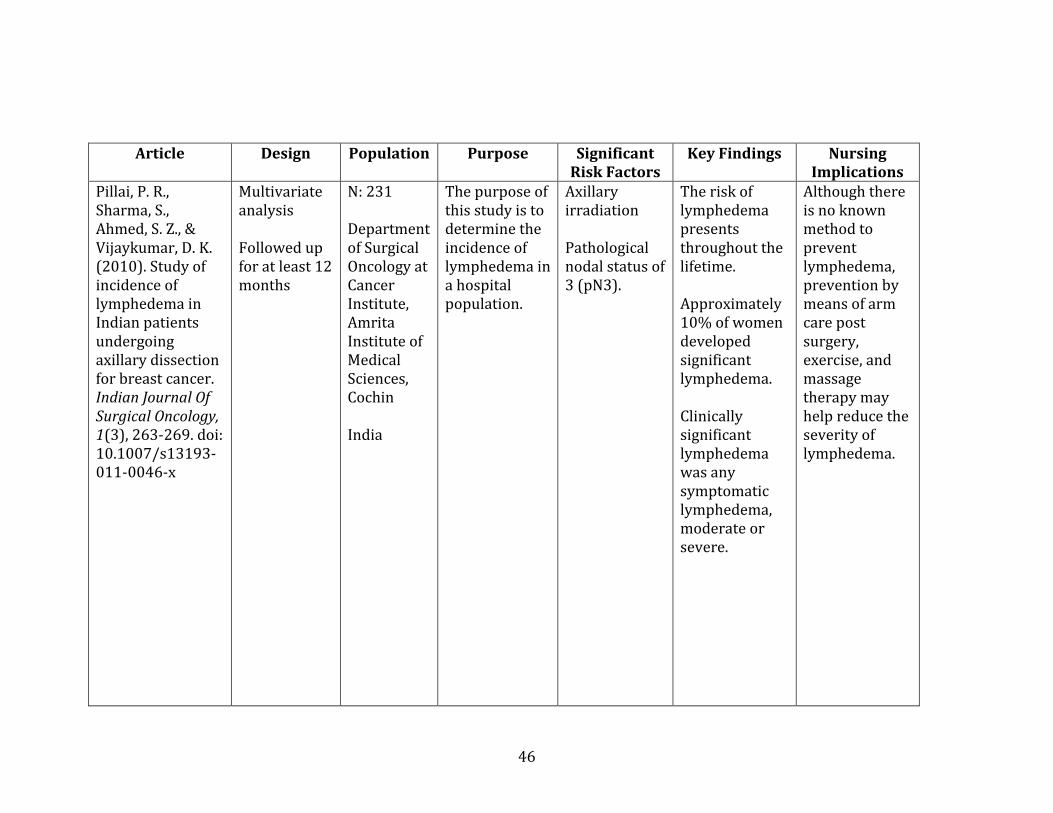
Pillai, P. R., Sharma, S., Ahmed, S. Z., & Vijaykumar, D. K. (2010). Study of incidence of lymphedema in Indian patients undergoing axillary dissection for breast cancer. Indian Journal Of Surgical Oncology, 1(3), 263-269. doi: 10.1007/s13193-011-0046-x
Multivariate analysis Followed up for at least 12 months
N: 231 Department of Surgical Oncology at Cancer Institute, Amrita Institute of Medical Sciences, Cochin India
The purpose of this study is to determine the incidence of lymphedema in a hospital population.
Axillary irradiation Pathological nodal status of 3 (pN3).
The risk of lymphedema presents throughout the lifetime. Approximately 10% of women developed significant lymphedema. Clinically significant lymphedema was any symptomatic lymphedema, moderate or severe.
Although there is no known method to prevent lymphedema, prevention by means of arm care post surgery, exercise, and massage therapy may help reduce the severity of lymphedema.
47
Article Design Population Purpose Significant Risk Factors
Key Findings Nursing Implications
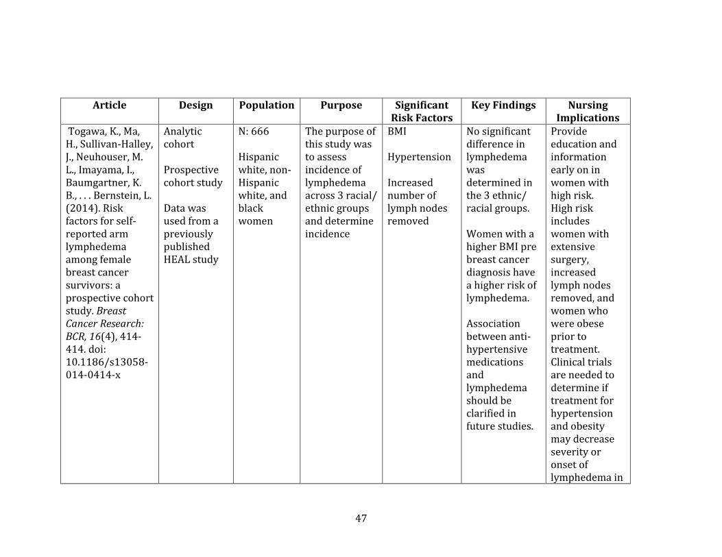
Togawa, K., Ma, H., Sullivan-Halley, J., Neuhouser, M. L., Imayama, I., Baumgartner, K. B., . . . Bernstein, L. (2014). Risk factors for self-reported arm lymphedema among female breast cancer survivors: a prospective cohort study. Breast Cancer Research: BCR, 16(4), 414-414. doi: 10.1186/s13058-014-0414-x
Analytic cohort Prospective cohort study Data was used from a previously published HEAL study
N: 666 Hispanic white, non-Hispanic white, and black women
The purpose of this study was to assess incidence of lymphedema across 3 racial/ ethnic groups and determine incidence
BMI Hypertension Increased number of lymph nodes removed
No significant difference in lymphedema was determined in the 3 ethnic/ racial groups. Women with a higher BMI pre breast cancer diagnosis have a higher risk of lymphedema. Association between anti-hypertensive medications and lymphedema should be clarified in future studies.
Provide education and information early on in women with high risk. High risk includes women with extensive surgery, increased lymph nodes removed, and women who were obese prior to treatment. Clinical trials are needed to determine if treatment for hypertension and obesity may decrease severity or onset of lymphedema in

48
Article Design Population Purpose Significant Risk Factors
Key Findings Nursing Implications
Continued post operative women.
49
REFERENCES
American Cancer Society, (2013). Cancer facts and figures 2013. Retrieved from
http://www.cancer.org/acs/groups/content/@epidemiologysurveilance/document
s/document/acspc-036845.pdf
American Cancer Society, (2015). Lymphedema: What every women with breast cancer
should know. Retrieved from
http://www.cancer.org/acs/groups/cid/documents/webcontent/002876-pdf.pdf
Armer, J. & Stewart, B. (2010). Post-breast cancer lymphedema: Incidence increases from
12 to 30 to 60 months. Lymphology, 43: 118-127.
Ay, A. A., Kutun, S., & Cetin, A. (2014). Lymphoedema after mastectomy for breast cancer:
Importance of supportive care. South African Journal of Surgery. Suid-Afrikaanse
Tydskrif Vir Chirurgie, 52(2), 41-44. doi: 10.7196/sajs.1908
Bates, D. O., Levick, J. R., & Mortimer, P. S. (1994). Starling pressures in the human arm and
their alteration in postmastectomy oedema. The Journal of Physiology, 477 ( Pt 2),
355-363.
Bates, D. O., Levick, J. R. & Mortimer, P.S. (1992). Subcutaneous interstitial fluid pressure
and arm volume in lymphoedema. International Journal of Microcirculation Clinical
and Experimental, 11, 359-373.
50
Beaulac S., McNair. L., Scott T., LaMorte, W., & Kavanah, M., (2002). Lymphedema and
quality of life in survivors of early-stage breast cancer. Archives of Surgery, 137(11),
1253–1257. doi: 10.1001/archsurg.137.11.1253.
Bennett Britton, T. M., Wallace, S. M. L., Wilkinson, I. B., Mortimer, P. S., Peters, A. M., &
Purushotham, A. D. (2009). Sympathetic nerve damage as a potential cause of
lymphoedema after axillary dissection for breast cancer. The British Journal of
Surgery, 96(8), 865-869. doi: 10.1002/bjs.6660
Böhler, F. K., Rhomberg, W., & Doringer, W. (1992). [Hypertension as risk factor for
increased rate of side effects in the framework of breast carcinoma irradiation].
Strahlentherapie Und Onkologie: Organ Der Deutschen Röntgengesellschaft ... [Et Al],
168(6), 344-349.
Breastcancer.org. (2014). Mastectomy vs. Lumpectomy.
Breastcancer.org. (2015a, March 25, 2014). from
http://www.breastcancer.org/treatment/lymphedema/reduce_risk/after
Breastcancer.org. (2015b, May 16, 2013). What is mastectomy?. Retrieved from
http://www.breastcancer.org/treatment/surgery/mastectomy/what_is
Brennan, M. J., & Weitz, J. (1992). Lymphedema 30 years after radical mastectomy.
American Journal of Physical Medicine & Rehabilitation / Association of Academic
Physiatrists, 71(1), 12-14.
Casley-Smith, J. R. (1994). Measuring and representing peripheral oedema and its
alterations. Lymphology, 27(2), 56-70.
51
Clark, B., Sitzia, J., & Harlow, W. (2005) Incidence and risk of arm oedema following
treatment for breast cancer: A three-year follow up study. QJM 98(5): 343-8
Cole, T. (2006). Risks and benefits of needle use in patients after axillary node surgery.
British Journal of Nursing (Mark Allen Publishing), 15(18), 969.
Deo, S. V. S., Ray, S., Rath, G. K., Shukla, N. K., Kar, M., Asthana, S., & Raina, V. (2004).
Prevalence and risk factors for development of lymphedema following breast cancer
treatment. Indian Journal of Cancer, 41(1), 8-12.
DiSipio, T., Rye, S., Newman, B., & Hayes, S. (2013). Incidence of unilateral arm
lymphoedema after breast cancer: A systematic review and meta-analysis. The
Lancet. Oncology, 14(6), 500-515. doi: 10.1016/S1470-2045(13)70076-7
Edwards, T. L. (2000). Prevalence and aetiology of lymphoedema after breast cancer
treatment in southern Tasmania. The Australian And New Zealand Journal of Surgery,
70(6), 412-418. doi: 10.5306/wjco.v5.i3.241
Fu, M. (2014). Breast cancer-related lymphedema: Symptoms, diagnosis, risk reduction,
and management. World Journal of Clinical Oncology. 5(3): 241-247.
Geller, B. M., Vacek, P. M., O'Brien, P., & Secker-Walker, R. H. (2003). Factors associated with
arm swelling after breast cancer surgery. Journal of Women's Health (15409996),
12(9), 921-930. doi: 10.1089/154099903770948159
Hack, T. F., Kwan, W. B., Thomas-Maclean, R. L., Towers, A., Miedema, B., Tilley, A., &
Chateau, D. (2010). Predictors of arm morbidity following breast cancer surgery.
Psycho-Oncology, 19(11), 1205-1212. doi: 10.1002/pon.1685
52
Harris, S. R., Hugi, M. R., Olivotto, I. A., & Levine, M. (2001). Clinical practice guidelines for
the care and treatment of breast cancer: 11. Lymphedema. CMAJ: Canadian Medical
Association Journal = Journal De L'association Medicale Canadienne, 164(2), 191-199.
Johansson K, Ohlsson. K., Ingvar C, Albertsson, M., & Ekdahl, C. (2002). Factors associated
with the development of arm lymphedema following breast cancer treatment: A
match pair case–control study. . Lymphology, 35, 59–71.
Keskin, O., Aksoy, S., Babacan, T., Sarici, F., Kertmen, N., Solak, M., . . . Altundag, K. (2013).
Impact of the obesity on lymph node status in operable breast cancer patients.
Journal of B.U.ON.: Official Journal of The Balkan Union of Oncology, 18(4), 824-830.
Kiel, K. D., & Rademacker, A. W. (1996). Early-stage breast cancer: Arm edema after wide
excision and breast irradiation. Radiology, 198(1), 279-283.
Kim, M., Kim, S. W., Lee, S. U., Lee, N. K., Jung, S.-Y., Kim, T. H., . . . Shin, K. H. (2013). A model
to estimate the risk of breast cancer-related lymphedema: Combinations of
treatment-related factors of the number of dissected axillary nodes, adjuvant
chemotherapy, and radiation therapy. International Journal of Radiation Oncology,
Biology, Physics, 86(3), 498-503. doi: 10.1016/j.ijrobp.2013.02.018
Kissan M, Queci della Rovere, G., Easton D, & Westbury G. (1986). Risk of lymphedema
following the treatment of breast cancer. Brittish Journal of Surgery, 73, 580.
McCredie, M. R., Dite, G. S., Porter, L., Maskiell, J., Giles, G. G., Phillips, K. A., . . . Hopper, J. L.
(2001). Prevalence of self-reported arm morbidity following treatment for breast
cancer in the Australian Breast Cancer Family Study. Breast (Edinburgh, Scotland),
10(6), 515-522.
53
Meeske, K. A., Sullivan-Halley, J., Smith, A. W., McTiernan, A., Baumgartner, K. B., Harlan, L.
C., & Bernstein, L. (2009). Risk factors for arm lymphedema following breast cancer
diagnosis in Black women and White women. Breast Cancer Research And
Treatment, 113(2), 383-391. doi: 10.1007/s10549-008-9940-5
National Cancer Institute, (n.d.). Clinical trials search results. Retrieved from
http://www.cancer.gov/aboutcancer/treatment/clinicaltrials/search/results?proto
colsearchid=6626363&vers=1
National Cancer Institute. (2014). Lymphedema. Retrieved 11/17/14, from
http://www.cancer.gov/cancertopics/pdq/supportivecare/lymphedema/healthpro
fessional
National Cancer Institute, (2014). SEER Cancer Statistics Factsheets: Breast Cancer.
Retrieved November 18, 2014. Retrieved from
http://seer.cancer.gov/statfacts/html/breast.html
National Cancer Institute, (2015). Cancer staging. Retrieved from
http://www.cancer.gov/about-cancer/diagnosis-staging/staging/staging-fact-sheet
National Institute of Health, (n.d. ). Body mass index table. Retrieved from
http://www.nhlbi.nih.gov/health/educational/lose_wt/BMI/bmi_tbl.pdf
National Lymphedema Network, (2015). What causes lymphedema?. Retrieved from
http://www.lymphnet.org/le-faqs/what-is-lymphedema/what-causes-lymphedema
Nesvold, I., Dahl A., Løkkevik. E., Marit Mengshoel, A., & Fosså, S. (2008). Arm and shoulder
morbidity in breast cancer patients after breast-conserving therapy versus
54
mastectomy. Acta Oncologica (Stockholm, Sweden), 47(5), 835-842. doi:
10.1080/02841860801961257
Norman, S. A., Miller, L. T., Erikson, H. B., Norman, M. F., & McCorkle, R. (2001).
Development and validation of a telephone questionnaire to characterize
lymphedema in women treated for breast cancer. Physical Therapy, 81(6), 1192-
1205.
Ozaslan, C., & Kuru, B. (2004). Lymphedema after treatment of breast cancer. American
Journal of Surgery, 187(1), 69-72.
Park, J. H., Lee, W. H., & Chung, H. S. (2008). Incidence and risk factors of breast cancer
lymphoedema. Journal of Clinical Nursing, 17(11), 1450-1459. doi: 10.1111/j.1365-
2702.2007.02187.x
Paskett ED, N. M., McCoy TP et al. (2007). The epidemiology of arm and hand swelling in
premenopausal breast cancer survivors. . Cancer Epidemiol Biomarkers Prev, 16,
775–782.
Petrek, J. A., Senie, R. T., Peters, M., & Rosen, P. P. (2001). Lymphedema in a cohort of breast
carcinoma survivors 20 years after diagnosis. Cancer, 92(6), 1368-1377.
Pillai, P. R., Sharma, S., Ahmed, S. Z., & Vijaykumar, D. K. (2010). Study of incidence of
lymphedema in Indian patients undergoing axillary dissection for breast cancer.
Indian Journal of Surgical Oncology, 1(3), 263-269. doi: 10.1007/s13193-011-0046-x
Ridner, S. H. (2002). Breast cancer lymphedema: pathophysiology and risk reduction
guidelines. Oncology Nursing Forum, 29(9), 1285-1293. doi: 10.1188/02.onf.1285-
1293
55
Segerström, K., Bjerle, P., Graffman, S., & Nyström, A. (1992). Factors that influence the
incidence of brachial oedema after treatment of breast cancer. Scandinavian Journal
of Plastic and Reconstructive Surgery and Hand Surgery / Nordisk Plastikkirurgisk
Forening [And] Nordisk Klubb For Handkirurgi, 26(2), 223-227.
Soran A, D’Angelo, G., Begovic M, Ardic, F., Harlak, A., Wieand, S., Vogel, V., & Johnson, R.
(2006). Breast cancer-related lymphedema – What are the significant predictors and
how do they affect the severity of lymphedema? . The Breast Journal, 12, 536–543.
Tengrup, I., Tennvall-Nittby, L., Christiansson, I., & Laurin, M. (2000). Arm morbidity after
breast-conserving therapy for breast cancer. Acta Oncologica (Stockholm, Sweden),
39(3), 393-397.
Togawa, K., Ma, H., Sullivan-Halley, J., Neuhouser, M. L., Imayama, I., Baumgartner, K. B., . . .
Bernstein, L. (2014). Risk factors for self-reported arm lymphedema among female
breast cancer survivors: A prospective cohort study. Breast Cancer Research: BCR,
16(4), 414-414. doi: 10.1186/s13058-014-0414-x
Werner RS, M. B., Petrek J et al (1991). Arm edema in conservatively managed breast
cancer: Obesity is a major predictive factor. Radiology, 180, 177–184.