Embed Size (px)
Citation preview

The role of a serralysin PrtA system in the
infection mechanism of an entomopathogen,
Photorhabdus
GABRIELLA FELFÖLDI
Eötvös Loránd University, Doctorate School of Biology
Head: Prof. Anna Erdei, CMHAS
Structural Biochemistry PhD Program
Head: Prof. László Gráf, MHAS
Supervisor:
Dr. István Venekei, associate professor, PhD
Eötvös Loránd University, Department of Biochemistry
2010

2
1. Table of content
1. Table of content ................................................................................................................ 2
2. Acknowledgements ........................................................................................................... 6
3. Abbreviations .................................................................................................................... 7
4. Introduction ....................................................................................................................... 8
4.1 Why an insect-pathogen infection model ...................................................................... 9
4.2 Manduca sexta: as the host in an infection model system ............................................ 9
4.3 Defense system of insects ............................................................................................. 9
4.3.1 Immune recognition ............................................................................................ 10
4.3.2 Cellular immune response ................................................................................... 10
4.3.3 Humoral immune response ................................................................................. 11
4.4 Photorhabdus as the ideal pathogen in an infection model system ............................ 16
4.4.1 Photorhabdus lives in symbiotic complexes with nematodes in the nature ........ 17
4.4.2 An impressive repertoire of virulence factors–strategy of “over-killing” ........... 19
4.4.2.1 Toxins ................................................................................................................. 19
4.4.2.2 Proteases ............................................................................................................. 21
4.5 Virulence factors as tools of investigation .................................................................. 22
4.6 Serralysisns as putative virulence factors ................................................................... 23
5. Goals of my thesis work.................................................................................................. 25
6. Materials and Methods .................................................................................................... 26
6.1 Material ....................................................................................................................... 26
6.1.1 Bacterium strains and culturing; preparation of bacteria for insect injection ..... 26
6.1.2 Insects; preparation of hemolymph plasma and fatbody samples ....................... 27
6.2 Methods....................................................................................................................... 28
6.2.1 Protease detection during culture growth and insect infection ........................... 28
6.2.1.1 Polyacrylamide gel electrophoresis and zymography ......................................... 28
6.2.1.2 Enzyme activity measurement ............................................................................ 28
6.2.1.3 PrtA purification ................................................................................................. 29
6.2.2 Separation and analysis of proteins cleaved by PrtA .......................................... 29
6.2.2.1 Preparation of “type” fractions from hemolymph ............................................... 29
6.2.2.2 Partial purification of PAT proteins .................................................................... 29
6.2.2.3 Purification of expressed Serpin-1 proteins ........................................................ 30
6.2.2.4 Blotting and N-terminal sequencing ................................................................... 31
6.2.2.5 Digestion of hemolymph and PAT proteins with PrtA ....................................... 31
6.2.3 RNA interference (RNAi) ................................................................................... 32

3
6.2.3.1 Total RNA isolation............................................................................................ 32
6.2.3.2 Generation of dsRNA of SPH-3 ......................................................................... 33
6.2.3.3 Insect injection protocol ..................................................................................... 34
6.2.3.4 RT-PCRs ............................................................................................................. 35
6.2.4 Examination of the effect of RNAi of SPH-3 in Manduca sexta ....................... 37
6.2.4.1 Western blotting ................................................................................................. 37
6.2.4.2 Mortality bioassay .............................................................................................. 37
6.2.4.3 Effect of RNAi knockdown of SPH-3 on PO activity by visual examination of
hemolymph ......................................................................................................................... 37
6.2.4.4 Phenoloxidase (PO) activity measurement ......................................................... 38
6.2.4.5 Nodule formation ................................................................................................ 38
6.2.4.6 Pathogen growth in vitro .................................................................................... 39
7. Results ............................................................................................................................ 40
7.1 Comparison of proteolytic activities produced by different Photorhabdus strains .... 40
7.1.1 Measuring enzymatic activity in culture ............................................................. 40
7.1.2 Enzyme activity measurement with chromogen oligopeptide substrate ............. 42
7.2 Measuring protease activity during infection ............................................................. 43
7.2.1 Infection of Galleria mellonella larvae with different Photorhabdus strains ..... 43
7.3 Investigation of natural substrates of PrtA ................................................................. 44
7.4 Isolation and identification of PAT proteins from Manduca sexta hemolymph ........ 46
7.4.1 Purification of PAT-110 ..................................................................................... 47
7.4.2 Purification of PAT-41 ....................................................................................... 50
7.4.3 Purification of PAT-52 protein ........................................................................... 51
7.4.4 Determination of N-terminal sequences of PAT proteins .................................. 52
7.4.5 Analysis of the cleavage products of PAT proteins ............................................ 53
7.5 Identification of the function of Serine Protease Homologue-3 (SPH-3) ................... 55
7.5.1 Induction and RNAi-mediated knockdown of SPH-3 in M. sexta ..................... 55
7.5.2 Mortality bioassay .............................................................................................. 57
7.5.3 SPH-3 is required for prophenoloxidase (PPO) synthesis .................................. 58
7.5.4 SPH-3 is required for nodule formation ............................................................. 61
7.5.5 SPH-3 is required for the synthesis of antimicrobial effectors but not of recognition proteins ............................................................................................................ 62
7.5.6 Knock-down of SPH-3 enhances the survival of Photorhabdus in hemolymph 63
8. Discussion ....................................................................................................................... 65
8.1 Investigation for a possible virulence factor ............................................................... 65
8.2 PrtA might function as an immune suppressor ........................................................... 66

4
8.3 SPH-3 might play an important role in the immune defenses of Manduca sexta ....... 68
9. Conclusion ...................................................................................................................... 72
10. Abstract ........................................................................................................................... 74
11. Összefoglalás .................................................................................................................. 76
12. References ....................................................................................................................... 78
List of Figures
Figure 1: Insect innate immunity ............................................................................................................... 16
Figure 2. Life cycle of Photorhabdus luminescens bacteria and Heterorhabditis bacteriophora nematode
symbiotic complex. .................................................................................................................................... 18
Figure 3 Recombinant plasmids for expressing M. sexta serpin-1 variants. .............................................. 31
Figure 4 Generation of dsRNA of SPH-3. For details see text .................................................................. 33
Figure 5. Sequence of injections of Manduca sexta for RNAi .................................................................. 34
Figure 6 Pheoloxydase activity measurement from Manduca sexta plasma samples ................................ 38
Figure 7: Comparison of protease secretion in the culture of seven Photorhabdus strains with SDS and
native PAGE-coupled zymography.. ......................................................................................................... 41
Figure 8 Proteolytic activity during Galleria mellonella infection with Nc19/1 Photorhabdus strain. ..... 44
Figure 9 DEAE Sephadex anion exchange chromatography of Manduca sexta non-immunized
hemolymph plasma. ................................................................................................................................... 45
Figure 10 Selective PrtA cleavage of proteins in the three “type” fractions (I-III) from DEAE Sephadex
chromathography. ...................................................................................................................................... 46
Figure 11: Procedure for the separation of PAT proteins.. ........................................................................ 47
Figure 12 Gel filtration of proteins in the 46 % (NH4)2SO4 precipitate on Sephacryl S-200 column........ 48
Figure 13 PAE Silica anion exchange chromatography of PAT-110 ........................................................ 49
Figure 14 Mono Q anion exchange chromatography of PAT-110. ........................................................... 49
Figure 15 Mono Q anion exchange chromathography of PAT-41. ........................................................... 50
Figure 16 Gel filtration of PAT-52 on Sephacryl S-200 column.. ............................................................. 51
Figure 17 Superdex-75 gelfiltration chromatography of PAT-52.. ............................................................ 52
Figure 18 Induction of SPH-3 encoding gene in Manduca sexta. ............................................................. 55
Figure 19 RNAi-mediated knockdown of SPH-3 in Manduca.. ................................................................ 56
Figure 20 Western blot analysis of hemolymph SPH-3 protein level. ....................................................... 57
Figure 21 Time course mortality bioassay following RNAi of SPH-3... ................................................... 58
Figure 22 RNAi of SPH-3 prevents melanization. .................................................................................... 59
Figure 23 Effect of dsSPH3 on PO activity. .............................................................................................. 60
Figure24 Number of melanotic nodules formed in M. sexta larvae. .......................................................... 61
Figure 25 RT-PCR results indicating levels in fat body of mRNAs encoding six microbial pattern
recognition protein genes and six antibacterial effector peptide genes ...................................................... 63
Figure 26 Pathogen growth assay. ............................................................................................................. 64
Figure 27 Proposed model of the role of SPH-3 in Manduca sexta immune pathways. ............................ 70

5
Figure 28 The possible roles of PrtA in the infection strategy of Photorhabdus ...................................... 72
List of Tables
Table 1 Examined Photorhabdus strains ................................................................................................... 27
Table 2 Nucleotide sequences of primers used in RT-PCR. ..................................................................... 36
Table 3: Protease secretion by 20 Photorhabdus strains and phase variants: summary of activities with
three detection methods. ............................................................................................................................ 42
Table 4 The time of first detection of PrtA in the hemolymph plasma samples from infected insects. .... 44
Table 5 N-terminal sequences of nine PAT proteins in comparison to the closest hits from the protein
database. .................................................................................................................................................... 53
Table 6 Results of the sequence comparison of SPH-3 with Spatzle, Gastrulation defective, Easer and
Snake. ........................................................................................................................................................ 71

6
2. Acknowledgements
I would like to express my appreciations and thanks to Prof. László Gráf, who
provided all conditions needed for my work at the Department of Biochemistry, as well
as in the Doctorate School of Biology, Structural Biochemistry Program.
My PhD work would have never been accomplished without the permanent
scientific and personal support of my supervisor and coordinator, Dr. István Venekei
who introduced me into this interesting and exciting research field who managed and
patronized my research on this subject throughout my study years. I am especially
grateful for his patient the situations when it was hard to carry on. Thanks all members
of our laboratory, especially to Dr Judit Marokházi whom I worked with during my
undergraduate and PhD studies, for helping, teaching and showing me the small secrets
of the lab works, and for becoming a real friend. I am also grateful to Dr. András
Patthy, for readily sequencing my protein samples and to the whole community of
Department of Biochemistry, for the supportive atmosphere, and in particular to Dr.
Máté Gyimesi, for his friendship and that I could count on him all the time.
I would like to thank Dr. Richard ffrench-Constant and to Dr. Stuart R.
Reynolds from the University of Bath, England for receiving me in their cheerful and
productive laboratory for a one-year Marie Curie Fellowship. Thanks to Dr Ioannis
Eleftherianos whom I worked with during this one productive year for supporting and
showing me new aspects of how to practice science and for creating a lively and
friendly environment every day.
Finally, I thank my family, my parents for supporting my undergraduate years
and encouraging me during the years of the graduate studies.

7
3. Abbreviations
BA Benzamidin
BLAST Basic Local Alignment Search Tool
BSA Bovine Serum Albumin
Bt Bacillus toxin
CAPS 3-cyclohexylamino-1-propanesulfonic acid;
Dabcyl 4-(4-dimethylaminophenylazo)benzoic acid;
DTT 1,4-dithiothreitol;
Edans 5-[(2-aminoethyl) amino] naphtalene-1 sulfonic acid;
EDTA ethylenediaminetetraacetic acid;
FuaALGPA 3-(2-furil)-akrilolil-Leu-Gly-Pro-Ala-OH
IJ infective juvenile;
mPBS modified phosphate-buffered saline;
NCBI National Center for Biotechnology Information
PAT PrtA substrate
PBS phosphate-buffered saline;
PAGE polyacrylamide gel electrophoresis;
PBS phosphate-buffered saline;
Php Photorhabdus protease
Pht Photorhabdus toxin
PMSF phenylmethylsulfonyl fluoride;
PrtA protease A;
Ptc phenylthiocarbamide;
PVDF polyvinylidene fluoride;
Rtx Repeats in ToXin;
SDS sodium dodecyl sulfate;
SPH-3 serine protease homologue 3
Succ. succinyl
Tc toxin complex
TRI reagent guanidine-isothiocyante reagent
TRIS 2-Amino-2-hydroxymethyl-propane-1,3-diol

8
4. Introduction
Pathogenic microorganisms are causative agents in many human diseases and
death and they also cause losses in agriculture by infecting plants and animals. Each
species of pathogens has a characteristic spectrum of hosts. The exploration of the
molecular interactions which determine virulence is important to understand host
specificity and might be instrumental in the prevention and treatments of microbial
infections.
Virulence factors of microorganisms belong to either microbial toxins or
microbial enzymes. Secreted enzymes may function as virulence factors which are
essential for survival and spread in the host. Among these enzymes, proteases can
neutralize the host’s defense systems by different ways. However, the roles of only few
of these enzymes in the pathomechanism have been documented. For example,
inhibitor A of Bacillus thuringiensis and the proteases of Serratia marcescens
specifically cleave insect immune proteins such as cecropins and attacins, while the
zinc metalloproteases of Bacteroides fragilis and Clostridium spp. have direct toxin
activity [1, 2]. ZapA, which is an extracellular metalloprotease of the uropathogenic
Proteus mirabilis, cleaves human immunoglobulins and antimicrobial peptides
associated with the innate immune response [3]. Other function of secreted enzymes
during infection includes invasion of the host and bioconversion of its tissues for
nutrient supply of the growing microorganism. Production of inhibitors as a component
of the immune response against the bacterial enzymes can provide protection for the
host.
Proteases function in proteolytic systems. A proteolytic system consists of a
protease, as well as its natural substrate(s) and its natural inhibitor(s). The full
understanding of the physiological role of proteases can be reached only via the
knowledge of the whole proteolytic system in which they participate. Since this is very
difficult it is not surprising that such systems of neither protease of the pathogens is
known with the exception of the mentioned substrate proteins and several inhibitors of
pathogen origin. My PhD work is part of research project which is aimed to explore the
role of the proteolytic system of a serralysin in Photorhabdus luminescens infection
mechanism.

9
4.1 Why an insect-pathogen infection model
One of the main factors in the successful examination of biological processes is
the selection of a relevant model system. The mammalian infection models of mouse
and rat are such since they share high similarity with the human body although their
maintenance is expensive and their experimental use is limited by ethical reasons.
Therefore, insect models are being developed for e.g., the studies of pathomechanism,
because these have the advantages of low cost, genetic and physiological malleability
and the lack of ethical problems involved in studying mammals [4]. On the other hand
the observations in insect systems can be instructive for studies in vertebrate-pathogen
bacteria systems, because many innate immune mechanisms are conserved throughout
the animal kingdom [5]. For example, in adult Drosophila melanogaster, homologues
of the transcription factor NF-κB are activated upon bacterial invasion in a process
mediated by the TOLL family of receptors similarly to immune cells in mammals
which induces the expression of defense genes [6, 7]. Studying bacterial – insect
interactions can also have agricultural significance through the development of
environment-friendly crop-protection technologies based on insect specific microbial
toxins and virulence strategies [8].
4.2 Manduca sexta: as the host in an infection model system
Manduca sexta (Tobacco hornworm, Lepidoptera) has been widely used as a
model for insect biochemical research due to its size and hemolymph volume: the last
instar larva reaches 10–12 g, and 1–2 mL of hemolymph (with approximately 106
hemocytes) can be collected from it. This species is also easy to rear in the laboratory.
Thus it is well suited for studies of hemocytes, hemolymph proteins and their
interactions [4, 5]. At the same time M. sexta larvae are sensitive to Photorhabdus
luminescens infection, so they are perfect model to study the pathogenic mechanism
and the interaction of the Photorhabdus virulence factor(s) and the insect immune
system, despite the fact that M. sexta lacks the genetics of Drosophila.
4.3 Defense system of insects
Metazoans have developed efficient mechanisms to eliminate microbial
invaders. Innate immunity is common to all metazoans and serves as the first-line of
defense. It consists of recognition of microorganisms by receptors, and rapid effector

10
mechanisms that involve phagocytosis, nodule formation, and encapsulation, activation
of proteolytic cascades and synthesis of potent antimicrobial peptides. The adaptive
immune system, as the second line of defense, is restricted to some 45,000 vertebrate
species, and involves the complex repertoire of immune receptors in lymphocytes
through somatic gene rearrangement and clonal expansion of activated lymphocytes
which endow memory. Since insects lack adaptive immunity, they are excellent model
organisms for studies of the innate immune system [5, 9, 10]. This system comprises
both cellular and humoral reactions (Fig. 1).
4.3.1 Immune recognition
Recognition of pathogens is the first step of the immune response which is
mediated by pattern recognition proteins (PRP) such as Hemolin (HEM), Peptidoglycan
recognition protein (PGRP), Immulectin-2 (IML-2), Pattern recognition serine
proteinase (PRSP) and β-1,3-glucan recognition proteins (βGRP-1 and βGRP-2) [5,
11]. These proteins bind to conserved pathogen-associated molecular pattern (PAMP)
molecules such as peptidoglycan, lipopolysaccharide, lipoteic acid and β-1,3-glucan
present on the surface of bacteria and fungi but not on the host cells, and trigger – after
a shorter or longer process of signal transduction - a protective response directly or
indirectly through the induction of antimicrobial genes [11, 12].
4.3.2 Cellular immune response
The initial cellular response to wound or invaders such as bacteria, fungi or
nematodes is mediated by circulating hemocytes, which are efficient in eliminating
particles by either phagocytosis, nodule formation or encapsulation. Phagocytosis is the
primary defense mechanism against number of bacteria below a certain threshold level.
When this threshold is surpassed, phagocytosis is augmented by nodule formation,
whereby both hemocytes and bacteria become entrapped in an extracellular matrix [13].
The resulting large cellular aggregates adhere to tissues, leave circulation and become
melanized. Around foreign objects which are too large to be phagocytosed (e.g.,
parasites, nematodes and mature nodules) the hemocytes initiate a multilayered cellular
capsule, a process called encapsulation [14]. Melanin deposits (see in humoral immune
response) form in the inner layers of the capsule that restricts growth and movement of
the offending organism and may result in its death (cellular melanotic encapsulation).
Two classes of hemocytes, the plasmatocytes and granulocytes (the primary

11
phagocytes) participate in these processes. The cellular encapsulation around foreign
organisms starts with granulocytes, followed by multiple layers of plasmatocytes, and
ends with a single layer of granulocytes [12].
4.3.3 Humoral immune response
The humoral responses of the insect immune system include melanization,
clotting of hemolymph and expression of genes encoding recognition proteins and
antimicrobial peptides.
An important component of both the cellular and humoral arms of the immune
response is the immune initiated synthesis of melanin by phenoloxidase. Recognition of
parasites or pathogens by pattern recognition receptors triggers a serial activation of
mostly unknown serine proteinase pathways, which are probably components of a
network. One such, not fully known pathway in this system leads to the activation of
prophenoloxidase- activating proteinase (PAP). PAP, also known as prophenoloxidase
activating enzyme (PPAE), is the terminal proteinase that converts inactive
prophenoloxidase (proPO) to active phenoloxidase (PO) [15]. PO catalyzes the
oxidation of phenols or diphenols to quinones, which then will polymerize non-
enzymatically to melanin, a toxic compound to microorganisms [16]. The quinone
intermediates may also participate in cuticle sclerotization, wound healing and killing
of parasites or pathogens entrapped in capsules during the cellular immune response.
Due to these important physiological functions, PO and proteolytic activation of
proPO have been investigated in many insects for more than three decades [17]. Our
understanding of this serine proteinase cascade is still incomplete, because the number
of proteolytic steps in the pathway is uncertain. The serine proteinases that directly
activate proPO (PAPs) have been isolated and cloned from several arthropod species
including Manduca sexta. Three PAPs from M. sexta are termed as PAP-1, PAP-2,
PAP-3 [18, 19, 20]. These serine proteases contain one or more clip domains. Clip
domains are 37–55 amino acid residue sequences consisting six highly conserved
cysteine residues forming three disulfide bonds. This structural unit is widely found in
arthropod serine proteinases and serine proteinase homologues. The clip-domain could
be a site for interactions of a proteinase with its upstream activator, its downstream
protein substrate, or a co-factor that regulates the enzyme’s activity [21]. PAPs are
synthesized as inactive zymogens and activated by other clip-domain serine
proteinase(s) upstream in the pro-PO cascade pathway (Fig. 1). The active PAPs are

12
regulated by specific inhibitors, because the quinons and reactive oxygen species
generated by uncontrolled spread of PPO activation would be harmful to the insect (see
later) [22].
Although some of the molecules of the immune protease network have been
found and characterized at molecular level, further constituents are supposed, and the
order and regulation of activity of the known proteinases are still poorly understood.
Our current knowledge is mainly limited to pathogen recognition, the activation of the
initial protease(s) and PPO activation. Two serine protease inhibitors, serpin-4 and
serpin-5, were used as probes to detect proteases that are activated upon exposure of
plasma to bacteria. It was found that they form complex with Hemolymph Proteinase -1
and -6 (HP-1 and HP-6), which are proteases upstream of PAP-s. The formation of
complex between the serpins and HP-1 and HP-6 blocked PPO activation [23]. In
another study seven known (HP1-HP4, PAP-1, PAP-2 and PAP-3) and 18 unknown
serine proteinases (HP-5-HP-22) were found in Manduca sexta fat body or hemocytes
cDNA library from larvae using degenerate primers, encoding two conserved regions in
S1 family of serine proteinases [24]. 15 proteinases have a pro-region longer than 90
residues, and nine of these contain 1 or 2 (regulatory) clip domains. HP-14 is the only
one with many modular structures such as five low density lipoprotein receptor class A
repeats, a Sushi domain, a unique Cys-rich region, and a serine proteinase-catalytic
domain making it similar to complement/MASP of mammals [25]. HP-14 exists in the
hemolymph as an inactive zymogen, pro-HP14, as a pattern recognition protein. It was
observed in vitro that pro-HP14 binds to β-1,3-glucan and β-GRP-2 and autoactivates
[26]. It was shown also in vitro that HP-14, generated by incubating proHP-14 with
beta-1,3-glucan and beta-1,3-glucan recognition protein-2, activated proHP-21 by
limited proteolysis. ProHP-21, a 51.1 kDa glycoprotein, contains an amino-terminal
clip domain, a linker region, and a carboxyl-terminal serine proteinase domain. Active
HP-21 cleaved PPO activating proteinase-2 precursor (proPAP-2), which activated PPO
in the presence of serine proteinase homolog-1 and 2 [27]. Thus, HP-14 activation is
the initial step of a PPO activation cascade, which represents the first fully elucidated
pathway in the immune proteinase system [26]. This mechanism is similar to the lectin-
mediated pathway for complement activation in mammals where mannose-binding
protein and mannose-binding protein-associated serine proteinase (MASP, which has
similar structure to pro-HP14) trigger serine proteinase cascade [26, 28]. These
similarities in the PPO activation pathway in arthropods and the complement system in

13
vertebrates also shows that proteinase autoactivation to trigger innate immune response
is an ancient evolutionary adaptation for defense against infection [28].
It was found in Manduca sexta that PAP-2 needs cofactor(s) to activate PPO,
which are two serine protease homologues (SPH-1 and SPH-2) [15, 17, 29]. These
associate with PAP and immulectin-2, a calcium dependent lectin that binds to
microbial lipopolysaccharide, forming a PPO activation complex, which – in this case -
contains IML-2, SPH, PAP, and PPO. These protein interactions may function to
localize proPO activation to the surface of pathogens or the site of entry for microbial
infections, and ensure a high local concentration of the quinone products of PO reaction
[15]. Serine protease homologues (SPHs) may function in this complex as recruiters of
PAP and PPO to the surface of pathogens or parasites.
SPHs contain an amino acid sequence that is clearly related to the chymotrypsin
family of serine proteinases, but the active site serine is replaced by a glycine residue
resulting in the loss of proteolytic activity. Numerous SPHs have been reported in both
invertebrates and vertebrates, but the physiological function of most of them remained
unknown. Many SPH-s contain an amino-terminal “clip” domain, like many active
serine proteinases in the hemolymph (see above) [15, 30]. Genome projects have
revealed that insects have large numbers of SPH genes. For example, Drosophila
melanogaster has no fewer than 63 genes encoding predicted SPHs [30] including the
well studied example, masquerade [31]. Masquerade-like SPHs, which include
Manduca sexta SPH-1 and SPH-2, have an N-terminal clip domain, and appear to play
a variety of roles in development and immunity [32, 33, 34]. Most of the SPHs lack the
clip domain. In Drosophila there are 47 such SPHs compared to only 16 SPHs with clip
domains [30]. In addition to the role of SPH-1 and 2 in M. sexta (above), clip domain
containing SPHs are important regulators of immune responses in other insects and in
invertebrates. They are involved in antimicrobial response in horseshoe crabs (Limulus
polyphemus), pattern recognition in a crayfish, somatic muscle attachment in
Drosophila embryos, and immune responses in the mosquito Anopheles gambiae [33,
35-37]. Clip domain SPHs in a coleopteran insect, Holotrichia diomphalia, the beetle
Tenebrio molitor, and the crab, Callinectes sapidus are proposed to have similar role in
PPO activation to that of SPH1 and 2 of M. sexta, while a clip-domain containing SPH
in the venom of a hymenopteran parasitoid, Cotesia rubecula inhibits PPO activation in
the hemolymph of its lepidopteran host [38-42]. Other known immune-related
functions of invertebrate clip domain-containing SPH-s include, cell adhesion (Penaeus

14
inonodon, Pacifastacus leniusculus) and opsonisation (Pacifastacus leniusculus) [36,
43-45]. In contrast to clip-domain SPHs, the role of the invertebrate non clip-domain
SPHs is as yet unknown. An immune related function of three such proteins is only
inferred from their enhanced expression during microbial infections. These are SPH-3
in Manduca sexta, an azurocidin-like protein in Trichoplusia ni (another lepidopteran),
and ISP15 in the mosquito Anopheles gambiae but the functions of these proteins
remain unknown [37, 46]. A range of as yet ill-defined roles played by mammalian
SPHs without clip domains involves hemoglobin scavenging, regulation of Langerhans
cell function and toxicity to Plasmodium falciparum trophozooites by haptoglobin,
suppression of dendritic cell function by hepatocyte growth factor, as well as both
signaling and antimicrobial activity by the human neutrophil granule protein,
azurocidin [47- 52].
Because quinones and melanin are toxic to both host tissues and pathogens, the
conversions of PPO to PO and PO activity have to be kept local to the place of
infection. The mechanism which is responsible for this includes a system of interacting
proteins (e.g., pattern recognition receptors, serine proteinases, serine proteinase
homologs/SPHs, serine proteinase inhibitors/serpins, and pro-PO) [17, 53]. Candidates
for regulators of PAPs are members of the serpin superfamily, the mammalian
counterparts of which are known to regulate proteinases involved in inflammation,
blood coagulation, and complement activation [5, 22, 54]. Serpins have been purified
from arthropods too, (Bombix mori, Manduca sexta, Aedes aegypti, Mythimna
unipuncta, and Tachypleus tridentatus). They regulate a number of biological processes
including hemolymph coagulation, proPO activation, and induced synthesis of
antimicrobial peptides. There are nearly 30 serpin genes in the Drosophila
melanogaster and 15 ones in Anopheles gambiae [55]. Serpins are 370-390 amino acid
residue proteins with a reactive site loop 30-40 residues from the carboxyl terminus
[56]. Serpins are irreversible covalent ‘suicide’ protease inhibitors [57]. When a
susceptible proteinase begins to cleave a specific bond (designated the P1-P1’ bond) in
the reactive site loop, the serpin undergoes massive and irreversible conformational
changes, in which the exposed reactive site loop sequence inserts into a β-sheet of the
protease [5, 57]. In M. sexta, six serpins have been identified so far [56, 58-60]. The
major serpin in the hemolymph of naive Manduca sexta larvae is the serpin-1, which is
actually a mixture of 12 variants (serpin-1A through -1K and -1Z), produced from a
single gene with alternative splicing [61]. These proteins are identical in sequence,

15
except for a region encoding the carboxyl-terminal, reactive site loop containing 40-46
residues [59]. The serpin-1 gene is composed of ten exons, with 12 alternate version in
the ninth [56]. It was shown by biochemical analysis of the recombinant serpins
produced by E. coli that the variation in reactive site loop sequences results in
differences in protease selectivity [61]. One of the serpin variants, serpin-1J can
efficiently block activation of M. sexta prophenoloxidase by the inhibition of PAP-3
[20]. To date, the physiological targets of the other 11 variants from serpin gene-1 are
unknown. Another serpin of M. sexta, serpin-3, also blocks PPO activation by
inhibiting PAP-1 and PAP-3 [22]. The biochemical analysis of serpin-6, which was
recently cloned from M. sexta showed that this recombinant serpin also inhibits PAP-3
efficiently [54]. Two new immune-responsive serpins, serpin-4 and serpin-5 have been
identified. They are able to inhibit PPO activation to different degrees but are not
efficient inhibitors of PAPs, suggesting that they inhibit serine proteases upstream of
PAPs in the activation cascade [2].
Serine proteinase cascade(s) also appears to lead to the activation of genes
encoding antimicrobial proteins and small, cationic peptides. The major site of the
synthesis of immune-induced antimicrobial peptides is the fat body (equivalent of the
mammalian liver) and they are secreted into the hemolymph where they act
synergistically to kill invading microorganisms [62]. Several distinct inducible
antimicrobial peptides (or peptide families) have been identified in insects. Their
activity spectra are directed either against fungi such as Drosomycins, Metchnikowin or
against Gram positive bacteria such as Defensins or against Gram negative bacteria
such as Attacins, Cecropins, Lebocins, Drosocins, Diptericins, Moricins, Lysozyme. It
is assumed that their combined activities largely contribute to the blocking of bacterial
growth in the hemolymph [62, 63]. The intracellular regulation and activation pathways
of these antimicrobial genes are intensively studied mainly in Drosophila melanogaster
due to its known genome sequence. These studies suggest that intracellular immune
signaling uses components which are similar, sometimes identical to those involved in
Drosophila embryogenesis, such as the Toll and Imd receptor families [9]. In contrast,
these studies have not revealed yet any of the extracellular components of the immune
signaling pathways. It can only be inferred that proteases, which are similar to Easter,
Snake (clip-domain serine proteinases), Gastrulation defective (a non-clip-domain
serine proteinase) and Spätzle (Toll ligand) might participate in them on the grounds
that these proteins form the signaling pathway to Toll receptor. Experiments also

16
revealed the fact that signalization in the innate immunity contains evolutionary
conserved elements throughout the animal kingdom: the activation of Toll-like
receptors – IL-1 receptor is a pathway leading to the induction of the antimicrobial
peptide genes in mammals [9].
Figure 1. Insect innate immunity based on [5, 64]. For details see text. Abbreviations: LPS, lipopolysaccharide; HEM, hemolin; IML-2, immunlectin-2; PGRP, peptidoglycan recognition protein; PRSP, pattern recognition serine proteainase; βGRP-1, βGRP-2, β-1,3-glucan recognition protein-1 and -2; proPAPs, zymogen of prophenoloxidase- activating proteinase; PAP, prophenoloxidase- activating proteinase; proSPHs, precursor of serine proteinase homologues; SPHs, serine proteinase homologues (SPH-1 and SPH-2); ProPO, prophenoloxidase; PO, phenoloxidase;
4.4 Photorhabdus as the ideal pathogen in an infection model system
Photorhabdus is Gram negative bacterium species in the family of
Enterobacteriaceae. It is highly virulent and toxic against insects. Such properties are
very important in studies of infection mechanisms because they offer a stable way of
generating infection. Also the infection can be relatively easily investigated in the
laboratory on Lepidopteran host models [4]. This bacterium is a close relative of the
human pathogen Yersinia pestis the cause of plague. As revealed by genomic analysis,

17
they share not only the chromosomal backbone of Enterobacteriaceae, but also many
putative mobile regions encoding virulence factors and proteins of unknown function
[65]. Thus, investigation of the Photorhabdus-insect interaction might be informative
for other pathogens, like Yersinia and Serratia entomophila, the causative agent of
“amber” disease in larvae of New Zealand grass grub [66, 67]. Moreover, some strains
of the genus Photorhabdus are capable of colonizing humans: in a few cases, as
causative agents, Photorhabdus asymbiotica were found in human infections [68].
Photorhabdus strains are intensively studied for their strong virulence and interesting
symbiotic life, as well as for their exploitation in the control of agricultural pest.
Photorhabdus can be a source of useful genes for transformation of crops. For example,
the gene encoding toxin A which is highly toxic to a variety of insects, including some
agriculturally important pests was successfully introduced in Arabidopsis thaliana to
generate the first transgenic plant containing Photorhabdus genes [69-71].
4.4.1 Photorhabdus lives in symbiotic complexes with nematodes in the nature
Photorhabdus forms a mutualistic symbiosis with entomopathogenic nematodes
of the family Heterorhabditidae. The Photorhabdus-Heterorhabditis complex has a
three-stage life cycle (Fig. 2). In the first symbiotic stage, the free-living infective form
of the nematode, called the infective juvenile (IJ) carries Photorhabdus bacteria in its
gut and actively seeks out for insect hosts. In the second, pathogenic stage, in which
susceptible insect host is killed by the combined action of the nematode and the
bacteria [66, 72]: the nematode gains access to the hemocoel of the insect via the
respiratory spiracles or digestive tract [73]. Upon reaching the insect blood system
nematode regurgitates the bacteria into the hemolymph. This is the service of the
nematode for the bacterium, which - in itself - is unable to invade the hemocoel and
infect the insect. Within the hemocoel, the bacteria first defeat the immune system then
start multiply and kill the host in most of the cases. In the third,
replicative/multiplicative stage the bacterium cells replicate rapidly and start converting
tissues of the cadaver into bacterial biomass using mixture of hydrolytic exoenzymes
(bioconversation). The cell density approaches the stationary–phase of culture.
Uncharacterized bacterial signals then stimulate nematode development and the IJ
converts into a self-fertile hermaphrodite. The hermaphrodites lay eggs that develop,
through juvenile larval stages J1–J4, into male and female nematodes which feed on the
bacterium. Thus, only a single infective juvenile is enough to establish a nematode
18
culture in the cadaver of the insect host. Nematode growth and development continues
for two to three generations until the cadaver as a nutrient source has been exhausted.
Then, the J2 juvenile nematodes, probably stimulated by environmental factors,
develop into the specialized IJ stage meanwhile reassociate with their symbiotic
bacterium partner [74]. When IJ carries the bacteria in its intestinal tract, then emerges
from the insect carcass to search and colonize a new insect host.
Photorhabdus maintains suitable conditions for nematode grow and
reproduction. These are optimal, when the natural symbiont bacterium partner
dominates the microbial flora. Therefore, Photorhabdus not only has to produce
different insecticidal toxins and virulence factors for overcoming the insect defense but
also several antibiotics, such as stilbene derivatives, anthraquinone derivatives,
genistine, a furan derivative and a phenol derivative to repel, overcome, or out-compete
either other microorganisms, as opportunistic colonists of the same insect, including
those that are already present, such as the native gut flora of the insect, as well as
scavengers [64, 72, 75].
Figure 2. Life cycle of Photorhabdus luminescens bacteria and Heterorhabditis
bacteriophora nematode symbiotic complex from [66]. For details see text.

19
4.4.2 An impressive repertoire of virulence factors–strategy of “over-killing”
4.4.2.1 Toxins
8-12 hours after its release into the open blood system (hemocoel) by its
symbiotic nematode partner, Photorhabdus starts multiply rapidly in spite of the
activation of the insect defense system. It has been suggested that the events that lead to
bacterial proliferation and insect death are due to numerous factors: the secretion of
proteases, lipases, lipopolysaccharide molecules and the anti-hemocyte properties of
the bacterial cell-surface as well as the production of a range of toxins of both oral and
injectable insecticidal activity such as the high molecular weight Toxin complexes
(Tc’s), the “makes caterpillars floppy” (Mcf) toxins, the “Photorhabdus-insect-related”
(Pir) toxins and other proteins encoded by the “Photorhabdus virulence cassettes”
(PVCs) [4, 76, 77] . Although the nematode may play a role in insect death, it is not
surprising that the bacteria alone are sufficient to cause the death of insect as it is seen
following the injection of 5-100 cells into the hemocoel.
We have little knowledge of the interaction between Photorhabdus and the
insect immune system. To escape from the first (mainly cellular) line of immune
defense, Photorhabdus has two possibilities: it can either hide from the cell-based
immune system by entering tissues where the phagocytosis less efficient, and starts
multiplying there, or it can suppress the hemocyte activity. It was shown with artificial
Photorhabdus infection of larval M. sexta that the midgut is colonized earlier than other
tissues. The colonization and the destruction of the midgut is important to stop the
insect feeding, but it might also provide shelter to hide from the majority of hemocytes
under the extracellular matrix, next to the midgut epithelium where the humoral
immune response is less efficient [4, 66]. Within this niche the bacteria express both the
gut active Toxin complex A (Tca, see later) and also an RTX-like metalloprotease PrtA
and induce the death of epithelium cells [4, 78]. It was also shown in vitro, that
Photorhabdus bacteria secret an unidentified, heat-stable protein into the supernatant
which is capable of inhibiting its own phagocytosis by M. sexta hemocytes. This
antiphagocytic factor was also detectable during infection, but the molecular base and
the role of this factor in modulating hemocytes behavior remains unclear [4, 66, 78].
The fact that the number of bacteria in the hemolymph increases exponentially after a
brief delay, shows that the hemocytes have a little overall effect in suppressing bacterial
growth [4, 66]. The immune system recognizes the presence of Photorhabdus and
mounts antimicrobial defenses that include cellular components (opsonisation,

20
encapsulation) as well as humoral ones (the transcription of several immune-related
genes), however, these are weak and eventually unsuccessful [79]. They can only slow
the progress of the infection temporarily. Besides the virulence factors that interfere
with the cellular immune response, others can be supposed that are against the
molecules in the humoral immune response [77, 79, 80]. Such a virulence factor is an
organic solvent-extractable inhibitor which targets two important host defense
functions, the enzyme phenoloxidase (PO) and nodule formation. It is a stilbene ((E)-
1,3-dihydroxy-2-(isopropyl)-5-(2-phenylethenyl)benzene) which also acts as an
inhibitor of the growth of microbial competitors both in vitro and in vivo [80].
Genomic sequence comparisons predicted more toxin and other virulence factor
genes in the genome of Photorhabdus luminescens subspecies laumondii strain TT01
than in any other bacterial genome sequenced to date. Many of these have not been
verified experimentally yet. For example, the deduced protein sequence of eight genes
is homologous to the Vibrio cholerae RtxA toxin which belongs to the RTX (repeat in
toxin) family of toxins, a group of related exotoxins produced by a variety of
pathogenic Gram-negative bacteria with hemolytic, leukotoxic, and leukocyte-
stimulating activities [65, 81, 82]. The products of two other loci, plu 4093-plu 4092
and plu 4437-plu 4436, were shown to encode insecticidal toxins when expressed in
recombinant Escherichia coli which were orally toxic against the caterpillar pest
Plutella xylostella, three mosquito and a lepidopteran species [65]. Similar gene pairs
are found in the genome of Photorhabdus asymbiotica and Yersinia intermedia too
[77]. The proteins encoded by these loci, are termed as ‘Photorhabdus insect related’
(Pir) proteins A and B binary toxins [83]. PirB shows similarity to both leptinotarsin
form Leptinotarsa decemLineata and the Cry pore-forming toxin produced by Bacillus
thuringiensis, which is widely used as insecticide. This indicates that the Pir proteins
can act as important virulence factors but the mode of their action and their role in the
infection process of Photorhabdus haven’t investigated yet [65, 83-85].
Another toxin gene is the ‘Makes caterpillars floppy’ or mcf1. Mcf1 acts on
both the gut and insect immune system. Injection of either E. coli carrying the mcf1
gene or by Mcf1 toxin itself into caterpillars results in a rapid (within 12 h) loss of
caterpillar body turgor and death of the larvae within 24 hours. Mcf1 may trigger
apoptosis [86, 87]. A homologue of mcf1, termed mcf2, is also identified in P.
luminescens strain W14 genome. E. coli expressing Mcf2 also kills insects after
injection [88].

21
Photorhabdus high molecular weight toxin complexes (Tc’s) are intensively
studied because these toxins have been suggested as useful alternatives to those derived
from Bacillus thuringiensis to generate insect resistant transgenic plants [67 ,77, 89-
93]. The Toxin complexes are large, multi-subunit orally active insecticidal toxins
produced by both gram-negative and gram-positive bacteria [94]. They have been
documented in a range of bacteria, some of which are insect-associated (Serratia
entomophilla, which colonizes insects during its life cycle; Yersinia pestis, the
causative agent of bubonic plague, which has a flea vector) and others that have no
obvious link with insects (Pseudomonas syringae a plant pathogen; Fibrobacter
succinogenes, a commensal of ruminants) [67]. These toxins are effective by both
injection and oral ingestion, causing larval death via disruption of the midgut
epithelium. The presence of the tc-like genes in both gram-negative and gram-positive
and also in bacteria not associated with insects implies that Tc-like proteins play
fundamental role in bacteria lifestyle.
4.4.2.2 Proteases
Secreted microbial enzymes can play diverse roles in the interaction between
pathogens and their hosts. Among putative virulence factors, secreted proteases are also
candidates for the role of pathogenicity determinants because they can help the survival
of the bacterium by neutralizing the immune system of the host. In previous studies
there were sporadic observations that unidentified proteases of bacteria can selectively
destroy insect antimicrobial proteins such as cecropins and attacins, which are known
to play an important role in host defense during the early phase of microbial infection
of Lepidopteran larvae, through their bacteriolytic effect [95-97].
The genome of the TT01 and W14 strains contain numerous sequences that
could be identified as protease- or peptidase-encoding genes [65, 98]. However, direct
experimental proof of their production is limited merely to several proteases. PrtA is
the best known of them. This enzyme is a 55 kDa, RTX (repeats-in-toxin)-like
metalloprotease belonging to the serralysin subfamily of metalloproteases. RTX-
proteases are secreted to the external medium through a Type I pathway via an ABC
transporter. These proteases are often secreted together with and linked to their cognate
inhibitors, such as Inh, also known as PrtI in the case of PrtA. The inhibitors are
sometimes encoded in the same operon [99]. Thus, the complete prtAIBCD operon
includes the prtA gene, which is found immediately upstream of a gene encoding its

22
own inhibitor, inh, and also three open reading frames, prtBCD, encoding the type I
secretion system for PrtA [99, 100]. Internal deletions in recombinant E. coli carrying
the whole prtAIBCD operon result in defective clones which indicates that all of the
genes in the prt operon is required for synthesis and secretion of PrtA into culture
medium [99]. The inh gene predicts a 14.8 kDa pre-protein, while the mature inhibitor
is a 11.9 kDa protein which accumulates in the periplasm. Inh has high specificity and
affinity for PrtA, and inhibits the protease in a one to one molar ratio [100].
Photorhabdus PrtA expression and presence was detected during culture growth
and infection by zymogram analysis [78]. Fluorescence, anti-PrtA immunreactivity
labels showed that during Photorhabdus infection of M. sexta PrtA is localized first in
the midgut, then in the fat body, muscle and their associated tracheae [4]. With a
development of highly sensitive and specific artificial substrate Dabcyl-EVYAVES-
Edans, it was possible to selectively measure activity in biological samples and
investigate the dynamics of PrtA production during Photorhabdus infection of Galleria
mellonella. PrtA was first detected 14 h post infection, and the activity was mainly in
the tissues and only ten hours later it appeared in the hemolymph [101].
These available data are insufficient to decide whether PrtA might function as a
virulence factor. To date, no natural substrate was identified for PrtA that would help us
to understand and prove its possible physiological role.
4.5 Virulence factors as tools of investigation
The long list of toxins makes understandable why Photorhabdus can kill so
effectively an insect. However, these can serve only one of the bacterial activities in the
process of taking over the body of the host with the purpose to convert it into a “culture
medium”. An equally important function is to survive the complex, well orchestrated
and usually very efficient defense mechanisms of insects. Such immune suppressive,
hiding, disguising functions of Photorhabdus are much less known than toxins which
are not against the immune response of host. Therefore, proteases are exceptionally
useful for a pathogen, because their peptide hydrolyzing capability provides a very
efficient way of inhibiting the function of the mostly proteinaceous carriers in the
immune response. Obviously, the simplest way of protection against such a molecular
weapon is the production of inhibitor by the host. Thus a tripartite interaction system
(proteolytic system) forms between the protease, its native substrate(s) and inhibitor(s).

23
If, indeed, immune suppression is the function of a proteolytic virulence factor,
then exploring its proteolytic system can provide an insight into the immune system of
a host. As for instance, among the target proteins new components of the immune
system can be discovered and through the effects of a selective (proteolytic)
inactivation of these targets (new or the known components of the immune system)
their interactions can be studied enhancing our understanding of the system, in addition
to understanding the role of a virulence factor.
4.6 Serralysins as putative virulence factors
The investigation of the function of PrtA through exploring its proteolytic
system is interesting from that respect too, that PrtA is a serralysin, because serralysins
are widely supposed as important virulence factors but without much hard evidence
about it.
Serralysins (named after a protease from Serratia marcescens) belong to clan
MA of metallopeptidases, the metzincins. They are in the M10B subfamily of matrix
metalloproteases (M10 family; MEROPS ID: M1.051), the group of enzymes of
prokaryotic origin. The catalytic Zn is bound in the N-terminal, metzincin type domain
which contains the HEXXHXXGXXH motif with three histidines that ligand Zn2+.
Beneath this structure is a conserved methionine containing turn, characteristic to
metzincins. The C-terminus of the protein contains an RTX (repeats-in-toxin) domain,
which includes a glycine-rich tandem repeats of RTX-like consensus sequence
GGXGXDX(L/I/F/V)X, essential for secretion [101]. Serralysins are secreted by a wide
range of microorganisms, including plant, insect and human pathogens [66, 103].
Serralysins have been identified from Serratia marcescens (serralysin), aeruginolysin
(an alkaline proteinase) from Pseudomonas aeruginosa, ZapA metalloprotease of
Proteus mirabilis and proteases A, B, C, G and W of various Erwinia strains and PrtA
from Photorhabdus [101].
Photorhabdus PrtA enzyme shows 66% similarity to the Pseudomonas aeruginosa
AprA protease (epralysin), however it was designated after a similar protease in
Erwinia chrysanthemi [99]. These enzymes are supposed to be virulence factors but,
except for some inhibitors that are secreted by the bacteria along with their enzymes,
the proteolytic system of neither of serralysin enzymes was explored. Only few
potential natural substrates have been found for ZapA of Proteus mirabilis and the
alkaline metalloprotease of Pseudomonas aeruginosa, which cleave human IgA and

24
IgG proteins and a proinflammatory cytokine, interleukin-6 (IL-6) in vitro [3, 104].
However, the relevance of these in vivo, in the pathogenicity is question because the
applied conditions, a large enzyme to substrate molar ratio and the very long incubation
time (1:10-1:6000 and 3-8 hours, respectively) do not specify a sensitive cleavage
which might be supposed for specific target proteins in a proteolytic system.

25
5. Goals of my thesis work
The research conducted in István Venekei’s laboratory investigates the proteolytic
system of Photorhabdus protease PrtA, as a tool to investigate the innate immune
system of insects. Joining this project, my task was to prove that PrtA might be a
virulence factor via suppressing the immune response. The main goal of my work was
to find target proteins to PrtA and to prove that they are immune proteins through their
identification and/or investigation of their function. Thus, I tried to investigate one
small segment of the role of PrtA in Photorhabdus luminescens infection mechanism
using the following biochemical and molecular biological approaches:
• Detection and comparison of the time course of proteolytic activities in different
Photorhabdus strains in culture growth using various biochemical methods.
• Establishing the earliest produced protease by different Photorhabdus strains is
during the bacterial growth in culture and after Galleria mellonella infection, in
vivo.
• Finding such proteins which are natural substrates of this enzyme in Manduca
sexta hemolymph.
• Identifying these natural substrates after partial purification which is sufficient
for N-terminal determination.
• Investigating the role of at least one target protein in Manduca sexta immunity
by inactivation with RNAi method.

26
6. Materials and Methods
6.1 Material
6.1.1 Bacterium strains and culturing; preparation of bacteria for insect
injection
The identities, taxonomic positions, and origins of Photorhabdus strains used in
the SDS-PAGE- and native PAGE-coupled zymography experiments and in Galleria
mellonella injection are summarized in Table1. These strains were obtained from the
entomopathogenic nematode/bacterium strain collection maintained at the Department
of Genetics, Eötvös Loránd University, Budapest. Escherichia coli strain DH5α and
Photorhabdus luminescens subsp. laumondii strain TT01 for Manduca sexta injections
were cultured at the Department of Biochemistry, University of Bath, Bath, United
Kingdom.
Single colonies were used as a starting material, from Luria-Bertani (LB) plates
that were grown on for 48 hours, at 30 ºC (Photorhabdus strains) or 37 ºC (E. coli).
Liquid cultures were grown in LB medium at the required temperature in a rotary
shaker at 300 rpm.
For injections, serial dilutions were prepared of overnight Photorhabdus and E.
coli cultures (Brecon/1, Hm/1, Hm/2, NC19/1, NC19/2, TT01 and DH5α) with sterile
phosphate-buffered saline (PBS solution; 137.9 mM NaCl, 2.7 mM KCl, 8.1 mM
Na2HPO4, 1.5 mM KH2PO4 pH 7.4) that contained 1 mM (final concentration)
phenylthiocarbamide (PBS-Ptc) to obtain 100 cells in 5.0 µl (G. mellonella injection) or
50 µl (M. sexta injection). The actual numbers of cells were estimated from the number
of colonies after plating 50 µl portions of the final dilutions onto LB agar plates. 5 µl of
each final cell dilution was injected into 10 fifth-instar larvae of Galleria mellonella or
50 µl into 10 fifth-instar Manduca larvae using a 100 µl disposable syringe with a 30-
gauge needle. PBS-injected insects served as controls.

27
Table 1. Examined Photorhabdus strains
Entomopathogenic symbiosis: natural partners Bacterium: Nematode: Origin of nematode
or the bacteria Photorhabdus Heterorhabditis
species subspecies strain species strain Geographic Laboratory luminescens laumondii TT01/1 bacteriophora TT01 Trinidad Boemare Brecon/1 bacteriophora Brecon Australia Gerritsen RH/1 bacteriophora RH USA Hurlbert RH/2 Hurlbert akhurstii Indica/1 indicus LN2 India Burnell luminescens Hm/1 Unknown, lost USA Boemare Hm/2 Boemare Hb/1 Hb USA Boemare Hb/2 Boemare temperata - NC19/1 bacteriophora NC1 USA, NC Ensign NC19/2 Ensign - WX6/1 Unknown, lost USA, WI Nealson WX6/2 Nealson - WX8/1 Unknown, lost USA, WI Nealson WX8/int Fodor WX8/2 Fodor temperata temperata HSH2/1 megidis NWE HSH-2 Germany Ehlers HSH2/2 Ehlers K122/1 downesii K122 Ireland Clarke K122/2 Clarke
6.1.2 Insects; preparation of hemolymph plasma and fat body samples
Fourth instar Galleria mellonella (greater wax moth, Lepidoptera) larvae were
obtained from a colony that was reared on bee-wax – pollen diet in our laboratory. For
detection of proteolytic activity in the hemolymph, 100 Photorhabdus cells were
injected into larvae, which were kept at 25°C. 5 µl of hemolymph samples were taken
through a pro-leg and were diluted immediately 5-fold with ice cold PBS-Ptc at 12, 22,
28, 42 h after injection.
Manduca sexta (tobacco hornworm, Lepidoptera) eggs were kindly provided by
the Department of Biochemistry, University of Bath, Bath, United Kingdom. Naïve
larvae were maintained individually on a wheat germ-based artificial diet at 25 oC
[105]. Injected larvae were kept at 25°C to determine survival. After surface
sterilization of the newly moulted, day 0, fifth-stage larvae with 70% ethanol, the
insects were bled and dissected to collect hemocytes and fat body, respectively.
Hemolymph samples were immediately taken into chilled sterile PBS-Ptc in 1:4
hemolymph to PBS ratio. The cellular fractions were sedimented at 5000 rpm for 15
minutes to obtain cell-free hemolymph plasma. The hemolymph plasma was used for

28
SDS and native PAGE coupled zymography, protein purification, phenoloxidase
activation assays, western analysis, while hemocytes were used for RNA extraction. Fat
body samples were collected into a pre-chilled sterile Eppendorf tube and used for
RNA isolation.
6.2 Methods
6.2.1 Protease detection during culture growth and insect infection
6.2.1.1 Polyacrylamide gel electrophoresis and zymography
To monitor purification steps, to detect the PrtA cleavage of hemolymph
proteins and to determine the mass of the PAT proteins, SDS-polyacrylamide gel
electrophoresis was performed using a 10% acrylamide, 0.26% bis-acrylamide
separation gel. The samples were run under reducing conditions, using 2×sample buffer
(3% SDS, 0,16 M TRIS-HCl pH 6.8, 20% glycerol, 0.01% bromphenol blue and 8.8
mg/mL DTT) at a 2:l sample:sample buffer ratio and heated for 5 min before loading.
To detect the protease activity, 0.025% casein (Sigma) or gelatin (Bloom 300,
Sigma) was copolymerized in the SDS or native gels (SDS or native PAGE coupled
zymography). Native gel electrophoresis was made with 10% acrylamide, 0.26% bis-
acrylamide in 0.38 M TRIS-HCl pH 8.8 buffer. The samples were mixed with sample
buffer (1.14 M TRIS-HCl pH 8.8, 0.01% bromphenol blue and 20% glycerol) at 2:1
sample: sample buffer ratio. After running, the zymograms were soaked in 2-4 changes
of 100 mL buffer solution containing 50 mM TRIS-HCl pH 8.0, 10 mM CaCl2 and 0.1
M NaCl in order to develop the proteolytic bands. Native zymograms were incubated in
two changes, the first for 20 minutes and the second for 60 minutes. SDS zymograms
were incubated in four changes, the first three for 20 minutes each, and the last one for
120 minutes. The gels were stained with Coomassie Brillant Blue R250 staining.
6.2.1.2 Enzyme activity measurement
The enzyme activities were measured in the culture supernatant at 30°C in 1.0
mL (final volume) of mixtures containing 50 mM TRIS-HCl pH 7.0, 10 mM CaCl2,
and 0.1 M NaCl (assay buffer). The final substrate concentrations were 50 µM for the
2-furylacryloyl (Fua)-blocked, photometric substrate Fua-Leu-Gly-Pro-Ala (Fua-
LGPA; Bachem). The reactions were started by addition of 150 µl of culture
supernatant and the decrease in absorbance was monitored at 324 nm until the end of
the reaction. The catalytic activity (kobs) was calculated by fitting the final portion of the

29
curves (where the remaining substrate concentration was less than 1/10 the Km (40 µM
for Php-B) to first-order kinetics by using the Origin 5.0 software (Microcal).
6.2.1.3 PrtA purification
The cloned PrtA was purified from E. coli Hb101 strains transformed with
pUC19 plasmid containing PrtA from Photorhabdus ssp. akhurstii W14 (a generous
gift from Richard ffrench-Constant, Department of Biology and Biochemistry,
University of Bath, Bath, United Kingdom). 100 mL LB medium was inoculated with
several colonies from a fresh LB plate, and incubated for 24 hours at 37ºC on a rotary
shaker at 200 rpm. The culture supernatant was dialyzed for four hours against 2×5 L of
buffer A (20 mM TRIS-HCl pH 8.0, 50 mM NaCl, 10 mM CaCl2). The dialyzate was
centrifuged with 8000 rpm for 15 minutes at 4 oC, then the clear supernatant was
loaded onto a 1.6×3 cm polyethylene-imine silica column (Matrex silica PAE 300,
Millipore) equilibrated with buffer A. The elution was with a 20 mL 0-1.0 M linear
NaCl gradient in buffer A with 0.5 mL/min flow rate. The PrtA containing fractions
were clean of other proteins. This procedure was worked out by Judit Marokházi in our
laboratory.
6.2.2 Separation and analysis of proteins cleaved by PrtA
6.2.2.1 Preparation of “type” fractions from hemolymph
The pH of 50 mL of hemolymph plasma was adjusted to pH 9.0 using 10 M
NaOH. After centrifugation (at 18,000 rpm for 15 min at 4oC), the clear supernatant
was loaded onto a 16 cm×1.0 cm DEAE Sephadex anion exchange column. The
proteins were eluted with a linear 0-0.9 M NaCl gradient in the equilibrating solution at
a flow rate of 0.5 mL/min. 1.2 mL fractions were collected and analyzed with SDS-
PAGE.
6.2.2.2 Partial purification of PAT proteins
50 mL of hemolymph sample was precipitated for 2 hours on ice with the
addition of saturated (NH4)2SO4 solution (pH 8.0) in two steps to 20% and 40% final
(NH4)2SO4 concentration. The precipitates were resuspended in 3.0 mL gel filtration
buffer (20 mM TRIS-HCl, pH 8.0, 1.0 mM benzamidine, 1.0 mM phenylthiocarbamide,
0.3 M sodium acetate pH 8.0). Before gel filtration, the samples were dialyzed against
2×3 L gel filtration buffer for 6 hours and then the insoluble material was sedimented

30
with centrifugation at 14,000 rpm for 20 min at 4 oC. The clear supernatant was applied
to 16/60 Sephacryl-S200 HR gel filtration column (Amersham Biosciences) or on
Superdex-75 analytical gel filtration column (Amersham Biosciences) and
chromatographed at a 0.5 mL/min flow rate. The protein content of the effluent was
monitored at 280 nm.
Before ion exchange chromatographies, the protein solutions were dialyzed
against 1×5 L ion exchange buffer (20 mM TRIS-HCl, pH 8.0, 1.0 mM EDTA, 1.0 mM
phenylthiocarbamide, 1.0 mM benzamidine) for 6 hours. Then, the insoluble material
was sedimented with centrifugation (as above). The Matrex Silica PAE 300 column
(Millipore; 9.0 x 1.0 cm) was eluted with a 0-05 M NaCl linear gradient in the ion
exchange buffer, whereas during chromatography on MonoQTM 5/50 GL FPLC column
(Amershan Biosciences) the following NaCl gradient was used in the ion exchange
buffer: 0-0.4 M NaCl within 35 minutes, then 0.4-0.5 M NaCl within 5 minutes. The
flow rate in ion exchange chromatographies was 0.5 mL/min. The protein content of
the effluent was monitored at 280 nm.
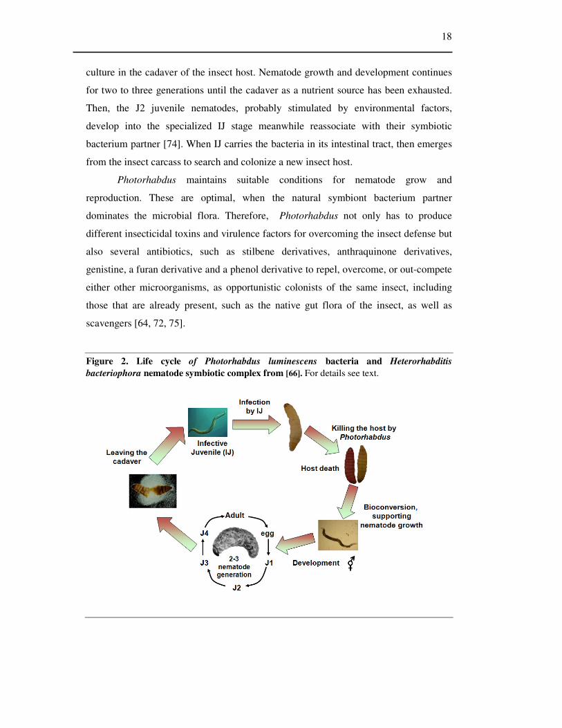
6.2.2.3 Purification of expressed Serpin-1 proteins
The purification of the twelve expressed Serpin-1 proteins were performed
using His select Nickel affinity columns (Sigma-Aldrich) according to [56] from 30 mL
cultures of E. coli XL1 blue strain, which was transformed with Bluescript plasmids
that contained the Serpin-1 variants (generous gift form Mike Kanost, Department of
Biochemistry, Kansas State University, U.S.A.; Fig. 3). For transformation 2 µl of
plasmid DNA was added to 50 µl of XL1 blue competent E. coli cells that were
incubated for 40 min on ice. The heat shock was at 42 oC for 1 min. Then, 1 mL of LB
was added to the cells, and incubated at 37 oC for 1 hour on a rotary shaker at 200 rpm.
The transformants were plated out on LB plates containing 0.1 µg/mL ampicillin and
the plates were incubated for overnight at 37 oC.
31
Figure 3. Recombinant plasmids for expressing M. sexta serpin-1 variants. Generous gift from Mike Kanost, Department of Biochemistry, Kansas State University, USA. A cDNA for serpin-1B in expression vector H6pQE-60 was used to reconstruct all 12 of the cDNA variants by substituting an equivalent restriction fragment from each variant cDNA. Open bar, plasmid vector; filled bar, constant regions of Manduca serpin-1 cDNA; shaded bar, the region of cDNA corresponding to exon 9; cross-hatched bar, vector sequence that differs, depending on how the original variant cDNA was cloned. X represents an EcoRI site for clones expressing serpin-1 variants B, F, and J’; X represents an XhoI site for clones expressing serpin-1 variants A, C, D, E, G, H, I, J, K, and Z [61].
6.2.2.4 Blotting and N-terminal sequencing
For N-terminal sequencing the partially purified PAT protein containing
samples were run in 10 % acrylamide-SDS gels under reducing conditions. Then the
gels were soaked for 10 minutes in transfer buffer (10mM CAPS (Sigma) pH 11.0, 10%
methanol), and blotted onto Immobilon-P PVDF Transfer Membrane (Millipore) at
200mA for 2 hours. The protein bands on the membrane were visualized by Coomassie
Brilliant Blue R-250 staining. The bands of PAT proteins were cut out and subjected to
Edman-sequencing in a Microtec-protein sequencer (Applied Biosystems) by András
Patthy from the ELTE-MTA Biotechnology Research Group. Identification through
database search for similar sequences was made with BLAST using the NCBI database.
6.2.2.5 Digestion of hemolymph and PAT proteins with PrtA
During the initial search for PrtA substrate proteins, hemolymph fractions that
contained 0.5-5.0 µg/mL protein, were exposed to digestion with 0.3 ng PrtA (~0.3 nM
final) or 30 ng chymotrypsin, trypsin and Clostridium collagenase (~60 nM final) at
room temperature in the presence of 50 mM Tris-HCl, (pH 8.0) 10 mM CaCl2 and 0.1
M NaCl (a reaction buffer in which all of the applied enzymes could exhibit their

32
highest activity on synthetic substrates). Samples were withdrawn at 45 and 90 minutes
of incubation. In order to find the PAT protein containing fractions after the various
isolation steps, samples of appropriate volumes were digested with 0.3 nM PrtA in 20
µl final volume at room temperature for 90 minutes. The purified serpin-1 variants
were subjected to PrtA cleavage at 1-4 µM Serpin-1 to 30 nM PrtA ratio (1.0-3.7 µg
serpin-1 to 30 ng PrtA) in the reaction buffer at room temperature for 90 minutes. All
the samples from the digestions were analyzed with SDS-PAGE.
6.2.3 RNA interference (RNAi)
6.2.3.1 Total RNA isolation
To isolate total RNA, 100 mg of dissected fat body and 30 mg of bled
hemocytes was homogenized in 500 µl of TRI reagent® (Sigma, UK) using a plastic
grinder. After homogenization the samples were centrifuged at 12,000 rpm for 10
minutes at 4°C. The removed supernatants were allowed to stand for 5 minutes at room
temperature before adding 200 µl of chloroform. The mixtures were vortexed for 15
seconds and were allowed to stand for 10 minutes at room temperature, then spinned at
12,000 rpm for 15 minutes at 4°C. The top aqueous phases were transferred to new
tubes, and 500 mL of isopropanol was added to them and mixed thoroughly. The
samples were incubated for 10 minutes at room temperature and centrifuged at 12,000
rpm for 10 minutes at 4°C. After removing the supernatants the RNA pellets were
resuspended in 1 mL of 70% ethanol, and sedimented at 7,500 rpm for 5 minutes at
4°C, then air-dried for 10 minutes. The resulted RNA preparation was dissolved in 20
µl of di-methyl-propyl carbonate (DMPC)-treated water, and treated with RNase free
DNaseI (Invitrogen, UK) (1 U/µL) to remove DNA contamination.

33
6.2.3.2 Generation of dsRNA of SPH-3
Figure 4. Generation of dsRNA of SPH-3. For details see text
To synthesize dsRNA of SPH-3, cDNA was amplified with RT-PCR on total
RNA extracted from fat body or hemocytes from insects previously injected with E.
coli to elicit immune response. RT-PCR was made using OneStep RT-PCR kit (Qiagen,
UK). Specific SPH-3 primers were used, which are shown in Table 2. The resulting
PCR product was cloned into pCR4-TOPO vector (Invitrogen, UK) and transformed
into E. coli one shot TOP10 chemically competent cells (Invitrogen, UK). The
transformants were plated on LB plates containing 1 µg/mL ampicillin (Sigma, UK). 10
colonies were inoculated into 5 mL LB broth and 1 µg /mL ampicillin and the cultures
were incubated for overnight (O/N) at 37 oC and then plasmid DNA was prepared with
QIAprep Spin Miniprep Kit (Qiagen, UK) following the manual’s protocol. The
minipreps were checked by sequencing for the correct nucleotide sequence and used as
a template to amplify the insert with T7 (TAATACGACTCACTATAGGG) and T3
(ATTAACCCTCACTAAAGGGA) primers by PCR (GenAmp Kit, UK). PCR
conditions: 34 cycles with the following 3 steps in each: 93 oC for 30 sec, 50 oC for 30

34
sec, 68 oC for 1 min then a final extension at 68 oC for 5 min. The PCR product was
purified with Montage PCR centrifugal filter Devices kit (Millipore, UK). These
purified PCR products were used to synthesize the sense and antisense RNAs using T3
and T7 ‘Megascript’ kits (Ambion, UK), respectively, according to the manufacturer’s
instructions. DNA templates were removed with RNase free DNaseI (Invitrogen) (1
U/µL), and the reaction products recovered and purified using lithium chloride
precipitation following the kit’s protocol. Single –stranded (ss) RNAs were dissolved in
DMPC treated water and complementary strands were annealed by combining equal
molar amount of each strand, heating to 70 oC for 15 min and cooling overnight at
room temperature. The annealing of ss-RNAs was found to be complete when
examined by agarose gel electrophoresis. dsRNAs were diluted to 2 µg/mL in DMPC
treated water and stored at -20 oC until required. The sequence of the dsRNA was from
base 19 to 675 (the entire coding region) of the SPH-3 cDNA (Fig.4). The negative
dsRNA control (dsCON) was synthesized with the same method as described above.
6.2.3.3 Insect injection protocol
For RNAi, 100 ng (50 µL, 2 µg/mL) of dsRNA of SPH-3 (dsSPH3) in DMPC-
treated water was injected into Manduca sexta larvae as primary injection, and then, 6
hours later with E. coli or Photorhabdus (TT01) as secondary injection. Controls used
DMPC-treated water without dsRNA in the primary injections and PBS without E. coli
or TT01 in the secondary injections. After treatment, insects were held at 25°C for 18
hours before isolation of fat body and hemocytes (Fig.5).
Figure 5. Sequence of injections of Manduca sexta for RNAi
35
6.2.3.4 RT-PCRs
To determine the transcription of each gene, semi-quantitative single-step
reverse transcription (RT)-PCR was performed with the ‘OneStep’ RT-PCR kit
(Qiagen, UK). Each reaction was carried out in a 50 µl volume containing 0.6 mM of
forward and reverse gene primers and 2 ng of total RNA template. Amplifications were
performed on a PTC- 100 thermal controller (MJ Research, USA) under the following
cycling conditions: at 50 oC for 30 min (reverse transcription step), 95 oC for 15 min
(initial PCR activation step), followed by 35 PCR cycles with the following steps in
each 94 oC for 30 sec (denaturation), 50 oC for 30 sec (annealing) and 72 oC for 1 min
(extension), then a final extension of 72 oC for 10 min. The different gene specific
forward and reverse primers are summarized in Table 2.
The RT-PCR product of ribosomal protein S3 (rpS3) mRNA (Manduca sexta
rpS3 is a constitutively expressed housekeeping gene) was used as a loading control to
make sure that the same amount of total RNA as a PCR template was used for each
experiment. RT-PCR control reactions for rpS3 were performed as outlined above.
PCR reaction without template as negative control (NC) was also carried out in each
case. PCR products were separated by 1% agarose gel electrophoresis. Expression of
immune-related genes and the extent of dsRNA induced gene silencing were assessed
using two insects in each treatment.

36
Table 2. Nucleotide sequences of primers used in RT-PCR. All sequences read 5’ to 3’, left to right
Gene Accession
No Primer Sequence
Product size (bp)
SPH3 AF413067 Forward CGTGGCACGATAATGTTGTT 657
Reverse AGTCGCTGCGTCAATGTATG
Hemolin U11879 Forward ACAGCAACAACACAGGTGAA 1273
Reverse TTAAGCAACAATCACGAGCG
IML-2 AF242202 Forward GACTCTTGCGAGTCGTGTGA 953
Reverse GACTGTTTGGGTCCTTTTCG
PGRP AF413068 Forward ACGGTATCACTTCCGTCCAC 516
Reverse CATTCTGGCCATCTCCTGAT
PRSP AY380790 Forward ACGGATGGCACCCTGGTGCAGCC 680
Reverse TTTGCAACACATCAACGTAAG
βGRP-1 AF177982 Forward CCTGACGCGAAGTTAGAAGC 792
Reverse AACGCGACCATATTTGAAGG
βGRP-2 AY135522 Forward TCTACCCCAAAGGCTTGAGA 802
Reverse CACCACCAGAGACCCACTTT
PPO L42556 Forward AAACAACTCCCAAACGATGC 889
Reverse TGTGCATGTTGTTGTGGATG
Attacin B BI262658 Forward GGTCACGGCGCTACTCTTAC 341
Reverse TTGGGCATCTCGAACTTCTT
Cecropin D BI262670 Forward TTCTTCGTCTTCGCTTGCTT 161
Reverse CCTTTGAAAATGGCGGTTG
Lebocin BI262726 Forward ACGTGCGTAGTGTGAACGAG 170
Reverse CGCAGATTATGAGTTACGACGA
Lysozyme BI262563 Forward TACAAACGCCACAAGTTCCA 309
Reverse AGGCTTGTAACCAAGGACGA
Moricin 2 BI262611 Forward TCGCGTTGTCGCTATTATTTT 327
Reverse CAGAAGATTCCGAAGGGAGA
rpS3 U12708 Forward CTGGCTGAGGATGGCTACTC 186
Reverse TTTCTCAGCGTACAGCTCCA

37
6.2.4 Examination of the effect of RNAi of SPH-3 in Manduca sexta
6.2.4.1 Western blotting
Manduca sexta hemolymph plasma samples were separated with standard SDS-
PAGE. After electrophoresis the gels were transferred electrophoretically (Mini Trans-
Blot Transfer Cell, Bio-Rad, Uk) to PVDF membranes (Bio-Rad, UK). The samples
were transferred in cold Towbin buffer containing 20% methanol, 0.18 M glycine, 25
mM TRIS at 100 volts for 60 min. The blot was blocked in 5% skimmed milk powder
(Marvel) in transblotting solution (TBS, 20 mM Tris, 0.5 M NaCl, pH 7.5) for 12 h at 4 oC. The blot was washed 3 times for 10 min in TBS, then incubated for 1 h at room
temperature in tween transblotting containing 3% milk powder (TTBS, 20 mM Tris,
0.5M NaCl, 0.1% Tween-20, pH 7.5) containing 1/10,000 dilution of the primary anti-
SPH-3 antibody which was raised in rabbit against the synthetic peptide
PQFKGRNTNYRNDI (corresponding to SPH-3 amino acid residues 135-148). After
subsequent washing (3 times for 10 min in TBS) the blot was incubated in TTBS
solution containing 3% milk powder, 1/10,000 dilution of horseradish peroxidase-
labelled goat anti-rabbit IgG secondary antibody (Upstate, UK) for 1 h at room
temperature. After further washing (3 times for 10 min in TBS) the bound antibodies
were detected using chemiluminescence Western blotting kit (Visualizer, Upstate, UK)
and the membrane was exposed to X-ray film (Biomax, Kodak, UK) for 1 sec to detect
the signals.
6.2.4.2 Mortality bioassay
In survival experiments, after insect injections (Fig. 5), the state of the insects
were checked at eight time points (12 h, 18 h, 21 h, 24 h, 36 h, 41 h, 48 h, 60 h) after
the second injection. The mortality was defined as failure to react to poking with a
needle. Ten insects were used for each treatment and the experiment was repeated three
times.
6.2.4.3 Effect of RNAi knockdown of SPH-3 on PO activity by visual
examination of hemolymph
18h after the bacterial infection insects were individually bled into pre-chilled
sterile polypropylene tubes to collect their total hemolymph fluids (approximately 500
µl per larva). Hemolymph samples were incubated for 1 h at room temperature. The
samples were visually evaluated for the development of their darkening.
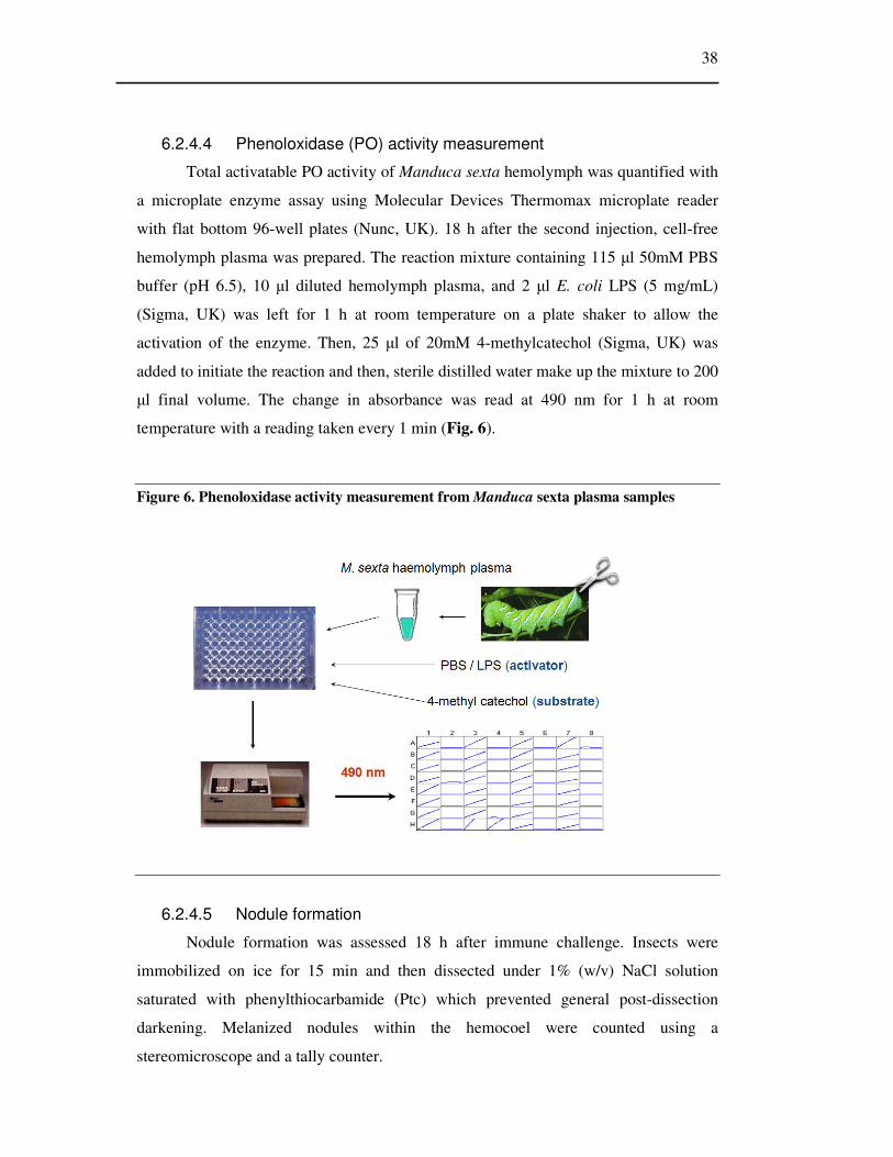
38
6.2.4.4 Phenoloxidase (PO) activity measurement
Total activatable PO activity of Manduca sexta hemolymph was quantified with
a microplate enzyme assay using Molecular Devices Thermomax microplate reader
with flat bottom 96-well plates (Nunc, UK). 18 h after the second injection, cell-free
hemolymph plasma was prepared. The reaction mixture containing 115 µl 50mM PBS
buffer (pH 6.5), 10 µl diluted hemolymph plasma, and 2 µl E. coli LPS (5 mg/mL)
(Sigma, UK) was left for 1 h at room temperature on a plate shaker to allow the
activation of the enzyme. Then, 25 µl of 20mM 4-methylcatechol (Sigma, UK) was
added to initiate the reaction and then, sterile distilled water make up the mixture to 200
µl final volume. The change in absorbance was read at 490 nm for 1 h at room
temperature with a reading taken every 1 min (Fig. 6).
Figure 6. Phenoloxidase activity measurement from Manduca sexta plasma samples
6.2.4.5 Nodule formation
Nodule formation was assessed 18 h after immune challenge. Insects were
immobilized on ice for 15 min and then dissected under 1% (w/v) NaCl solution
saturated with phenylthiocarbamide (Ptc) which prevented general post-dissection
darkening. Melanized nodules within the hemocoel were counted using a
stereomicroscope and a tally counter.

39
6.2.4.6 Pathogen growth in vitro
To determine the ability of P. luminescens to grow in the presence of plasma
insects were bled individually 18 h after the bacterial infections, and cell free plasma
was prepared following the addition of 20 mM Ptc. All samples were inoculated with ~
103 cells (3 µL) of P. luminescens and incubated at 30°C with constant shaking for 24
h. Growth was estimated as optical density at 600 nm.

40
7. Results
7.1 Comparison of proteolytic activities produced by different
Photorhabdus strains
7.1.1 Measuring enzymatic activity in culture
Photorhabdus bacteria had been found as protease producers however the
number and the type of secreted proteases and the dynamics of production remained
unknown. My goal was to find a protease, which might be a pathogenic factor during
Photorhabdus infection. Such an enzyme must be secreted early and in all strains of
Photorhabdus. Therefore, I investigated 20 Photorhabdus strains and compared their
protease production with zymography. I focused on proteases that could be detected in
the first 48 h of culture and infection and could, therefore, have a role in virulence. For
the detection of protease activity I used both SDS- and native PAGE coupled
zymography in the presence of casein or gelatin substrates. (Fig.7, Table 3).
Photorhabdus bacteria produced more than one protease in the examined period but I
couldn’t find any other enzyme, which would be secreted earlier than a 55 kDa
protease, PrtA. PrtA activity was observed after SDS-PAGE coupled zymography in
almost all strains except for several secondary phase variants. There were slight
variations in the level of PrtA activity and in the time of occurrence among strains and
among growth conditions. In case of four strains – Hm/2, Wx6/2, HSH-2/1 and HSH-
2/2 – I couldn’t detect any PrtA activity with either SDS or native gel electrophoresis.
In case of the most intensive PrtA producers (Brecon/1 and Nc19/2) the activity
appeared from 12 - 24 h of culture (mid-logarithmic – late-logarithmic growth phase)
and remained detectable for the 44 h of culturing. With the exception of Hm/2 and
K122/2 strains, there were at least two activity bands in the molecular mass range from
50 to 55 kDa. The smaller species, which were thought to be molecular variants of
PrtA, were not present at the beginning of secretion. In each case, two dominant forms,
a larger 55-kDa form (PrtA1) and a 2- to 4-kDa smaller form (PrtA2), could be
distinguished. The ratio of these forms and the occurrence of other forms varied slightly
with different cultures of the same strain, but the PrtA1 form was always the most
abundant (or most active). I also found with SDS-PAGE coupled zymography in the
cultures of strains Brecon/1 and K122/1 but not in the cultures of the other strains,
another major activity, a 37-kDa protease termed as Php-C. Depending on the strain, it
occurred from 21 to 40 hours of culturing (in the late logarithmic and early stationary

41
phases). In strain Brecon/1, Php-C also showed size variation and was resolved by
SDS-PAGE as a larger 37-kDa form (Php-C1) and a fainter, smaller and substantially
less active (or abundant) 35-kDa form (Php-C2). Although, Php-C was detected by
SDS-PAGE coupled zymography in only two strains, the native PAGE coupled
zymography showed that it was actually produced by fifteen strains (Table 3).
Figure 7. Comparison of protease secretion in the culture of seven Photorhabdus strains (Hm/1; Hm/2, Nc19/1; Nc19/2; K121/1; K121/2 and Brecon) with SDS- and native PAGE coupled zymography. Detection of protease secretion with SDS-PAGE coupled zymography in the presence of gelatin substrate (A) and casein substrate (C); and with native PAGE coupled zymography in the presence of gelatin (B) and casein substrates (D). The numbers on the top of the gels indicate the sampling time point from culture growth. A1 and A2 are molecular variants of PrtA; C1 and C2 are molecular variants of PhpC (C).

42
Table 3. Protease secretion by 20 Photorhabdus strains and phase variants: summary of activities with three detection methods. Intensity of the highest activity: -, not detectable; (±), very weak; (+), weak; +, well easily detectable; ++, strong. The times of first detection are indicated in parentheses, as follows: ml, mid-logarithmic phase (OD600 nm between 1.0 and 4.8; h 12 to 21); ll, late logarithmic phase (optical density at 600 nm between 4.8 and 5.3; h 21 to 40); s, early stationary phase (optical density at 600 nm between 5.3 and 6.6; h 40 to 50). On Fua-LGPA substrate the values were determined in supernatants having optical densities at 600 nm between 4.9 and 8.1 (43-h cultures). kobs is the first-order rate constant and is expressed in seconds-1 (see Materials and Methods).
7.1.2 Enzyme activity measurement with chromogen oligopeptide substrate
I measured and compared the time course of proteolytic activity of 20 strains on
Fua-Leu-Gly-Pro-Ala (Fua-LGPA) bacterial collagenase substrate. This artificial
chromogen substrate is used to measure the activity of proteases that have primary
preferences for amino acids that are C terminal to the scissile bond. Hydrolysis of that
substrate at the Leu-Gly bond causes a shift in the absorption spectrum of the Fua
chromophore group [106]. I found that all the tested strains produce an enzyme with
Fua-ALGPA-ase activity in the late logarithmic and early stationary growth phases
(Table 3). Later in the stationary phase (40 to 50 hours, depending on the strain) the
activity did not increase further. The Fua-LGPA-ase enzyme, termed as Php-B, was
purified and proved to be a 74-kDa intracellular metalloenzyme, known as OpdA,

43
which could not be detected by either SDS-PAGE or native PAGE coupled
zymography [107].
7.2 Measuring protease activity during infection
7.2.1 Infection of Galleria mellonella larvae with different Photorhabdus strains
In order to investigate the presence of protease activities during infection, I took
hemolymph plasma samples from G. mellonella larvae infected with five different
Photorhabdus strains, Brecon/1, Hm/1, Hm/2, Nc19/1 or Nc19/2, for detection of PrtA
and Php-C with native and SDS-PAGE-coupled zymography. Choosing these strains I
tested how the differences in protease production in culture manifest during infection.
In culture growth from Hm/1 and Hm/2 phase variant strains, the Hm/1 secrets both
PrtA and Php-C activities, while Hm/2 produces neither of them (no enzyme activity
was detectable with SDS- and native PAGE coupled zymography Fig. 7).
I injected about 100 cells into each G. mellonella larvae and detected the
activities of both PrtA and Php-C the earliest at 12 to 42 hours postinfection,
respectively (Fig. 8). While PrtA was produced again by every strain, I observed Php-C
only in the hemolymph plasma of Brecon infected G. mellonella 42 h postinfection
(Table 4). From these experiments I concluded that PrtA was the earliest secreted
enzyme that I could detect with my methods both in culture and infection.
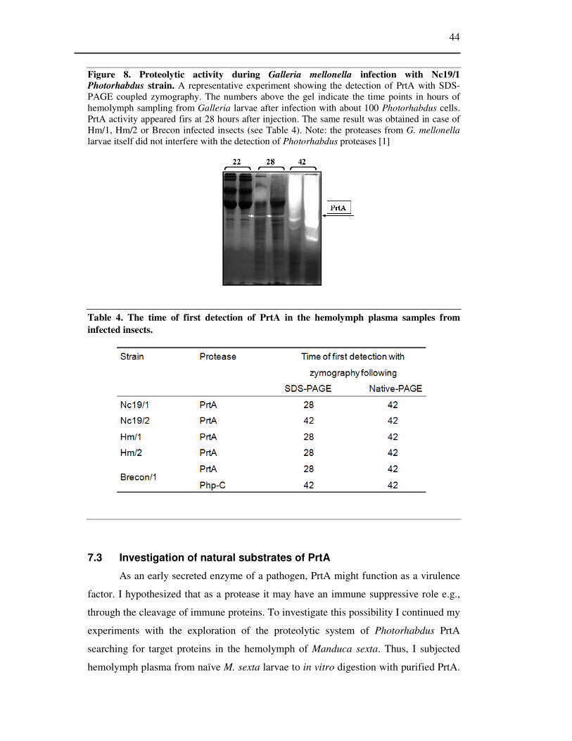
44
Figure 8. Proteolytic activity during Galleria mellonella infection with Nc19/1 Photorhabdus strain. A representative experiment showing the detection of PrtA with SDS-PAGE coupled zymography. The numbers above the gel indicate the time points in hours of hemolymph sampling from Galleria larvae after infection with about 100 Photorhabdus cells. PrtA activity appeared firs at 28 hours after injection. The same result was obtained in case of Hm/1, Hm/2 or Brecon infected insects (see Table 4). Note: the proteases from G. mellonella larvae itself did not interfere with the detection of Photorhabdus proteases [1]
Table 4. The time of first detection of PrtA in the hemolymph plasma samples from infected insects.
7.3 Investigation of natural substrates of PrtA
As an early secreted enzyme of a pathogen, PrtA might function as a virulence
factor. I hypothesized that as a protease it may have an immune suppressive role e.g.,
through the cleavage of immune proteins. To investigate this possibility I continued my
experiments with the exploration of the proteolytic system of Photorhabdus PrtA
searching for target proteins in the hemolymph of Manduca sexta. Thus, I subjected
hemolymph plasma from naïve M. sexta larvae to in vitro digestion with purified PrtA.
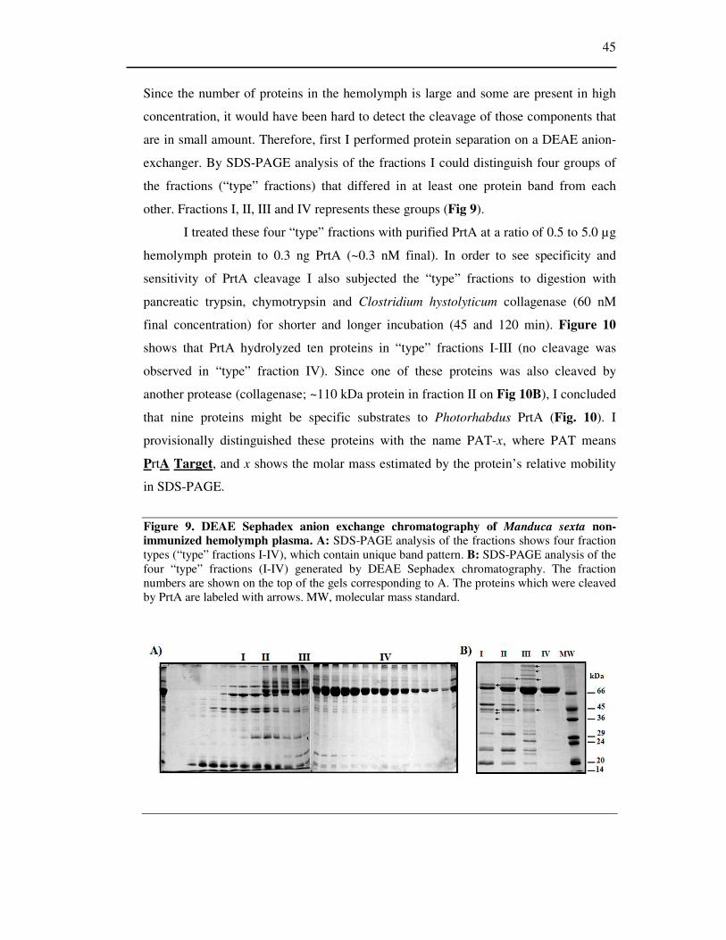
45
Since the number of proteins in the hemolymph is large and some are present in high
concentration, it would have been hard to detect the cleavage of those components that
are in small amount. Therefore, first I performed protein separation on a DEAE anion-
exchanger. By SDS-PAGE analysis of the fractions I could distinguish four groups of
the fractions (“type” fractions) that differed in at least one protein band from each
other. Fractions I, II, III and IV represents these groups (Fig 9).
I treated these four “type” fractions with purified PrtA at a ratio of 0.5 to 5.0 µg
hemolymph protein to 0.3 ng PrtA (~0.3 nM final). In order to see specificity and
sensitivity of PrtA cleavage I also subjected the “type” fractions to digestion with
pancreatic trypsin, chymotrypsin and Clostridium hystolyticum collagenase (60 nM
final concentration) for shorter and longer incubation (45 and 120 min). Figure 10
shows that PrtA hydrolyzed ten proteins in “type” fractions I-III (no cleavage was
observed in “type” fraction IV). Since one of these proteins was also cleaved by
another protease (collagenase; ~110 kDa protein in fraction II on Fig 10B), I concluded
that nine proteins might be specific substrates to Photorhabdus PrtA (Fig. 10). I
provisionally distinguished these proteins with the name PAT-x, where PAT means
PrtA Target, and x shows the molar mass estimated by the protein’s relative mobility
in SDS-PAGE.
Figure 9. DEAE Sephadex anion exchange chromatography of Manduca sexta non-immunized hemolymph plasma. A: SDS-PAGE analysis of the fractions shows four fraction types (“type” fractions I-IV), which contain unique band pattern. B: SDS-PAGE analysis of the four “type” fractions (I-IV) generated by DEAE Sephadex chromatography. The fraction numbers are shown on the top of the gels corresponding to A. The proteins which were cleaved by PrtA are labeled with arrows. MW, molecular mass standard.

46
Figure 10. Selective PrtA cleavage of proteins in the three “type” fractions (I-III) from DEAE Sephadex chromatography. SDS-PAGE analysis. The conditions of the reactions shown above the lanes are the type of the protease applied (control is without protease) and incubation time. Arrows point to PAT proteins that are selectively cleaved by PrtA. The numbers are estimated to the molecular masses of PAT proteins. These are not shown for three proteins, which are larger than PAT-170 in type fraction III because of the imprecision of estimation. Molecular weights (kDa) are indicated on the right hand side of each gels.
7.4 Isolation and identification of PAT proteins from Manduca sexta
hemolymph
To understand the possible role of PrtA during the infection process of
Photorhabdus I investigated the substrate side in the proteolytic system of PrtA further
with the isolation and identification of its target proteins (PAT proteins). To this end I
developed a purification procedure for PAT proteins (Fig. 11) to reach such a
separation level that is sufficient for their N-terminal sequencing after SDS-PAGE and
blotting and thus, for their identification from the protein database.

47
Figure 11. Procedure for the separation of PAT proteins. PAT protein names in boldface letters in a purification step indicate those proteins that were detectable following that step.
7.4.1 Purification of PAT-110
I started the purification of PrtA target proteins with a two-step (NH4)2SO4
precipitation. The 46 % precipitate contained all of the PAT proteins, including PAT-
110, PAT-90 and PAT-41, but not PAT-52, which remained in the supernatant. The
second purification step to separate PAT-110 from the other hemolymph proteins was
gel filtration of proteins in the 46 % (NH4)2SO4 precipitate on Sephacryl S-200 column
(Fig. 12). This step separated efficiently PAT-110 from PAT-41 and a number of other
hemolymph proteins, but PAT-90 remained with PAT-110. From this step I made the
further purifications of PAT-110 – PAT-90 and PAT-41 separately (Fig. 11).

48
Figure 12. Gel filtration of proteins in the 46 % (NH4)2SO4 precipitate on Sephacryl S-200 column. SDS-PAGE analysis of the fractions. Arrowheads indicate the appearance of PAT-110, PAT-90 and PAT-41 during gel filtration procedure. The fractions containing PAT-110 and PAT-41 are indicated on the top of the gel.
For further purification of PAT-110, I combined the fractions of gel filtration
containing PAT-110 (and PAT-90) and subjected to PAE Silica chromatography. The
SDS-PAGE analysis of the fractions shows that PAT-110 and PAT-90 remained in the
same fractions, but they were pure from many other hemolymph proteins (Fig.13A).
When I treated the fractions containing PAT-110 and PAT-90 with PrtA (Fig.13B),
only these two proteins were hydrolyzed. Their hydrolysis was complete under the
applied conditions (0.3 ng PrtA to 0.5-5.0 µg hemolymph protein ratio and 90 min
incubation time) showing that they were very sensitive to PrtA digestion. The cleavage
products of PAT-110, a 27 kDa and a 20 kDa protein, that accumulated temporarily
indicated that PrtA can have more than one cleavage site in that protein.

49
Figure 13. PAE Silica anion exchange chromatography of PAT-110. A: SDS-PAGE analysis of the fractions. Arrowheads indicate the appearance of PAT-110 and PAT-90 during the chromatography procedure. The fractions containing PAT-110 is indicated on the top of the gel. B: SDS-PAGE analysis of PrtA treatment of three fractions (1, 2, 3). The fraction numbers above the lane correspond to A. Incubation in the presence (+) or absence (-) of PrtA is indicated on the top of the gel. Arrowheads show the PAT-110 cleavage products.
Fractions from PAE chromatography containing PAT-110 and PAT-90 were
still contaminated with several other proteins. To eliminate these and obtain pure PAT-
110 and PAT-90 fractions I performed Mono Q anion exchanger chromatography.
PAT-110 was eluted in a pure form from the column at approximately 0.2 M NaCl
concentration, whereas PAT-90 was lost (Fig. 14). Although the yield was very low,
the amount was sufficient for blotting and N-terminal sequencing.
Figure 14. Mono Q anion exchange chromatography of PAT-110. A: Profile of protein elution. The fractions (1, 2) containing PAT-110 was eluted from the column at 16 min. B: SDS-PAGE analysis of the fractions. The fractions containing PAT-110 are indicated on the top of the gel. The fraction numbers above the lane correspond to A.

50
7.4.2 Purification of PAT-41
After gel filtration of proteins in the 46 % (NH4)2SO4 precipitate on Sephacryl
S-200 column I could separate PAT-41 from PAT-110 and PAT-90 (Fig. 12). Then, I
purified further PAT-41 with MonoQ FPLC chromatography (Fig. 15A). When I
probed these fractions for PrtA cleavable proteins, surprisingly I found seven instead of
PAT-41 only. They were- in addition to PAT-41 (from now on PAT-41a) - PAT41b,
PAT-63, PAT-54a, PAT-54b, PAT-54c, PAT-35ab (Fig.15B). They might be in the
(non-immune) hemolymph in such a small amount that prior to separation and
concentration with ion exchange chromatography they remained undetectable with
Coomassie staining of acrylamide gels. Together with these, the number of proteins that
were cleaved by PrtA increased to fifteen. The purity of some of them was suitable for
blotting and N-terminal sequencing.
Figure 15. Mono Q anion exchange chromatography of PAT-41. A: SDS-PAGE analysis of the resulted fractions. Fraction numbers are shown above the lanes (1-8). The bands of PAT-proteins are labeled as follows: (1) PAT-63; (2) PAT-54a; (3) PAT-54b; (4) PAT-54c; (5) PAT-41a; (6) PAT-41b; (7) PAT-35a and b. B: SDS-PAGE analysis of PrtA treated fractions (+) and controls without PrtA (-). The fraction numbers above the lane correspond to A. Numbers with primes indicate putative degradation products of PAT-proteins of the corresponding number.

51
7.4.3 Purification of PAT-52 protein
After 46% precipitation with ammonium sulfate PAT-52 was separated from
PAT-110 and other PAT proteins (Fig. 11) and remained in the supernatant. Then, it
was precipitated with additional 20% (NH4)2SO4. I resuspended the precipitate and
purified further first with gel filtration on a Sephacryl S-200 column (Fig. 16A). The
SDS gel analysis shows that PAT-52 was separated from the proteins with higher
molecular weight but not from the smaller ones. The treatment of the fractions with
PrtA showed that PAT -52 was the only protein which was cleaved by PrtA, although it
did not hydrolyzed completely (see later) (Fig. 16B).
Figure 16. Gel filtration of PAT-52 on Sephacryl S-200 column. A: SDS-PAGE analysis of the fractions. The fraction numbers are shown above the lanes (1-4). The fractions containing PAT-52 are indicated on the top of the gel. B: SDS-PAE analysis of the PrtA treated (+) and non-treated control (-) fractions. The fraction number (1) above the lane corresponds to A.
I continued the purification with fractions of Sephacryl S200 column which
contained PAT-52 (Fig. 16) on a Superdex-75 gel filtration column to separate PAT-52
further from the minor hemolymph proteins still present. Fig. 17 sows that the level of
the resulted purity was sufficient for blotting and N-terminal sequencing.
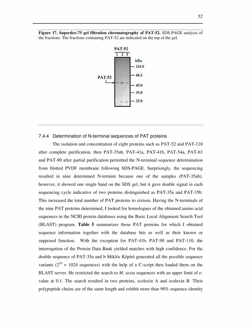
52
Figure 17. Superdex-75 gel filtration chromatography of PAT-52. SDS-PAGE analysis of the fractions. The fractions containing PAT-52 are indicated on the top of the gel.
7.4.4 Determination of N-terminal sequences of PAT proteins
The isolation and concentration of eight proteins such as PAT-52 and PAT-110
after complete purification, then PAT-35ab, PAT-41a, PAT-41b, PAT-54a, PAT-63
and PAT-90 after partial purification permitted the N-terminal sequence determination
from blotted PVDF membrane following SDS-PAGE. Surprisingly, the sequencing
resulted in nine determined N-termini because one of the samples (PAT-35ab),
however, it showed one single band on the SDS gel, but it gave double signal in each
sequencing cycle indicative of two proteins distinguished as PAT-35a and PAT-35b.
This increased the total number of PAT proteins to sixteen. Having the N-terminals of
the nine PAT proteins determined, I looked for homologues of the obtained amino acid
sequences in the NCBI protein databases using the Basic Local Alignment Search Tool
(BLAST) program. Table 5 summarizes those PAT proteins for which I obtained
sequence information together with the database hits as well as their known or
supposed function. With the exception for PAT-41b, PAT-90 and PAT-110, the
interrogation of the Protein Data Bank yielded matches with high confidence. For the
double sequence of PAT-35a and b Miklós Képíró generated all the possible sequence
variants (210 = 1024 sequences) with the help of a C-script then loaded them on the
BLAST server. He restricted the search to M. sexta sequences with an upper limit of e-
value at 0.1. The search resulted in two proteins, scolexin A and scolexin B. Their
polypeptide chains are of the same length and exhibit more than 90% sequence identity

53
explaining why PAT-35 a and b remained together during the isolation steps and in
SDS-PAGE.
Regarding the function of each identified PAT proteins I can conclude that all
of them seem to have immune-related functions, which either had been known,
supposed or was proven later as in the case of PAT-41a (see below). (The sequencings
were made by András Patthy).
Table 5. N-terminal sequences of nine PAT proteins in comparison to the closest hits from the protein database. Abbreviations: SPH-3, serine protease homologue-3; HAIP, hemocyte aggregation inhibitor protein; β-GRP-2, β-1,3 glucan recognition protein-2.
PAT protein N-terminal sequence Database sequence
Database name
BLAST e value
(Supposed) function
Accession number
PAT-35ab DANDIDIQQN GPGANQLNLK DPGANDIQLN Scolexin A 4x10⁻² Immune AAD14591 GANDIQLNQK Scolexin B 6x10⁻² Immune AAD14592 PAT-41 DQGSPDLPLATEPPT DQGSPDLPLATEPPT SPH-3 3x10⁻⁴ Immune AAO21508 PAT-41b GVIKEESI Unknown Unknown PAT-52 ETDLQKILRESNDQFTA ETDLQKILRESNDQFTA Serpin-1 1x10⁻⁵ Immune AAC47343 PAT-54 TPASVPRKVLXYYDSK TPASVPXKVLXYYDSK HAIP 2x10⁻³ Immune AAB32418 PAT-63 LEVPDALKEAIYPKG LEVPDALKEAIYPKG β-GRP-2 2X10⁻² Immune AAN10151 PAT-90 QXNESLVII Unknown Unknown PAT-110 QRXESLVISXVEDXGXAAI Unknown Unknown
7.4.5 Analysis of the cleavage products of PAT proteins
As PAT proteins became more concentrated and better isolated from other
proteins, some features of their cleavage could be observed. For example PAT-52 could
never be hydrolyzed completely (Fig.16B), even a longer (120 minute) exposure to
PrtA. In contrast, PAT-110 and 90 proved to be very sensitive: they were completely
cleaved in the presence of even 0.03 ng PrtA within less than 40 minutes. Importantly,
when I exposed pure PAT-110 to trypsin or chymotrypsin digestion under the same

54
conditions as those with PrtA, it remained intact showing that this protein (like other
PAT proteins) is not generally sensitive for proteolysis.
The appearance of cleavage products was mostly temporary indicating a further
degradation by either PrtA or contaminating proteinases that were present in the
hemolymph and could start acting on PAT proteins only after the initial cleavage by
PrtA. However, the cleavage product of PAT-170, 54a, 41a, 35a,b and especially PAT-
52 has longer half life suggesting that these might not be degraded further by PrtA. In
the case of the latter the cleavage product was just a little smaller than the intact PAT-
52 (Fig.16B) showing that PrtA clips only 10-15 amino acids from one of the
molecular termini (see below).
I investigated the cleavage of PAT-52 (M. sexta serpin-1) further to explain why
a fraction of this protein always remained uncleaved by PrtA. This protein is present in
the hemolymph in twelve C-terminal variants [56], and even the purified fraction might
contain several of them. Therefore, the most likely explanation is that only some of the
variants are sensitive for PrtA cleavage. To verify this and to establish which ones are
cleaved, I expressed and purified all of the serpin-1 variants and exposed them to PrtA
digestion. Several variants (B, E, F, J, and Z) were, indeed, not digested; the others in
turn were hydrolyzed with an occasional accumulation of 1.0-1.5 kDa smaller, short
lived cleavage products. PrtA must discriminate serpin-1 variants through their
differential, 40-50 amino acid long C-terminal end, which contains the reactive site
loop, confers the protease selectivity [61] and – as it is revealed by their different PrtA
sensitivity – also the susceptibility to proteolytic cleavage by a noncognate (i.e. non
serine) protease. The N-terminal sequence of the cleaved and the uncleaved forms was
the same, which proves that PrtA clipped 10-15 amino acids from the C-terminus of
serpin-1. I obtained the same result when the N-terminal sequence of PAT-52, partially
purified from M. sexta hemolymph and exposed to PrtA hydrolysis, was determined. It
was the same as that of the intact protein (ETDLQKILRESNDQFTA).

55
7.5 Identification of the function of Serine Protease Homologue-3
I identified PAT-41a as Serine Protease Homologue-3 (SPH-3), a M. sexta
hemolymph protein of unknown function. However, it was reported as “immune
related” on the basis that it has proved to be immune inducible [108]. To verify its
function in the immune system of M. sexta I exploited RNA interference technique.
7.5.1 Induction and RNAi-mediated knockdown of SPH-3 in M. sexta
First, to corroborate that SPH-3 is an immune inducible gene in Manduca sexta
I carried out RT-PCR using specific SPH-3 primers and RNA extracts from fat body
and hemocytes. The RT-PCR result was consistent with earlier observations that SPH-3
was expressed in naïve, non-immune induced Manduca larvae at a very low level, but
it was markedly up-regulated in both fat body and hemocytes of insects which had
previously been challenged with either non-pathogenic E. coli or pathogenic
Photorhabdus TT01 bacteria (Fig. 18). Thus, no SPH-3 transcription was seen in
negative controls including insects injected with PBS or untreated insects.
Figure 18. Induction of SPH-3 encoding gene in Manduca sexta. RT-PCR on fat body and hemocyte total RNA was extracted 18 h after bacterial challenge with E.coli (EC) or Photorhabdus (TT01) or injection of PBS. A no-template RT-PCR control (NC) and the non-treated (NT) negative control are included. Manduca sexta ribosomal protein gene S3 (rpS3) was used as a loading control. The sizes of PCR products are indicated. (For the specific primers used see Table 2)
Second, to investigate if SPH-3 is required for normal immune function I
knocked down SPH-3 in Manduca sexta with RNA interference. For the experiments I
constructed a dsRNA reagent specific for SPH-3 (dsSPH3). When I checked SPH-3

56
expression (as above) 18 h after dsRNA treatment I found complete knockdown i.e.
upon injection with either non-pathogen or pathogen microorganism the gene of SPH-3
remained inactive (Fig. 19). This inhibition in the induction of SPH-3 gene was specific
to dsSPH3 treatment, and it was not observed on treatment with dsRNA of a plant
catalase (dsCON; Fig. 19). This also showed that the inhibition of SPH-3 gene
induction was not a general effect of dsRNA injection.
Figure 19. RNAi-mediated knockdown of SPH-3 in Manduca. 6 h before bacterial challenge insects were pretreated with the injection of dsRNAs of either SPH-3 (dsSPH3) or catalase (dsCON as dsRNA control), or of DMPC-treated water (W as injection control). The immune challenge was the injection of 103 cells of either E. coli (EC) or Photorhabdus luminescens TT01 (TT01). As negative control (no bacterial challenge), insects were pretreated the same way, but 6 hours later they were injected with PBS (PBS). Total RNA was extracted from fat body 18 h after second injection (with bacterial challenge or PBS). Non treated control (NT) and RT-PCR control (NC) are also shown. (For further details see Methods). Manduca sexta ribosomal protein gene S3 (rpS3) was used as a loading control. The sizes of PCR products are indicated. RT-PCR was performed in duplicate for each treatment; results for single individuals are shown.
Then, I investigated the effect of dsSPH3 treatment on protein level also, using
immunoblot with anti-SPH-3 antibody. The result was in a complete agreement with
that of RT-PCR: SPH-3 protein level in hemolymph plasma is also strongly reduced by
RNAi knockdown of SPH-3 (dsSPH3), but not by dsCON. At the same time I observed
very low levels of SPH-3 protein in the non-induced controls which means that there is
some SPH-3 expression even in naive larvae, which correlates with the fact that I
purified SPH-3 protein from naïve M. sexta hemolymph plasma (Fig. 20).

57
Figure 20. Western blot analysis of hemolymph SPH-3 protein level, using specific anti-SPH-3 antibody. The treatments and injection protocol were the same as given in Figure 19. (For further details see Methods.) The experiment was made in duplicate for each treatment; results for single individuals are shown. Specificity of staining was checked by omitting the primary antibody, which led to complete absence of bands (not shown).
7.5.2 Mortality bioassay
Having knocked-down of SPH-3 transcription successfully the next question
was whether it has an effect on M. sexta mortality. To test this I carried out mortality
bioassay in which M. sexta larvae were injected first with either DMPC-treated water
(W), dsRNA control (dsCON) or SPH-3 dsRNA (dsSPH3) and 6 h later with 103 cells
of P. luminescens TT01 or PBS. As it was expected from earlier results 103 cells of P.
luminescens TT01 killed Manducas within 48 h when they were pretreated with water
or dsCON before TT01 injection [80]. By contrast RNAi knockdown of SPH-3 (dsSPH
pretreatment) caused a drastic decrease in the ability of the insect to survive
experimental Photorhabdus infections since all insects died within 24 h. The time for
50% survival decreased from 24-36 h in control insects to only 12-21 h in the dsSPH3-
treated insects. The difference in the number of survivors at 21 h was highly significant.
Injection of dsRNAs alone without bacterial infection (dsSPH+PBS), or injection of
water and then PBS (W+PBS) did not cause death. Similarly when nonpathogenic E.
coli was used instead of Photorhabdus no insect death was observed and the survival
rate was the same as that of non-pretreated insects (Fig. 21).

58
Figure 21. Time course mortality bioassay following RNAi of SPH-3. A: RNAi-mediated knockdown of SPH-3 is associated with decreased survival following Photorhabdus (TT01) exposure. The injection protocol of insects was the same as given in the legend to Figure 19. Asterisks show significant differences between SPH-3 knock-down insects and the infected control groups. B: The survival rate of insects infected with nonpathogenic E. coli was 100%. Groups of ten insects were pre-treated with dsRNA 6 h before administering the same dose of E. coli as the lethal inoculum of Photorhabdus. Uninfected control groups were the same in both (A;B) experiments (W+PBS, dsSPH+PBS, dsCON+PBS, and NT). The experiment was repeated three times with same results.
7.5.3 SPH-3 is required for prophenoloxidase (PPO) synthesis
Since phenoloxidase (PO) activation in hemolymph plasma is an important and
easily-observed component of immune defense, I examined PO activity in cell-free
hemolymph of insects infected with bacteria. Prophenoloxidase (PPO), the proenzyme
of phenoloxidase (PO) is constitutively present in hemolymph plasma at a low level
and is overtrancribed on immune challenge (see Introduction). To test whether
knocking down of SPH-3 by RNAi affects PO level, first I examined hemolymph total
PO activity visually, through the observation of hemolymph melanization (Fig. 22).
RNAi knock-down of SPH-3 followed by infection with E. coli caused hemolymph of
treated insects to remain unmelanized after bleeding (dsSPH3+EC). By comparison
hemolymph from infected insects given control dsRNA or water, turned black within
one hour (dsCON+EC; W+EC). I also examined hemolymph from insects infected with
Photorhabdus. In the case of infected controls (dsCON+TT01; W+TT01), the result is
complicated by the fact that Photorhabdus produces molecules that inhibit plasma PO
[80]. Consequently, the extent of PO-mediated darkening observed in hemolymph from
these insects was less than in the case of E. coli-infected insects. Nevertheless,

59
hemolymph from Photorhabdus-infected insects pre-treated with SPH-3 dsRNA
darkened much less than that of infected controls. This might be caused by the inhibitor
of PO produced by TT01, the lack of PPO activation or the suppression of PPO
production. Under the conditions of this assay, hemolymph from uninfected controls
did not darken at all.
Figure 22. RNAi of SPH-3 prevents melanization. PO activity as shown by visual examination of hemolymph exposed to air for 1 h after bleeding. Treatments and controls were as described in the legend to Fig. 19. Note the reduced melanization in the hemolymph from Photorhabdus-infected insects (W+TT01 or dsCON+TT01) compared to E. coli-infected individuals (W+EC, dsCON+EC) (for further explanation see text).
Thus, the effect of SPH-3 knock down was similar to that of Photorhabdus
injection regarding PO activity and melanization. This raised the immediate question
whether SPH-3 is required for either the activation or the synthesis of prophenoloxidase
(PPO). The hypothesis was that if SPH-3 is primarily required for PPO activation, then
the total amount of activatable PO should not be reduced in the SPH-3 knock-down
insects, regardless of whether they have been infected or not. On the other hand, if the
main role of SPH-3 is to allow the synthesis of additional PPO (i.e. it participates in the
induction of the gene upon infection), then the total amount of activatable PO in the
SPH-3 knockdown insects should be markedly lower than in control insects (without
knock down) when both types of insect have been exposed to infection. At the same
time the dsSPH3 treated and untreated insects should not be different in activatable PO
in the absence of bacterial infection from the negative controls i.e. to insects without
bacterial challenge (W+PBS, dsCON+PBS and dsSPH3+PBS). To test this I carried out
PO measurements to determine PO and PPO levels in the hemolymph. For the latter
PPO conversion to PO was induced in vitro by the PPO-activating agent, bacterial
lipopolysaccharide (LPS) (Fig. 23A). When total PO activities from dsSPH3 pretreated

60
and E. coli or Photorhabdus TT01 injected insects were compared, they did not differ
and were similar to the activity of negative controls (W+PBS; dsCON+PBS;
dsSPH+PBS). However, the difference was substantial relative to positive controls (no
dsSPH3 pretreatment+ E. coli infection), because these latter exhibited a significant
increase in PO activity upon bacterial challenge. When Photorhabdus TT01 was used
for infection in the positive control, the increase in PO activity was much less probably
due to the PO inhibitor of Photorhabdus TT01 [80] (and perhaps the suppression of
SPH-3). The simplest interpretation of this result is that total PO activity is lower in
dsSPH3-treated insects, because there is less proenzyme (PPO) in the hemolymph
plasma. This might be because SPH-3 is needed to PPO synthesis rather than to PO
activation. To investigate this possibility I used RT-PCR to detect the mRNA level of
PPO in fat body cells (Fig. 23B). The results show that the treatment with dsRNA of
SPH-3, indeed, down-regulated the mRNA level of PPO, so that not only its induction
was suppressed after bacterial infection but also the basal PPO mRNA level was also
abrogated.
Figure 23. Effect of dsSPH3 on PO activity. The protocol of pretreatment and infection of insects as well as hemolymph sampling were the same as given in the legends to Figure 19. (For further details and mRNA extraction from fat body see Methods). A: Total PO activity measurement in hemolymph plasma samples on 4-methylcatechol after 60 minutes activation of PPO with the addition of LPS. B: Fat body PPO mRNA levels detected with RT-PCR

61
7.5.4 SPH-3 is required for nodule formation
Like other insects, M. sexta caterpillars form melanotic nodules around sites of
bacterial invasion. This is an important defense mechanism against infection in which
PO also takes part [78]. Following the same protocol of treatment and infections as
above, I investigated the number of melanotic nodules formed in vivo in M. sexta
larvae. Insects pre-treated with SPH-3 dsRNA (dsSPH3) have fewer nodules than water
(W) or dsRNA control (dsCON) pre-treated insects (Fig. 24). This result was consistent
with the reduction in the amount of PPO in the hemolymph of the SPH-3 knock down
insects but it cannot be excluded that the smaller number of nodules may have been due
to another way of SPH-3 participation in nodule formation (e.g. via influencing the
production of cell adhesion factors).
Figure 24. Number of melanotic nodules formed in M. sexta larvae. The experimental conditions (insect pretreatment infection and the time of sampling were the same as described in the legends to Figure 19.) Five insects were used for each treatment and each assay was replicated three times. Asterisks indicate significant differences between the SPH-3 knock-down insects and the two control groups.

62
7.5.5 SPH-3 is required for synthesis of antimicrobial effectors but not
recognition proteins
The results above showed that SPH-3 plays an important role in M. sexta
immune responses to Gram-negative bacteria. Thus, the next question was how basic
the role is, i.e.; how wide the range of processes is that SPH-3 is involved in? Therefore
first I looked by RT-PCR if knocking-down of SPH-3 influenced the mRNA level of
the known immune effector antimicrobial proteins and peptides other than PPO/PO,
such as Attacin, Cecropin, Lebocin, Lysozyme, and Moricin which are induced by
immune challenge in M. sexta. I found that the SPH-3 RNAi treatment markedly
reduced or abrogated the immune-stimulated transcription of all these genes (Fig. 25).
Essentially they remained silent even upon bacterial infection. This shows that the
induced expression of SPH-3 is required for immune signaling upstream of events that
control the expression of a wide range of immune effectors. Next, I tested whether
RNAi of SPH-3 influenced the function of immune related recognition proteins also. I
carried out a series of RT-PCRs using specific primers to detect mRNA encoding six
different recognition proteins. Hemolin, Immulectin-2, Peptidoglycan Recognition
Protein (PGRP), Pattern Recognition Serine Protease (PRSP, also known as HP-14,
hemolymph proteinase-14) are recognition proteins in M. sexta, which are immune
inducible after bacterial challenge while other two recognition proteins, β-1-3-glucan
recognition protein-1 (β-GRP-1) and β-1-3-glucan recognition protein -2 (β-GRP-2) are
constitutively expressed. I found that RNAi of SPH-3 had no effect on the mRNA
levels of any of these proteins (Fig. 25)
Thus, the simplest explanation of my observations is that SPH-3 participates
somehow in the signal mediation from the recognition of pathogen towards the control
of genes of the immune effectors.

63
Figure 25. RT-PCR results indicating levels in fat body of mRNAs encoding six microbial pattern recognition protein genes: Hemolin (HEM), Immulectin-2 (IML-2), Peptidoglycan
Recognition Protein 1A (PGRP-1A), Pattern Recognition Serine Proteinase (PRSP) and β-1-3-
glucan recognition proteins (β-GRP-1 and -2), and six antibacterial effector peptide genes: Attacin (ATT), Cecropin (CEC), Lebocin (LEB), Lysozyme (LYS), Moricin (MOR). The pretreatment, infection and sampling protocol of M. sexta larvae was the same as given in the legends to Figure 19 (for further details see Methods). Manduca sexta ribosomal protein gene S3 (rpS3) was used as a loading control. Size of PCR products is indicated. RT-PCR was assessed in duplicate for each treatment; results for a single individual are shown.
7.5.6 Knock-down of SPH-3 enhances the survival of Photorhabdus in
hemolymph
Since the role of immune inducible antimicrobial effectors is to restrict the
growth of invading microbes in the insect’s hemolymph [10], the results above showing
the down regulation of all the examined effector genes imply that the knock-down of

64
SPH-3 expression can lead to enhanced growth of pathogenic bacteria. To test this, I
pre-treated insects as above and then induced antimicrobial effector synthesis by
infecting them with either E. coli or Photorhabdus as previously. 18 h post-infection I
prepared bacterium-free plasma from these insects by centrifugation and inoculated
them with Photorhabdus TT01 bacteria. I found as expected, that the pre-exposure of
insects to E. coli or Photorhabdus infection i.e. the immunization significantly reduced
the growth of Photorhabdus in the plasma, but this immunizing effect was completely
abrogated by prior knockdown of SPH-3 (Fig. 26) so that Photorhabdus grew to the
same extent in the plasma of dsRNA of SPH-3 pre-treated insects as in the plasma of
naïve insects (that had not been exposed to bacteria).
Figure 26. Pathogen growth assay. Final cell number (optical density at 600 nm, OD600) of Photorhabdus after 18 h growth in cell-free plasma collected from variously treated M. sexta larvae. The protocol of pretreatment, infection and hemolymph sample taking was the same as described in the legends to Figure 19. Asterisks show values that do not differ from each other, but which differ significantly from all other values.

65
8. Discussion
Proteolytic systems might play an important role in the pathomechanism of
infections. The role of proteases in biological processes can only be understood via
examining the whole proteolytic system in which they participate. My PhD work is part
of a study aimed to explore the possible role of a proteolytic system in bacterial
virulence using a natural host-pathogen model system, which is between a highly
pathogenic bacterium, Photorhabdus luminescens, and an insect host, Manduca sexta,
and to gain insight into the pathogenicity with biochemical and molecular approaches.
To explore the proteolytic systems of Photorhabdus, I started my work with the
investigation of a Photorhabdus protease, PrtA whether it might function as a virulence
factor. Then, in order to obtain some information about its target protein preference
and, through this, the potential role in the infection of this enzyme, I looked for and
identified its natural substrates in M. sexta hemolymph. Finally, I investigated the role
of one of the PrtA targets, SPH-3, in M. sexta immune response if it reveals new
aspects of the insect immune defense.
8.1 Investigation for a possible virulence factor
As a first step in the investigation of the proteolytic system of Photorhabdus,
which may be involved in the pathogenic process, I examined the protease production
of 20 Photorhabdus strains, including eight phase variant pairs. This was the first time
when such a high number of Photorhabdus strains was compared for their protease
production biochemically, using a combination of different detection methods. The
hypothesis was that a protease which might function as a virulence factor should be the
earliest produced enzyme by all strains. I found three major activities during the
bacterial growth in culture, termed as PrtA, Php-B and Php-C. Php-B was produced by
all strains while PrtA and Php-C production exhibited substantial and seemingly strain
specific differences; ranging from undetectable (HSH/2 strain) to a very intensive
production in both phase variants of a strain NC19 (Table 4). In all cases where I
detected enzyme activity, the enzyme responsible for the most prominent zymographic
activity, was also the enzyme that was secreted earliest in both culture growth and
infection. This was PrtA, an Rtx-like protease belonging to the serralysin subfamily of
metallopeptidases. These results suggested that PrtA can be a protease which might
function as a virulence factor although it was not detected in all strains. The lack of

66
PrtA activity was rather characteristic for the secondary phase variant Photorhabdus
strains which are, at the same time, very pathogenic to insect hosts, but they are not
able to form symbiosis with nematodes. The simplest explanation for larger differences
or the absence or presence of PrtA activity might be the formation of an enzyme-
inhibitor complex, (like the one between PrtA and the Inh protein of strain W14) as it
has already been proposed as a potential cause of the reduced protease activity of some
Photorhabdus strains or secondary-phase variants [100, 109]. While this might be the
interpretation for a reduced (or missing) PrtA activity detected with native PAGE-
coupled zymography, it cannot explain the missing zymographic activity observed after
SDS-PAGE, because the presence of SDS disrupts the non-covalent enzyme- inhibitor
complex and the enzyme can be well separated from the inhibitor by the time of
zymogram development. Further possibilities are a non-functional secretion or missing
or inactive PrtA gene. I did not test whether there was a non-functioning secretion
(resulting in an accumulation of PrtA in the cytosol), and I did not examine either the
activity of the gene or the possibility of a differential posttranslational regulation [110],
which would have been needed to clarify the cause of variations in PrtA production.
The fact that Photorhabdus bacteria are extremely virulent specialist pathogens
of insects – in the laboratory only 10-100 bacteria are sufficient to kill the host [111] –
must involve a mechanism by which the bacterium can evade the immune defense and
PrtA can still be one of the factors participating in it. In support of this assumption are
the facts that Photorhabdus starts producing PrtA early during infection (see above),
and PrtA did not exhibit activity on native proteins (fibrinogen, albumin and collagen
types I and IV) [101], which might have been expected if it had function in the
bioconversion of host tissues as a non-specific protease [78, 99], or if it were involved
in the degradation of extracellular matrix [4]. The contribution of PrtA to pathogenicity
does not include a direct toxic effect either [99], as in the case of several other
metalloproteases which are lethal toxins.
8.2 PrtA might function as an immune suppressor
To prove that PrtA is a virulence factor with e.g., with immune suppressive
function, I looked for potential target proteins in the hemolymph of Manduca sexta that
are specifically cleaved by PrtA.
I obtained sequence information for nine of the sixteen proteins that were
sensitive to PrtA treatment in vitro, and by searching databases I could identify six of

67
them (Table 5). PAT-63 is the β-1,3 glucan recognition protein-2 (β-1,3-GRP-2) of M.
sexta [112]. Similar proteins have been found in different families of invertebrates
[112-116]. By binding β-1,3-glucans (e.g., laminarin and crudlan) and/or
lipopolysaccharides, such pattern recognition proteins function as immune receptors
which trigger proteolytic cascade(s) for the activation of prophenoloxidase [53, 112,
113, 115-117] and an unknown signaling pathway for the production of immune-
inducible antibacterial peptides and proteins [115]. PAT-54a, the hemocyte aggregation
inhibitor protein (HAIP) had been found through its effect on hemocytes in vitro [118].
It might be involved in cellular immune responses like nodulation and encapsulation of
pathogens. PAT-35 a and b, Manduca sexta scolexin A and B, are chymotrypsin-like
proteinases [119, 120] which are thought to induce coagulation reactions during
nodulation and encapsulation. PAT-52, Manduca sexta serpin-1, was found to inhibit in
vitro various serine proteinases [58, 61]. Of the twelve C-terminal sequence variants,
which are generated via alternative splicing [105], functions were found only for
serpin-1I and J, which inhibit hemolymph proteinase-14 [53] and prophenoloxidase-
activating proteinase-3 [20], respectively. Thus, they are involved in the regulation of
one of the most important instant immune responses of insects, melanization which is
caused by phenoloxidase activity. The twelve serpin-1 variants, which differ only in
their 40-50 amino acids long C-terminal sequence, were cleaved with different rates by
PrtA. This specifically shows a differential sensitivity to PrtA, while generally indicates
that the C-terminal segment, which confers the protease selectivity to serpin-1, may
also be the determinant of their stability towards a non-cognate protease. PAT-41,
serine proteinase homolog-3 (SPH-3), belongs to a large group of proteins that are
similar to serine proteinases in their amino acid sequence but are catalytically inactive
due to the replacement of the catalytic residue(s) [30]. Members of the group are also
found in vertebrates. Most of them contain an N-terminal, disulfide knotted extension
called clip-domain. With the exception for several clip-domain SPH-s, their function is
unknown. SPH-3, a non-clip-domain SPH, has been found as an immune inducible
protein [22]. I suppose that PAT-41, 110 and (most of) the other, unidentified PAT
proteins also have immune related function, and may be new, as yet unknown
participants in the immune system.

68
8.3 SPH-3 might play an important role in the immune defenses of
Manduca sexta
Among the PrtA target proteins SPH-3, a serine proteinase homologue (PAT-
41a) had originally been found in a survey of immune-induced mRNAs in M. sexta fat
body cells by M. Kanost’s group but then they did not investigate its function in the
immune response [108]. Thus, to find further evidence for the possible role(s) of PrtA
in the infection mechanism of Photorhabdus through studying the function of its target
proteins, SPH-3 seemed an attractive choice. An additional interest in such study was
the fact that the function of only several SPH-s is known despite their abundance in the
animal kingdom.
I employed SPH-3 specific dsRNA treatment of M. sexta larvae to suppress
SPH-3 production through systemic RNAi, a method which had previously been used
for the investigation of various immune proteins [10, 11, 79]. In those studies it was
found that knock-down of individual effectors in M. sexta using RNAi generally had
only a small effect on the insect’s ability to resist Photorhabdus infection. In contrast,
suppressing expression of individual recognition genes (PRPs – e.g.; Hemolin, IML-2
and PGRP) increased substantially the death rates. This is in accord with the
supposition that there are only few pathways from immune detection, each of which
probably control a number of effector functions. I have shown that the effect of SPH-3
knock-down on resistance to Photorhabdus in itself is large, and thus SPH-3
functionally resembles a pattern recognition protein (PRP) rather than an antimicrobial
effector. However, the unusually large impact of SPH-3 inactivation on M. sexta
survival raised an alternative possibility also, that SPH-3 can play a role in the signal
mediation, between the recognition and effector functions. Consistent with such a role,
I found that inhibition of SPH-3 expression had no effect on the mRNA levels of any of
four examined PRPs, but that SPH-3 knock-down strongly repressed the immune-
related expression of all five tested antimicrobial effectors. I confirmed that the
decreased ability to resist Photorhabdus is due to the hemolymph plasma’s lower
content of antimicrobial peptides and proteins, by showing that SPH-3 knock-down
leads to enhanced ability of Photorhabdus (and E. coli) to grow in M. sexta hemolymph
plasma in vitro.
Among the reduced antimicrobial functions were the diminished hemolymph
total PO activity in infected (but not uninfected) insects, showing that SPH-3 is
required for the up regulation of PPO synthesis.
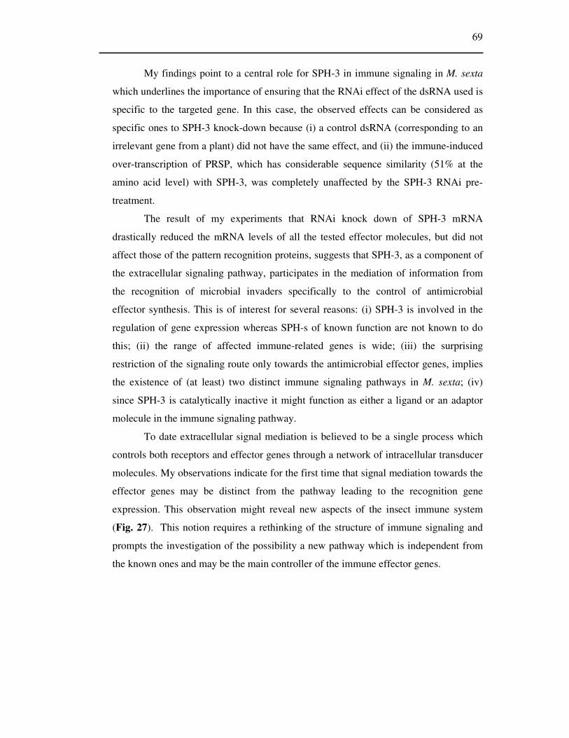
69
My findings point to a central role for SPH-3 in immune signaling in M. sexta
which underlines the importance of ensuring that the RNAi effect of the dsRNA used is
specific to the targeted gene. In this case, the observed effects can be considered as
specific ones to SPH-3 knock-down because (i) a control dsRNA (corresponding to an
irrelevant gene from a plant) did not have the same effect, and (ii) the immune-induced
over-transcription of PRSP, which has considerable sequence similarity (51% at the
amino acid level) with SPH-3, was completely unaffected by the SPH-3 RNAi pre-
treatment.
The result of my experiments that RNAi knock down of SPH-3 mRNA
drastically reduced the mRNA levels of all the tested effector molecules, but did not
affect those of the pattern recognition proteins, suggests that SPH-3, as a component of
the extracellular signaling pathway, participates in the mediation of information from
the recognition of microbial invaders specifically to the control of antimicrobial
effector synthesis. This is of interest for several reasons: (i) SPH-3 is involved in the
regulation of gene expression whereas SPH-s of known function are not known to do
this; (ii) the range of affected immune-related genes is wide; (iii) the surprising
restriction of the signaling route only towards the antimicrobial effector genes, implies
the existence of (at least) two distinct immune signaling pathways in M. sexta; (iv)
since SPH-3 is catalytically inactive it might function as either a ligand or an adaptor
molecule in the immune signaling pathway.
To date extracellular signal mediation is believed to be a single process which
controls both receptors and effector genes through a network of intracellular transducer
molecules. My observations indicate for the first time that signal mediation towards the
effector genes may be distinct from the pathway leading to the recognition gene
expression. This observation might reveal new aspects of the insect immune system
(Fig. 27). This notion requires a rethinking of the structure of immune signaling and
prompts the investigation of the possibility a new pathway which is independent from
the known ones and may be the main controller of the immune effector genes.

70
Figure 27. Proposed model of the role of SPH-3 in Manduca sexta immune pathways. Proposed model for the role of SPH3 in Manduca immune pathways. SPH-3 acts downstream of the pattern recognition proteins and is involved in signal mediation from pathogen recognition towards the gene regulation of immune effectors, but not of recognition proteins.
The existence of intracellular immune signaling pathways is already well
established in Drosophila, where pathways form the Toll and Imd receptors each
regulate the induced expression of partially overlapping sets of effector genes in
response to fungal/Gram positive and Gram negative bacterial infections, respectively
[121]. However, the extracellular part of these pathways, i.e. the link between the
recognition of microorganisms by the PRR and the activation of the intracellular Toll
(and also the Imd) pathway is still missing. In the developing Drosophila, Toll
activation is triggered by a cascade of serine proteases consisting of Gastrulation
defective, Snake and Easter, a process which is similar to the coagulation cascade in
mammalian blood. This cascade ends with the cleavage of the clip domain of the
cytokine-like protein Spätzle [122]. Being catalytically inactive, SPH-3 cannot function
as an enzyme in a proteolytic signaling cascade. It is possible; however, that it is a
functional counterpart in Manduca of Spätzle (or a related molecule) in Drosophila, i.e.
a ligand in an extracellular signaling pathway towards an unknown receptor. A Spätzle
homologue is present in Bombyx mori (also a Lepidopteran, like M. sexta) and the

71
involvement of Spätzle homologue in the signaling pathway has been shown in both
Bombyx and Manduca [123]. A Spätzle processing enzyme is present in Bombyx [124].
The possibility of a functional similarity between SPH-3 and Spätzle is not
supported by the pairwise amino acid sequence comparison. It showed no significant
similarity between SPH-3 and Spätzle (either Drosophila or Bombyx) but, surprisingly,
does show similarity between SPH-3 and Easter, which is a catalytically active, clip
domain containing serine proteinase of Drosophila in the signaling pathway towards to
Toll receptor. The sequences are 21.7% identical even when the full (clip domain
containing) sequence of Easter is included in the comparison. (Without the clip domain
the identity is 25.3%; Table 6)
Table 6. Results of the sequence comparison of SPH-3 with Spätzle, Gastrulation defective, Easer and Snake.
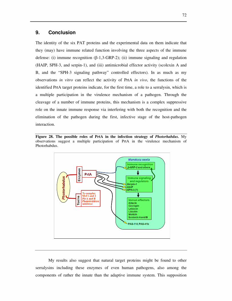
72
9. Conclusion
The identity of the six PAT proteins and the experimental data on them indicate that
they (may) have immune related function involving the three aspects of the immune
defense: (i) immune recognition (β-1,3-GRP-2); (ii) immune signaling and regulation
(HAIP, SPH-3, and serpin-1), and (iii) antimicrobial effector activity (scolexin A and
B, and the “SPH-3 signaling pathway” controlled effectors). In as much as my
observations in vitro can reflect the activity of PrtA in vivo, the functions of the
identified PrtA target proteins indicate, for the first time, a role to a serralysin, which is
a multiple participation in the virulence mechanism of a pathogen. Through the
cleavage of a number of immune proteins, this mechanism is a complex suppressive
role on the innate immune response via interfering with both the recognition and the
elimination of the pathogen during the first, infective stage of the host-pathogen
interaction.
Figure 28. The possible roles of PrtA in the infection strategy of Photorhabdus. My observations suggest a multiple participation of PrtA in the virulence mechanism of Photorhabdus.
My results also suggest that natural target proteins might be found to other
serralysins including these enzymes of even human pathogens, also among the
components of rather the innate than the adaptive immune system. This supposition

73
seems reasonable also because some innate immune mechanisms are conserved and
many are similar throughout the animal kingdom. The first challenge for the pathogens
during an infection is the innate immune response, thus it might be a winning strategy,
and effective virulence mechanism to weaken or destroy the components of it.

74
10. Abstract
In my thesis work I used an easily accessible, low cost insect-entomopathogen
bacterium model system for studying the role of a protease in the virulence mechanism.
The insect, Manduca sexta is widely used as a model for insect biochemical research
due to its size and hemolymph volume, while the bacterium, Photorhabdus, is an
intensively studied pathogen for its nematode-symbiotic lifecycle and very high
virulence.
Despite the fact that proteases can play various roles in establishing and
propagating infection, their function in these processes is rarely investigated and
therefore not known. The assessment of these is possible only through determining at
least the target proteins in their proteolytic system.
To find a virulence associated protease, I investigated the protease production of
P. luminescens with various methods, during both culturing of 20 strains and infection.
Three proteolytic activities could be distinguished this way. One of these enzymes was
PrtA, a metzincin metalloprotease, a member of the serralysin family which was the
earliest secreted enzyme by Photorhabdus in both culture growth and during infection.
It was hypothesized that PrtA might function as a virulence factor. To prove this, I
investigated its natural substrates in Manduca sexta hemolymph. I found sixteen PrtA
target (PAT) proteins which were selectively cleaved by PrtA. I purified nine of them
partially and one of them fully. With the help of their N-terminal sequence I could
identify six PAT proteins from the NCBI protein database. Each of these has immune
related function involving the three aspects of immune defense: recognition, signal
mediation and effector functions.
I investigated further one of the PrtA target proteins, SPH-3 (serine protease
homolog-3) which had been known as an immune-inducible protein, but its function
had not been studied. I found that SPH-3 plays a very important role in the immune
signaling in Manduca sexta: RNAi knock down of SPH-3 mRNA drastically increased
the mortality of the insects infected with Photorhabdus. This was accompanied by
dramatically reduced mRNA levels of all the tested immune effector (but not the
pattern recognition, immune receptor) molecules. I concluded from this that SPH-3 act
as an extracellular component in a signaling pathway, which is responsible for the
control of antimicrobial effector synthesis. Such a group specific immune signaling,

75
which distinguishes the immune receptor from the immune effector genes is not known
yet in the insect immune system.
Based on my results I suppose that PAT-41, 110 and (most of) the other,
unidentified PAT proteins also have immune related function, and may be new, as yet
unknown components in the immune system. In conclusion, my observations support
the assumption that PrtA is a virulence factor of Photorhabdus which might have an
immune-suppressive role during infection.

76
11. Összefoglalás
Munkám során olyan Manduca-Photorhabdus rovar-entomopatogén baktérium
modellrendszert használtam egy proteáz virulenciában betöltött szerepének
tanulmányozására, ami széles körben használt, könnyen hozzáférhető és alacsony
költségű. Manduca sexta méretének, és a belőle nyerhető hemolimfa mennyiségének
köszönhetően a biokémiai kutatásokban széles körben alkalmazott rovar modell. A
Photorhabdus rovarpatogén baktériumokat sokan vizsgálják erős virulenciájuk, és
érdekes fonalféreg-szimbióta életmódjuk következtében.
Annak ellenére, hogy a fertőzések kialakításában a bakteriális fehérjebontó
enzimeknek sokféle szerepe lehet, funkciójuk csak kevéssé vizsgált, így szinte nem is
ismert. A patomechanizmusban betöltött szerepüket szubsztrátspecifitásuk és
proteolitikus rendszereik felderítésével lehetne jobban megismerni.
Első célom egy olyan proteáz keresése volt, amely részt vehet a Photorhabdus
által kialakított fertőzésben. 20 Photorhabdus baktérium törzs proteáz termelését
hasonlítottam össze különböző módszerekkel kultúrában, és fertőzést követően
egyaránt. Három proteolitikus enzimet különítettem el. Ezek közül a PrtA, egy
serralysin metalloproteáz volt az, ami a legkorábban termelődött mind kultúrában mind
a fertőzést követően egyaránt. A hipotézisem szerint az az enzim, amely a legkorábban
termelődik, tehát a PrtA, lehet az a proteáz, amely virulencia faktorként fontos szerepet
játszhat a fertőzés kialakításában. Ennek bizonyítására olyan természetes szubsztrát
fehérjéket kerestem Manduca sexta hemolimfában, amelyeket a PrtA szelektíven hasít.
Összesen tizenhat ilyen célfehérjét találtam, amelyek közül kilencet teljesen, illetve
részlegesen megtisztítottam. N-terminális szekvenciájuk alapján hat fehérjét tudtam
azonosítani az NCBI fehérje adatbázisból. Mind a hat azonosított protein immun-
fehérjének bizonyult.
Továbbiakban az egyik PrtA target fehérjének, az SPH-3-nak (szerin proteáz
homológ-3) a rovar immunrendszerben betöltött lehetséges szerepét vizsgáltam.
Kísérleteim alapján elmondható, hogy az SPH-3 jelentős szerepet tölt be a M. sexta
immun-szignalizációs útvonalában; az SPH-3 RNSi-vel történő kiütését követően
jelentősen megnőtt a rovarok Photorhabdus fertőzést követő mortalitása. Ezzel
egyidejűleg az immunválaszt hordozó, összes jelenleg ismert antimikrobiális fehérje és
peptid génjének expressziója leállt, míg az immun receptorok expressziója, ill.
indukciója változatlan maradt. Ebből arra következtettem, hogy az SPH-3 központi

77
szerepet tölthet be olyan sejten kívüli immunjelátviteli folyamatban, amely az
antimikrobiális effektorok szintéziséért felelős. Ilyen specifikus immun-szignalizációs
útvonalra, amely megkülönbözteti az immun receptor és az immun effektor gének
indukálását, eddig még nem volt példa a rovarok immunrendszerében.
Eddigi eredményeim alapján feltételezem, hogy az adatbázisban nem szerepelő
PrtA target fehérjék (PAT-41 és PAT-110), ugyancsak immun-fehérjék, amelyek új,
vagy eddig még ismeretlen résztvevői lehetnek az immunrendszernek. Összegzésként
elmondható, hogy megfigyeléseim alátámasztják azt a feltételezésemet, hogy a PrtA
proteáz rendelkezhet immunszuppresszív szereppel a Photorhabdus fertőzési
mechanizmusában.

78
12. References
1. Judit Marokhazi, Katalin Lengyel, Szilvia Pekaár, Gabriella Felfoldi, Andras Patthy, Laszlo Graf, Andras Fodor and Istvan Venekei (2004) Comparison of Proteolytic Activities Produced by Entomopathogenic Photorhabdus Bacteria: Strain- and Phase-Dependent Heterogeneity in Composition and Activity of Four Enzymes. Applied and Environmental Microbiology 70, 7311–7320
2. Tong, Y., and Kanost, M. R. (2005) Manduca sexta serpin-4 and serpin-5 inhibit the prophenol oxidase activation pathway: cDNA cloning, protein expression, and characterization. J. Biol. Chem. 280, 14923–14931
3. Belas, R., Manos, J. and Suvanasuthi, R. (2004) Proteus mirabilis ZapA metalloprotease degrades a broad spectrum of substrates, including antimicrobial peptides. Infect Immun. 72, 5159-5167
4. Silva, C. P., Waterfield, N. R., Daborn, P. J., Dean, P., Chilver, T., Au, C. P., Sharma, S., Potter, U., Reynolds, S. E. and ffrench-Constant, R. H. (2002) Bacterial infection of a model insect: Photorhabdus luminescens and Manduca sexta. Cell Microbiol. 4, 329-339
5. Michael R. Kanost, Haobo Yiang, Xiao-Qiang Yu (2004) Innate immune response of lepidopteran insect, Manduca sexta. Immunological Reviews; 198, 97-105
6. Lemaitre B, Nicolas E, Michaut L, Reichhart JM, Hoffmann JA. (1996) The dorsoventral regulatory gene cassette spatzle/Toll/cactus controls the potent antifungal response in Drosophila adults. Cell 86, 973–83.
7. R, Preston-Hurlburt P, Janeway CA Jr. (1997) A Inhuman homologue of the Drosophila Toll protein signals Dictyactivation of adaptive immunity. Nature; 388, 394–397
8. Abanti Chattopadhyay, N. B. Bhatnagar, and Rakesh Bhatnagar (2004) Bacterial Insecticidal Toxins. Critical Reviews in Microbiology; 30, 33–54
9. Jules A Hoffman (2003) The immune response of Drosophila. Nature; 426, 33-38 10. Eleftherianos I, Marokhazi J, Millichap PJ, Hodgkinson AJ, Sriboonlert A, ffrench-Constant
RH, Reynolds SE (2006) Prior infection of Manduca sexta with non-pathogenic Escherichia coli elicits immunity to pathogenic Photorhabdus luminescens: roles of immune-related proteins shown by RNA interference. Insect Biochem Mol Biol. 36; 517-25
11. Eleftherianos I, Gökçen F, Felföldi G, Millichap PJ, Trenczek TE, ffrench-Constant RH, Reynolds SE. (2007) The immunoglobulin family protein Hemolin mediates cellular immune responses to bacteria in the insect Manduca sexta. Cell Microbiol. 9; 1137-47
12. Ling E, Yu XQ. (2006) Cellular encapsulation and melanization are enhanced by immulectins, pattern recognition receptors from the tobacco hornworm Manduca sexta. Dev Comp Immunol. 30; 289-99
13. Peter E Dunn (1986) Biochemical aspects of insect immunology Ann. Rev. Entomol. 31 321-39 14. Dunphy G. B., Thurston G. S. (Editors: Gaugler R., Kaya H. K.) (1990) Insect Immunity.
Entomopathogen Nematodes in Biological Conrrol. Boca Raton, Florida, Crc Press 301-323 15. Xiao-Qiang Yu a, Haobo Jiang b, Yang Wang b, Michael R. Kanost (2003) Nonproteolytic
serine proteinase homologs are involved in prophenoloxidase activation in the tobacco hornworm, Manduca sexta. Insect Biochemistry and Molecular Biology 33, 197–208
16. Kenneth Söderhall and Lage Cerenius (1998) Role of the prophenoloxidase-activating sytrem in invertebrate immunity. Current Opinion in Immunology; 10, 23-28
17. Yang Wang, Haobo Jiang (2004) Phenoloxidase (proPO) activation in Manduca sexta: an analysis of molecular interaction among proPO, proPO-activating proteinase-3 and a cofactor. Insect Biochemistry and Molecular Biology; 34, 731-742
18. Jiang, H., Wang, Y., Kanost, M.R. (1998) Pro-phenol oxidase activating proteinase from an insect, Manduca sexta: a bacteriainducible protein similar to Drosophila easter. Proc. Nat. Acad. Sci. 95, 12220–12225
19. Jiang, H., Wang, Y., Yu, X.-Q., Kanost, M.R. (2003) Prophenoloxidase- activating proteinase-2 (PAP-2) from hemolymph of Manduca sexta: a bacteria-inducible serine proteinase containing two clip domains. J. Biol. Chem. 278, 3552–3561.
20. Jiang, H., Wang, Y., Yu, X.-Q., Zhu, Y., Kanost, M.R. (2003) Prophenoloxidase-activating proteinase-3 (PAP-3) from Manduca sexta hemolymph: a clip-domain serine proteinase regulated by serpin-1J and serine proteinase homologs. Insect Biochem. Mol. Biol. 33, 1049–1060.
21. Jiang, Michael R. Kanost (2000) The clip-domain family of serine proteinases in arthropods. Insect Biochemistry and Molecular Biology 30, 95–105

79
22. Zhu Y, Wang Y, Gorman MJ, Jiang H, Kanost MR. (2003) Manduca sexta serpin-3 regulates prophenoloxidase activation in response to infection by inhibiting prophenoloxidase activating enzyme. J. Biol Chem. 278 46556-46564.
23. Youren Tong‡, Haobo Jiang§, and Michael R. Kanost (2005) Identification of Plasma Proteases Inhibited by Manduca sexta Serpin-4 and Serpin-5 and Their Association with Components of the Prophenol Oxidase Activation Pathway. J. Biol. Chem. 280, 14932–14942,
24. Haobo Jianga, Yang Wanga, Yongli Gub, Xiaoping Guoa, Zhen Zoua, Frank Scholzc, Tina E. Trenczekc, Michael R. Kanost (2005) Molecular identification of a bevy of serine proteinases in Manduca sexta hemolymph Insect Biochemistry and Molecular Biology; 35 931–943
25. Chuanyi Ji, Yang Wang, Xiaoping Guo‡, Steve Hartson, and Haobo Jiang (2004) A Pattern Recognition Serine Proteinase Triggers the Prophenoloxidase Activation Cascade in the Tobacco Hornworm, Manduca sexta J. Biol. Chem. 279, 34101- 34106,
26. Yang Wang and Haobo Jiang (2006) Interaction of β-1,3-Glucan with Its Recognition Protein Activates Hemolymph Proteinase 14, an Initiation Enzyme of the Prophenoloxidase Activation System in Manduca sexta J. Biol. Chem 281, 9271-9278
27. Wang Y, Jiang H (2007) Reconstitution of a branch of the Manduca sexta prophenoloxidase activation cascade in vitro: Snake-like hemolymph proteinase 21 (HP21) cleaved by HP14 activates prophenoloxidase-activating proteinase-2 precursor. Insect Biochem Mol Biol. 37, 1015-25.
28. Matsushita, M., Fujita, T. (2001) Ficolins and the lectin complement pathway. Immunological Reviews; 180, 78-85.
29. Ashida, M., Brey, P.T., (1998). Recent advances on the research of the insect prophenoloxidase cascade. In: Brey, P.T., Hultmark, D. (Eds.), Molecular Mechanisms of Immune Responses in Insects. Chapman and Hall, London, 135–172.
30. Ross J, Jiang H, Kanost MR, Wang Y. (2003) Serine proteases and their homologs in the Drosophila melanogaster genome: an initial analysis of sequence conservation and phylogenetic relationships. Gene; 304, 117-131.
31. Murugasuoei, B., et al., Masquerade, a Protein with Homology to Serine Proteases, Plays a Role in the Development of the Drosophila Nerve, Muscle and Tracheal Systems. Journal of Neurochemistry, 1994. 63: p. S8-S8.
32. Muta T, Hashimoto R, Miyata T, Nishimura H, Toh Y, Iwanaga S. (1990) Proclotting Enzyme from Horseshoe-Crab Hemocytes - cDNA Cloning, Disulfide Locations, and Subcellular-Localization. Journal of Biological Chemistry; 265, 22426-22433.
33. Murugasu-Oei B, Rodrigues V, Yang X, Chia W. (1995) Masquerade: a novel secreted serine protease-like molecule is required for somatic muscle attachment in the Drosophila embryo. Genes Dev. 15; 139-54.
34. MurugasuOei, B., R Balakrishnan, X Yang, W Chia, V Rodrigues (1996) Mutations in masquerade, a novel serine-protease-like molecule, affect axonal guidance and taste behavior in Drosophila. Mechanisms of Development; 57, 91-101.
35. Kawabata, S., Tokunaga, F., Kugi, Y., Motoyama, S., Miura, Y., Hirata, M., Iwanaga, S., (1996) Limulus factor D, a 43 kDa protein isolated from horseshoe crab hemocytes, is a serine proteinase homologue with antimicrobial activity. FEBS Lett. 398, 146–150.
36. Huang, T., Wang, H., Lee, S.Y., Johansson, M.W., So¨derha¨ll, K., Cerenius, L., (2000) A cell adhesion protein from the crayfish Pacifastacus leniusculus, a serine proteinase homologue similar to Drosophila masquerade. J. Biol. Chem. 275, 9996–10001.
37. Dimopoulos, G., Richman, A., Muller, H.M., Kafatos, F.C., (1997) Molecular immune responses of the mosquito Anopheles gambiae to bacteria and malaria parasites. Proc. Natl. Acad. Sci. USA 94, 11508–11513.
38. Kwon TH, Kim MS, Choi HW, Joo CH, Cho MY, Lee BL, (2000) A masquerade-like serine proteinase homologue is necessary for phenoloxidase activity in the coleopteran insect, Holotrichia diomphalia larvae. European Journal of Biochemistry; 267(20), 6188-6196.
39. Kim, M. S., Choi, H. W., Joo, C. H., Cho, M. Y., and Lee, B. L., (2002) A new easter-type serine protease cleaves a masquerade-like protein during prophenoloxidase activation in Holotrichia diomphalia larvae. Journal of Biological Chemistry; 277(42), 39999-40004.
40. Kum Young Lee, Rong Zhang, Moon Suk Kim, Ji Won Park, Ho Young Park, Shun-ichiro Kawabata, Bok Luel Lee (2002) A zymogen form of masquerade-like serine proteinase homologue is cleaved during pro-phenoloxidase activation by Ca2+ in coleopteran and Tenebrio molitor larvae. European Journal of Biochemistry; 269(17) 4375-4383.

80
41. Buda, E.S. and T.H. Shafer (2005) Expression of a serine proteinase homolog prophenoloxidase-activating factor from the blue crab, Callinectes sapidus. Comparative Biochemistry and Physiology B-Biochemistry & Molecular Biology; 140(4) 521-531.
42. Asgari, S., Zhang, G. M., Zareie, R. and Schmidt, O. (2003) A serine proteinase homolog venom protein from an endoparasitoid wasp inhibits melanization of the host hemolymph. Insect Biochemistry and Molecular Biology; 33(10) 1017-1024.
43. Achara Rattanachai, Ikuo Hirono, Tsuyoshi Ohira, Yukinori Takahashi and Takashi Aoki (2005) Peptidoglycan inducible expression of a serine proteinase homologue from kuruma shrimp (Marsupenaeus japonicus). Fish & Shellfish Immunology; 18(1) 39-48.
44. Lin CY, Hu KY, Ho SH, Song YL. (2006) Cloning and characterization of a shrimp clip domain serine protease homolog (c-SPH) as a cell adhesion molecule. Developmental and Comparative Immunology; 30(12) 1132-1144.
45. Lee, S.Y. and K. Soderhall (2001) Characterization of a pattern recognition protein, a masquerade-like protein, in the freshwater crayfish Pacifastacus leniusculus. Journal of Immunology; 166(12) 7319-7326.
46. Kang, D. Lundström A, Liu G, Steiner H, (2002) An azurocidin-like protein is induced in Trichoplusia ni larval gut cells after bacterial challenge. Developmental and Comparative Immunology; 26 495-503.
47. Xie Y, Li Y, Zhang Q, Stiller MJ, Wang CL, Streilein JW. (2000) Haptoglobin is a natural regulator of Langerhans cell function in the skin. Journal of Dermatological Science; 24 25-37.
48. Imrie, H., D.J.P. Ferguson, and K.P. Day, (2004) Human serum haptoglobin is toxic to Plasmodium falciparum in vitro. Molecular and Biochemical Parasitology; 133(1) 93-98.
49. A Kurosky, D R Barnett, T H Lee, B Touchstone, R E Hay, M S Arnott, B H Bowman, and W M Fitch (1980) Covalent Structure of Human Haptoglobin - a Serine Protease Homolog. Proceedings of the National Academy of Sciences of the United States of America-Biological Sciences; 77(6) 3388-3392.
50. Katsuhide Okunishi, Makoto Dohi, Kazuyuki Nakagome, Ryoichi Tanaka, Shinya Mizuno, Kunio Matsumoto, Jun-ichi Miyazaki, Toshikazu Nakamura and Kazuhiko Yamamoto (2005) A novel role of hepatocyte growth regulator as an immune regulator through suppressing dendritic cell function. Journal of Immunology; 175(7) 4745-4753.
51. T. Nakamura, T. Nishizawa, M. Hagiya, T. Seki, M. Shimonishi, A. Sugimura, K. Tashiro, S. Shimizu., (1989) Molecular-Cloning and Expression of Human Hepatocyte Growth-Factor. Nature; 342 (6248) 440-443.
52. Watorek, W., (2003) Azurocidin - inactive serine proteinase homolog acting as a multifunctional inflammatory mediator. Acta Biochimica Polonica; 50(3) 743-752.
53. Yang Wang and Haobo Jiang (2006) Interaction of β-1,3-Glucan with Its Recognition Protein Activates Hemolymph Proteinase 14, an Initiation Enzyme of the Prophenoloxidase Activation System in Manduca sexta The Journal of Biological Chemistry; 281 9271–9278,
54. Yang Wang, Haobo Jiang (2004)Purification and characterization of Manduca sexta serpin-6: a serine proteinase inhibitor that selectively inhibits prophenoloxidase-activating proteinase-3 Insect Biochemistry and Molecular Biology 34 387–395
55. Zhen Zou and Haobo Jiang (2005) Manduca sexta Serpin-6 Regulates Immune Serine Proteinases PAP-3 and HP8 J. Biol. Chem. 280, 14341–14348,
56. H., Wang Y., Huang Y., Mulnix A. B., Kadel J., Cole K., Kanost M. (1996) Organization of serpin gene-1 from Manduca sexta Journal of Biological Chemistry 271, 28017-28023
57. Sheng Ye, Elizabeth J Goldsmith, (2001) Serpins and other covalent protease inhibitors, Current Opinion in Structural Biology 11, 740-745
58. Michael R. Kanost, Sarvamangala V. Prasad, Michael A. Wells, (1989) Primary structure of a member of the serpin superfamily of proteinase inhibitors from an insect, Maduca sexta J. Biol. Chem 264 965-972
59. Haobo Jiang, Yang Wang, and Michael R. Kanost (1994) Mutually Exclusive Exon Use and Reactive Center Diversityin Insect Serpins J. Biol. Chem. 269, 55-58,
60. Gan, H., Wang, Y., Jiang, H., Mita, K., and Kanost, M. R. (2001) Insect Biochem. Mol. Biol. 31, 887–898
61. Haobo Jiang and Michael R. Kanost (1997) Characterization and Functional Analysis of 12 Naturally Occurring Reactive Site Variants of Serpin-1 from Manduca sexta J. Biol. Chem. 272, 1082–1087
62. Jules Hoffmann, Jean-Marc Reichhart, Charles Hetru, (1996) Innate immunity in higher insects Current opinion in immunology 8-13

81
63. Michael R. Kanost, J. K. Kawooya, J. H. Law, R. O. Ryan, M. C. van Hausde and R. Ziegler (1990) Insect Haemolymph Proteins Advances in Insect Physiology; 22, 299-366
64. Yang Wang, Haobo Jiang (2008) A positive feedback mechanism in the Manduca sexta prophenoloxidase activation system Insect Biochemistry and Molecular Biology; 38, 763– 769
65. Duchaud, E., Rusniok, C., Frangeul, L., Buchrieser, C., Givaudan, A., Taourit, S., Bocs, S., Boursaux-Eude, C., Chandler, M., Charles, J. F., Dassa, E., Derose, R., Derzelle, S., Freyssinet, G., Gaudriault, S., Medigue, C., Lanois, A., Powell, K., Siguier, P., Vincent, R., Wingate, V., Zouine, M., Glaser, P., Boemare, N., Danchin, A. and Kunst, F. (2003) The genome sequence of the entomopathogenic bacterium Photorhabdus luminescens. Nat Biotechnol. 21, 1307-1313
66. ffrench-Constant, R., Waterfield, N., Daborn, P., Joyce, S., Bennett, H., Au, C., Dowling, A., Boundy, S., Reynolds, S. and Clarke, D. (2003) Photorhabdus: towards a functional genomic analysis of a symbiont and pathogen. FEMS Microbiol Rev. 26, 433-456
67. Richard H. ffrench-Constant and Nicholas Waterfield (2006) An ABC guide to the bacterial toxin complexes. Advances in Applied Microbiology; 58, 169-183
68. Farmer, J. J., 3rd, Jorgensen, J. H., Grimont, P. A., Akhurst, R. J., Poinar, G. O., Jr., Ageron, E., Pierce, G. V., Smith, J. A., Carter, G. P., Wilson, K. L. and et al. (1989) Xenorhabdus luminescens (DNA hybridization group 5) from human clinical specimens. J Clin Microbiol. 27, 1594-1600
69. Keneth H. Nelson, Thomas M. Schmidt, Bruce Bleakley. (1990) Physiology and Biochemistry of Xenorhabdus Entomopathogenic; Nematodes in Biological control; Editors: Gaugler R., Kaya H. K.) Boca Raton, CRC Press 271-284
70. Karine Brugirard-Ricaud, Eric Duchaud, Alain Givaudan, Pierre Alain Girard, Frank Kunst, Noel Boemare, Michel Brehélin and Robert Zumbihl (2005) Site-specific antiphagocytic function of the Photorhabdus luminescens type III secretion system during insect colonization Cellular Microbiolog; 7 (3), 363–371
71. Liu D, Burton S, Glancy T, Li ZS, Hampton R, Meade T, Merlo DJ. (2003) Insect resistance conferred by 283-kDa Photorhabdus luminescens protein TcdA in Arabidopsis thaliana. Nature Biotechnology; 21 1222-8.
72. Steven Forst, Barbara Dowds, Noel Boemare, Erico Stackebrandt (1997) Xenorhabdus and Photorhabdus spp.: Bugs that kill bugs. Annual Review of Microbiology 51: 47-72
73. Steven Forst, Kenneth Nealson (1996) Molecular biology of the Symbiotic-pathogenic Bacteria Xenorhabdus spp. and Photorhabdus spp. Microbiological Reviews 60, 21-43 (1996)
74. Robert J. Watson, Susan A. Joyce, Georgette V. Spencer and David J. Clarke (2005) The exbD gene of Photorhabdus temperate is required for full virulence in insects and symbiosis with the nematode Heterorhabditis Molecular Microbiology; 56 (3), 763–773
75. Kaiji Hu, John M. Webster (2000) Antibiotic production in relation to bacterial growth and nematode development in Photorhabdus-Heterorhabditis infected Galleria mellonella larvae FEMS Microbiology Letters 189, 219-223
76. Lining Guo, Raymond O. Fating III, Gregory L. Orr, Barry W. Schafer, James A. Strickland,Kitisri Sukhapinda, Aaron T. Woodsworth, and James K. Petell (1999) Photorhabdus luminescens W-14 Insecticidal activity consists of at least two similar but distinct proteins. The Jurnal of Biological chemistry; 274 (14), 9836-9842
77. Richard H. ffrench-Constant, Andrea Dowlingb, Nicholas R. Waterfield (2007) Insecticidal toxins from Photorhabdus bacteria and their potential use in agriculture. Toxicon. 15, 436-51.
78. Daborn, P. J., Waterfield, N., Blight, M. A. and ffrench-Constant, R. H. (2001) Measuring Virulence Factor Expression by the Pathogenic Bacterium Photorhabdus luminescens in Culture and during Insect Infection. J Bacteriol. 183, 5834-5839
79. 20. Eleftherianos I, Millichap PJ, ffrench-Constant RH, Reynolds SE. (2006) RNAi suppression of recognition protein mediated immune responses in the tobacco hornworm Manduca sexta causes increased susceptibility to the insect pathogen Photorhabdus. Dev Comp Immunol. 30(12) 1099-107.
80. Ioannis Eleftherianos, Sam Boundy, Susan A. Joyce, Shazia Aslam, James W. Marshall, Russell J. Cox, Thomas J. Simpson, David J. Clarke, Richard H. ffrench-Constant, and Stuart E. Reynolds (2007) An antibiotic produced by an insect pathogenic bacterium suppresses host defenses through phenoloxidase inhibition PNAS 104, 2419–2424
81. Wei Lin, Karla Jean Fullner, Rebecca Clayton, Jessica A. Sexton, Marc B. Rogers, Kerstin E. Calia, Stephen B. Calderwood, Claire Fraser, John J. Mekalanos (1999) Identification of a Vibrio cholerae RTX toxin gene cluster that is tightly linked to the cholera toxin prophage PNAS 96, 1071–1076

82
82. Karla Jean Fullner and John J. Mekalanos (2000) In vivo covalent cross-linking of cellular actin by the Vibrio cholerae RTX toxin The EMBO Jurnal; .19, 5315-5323
83. Nicholas Waterfield, Shizuo George Kamita, Bruce D. Hammock and Richard ffrench-Constant (2005) The Photorhabdus Pir toxins are similar to a developmentally regulated insect protein but show no juvenile hormone esterase activity FEMS Microbiology Letters; 245, 47-52
84. Crosland RD, Hsiao TH, McClure WO., (1984) Purification and characterization of beta-leptinotarsin-h, an activator of presynaptic calcium channels. Biochemistry; 23(4), 734-41
85. Jiménez-Juárez N, Muñoz-Garay C, Gómez I, Saab-Rincon G, Damian-Almazo JY, Gill SS, Soberón M, Bravo A. (2007) Bacillus thuringiensis Cry1Ab Mutants Affecting Oligomer Formation Are Non-toxic to Manduca sexta Larvae J Biol Chem. 282(29), 21222-9.
86. P. J. Daborn, N. Waterfield, C. P. Silva, C. P. Y. Au, S. Sharma, and R. H. ffrench-Constant (2002) A single Photorhabdus gene, makes caterpillars floppy (mcf ), allows Escherichia coli to persist within and kill insects. PNAS; 99, 10742–10747
87. Dowling AJ, Daborn PJ, Waterfield NR, Wang P, Streuli CH, ffrench-Constant RH. (2004) The insecticidal toxin Makes caterpillars floppy (Mcf) promotes apoptosis in mammalian cells. Cell Microbiol. 4, 345-53.
88. Nicholas R. Waterfield, Phillip J. Daborn, Andrea J. Dowling, Guowei Yang, Michelle Hares, Richard H. ffrench-Constant (2003) The insecticidal toxin Makes caterpillars floppy 2 (Mcf2) shows similarity to HrmA, an avirulence protein from plant pathogen FEMS Microbiology Letters 229 265-270
89. Bowen D, Rocheleau TA, Blackburn M, Andreev O, Golubeva E, Bhartia R, ffrench-Constant RH. (1998) Insecticidal toxins from the bacterium Photorhabdus luminescens. Science; 26;280(5372), 2129-32.
90. Blackburn, M., Golubeva, E., Bowen, D. and Ffrench-Constant, R. H. (1998) A novel insecticidal toxin from Photorhabdus luminescens, toxin complex a (Tca), and its histopathological effects on the midgut of Manduca sexta. Appl Environ Microbiol. 64, 3036-3041
91. Waterfield N, Hares M, Yang G, Dowling A, ffrench-Constant R. (2005) Potentiation and cellular phenotypes of the insecticidal Toxin complexes of Photorhabdus bacteria. Cell Microbiol. 3, 373-82.
92. Nicholas R.Waterfield,David J. Bowen, Jacqueline D. Fetherston, Robert D. Perry and Richard H. ffrench-Constant (2001) The tc genes of Photorhabdus: a growing family Trends in Microbiology; 9, 185-191
93. Haq SK, Atif SM, Khan RH (2004) Protein proteinase inhibitor genes in combat against insects, pests, and pathogens: natural and engineered phytoprotection. Arch Biochem Biophys. 431(1), 145-59.
94. Bowen DJ, Ensign JC (1998) Purification and characterization of a high-molecular-weight insecticidal protein complex produced by the entomopathogenic bacterium photorhabdus luminescens. Appl Environ Microbiol. 8, 3029-35.
95. Dalhammar G, Steiner H. (1984) Characterization of inhibitor A, a protease from Bacillus thuringiensis which degrades attacins and cecropins, two classes of antibacterial proteins in insects. Eur J Biochem. 139(2), 247-52.
96. Andrejko M. (1999) Exoproteinases of the type A in pathogenesis of insect bacterial diseases. Folia Biol (Krakow), 47(3-4), 135-41.
97. Park SY, Kim KM, Lee JH, Seo SJ, Lee IH. Extracellular gelatinase of Enterococcus faecalis destroys a defense system in insect hemolymph and human serum. Infect Immun. 2007 Apr;75(4):1861-9. Epub 2007 Jan 29
98. RH ffrench-Constant, N. waterfield, V. Burland, NT Perna, PJ Daborn, D. Bowen, and FR Blattner (2000) A Genomic Sample Sequence of the Entomopathogenic Bacterium Photorhabdus luminescens W14: Potential Implications for Virulence Applied and Environmental Microbiology, 66, 3310–3329
99. David J. Bowen, Thomas A. Rocheleau, Cathy K. Grutzmacher, Laurence Meslet, Michelle Valens, Daniel Marble, Andrea Dowling, Richard ffrench-Constant and Mark A. Blight Genetic and biochemical characterization of PrtA, an RTX-like metalloprotease from Photorhabdus Microbiology (2003), 149, 1581–1591
100. Valens M, Broutelle AC, Lefebvre M, Blight MA :A zinc metalloprotease inhibitor, Inh, from the insect pathogen Photorhabdus luminescens Microbiology. 2002 Aug;148(Pt 8):2427-37
101. Marokházi J, Mihala N, Hudecz F, Fodor A, Gráf L, Venekei I. (2007) Cleavage site analysis of a serralysin-like protease, PrtA, from an insect pathogen Photorhabdus luminescens and development of a highly sensitive and specific substrate. FEBS J. 274(8), 1946-56

83
102. Handbook of Proteolytic Enzymes (1998) Academic Press, London. 103. Electronic source: http://merops.sanger.ac.uk 104. Matheson NR, Potempa J, Travis J (2006) Interaction of a novel form of Pseudomonas
aeruginosa alkaline protease (aeruginolysin) with interleukin-6 and interleukin-8. Biol Chem. 387(7), 911-5
105. Reynolds, S.E. Nottingham, S.F., Stephens, A.E. (1985) Food and water economy and its relation to growth in 5-th larvae of the tobacco hornworm, Manduca sexta. J. Insect Physiol. 31, 119-127
106. Van Wart, H. E., and D. R. Steinbrink (1981) A continuous spectrophotometric assay for Clostridium histolyticum collagenase. Anal. Biochem. 113: 356–365.
107. Marokhazi, J., G. Koczan, F. Hudecz, L. Graf, A. Fodor, and I. Venekei (2004) Enzymic characterization with progress curve analysis of a collagen peptidase from an enthomopathogenic bacterium, Photorhabdus luminescens.Biochem. J. 379, 633–640.
108. Zhu,Y., T.J.Johnson, A.A.Myers and M.R.Kanost (2003). Identification by subtractive suppression hybridization of bacteria-induced genes expressed in Manduca sexta fat body. Insect Biochem. Mol. Biol. 33,541-559.
109. Wee, K. E., C. R. Yonan, and F. N. Chang (2000) A new broad-spectrum protease inhibitor from the entomopathogenic bacterium Photorhabdus luminescens. Microbiology 146, 3141–3147.
110. Wang, H., and B. C. Dowds (1993) Phase variation in Xenorhabdus luminescens: cloning and sequencing of the lipase gene and analysis of its expression in primary and secondary phases of the bacterium. J. Bacteriol. 175, 1665– 1673
111. Forst, S., Dowds, B., Boemare, N. & Stackebrandt, E. (1997) Annual Review of Microbiology 51, 47-72.
112. Ma,C. and M.R.Kanost (2000). A β1,3-glucan recognition protein from an insect, Manduca sexta, agglutinates microorgansims and activates the phenoloxidase cascade. J.Biol.Chem. 275, 7505-7514.
113. Beschin,A., M.Bilej, F.Hanssens, J.Raymakers, E.Van Dyck, H.Revets, L.Brys, J.Gomez, P.De Baetselier and M.Timmermans (1998) Identification and cloning of a glucan- and lipopolisaccharide binding protein from Eisenia foetia earthworm involved in the activation of prophenoloxidase cascade. J.Biol.Chem. 273, 24948-24954.
114. Kim,Y.-S., J.-H.Ryu, S.-J.Han, K.-H.Choi, K.-B.Nam, I.-H.Jang, B.Lemaitre, P.T.Brey and W.-J.Lee (2000) Gram-negative bacteria-binding protein, a pattern recognition receptor for lipopolisaccharide and β-1,3-glucan that mediates the signalling for the induction of innate immune genes in Drosophila melanogaster cells. J.Biol.Chem. 275, 32721-32727.
115. Lee,S.Y., R.Wang and K.Söderhall (2000) A lipopolisaccharide- and β-1,3-glucan-binding protein from haemocytes of the freshwater crayfish Pacifastacus leniusculus. J.Biol.Chem. 275, 1337-1343.
116. Yoshida,H., K.Kinoshita and M.Ashida (1996) Purification of a peptidoglycan recognition protein from Hemolymph of the silkworm, Bombyx mori. J.Biol.Chem. 271, 13854-13860
117. Jiang,H., C.Ma, Z.-Q.Lu and M.R.Kanost (2004) β-1,3-glucan recognition protein-2 (βGRP-2) from Manduca sexta: an acute-phase protein that binds β -1,3-glucan and lipoteichoic acid to aggregate fungi and bacteria and stimulate prophenoloxidase activation. Insect Biochem. Mol. Biol. 34. 89-100.
118. Kanost,M.R., M.K.Zepp, N.E.Ladendorff and L.A.Anderrsson (1994) Isolation and characterization of a hemocyte aggregation inhibitor from hemolymph of Manduca sexta larvae. Arch. Insect Biochem. Physiol. 27, 123-136.
119. Finnerty,C.M., P.A.Karplus and R.R.Granados (1999) The insect immune protein scolexin is a novel serine proteinase homolog. Protein Sci. 8, 242-248.
120. Kyriakides,T.R., J.L.McKillip and K.D.Spence (1995) Biochemical characterization, developmental expression and induction of the immune protein scolexin from Manduca sexta. Arch. Insect Biochem. Physiol. 29, 269-280.
121. Bruno Lemaitre and Jules Hoffmann 2007 The Host Defense of Drosophila melanogaster Annu. Rev. Immunol. 2007. 25:697–743
122. Vincent Leclerc Jean-Marc Reichhart. 2004. The immune response of Drosophila Melanogaster Immunological Reviews Vol. 198: 59–71
123. Proteolytic activation of pro-spätzle is required for the induced transcription of antimicrobial peptide genes in lepidopteran insects. Wang Y, Cheng T, Rayaprolu S, Zou Z, Xia Q, Xiang Z, Jiang H. Dev Comp Immunol. 2007;31(10):1002-12. Epub 2007 Feb 12.

84
124. A Spätzle-processing enzyme required for toll signaling activation in Drosophila innate immunity. Jang IH, Chosa N, Kim SH, Nam HJ, Lemaitre B, Ochiai M, Kambris Z, Brun S, Hashimoto C, Ashida M, Brey PT, Lee WJ. Dev Cell. 2006 Jan;10 (1):45-55.