Embed Size (px)
Citation preview
JBUON 2018; 23 (Suppl 1): S122-S131ISSN: 1107-0625, online ISSN: 2241-6293 • www.jbuon.comE-mail: [email protected]
ORIGINAL ARTICLE
Correspondence to: Calin Cainap, MD, PhD. University of Medicine and Pharmacy “I.Hatieganu”, Cluj-Napoca, and Oncology Institute “Prof Dr Ion Chiricuta”, Republicii 34-36 str, 400015 PO, Cluj-Napoca, Romania. Tel: +40 724543672, E-mail: [email protected]: 29/05/2018; Accepted: 22/06/2018
The role of biomarkers and echocardiography in the evaluation of cardiotoxicity risk in children treated for leukemiaDiana Raluca Maniu1,2, Cristina Blag6,7*, Gheorghe Popa6,7, Madalina Bota6,7, Catalin Vlad2,3*, Calin Cainap2,4, Ovidiu Balacescu5, Laura Pop8, Simona Sorana Cainap1,7
1Emergency County Hospital for Children, Pediatric Clinic no 2, Department of Pediatric Cardiology, Cluj-Napoca, Romania; 2University of Medicine and Pharmacy “I.Hatieganu”, Department No 11 – Oncology, Cluj-Napoca, Romania; 3Institute of Oncology “I.Chiricuta”, Department of Oncological Surgery, Cluj-Napoca, Romania; 4Institute of Oncology “I.Chiricuta”, Department of Medical Oncology, Cluj-Napoca, Romania; 5Institute of Oncology “I.Chiricuta”, Department of Functional Genomics, Proteomics and Experimental Pathology, Cluj-Napoca, Romania; 6Emergency County Hospital for Children, Pediatric Clinic no 2, Department of Pediatric Oncology, Cluj-Napoca, Romania; 7University of Medicine and Pharmacy “I.Hatieganu”, Department No 9 – Mother & Child, Cluj-Napoca, Romania; 8University of Medicine and Pharmacy “I.Hatieganu”, The Research Center for Advanced Medicine MEDFUTURE, Cluj-Napoca, Romania.
*These authors contributed equally in this work.
Summary
Purpose: To describe the high-risk profile group, susceptible to develop anthracycline-induced cardiomyopathy in chil-dren with acute leukemia.
Methods: The study involved 35 pediatric patients diag-nosed with acute lymphoblastic (ALL) or acute myeloblastic leukemia (AML), from March 2014 to December 2016. Se-rologic markers used for the analysis of cardiac dysfunction were troponin T, NT-proBNP and PCRhs. Also, the patients have had echocardiographic evaluation at the beginning of treatment to determine LVEF, SF and A, E, E’ Doppler waves.
Results: Positive linear correlation was shown between NT-proBNP and leukocyte values, NT-proBNP and blast cells value, and NT-proBNP and LDH. Significant linear nega-tive correlations between LVEF with leukocyte values, blast
cells values, LDH, SF and leukocyte values, LVEF and NT-proBNP values and LVEF and troponin T values were also identified. A weak negative correlation between E/E’ ratio and blast cells values has been observed. All of these correlations were statistically significant (p<0.05).
Conclusions: Leukocyte value, as well as the other serologi-cal markers assessed (NT-proBNP, Troponin T), are useful tools to evaluate the risk of anthracycline-induced cardiotox-icity. The variation of the biological markers at the begin-ning of the cytotoxic treatment confirms the presence of an early myocardial dysfunction, emphasizing the importance of systematic evaluation of this particular group of patients.
Key words: biomarker, cancer, cardiotoxicity, chemotherapy, leukemia, pediatric patients
Introduction
Chemotherapy-induced cardiotoxicity is an im-portant emerging health issue in cancer survivors. This is even more notable in pediatric cancer survi-vors, in whom the toxic effects of the antineoplastic treatment can affect early the myocardial tissue. Also, the longer life expectancy increases the im-
pact of these long-term side effects on their overall quality of life and health status. Considering the increase in childhood cancer survivor rates (from 50% 5-year survival rate in the 1970s, to approxi-mately 80% in 2016), cardiomyopathy is expected to be diagnosed more frequently in the future [1,2].
This work by JBUON is licensed under a Creative Commons Attribution 4.0 International License.
Biomarkers and echocardiography to assess anthracyclines’ cardiotoxicity risk 123
JBUON 2018; 23 (Suppl 1): S123
Neoplastic pathology is generally rare in chil-dren, its annual incidence in the USA being 1.6 per 100,000 children [1,3]. However, its incidence con-tinues to increase, cancer being the main cause of death caused by disease in children (12.8%) [1,2,4]. Of all cancers, hematopoietic cancers represent the most frequent type of cancer in this age group, up to 40% of all pediatric cancers. Leukemia, espe-cially ALL, is the most frequent cancer diagnosis encountered in children [3]. Chemotherapy is the main treatment method used in pediatric leukemia, one of the main drug groups used being anthracyclines [5,6]. All chem-otherapeutic agents are known for their toxicity on rapidly growing tissues (hair, gastro-intestinal mucosa), but anthracyclines are also well known for their toxicity on the myocardial tissue [7-9]. As shown by the Childhood Cancer Survivor Study, patients treated with anthracyclines in their youth, have an 8-fold higher risk of death caused by car-diac pathology as compared to the general popula-tion [10]. It has been estimated that in the present time there are over 363,000 survivors of pediatric cancers, with over 60% of them presumed to have been exposed to anthracyclines [9]. Anthracycline-induced cardiotoxicity can pre-sent during the cytotoxic treatment as an acute event (myocarditis, pericarditis or even acute heart failure), or after a short time from treatment ces-sation, or, most often as a late side effect of the
cytotoxic drugs [6]. Chronic cardiotoxicity mani-fests as a progressive decrease in cardiac function, leading most often to chronic heart failure (CHF) with a very poor prognosis. Diagnosing this form of cardiomyopathy in its symptomatic form offers little possibilities regarding its treatment, affecting severely these patients’ quality of life [6,11]. All these considered, it has proven to be ex-tremely important to identify patients at risk of developing anthracycline-induced cardiomyopathy even before they start developing signs or symp-toms of cardiac dysfunction [12]. This could allow the development of specific treatment protocols for these patients in order to slow down the progres-sion to heart failure, or even prevent it.
Methods
The present study involved 35 pediatric patients di-agnosed with ALL or AML, from March 2014 to Decem-ber 2016. All the patients belonged to the Hemato-On-cology department of the Children’s Emergency Hospital from Cluj-Napoca. Patients’ data are listed in Table 1.
Inclusion criteria
• Children between 1 and 18 years old, with histologi-cally positive diagnosis of ALL or AML.
• Chemotherapy protocol, which involved administra-tion of anthracyclines.
• Biological parameters within acceptable limits in or-der to permit chemotherapy (hematologic, hepatic, renal function).
• Informed consent signed by both parents, approved by the Ethics Commission from Cluj-Napoca Univer-sity of Medicine and Pharmacy.
Exclusion criteria
• Mediastinal radiotherapy in the patient’s history.• Left ventricular ejection fraction (LVEF) <50% at the
beginning of chemotherapy.• Preexisting heart diseases or arterial hypertension.• Collagenosis and other systemic diseases.
Echocardiographic evaluation
The echocardiographs were performed using a VIV-ID S5 echocardiograph (General Electric). Heart function was assessed using two windows: parasternal long axis and apical four chambers. The systolic function of the left ventricle was evaluated by calculating the LVEF and SF (shortening fraction) using two methods: M mode and volumetric method. The diastolic function of the left and right ventricle was assessed using Pulsed Wave Doppler (PW) at the apex of the mitral and tricuspid valve, from the apical four chamber window, with the patient in supine position. The E/A ratio was measured. To enhance the accuracy, Tissue Doppler Imaging (TDI) was performed, with the cursor being placed on the lateral side of the mitral and tricuspid valve. The E/E’ ratio was measured, thus ena-bling the evaluation of LV filling pattern.
Characteristics n (%)
Gender
Male 17 (49)
Female 18 (51)
Age at diagnosis (years)
1-10 25 (71)
>10 10 (29)
Mean ± SD 7.4 ± 4.35
Diagnosis
ALL 25 (71)
T cell subtype 1
AML 9 (25)
M0 3
M7 1
Mixed type 1 (3)
Risk group
SRG 13 (37.14)
MRG 12 (34.29)
HRG 10 (28.57)
SD: standard deviation, ALL: acute lymphoblastic leukemia, AML: acute myeloblastic leukemia, SRG: standard risk group, MRG: medium risk group, HRG: high risk group
Table 1. Demographic and clinical characteristics
Biomarkers and echocardiography to assess anthracyclines’ cardiotoxicity risk124
JBUON 2018; 23 (Suppl 1): S124
Serologic markers
Serologic markers used for the analysis of cardiac dysfunction were: troponin T, NT-proBNP and PCRhs. They were determined using the TNT hSST Roche kit, the NT-proBNP Roche kit and the C-reactive Proteing la-tex High Sensitive Assay (Roche). The device used in the evaluation of these biomarkers was COBAS 6000(e601)/Roche/Germany.
Statistics
Data obtained was analyzed using Office Excel 2013. Normal data distribution was reported as mean ± stand-
arddeviation. Student-t- two-tailed tests were used in order to evaluate the statistical significance of the cor-relation coefficients. Also, chi-square test and regression analysis were used in order to determine relationships between qualitative variables. A p value <0.05 was con-sidered to be statistically significant.
Results
Characteristics of the studied group are shown in Table 1. Median age of the included patients was 7.4 ± 4.35 years, with 2 peaks of incidences: 4-5 and 12-13 years. The pathologies were represented by ALL (72% of the patients), 25% with AML, and 3% with mixed pathology. Of the patients, 37.14% were standard risk patients, 34.29% medium risk and 28.57% high risk. Eight of the 35 patients died during the study, 4 due to progressive disease and 4 from sepsis sec-ondary to aplasia during chemotherapy. All biological markers analyzed had a normal distribution in the studied group. The mean value and the standard deviation (SD) are shown in Table 2. Also, the echocardiographic parameters had a normal distribution in the studied group, with their mean and SD are listed in Table 3.
Associations between specific cardiac biomarkers val-ues at diagnosis and other biological parameters
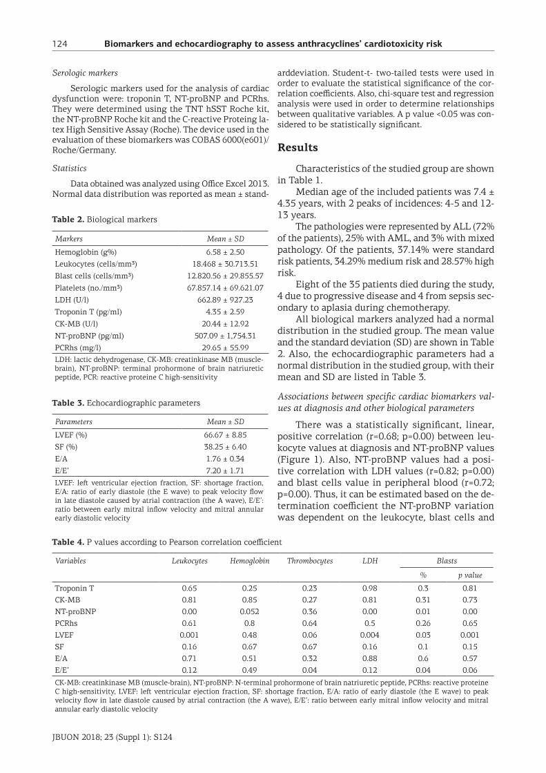
There was a statistically significant, linear, positive correlation (r=0.68; p=0.00) between leu-kocyte values at diagnosis and NT-proBNP values (Figure 1). Also, NT-proBNP values had a posi-tive correlation with LDH values (r=0.82; p=0.00) and blast cells value in peripheral blood (r=0.72; p=0.00). Thus, it can be estimated based on the de-termination coefficient the NT-proBNP variation was dependent on the leukocyte, blast cells and
Markers Mean ± SD
Hemoglobin (g%) 6.58 ± 2.50
Leukocytes (cells/mm3) 18.468 ± 30.713.51
Blast cells (cells/mm3) 12.820.56 ± 29.855.57
Platelets (no./mm3) 67.857.14 ± 69.621.07
LDH (U/l) 662.89 ± 927.23
Troponin T (pg/ml) 4.35 ± 2.59
CK-MB (U/l) 20.44 ± 12.92
NT-proBNP (pg/ml) 507.09 ± 1,754.31
PCRhs (mg/l) 29.65 ± 55.99LDH: lactic dehydrogenase, CK-MB: creatinkinase MB (muscle-brain), NT-proBNP: terminal prohormone of brain natriuretic peptide, PCR: reactive proteine C high-sensitivity
Table 2. Biological markers
Parameters Mean ± SD
LVEF (%) 66.67 ± 8.85
SF (%) 38.25 ± 6.40
E/A 1.76 ± 0.34
E/E’ 7.20 ± 1.71
LVEF: left ventricular ejection fraction, SF: shortage fraction, E/A: ratio of early diastole (the E wave) to peak velocity flow in late diastole caused by atrial contraction (the A wave), E/E’: ratio between early mitral inflow velocity and mitral annular early diastolic velocity
Table 3. Echocardiographic parameters
Variables Leukocytes Hemoglobin Thrombocytes LDH Blasts
% p value
Troponin T 0.65 0.25 0.23 0.98 0.3 0.81CK-MB 0.81 0.85 0.27 0.81 0.31 0.73NT-proBNP 0.00 0.052 0.36 0.00 0.01 0.00PCRhs 0.61 0.8 0.64 0.5 0.26 0.65LVEF 0.001 0.48 0.06 0.004 0.03 0.001SF 0.16 0.67 0.67 0.16 0.1 0.15E/A 0.71 0.51 0.32 0.88 0.6 0.57E/E’ 0.12 0.49 0.04 0.12 0.04 0.06
CK-MB: creatinkinase MB (muscle-brain), NT-proBNP: N-terminal prohormone of brain natriuretic peptide, PCRhs: reactive proteine C high-sensitivity, LVEF: left ventricular ejection fraction, SF: shortage fraction, E/A: ratio of early diastole (the E wave) to peak velocity flow in late diastole caused by atrial contraction (the A wave), E/E’: ratio between early mitral inflow velocity and mitral annular early diastolic velocity
Table 4. P values according to Pearson correlation coefficient
Biomarkers and echocardiography to assess anthracyclines’ cardiotoxicity risk 125
JBUON 2018; 23 (Suppl 1): S125
LDH values variation. A mean value NT-proBNP for included patients was 507.09±1754.32 pg/ml with a high normal value considered to be 125 pg/ml. Considering high normal level value of 14 pg/ml for troponin T, all included patients had values within normal range, the mean value for included group being 4.35±2.59 pg/ml. On the contrary, for CK MB, for a high normal level of 24 UI/L, 35.39% of the included patients had values higher than the normal limits at diag-nostis (upper level 24Ul/L, 20.44±12.92 U/l). CRPhs above 3 mg/l was seen in 53% of the included patients (mean value 29.65±55.99 mg/l). Out of the total 35 patients included in our study, 14.71% had none of the cardiac function biomarkers above the normal limit, 41.17% had one marker above the normal range, 35.29% had two markers above the limits, and 8.82% had three markers above the normal values. Other correlations between hematological pa-rameters and Troponin T, CK-MB or PCRhs values were not statistically significant (p>0.05; Table 4).
Associations between echocardiographic and hemato-logical parameters
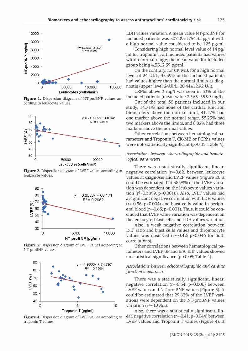
There was a statistically significant, linear, negative correlation (r=-0.62) between leukocyte values at diagnosis and LVEF values (Figure 2). It could be estimated that 38.99% of the LVEF varia-tion was dependent on the leukocyte values varia-tion (r2=0.3899; p=0.0016). Also, LVEF values had a significant negative correlation with LDH values (r=-0.56; p=0.004) and blast cells value in periph-eral blood (r=-0.63; p=0.001). Thus, it could be con-cluded that LVEF value variation was dependent on the leukocyte, blast cells and LDH values variation. Also, a weak negative correlation between E/E’ ratio and blast cells values and thrombocyte values was observed (r=-0.42; p=0.046 for both correlations). Other correlations between hematological pa-rameters and LVEF, SF and E/A, E/E’ values showed no statistical significance (p >0.05; Table 4).
Associations between echocardiographic and cardiac function biomarkers
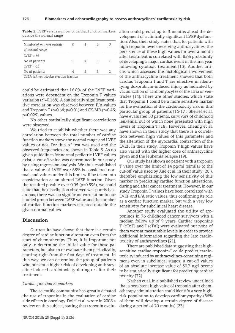
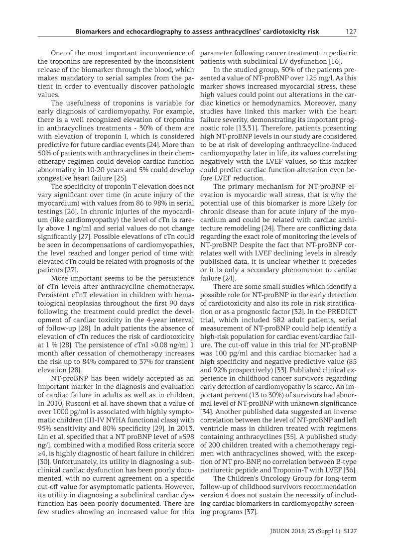
There was a statistically significant, linear, negative correlation (r=-0.54; p=0.006) between LVEF values and NT-pro BNP values (Figure 3). It could be estimated that 29.62% of the LVEF vari-ations were dependent on the NT-proBNP values variation (r2=0.2962). Also, there was a statistically significant, lin-ear, negative correlation (r=-0.41; p=0.044) between LVEF values and Troponin T values (Figure 4). It
Figure 1. Dispersion diagram of NT-proBNP values ac-cording to leukocyte values.
Figure 2. Dispersion diagram of LVEF values according to leukocyte values.
Figure 3. Dispersion diagram of LVEF values according to NT-proBNP values.
Figure 4. Dispersion diagram of LVEF values according to troponin T values.
Biomarkers and echocardiography to assess anthracyclines’ cardiotoxicity risk126
JBUON 2018; 23 (Suppl 1): S126
could be estimated that 16.8% of the LVEF vari-ations were dependent on the Troponin T values variation (r2=0.168). A statistically significant posi-tive correlation was observed between E/A values and Troponin T (r=0.64; p=0.01) and CK-MB (r=0.45; p=0.029) values. No other statistically significant correlations were observed. We tried to establish whether there was any correlation between the total number of cardiac function markers above the normal range and LVEF values or not. For this, x2 test was used and the observed frequencies are shown in Table 5. As no given guidelines for normal pediatric LVEF values exist, a cut-off value was determined in our study by using regression analysis. We thus established that a value of LVEF over 65% is considered nor-mal, and values under this limit will be taken into consideration as an altered LVEF function. Given the resulted p value over 0.05 (p=0.396), we could state that the distribution observed was purely haz-ardous, there was no significant correlation in our studied group between LVEF value and the number of cardiac function markers situated outside the given normal values.
Discussion
Our results have shown that there is a certain degree of cardiac function alteration even from the start of chemotherapy. Thus, it is important not only to determine the initial value for these pa-rameters, but also to re-evaluate them periodically, starting right from the first days of treatment. In this way, we can determine the group of patients who present a higher risk of developing anthracy-cline-induced cardiotoxicity during or after their treatment.
Cardiac function biomarkers
The scientific community has greatly debated the use of troponins in the evaluation of cardiac side effects in oncology. Dolci et al. wrote in 2008 a review on this subject, stating that troponin evalu-
ation could predict up to 3 months ahead the de-velopment of a clinically significant LVEF dysfunc-tion. Also, their study states that, for patients with high troponin levels receiving anthracyclines, the persistence of these high values for over a month after treatment is correlated with 85% probability of developing a major cardiac event in the first year following cytotoxic treatment [13]. Another arti-cle, which assessed the histological involvement of the anthracycline treatment showed that both cardiac Troponin I and T are effective in identi-fying doxorubicin-induced injury as indicated by vacuolization of cardiomyocytes of the atria or ven-tricles [14]. There are other studies, which state that Troponin I could be a more sensitive marker for the evaluation of the cardiotoxicity risk in this particular group of patients [15-17]. Sherief et al. have evaluated 50 patients, survivors of childhood leukemia, out of which none presented with high levels of Troponin T [18]. However, Cheung et al. have shown in their study that there is a correla-tion between high values of this parameter and the alteration of the myocardial contraction of the LVEF. In their study, Troponin T high values have also varied with the higher dose of anthracycline given and the leukemia relapse [19]. Our study has shown no patient with a troponin T value over the limit of 14 pg/ml (similar to the cut-off value used by Xue et al. in their study [20]), therefore emphasizing the low sensitivity of this marker in predicting cardiac function alterations, during and after cancer treatment. However, in our study Troponin T values have been correlated with LVEF and E/A ratio values, thus confirming its role as a cardiac function marker, but with a very low sensitivity for subclinical heart disease. Another study evaluated the utility of tro-ponines in 76 childhood cancer survivors with a median follow up of 9 years. Cardiac troponins T (cTnT) and I (cTnI) were evaluated but none of them were at measurable levels in order to provide additional information regarding the late cardio-toxicity of anthracyclines [21]. There are published data suggesting that high-sensitive cardiac troponin-I could predict cardio-toxicity induced by anthracyclines-containing regi-mens even in subclinical stages. A cut-off values of an absolute increase value of 30.7 ng/l seems to be statistically significant for predicting cardiac toxicity [22]. Nathan et al. in a published review underlined that a persistent high value of troponin after chem-otherapy administration could identify a very high-risk population to develop cardiomyopathy (86% of them will develop a certain degree of disease during a period of 20 months) [23].
Number of markers outside of normal range
0 1 2 3
LVEF ≤ 65
No of patients 1 2 5 1
LVEF > 65
No of patients 4 5 3 2LVEF: left ventricular ejection fraction
Table 5. LVEF versus number of cardiac function markers outside the normal range
Biomarkers and echocardiography to assess anthracyclines’ cardiotoxicity risk 127
JBUON 2018; 23 (Suppl 1): S127
One of the most important inconvenience of the troponins are represented by the inconsistent release of the biomarker through the blood, which makes mandatory to serial samples from the pa-tient in order to eventually discover pathologic values. The usefulness of troponins is variable for early diagnosis of cardiomyopathy. For example, there is a well recognized elevation of troponins in anthracyclines treatments - 30% of them are with elevation of troponin I, which is considered predictive for future cardiac events [24]. More than 50% of patients with anthracyclines in their chem-otherapy regimen could develop cardiac function abnormality in 10-20 years and 5% could develop congestive heart failure [25]. The specificity of troponin T elevation does not vary significant over time (in acute injury of the myocardium) with values from 86 to 98% in serial testings [26]. In chronic injuries of the myocardi-um (like cardiomyopathy) the level of cTn is rare-ly above 1 ng/ml and serial values do not change significantly [27]. Possible elevations of cTn could be seen in decompensations of cardiomyopathies, the level reached and longer period of time with elevated cTn could be related with prognosis of the patients [27]. More important seems to be the persistence of cTn levels after anthracycline chemotherapy. Persistent cTnT elevation in children with hema-tological neoplasias throughout the first 90 days following the treatment could predict the devel-opment of cardiac toxicity in the 4-year interval of follow-up [28]. In adult patients the absence of elevation of cTn reduces the risk of cardiotoxicity at 1 % [28]. The persistence of cTnI >0.08 ng/ml 1 month after cessation of chemotherapy increases the risk up to 84% compared to 37% for transient elevation [28]. NT-proBNP has been widely accepted as an important marker in the diagnosis and evaluation of cardiac failure in adults as well as in children. In 2010, Rusconi et al. have shown that a value of over 1000 pg/ml is associated with highly sympto-matic children (III-IV NYHA functional class) with 95% sensitivity and 80% specificity [29]. In 2013, Lin et al. specified that a NT proBNP level of ≥598 ng/l, combined with a modified Ross criteria score ≥4, is highly diagnostic of heart failure in children [30]. Unfortunately, its utility in diagnosing a sub-clinical cardiac dysfunction has been poorly docu-mented, with no current agreement on a specific cut-off value for asymptomatic patients. However, its utility in diagnosing a subclinical cardiac dys-function has been poorly documented. There are few studies showing an increased value for this
parameter following cancer treatment in pediatric patients with subclinical LV dysfunction [16]. In the studied group, 50% of the patients pre-sented a value of NT-proBNP over 125 mg/l. As this marker shows increased myocardial stress, these high values could point out alterations in the car-diac kinetics or hemodynamics. Moreover, many studies have linked this marker with the heart failure severity, demonstrating its important prog-nostic role [13,31]. Therefore, patients presenting high NT-proBNP levels in our study are considered to be at risk of developing anthracycline-induced cardiomyopathy later in life, its values correlating negatively with the LVEF values, so this marker could predict cardiac function alteration even be-fore LVEF reduction. The primary mechanism for NT-proBNP el-evation is myocardic wall stress, that is why the potential use of this biomarker is more likely for chronic disease than for acute injury of the myo-cardium and could be related with cardiac archi-tecture remodeling [24]. There are conflicting data regarding the exact role of monitoring the levels of NT-proBNP. Despite the fact that NT-proBNP cor-relates well with LVEF declining levels in already published data, it is unclear whether it precedes or it is only a secondary phenomenon to cardiac failure [24]. There are some small studies which identify a possible role for NT-proBNP in the early detection of cardiotoxicity and also its role in risk stratifica-tion or as a prognostic factor [32]. In the PREDICT trial, which included 582 adult patients, serial measurement of NT-proBNP could help identify a high-risk population for cardiac event/cardiac fail-ure. The cut-off value in this trial for NT-proBNP was 100 pg/ml and this cardiac biomarker had a high specificity and negative predictive value (85 and 92% prospectively) [33]. Published clinical ex-perience in childhood cancer survivors regarding early detection of cardiomyopathy is scarce. An im-portant percent (13 to 30%) of survivors had abnor-mal level of NT-proBNP with unknown significance [34]. Another published data suggested an inverse correlation between the level of NT-proBNP and left ventricle mass in children treated with regimens containing anthracyclines [35]. A published study of 200 children treated with a chemotherapy regi-men with anthracyclines showed, with the excep-tion of NT pro-BNP, no correlation between B-type natriuretic peptide and Troponin-T with LVEF [36]. The Children’s Oncology Group for long-term follow-up of childhood survivors recommendation version 4 does not sustain the necessity of includ-ing cardiac biomarkers in cardiomyopathy screen-ing programs [37].
Biomarkers and echocardiography to assess anthracyclines’ cardiotoxicity risk128
JBUON 2018; 23 (Suppl 1): S128
Regarding CK-MB values, 36% of the included patients presented with values over 24 U/l in our study. Many studies have shown that anthracycline treatment can cause not only late-onset CHF, but also acute cardiotoxicity presenting as coronary vasospasm or even acute myocardial infarction [38]. Although rare, these acute side effects are life-threatening, so it is vital to identify them as soon as possible, by obtaining an ECG prior and during anthracycline treatment and by evaluating CK-MB values periodically. On the other hand, an increased value for this marker could be the ex-pression of cardiac ischemia in the context of a later-onset CHF, proving to be useful in monitor-ing these high-risk patients. In children exposed to anthracyclines in a small study which included 22 patients, CK-MB was in normal range before and during 72 hrs of treatment [39]. CK-MB remained within normal range even if the pediatric patients followed a high dose chemotherapy regimen need-ed in hematopoietic stem cell transplantation [40]. If the exposed total dose of anthracyclines is taken into account, CK-MB variations seem to be without statistical significance in a study that included 131 patients [41]. PCRhs values are closely correlated with the risk of acute cardiac events. In the studied group, more than half of the patients presented values above 3 mg/l of this marker, putting themselves in the high-risk group for these types of cardiac pa-thology [42]. Thus, PCRhs could be used as a mark-er for acute anthracycline cardiotoxicity. However, cancer patients are known to have an increased pro-inflammatory status, which could in turn explain the increase PCRhs values. This is why periodic evaluation of this marker could help distinguish between the two causes, indicating the high-risk patients which need more careful future monitor-ing. In a published randomized study of 205 pedi-atric patients with anthracyclines chemo regimen, PCRhs level was similar between groups through all the study time and were not statistically associ-ated with any echocardiographic variables [43].
Hematologic parameters and cardiac function
In the studied group, leukocyte value at di-agnosis has proven to have an important role in the later development of a CMP (cardiomyopathy). Leukemia patients are classified into three risk groups according to a number of criteria, one of which is the number of leukocytes at diagnosis. A leukocyte count at diagnosis of over 50000/mm3 includes the patient in the high-risk group (HRG), meaning a more aggressive treatment, thus a high-er total dose of anthracyclines. However, a high level of leukocytes at diagnosis could also imply
myocardial infiltration by cancer cells, thus leading to impaired cardiac function. This is why the sen-sitivity and specificity of this marker in predicting future cardiac dysfunction is hard to assess. In our study, hematological parameters (leukocyte, LDH and blast cell values) have been correlated posi-tively with NT-proBNP values and negatively with LVEF values. Also, E/E’ values have been correlated negatively with blast cell values. This is of high importance, as it points out the degree in which the level of malignant cell presence in the body af-fects cardiac function even in the absence of an on-cological treatment. This might signal infiltration of the myocardium with malignant cells, meaning that all these patients suffer a pre-treatment altera-tion of their cardiac function, which would then be amplified by the cytotoxic treatment. This is of vital importance since, by proving the existence of this already influenced cardiac status, would urge the initiation of a cardio-protective treatment right from the diagnosis. Also, considering that the higher the number of leukocytes, thus the patient being in the high risk group, the higher the given dose of anthracycline will be, it is noticeable that these are the patients that will require the most intensive cardiac monitoring, and also long-term cardio-protective treatment.
Echocardiographic parameters
With regard to the echocardiographic evalua-tion, it is widely accepted (class I recommendation) that children receiving cardiotoxic chemotherapeu-tic agents should have a baseline evaluation as well as frequent follow-ups, in order to determine sub-clinical cardiac ischemia [43]. The measurement of the echocardiographic LVSF and LVEF are both non-invasive and available in most pediatric oncol-ogy centers, being the most widely used diagnos-tic method for detecting cardiotoxicity in children. However, these measurements have their limita-tions, their value depending on the exact methods used to obtain the LVSF or the LVEF [44]. Moreover, no studies have evaluated the predictive value of the echocardiographic LVSF as a surrogate marker for the future development of clinical heart failure after anthracycline therapy [45], as was confirmed by an extensive literature search. Generally, the cut-off value for SF is 28-29% and for LVEF 60-65%. The same cut-off values have been consid-ered in this study. A more accurate approach is to measure the difference between their values from one examination to another: a decrease of LVEF of over 10% being considered significant for an altera-tion of the heart’s function. The present study has established its cut-off value of 65% for LVEF using regression analysis in the given patient group.
Biomarkers and echocardiography to assess anthracyclines’ cardiotoxicity risk 129
JBUON 2018; 23 (Suppl 1): S129
Recent studies have shown lack of sensitiv-ity of the classical echocardiographic parameters (LVEF, SF) in the early diagnosis of cardiac dys-function. The reduction of LVEF is considered a late phenomenon, which is a clinical result of the failure of myocardium systolic function recover-ing. McKillop et al. determined that an abnormal radionuclide LVEF at rest (≤45%) had a sensitiv-ity of 53% and a specificity of 75% for detecting patients at moderate or high risk of developing CHF, with SF presenting an even lower sensitivity and specificity value [46]. Our study is consistent with these findings, proving an increased sensitiv-ity of the biological markers as compared to the echocardiographic ones. However, this study has shown that there is not an exact biological profile that could predict a future decrease of cardiac func-tion, as each patient presents with a particular set of altered biomarkers. One of the most important issues regarding echocardiography is represented by inter-operator variability. The comparative analysis on risk groups of LVEF and SF showed a difference of about 3% and 4%, respectively, between the mean values for these parameters in the standard risk group (SRG) and high-risk group (HRG), but this difference was not statistically significant (p>0.05). However, it was noticed that in the case of ultrasound determi-nations, patients included in HRG are more likely to develop subsequent cardiotoxicity, with initial-ly LVEF and SF slightly lower than those in theSRG. With regard to the values of E and A waves, determined by PW, and E’ respectively, determined by the TDI method, normal values were considered those proposed by Eidem et al. in 2004 [47]. For adults, four diastolic filling patterns are known de-pending on the E/A and E/E’ ratio: normal, pseudo-normal diastolic filling, impaired LV relaxation, and restrictive diastolic dysfunction. For children, there is currently no similar classification according to the echocardiographic parameters. However, due to the relative stabilization of diastolic velocities in children around 3 years of age, it is considered that the models used in adults can be applied to children over this age. In the studied group we identified three such patients, who had diastolic dysfunction according to the above-mentioned classification: two patients with impaired relaxation, presenting E/A and E/E’ values below the normal limits, and one patient presenting with a restrictive dysfunc-tion pattern with both parameters exceeding the maximum limit for their age group. In recent studies, PW and TDI parameters are considered highly useful in the early diagnosis
of diastolic dysfunction. Cengiz et al., declare in their study that the TDI method allows an early and accurate identification of diastolic dysfunction in patients treated with anthracyclines, the sen-sitivity of which increases with the time elapsed since cessation of treatment [48]. Also, Sherief et al. found a variation of TDI parameters in 26% of the 50-childhood leukemia surviving patients included in their study [18]. They point out that alteration of these parameters was also found in patients who had normal cardiac function in basal echocardiog-raphy. On the other hand, although Doppler evalu-ation improves early detection of cardiac dysfunc-tion in these patients, there is limited knowledge regarding the normal values of these parameters, as well as the interpretation of values situated out-side the reference limits in children [49,50]. This makes it difficult to diagnose cardiac dysfunction using PW and TDI, increasing the dependence on the examiner’s expertise and interpretation.
Limitations and Conclusion
The present study was mainly limited in terms of the number of patients and their adherence to the study. Of the initial number of 35 patients, only 24 were present for echocardiography. For these reasons, the results are difficult to extrapolate. Our results offer a pre-chemotherapy image of pediatric oncological patients, in which even the leukemic cell load could determine a preexisting subclini-cal cardiomyopathy which could be over-expressed during specific treatment, as statistically demon-strated correlation between blastic load and NT-proBNP and E/E’ values negatively correlated with blast cell values. This hypothesis could change the cardio-protective strategy by initiating the cardio-protective treatment right from the diagnosis. It is also necessary to monitor these patients at the end of treatment as well as 1 and 2 years after the end of treatment. In this way the predictive value of the biological markers and the echocardiographic pa-rameters in anticipating cardiac dysfunction, could be determined properly.
Acknowledgements
Knowledge transfer of bio-genomics in oncol-ogy and related domains in clinical applications - BIOGENONCO, MySMIS Code: 105774, Financing contract No: 10/01.09.2016.
Conflict of interests
The authors declare no conflict of interests.
Biomarkers and echocardiography to assess anthracyclines’ cardiotoxicity risk130
JBUON 2018; 23 (Suppl 1): S130
References
1. DeSantis CE, Lin CC, Mariotto AB et al. Cancer treat-ment and survivorship statistics. CA Cancer J Clin 2014;64:252-71.
2. Smith MA, Altekruse SF, Adamson PC, Reaman GH, Seibel NL. Declining childhood and adolescent cancer mortality. Cancer 2014;120:2497-2506.
3. Tubergen DG, Bleyr A, Ritchey AK. The Leukemias. In: Kliegman RM, Stanton BMD, Geme JSt, Schor NF, Behrman RE (Eds): Nelson textbook of pediatrics (19th Edn). Saunders, Philadelphia, 2011, pp 1732-9.
4. Campana D, Pui CH. Childhood leukemia. In: Niederhu-ber JE, Armitage JO, Dorshow JH, Kastan MB, Tepper JE (Eds): Abeloff’s Clinical Oncology (5th Edn). Saunders, Philadelphia, 2014, pp 1849-72.
5. Lipshultz SE, Adams MJ, Colan SD et al. Long-term cardiovascular toxicity in children, adolescents, and young adults who receive cancer therapy: pathophysi-ology, course, monitoring, management, prevention, and research directions: a scientific statement from the American Heart Association. Circulation 2013;128: 1927-95.
6. Fulbright JM. Review of Cardiotoxicity in Pediatric Cancer Patients: During and after Therapy. Cardiol Res Pract 2011;2011:942090-9.
7. Volkova M, Russell R. Anthracycline Cardiotoxicity: Prevalence, Pathogenesis and Treatment. Curr Cardiol Rev 2011;7:214-20.
8. Late Effects of Treatment for Childhood Cancer (PDQ®). PDQ Cancer Information Summaries. Available from: https://www.ncbi.nlm.nih.gov/books/NBK65832/. Last accessed 5th May 2018.
9. Lipshultz SE, Alvarez JA, Scully RE. Anthracycline as-sociated cardiotoxicity in survivors of childhood can-cer. Heart 2008;94:525-33.
10. Armenian SH, Robison LL. Childhood Cancer Survi-vorship: An Update on Evolving Paradigms for Un-derstanding Pathogenesis and Screening for Therapy-Related Late Effects. Curr Opin Pediatr 2013;25:16-22.
11. Albini A, Pennesi G, Donatelli F, Cammarota R, De Flora S, Noonan DM. Cardiotoxicity of Anticancer Drugs: The Need for Cardio-Oncology and Cardio-Oncological Pre-vention. J Natl Cancer Inst 2010;102:14-25.
12. Monsuez JJ. Detection and prevention of cardiac com-plications of cancer chemotherapy. Arch Cardiovasc Dis 2012;105:593-604.
13. Dolci A, Dominici R, Cardinale D, Sandri MT, Pan-teghini M. Biochemical markers for prediction of chemotherapy-induced cardiotoxicity: systematic re-view of the literature and recommendations for use. Am J Clin Pathol 2008;130:688-95.
14. Reagan WJ, York M, Berridge B, Schultze E, Walker D, Pettit S. Comparison of cardiac troponin I and T, including the evaluation of an ultrasensitive assay, as indicators of doxorubicin-induced cardiotoxicity. Toxi-col Pathol 2013;41:1146-58.
15. Cardinale D, Sandri MT, Colombo A et al. Prognostic value of troponin I in cardiac risk stratification of can-cer patients undergoing high-dose chemotherapy. Cir-culation 2004;109:2749-54.
16. Soker M, Kervancioglu M. Plasma concentrations of NT-pro-BNP and cardiac troponin-I in relation to dox-orubicin-induced cardiomiopathy and cardiac function in childhood malignancy. Saudi Med J 2005;26:1197-1202.
17. Sawaya H, Sebag IA, Plana JC et al. Assessment of echocardiography and biomarkers for the extended cardiotoxicity in patients treated with anthracyclines, taxanes, and trastuzumab. Circ Cardiovasc Imaging 2012;5:596-603.
18. Sherief LM, Kamal AG, Khalek EA, Kamal NM, Soliman AA, Esh AM. Biomarkers and early detection of late onset anthracycline-induced cardiotoxicity in children. Hematology 2012;17:151-6.
19. Cheung Y, Yu W, Cheuk DK et al. Plasma high sensitiv-ity Troponin T Levels in Adult Survivors of Childhood Leukaemias: Determinants and Associations with Car-diac Function. PLoS ONE 2013;8:e77063.
20. Xue K, Gu JJ, Zhang Q et al. Cardiotoxicity as Indicated by LVEF and Troponin T Sensitivity Following Two Anthracycline-Based Regimens in Lymphoma: Results from a Randomized Prospective Clinical Trial. Onco-target 2016;7:32519-31.
21. Ylänen K, Poutanen T, Savukoski T, Eerola A, Vetten-ranta K. Cardiac biomarkers indicate a need for sensi-tive cardiac imaging among long-term childhood can-cer survivors exposed to anthracyclines. Acta Paediatr 2015;104:313-9.
22. Jones M, O’Gorman P, Kelly C, Mahon N, Fitzgibbon MC. High-sensitive cardiac troponin-I facilitates timely detection of subclinical anthracycline-mediated cardiac injury. Ann Clin Biochem 2017;54:149-157.
23. Nathan PC, Amir E, Abdel-Qadir H. Cardiac Outcomes in Survivors of Pediatric and Adult Cancers. Can J Car-diol 2016;32:871-80.
24. Wittles RM. Biomarkers as Predictors of Cardiac Tox-icity From Targeted Cancer Therapies. J Card Fail 2016;22:459-64.
25. Cardinale D, Colombo A, Sandri MT et al. Prevention of High-Dose Chemotherapy-Induced Cardiotoxicity in High-Risk Patients by Angiotensin-Converting Enzyme Inhibition. Circulation 2006;114:2474-81.
26. Daubert MA, Jeremias A. The utility of troponin meas-urement to detect myocardial infarction: review of the current findings. Vasc Health Risk Manag 2010;6:691-9.
27. Muthu V, Kozman H, Liu K, Smulyan H, Villarreal D. Cardiac troponins: bench to bedside interpretation in cardiac disease. Am J Med Sci 2014;347:331-7.
28. Henri C, Heinonen T, Tardif JC. The Role of Biomark-ers in Decreasing Risk of Cardiac Toxicity after Cancer Therapy. Biomark Cancer 2016;8(Suppl 2):39-45.
29. Rusconi P, Ludwig D, Ratnasamy C et al. Serial meas-urements of serum NT-proBNP as markers of left ven-tricular systolic function and remodeling in children with heart failure. Am Heart J 2010;160:776-83.
30. Lin C, Zeng X, Jiang S et al. Role of the NT-proBNP level in the diagnosis of pediatric heart failure and in-vestigation of novel combined diagnostic criteria. Exp Ther Med 2013;6:995-9.
Biomarkers and echocardiography to assess anthracyclines’ cardiotoxicity risk 131
JBUON 2018; 23 (Suppl 1): S131
31. Nakamae H, Tsumura K, Akahori M et al. QT disper-sion correlates with systolic rather than diastolic pa-rameters in patients receiving anthracycline treatment. Intern Med 2004;43:379-87.
32. Lenihan DJ, Stevens PL, Massey M et al. The Utility of Point-of-Care Biomarkers to Detect Cardiotoxicity Dur-ing Anthracycline Chemotherapy: A Feasibility Study. J Card Fail 2016;22:433-8.
33. Stevens PL, Lenihan DJ. Cardiotoxicity due to Chemo-therapy: the Role of Biomarkers. Curr Cardiol Rep 2015;17:603.
34. Zidan A, Sherief LM, El-Sheikh A et al. NT-proBNP as early marker of subclinical late cardiotoxicity after dox-orubicin therapy and mediastinal irradiation in child-hood cancer survivors. Dis Markers 2015;2015:513219.
35. Germanakis I, Kalmanti M, Parthenakis F, Nikitovic D, Stiakaki E. Correlation of plasma N-terminal pro-brain natriuretic peptide levels with left ventricle mass in children treated with anthracyclines. Int J Cardiol 2006;108:212-5.
36. Armenian SH, Gelehrter SK, Vase T et al. Screening for cardiac dysfunction in anthracycline-exposed child-hood cancer survivors. Clin Cancer Res 2014;20:6314-23.
37. Long-term follow-up guidelines for Survivors of Child-hood, Adolescent, and Young Adult Cancers. Children’s Oncology Group. Available from: https://www.survivor-shipguidelines.org/pdf/LTFUGuidelines. Last accessed 5th May 2018.
38. Jarfelt M, Andersen NH, Hasle H. Is it possible to cure childhood acute myeloid leukaemia without significant cardiotoxicity? Br J Haematol 2016;175:577-87.
39. Fink FM, Genser N, Fink C et al. Cardiac troponin T and creatine kinase MB mass concentrations in children receiving anthracycline chemotherapy. Med Pediatr Oncol 1995;25:185-9.
40. Ozturk G, Tavil B, Ozguner M et al. Evaluation of Car-diac Markers in Children Undergoing Hematopoietic Stem Cell Transplantation. J Clin Lab Anal 2015;29:259-62.
41. Hu H, Zhang W, Huang D, Yang Q, Li J, Gao Y. Car-diotoxicity of anthracycline (ANT) treatment in chil-
dren with malignant tumors. Pediatr Hematol Oncol 2018;0:1-10.
42. Pearson TA, Mensah GA, Alexander RW et al. Markers of inflammation and cardiovascular disease: application to clinical and public health practice: A statement for healthcare professionals from the Centers for Disease Control and Prevention and the American Heart As-sociation. Circulation 2003;107:499-511.
43. Lipshultz SE, Miller TL, Scully RE et al. Changes in cardiac biomarkers during doxorubicin treatment of pediatric patients with high-risk acute lymphoblastic leukemia: associations with long-term echocardio-graphic outcomes. J Clin Oncol 2012;30:1042-9.
44. Thavendiranathan P, Grant AD, Negishi T, Plana JC, Popovic ZB, Marwick TH. Reproducibility of echocar-diographic techniques for sequential assessment of left ventricular ejection fraction and volumes:application to patients undergoing cancer chemotherapy. J Am Coll Cardiol 2013;61:77-84.
45. Lipshultz SE, Colan SD. Cardiovascular trials in long-term survivors of childhood cancer. J Clin Oncol 2004;22:769-73.
46. McKillop JH, Bristow MR, Goris ML, Billingham ME, Bockemuehl K. Sensitivity and specificity of radionu-clide ejection fractions in doxorubicin cardiotoxicity. Am Heart J 1982;106:1048-56.
47. Eidem BW, McMahon CJ, Cohen RR et al. Impact of cardiac growth on Doppler tissue imaging velocities: a study in healthy children. J Am Soc Echocardiography 2004;17:212-1.
48. Bayram C, Cetin I, Tavil B et al. Evaluation of Cardio-toxicity by Tissue Doppler Imaging in Childhood Leu-kemia Survivors Treated with Low-Dose Anthracycline. Pediatr Cardiol 2015;36:862-6.
49. Baysal T, Koksal Y, Oran B, Sen M, Unal E, Cimen D. Cardiac functions evaluated with tissue Doppler imag-ing in childhood cancers treated with anthracyclines. Pediatr Hematol Oncol 2010;27:13-23.
50. Cantinotti M, Lopez L. Nomograms for blood flow and tissue Doppler velocities to evaluate diastolic function in childern: a critical review. J Am Soc Echocardiogra-phy 2013;26:126-41.