Embed Size (px)
Citation preview
metabolites
H
OH
OH
Review
The Role of Metabolic Enzymes in the Regulationof Inflammation
Wesley H. Godfrey and Michael D. Kornberg *
Department of Neurology, Johns Hopkins University School of Medicine, Baltimore, MD 21205, USA;[email protected]* Correspondence: [email protected]
Received: 4 September 2020; Accepted: 19 October 2020; Published: 26 October 2020�����������������
Abstract: Immune cells undergo dramatic metabolic reprogramming in response to external stimuli.These metabolic pathways, long considered as simple housekeeping functions, are increasinglyunderstood to critically regulate the immune response, determining the activation, differentiation,and downstream effector functions of both lymphoid and myeloid cells. Within the complexmetabolic networks associated with immune activation, several enzymes play key roles in regulatinginflammation and represent potential therapeutic targets in human disease. In some cases,these enzymes control flux through pathways required to meet specific energetic or metabolicdemands of the immune response. In other cases, key enzymes control the concentrations ofimmunoactive metabolites with direct roles in signaling. Finally, and perhaps most interestingly,several metabolic enzymes have evolved moonlighting functions, with roles in the immune responsethat are entirely independent of their conventional enzyme activities. Here, we review key metabolicenzymes that critically regulate inflammation, highlighting mechanistic insights and opportunitiesfor clinical intervention.
Keywords: immunometabolism; inflammation; metabolism
1. Introduction
Immunologic responses are complex and finely tuned. Immune cells of multiple types mustintegrate an array of external signals and coordinate with one another to produce responses that areappropriately targeted and effective, leading to profound yet precisely timed changes in proliferationand function. The past two decades have produced recognition that cellular metabolism, previouslyrelegated to a housekeeping role, in fact critically regulates immune functions. Immunologic signalsproduce a broad reprogramming of metabolic pathways, which drives changes in immune cell activation,differentiation, and effector functions. This regulation of immune responses by metabolic pathways,termed “immunometabolism”, has become a major focus of research, with a goal of identifyingpathways that can be targeted in human diseases, such as autoimmune diseases characterized bydysregulated inflammation.
Our mechanistic understanding of the role of metabolism in immunology is ever growing,but certain common principles have come into focus. Metabolic reprogramming appears necessary toprovide precursors and meet the energy demands unique to specific immunologic states. Moreover,metabolites themselves can act as signaling molecules that directly modulate inflammatory responses.Within this framework, metabolic enzymes have been identified that regulate inflammation bycontrolling flux into key pathways and/or the altering levels of immunoactive metabolites. As anexample of the beauty and efficiency of evolution, several such enzymes have been co-opted as“moonlighting” proteins, playing roles in immunologic signaling pathways entirely independent oftheir conventional enzyme activities. As critical regulators of inflammation, these enzymes represent
Metabolites 2020, 10, 426; doi:10.3390/metabo10110426 www.mdpi.com/journal/metabolites
Metabolites 2020, 10, 426 2 of 22
potential therapeutic targets in human disease. Animal and, in some cases, human studies support theplausibility of pharmacologically targeting metabolic enzymes to modulate immunity [1].
Here, we will review the growing list of metabolic enzymes that critically regulateimmune responses, highlighting their susceptibility to pharmacologic interventions in animalsand humans. Given the breadth of metabolic processes shown to impact immune function, we havestructured our review based on several key metabolic pathways.
2. mTOR and AMPK—The Master Regulators of Metabolism
Mechanistic target of rapamycin (mTOR) is a serine/threonine kinase with major regulatory powersover cell survival, growth, and metabolism. mTOR, which can exist within two distinct complexes(mTORC1 and mTORC2), integrates information about cellular energy and nutrient availability withexternal stimuli to produce broad effects, including the induction of nutrient transporter expression,promoting the activity of glycolytic enzymes, increasing lipid synthesis, inducing ribosome synthesis,and favoring the transcription and translation of various proteins [2]. Indeed, the mechanisms by whichmTOR induces metabolic reprogramming both within immune cells and beyond are innumerable,and a full accounting is beyond the scope of this review. However, it is crucial to note that mTORacts as a master regulator of immune responses, positioning both lymphoid and myeloid cells forthe pro-inflammatory state by inducing broad metabolic changes. In T cells, mTOR activation occursdownstream of AKT/PI3K signaling in response to the co-stimulation of the T cell receptor (TCR) andCD28 [3]. In myeloid cells, mTOR is activated downstream of Toll-like receptors (TLRs) and cytokinereceptors [4]. Within both myeloid and lymphoid cells, mTOR signaling is critical for the upregulationof metabolic pathways required for inflammatory activation and effector functions, including glycolysis,the pentose phosphate pathway, and glutaminolysis, acting largely through the transcription factorsHIF-1α and Myc. Rapamycin, a well-known inhibitor of mTOR, is used as an immunosuppressivedrug to prevent organ transplant rejection [5].
Signaling downstream of mTORC1 and mTORC2 differentially regulates CD4+ T cell differentiation.A deficiency of mTOR, which impacts both complexes, impairs the differentiation of T helper (Th) 1,Th2, and Th17 cells, while promoting the differentiation of regulatory T (Treg) cells [6]. Both mTORC1and mTORC2 inhibit Treg differentiation, and a deficiency of both complexes is required for enhancedFoxp3+ Treg production. The selective impairment of mTORC1 signaling through a deficiency of itsupstream regulator Rheb was found to prevent Th1 and Th17 differentiation while promoting Th2differentiation, while a reciprocal effect on differentiation was observed with mTORC2 deficiency [7].However, another study found that CD4+ cells deficient in mTORC2 failed to differentiate into eitherTh1 or Th2 cells [8].
The counterbalance to mTOR in metabolic regulation is AMP-activated protein kinase (AMPK),which reciprocally inhibits mTOR signaling and activates opposing metabolic pathways. AMPK turnsoff mTOR by activating the TSC1/TSC2 complex [9] and phosphorylating Raptor, the mTOR bindingpartner [10]. While mTOR responds to nutrient excess and promotes anabolic processes, AMPK isactivated by an increase in the intracellular AMP/ATP ratio (reflective of energy/nutrient depletion)and turns on catabolic processes to restore energy balance [11]. It shuts down gluconeogenesis,lipid synthesis, and protein synthesis while turning on fatty acid oxidation to regenerate the cell’ssupply of ATP [12]. Most notably, AMPK accomplishes a shift away from lipid synthesis to fatty acidoxidation by phosphorylating and thereby inactivating acetyl-CoA carboxylase (ACC) [13].
The precise role of AMPK in inflammation is nuanced and an area of active investigation.Although AMPK is not required for T cell development or homeostasis [14,15], AMPK activity istransiently increased post-TCR activation [16]. AMPK is crucial for T cell effector responses [14,17,18]and also regulates memory CD8+ T cell development [19] and the recall response [20]. At the same time,AMPK activation can prevent pathological inflammation. AICAR, an activator of AMPK, preventedsepsis in murine models [21] and ameliorated models of ulcerative colitis [22] and multiple sclerosis [23].Metformin, which acts in part via AMPK activation, was shown to limit inflammation in models of
Metabolites 2020, 10, 426 3 of 22
lupus [24] and allograft rejection [25]. By contrast, inhibiting AMPK with compound C exacerbatedsepsis in murine models [21].
3. Glycolysis
The importance of glycolytic reprogramming in immune activation was one of the earliestobservations in immunometabolism, beginning with the discovery that the immune challenge ofnaïve T cells produces an upregulation of glycolysis critical for T cell effector functions [26–28].This increase in glycolytic flux, with the preferential conversion of pyruvate to lactate rather thanoxidation in mitochondria, is akin to the Warburg effect first described in cancer and broadlycharacterizes the inflammatory response in both adaptive and innate immune cells [29]. As noted above,glycolytic reprogramming critically depends on mTOR activation and the downstream transcriptionfactors HIF-1α and Myc [30–33].
Although this switch toward aerobic glycolysis has been consistently linked with the differentiationand effector functions of inflammatory cells, glycolytic reprogramming also impacts the functionof regulatory cell types. For instance, the upregulation of glycolysis in response to TLR activationor GLUT1 overexpression was shown to increase the proliferation of Treg cells but impair theirsuppressive functions [34]. More recently, thymus-derived Treg cells were found to increase glycolysisfollowing TNF receptor 2 (TNFR2) stimulation in a manner that enhanced both proliferation andsuppressive function, although these cells oxidized pyruvate rather than secreting lactate in aWarburg-like manner [35]. Similarly, although the upregulation of OXPHOS is critically importantfor the alternative activation of anti-inflammatory M2 macrophages [36], some recent reports usingthe glycolysis inhibitor 2-deoxyglucose (2-DG) suggested that glycolysis is also required in these cellsto support OXPHOS and fatty acid synthesis [37–39]. However, a more recent study suggested thatglycolysis is dispensable for M2 differentiation, with the inhibitory actions of 2-DG based on off-targeteffects independent of glycolysis [40].
Several hypotheses are widely held regarding the requirement for glycolytic upregulation followingimmune activation. For instance, one argument is that increased glycolysis provides critical biomass bysupplying precursors necessary for nucleotide, lipid, and protein synthesis. Another argument is thatthe rapid kinetics of glycolysis support increased bioenergetic requirements by providing more ATPper second despite less efficiency relative to oxidative phosphorylation. Although these hypotheses arebiologically plausible, it has become clear that additional mechanisms contribute to the requirementfor upregulated glycolysis in inflammatory responses. Thus, while some glycolytic enzymes appear tobe key regulators of inflammation simply by controlling metabolic flux, others possess unique andunexpected moonlighting roles or control the supply of metabolites that act not simply as precursorsbut also as signaling molecules.
Below, we highlight a number of glycolytic enzymes demonstrated to critically regulateinflammatory responses (Figure 1).
3.1. Hexokinase
Hexokinase catalyzes the first step in glycolysis, the conversion of glucose to glucose-6-phosphate.As a critical regulator of glycolytic flux, the transcription of hexokinase, particularly hexokinase 2,is upregulated downstream of TCR and IL-2 receptor signaling. This is achieved by the activationof mTOR and the transcription factors HIF-1α and Myc [3,4,41]. Hexokinase is also particularlyimportant for the HIV infection of macrophages by supporting the survival of HIV-infected cells [42].The pharmacologic inhibition of hexokinase enzyme activity dampens the inflammatory response.The glycolytic inhibitor 2-DG, which indirectly inhibits hexokinase through the competitive inhibitionof the downstream enzyme phosphoglucoisomerase, impairs T cell proliferation and effectorfunctions [27,28]; shifts the balance between effector, regulatory, and memory T cells [31,43]; prevents thepro-inflammatory activation of dendritic cells [44–46] and macrophages [47]; and produces benefit inanimal models of autoimmunity such as lupus [24] and rheumatoid arthritis (RA) [48]. Furthermore,
Metabolites 2020, 10, 426 4 of 22
the direct hexokinase inhibitor 3-bromopyruvate prevents immune activation and attenuates diseasein murine models of RA [49] and multiple sclerosis (MS) [50].Metabolites 2020, 10, x FOR PEER REVIEW 7 of 22
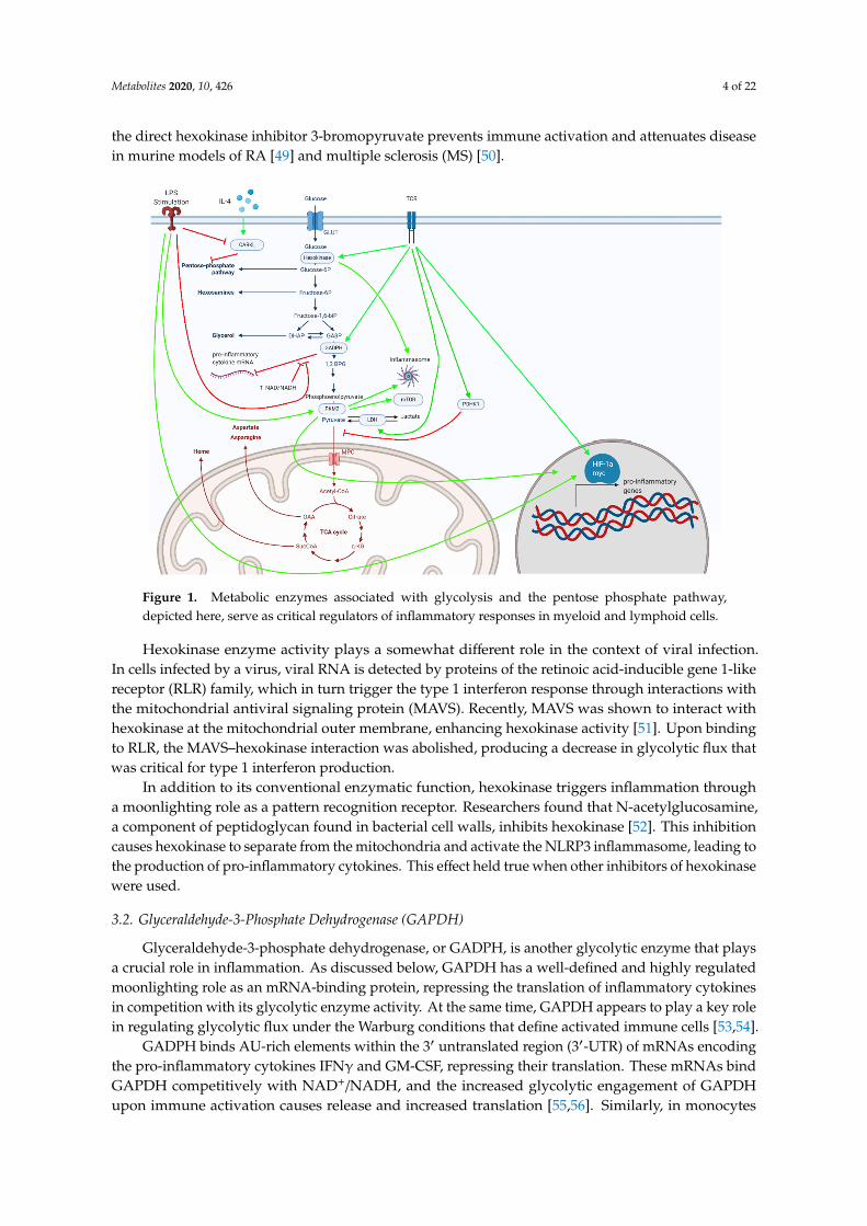
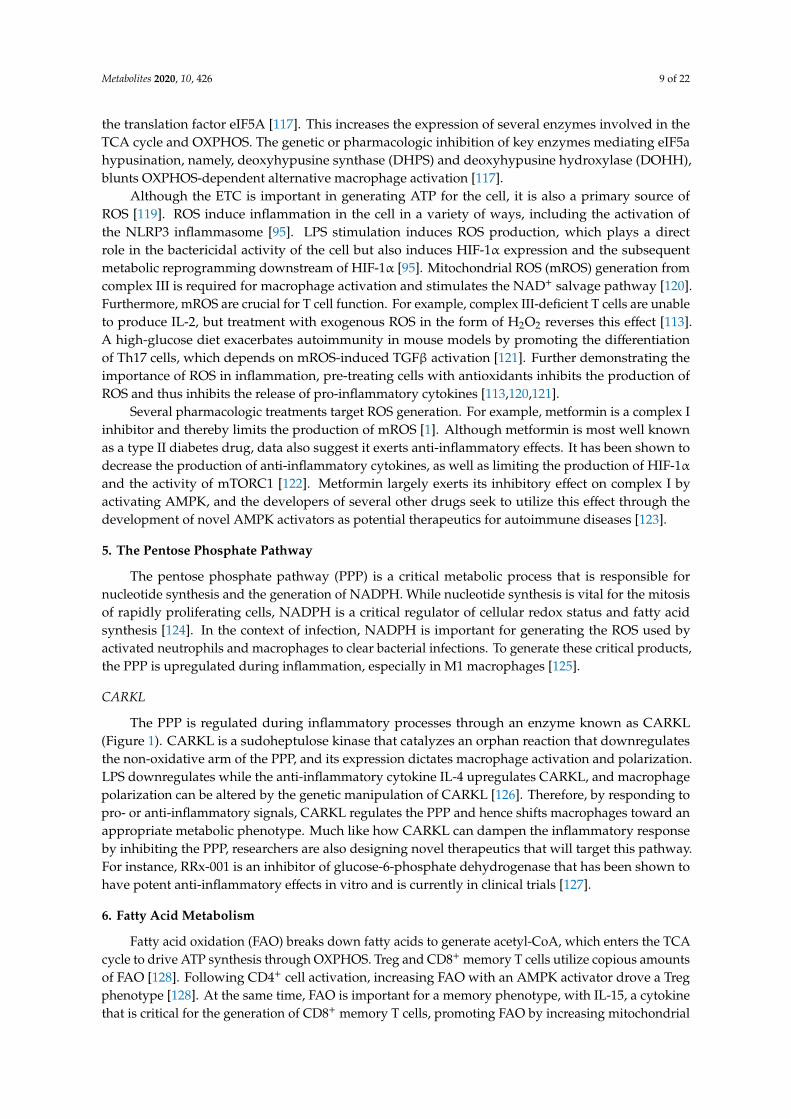
Figure 1. Metabolic enzymes associated with glycolysis and the pentose phosphate pathway, depicted here, serve as critical regulators of inflammatory responses in myeloid and lymphoid cells.
4. Mitochondrial Metabolism—TCA Cycle and Electron Transport Chain
Although glycolytic reprogramming represents a common theme among pro-inflammatory immune cells, mitochondrial metabolism also plays a vital role in inflammatory responses. The activation of naïve T cells leads to increased oxidative phosphorylation (OXPHOS) [95], and memory lymphocytes generate modified mitochondrial networks that allow them to better carry out the TCA cycle and OXPHOS [96]. By contrast, macrophages and dendritic cells experience a drastic reduction in OXPHOS upon inflammatory activation, yet this disruption of the TCA cycle and electron transport chain (ETC) nonetheless plays a key role in inflammation by providing metabolites and reactive oxygen species (ROS) that serve as signaling molecules [47,95,97]. Oxidative metabolism skews toward an anti-inflammatory phenotype in myeloid cells [36], as further evidenced by the fact that monocytes found within a Staphylococcus aureus biofilm primarily used OXPHOS, while OXPHOS inhibition via the nanoparticle delivery of oligomycin skewed cells toward a glycolytic phenotype and induced bacterial clearance [98].
In this section, we will focus on key enzymes within the TCA cycle and ETC that regulate inflammatory responses.
4.1. TCA Cycle
Fed by glucose, protein, and fatty acids, the TCA cycle generates electron donors (NADH and FADH2) to support OXPHOS through the ETC. However, in addition to its role in energy generation, the TCA cycle produces metabolites with key roles in inflammation. For instance, the inflammatory activation of macrophages leads to increased levels of citrate, succinate, and itaconate, each of which plays specific roles in the immune response [97]. While a comprehensive discussion of the roles of these metabolites is beyond our scope, we will highlight key enzyme targets that regulate the availability of these metabolites or their downstream effects relevant to inflammation.
4.1.1. Isocitrate Dehydrogenase (IDH)
Figure 1. Metabolic enzymes associated with glycolysis and the pentose phosphate pathway,depicted here, serve as critical regulators of inflammatory responses in myeloid and lymphoid cells.
Hexokinase enzyme activity plays a somewhat different role in the context of viral infection.In cells infected by a virus, viral RNA is detected by proteins of the retinoic acid-inducible gene 1-likereceptor (RLR) family, which in turn trigger the type 1 interferon response through interactions withthe mitochondrial antiviral signaling protein (MAVS). Recently, MAVS was shown to interact withhexokinase at the mitochondrial outer membrane, enhancing hexokinase activity [51]. Upon bindingto RLR, the MAVS–hexokinase interaction was abolished, producing a decrease in glycolytic flux thatwas critical for type 1 interferon production.
In addition to its conventional enzymatic function, hexokinase triggers inflammation througha moonlighting role as a pattern recognition receptor. Researchers found that N-acetylglucosamine,a component of peptidoglycan found in bacterial cell walls, inhibits hexokinase [52]. This inhibitioncauses hexokinase to separate from the mitochondria and activate the NLRP3 inflammasome, leading tothe production of pro-inflammatory cytokines. This effect held true when other inhibitors of hexokinasewere used.
3.2. Glyceraldehyde-3-Phosphate Dehydrogenase (GAPDH)
Glyceraldehyde-3-phosphate dehydrogenase, or GADPH, is another glycolytic enzyme that playsa crucial role in inflammation. As discussed below, GAPDH has a well-defined and highly regulatedmoonlighting role as an mRNA-binding protein, repressing the translation of inflammatory cytokinesin competition with its glycolytic enzyme activity. At the same time, GAPDH appears to play a key rolein regulating glycolytic flux under the Warburg conditions that define activated immune cells [53,54].
GADPH binds AU-rich elements within the 3′ untranslated region (3′-UTR) of mRNAs encodingthe pro-inflammatory cytokines IFNγ and GM-CSF, repressing their translation. These mRNAs bindGAPDH competitively with NAD+/NADH, and the increased glycolytic engagement of GAPDHupon immune activation causes release and increased translation [55,56]. Similarly, in monocytes
Metabolites 2020, 10, 426 5 of 22
and macrophages, GADPH represses the translation of TNFα mRNA, which is reversed upon LPSexposure [57]. The relieved repression of cytokine mRNA translation by GAPDH therefore links theupregulation of glycolysis with inflammatory cytokine production.
The moonlighting role of GAPDH as an mRNA-binding protein is regulated bypost-translational modification. Malonylation is a lysine modification induced by malonyl-CoA.The malonylation of GADPH occurs downstream of LPS stimulation in monocytes and macrophages,increasing GAPDH enzyme activity while decreasing its mRNA binding capacity, thus allowinginflammatory cytokines such as IFN-γ, IL-6, and TNF-α to be translated into protein [58].
Although not a rate-limiting enzyme under basal conditions, GADPH becomes rate-limitingunder Warburg conditions [53,54,59], which may have direct relevance to its role in regulatingimmune responses. Dimethyl fumarate (DMF), an immunomodulatory drug FDA-approved forthe treatment of MS, post-translationally modifies GAPDH at its active site and inactivates itsenzymatic activity [60]. The enzymatic inhibition of GAPDH by DMF inhibits glycolysis in activated,but not resting, immune cells and mediates the anti-inflammatory effects of the drug. Interestingly,DMF does not act by altering GAPDH–mRNA binding. Subsequent work demonstrated that itaconate,an anti-inflammatory metabolite derived from the TCA cycle, similarly inactivates GAPDH enzymeactivity [61]. These findings suggest that GAPDH enzyme activity is required for pro-inflammatoryresponses independent of mRNA binding, either by limiting glycolytic flux in a general sense or byregulating the levels of immunoactive metabolites. For instance, the inhibition of GAPDH increasesconcentrations of methylglyoxal, which has been shown to dampen inflammation by acting on theKEAP1–NRF2 axis [62].
3.3. Enolase
Enolase is responsible for the ninth step of glycolysis, converting 2-phosphoglycerate tophosphoenolpyruvate. At the same time, enolase is an abundant protein on bacterial cell surfaces thatbinds to plasminogen, thus allowing bacteria to invade the host organism [63]. Enolase has been shownto be crucial for the virulence of several strains of bacteria, as evidenced by data showing that inhibitinginteraction between enolase and plasminogen [64] or immunizing against enolase prior to challengewith pathogenic bacteria [65] significantly altered the progression of the infection in mouse models.The binding of α-enolase to plasminogen has been shown to be important for the recruitment ofmacrophages in inflammatory lung disease [66]. Additionally, enolase plays a role in Treg generationthrough a moonlighting function as a transcriptional regulator. One study found that enolase localizesto the nuclei of T cells to generate Tregs in the periphery. In the nucleus, enolase binds to regulatoryregions of FOXP3 and directly affects the expression of the splicing variant Foxp3-E2, which wascorroborated in peripheral blood samples from patients with type 2 diabetes and relapsing-remittingmultiple sclerosis [67].
3.4. Pyruvate Kinase M2 (PKM2)
Pyruvate kinase (PK) converts phosphoenolpyruvate into pyruvate, a rate-limiting step and thefinal reaction of glycolysis. Multiple isoforms of PK exist. The M1 isoform (PKM1) is constitutivelyexpressed in most differentiated tissues under basal conditions and exists as a tetramer with highglycolytic activity. The M2 isoform of pyruvate kinase (PKM2) is preferentially expressed underWarburg conditions, such as in cancer cells and activated immune cells, and contributes to theinflammatory response through multiple mechanisms [68]. Unlike PKM1, PKM2 exists either as atetramer with high glycolytic activity, or as a dimer with low glycolytic activity. Perhaps paradoxically,it is the low-activity PKM2 dimer that promotes aerobic glycolysis and inflammation, largely throughnon-glycolytic moonlighting functions.
Pro-inflammatory stimuli, such as LPS stimulation of macrophages and TCR ligation in T cells,increase the expression of PKM2 [69,70]. PKM2 in turn activates mTORC1 by phosphorylating the mTORinhibitor AKT1 substrate 1 (AKT1S1) [71] and increasing serine synthesis from the glycolytic metabolite
Metabolites 2020, 10, 426 6 of 22
3-phosphoglycerate [72]. Despite its intrinsically lower enzyme activity, PKM2 upregulates glycolysisthrough moonlighting functions as a transcriptional co-activator, promoting the transcriptional programof HIF-1α [73]. HIF-1α increases the expression of PKM2. The dimer form of PKM2 in turn bindsto HIF-1α, translocates to the nucleus, and enhances the transcription of HIF-1α target genes.
The non-canonical moonlighting activity of PKM2 as a transcriptional co-activator of HIF-1α iscrucial for the inflammatory activation of both macrophages and T lymphocytes. Following the LPSstimulation of macrophages, dimerized nuclear PKM2 drives a pro-inflammatory transcriptionalprogram that includes IL-1β induction. Small molecules such as TEPP-46, which induce thetetramerization of PKM2 and thereby promote its canonical enzyme activity while inhibiting itsnuclear functions, inhibit LPS-induced glycolytic reprogramming and inflammatory functions whilepromoting the expression of anti-inflammatory cytokines such as IL-10 [70]. Similarly, the treatment ofCD4+ T cells with TEPP-46 blocks PKM2 nuclear translocation, prevents glycolytic reprogramming,and reduces T cell activation, proliferation, and cytokine production [74]. Treatment with TEPP-46prevents the differentiation of pro-inflammatory Th1 and Th17 cells and attenuates disease in theexperimental autoimmune encephalomyelitis (EAE) mouse model of autoimmune neuroinflammation,identifying PKM2 as a potential therapeutic target. Two other groups similarly found that a geneticdeficiency of PKM2 protects mice from EAE. One of these demonstrated that the shRNA-mediatedknockdown of PKM2 in isolated CD4+ cells reduced glycolysis and Th1/Th17 differentiation while alsolimiting their pathogenicity in an adoptive transfer model of EAE [75]. The other group found that theCD4+ T cell-specific knockout of PKM2 impaired Th17 differentiation and attenuated the course ofactive-immunization EAE, though they observed that PKM2 was required for Th17 differentiationthrough the activation of the transcription factor STAT3 rather than through actions on HIF-1α andmetabolic reprogramming [76]. Similarly, another group found that PKM2 upregulates IL-17 productionin CD4+ cells via STAT3 in response to lactate uptake [77].
In addition to its nuclear functions, PKM2 has been shown to directly activate the NLRP3inflammasome in macrophages [78]. AIM2 and NLRP3 inflammasome activation were prevented bothby the genetic deletion of PKM2 and a pharmacologic inhibitor of PKM2 enzyme activity, suggestingthat its canonical enzyme activity may be important. In natural killer (NK) cells, PKM2 regulates theinflammatory response independent of HIF-1α and its nuclear activities, regulating redox status bycontrolling the flux of upstream glycolytic metabolites into the pentose phosphate pathway for thegeneration of NADPH [79]. As such, canonical PKM2 enzyme activity may be important in certain celltypes and/or under specific conditions.
3.5. Pyruvate Dehydrogenase Kinase 1 (PDHK1)
As the end product of glycolysis, pyruvate has two potential fates—it can either enter the pyruvatedehydrogenase (PDH) complex to become acetyl-CoA and enter the TCA cycle, or be converted tolactate by lactate dehydrogenase (LDH). When pyruvate is reduced to lactate, it regenerates NAD+,thus allowing glycolysis and the Warburg phenotype to continue. In both CD4+ and CD8+ T cells,TCR stimulation increases the expression and activity of pyruvate dehydrogenase kinase 1 (PDHK1),a kinase that inhibits PDH and thereby diverts pyruvate away from the TCA cycle and toward lactateproduction [80,81]. PDHK1 activity is high in pro-inflammatory Th17 cells but low in Treg cells.The inhibition of PDHK1 with the small molecule dichloroacetate (DCA) limits aerobic glycolysisand promotes pyruvate entry into the TCA cycle, thereby limiting Th17 and augmenting Treggeneration from naïve CD4+ cells and inhibiting inflammatory cytokine production in CD8+ cells.Treatment with DCA in vivo produces benefits in animal models of multiple autoimmune diseases,including inflammatory bowel disease, RA, MS, and asthma [81–83]. These findings implicate PDHK1not only as a key regulator of aerobic glycolysis and inflammation, but also as a potential therapeutictarget in autoimmunity.
Metabolites 2020, 10, 426 7 of 22
3.6. Lactate Dehydrogenase (LDH)
As noted above, the conversion of pyruvate to lactate by LDH regenerates NAD+ and maintainshigh glycolytic flux under Warburg conditions. Through several mechanisms, LDH criticallyregulates cellular inflammatory responses. The pro-inflammatory activation of CD4+ cells leadsto increased expression of LDH-A, an isoform of LDH with high enzymatic activity. LDH-A activitypromotes the inflammatory response by maintaining high levels of acetyl-CoA, which in turnpromotes histone acetylation and the transcription of IFNγ [84]. LDH-A-deficient mice wereprotected from autoimmune attacks. The inhibition of LDH with the small molecule FX11(3-dihydroxy-6-methyl-7-(phenylmethyl)-4-propylnaphthalene-1-carboxylic acid) has been shown toinhibit the release of pro-inflammatory cytokines in macrophages [85]. Similar to GAPDH, LDH hasalso been found to bind and repress the translation of mRNAs encoding inflammatory cytokines,releasing them upon the engagement of its enzymatic activity [80,86]. Finally, lactate itself has beenshown to play a direct signaling role in inflammation. In CD4+ cells, lactate uptake via the transporterSLC5A12 was shown to promote Th17 differentiation and prevent migratory egress from sites ofinflammation, and the blockade of lactate uptake attenuated disease in a model of autoimmunearthritis [77]. In many other studies, however, lactate has been found to produce anti-inflammatoryeffects in both lymphocytes and macrophages. Earlier studies demonstrated that lactate produced inthe tumor microenvironment (TME) has suppressive effects on effector and cytotoxic T cells [87,88].By contrast, it was recently reported that Foxp3 induces metabolic changes in Treg cells that allowthem to survive and function in a low-glucose, high-lactate TME [89]. Another study found thatlactate specifically promotes Foxp3 expression [90]. Through a variety of mechanisms, extracellularlactate similarly induces regulatory phenotypes in tumor-associated macrophages, contributing totumor evasion [91–93]. More recently, lactate was found to produce a post-translational modificationof histones (lactylation) that regulates transcription and shifts cells toward an anti-inflammatory M2phenotype as a late event following LPS stimulation [94]. Interestingly, in the context of viral infection,lactate dampens type 1 interferon production by infected cells through a direct interaction withMAVS [51]. As such, mice deficient in LDH-A displayed a greater type 1 interferon response andheightened resistance to infection with vesicular stomatitis virus.
4. Mitochondrial Metabolism—TCA Cycle and Electron Transport Chain
Although glycolytic reprogramming represents a common theme among pro-inflammatoryimmune cells, mitochondrial metabolism also plays a vital role in inflammatory responses.The activation of naïve T cells leads to increased oxidative phosphorylation (OXPHOS) [95], and memorylymphocytes generate modified mitochondrial networks that allow them to better carry out the TCAcycle and OXPHOS [96]. By contrast, macrophages and dendritic cells experience a drastic reductionin OXPHOS upon inflammatory activation, yet this disruption of the TCA cycle and electron transportchain (ETC) nonetheless plays a key role in inflammation by providing metabolites and reactive oxygenspecies (ROS) that serve as signaling molecules [47,95,97]. Oxidative metabolism skews toward ananti-inflammatory phenotype in myeloid cells [36], as further evidenced by the fact that monocytesfound within a Staphylococcus aureus biofilm primarily used OXPHOS, while OXPHOS inhibition viathe nanoparticle delivery of oligomycin skewed cells toward a glycolytic phenotype and inducedbacterial clearance [98].
In this section, we will focus on key enzymes within the TCA cycle and ETC that regulateinflammatory responses.
4.1. TCA Cycle
Fed by glucose, protein, and fatty acids, the TCA cycle generates electron donors(NADH and FADH2) to support OXPHOS through the ETC. However, in addition to its role inenergy generation, the TCA cycle produces metabolites with key roles in inflammation. For instance,
Metabolites 2020, 10, 426 8 of 22
the inflammatory activation of macrophages leads to increased levels of citrate, succinate, and itaconate,each of which plays specific roles in the immune response [97]. While a comprehensive discussion ofthe roles of these metabolites is beyond our scope, we will highlight key enzyme targets that regulatethe availability of these metabolites or their downstream effects relevant to inflammation.
4.1.1. Isocitrate Dehydrogenase (IDH)
The inflammatory activation of macrophages leads to a decreased activity of isocitratedehydrogenase (IDH), potentially via both transcriptional repression [47] and inactivation by nitricoxide (NO)-mediated S-nitrosylation [99]. This “break” in the TCA cycle leads to the accumulationof citrate and itaconate, although it must be noted that aconitase, rather than IDH, has recently beenimplicated as the target of NO mediating this break [100]. As discussed below, accumulated citrate canbe converted to itaconate within mitochondria or be transported to the cytosol. Within the cytosol,citrate serves several functions regulated by key enzymes. ATP-citrate lyase (ACLY), which convertscitrate to oxaloacetate and acetyl-CoA, contributes to NO and ROS production through an unknownmechanism [101] and generates a pool of acetyl-CoA serving histone acetylation [102], lipogenesis [103],and malonylation [104].
4.1.2. Immune-Responsive Gene 1 Protein (IRG1)
Immune-responsive gene 1 (IRG1) is responsible for catalyzing the conversion of cis-aconitate(an intermediate of the TCA cycle) into itaconic acid [105]. The transcription of IRG1 is upregulatedin the pro-inflammatory state in macrophages as a response to various stimuli, such as IFNγ, LPS,and TNFα [106]. During inflammation, IRG1 produces itaconate, which has a direct bactericidalrole by altering bacterial metabolism [107]. However, other studies have shown that itaconate hasanti-inflammatory effects via multiple mechanisms [105,108], such as the inhibition of succinatedehydrogenase (SDH) [109] and post-translational modification of key protein targets such asKEAP1 [110] and GAPDH [61].
4.1.3. Succinate Dehydrogenase (SDH)
SDH is a key enzyme in the TCA cycle, converting succinate into fumarate [97]. SDH plays a rolein generating a pro-inflammatory phenotype by contributing to the production of pro-inflammatorycytokines such as IL-1β and reactive oxygen species (ROS) in macrophages in vitro; the inhibition ofSDH produces anti-inflammatory effects, as discussed above in the context of itaconate [109,111].
4.2. Electron Transport Chain (ETC) and ROS
The generation of ATP from OXPHOS is mediated by the ETC, which accepts electrons from NADHand FADH2 produced from the TCA cycle and generates ATP via ATP synthase. Because OXPHOSincreases in lymphocytes after activation, the electron transport chain is required for lymphocytesto be properly activated, and ATP synthase activity is one of the most important parts of thisprocess [112]. When complex IV is knocked out, T cell activation is inhibited [112], while complex IIIdeficiency prevents T cell proliferation in vivo and in vitro [113]. At the same time, bacterial RNAstimulates complex II activity in macrophages, and the inhibition of complex II with a smallmolecule inhibitor significantly increased death rates due to increasing rates of sepsis and thedecreased release of pro-inflammatory cytokines when mice were infected with Salmonella enterica [114].Furthermore, HIF-1α and IL-1β are linked to the production of NO, which can turn off the ETC inmacrophages [115]. The ETC is also the target of novel pharmacological interventions for autoimmunity,such as LYC-30937-ec, an F1F0 ATP synthesis inhibitor, which is in clinical trials for ulcerative colitis [116].
The upregulation of OXPHOS plays an important role in the alternative activation of macrophagesin response to signals such as IL-4 [36]. An important regulator of OXPHOS in this context is thepolyamine–eIF5A–hypusine axis [117]. Hypusine, a natural amino acid derived from the polyaminespermidine [118], which itself is a downstream metabolite of arginine, post-translationally modifies
Metabolites 2020, 10, 426 9 of 22
the translation factor eIF5A [117]. This increases the expression of several enzymes involved in theTCA cycle and OXPHOS. The genetic or pharmacologic inhibition of key enzymes mediating eIF5ahypusination, namely, deoxyhypusine synthase (DHPS) and deoxyhypusine hydroxylase (DOHH),blunts OXPHOS-dependent alternative macrophage activation [117].
Although the ETC is important in generating ATP for the cell, it is also a primary source ofROS [119]. ROS induce inflammation in the cell in a variety of ways, including the activation ofthe NLRP3 inflammasome [95]. LPS stimulation induces ROS production, which plays a directrole in the bactericidal activity of the cell but also induces HIF-1α expression and the subsequentmetabolic reprogramming downstream of HIF-1α [95]. Mitochondrial ROS (mROS) generation fromcomplex III is required for macrophage activation and stimulates the NAD+ salvage pathway [120].Furthermore, mROS are crucial for T cell function. For example, complex III-deficient T cells are unableto produce IL-2, but treatment with exogenous ROS in the form of H2O2 reverses this effect [113].A high-glucose diet exacerbates autoimmunity in mouse models by promoting the differentiationof Th17 cells, which depends on mROS-induced TGFβ activation [121]. Further demonstrating theimportance of ROS in inflammation, pre-treating cells with antioxidants inhibits the production ofROS and thus inhibits the release of pro-inflammatory cytokines [113,120,121].
Several pharmacologic treatments target ROS generation. For example, metformin is a complex Iinhibitor and thereby limits the production of mROS [1]. Although metformin is most well knownas a type II diabetes drug, data also suggest it exerts anti-inflammatory effects. It has been shown todecrease the production of anti-inflammatory cytokines, as well as limiting the production of HIF-1αand the activity of mTORC1 [122]. Metformin largely exerts its inhibitory effect on complex I byactivating AMPK, and the developers of several other drugs seek to utilize this effect through thedevelopment of novel AMPK activators as potential therapeutics for autoimmune diseases [123].
5. The Pentose Phosphate Pathway
The pentose phosphate pathway (PPP) is a critical metabolic process that is responsible fornucleotide synthesis and the generation of NADPH. While nucleotide synthesis is vital for the mitosisof rapidly proliferating cells, NADPH is a critical regulator of cellular redox status and fatty acidsynthesis [124]. In the context of infection, NADPH is important for generating the ROS used byactivated neutrophils and macrophages to clear bacterial infections. To generate these critical products,the PPP is upregulated during inflammation, especially in M1 macrophages [125].
CARKL
The PPP is regulated during inflammatory processes through an enzyme known as CARKL(Figure 1). CARKL is a sudoheptulose kinase that catalyzes an orphan reaction that downregulatesthe non-oxidative arm of the PPP, and its expression dictates macrophage activation and polarization.LPS downregulates while the anti-inflammatory cytokine IL-4 upregulates CARKL, and macrophagepolarization can be altered by the genetic manipulation of CARKL [126]. Therefore, by responding topro- or anti-inflammatory signals, CARKL regulates the PPP and hence shifts macrophages toward anappropriate metabolic phenotype. Much like how CARKL can dampen the inflammatory responseby inhibiting the PPP, researchers are also designing novel therapeutics that will target this pathway.For instance, RRx-001 is an inhibitor of glucose-6-phosphate dehydrogenase that has been shown tohave potent anti-inflammatory effects in vitro and is currently in clinical trials [127].
6. Fatty Acid Metabolism
Fatty acid oxidation (FAO) breaks down fatty acids to generate acetyl-CoA, which enters the TCAcycle to drive ATP synthesis through OXPHOS. Treg and CD8+ memory T cells utilize copious amountsof FAO [128]. Following CD4+ cell activation, increasing FAO with an AMPK activator drove a Tregphenotype [128]. At the same time, FAO is important for a memory phenotype, with IL-15, a cytokinethat is critical for the generation of CD8+ memory T cells, promoting FAO by increasing mitochondrial
Metabolites 2020, 10, 426 10 of 22
biogenesis and increasing the expression of carnitine palmitoyl transferase (CPT1a), the rate-limitingstep of mitochondrial long-chain fatty acid oxidation (FAO) [129] (Figure 2). It should be noted,however, that a recent report employing a genetic deficiency of CPT1a suggested that the enzyme isdispensable for T cell activation and the generation of memory CD8+ and Treg cells, and that AMPKactivation augments Treg differentiation independent of CPT1a [130]. Other studies have found thatTNF receptor-associated factor 6 (TRAF6), an adaptor protein in the TNF-receptor superfamily, is alsocritical for CD8+ memory T cell generation [19]. Additionally, one of the most important mechanismsfor upregulating FAO in memory T cells involves mobilizing fatty acids through lysosomal hydrolaseLAL (lysosomal acid lipase) [131].
Metabolites 2020, 10, x FOR PEER REVIEW 10 of 22
studies have found that TNF receptor-associated factor 6 (TRAF6), an adaptor protein in the TNF-receptor superfamily, is also critical for CD8+ memory T cell generation [19]. Additionally, one of the most important mechanisms for upregulating FAO in memory T cells involves mobilizing fatty acids through lysosomal hydrolase LAL (lysosomal acid lipase) [131].
In macrophages, M1 macrophages primarily utilize glycolysis, while M2 macrophages heavily rely on FAO. Overexpressing CPT1a, the rate limiting factor in long-chain FAO, in macrophages in vitro reduced the release of pro-inflammatory cytokines and promoted M2 polarization [132]. Researchers have sought to therapeutically target this pathway with the purported CPT1a inhibitor etomoxir, which mitigated MS symptoms in the EAE mouse model [133] and prevented graft versus host disease in a murine model [134]. However, etomoxir has recently been demonstrated to have off-target immunomodulatory effects beyond CPT1a, which were suggested to be the reason for discrepancies between studies employing genetic and pharmacologic inhibition to study the role of CPT1a in T cell function [130].
Fatty acid synthesis also plays a crucial role in inflammation. In dendritic cells (DCs), TLR activation upregulates fatty acid synthesis, which supports the expansion of the endoplasmic reticulum and Golgi and is required for DC activation [135]. Fatty acid synthesis is also required for proper T cell activation [136]. In T cells, the ratio of cholesterol in the cell membrane is important for proper activation, and inhibiting the cholesterol esterification enzyme acetyl-CoA acetyltransferase (ACAT1) generated a more robust effector response [137]. Acetyl-CoA carboxylase 1 (ACC1) is the rate-limiting factor in fatty acid synthesis, and T cells deficient in ACC1 had impaired proliferation and effector capacity [138] (Figure 2). Interestingly, ACC1 deficiency does not affect Treg function and instead promotes Treg differentiation over Th17 polarization [139]. Fatty acid synthesis can be pharmacologically targeted with the ACC1 inhibitor soraphen A, which alleviated disease severity and symptom onset in EAE by inhibiting Th17 polarization.
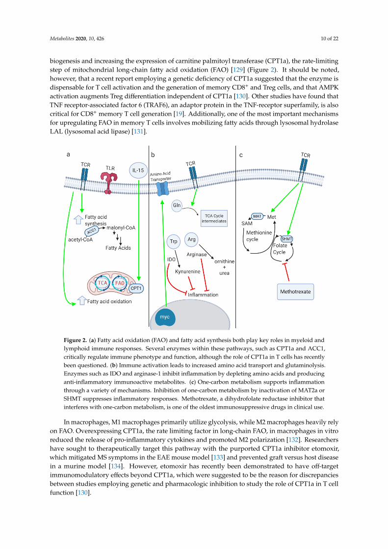
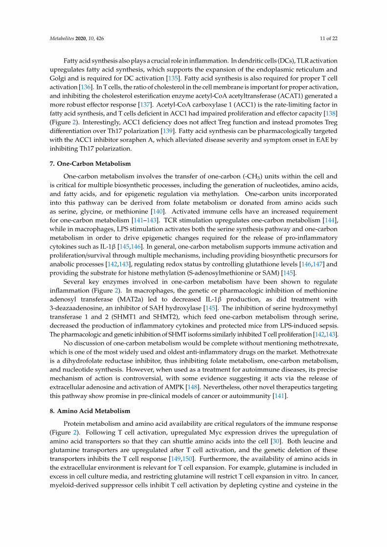
Figure 2. (a) Fatty acid oxidation (FAO) and fatty acid synthesis both play key roles in myeloid and lymphoid immune responses. Several enzymes within these pathways, such as CPT1a and ACC1, critically regulate immune phenotype and function, although the role of CPT1a in T cells has recently been questioned. (b) Immune activation leads to increased amino acid transport and glutaminolysis. Enzymes such as IDO and arginase-1 inhibit inflammation by depleting amino acids and producing
Figure 2. (a) Fatty acid oxidation (FAO) and fatty acid synthesis both play key roles in myeloid andlymphoid immune responses. Several enzymes within these pathways, such as CPT1a and ACC1,critically regulate immune phenotype and function, although the role of CPT1a in T cells has recentlybeen questioned. (b) Immune activation leads to increased amino acid transport and glutaminolysis.Enzymes such as IDO and arginase-1 inhibit inflammation by depleting amino acids and producinganti-inflammatory immunoactive metabolites. (c) One-carbon metabolism supports inflammationthrough a variety of mechanisms. Inhibition of one-carbon metabolism by inactivation of MAT2a orSHMT suppresses inflammatory responses. Methotrexate, a dihydrofolate reductase inhibitor thatinterferes with one-carbon metabolism, is one of the oldest immunosuppressive drugs in clinical use.
In macrophages, M1 macrophages primarily utilize glycolysis, while M2 macrophages heavily relyon FAO. Overexpressing CPT1a, the rate limiting factor in long-chain FAO, in macrophages in vitroreduced the release of pro-inflammatory cytokines and promoted M2 polarization [132]. Researchershave sought to therapeutically target this pathway with the purported CPT1a inhibitor etomoxir,which mitigated MS symptoms in the EAE mouse model [133] and prevented graft versus host diseasein a murine model [134]. However, etomoxir has recently been demonstrated to have off-targetimmunomodulatory effects beyond CPT1a, which were suggested to be the reason for discrepanciesbetween studies employing genetic and pharmacologic inhibition to study the role of CPT1a in T cellfunction [130].
Metabolites 2020, 10, 426 11 of 22
Fatty acid synthesis also plays a crucial role in inflammation. In dendritic cells (DCs), TLR activationupregulates fatty acid synthesis, which supports the expansion of the endoplasmic reticulum andGolgi and is required for DC activation [135]. Fatty acid synthesis is also required for proper T cellactivation [136]. In T cells, the ratio of cholesterol in the cell membrane is important for proper activation,and inhibiting the cholesterol esterification enzyme acetyl-CoA acetyltransferase (ACAT1) generated amore robust effector response [137]. Acetyl-CoA carboxylase 1 (ACC1) is the rate-limiting factor infatty acid synthesis, and T cells deficient in ACC1 had impaired proliferation and effector capacity [138](Figure 2). Interestingly, ACC1 deficiency does not affect Treg function and instead promotes Tregdifferentiation over Th17 polarization [139]. Fatty acid synthesis can be pharmacologically targetedwith the ACC1 inhibitor soraphen A, which alleviated disease severity and symptom onset in EAE byinhibiting Th17 polarization.
7. One-Carbon Metabolism
One-carbon metabolism involves the transfer of one-carbon (-CH3) units within the cell andis critical for multiple biosynthetic processes, including the generation of nucleotides, amino acids,and fatty acids, and for epigenetic regulation via methylation. One-carbon units incorporatedinto this pathway can be derived from folate metabolism or donated from amino acids suchas serine, glycine, or methionine [140]. Activated immune cells have an increased requirementfor one-carbon metabolism [141–143]. TCR stimulation upregulates one-carbon metabolism [144],while in macrophages, LPS stimulation activates both the serine synthesis pathway and one-carbonmetabolism in order to drive epigenetic changes required for the release of pro-inflammatorycytokines such as IL-1β [145,146]. In general, one-carbon metabolism supports immune activation andproliferation/survival through multiple mechanisms, including providing biosynthetic precursors foranabolic processes [142,143], regulating redox status by controlling glutathione levels [146,147] andproviding the substrate for histone methylation (S-adenosylmethionine or SAM) [145].
Several key enzymes involved in one-carbon metabolism have been shown to regulateinflammation (Figure 2). In macrophages, the genetic or pharmacologic inhibition of methionineadenosyl transferase (MAT2a) led to decreased IL-1β production, as did treatment with3-deazaadenosine, an inhibitor of SAH hydroxylase [145]. The inhibition of serine hydroxymethyltransferase 1 and 2 (SHMT1 and SHMT2), which feed one-carbon metabolism through serine,decreased the production of inflammatory cytokines and protected mice from LPS-induced sepsis.The pharmacologic and genetic inhibition of SHMT isoforms similarly inhibited T cell proliferation [142,143].
No discussion of one-carbon metabolism would be complete without mentioning methotrexate,which is one of the most widely used and oldest anti-inflammatory drugs on the market. Methotrexateis a dihydrofolate reductase inhibitor, thus inhibiting folate metabolism, one-carbon metabolism,and nucleotide synthesis. However, when used as a treatment for autoimmune diseases, its precisemechanism of action is controversial, with some evidence suggesting it acts via the release ofextracellular adenosine and activation of AMPK [148]. Nevertheless, other novel therapeutics targetingthis pathway show promise in pre-clinical models of cancer or autoimmunity [141].
8. Amino Acid Metabolism
Protein metabolism and amino acid availability are critical regulators of the immune response(Figure 2). Following T cell activation, upregulated Myc expression drives the upregulation ofamino acid transporters so that they can shuttle amino acids into the cell [30]. Both leucine andglutamine transporters are upregulated after T cell activation, and the genetic deletion of thesetransporters inhibits the T cell response [149,150]. Furthermore, the availability of amino acids inthe extracellular environment is relevant for T cell expansion. For example, glutamine is included inexcess in cell culture media, and restricting glutamine will restrict T cell expansion in vitro. In cancer,myeloid-derived suppressor cells inhibit T cell activation by depleting cystine and cysteine in the
Metabolites 2020, 10, 426 12 of 22
tumor microenvironment [151]. As noted above, serine availability, derived both from exogenouspools and de novo synthesis, critically regulates T cell and macrophage activation [142,143,145,146].
There are several enzymes that play a key role in regulating amino acid availability to control theimmune response. Indoleamine 2,3-dioxygenase (IDO) depletes tryptophan, thereby inhibitingT cell responses [152]. IDO is expressed at high levels in the placenta to prevent T cellactivation and promote feto-maternal tolerance [153]. Kynurenine, the downstream metaboliteof IDO, is also immunosuppressive, and cancer researchers are seeking to therapeutically alter thispathway [154]. Tryptophan metabolism via the kynurenine pathway also regulates macrophagefunction by controlling de novo NAD+ synthesis [155]. The genetic or pharmacologic inhibitionof quinolate phosphoribosyltransferase (QPRT), which provides the NAD+ precursor nicotinic acidmononucleotide (NaMN), augments the pro-inflammatory response to LPS. Conversely, the ectopicexpression of QPRT dampens inflammation and promotes a homeostatic macrophage phenotype.Arginase-1 is preferentially expressed in M2 macrophages, and this enzyme depletes arginine,hence preventing the formation of the pro-inflammatory nitric oxide generated by M1 macrophages [156,157].During infection with Mycobacterium tuberculosis, macrophages in the lung granulomas pathognomonicfor the disease produce high levels of Arg1 to control the inflammatory response [158].
Glutaminolysis is the process by which the cell breaks down glutamine and converts itinto TCA cycle intermediates and other metabolites. In activated T cells, glutaminolysis isupregulated in a Myc-dependent manner; this upregulation of glutamine catabolism replenishesTCA cycle intermediates, fuels polyamine synthesis, and coordinates with glucose catabolism tosupport amino acid, nucleotide, and lipid biosynthesis [30]. Moreover, glutamate oxaloacetatetransaminase 1 (GOT1), an enzyme involved in glutamine metabolism, exerts pro-inflammatoryeffects by producing 2-hydroxyglutarate, which hinders the expression of FOXP3 and thus blocksthe formation of Tregs. Researchers have tried inhibiting glutamine metabolism with the glutamineanalog 6-diazo-5-oxo-L-norleucine (DON), and this molecule suppressed inflammation in mousemodels of acute lung injury [159] and prevented allograft rejection [25]. Although inhibitingglutamine metabolism in models of autoimmunity alleviates the autoimmune phenotype,in cancer models, inhibiting glutamine metabolism generates a more potent anti-tumorimmune response. Researchers found that in several cancer models, inhibiting glutamine metabolismwith DON or a related prodrug generated effector CD8+ T cells capable of a robust anti-tumor response.Glutamine antagonism selectively upregulated OXPHOS in tumor-infiltrating CD8+ cells via anincreased activity of acyl-coenzyme A (CoA) synthetase short-chain family member 1 (ACSS1),allowing the fueling of the TCA cycle through acetate [160].
9. Autophagy
Autophagy is the metabolic process by which cells degrade and recycle cellular components.Autophagy has been shown to be important for immune cell activation; the knockout of theautophagy-essential gene Atg3 resulted in apoptosis and inhibited the activation of murine immunecells in vitro [161]. This study also showed that hematopoietic cells use autophagy to provide lipids tothe cell when they become metabolically stressed. Following TCR or TLR activation, immune cellsupregulate autophagy to generate the nutrients needed for the metabolic demands of the cell [162].On the other hand, autophagy also plays a role in inducing cell death during HIV infection, with the HIVglycoprotein gp120 binding to CXCR4 on T cells and inducing apoptosis, but this effect was abrogated bythe deletion of autophagy-critical enzymes such as Beclin-1 or Atg7 [163]. Autophagy also plays a majorrole in immune cell differentiation [164]. Autophagy is turned off by mTOR and turned on by AMPK,which also allows the cell to regulate Treg and memory T cell fate by using autophagy to generatethe lipids needed for the FAO-dependent metabolic phenotype of these cells [164]. Autophagy alsocontrols inflammatory responses in myeloid cells; the deletion of autophagy-critical genes promotedan M1 inflammatory phenotype and inhibited M2 polarization [165]. Autophagy is also relevant forthe neutrophil response to infection, as it upregulates cell-intrinsic survival mechanisms [166].
Metabolites 2020, 10, 426 13 of 22
10. Conclusions
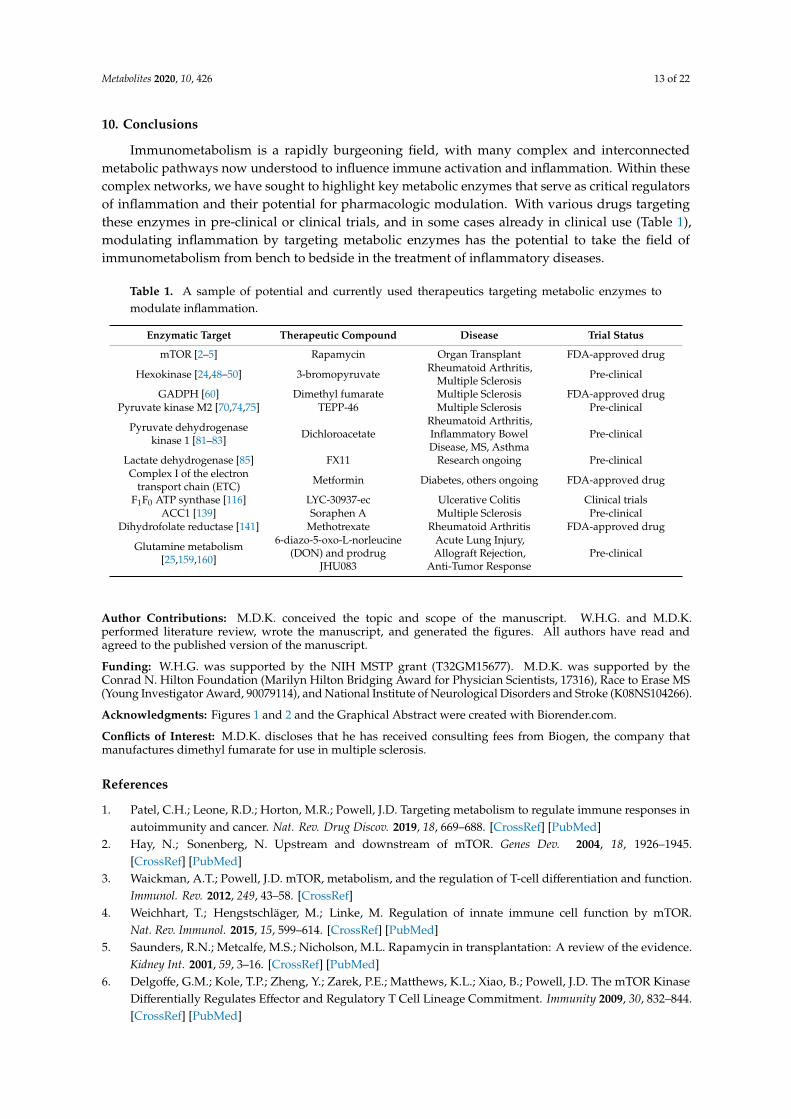
Immunometabolism is a rapidly burgeoning field, with many complex and interconnectedmetabolic pathways now understood to influence immune activation and inflammation. Within thesecomplex networks, we have sought to highlight key metabolic enzymes that serve as critical regulatorsof inflammation and their potential for pharmacologic modulation. With various drugs targetingthese enzymes in pre-clinical or clinical trials, and in some cases already in clinical use (Table 1),modulating inflammation by targeting metabolic enzymes has the potential to take the field ofimmunometabolism from bench to bedside in the treatment of inflammatory diseases.
Table 1. A sample of potential and currently used therapeutics targeting metabolic enzymes tomodulate inflammation.
Enzymatic Target Therapeutic Compound Disease Trial Status
mTOR [2–5] Rapamycin Organ Transplant FDA-approved drug
Hexokinase [24,48–50] 3-bromopyruvate Rheumatoid Arthritis,Multiple Sclerosis Pre-clinical
GADPH [60] Dimethyl fumarate Multiple Sclerosis FDA-approved drugPyruvate kinase M2 [70,74,75] TEPP-46 Multiple Sclerosis Pre-clinical
Pyruvate dehydrogenasekinase 1 [81–83] Dichloroacetate
Rheumatoid Arthritis,Inflammatory BowelDisease, MS, Asthma
Pre-clinical
Lactate dehydrogenase [85] FX11 Research ongoing Pre-clinicalComplex I of the electron
transport chain (ETC) Metformin Diabetes, others ongoing FDA-approved drug
F1F0 ATP synthase [116] LYC-30937-ec Ulcerative Colitis Clinical trialsACC1 [139] Soraphen A Multiple Sclerosis Pre-clinical
Dihydrofolate reductase [141] Methotrexate Rheumatoid Arthritis FDA-approved drug
Glutamine metabolism[25,159,160]
6-diazo-5-oxo-L-norleucine(DON) and prodrug
JHU083
Acute Lung Injury,Allograft Rejection,
Anti-Tumor ResponsePre-clinical
Author Contributions: M.D.K. conceived the topic and scope of the manuscript. W.H.G. and M.D.K.performed literature review, wrote the manuscript, and generated the figures. All authors have read andagreed to the published version of the manuscript.
Funding: W.H.G. was supported by the NIH MSTP grant (T32GM15677). M.D.K. was supported by theConrad N. Hilton Foundation (Marilyn Hilton Bridging Award for Physician Scientists, 17316), Race to Erase MS(Young Investigator Award, 90079114), and National Institute of Neurological Disorders and Stroke (K08NS104266).
Acknowledgments: Figures 1 and 2 and the Graphical Abstract were created with Biorender.com.
Conflicts of Interest: M.D.K. discloses that he has received consulting fees from Biogen, the company thatmanufactures dimethyl fumarate for use in multiple sclerosis.
References
1. Patel, C.H.; Leone, R.D.; Horton, M.R.; Powell, J.D. Targeting metabolism to regulate immune responses inautoimmunity and cancer. Nat. Rev. Drug Discov. 2019, 18, 669–688. [CrossRef] [PubMed]
2. Hay, N.; Sonenberg, N. Upstream and downstream of mTOR. Genes Dev. 2004, 18, 1926–1945.[CrossRef] [PubMed]
3. Waickman, A.T.; Powell, J.D. mTOR, metabolism, and the regulation of T-cell differentiation and function.Immunol. Rev. 2012, 249, 43–58. [CrossRef]
4. Weichhart, T.; Hengstschläger, M.; Linke, M. Regulation of innate immune cell function by mTOR.Nat. Rev. Immunol. 2015, 15, 599–614. [CrossRef] [PubMed]
5. Saunders, R.N.; Metcalfe, M.S.; Nicholson, M.L. Rapamycin in transplantation: A review of the evidence.Kidney Int. 2001, 59, 3–16. [CrossRef] [PubMed]
6. Delgoffe, G.M.; Kole, T.P.; Zheng, Y.; Zarek, P.E.; Matthews, K.L.; Xiao, B.; Powell, J.D. The mTOR KinaseDifferentially Regulates Effector and Regulatory T Cell Lineage Commitment. Immunity 2009, 30, 832–844.[CrossRef] [PubMed]
Metabolites 2020, 10, 426 14 of 22
7. Delgoffe, G.M.; Pollizzi, K.N.; Waickman, A.T.; Heikamp, E.; Meyers, D.J.; Horton, M.R.; Powell, J.D.The kinase mTOR regulates the differentiation of helper T cells through the selective activation of signalingby mTORC1 and mTORC2. Nat. Immunol. 2011, 12, 295–304. [CrossRef]
8. Lee, K.; Gudapati, P.; Dragovic, S.; Spencer, C.; Joyce, S.; Killeen, N.; Boothby, M. Mammalian targetof rapamycin protein complex 2 regulates differentiation of Th1 and Th2 cell subsets via distinctsignaling pathways. Immunity 2010, 32, 743–753. [CrossRef]
9. Inoki, K.; Zhu, T.; Guan, K.L. TSC2 mediates cellular energy response to control cell growth and survival.Cell 2003, 115, 577–590. [CrossRef]
10. Gwinn, D.M.; Shackelford, D.B.; Egan, D.F.; Mihaylova, M.M.; Mery, A.; Vasquez, D.S.; Turk, B.E.; Shaw, R.J.AMPK phosphorylation of raptor mediates a metabolic checkpoint. Mol. Cell 2008, 30, 214–226. [CrossRef]
11. Hardie, D.G. AMP-activated protein kinase-an energy sensor that regulates all aspects of cell function. GenesDev. 2011, 25, 1895–1908. [CrossRef]
12. Herzig, S.; Shaw, R.J. AMPK: Guardian of metabolism and mitochondrial homeostasis. Nat. Rev. Mol.Cell Biol. 2018, 19, 121–135. [CrossRef]
13. Davies, S.P.; Sim, A.T.R.; Hardie, D.G. Location and function of three sites phosphorylated on rat acetyl-CoAcarboxylase by the AMP-activated protein kinase. Eur. J. Biochem. 1990, 187, 183–190. [CrossRef] [PubMed]
14. Blagih, J.; Coulombe, F.; Vincent, E.E.; Dupuy, F.; Galicia-Vázquez, G.; Yurchenko, E.; Raissi, T.C.;vanderWindt, G.J.W.; Viollet, B.; Pearce, E.L.; et al. The energy sensor AMPK regulates T Cell metabolicadaptation and effector responses invivo. Immunity 2015, 42, 41–54. [CrossRef]
15. Mayer, A.; Denanglaire, S.; Viollet, B.; Leo, O.; Andris, F. AMP-activated protein kinase regulates lymphocyteresponses to metabolic stress but is largely dispensable for immune cell development and function.Eur. J. Immunol. 2008, 38, 948–956. [CrossRef] [PubMed]
16. Tamás, P.; Hawley, S.A.; Clarke, R.G.; Mustard, K.J.; Green, K.; Hardie, D.G.; Cantrell, D.A. Regulation of theenergy sensor AMP-activated protein kinase by antigen receptor and Ca2+ in T lymphocytes. J. Exp. Med.2006, 203, 1665–1670. [CrossRef] [PubMed]
17. Ma, E.H.; Poffenberger, M.C.; Wong, A.H.T.; Jones, R.G. The role of AMPK in T cell metabolism and function.Curr. Opin. Immunol. 2017, 46, 45–52. [CrossRef] [PubMed]
18. Rao, E.; Zhang, Y.; Zhu, G.; Hao, J.; Persson, X.M.T.; Egilmez, N.K.; Suttles, J.; Li, B. Deficiency of AMPK inCD8+ T cells suppresses their anti-tumor function by inducing protein phosphatase-mediated cell death.Oncotarget 2015, 6, 7944–7958. [CrossRef]
19. Pearce, E.L.; Walsh, M.C.; Cejas, P.J.; Harms, G.M.; Shen, H.; Wang, L.S.; Jones, R.G.; Choi, Y. Enhancing CD8T-cell memory by modulating fatty acid metabolism. Nature 2009, 460, 103–107. [CrossRef]
20. Rolf, J.; Zarrouk, M.; Finlay, D.K.; Foretz, M.; Viollet, B.; Cantrell, D.A. AMPKα1: A glucose sensor thatcontrols CD8 T-cell memory. Eur. J. Immunol. 2013, 43, 889–896. [CrossRef]
21. Escobar, D.A.; Botero-Quintero, A.M.; Kautza, B.C.; Luciano, J.; Loughran, P.; Darwiche, S.; Rosengart, M.R.;Zuckerbraun, B.S.; Gomez, H. Adenosine monophosphate-activated protein kinase activation protects againstsepsis-induced organ injury and inflammation. J. Surg. Res. 2015, 194, 262–272. [CrossRef] [PubMed]
22. Bai, A.; Ma, A.G.; Yong, M.; Weiss, C.R.; Ma, Y.; Guan, Q.; Bernstein, C.N.; Peng, Z. AMPK agonistdownregulates innate and adaptive immune responses in TNBS-induced murine acute and relapsing colitis.Biochem. Pharmacol. 2010, 80, 1708–1717. [CrossRef] [PubMed]
23. Paintlia, A.S.; Paintlia, M.K.; Singh, I.; Singh, A.K. Immunomodulatory effect of combination therapy withlovastatin and 5-aminoimidazole-4-carboxamide-1-β-D-ribofuranoside alleviates neurodegeneration inexperimental autoimmune encephalomyelitis. Am. J. Pathol. 2006, 169, 1012–1025. [CrossRef] [PubMed]
24. Yin, Y.; Choi, S.C.; Xu, Z.; Perry, D.J.; Seay, H.; Croker, B.P.; Morel, L. Normalization of CD4+ T cell metabolismreverses lupus. Sci. Transl. Med. 2015, 7, 274ra18. [CrossRef] [PubMed]
25. Lee, C.F.; Lo, Y.C.; Cheng, C.H.; Furtmüller, G.J.; Oh, B.; Andrade-Oliveira, V.; Thomas, A.G.; Bowman, C.E.;Slusher, B.S.; Wolfgang, M.J.; et al. Preventing allograft rejection by targeting immune metabolism. Cell Rep.2015, 13, 760–770. [CrossRef]
26. Frauwirth, K.A.; Riley, J.L.; Harris, M.H.; Parry, R.V.; Rathmell, J.C.; Plas, D.R.; Elstrom, R.L.;June, C.H.; Thompson, C.B. The CD28 signaling pathway regulates glucose metabolism. Immunity 2002,16, 769–777. [CrossRef]
27. Cham, C.M.; Driessens, G.; O’Keefe, J.P.; Gajewski, T.F. Glucose deprivation inhibits multiple key geneexpression events and effector functions in CD8+ T cells. Eur. J. Immunol. 2008, 38, 2438–2450. [CrossRef]
Metabolites 2020, 10, 426 15 of 22
28. Cham, C.M.; Gajewski, T.F. Glucose Availability regulates IFN-γ production and p70S6 Kinase activation inCD8 + effector T Cells. J. Immunol. 2005, 174, 4670–4677. [CrossRef]
29. Kornberg, M.D. The immunologic Warburg effect: Evidence and therapeutic opportunities in autoimmunity.Wiley Interdiscip. Rev. Syst. Biol. Med. 2020, 12, e1486. [CrossRef]
30. Wang, R.; Dillon, C.P.; Shi, L.Z.; Milasta, S.; Carter, R.; Finkelstein, D.; McCormick, L.L.; Fitzgerald, P.;Chi, H.; Munger, J.; et al. The transcription factor myc controls metabolic reprogramming uponT lymphocyte activation. Immunity 2011, 35, 871–882. [CrossRef]
31. Shi, L.Z.; Wang, R.; Huang, G.; Vogel, P.; Neale, G.; Green, D.R.; Chi, H. HIF1α-dependent glycolyticpathway orchestrates a metabolic checkpoint for the differentiation of TH17 and Treg cells. J. Exp. Med. 2011,208, 1367–1376. [CrossRef]
32. Byles, V.; Covarrubias, A.J.; Ben-Sahra, I.; Lamming, D.W.; Sabatini, D.M.; Manning, B.D.; Horng, T.The TSC-mTOR pathway regulates macrophage polarization. Nat. Commun. 2013, 4, 1–11. [CrossRef] [PubMed]
33. Saeed, S.; Quintin, J.; Kerstens, H.H.D.; Rao, N.A.; Aghajanirefah, A.; Matarese, F.; Stunnenberg, H.G.Epigenetic programming of monocyte-to-macrophage differentiation and trained innate immunity. Science2014, 345. [CrossRef]
34. Gerriets, V.A.; Kishton, R.J.; Johnson, M.O.; Cohen, S.; Siska, P.J.; Nichols, A.G.; Rathmell, J.C. Foxp3 andtoll-like receptor signaling balance T reg cell anabolic metabolism for suppression. Nat. Immunol. 2016,17, 1459–1466. [CrossRef]
35. de Kivit, S.; Mensink, M.; Hoekstra, A.T. Stable human regulatory T cells switch to glycolysis following TNFreceptor 2 costimulation. Nat. Metab. 2020, 2, 1046–1061. [CrossRef]
36. Vats, D.; Mukundan, L.; Odegaard, J.I.; Zhang, L.; Smith, K.L.; Morel, C.R.; Chawla, A. Oxidative metabolismand PGC-1β attenuate macrophage-mediated inflammation. Cell Metab. 2006, 4, 13–24. [CrossRef] [PubMed]
37. Covarrubias, A.J.; Aksoylar, H.I.; Yu, J. Akt-mTORC1 signaling regulates Acly to integrate metabolic input tocontrol of macrophage activation. eLife 2016, 5, e11612. [CrossRef]
38. Huang, S.C.C.; Smith, A.M.; Everts, B. Metabolic reprogramming mediated by the mTORC2-IRF4 signalingaxis is essential for macrophage alternative activation. Immunity 2016, 45, 817–830. [CrossRef]
39. Zhao, Q.; Chu, Z.; Zhu, L. 2-Deoxy-d-glucose treatment decreases anti-inflammatory M2 macrophagepolarization in mice with tumor and allergic airway inflammation. Front. Immunol. 2017, 8, 637. [CrossRef]
40. Wang, F.; Zhang, S.; Vuckovic, I. Glycolytic Stimulation Is Not a Requirement for M2 Macrophage Differentiation.Cell Metab. 2018, 28, 463–475. [CrossRef]
41. Gnanaprakasam, J.N.R.; Wang, R. MYC in regulating immunity: Metabolism and beyond. Genes 2017, 8, 88.[CrossRef] [PubMed]
42. Sen, S.; Kaminiski, R.; Deshmane, S.; Langford, D.; Khalili, K.; Amini, S.; Datta, P.K. Role of hexokinase-1 inthe survival of hiv-1-infected macrophages. Cell Cycle 2015, 14, 980–989. [CrossRef] [PubMed]
43. Sukumar, M.; Liu, J.; Ji, Y.; Subramanian, M.; Crompton, J.G.; Yu, Z.; Roychoudhuri, R.; Palmer, D.C.;Muranski, P.; Karoly, E.D.; et al. Inhibiting glycolytic metabolism enhances CD8+ T cell memory andantitumor function. J. Clin. Investig. 2013, 123, 4479–4488. [CrossRef] [PubMed]
44. Everts, B.; Pearce, E.J. Metabolic control of dendritic cell activation and function: Recent advances andclinical implications. Front. Immunol. 2014, 5, 203. [PubMed]
45. Everts, B.; Amiel, E.; van der Windt, G.J.W.; Freitas, T.C.; Chott, R.; Yarasheski, K.E.; Pearce, E.L.; Pearce, E.J.Commitment to glycolysis sustains survival of NO-producing inflammatory dendritic cells. Blood 2012,120, 1422–1431. [CrossRef]
46. Krawczyk, C.M.; Holowka, T.; Sun, J.; Blagih, J.; Amiel, E.; DeBerardinis, R.J.; Cross, J.R.; Jung, E.;Thompson, C.B.; Jones, R.G.; et al. Toll-like receptor-induced changes in glycolytic metabolism regulatedendritic cell activation. Blood 2010, 115, 4742–4749. [CrossRef]
47. Tannahill, G. Succinate is an inflammatory signal that induces IL-1beta through HIF-1alpha. Nature 2013,496, 238–242. [CrossRef]
48. Abboud, G.; Choi, S.C.; Kanda, N.; Zeumer-Spataro, L.; Roopenian, D.C.; Morel, L. Inhibition of glycolysisreduces disease severity in an autoimmune model of rheumatoid arthritis. Front. Immunol. 2018, 9. [CrossRef]
49. Okano, T.; Saegusa, J.; Nishimura, K.; Takahashi, S.; Sendo, S.; Ueda, Y.; Morinobu, A.3-bromopyruvate ameliorate autoimmune arthritis by modulating Th17/Treg cell differentiation andsuppressing dendritic cell activation. Sci. Rep. 2017, 7, 1–10. [CrossRef]
Metabolites 2020, 10, 426 16 of 22
50. Seki, S.M.; Stevenson, M.; Rosen, A.M.; Arandjelovic, S.; Gemta, L.; Bullock, T.N.J.; Gaultier, A. Lineage-specificmetabolic properties and vulnerabilities of T Cells in the demyelinating central nervous system. J. Immunol.2017, 198, 4607–4617. [CrossRef] [PubMed]
51. Zhang, W.; Wang, G.; Xu, Z.G.; Tu, H.; Hu, F.; Dai, J.; Lin, H.K. Lactate is a natural suppressor of RLRsignaling by targeting MAVS. Cell 2019, 178, 176–189.e15. [CrossRef] [PubMed]
52. Wolf, A.J.; Reyes, C.N.; Liang, W.; Becker, C.; Shimada, K.; Wheeler, M.L.; Cho, H.C.; Popescu, N.I.;Coggeshall, K.M.; Arditi, M.; et al. Hexokinase is an innate immune receptor for the detection ofbacterial peptidoglycan. Cell 2016, 166, 624–636. [CrossRef] [PubMed]
53. Liberti, M.V.; Dai, Z.; Wardell, S.E.; Baccile, J.A.; Liu, X.; Gao, X.; Baldi, R.; Mehrmohamadi, M.; Johnson, M.O.;Madhukar, N.S.; et al. A predictive model for selective targeting of the Warburg effect through GAPDHinhibition with a natural product. Cell Metab. 2017, 26, 648–659. [CrossRef] [PubMed]
54. Shestov, A.A.; Liu, X.; Ser, Z.; Cluntun, A.A.; Hung, Y.P.; Huang, L.; Kim, D.; Le, A.; Yellen, G.; Albeck, J.G.; et al.Quantitative determinants of aerobic glycolysis identify flux through the enzyme GAPDH as a limiting step.eLife 2014, 3, 1–18. [CrossRef]
55. Nagy, E.; Rigby, W.F.C. Glyceraldehyde-3-phosphate dehydrogenase selectively binds AU-rich RNA in theNAD+-binding region (Rossmann fold). J. Biol. Chem. 1995, 270, 2755–2763. [CrossRef]
56. Chang, C.H.; Curtis, J.D.; Maggi, L.B.; Faubert, B.; Villarino, A.V.; O’Sullivan, D.; Huang, S.C.C.;van der Windt, G.J.W.; Blagih, J.; Qiu, J.; et al. Posttranscriptional control of T cell effector functionby aerobic glycolysis. Cell 2013, 153, 1239. [CrossRef]
57. Millet, P.; Vachharajani, V.; McPhail, L.; Yoza, B.; McCall, C.E. GAPDH binding to TNF-alpha mRNAcontributes to posttranscriptional repression in monocytes: A novel mechanism of communication betweeninflammation and metabolism. J. Immunol. 2016, 196, 2541–2551. [CrossRef]
58. Galván-Peña, S.; Carroll, R.G.; Newman, C.; Hinchy, E.C.; Palsson-McDermott, E.; Robinson, E.K.;Covarrubias, S.; Nadin, A.; James, A.M.; Haneklaus, M.; et al. Malonylation of GAPDH is an inflammatorysignal in macrophages. Nat. Commun. 2019, 10, 1–11. [CrossRef]
59. Yun, J.; Mullarky, E.; Lu, C.; Bosch, K.N.; Kavalier, A.; Rivera, K.; Roper, J.; Chio, I.I.C.; Giannopoulou, E.G.;Rago, C.; et al. Vitamin C selectively kills KRAS and BRAF mutant colorectal cancer cells by targeting GAPDH.Science 2015, 350, 1391–1396. [CrossRef]
60. Kornberg, M.D.; Bhargava, P.; Kim, P.M.; Putluri, V.; Snowman, A.M.; Putluri, N.; Calabresi, P.A.;Snyder, S.H. Dimethyl fumarate targets GAPDH and aerobic glycolysis to modulate immunity. Science 2018,360, 449–453. [CrossRef]
61. Liao, S.T.; Han, C.; Xu, D.Q.; Fu, X.W.; Wang, J.S.; Kong, L.Y. 4-Octyl itaconate inhibits aerobic glycolysis bytargeting GAPDH to exert anti-inflammatory effects. Nat. Commun. 2019, 10, 1–11. [CrossRef] [PubMed]
62. Bollong, M.J.; Lee, G.; Coukos, J.S.; Yun, H.; Zambaldo, C.; Chang, J.W.; Chin, E.N.; Ahmad, I.;Chatterjee, A.K.; Lairson, L.L.; et al. A metabolite-derived protein modification integrates glycolysiswith KEAP1–NRF2 signalling. Nature 2018, 562, 600–604. [CrossRef]
63. Eberhard, T.; Kronvall, G.; Ullberg, M. Surface bound plasmin promotes migration of Streptococcuspneumoniae through reconstituted basement membranes. Microb. Pathog. 1999, 26, 175–181.[CrossRef] [PubMed]
64. Bergmann, S.; Wild, D.; Diekmann, O.; Frank, R.; Bracht, D.; Chhatwal, G.S.; Hammerschmidt, S.Identification of a novel plasmin(ogen)-binding motif in surface displayed α-enolase ofStreptococcus pneumoniae. Mol. Microbiol. 2003, 49, 411–423. [CrossRef] [PubMed]
65. Sha, J.; Erova, T.E.; Alyea, R.A.; Wang, S.; Olano, J.P.; Pancholi, V.; Chopra, A.K. Surface-expressedenolase contributes to the pathogenesis of clinical isolate ssu of Aeromonas hydrophilaa. J. Bacteriol. 2009,191, 3095–3107. [CrossRef]
66. Wygrecka, M.; Marsh, L.M.; Morty, R.E.; Henneke, I.; Guenther, A.; Lohmeyer, J.; Markart, P.; Preissner, K.T.Enolase-1 promotes plasminogen-mediated recruitment of monocytes to the acutely inflamed lung. Blood2009, 113, 5588–5598. [CrossRef]
67. De Rosa, V.; Galgani, M.; Porcellini, A.; Colamatteo, A.; Santopaolo, M.; Zuchegna, C.; Matarese, G.Glycolysis controls the induction of human regulatory T cells by modulating the expression of FOXP3 exon 2splicing variants. Nat. Immunol. 2015, 16, 1174–1184. [CrossRef]
68. Tamada, M.; Suematsu, M.; Saya, H. Pyruvate kinase M2: Multiple faces for conferring benefits on cancer cells.Clin. Cancer Res. 2012, 18, 5554–5561. [CrossRef]
Metabolites 2020, 10, 426 17 of 22
69. Cao, Y.; Rathmell, J.C.; Macintyre, A.N. Metabolic reprogramming towards aerobic glycolysis correlates withgreater proliferative ability and resistance to metabolic inhibition in CD8 versus CD4 T cells. PLoS ONE 2014,9, e104104. [CrossRef]
70. Palsson-Mcdermott, E.M.; Curtis, A.M.; Goel, G.; Lauterbach, M.A.R.; Sheedy, F.J.; Gleeson, L.E.;van den Bosch, M.W.M.; Quinn, S.R.; Domingo-Fernandez, R.; Johnson, D.G.W.; et al. Pyruvate kinaseM2 regulates hif-1α activity and il-1β induction and is a critical determinant of the Warburg effect inLPS-activated macrophages. Cell Metab. 2015, 21, 65–80. [CrossRef]
71. He, C.L.; Bian, Y.Y.; Xue, Y.; Liu, Z.X.; Zhou, K.Q.; Yao, C.F.; Lin, Y.; Zou, H.F.; Luo, F.X.; Qu, Y.Y.; et al.Pyruvate Kinase M2 Activates mTORC1 by Phosphorylating AKT1S1. Sci. Rep. 2016, 6. [CrossRef] [PubMed]
72. Ye, J.; Mancuso, A.; Tong, X.; Ward, P.S.; Fan, J.; Rabinowitz, J.D.; Thompson, C.B. Pyruvate kinase M2promotes de novo serine synthesis to sustain mTORC1 activity and cell proliferation. Proc. Natl. Acad.Sci. USA 2012, 109, 6904–6909. [CrossRef] [PubMed]
73. Luo, W.; Hu, H.; Chang, R.; Zhong, J.; Knabel, M.; O’Meally, R.; Cole, R.N.; Pandey, A.; Semenza, G.L.Pyruvate kinase M2 is a PHD3-stimulated coactivator for hypoxia-inducible factor 1. Cell 2011, 145, 732–744.[CrossRef] [PubMed]
74. Angiari, S.; Runtsch, M.C.; Sutton, C.E.; Pearce, E.L.; Mills, K.H.G.; O’neill, L.A.J. Pharmacological activationof pyruvate kinase M2 inhibits T Cell pathogenicity and suppresses autoimmunity. Cell Metab. 2020,31, 391–405. [CrossRef]
75. Kono, M.; Maeda, K.; Stocton-Gavanescu, I.; Pan, W.; Umeda, M.; Katsuyama, E.; Burbano, C.; Orite, S.Y.K.;Vukelic, M.; Tsokos, M.G.; et al. Pyruvate kinase M2 is requisite for Th1 and Th17 differentiation. JCI Insight2019, 4. [CrossRef]
76. Damasceno, L.E.A.; Prado, D.S.; Veras, F.P.; Fonseca, M.M.; Toller-Kawahisa, J.E.; Rosa, M.H.; Alves-Filho, J.C.PKM2 promotes Th17 cell differentiation and autoimmune inflammation by fine-tuning STAT3 activation.J. Exp. Med. 2020, 217. [CrossRef]
77. Pucino, V.; Certo, M.; Bombardieri, M.; Pitzalis, C. Lactate buildup at the site of chronic inflammationpromotes disease by inducing CD4+ T Cell Metabolic Rewiring. Cell Metab. 2019, 30, 1055–1074. [CrossRef]
78. Xie, M.; Yu, Y.; Kang, R.; Zhu, S.; Yang, L.; Zeng, L.; Sun, X.; Yang, M.; Billiar, T.R.; Wang, H.; et al.PKM2-Dependent glycolysis promotes NLRP3 and AIM2 inflammasome activation. Nat. Commun. 2016,7, 1–13. [CrossRef]
79. Walls, J.F.; Subleski, J.J.; Palmieri, E.M.; Gonzalez Cotto, M.; Gardiner, C.M.; McVicar, D.W.; Finlay, D.K.Metabolic but not transcriptional regulation by PKM2 is important for Natural Killer cell responses. eLife2020, 9. [CrossRef]
80. Menk, A.; Scharping, N.E.; Moreci, R.S.; Zeng, X.; Guy, C.; Salvatore, S.; Bae, H.; Xie, J.; Young, H.A.;Wendell, S.G.; et al. Early TCR signaling induces rapid aerobic glycolysis enabling distinct acute T celleffector functions. Cell Rep. 2018, 22, 1509–1521. [CrossRef]
81. Gerriets, V.A.; Kishton, R.J.; Nichols, A.G.; MacIntyre, A.N.; Inoue, M.; Ilkayeva, O.; Winter, P.S.; Liu, X.;Priyadharshini, B.; Slawinska, M.E.; et al. Metabolic programming and PDHK1 control CD4+ T cell subsetsand inflammation. J. Clin. Investig. 2015, 125, 194–207. [CrossRef] [PubMed]
82. Bian, L.; Josefsson, E.; Jonsson, I.M.; Verdrengh, M.; Ohlsson, C.; Bokarewa, M.; Tarkowski, A.; Magnusson, M.Dichloroacetate alleviates development of collagen II-induced arthritis in female DBA/1 mice. Arthritis Res.2009, 11. [CrossRef] [PubMed]
83. Ostroukhova, M.; Goplen, N.; Karim, M.Z.; Michalec, L.; Guo, L.; Liang, Q.; Alam, R. The role oflow-level lactate production in airway inflammation in asthma. Am. J. Physiol. Lung Cell. Mol. Physiol.2012, 302. [CrossRef]
84. Peng, M.; Yin, N.; Chhangawala, S.; Xu, K.; Leslie, C.S.; Li, M.O. Aerobic glycolysis promotes T helper 1 celldifferentiation through an epigenetic mechanism. Science 2016, 354, 481–484. [CrossRef]
85. Kaushik, D.K.; Bhattacharya, A.; Mirzaei, R.; Rawji, K.S.; Ahn, Y.; Rho, J.M.; Yong, V.W. Enhanced glycolyticmetabolism supports transmigration of brain-infiltrating macrophages in multiple sclerosis. J. Clin. Investig.2019, 129, 3277–3292. [CrossRef] [PubMed]
86. Pioli, P.A.; Jonell Hamilton, B.; Connolly, J.E.; Brewer, G.; Rigby, W.F.C. Lactate dehydrogenase is an AU-richelement-binding protein that directly interacts with AUF1. J. Biol. Chem. 2002, 277, 35738–35745. [CrossRef]
Metabolites 2020, 10, 426 18 of 22
87. Calcinotto, A.; Filipazzi, P.; Grioni, M.; Iero, M.; De Milito, A.; Ricupito, A.; Rivoltini, L. Modulation ofmicroenvironment acidity reverses anergy in human and murine tumor-infiltrating T lymphocytes. Cancer Res.2012, 72. [CrossRef]
88. Fischer, K.; Hoffmann, P.; Voelkl, S.; Meidenbauer, N.; Ammer, J.; Edinger, M.; Kreutz, M. Inhibitory effect oftumor cell-derived lactic acid on human T cells. Blood 2007, 109, 3812–3819. [CrossRef]
89. Angelin, A.; Gil-de-Gómez, L.; Dahiya, S.; Jiao, J.; Guo, L.; Levine, M.H.; Beier, U.H. Foxp3 ReprogramsT Cell Metabolism to Function in Low-Glucose, High-Lactate Environments. Cell Metab. 2017,25, 1282–1293. [CrossRef]
90. Comito, G.; Iscaro, A.; Bacci, M.; Morandi, A.; Ippolito, L.; Parri, M.; Chiarugi, P. Lactate modulates CD4 +
T-cell polarization and induces an immunosuppressive environment, which sustains prostate carcinomaprogression via TLR8/miR21 axis. Oncogene 2019, 38, 3681–3695. [CrossRef]
91. Colegio, O.R.; Chu, N.Q.; Szabo, A.L.; Chu, T.; Rhebergen, A.M.; Jairam, V.; Medzhitov, R.Functional polarization of tumour-associated macrophages by tumour-derived lactic acid. Nature 2014, 513,559–563. [CrossRef] [PubMed]
92. Liu, N.; Luo, J.; Kuang, D.; Xu, S.; Duan, Y.; Xia, Y.; Yang, X.P. Lactate inhibits ATP6V0d2 expression intumor-associated macrophages to promote HIF-2α–mediated tumor progression. J. Clin. Investig. 2019, 129,631–646. [CrossRef] [PubMed]
93. Bohn, T.; Rapp, S.; Luther, N.; Klein, M.; Bruehl, T.J.; Kojima, N.; Bopp, T. Tumor immunoevasionvia acidosis-dependent induction of regulatory tumor-associated macrophages. Nat. Immunol. 2018,56, KV-092. [CrossRef]
94. Zhang, D.; Tang, Z.; Huang, H.; Zhou, G.; Cui, C.; Weng, Y.; Liu, W.; Kim, S.; Lee, S.; Perez-Neut, M.; et al. Metabolicregulation of gene expression by histone lactylation. Nature 2019, 574, 575–580. [CrossRef] [PubMed]
95. Mehta, M.M.; Weinberg, S.E.; Chandel, N.S. Mitochondrial control of immunity: Beyond ATP. Nat. Rev. Immunol.2017, 17, 608–620. [CrossRef]
96. Bailis, W.; Shyer, J.A.; Zhao, J.; Canaveras, J.C.G.; al Khazal, F.J.; Qu, R.; Steach, H.R.; Bielecki, P.; Khan, O.;Jackson, R.; et al. Distinct modes of mitochondrial metabolism uncouple T cell differentiation and function.Nature 2019, 571, 403–407. [CrossRef]
97. Ryan, D.G.; O’Neill, L.A.J. Krebs cycle reborn in macrophage immunometabolism. Annu. Rev. Immunol.2020, 38, 289–313. [CrossRef]
98. Yamada, K.J.; Heim, C.E.; Xi, X.; Attri, K.S.; Wang, D.; Zhang, W.; Singh, P.K.; Bronich, T.K.; Kielian, T.Monocyte metabolic reprogramming promotes pro-inflammatory activity and Staphylococcus aureusbiofilm clearance. PLoS Pathog. 2020, 16, e1008354. [CrossRef] [PubMed]
99. Bailey, J.D.; Diotallevi, M.; Nicol, T.; McNeill, E.; Shaw, A.; Chuaiphichai, S.; Hale, A.; Starr, A.; Nandi, M.;Stylianou, E.; et al. Nitric Oxide Modulates Metabolic Remodeling in Inflammatory Macrophages throughTCA Cycle Regulation and Itaconate Accumulation. Cell Rep. 2019, 28, 218–230. [CrossRef]
100. Palmieri, E.M.; Gonzalez-Cotto, M.; Baseler, W.A.; Davies, L.C.; Ghesquière, B.; Maio, N.; Rice, C.M.;Rouault, T.A.; Cassel, T.; Higashi, R.M.; et al. Nitric oxide orchestrates metabolic rewiring in M1 macrophagesby targeting aconitase 2 and pyruvate dehydrogenase. Nat. Commun. 2020, 11, 1–17. [CrossRef]
101. Infantino, V.; Convertini, P.; Cucci, L.; Panaro, M.A.; di Noia, M.A.; Calvello, R.; Palmieri, F.; Iacobazzi, V.The mitochondrial citrate carrier: A new player in inflammation. Biochem. J. 2011, 438, 433–436. [CrossRef]
102. Wellen, K.E.; Hatzivassiliou, G.; Sachdeva, U.M.; Bui, T.V.; Cross, J.R.; Thompson, C.B. ATP-citrate lyaselinks cellular metabolism to histone acetylation. Science 2009, 324, 1076–1080. [CrossRef] [PubMed]
103. Meiser, J.; Krämer, L.; Sapcariu, S.C.; Battello, N.; Ghelfi, J.; D’Herouel, A.F.; Skupin, A.; Hiller, K.Pro-inflammatory macrophages sustain pyruvate oxidation through pyruvate dehydrogenase for the synthesisof itaconate and to enable cytokine expression. J. Biol. Chem. 2016, 291, 3932–3946. [CrossRef] [PubMed]
104. Witkowski, A.; Thweatt, J.; Smith, S. Mammalian ACSF3 protein is a malonyl-CoA synthetase that suppliesthe chain extender units for mitochondrial fatty acid synthesis. J. Biol. Chem. 2011, 286, 33729–33736.[CrossRef] [PubMed]
105. O’Neill, L.A.J.; Artyomov, M.N. Itaconate: The poster child of metabolic reprogramming inmacrophage function. Nat. Rev. Immunol. 2019, 19, 273–281. [CrossRef] [PubMed]
106. Tallam, A.; Perumal, T.M.; Antony, P.M.; Jäger, C.; Fritz, J.V.; Vallar, L.; Balling, R.; del Sol, A.; Michelucci, A.Gene regulatory network inference of immunoresponsive gene 1 (IRG1) identifies interferon regulatory factor1 (IRF1) as its transcriptional regulator in mammalian macrophages. PLoS ONE 2016, 11. [CrossRef] [PubMed]
Metabolites 2020, 10, 426 19 of 22
107. Michelucci, A.; Cordes, T.; Ghelfi, J.; Pailot, A.; Reiling, N.; Goldmann, O.; Binz, T.; Wegner, A.; Tallam, A.;Rausell, A.; et al. Immune-responsive gene 1 protein links metabolism to immunity by catalyzing itaconicacid production. Proc. Natl. Acad. Sci. USA 2013, 110, 7820–7825. [CrossRef] [PubMed]
108. Li, R.; Zhang, P.; Wang, Y.; Tao, K. Itaconate: A metabolite regulates inflammation response and oxidative stress.Oxidative Med. Cell. Longev. 2020, 2020. [CrossRef]
109. Lampropoulou, V.; Sergushichev, A.; Bambouskova, M.; Nair, S.; Vincent, E.E.; Loginicheva, E.;Cervantes-Barragan, L.; Ma, X.; Huang, S.C.C.; Griss, T.; et al. Itaconate links inhibition of succinatedehydrogenase with macrophage metabolic remodeling and regulation of inflammation. Cell Metab. 2016,24, 158–166. [CrossRef]
110. Mills, E.L.; Ryan, D.G.; Prag, H.A.; Dikovskaya, D.; Menon, D. Itaconate is an anti-inflammatory metabolitethat activates Nrf2 via alkylation of KEAP1. Nature 2018, 556, 113–117. [CrossRef]
111. Mills, E.L.; Kelly, B.; Logan, A.; Costa, A.S.H.; Varma, M.; Bryant, C.E.; Tourlomousis, P.; Däbritz, J.H.M.;Gottlieb, E.; Latorre, I.; et al. Succinate dehydrogenase supports metabolic repurposing of mitochondria todrive inflammatory macrophages. Cell 2016, 167, 457–470. [CrossRef]
112. Tarasenko, T.N.; Pacheco, S.E.; Koenig, M.K.; Gomez-Rodriguez, J.; Kapnick, S.M.; Diaz, F.; Zerfas, P.M.;Barca, E.; Sudderth, J.; DeBerardinis, R.J.; et al. Cytochrome c oxidase activity is a metabolic checkpointthat regulates cell fate decisions during T cell activation and differentiation. Cell Metab. 2017, 25, 1254–1268.[CrossRef] [PubMed]
113. Sena, L.A.; Li, S.; Jairaman, A.; Prakriya, M.; Ezponda, T.; Hildeman, D.A.; Wang, C.R.; Schumacker, P.T.;Licht, J.D.; Perlman, H.; et al. Mitochondria are required for antigen-specific t cell activation through reactiveoxygen species signaling. Immunity 2013, 38, 225–236. [CrossRef] [PubMed]
114. Garaude, J.; Acín-Pérez, R.; Martínez-Cano, S.; Enamorado, M.; Ugolini, M.; Nistal-Villán, E.;Hervás-Stubbs, S.; Pelegrín, P.; Sander, L.E.; Enríquez, J.A.; et al. Mitochondrial respiratory-chain adaptationsin macrophages contribute to antibacterial host defense. Nat. Immunol. 2016, 17, 1037–1045. [CrossRef]
115. Clementi, E.; Brown, G.C.; Feelisch, M.; Moncada, S. Persistent inhibition of cell respiration by nitric oxide:Crucial role of S-nitrosylation of mitochondrial complex I and protective action of glutathione. Proc. Natl.Acad. Sci. USA 1998, 95, 7631–7636. [CrossRef] [PubMed]
116. NCT02762500. Available online: www.clinicaltrials.gov (accessed on 6 July 2020).117. Puleston, D.J.; Buck, M.D.; Klein, R.I.; Pearce, E.J.; Balabanov, S.; Pearce, E.L. Polyamines and eIF5A
hypusination modulate mitochondrial respiration and macrophage activation. Cell Metab. 2019, 30, 352–363.[CrossRef] [PubMed]
118. Park, M.H.; Wolff, E.C. Hypusine, a polyamine-derived amino acid critical for eukaryotic translation.J. Biol. Chem. 2018, 293, 18710–18718. [CrossRef] [PubMed]
119. Zhao, R.Z.; Jiang, S.; Zhang, L.; Yu, Z. Mitochondrial electron transport chain, ROS generation anduncoupling (Review). Int. J. Mol. Med. 2019, 44, 3–15. [CrossRef]
120. Cameron, A.M.; Castoldi, A.; Sanin, D.E.; Flachsmann, L.J.; Field, C.S.; Puleston, D.J.; Kyle, R.L.;Patterson, A.E.; Hässler, F.; Buescher, J.M.; et al. Inflammatory macrophage dependence on NAD +
salvage is a consequence of reactive oxygen species–mediated DNA damage. Nat. Immunol. 2019,20, 420–432. [CrossRef]
121. Zhang, D.; Jin, W.; Wu, R.; Li, J.; Park, S.A.; Tu, E.; Zanvit, P.; Xu, J.; Liu, O.; Cain, A.; et al. High GlucoseIntake Exacerbates Autoimmunity through Reactive-Oxygen-Species-Mediated TGF-β Cytokine Activation.Immunity. 2019 51, 671–681. [CrossRef]
122. Park, M.; Lee, S.; Moon, S. Metformin attenuates graft-versus-host disease via restricting mammaliantarget of rapamycin/signal transducer and activator of transcription 3 and promoting adenosinemonophosphate-activated protein kinase-autophagy for the balance between T helper 17 and Tregs. Transl. Res.2016, 173, 115–130. [CrossRef]
123. O’Neill, L.A.J.; Kishton, R.J.; Rathmell, J. A guide to immunometabolism for immunologists. Nat. Rev. Immunol.2016, 16, 553–565. [CrossRef]
124. Nagy, C.; Haschemi, A. Time and demand are two critical dimensions of immunometabolism: The process ofmacrophage activation and the pentose phosphate pathway. Front. Immunol. 2015, 6. [CrossRef]
125. Gaber, T.; Strehl, C.; Buttgereit, F. Metabolic regulation of inflammation. Nat. Rev. Rheumatol. 2017, 13,267–279. [CrossRef] [PubMed]
Metabolites 2020, 10, 426 20 of 22
126. Haschemi, A.; Kosma, P.; Gille, L.; Evans, C.R.; Burant, C.F.; Starkl, P.; Knapp, B.; Haas, R.; Schmid, J.A.;Jandl, C.; et al. The sedoheptulose kinase CARKL directs macrophage polarization through control ofglucose metabolism. Cell Metab. 2012, 15, 813–826. [CrossRef] [PubMed]
127. Oronsky, B.; Scicinski, J.; Ning, S.; Peehl, D.; Oronsky, A.; Cabrales, P.; Bednarski, M.; Knox, S. RRx-001,A novel dinitroazetidine radiosensitizer. Investig. New Drugs 2016, 34, 371–377. [CrossRef]
128. Michalek, R.D.; Gerriets, V.A.; Jacobs, S.R.; Macintyre, A.N.; MacIver, N.J.; Mason, E.F.; Sullivan, S.A.;Nichols, A.G.; Rathmell, J.C. Cutting edge: Distinct glycolytic and lipid oxidative metabolic programs areessential for effector and regulatory CD4 + T Cell Subsets. J. Immunol. 2011, 186, 3299–3303. [CrossRef]
129. van der Windt, G.J.W.; Everts, B.; Chang, C.H.; Curtis, J.D.; Freitas, T.C.; Amiel, E.; Pearce, E.J.; Pearce, E.L.Mitochondrial respiratory capacity is a critical regulator of cd8 + t cell memory development. Immunity2012, 36, 68–78. [CrossRef]
130. Raud, B.; Roy, D.G.; Divakaruni, A.S. Etomoxir actions on regulatory and memory T cells are independent ofCpt1a-mediated fatty acid oxidation. Cell Metab. 2018, 28, 504–515. [CrossRef]
131. O’Sullivan, D.; vanderWindt, G.W.J.; Huang, S.C.C.; Curtis, J.D.; Chang, C.H.; Buck, M.D.L.; Qiu, J.;Smith, A.M.; Lam, W.Y.; DiPlato, L.M.; et al. Memory CD8 + T Cells use cell-intrinsic lipolysis to support themetabolic programming necessary for development. Immunity 2014, 41, 75–88. [CrossRef] [PubMed]
132. Malandrino, M.I.; Fucho, R.; Weber, M.; Calderon-Dominguez, M.; Mir, J.F.; Valcarcel, L.; Escoté, X.;Gómez-Serrano, M.; Peral, B.; Salvadó, L.; et al. Enhanced fatty acid oxidation in adipocytes and macrophagesreduces lipid-induced triglyceride accumulation and inflammation. Am. J. Physiol. Endocrinol. Metab. 2015,308, E756–E769. [CrossRef]
133. Shriver, L.P.; Manchester, M. Inhibition of fatty acid metabolism ameliorates disease activity in an animalmodel of multiple sclerosis. Sci. Rep. 2011, 1. [CrossRef]
134. Byersdorfer, C.A.; Tkachev, V.; Opipari, A.W.; Goodell, S.; Swanson, J.; Sandquist, S.; Glick, G.D.;Ferrara, J.L.M. Effector T cells require fatty acid metabolism during murine graft-versus-host disease.Blood 2013, 122, 3230–3237. [CrossRef]
135. Everts, B.; Amiel, E.; Huang, S.C.C.; Smith, A.M.; Chang, C.H.; Lam, W.Y.; Redmann, V.; Freitas, T.C.; Blagih, J.;van der Windt, G.J.W.; et al. TLR-driven early glycolytic reprogramming via the kinases TBK1-IKKε supportsthe anabolic demands of dendritic cell activation. Nat. Immunol. 2014, 15, 323–332. [CrossRef] [PubMed]
136. Angela, M.; Endo, Y.; Asou, H.K.; Yamamoto, T.; Tumes, D.J.; Tokuyama, H.; Yokote, K.; Nakayama, T.Fatty acid metabolic reprogramming via mTOR-mediated inductions of PPARγ directs early activation ofT cells. Nat. Commun. 2016, 7. [CrossRef] [PubMed]
137. Yang, W.; Bai, Y.; Xiong, Y.; Zhang, J.; Chen, S.; Zheng, X.; Meng, X.; Li, L.; Wang, J.; Xu, C.; et al.Potentiating the antitumour response of CD8+ T cells by modulating cholesterol metabolism. Nature 2016,531, 651–655. [CrossRef] [PubMed]
138. Lee, J.; Walsh, M.C.; Hoehn, K.L.; James, D.E.; Wherry, E.J.; Choi, Y. Regulator of fatty acid metabolism,acetyl coenzyme a carboxylase 1, controls T cell immunity. J. Immunol. 2014, 192, 3190–3199.[CrossRef] [PubMed]
139. Berod, L.; Friedrich, C.; Nandan, A.; Freitag, J.; Hagemann, S.; Harmrolfs, K.; Sandouk, A.; Hesse, C.;Castro, C.N.; BäHre, H.; et al. De novo fatty acid synthesis controls the fate between regulatory T and Thelper 17 cells. Nat. Med. 2014, 20, 1327–1333. [CrossRef]
140. Fox, J.T.; Stover, P.J. Chapter 1 Folate-Mediated One-Carbon Metabolism. Vitam. Horm. 2008, 79, 1–44.141. Ducker, G.S.; Rabinowitz, J.D. One-carbon metabolism in health and disease. Cell Metab. 2017, 25, 27–42. [CrossRef]142. Ron-Harel, N.; Santos, D.; Ghergurovich, J.M.; Sage, P.T.; Reddy, A.; Lovitch, S.B.; Dephoure, N.;
Satterstrom, F.K.; Sheffer, M.; Spinelli, J.B.; et al. Mitochondrial biogenesis and proteome remodelingpromote one-carbon metabolism for T cell activation. Cell Metab. 2016, 24, 104–117. [CrossRef]
143. Ma, E.H.; Bantug, G.; Griss, T.; Condotta, S.; Johnson, R.M.; Samborska, B.; Mainolfi, N.; Suri, V.; Guak, H.;Balmer, M.L.; et al. Serine is an essential metabolite for effector t cell expansion. Cell Metab. 2017,25, 345–357. [CrossRef]
144. Ron-Harel, N.; Notarangelo, G.; Ghergurovich, J.M.; Paulo, J.A.; Sage, P.T.; Santos, D.; Kyle Satterstrom, F.;Gygi, S.P.; Rabinowitz, J.D.; Sharpe, A.H.; et al. Defective respiration and one-carbon metabolism contributeto impaired naïve T cell activation in aged mice. Proc. Natl. Acad. Sci. USA 2018, 115, 13347–13352. [CrossRef]
Metabolites 2020, 10, 426 21 of 22
145. Yu, W.; Wang, Z.; Zhang, X.; Wu, Y.; Correspondence, D.W. One-carbon metabolism supportsS-adenosylmethionine and histone methylation to drive inflammatory macrophages. Mol. Cell 2019,75, 1147–1160. [CrossRef] [PubMed]
146. Rodriguez, A.E.; Ducker, G.S.; Billingham, L.K.; Martinez, C.A.; Mainolfi, N.; Suri, V.; Friedman, A.;Manfredi, M.G.; Weinberg, S.E.; Rabinowitz, J.D.; et al. Serine metabolism supports macrophageIL-1β production. Cell Metab. 2019, 29, 1003–1011. [CrossRef] [PubMed]
147. Ducker, G.S.; Chen, L.; Morscher, R.J.; Ghergurovich, J.M.; Esposito, M.; Teng, X.; Kang, Y.; Rabinowitz, J.D.Reversal of cytosolic one-carbon flux compensates for loss of the mitochondrial folate pathway. Cell Metab.2016, 23, 1140–1153. [CrossRef]
148. Pålsson-McDermott, E.M.; O’Neill, L.A.J. Targeting immunometabolism as an anti-inflammatory strategy.Cell Res. 2020, 30, 300–314. [CrossRef]
149. Sinclair, L.V.; Rolf, J.; Emslie, E.; Shi, Y.B.; Taylor, P.M.; Cantrell, D.A. Control of amino-acid transport byantigen receptors coordinates the metabolic reprogramming essential for T cell differentiation. Nat. Immunol.2013, 14, 500–508. [CrossRef] [PubMed]
150. Nakaya, M.; Xiao, Y.; Zhou, X.; Chang, J.H.; Chang, M.; Cheng, X.; Blonska, M.; Lin, X.; Sun, S.C.Inflammatory T cell responses rely on amino acid transporter ASCT2 facilitation of glutamine uptake andmTORC1 kinase activation. Immunity 2014, 40, 692–705. [CrossRef] [PubMed]
151. Srivastava, M.K.; Sinha, P.; Clements, V.K.; Rodriguez, P.; Ostrand-Rosenberg, S. Myeloid-derived suppressorcells inhibit T-cell activation by depleting cystine and cysteine. Cancer Res. 2010, 70, 68–77. [CrossRef]
152. Munn, D.H.; Zhou, M.; Attwood, J.T.; Bondarev, I.; Conway, S.J.; Marshall, B.; Brown, C.; Mellor, A.L.Prevention of allogeneic fetal rejection by tryptophan catabolism. Science 1998, 281, 1191–1193.[CrossRef] [PubMed]
153. Sedlmayr, P.; Blaschitz, A.; Stocker, R. The role of placental tryptophan catabolism. Front. Immunol. 2014,5, 230. [CrossRef] [PubMed]
154. Triplett, T.A.; Garrison, K.C.; Marshall, N.; Donkor, M.; Blazeck, J.; Lamb, C.; Qerqez, A.; Dekker, J.D.; Tanno, Y.;Lu, W.C.; et al. Reversal of indoleamine 2,3-dioxygenase–Mediated cancer immune suppression by systemickynurenine depletion with a therapeutic enzyme. Nat. Biotechnol. 2018, 36, 758. [CrossRef] [PubMed]
155. Minhas, P.S.; Liu, L.; Moon, P.K.; Joshi, A.U.; Dove, C.; Mhatre, S.; Contrepois, K.; Wang, Q.; Lee, B.A.;Coronado, M.; et al. Macrophage de novo NAD+ synthesis specifies immune function in agingand inflammation. Nat. Immunol. 2019, 20, 50–63. [CrossRef]
156. Albina, J.E.; Mills, C.D.; Barbul, A.; Thirkill, C.E.; Henry, W.L.; Mastrofrancesco, B.; Caldwell, M.D.Arginine metabolism in wounds. Am. J. Physiol. Endocrinol. Metab. 1988, 254. [CrossRef]
157. El-Gayar, S.; Thüring-Nahler, H.; Pfeilschifter, J.; Röllinghoff, M.; Bogdan, C. Translational control of induciblenitric oxide synthase by il-13 and arginine availability in inflammatory macrophages. J. Immunol. 2003,171, 4561–4568. [CrossRef]
158. Duque-Correa, M.A.; Kühl, A.A.; Rodriguez, P.C.; Zedler, U.; Schommer-Leitner, S.; Rao, M.; Weiner, J.;Hurwitz, R.; Qualls, J.E.; Kosmiadi, G.A.; et al. Macrophage arginase-1 controls bacterial growth andpathology in hypoxic tuberculosis granulomas. Proc. Natl. Acad. Sci. USA 2014, 111, E4024–E4032. [CrossRef]
159. Vigeland, C.L.; Beggs, H.S.; Collins, S.L.; Chan-Li, Y.; Powell, J.D.; Doerschuk, C.M.; Horton, M.R. Inhibition ofglutamine metabolism accelerates resolution of acute lung injury. Physiol. Rep. 2019, 7. [CrossRef]
160. Leone, R.D.; Zhao, L.; Englert, J.M.; Sun, I.M.; Oh, M.H.; Sun, I.H.; Arwood, M.L.; Bettencourt, I.A.; Patel, C.H.;Wen, J.; et al. Glutamine blockade induces divergent metabolic programs to overcome tumor immune evasion.Science 2019, 366, 1013–1021. [CrossRef]
161. Altman, B.J.; Jacobs, S.R.; Mason, E.F.; Michalek, R.D.; MacIntyre, A.N.; Coloff, J.L.; Ilkayeva, O.;Jia, W.; He, Y.W.; Rathmell, J.C. Autophagy is essential to suppress cell stress and to allowBCR-Abl-mediated leukemogenesis. Oncogene 2011, 30, 1855–1867. [CrossRef]
162. Mcleod, I.X.; Jia, W.; He, Y.W. The contribution of autophagy to lymphocyte survival and homeostasis.Immunol. Rev. 2012, 249, 195–204. [CrossRef] [PubMed]
163. Espert, L.; Denizot, M.; Grimaldi, M.; Robert-Hebmann, V.; Gay, B.; Varbanov, M.; Codogno, P.;Biard-Piechaczyk, M. Autophagy is involved in T cell death after binding of HIV-1 envelope proteinsto CXCR4. J. Clin. Investig. 2006, 116, 2161–2172. [CrossRef] [PubMed]
164. Riffelmacher, T.; Richter, F.C.; Simon, A.K. Autophagy dictates metabolism and differentiation of inflammatoryimmune cells. Autophagy 2018, 14, 199–206. [CrossRef]

Metabolites 2020, 10, 426 22 of 22
165. Kang, Y.H.; Cho, M.H.; Kim, J.Y.; Kwon, M.S.; Peak, J.J.; Kang, S.W.; Yoon, S.Y.; Song, Y. Impaired macrophageautophagy induces systemic insulin resistance in obesity. Oncotarget 2016, 7, 35577–35591. [CrossRef]
166. Mouttie, L.E.; Vu, T.; Lineburg, K.E.; Kuns, R.D.; Bagger, F.O.; Teal, B.E.; Lor, M.; Boyle, G.M.;Bruedigam, C.; Mintern, J.D.; et al. Autophagy is required for stem cell mobilization by G-CSF. Blood2015, 125, 2933–2936. [CrossRef]
Publisher’s Note: MDPI stays neutral with regard to jurisdictional claims in published maps and institutionalaffiliations.
© 2020 by the authors. Licensee MDPI, Basel, Switzerland. This article is an open accessarticle distributed under the terms and conditions of the Creative Commons Attribution(CC BY) license (http://creativecommons.org/licenses/by/4.0/).