Embed Size (px)
Citation preview
The role of photomedicine in gynecological oncology
Xiaojun ChenObstetrics and Gynecology Hospital Fudan University, Shanghai China
Tutor:Frank LudickeGeneva Foundation for Medical Education and Research, Geneva Switizerland
From Research to Practice: Postgraduate Training in Reproductive Health / Chronic Disease Geneva 2003

photomedicine
IntroductionMechanismApplication of PDD in gynecological neoplasmsApplication of PDT in gynecological neoplasms

Introduction/Mechanism
light
Tumor
tissue
Photodynamic diagnosis
PDDFluorescence energy wave length
light-activated photosensitizer
superoxide anions singlet oxygen
cell necrosis
induced cell apoptosis
obstruction of blood vessels
immune effect
affect cellular- extracellularmatrix interactions
Tumor destruction
Photodynamic therapy
PDT
Photosensitizer
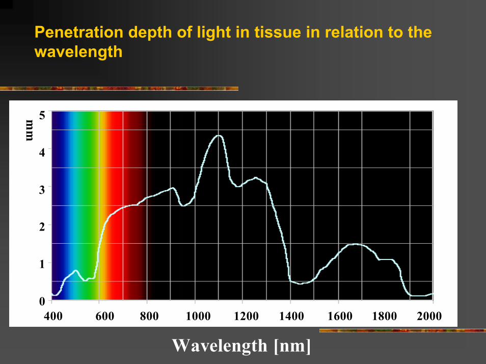
Penetration depth of light in tissue in relation to the wavelength
Wavelength [ nm ]
mm
400 600 800 1000 1200 1400 1600 1800 2000
5
4
3
2
1
0
Photosensitizer
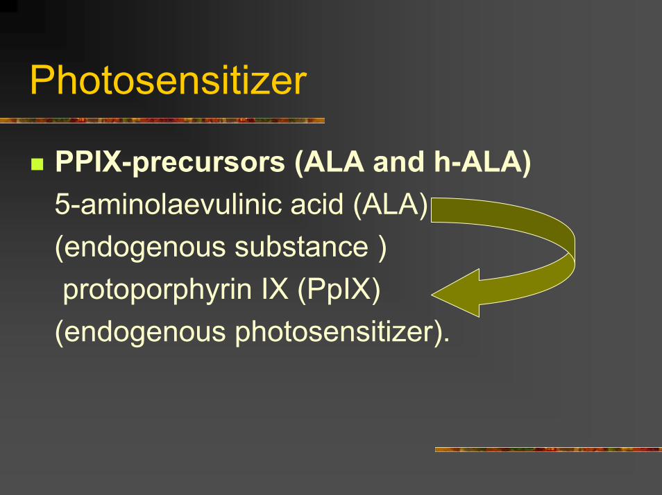
PPIX-precursors (ALA and h-ALA)5-aminolaevulinic acid (ALA)(endogenous substance )protoporphyrin IX (PpIX) (endogenous photosensitizer).
Photosensitizer
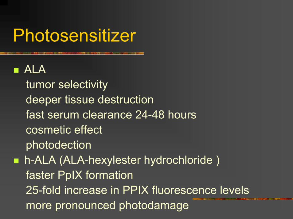
ALAtumor selectivitydeeper tissue destructionfast serum clearance 24-48 hourscosmetic effectphotodectionh-ALA (ALA-hexylester hydrochloride )faster PpIX formation 25-fold increase in PPIX fluorescence levelsmore pronounced photodamage

PDD Early Tumor Detection
Urology
Pneumology
Neurosurgery

PDT
Henta et.al . British Journal of Dermatology 1999 141(2) 347

PHOTOMEDICINE
GYNECOLOGICAL ONCOLOGY
PDD in gynecological neoplasms
CINendometrial cancerintraperitoneal metastasis of ovarian cancer

PDD in CIN
early detection noninvasive staging of CIN
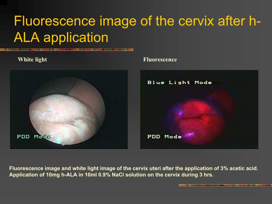
Fluorescence image of the cervix after h-ALA application
White light Fluorescence
Fluorescence image and white light image of the cervix uteri after the application of 3% acetic acid. Application of 10mg h-ALA in 10ml 0.9% NaCl solution on the cervix during 3 hrs.
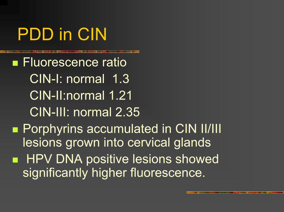
PDD in CINFluorescence ratioCIN-I: normal 1.3 CIN-II:normal 1.21 CIN-III: normal 2.35
Porphyrins accumulated in CIN II/III lesions grown into cervical glandsHPV DNA positive lesions showed significantly higher fluorescence.

PDD in CIN
specificity : fluorescence spectroscopy 75% colposcopy 50%
sensitivity :95%Double ratio (DR) fluorescence imaging technique

PDD in ovarian cancer
improves visualization and guides treatment of small cancerous nodules (0.3 mm)

PDD in ovarian cancer
In vivo fluorescence and light images of peritoneal tumor nodules. Fluorescence wasexcited using an endoscope (with D-light) after ip administration of ALA in an ovariancancer rat (Fischer 344) model.

PDD in endometrial cancer
Malignant endometrial epithelial cells showed significant higher fluorescence of PpIX than normal epithelial cells after incubation with 1 mg ALA The well-differentiated cancer cells produced significantly more PpIX than the poorly differentiated cancer cells.

PDT in gynecological neoplasms
Cervical neoplasms Vulvar and vaginal neoplasmsOvarian cancerEndometrial cancer
PDT in cervical neoplasms
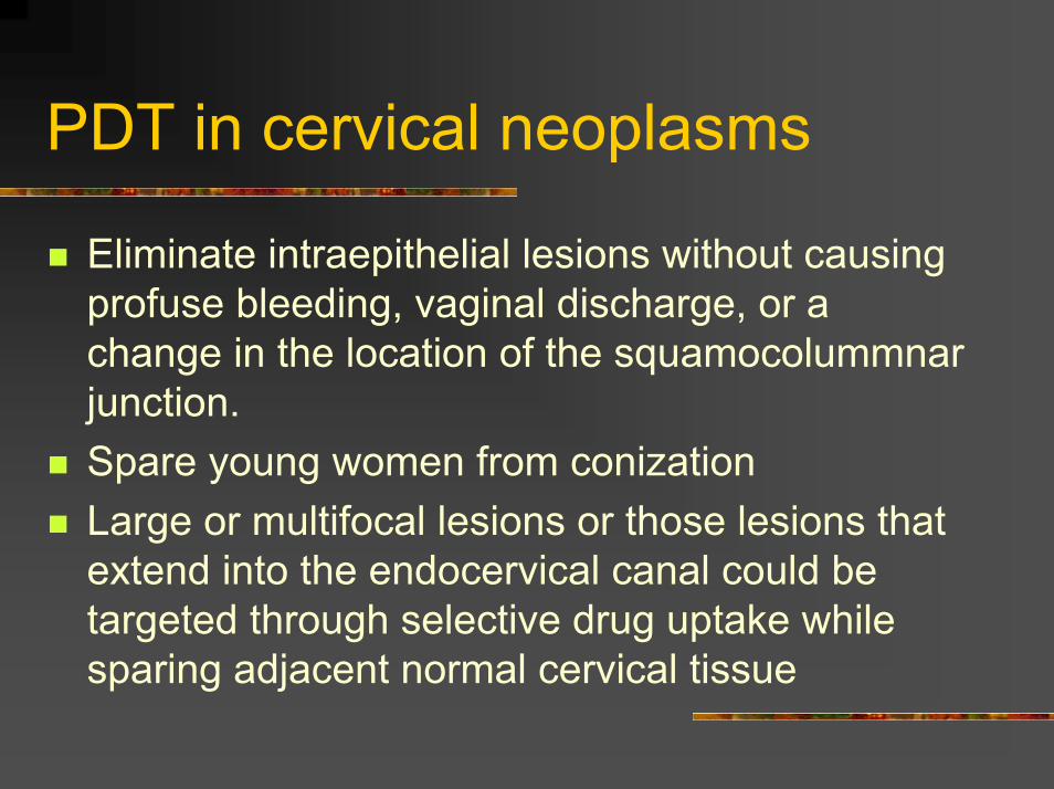
Eliminate intraepithelial lesions without causing profuse bleeding, vaginal discharge, or a change in the location of the squamocolummnarjunction. Spare young women from conizationLarge or multifocal lesions or those lesions that extend into the endocervical canal could be targeted through selective drug uptake while sparing adjacent normal cervical tissue

PDT in CIN
3(25%)5(42%)4(33%)210PDT
4(31%)5(38%)4(31%)112Placebo
Apparent progression
No change
Normal I/IIIGroup
Outcome (3 months after PDT)Pretreatment diagnosis
Adrian A 2003

PDT in CIN
CIN IIISuccess rate was 31% (10/32) 12 months after treatment

PDT in vulvar neoplasms

PDT in vular neoplasms
as effective as conventional treatments (laser evaporation and excision) for condyloma and VIN shorter healing time (2 weeks)less pain excellent cosmetic results Lower grades (VIN I) vs high grades (VIN II-III) monofocal and bifocal vs multifocalpigmented and hyperkeratotic lesions respond poorly

PDT in ovarian cancer
Diffuse intra-abdominal metastases have been successfully treated with PDT in a mouse model. Minimally invasive debulking of nonresectablepelvic tumors was effective in a rat ovarian cancer model Wierrani et al. m-THPC mediated PDT for two recurrent ovarian caner patients and one patient following surgical tumor debulking. After more than 2 years all three patients remained free of relapses

PDT in ovarian cancer
“conjugated phototherapy”photoimmunotherapyphotochemotherapy

PDT in endometrial cancerKoren 1996
-3NR
Month after treatment
5-CR-REC
69total
16CR
121
7 endometrial carcinomas stage Ia2 with recurrent endomerial carcinoma at vagina

Discussion
a promising tool for early detection of superficial gynecological neoplasmearly detection and noninvasive staging of CINdetecting intraperitoneal macroscopically invisible ovarian cancer nodules

Discussion
a better choice for VIN than conventional treatment CIN ?Ovarian cancer?Endometrial cancer?Conjugated photosensitzers

Discussion
Further well designed, large sample size clinical trials are needed!!!