Embed Size (px)
Citation preview

Chapter 3
The Role of Placental Exosomesin Gestational Diabetes Mellitus
Carlos Salomon, Luis Sobrevia, Keith Ashman,Sebastian E. Illanes, Murray D. Mitchell andGregory E. Rice
Additional information is available at the end of the chapter
http://dx.doi.org/10.5772/55298
1. Introduction
Gestational Diabetes Mellitus (GDM) affects ~5% of all pregnancies and parallels the globalincrease in obesity and type 2 diabetes. In the USA alone, GDM affects more than 135,000pregnancies per year. Lifestyle changes that impact adversely on caloric balance are thoughtto be a contributing factor in this emerging pandemic [1, 2]. The current ‘gold standard’ forthe diagnosis of GDM is the oral glucose tolerance test (OGTT) at 24–28 weeks of gestation [3,4]. When GDM is diagnosed in the late second or early third trimester of pregnancy the‘pathology’ is most likely well-established and the possibility to reverse or limit potentialadverse effects on perinatal outcomes may be limited [ 5]. Early detection of predisposition toand/or onset of GDM, thus, is the first step in developing, evaluating and implementingefficacious treatment. If such early detection tests were available, they would represent a majoradvance and contribution to the discipline and afford the opportunity to evaluate alternatetreatment and clinical management strategies to improve health outcomes for both motherand baby. Based upon recent technological developments and studies, we consider it realisticthat a clinically useful antenatal screening test can be developed. Unlike diseases such as cancerwhere biomarkers need to be exquisitely specific, a useful antenatal screening test wouldideally be highly sensitive, but not necessarily highly specific. The consequence of a falsepositive would be no worse than an erroneous triage to high-risk care.
Recent studies highlight the putative utility of tissue-specific nanovesicles (e.g. exosomes) inthe diagnosis of disease onset and treatment monitoring [6-11]. To date there is a paucity of
© 2013 Salomon et al.; licensee InTech. This is an open access article distributed under the terms of theCreative Commons Attribution License (http://creativecommons.org/licenses/by/3.0), which permitsunrestricted use, distribution, and reproduction in any medium, provided the original work is properly cited.

data defining changes in the release, role and diagnostic utility of placenta-derived nanovesi‐cles (e.g. exosomes) in pregnancies complicated by GDM.
The aim of this brief commentary, thus, is to review the biogenesis, isolation and role ofnanovesicles; and their release from the placenta. Placental exosomes may engage in paracel‐lular interactions (i.e. local cell-to-cell communication between the cell constituents of theplacenta and contiguous maternal tissues) and/or distal interactions (i.e. involving the releaseof placental exosomes into biological fluids and their transport to a remote site of action).
2. Exosome biogenesis and composition
2.1. Biogenesis
Exosomes are small [40-100 nm) membrane vesicles that are released following the exocytoticfusion of multi-vesicular bodies with the cell membrane (Figure 1). They are characterised by:a cup-shaped form: (a) a buoyant density of 1.13-1.19 g⁄ml [12, 13], (b) endosomal origin, and(c) the enrichment of late endosomal membrane markers including Tsg101, CD63, CD9 andCD81 [7, 14, 15]. While the process(es) of exosome formation remains to be fully elucidated,available data support an endosomal origin and formation by the inward budding of multi-vesicular bodies [16] (see Figure 2). Exosomes may also be directly transported from the Golgicomplex to multi-vesicular bodies [14].
Figure 1. Electron micrograph of circulating exosomes. Exosomes are 40-100 nm membrane vesicles with a densityranging from 1.13-1.19 g/ml, characterised by a cup-shaped form and secreted by most cell types in vivo and in vitro.Villous chorionic explant-derived exosomes were isolated by ultracentrifugation and purified using a sucrose gradient.Scale bar = 100 nm.
Gestational Diabetes - Causes, Diagnosis and Treatment30
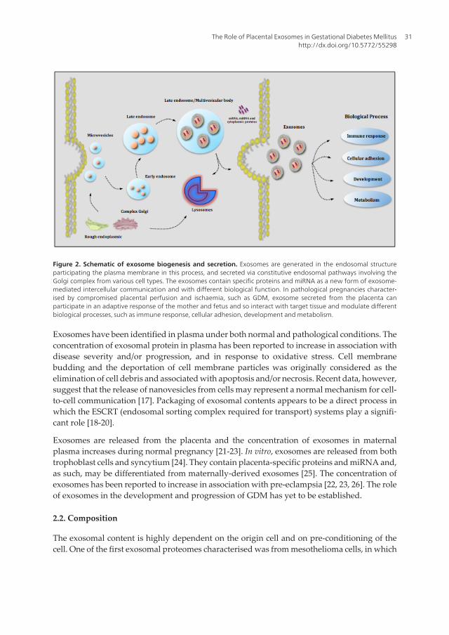
Figure 2. Schematic of exosome biogenesis and secretion. Exosomes are generated in the endosomal structureparticipating the plasma membrane in this process, and secreted via constitutive endosomal pathways involving theGolgi complex from various cell types. The exosomes contain specific proteins and miRNA as a new form of exosome-mediated intercellular communication and with different biological function. In pathological pregnancies character‐ised by compromised placental perfusion and ischaemia, such as GDM, exosome secreted from the placenta canparticipate in an adaptive response of the mother and fetus and so interact with target tissue and modulate differentbiological processes, such as immune response, cellular adhesion, development and metabolism.
Exosomes have been identified in plasma under both normal and pathological conditions. Theconcentration of exosomal protein in plasma has been reported to increase in association withdisease severity and/or progression, and in response to oxidative stress. Cell membranebudding and the deportation of cell membrane particles was originally considered as theelimination of cell debris and associated with apoptosis and/or necrosis. Recent data, however,suggest that the release of nanovesicles from cells may represent a normal mechanism for cell-to-cell communication [17]. Packaging of exosomal contents appears to be a direct process inwhich the ESCRT (endosomal sorting complex required for transport) systems play a signifi‐cant role [18-20].
Exosomes are released from the placenta and the concentration of exosomes in maternalplasma increases during normal pregnancy [21-23]. In vitro, exosomes are released from bothtrophoblast cells and syncytium [24]. They contain placenta-specific proteins and miRNA and,as such, may be differentiated from maternally-derived exosomes [25]. The concentration ofexosomes has been reported to increase in association with pre-eclampsia [22, 23, 26]. The roleof exosomes in the development and progression of GDM has yet to be established.
2.2. Composition
The exosomal content is highly dependent on the origin cell and on pre-conditioning of thecell. One of the first exosomal proteomes characterised was from mesothelioma cells, in which
The Role of Placental Exosomes in Gestational Diabetes Mellitushttp://dx.doi.org/10.5772/55298
31

38 different proteins were identified [27]. Studies in cancer cells show the great variability ofproteins expressed in exosomes [28-32]. In exosomes isolated from a human first trimester cellline (Sw7 1) Atay et al., using an ion trap mass spectrometry approach, identified proteinsimplicated in a wide range of cellular processes including: cytoskeleton structure (adhesion,membrane transport, and fusion), ion channels, lysosomal degradation, molecular chaperones,amino-acid metabolism, carbohydrate metabolism, lipid metabolism, oxydo-reductaseactivity, protein synthesis and post-translational modifications, ubiquitin modifiers, signaltransduction, transcription factors and regulators, DNA replication, chromatin structural andregulatory proteins, mRNA splicing, transcription/translation, post-translational proteinmodification enzymes, nuclear structural proteins, integrins, complement and coagulation,immune function, iron transport, and ER specific proteins. This study provides the firstextensive analysis of the proteome of the exosome-derived trophoblast cells [7]. The dataobtained in this study, highlights the extent of putative functional interactions that may bemediated by exosomes.
While the composition of exosomes appears to be cell-specific, a subset of common proteinshas been identified. The lipid bilayer is composed of sphingomyelin [33, 34]. Among the mostcommonly used markers for characterisation of exosomes are tetraspanin proteins, including:CD63, CD81, CD9, and CD82. Other families of common proteins in all exosomes includechaperone proteins such as: Hsc70 and Hsp90; cytoskeletal proteins including actin, tubulinand myosin; transport proteins; and annexins [35]. Exosomes derived from antigen-presentingcells (APC) express MHC-I and MHC-II on their surface [36-38]. During exosome biogenesis,the phospholipid/protein ratio of exosomes may be regulated by the Golgi membranes [39].
Significantly, single cell types display the capacity to generate different subpopulations ofexosomes. Laulagnier et al. 2005 demonstrated that RBL-2H3 cells (basophilic leukemia cellline) released two main subpopulations of exosomes that can be discriminated by protein andlipid contents. The first subpopulation contains phospholipids obtained mainly from granulesand the second contains phospholipids from Golgi. In addition, proteins CD63, MHC-II, CD81-containing exosomes accounted for 47%, 32%, and 21%, respectively, of total exosomes [39, 40].
3. Isolation of exosomes
Exosome research is a burgeoning discipline with over 2000 articles published in the last 3years. The putative role of exosomes spans from intracellular signaling to biomarkers of disease[41]. Germane to any study seeking to elucidate the physiological or pathophysiological roleof exosomes is their specific isolation. Several methods for isolating exosomes have beendeveloped and partially characterised. These isolation methods are primarily based on particlesize and density. By definition, exosomes are nanovesicles with a diameter of 30-100 nm.Typically they display a density of 1.12 to 1.19 g/ml and express characteristic cell-surfacemarkers.
The most common method of separation involves a series of differential centrifugation toremove intact cells and debris, and nuclei followed by size selective filtration (0.2 μm pore
Gestational Diabetes - Causes, Diagnosis and Treatment32
size) and sedimentation by ultracentrifugation (e.g. at 110,000 g for 1-2 hours [42, 43]. Exosomesmay be further purified by differential sedimentation on sucrose gradients or sucrose-deuterium oxide (D2O) [ 7]. Alternative methods utilise size exclusion chromatography anddensity gradient centrifugation [44] or solid-phase immunoaffinity capture (i.e. anti-MHC-IIDynabeads) [38, 43, 45]. In the absence of specific, cell surface exosome markers, the veracityof immuno-affinity methods remains to be established.
More recently a commercial kit for the isolation of the exosomes has been released (Exo‐QuickTM, System Biosciences). The isolation process involves a simple one-step precipitation[43, 46]. While the commercial kit provides significant advantage with respect to processingtime, the resulting preparation may not be equivalent to that obtained by ultracentrifugationand differential segmentation. In our own laboratory, parallel preparations of exosomes usingboth methods reveal differences in the biophysical characteristics of the exosomes isolated.Exosome preparations isolated using the commercial kit were characterised by a great rangein particle diameter (30-300 nm, as estimated by transmission electron microscopy (TEM) andhave a higher protein content than similar preparations isolated using the ultracentrifugationmethod. In addition, analysis of protein patterns in SDS-PAGE electrophoresis and westernblot against CD63, CD81 and CD9 show similar characteristics between exosomes fromultracentrifuge and ExoQuickTM methods, however, we had to dilute ExoQuickTM samples ~10 times to obtain comparable concentrations with ultracentrifuge methods in exosomesisolated from trophoblastic cells (see Figure 3). Differences in exosome protein and mRNAcontent and functional activity between different preparations remain to be established.
Figure 3. Typical characteristics of exosomes isolated from trophoblast cells. (A) Exosome protein pattern analy‐sis. 10 ug of exosome proteins and trophoblast cell lysate were separated on 4-12% SDS-PAGE and stained with Sim‐plyBlue™ SafeStain (Invitrogen). (B) Western blot characterisation of exosomes with antibodies against CD63, CD81and CD9 for 4 different samples isolated with ultracentrifuge or ExoQuick™ methods.
The Role of Placental Exosomes in Gestational Diabetes Mellitushttp://dx.doi.org/10.5772/55298
33

4. The role for exosomes in cell-to-cell communication
Recently, evidence supporting a role for exosomes in cell-to-cell communication has beenobtained [47, 48]. Exosome release may represent a significant, hitherto unappreciated,communication mechanism between cells, host cell and microbes [47].
For example, exosome function as a carrier of specific molecules such as mRNA and miRNAcan interact with neighbouring cells or travel long distances in the bloodstream to reprogramthe phenotype and regulate their function [40]. In the placenta, exosome- derived trophoblasticcells are able to reprogram monocytes to secrete specific cytokine profiles independent of cell-to-cell contact [8]. Placental-derived exosomes may also play a role in modulating immuno‐logical responses through the induction of lymphocyte apoptosis, [21, 44, 49].
4.1. Information encoding by exosomes
Exosomes have been reported to express a diverse range of cell surface receptors, proteins(including, heat shock proteins, cytoskeletal proteins, adhesion molecules, membrane trans‐port and fusion proteins), mRNA and miRNA with the potential to affect the acute and long-term function of the cells with which they interact [50]. In addition, in the absence of energyproduction, normal membrane phospholipid asymmetry is lost and amniophospholipidstranslocate to the outer leaflet of the cell membrane and generate a fusogenic and pro-coagulantsurface. Given that exosomes circulate in blood, these fusogenic moieties may be masked by,for example, annexin V.
In vitro effects of exposing cells to exosomal proteins has been reported and include: inductionof differentiation of stem cells [51], suppression of activation of natural killer cells andmacrophages [52, 53], and stimulation of cell migration [8, 54]. Putative roles of exosomes,thus, include cell differentiation, immunomodulation and migration [55]. Exosomes are notmerely inert fragments of cell membrane but display capacity to affect cell function at remoteloci and possibly be a source of disease biomarkers.
Exosomes also contain miRNA that may transfer to other cells and alter the expression of thetranscriptome and ultimately cell phenotype. miRNAs are a class of small non-coding RNAsthat function as translational repressors involved in a variety of physiological and pathologicalprocesses in animals [56, 57]. They act via binding to messenger RNA and, thus, prevent thetranslation of the encoded protein. Previous studies have reported that miRNAs are involvedin the pathogenesis of diabetes and are required for pancreatic development and the regulationof glucose-stimulated insulin secretion [50, 58]. Moreover, differences in the expression ofmiRNA such as miR-146a, miR-21, miR-29a, miR-34a, miR-222, and miR-375 have beenreported in pancreatic β-cells, liver, adipose tissue, and/or skeletal muscle of animal modelsof type 1 or type 2 diabetes [59]. Another study found that miR-20b, miR-21, miR-24, miR-15a,miR-126, miR-191, miR-197, miR-223, miR-320, and miR-486 were lower in prevalent type 2diabetes [60].
Gestational Diabetes - Causes, Diagnosis and Treatment34
4.2. Placental exosome release and effects
4.2.1. Placental exosome release
Exosomes are released by the placenta during pregnancy and their release may correlate withpregnancy outcome. The syncytiotrophoblasts and cytotrophoblasts are the most abundantcell types of the human placenta and sense and regulate oxygen and nutritional exchangebetween mother and fetus during the pregnancy [61, 62]. Pathologies of pregnancy includingpreeclampsia, intrauterine growth restriction (IUGR) and GDM are associated with placentaldysfunction [4, 63, 64] and may display differential and specific exosome release profiles.
It has been established that the concentration of exosomes in maternal peripheral blood isgreater than that observed in non-pregnant women [21]. In this study, exosomes of placentalorigin were specifically isolated from the maternal blood using anti-PLAP (anti-placental-typealkaline phosphatase) conjugated to agarose micro-beads. In peripheral blood mononuclearcells (PBMC), placental exosomes suppressed T signalling components such as CD3-zeta andJAK3, while inducing SOCS-2 [2 1]. These results are consistent with those of Taylor et al. [44,65] who demonstrated the presence and composition of placenta-derived exosomes inmaternal circulation along with their effects on T cell activation markers. Exosomes appear toplay an essential role in preventing an excessive immune response and in the development ofautoimmunity in human pregnancy.
Recently, it has been demonstrated that placental miRNAs circulate in the blood of pregnantwomen [66, 67]. For example, maternal plasma concentration of placental miRNA-141increases with gestational age [66]. Placenta-specific miRNA-517A is released from chorionicvillous trophoblasts into maternal circulation, where it may affect maternal tissues (e.g.maternal endothelium) during pregnancy [25]. There is a paucity of data, however character‐ising the release of exosome from endothelial cells during normal and pathological pregnan‐cies. It will be important to determine if the placenta communicates with the maternalendothelium via microvesicles, and, if so, to elucidate the role and mechanism of action ofexosome pathologies associated with endothelial dysfunction, such as GDM. Placenta-derivedmiRNAs, therefore, may be of utility as biomarkers of placental function and/or pregnancyoutcome. It remains to be elucidated how much of this “free” miRNA and mRNA is actuallycontained within exosomes and thereby confers stability. Indeed, the exact mechanismsinvolved in the release of miRNA from the placenta remain to be established. A recent study,however, reported that miRNAs are selectively packaged into microvesicles and are activelysecreted [68-70]. miRNAs are also released from the syncytiotrophoblast to the maternalcirculation in the pregnancy packaged inside exosomes [25, 71].
4.2.2. Effects
There remains a paucity of data about the effect of placenta-derived exosomes on both fetusand mother. The available data, however, support a role for placental exosomes in mediatingcommunication at the materno-fetal interface and, possibly, at the distal site within the mother.Recent data show that trophoblast-derived exosomes induce proinflammatory cytokines suchIL-1 β in human macrophages cells [8]. Furthermore, in vitro exposure of PBMC and dendritic
The Role of Placental Exosomes in Gestational Diabetes Mellitushttp://dx.doi.org/10.5772/55298
35

cells to exosomal proteins induce differentiation of stem cells; suppression of activation ofnatural killer cells and macrophages; and stimulation of cell migration [53, 72, 73]. Interest‐ingly, protein analysis revealed that exosome release from trophoblast cells increases with lowoxygen tension and their exosome promote the cell migration in extravillous cytotrophoblast(HTR- 8) (Salomon et al. manuscript in preparation).
5. Exosomes and GDM
Exosomes released from the placentae of women with GDM may alter maternal physiology.Via a process of exosomal placento-maternal transfection a “payload” of receptors, proteinsand/or oligonucleotides” that have been specifically pre-conditioned by the GDM placentamay be delivered to maternal response systems. Such mediators include: vascular, pancreaticand adipose tissues, and the innate immune response system. The extent and impact ofplacenta-derived exosomes on maternal physiology, however, remains to be elucidated.
In addition to a placento-maternal transfection pathway, trophoblasts or placental mesenchymalstem cells (MSCs) may induce paracellular effects in association with GDM. For example, inplacental villi, exosomes released by perivascular MSCs may alter transport activity withinthe placental vascular endothelium (e.g. the glucose transport GLUT 3) and thus the deliveryof energy substrates to the fetus.
In support of the role of exosomes in modulating glucose homeostasis, Deng et al., reportedthat exosomes isolated from adipose tissue induce differentiation of monocytes into activatedmacrophages and promote insulin resistance in an obese mouse model [74]. Exosomes isolatedfrom mouse insulinoma induce the secretion of inflammatory cytokines including IL-6 andTNF-α in splenocytes cultured from non-obese diabetic mice (NOD) [75]. In this regards, thesecytokines as well as other inflammatory mediators play an important role in glucose toleranceand insulin sensitivity dysregulation in women with previous GDM.
5.1. The effects of hyperglycaemia and oxidative stress on exosome release
GDM is a state of hyperglycaemia and increased oxidative stress [76]. In addition, hypergly‐caemia-induced oxidative stress makes an important contribution to the aetiology of GDM[77], with consequences for both mother and baby [78]. In support of an aetiological role ofhypoglycaemia and attendant oxidative stress in poor pregnancy outcome, the HAPO studyreported a strong and continuous association between maternal glucose concentrations andpregnancy outcome and confirmed a relationship between birth weight and maternal hyper‐glycaemia [79, 80].
Reactive oxygen species (ROS) include oxygen ions such as superoxide ions and hydrogenperoxide (H2O 2) that are generated continuously during cellular metabolism. GDM pregnan‐cies are characterised by an overproduction of ROS and free radicals and impaired antioxidantcapacity [81]. Oxidative stress and increased exosome release are common features of manypathologies including: cancer, kidney disease, hypertension, and preeclampsia. It remains to
Gestational Diabetes - Causes, Diagnosis and Treatment36
be established whether or not exosome release in these circumstances is a paraphenomenonof, or an adaptive response to, increased ROS formation and oxidative stress.
GDM is a syndrome that leads to feto-placental vascular endothelial dysfunction involvinghigher nitric oxide (NO) concentrations and increases of oxidative state and vascular resistance[63, 82]. Exosomes – endothelial cell interactions may result in activation of NO synthesis via anumber of mechanisms. Exosomes isolated from platelets obtained from patients with septicinduced endothelial dysfunction through the NADPH oxidase-dependent release of superox‐ide and have been implicated in the induction of NO and peroxinitrite. NO synthase is alsoinduced by miR-203. miRNA-203 has been identified in exosomes [83]. It remains to be establish‐ed whether or not miRNA-203 is present in exosomes isolated from women with GDM.
In non-gestational tissues, the available evidence supports an active role for exosomes inregulating cellular redox status. For example, oxidative stress enhances exosome release fromJurkat and Raji cell lines and the resultant increase in NKG2D receptor bioactivity impairscytotoxic response [84]. In 3T3-L1 adipocytes, oxidative stress increases microvesicle release[85]. Melanoma-derived exosomes induce ROS production in T cells compared to exosomesfrom normal cells, suppressing the immune response and improving carcinogenic invasion[86]. Exosomes isolated from mouse mast cells MC/9 exposed to oxidative stress alter theresponse of others cells to oxidative stress [87], increasing their resistance to oxidative stressand reducing cell death. Interestingly, the mRNA content of exosomes produced underoxidative stress conditions differ from those produced under normal conditions. These dataare consistent with the observations of Atay et al. [7, 8], Luo et al., [25] and Taylor et al. [83] whosimilarly report cell- and condition-specific variation in exosomal protein, mRNA and miRNAcontent.
5.2. Exosome biomarkers of GDM
In addition to their putative functional involvement in the pathophysiology of pregnancy,placental-derived exosomes may be of utility as diagnostic markers of GDM in asymptomaticwomen.
In 2011, the American Diabetes Association (ADA) and the International Association ofDiabetes and Pregnancy Study Groups (IADPSG) revised recommendations regarding GDM.It is now recommended that patients at increased risk for type 2 diabetes be screened fordiabetes using standard diagnostic criteria at their first prenatal visit (ADA 201 2). Currently,GDM is diagnosed in the late second or early third trimester of pregnancy. Pathology isprobably already established by this time and reversal of the potential adverse perinataloutcomes may be limited. The lack of a reliable early test for GDM has hampered the devel‐opment of useful intervention therapies that may impact not only on the acute but also thelong-term health outcomes [88-90]. Thus, there is a need to diagnose and predict GDM earlierso that appropriate management can be initiated and tailored to the needs of the patient inorder to minimise perinatal complications and their sequelae.
Currently, the diagnosis of GDM is between 24-28 weeks of gestation by an oral glucosetolerance test. The aim of the treatment for GDM is to maintain the glucose level in euglycaemia
The Role of Placental Exosomes in Gestational Diabetes Mellitushttp://dx.doi.org/10.5772/55298
37

with dietary modifications or in some cases with insulin therapy, however, when it is diag‐nosed, the pathology is established and the clinical and obstetric management is limited [5, 91].
The quantitation of exosomes and/or exosome-specific content may be of diagnostic utility [14,43, 83, 92, 93]. Exosomes are found in all body fluids tested to date including blood, urine,saliva and breast milk. They can be obtained by minimally invasive methods (blood) or non-invasive methods (using urine or saliva) [94]. Several studies have demonstrated the putativeutility of exosomes as biomarkers, particularly in cancers, where exosomal protein is correlatedwith disease burden.
The measurement of exosomal miRNA in biofluids has proven of utility in cases of lung cancer,colorectal cancer, prostate cancer and diabetes [95-99].
Zhao et al. isolated miRNA from blood circulation at 16-19 gestational weeks. Interestingly,these authors found that the expression of miRNA-132, miRNA-29a and miRNA-22 weredecreased in GDM women compared with normal pregnancies in similar gestational weeks[70]. Finally, there are few reports suggesting that mesenchymal stem cell and trophoblastcells-derived exosomes may serve as therapeutic agents for use in regenerative medicine torepair damaged tissue [100, 101].
Finally, in normal pregnancies, the placenta secretes significant amounts of macro- and microve‐sicles, including exosomes [22, 26]. We suggest that in pregnancies complicated by GDM,oxidative stress and hyperglycaemia increase the release of exosomes from the placenta into thematernal circulation during in the first trimester of pregnancy. The quantification and character‐ised of exosomes in the blood of these pre-symptomatic women, thus, may be of utility as an earlybiomarker of disease onset. Furthermore, we propose that during first trimester, pre-symptomat‐ic women who subsequently develop GDM: have higher plasma concentrations of placental-derived exosomes; and a different exosomal protein and miRNA profile than women whoexperience a normoglycaemic pregnancy. These characteristics could potentially be used fordiagnostic markers for exosome profiling to screen asymptomatic populations.
Acknowledgements
CS holds a Postdoctoral Fellowship at The University of Queensland Centre for ClinicalResearch, Brisbane, Australia. GER was in receipt of an NHMRC Principal Research Fellow‐ship. The work described herein was partially funded by a CIEF grant (University of Queens‐land), a Smart Futures Fund grant (Department of Employment, Economic Development andInnovation, Queensland Government) and a Translating Health Discovery into ClinicalApplications SuperScience Award (Department of Industry, Innovation, Science, Research andTertiary Education, Australian Government).
This investigation was supported by CONICYT (ACT-73 PIA, Pasantía Doctoral en el Extra‐njero BECAS Chile), FONDECYT (1110977). CS hold CONICYT-PhD fellowships and Facultyof Medicine/PUC-PhD fellowships.
Gestational Diabetes - Causes, Diagnosis and Treatment38

Author details
Carlos Salomon1,2, Luis Sobrevia1, Keith Ashman2, Sebastian E. Illanes3,Murray D. Mitchell2 and Gregory E. Rice2
1 Cellular and Molecular Physiology Laboratory (CMPL), Division of Obstetrics and Gynae‐cology, School of Medicine, Faculty of Medicine, Pontificia Universidad Católica de Chile,Santiago, Chile
2 University of Queensland Centre for Clinical Research, University of Queensland, Her‐ston, Queensland, Australia
3 Department of Obstetric and Gynaecology, Universidad de los Andes, Santiago, Chile
References
[1] Ferrara A, Kahn HS, Quesenberry CP, Riley C, Hedderson MM. An increase in theincidence of gestational diabetes mellitus: Northern California, 1991-2000. Obstetricsand gynecology. 2004;103 ( 3):526-33. Epub 2004/03/03.
[2] Robitaille J, Grant AM. The genetics of gestational diabetes mellitus: evidence for re‐lationship with type 2 diabetes mellitus. Genetics in medicine : official journal of theAmerican College of Medical Genetics. 2008;10 ( 4):240-50. Epub 2008/04/17.
[3] Diagnosis and classification of diabetes mellitus. Diabetes care. 2012;35 Suppl1:S64-71. Epub 2012/01/04.
[4] Salomon C, Westermeier F, Puebla C, Arroyo P, Guzman-Gutierrez E, Pardo F, et al.Gestational diabetes reduces adenosine transport in human placental microvascularendothelium, an effect reversed by insulin. PloS one. 2012;7 ( 7):e40578. Epub2012/07/19.
[5] Agarwal MM, Weigl B, Hod M. Gestational diabetes screening: the low-cost algo‐rithm. International journal of gynaecology and obstetrics: the official organ of theInternational Federation of Gynaecology and Obstetrics. 2011;115 Suppl 1:S30-3.Epub 2011/12/07.
[6] Chen Y, Ge W, Xu L, Qu C, Zhu M, Zhang W, et al. miR-200b is involved in intestinalfibrosis of Crohn's disease. International journal of molecular medicine. 2012;29 ( 4):601-6. Epub 2012/02/02.
[7] Atay S, Gercel-Taylor C, Kesimer M, Taylor DD. Morphologic and proteomic charac‐terization of exosomes released by cultured extravillous trophoblast cells. Experi‐mental cell research. 2011;317 ( 8):1192-202. Epub 2011/02/01.
The Role of Placental Exosomes in Gestational Diabetes Mellitushttp://dx.doi.org/10.5772/55298
39

[8] Atay S, Gercel-Taylor C, Suttles J, Mor G, Taylor DD. Trophoblast-derived exosomesmediate monocyte recruitment and differentiation. Am J Reprod Immunol. 2011;65( 1):65-77. Epub 2010/06/22.
[9] Armitage JA, Poston L, Taylor PD. Developmental origins of obesity and the meta‐bolic syndrome: the role of maternal obesity. Frontiers of hormone research.2008;36:73-84. Epub 2008/01/31.
[10] Taylor DD, Gercel-Taylor C. Tumour-derived exosomes and their role in cancer-asso‐ciated T-cell signalling defects. British journal of cancer. 2005;92 ( 2):305-11. Epub2005/01/19.
[11] Simpson RJ, Jensen SS, Lim JW. Proteomic profiling of exosomes: current perspec‐tives. Proteomics. 2008;8 (1 9):4083-99. Epub 2008/09/10.
[12] Mignot G, Roux S, Thery C, Segura E, Zitvogel L. Prospects for exosomes in immu‐notherapy of cancer. Journal of cellular and molecular medicine. 2006;10 ( 2):376-88.Epub 2006/06/27.
[13] Miranda KC, Bond DT, McKee M, Skog J, Paunescu TG, Da Silva N, et al. Nucleicacids within urinary exosomes/microvesicles are potential biomarkers for renal dis‐ease. Kidney international. 2010;78 ( 2):191-9. Epub 2010/04/30.
[14] Mincheva-Nilsson L, Baranov V. The role of placental exosomes in reproduction. AmJ Reprod Immunol. 2010;63 ( 6):520-33. Epub 2010/03/25.
[15] Keller S, Ridinger J, Rupp AK, Janssen JW, Altevogt P. Body fluid derived exosomesas a novel template for clinical diagnostics. Journal of translational medicine.2011;9:86. Epub 2011/06/10.
[16] Simons M, Raposo G. Exosomes--vesicular carriers for intercellular communication.Current opinion in cell biology. 2009;21 ( 4):575-81. Epub 2009/05/16.
[17] Ludwig AK, Giebel B. Exosomes: small vesicles participating in intercellular commu‐nication. The international journal of biochemistry & cell biology. 2012;44 ( 1):11-5.Epub 2011/10/26.
[18] Wegner CS, Rodahl LM, Stenmark H. ESCRT proteins and cell signalling. Traffic.2011;12 (1 0):1291-7. Epub 2011/04/27.
[19] Stuffers S, Sem Wegner C, Stenmark H, Brech A. Multivesicular endosome biogene‐sis in the absence of ESCRTs. Traffic. 2009;10 ( 7):925-37. Epub 2009/06/06.
[20] Stuffers S, Brech A, Stenmark H. ESCRT proteins in physiology and disease. Experi‐mental cell research. 2009;315 ( 9):1619-26. Epub 2008/11/18.
[21] Sabapatha A, Gercel-Taylor C, Taylor DD. Specific isolation of placenta-derived exo‐somes from the circulation of pregnant women and their immunoregulatory conse‐quences. Am J Reprod Immunol. 2006;56 (5- 6):345-55. Epub 2006/11/02.
Gestational Diabetes - Causes, Diagnosis and Treatment40

[22] Redman CW, Sargent IL. Circulating microparticles in normal pregnancy and pre-eclampsia. Placenta. 2008;29 Suppl A:S73-7. Epub 2008/01/15.
[23] Orozco AF, Lewis DE. Flow cytometric analysis of circulating microparticles in plas‐ma. Cytometry Part A : the journal of the International Society for Analytical Cytolo‐gy. 2010;77 ( 6):502-14. Epub 2010/03/18.
[24] Atay S, Gercel-Taylor C, Taylor DD. Human trophoblast-derived exosomal fibronec‐tin induces pro-inflammatory IL-1beta production by macrophages. Am J ReprodImmunol. 2011;66 ( 4):259-69. Epub 2011/03/18.
[25] Luo SS, Ishibashi O, Ishikawa G, Ishikawa T, Katayama A, Mishima T, et al. Humanvillous trophoblasts express and secrete placenta-specific microRNAs into maternalcirculation via exosomes. Biology of reproduction. 2009;81 ( 4):717-29. Epub2009/06/06.
[26] Redman CW, Tannetta DS, Dragovic RA, Gardiner C, Southcombe JH, Collett GP, etal. Review: Does size matter? Placental debris and the pathophysiology of pre-eclampsia. Placenta. 2012;33 Suppl:S48-54. Epub 2012/01/06.
[27] Hegmans JP, Bard MP, Hemmes A, Luider TM, Kleijmeer MJ, Prins JB, et al. Proteo‐mic analysis of exosomes secreted by human mesothelioma cells. The American jour‐nal of pathology. 2004;164 ( 5):1807-15. Epub 2004/04/28.
[28] Welton JL, Khanna S, Giles PJ, Brennan P, Brewis IA, Staffurth J, et al. Proteomicsanalysis of bladder cancer exosomes. Molecular & cellular proteomics : MCP. 2010;9( 6):1324-38. Epub 2010/03/13.
[29] Pisitkun T, Gandolfo MT, Das S, Knepper MA, Bagnasco SM. Application of systemsbiology principles to protein biomarker discovery: Urinary exosomal proteome in re‐nal transplantation. Proteomics Clinical applications. 2012;6 (5- 6):268-78. Epub2012/05/30.
[30] Gonzales PA, Pisitkun T, Hoffert JD, Tchapyjnikov D, Star RA, Kleta R, et al. Large-scale proteomics and phosphoproteomics of urinary exosomes. Journal of the Ameri‐can Society of Nephrology : JASN. 2009;20 ( 2):363-79. Epub 2008/12/06.
[31] Li Y, Zhang Y, Qiu F, Qiu Z. Proteomic identification of exosomal LRG1: a potentialurinary biomarker for detecting NSCLC. Electrophoresis. 2011;32 (1 5):1976-83. Epub2011/05/11.
[32] Zhang Y, Li Y, Qiu F, Qiu Z. Comprehensive analysis of low-abundance proteins inhuman urinary exosomes using peptide ligand library technology, peptide OFFGELfractionation and nanoHPLC-chip-MS/MS. Electrophoresis. 2010;31 (23-2 4):3797-807.Epub 2010/11/18.
[33] Laulagnier K, Motta C, Hamdi S, Roy S, Fauvelle F, Pageaux JF, et al. Mast cell- anddendritic cell-derived exosomes display a specific lipid composition and an unusual
The Role of Placental Exosomes in Gestational Diabetes Mellitushttp://dx.doi.org/10.5772/55298
41

membrane organization. The Biochemical journal. 2004;380(Pt 1):161-71. Epub2004/02/18.
[34] Wubbolts R, Leckie RS, Veenhuizen PT, Schwarzmann G, Mobius W, Hoernschemey‐er J, et al. Proteomic and biochemical analyses of human B cell-derived exosomes.Potential implications for their function and multivesicular body formation. TheJournal of biological chemistry. 2003;278 (1 3):10963-72. Epub 2003/01/10.
[35] Keller S, Sanderson MP, Stoeck A, Altevogt P. Exosomes: from biogenesis and secre‐tion to biological function. Immunology letters. 2006;107 ( 2):102-8. Epub 2006/10/28.
[36] Raposo G, Nijman HW, Stoorvogel W, Liejendekker R, Harding CV, Melief CJ, et al.B lymphocytes secrete antigen-presenting vesicles. The Journal of experimental med‐icine. 1996;183 ( 3):1161-72. Epub 1996/03/01.
[37] Denzer K, van Eijk M, Kleijmeer MJ, Jakobson E, de Groot C, Geuze HJ. Folliculardendritic cells carry MHC class II-expressing microvesicles at their surface. J Immu‐nol. 2000;165 ( 3):1259-65. Epub 2000/07/21.
[38] Clayton A, Court J, Navabi H, Adams M, Mason MD, Hobot JA, et al. Analysis of an‐tigen presenting cell derived exosomes, based on immuno-magnetic isolation andflow cytometry. Journal of immunological methods. 2001;247 (1- 2):163-74. Epub2001/01/11.
[39] Laulagnier K, Vincent-Schneider H, Hamdi S, Subra C, Lankar D, Record M. Charac‐terization of exosome subpopulations from RBL-2H3 cells using fluorescent lipids.Blood cells, molecules & diseases. 2005;35 ( 2):116-21. Epub 2005/07/19.
[40] Denzer K, Kleijmeer MJ, Heijnen HF, Stoorvogel W, Geuze HJ. Exosome: from inter‐nal vesicle of the multivesicular body to intercellular signaling device. Journal of cellscience. 2000;113 Pt 19:3365-74. Epub 2000/09/14.
[41] Mathivanan S, Fahner CJ, Reid GE, Simpson RJ. ExoCarta 2012: database of exosomalproteins, RNA and lipids. Nucleic acids research. 2012;40(Database issue):D1241-4.Epub 2011/10/13.
[42] Tauro BJ, Greening DW, Mathias RA, Ji H, Mathivanan S, Scott AM, et al. Compari‐son of ultracentrifugation, density gradient separation, and immunoaffinity capturemethods for isolating human colon cancer cell line LIM1863-derived exosomes.Methods. 2012;56 ( 2):293-304. Epub 2012/01/31.
[43] Taylor DD, Zacharias W, Gercel-Taylor C. Exosome isolation for proteomic analysesand RNA profiling. Methods Mol Biol. 2011;728:235-46. Epub 2011/04/07.
[44] Taylor DD, Akyol S, Gercel-Taylor C. Pregnancy-associated exosomes and theirmodulation of T cell signaling. J Immunol. 2006;176 ( 3):1534-42. Epub 2006/01/21.
[45] Thery C, Amigorena S, Raposo G, Clayton A. Isolation and characterization of exo‐somes from cell culture supernatants and biological fluids. Current protocols in cell
Gestational Diabetes - Causes, Diagnosis and Treatment42

biology / editorial board, Juan S Bonifacino (et al). 2006;Chapter 3:Unit 3 22. Epub2008/01/30.
[46] Yamada T, Inoshima Y, Matsuda T, Ishiguro N. Comparison of Methods for IsolatingExosomes from Bovine Milk. The Journal of veterinary medical science / the JapaneseSociety of Veterinary Science. 2012. Epub 2012/07/13.
[47] Deatherage BL, Cookson BT. Membrane vesicle release in bacteria, eukaryotes, andarchaea: a conserved yet underappreciated aspect of microbial life. Infection and im‐munity. 2012;80 ( 6):1948-57. Epub 2012/03/14.
[48] Southcombe J, Tannetta D, Redman C, Sargent I. The immunomodulatory role ofsyncytiotrophoblast microvesicles. PloS one. 2011;6 ( 5):e20245. Epub 2011/06/03.
[49] Bobrie A, Colombo M, Raposo G, Thery C. Exosome secretion: molecular mecha‐nisms and roles in immune responses. Traffic. 2011;12 (1 2):1659-68. Epub 2011/06/08.
[50] Ambros V. The functions of animal microRNAs. Nature. 2004;431 (700 6):350-5. Epub2004/09/17.
[51] Zhang HC, Liu XB, Huang S, Bi XY, Wang HX, Xie LX, et al. Microvesicles derivedfrom human umbilical cord mesenchymal stem cells stimulated by hypoxia promoteangiogenesis both in vitro and in vivo. Stem cells and development. 2012. Epub2012/07/31.
[52] Zhang HG, Zhuang X, Sun D, Liu Y, Xiang X, Grizzle WE. Exosomes and immunesurveillance of neoplastic lesions: a review. Biotechnic & histochemistry : officialpublication of the Biological Stain Commission. 2012;87 ( 3):161-8. Epub 2012/01/06.
[53] Mincheva-Nilsson L, Nagaeva O, Chen T, Stendahl U, Antsiferova J, Mogren I, et al.Placenta-derived soluble MHC class I chain-related molecules down-regulateNKG2D receptor on peripheral blood mononuclear cells during human pregnancy: apossible novel immune escape mechanism for fetal survival. J Immunol. 2006;176( 6):3585-92. Epub 2006/03/07.
[54] Lotvall J, Valadi H. Cell to cell signalling via exosomes through esRNA. Cell adhe‐sion & migration. 2007;1 ( 3):156-8. Epub 2007/07/01.
[55] Vrijsen KR, Sluijter JP, Schuchardt MW, van Balkom BW, Noort WA, Chamuleau SA,et al. Cardiomyocyte progenitor cell-derived exosomes stimulate migration of endo‐thelial cells. Journal of cellular and molecular medicine. 2010;14 ( 5):1064-70. Epub2010/05/15.
[56] Breving K, Esquela-Kerscher A. The complexities of microRNA regulation: mirander‐ing around the rules. The international journal of biochemistry & cell biology.2010;42 ( 8):1316-29. Epub 2009/10/06.
[57] Rottiers V, Naar AM. MicroRNAs in metabolism and metabolic disorders. Nature re‐views Molecular cell biology. 2012;13 ( 4):239-50. Epub 2012/03/23.
The Role of Placental Exosomes in Gestational Diabetes Mellitushttp://dx.doi.org/10.5772/55298
43

[58] Poy MN, Eliasson L, Krutzfeldt J, Kuwajima S, Ma X, Macdonald PE, et al. A pancre‐atic islet-specific microRNA regulates insulin secretion. Nature. 2004;432 (701 4):226-30. Epub 2004/11/13.
[59] Guay C, Roggli E, Nesca V, Jacovetti C, Regazzi R. Diabetes mellitus, a microRNA-related disease? Translational research : the journal of laboratory and clinical medi‐cine. 2011;157 ( 4):253-64. Epub 2011/03/23.
[60] Zampetaki A, Kiechl S, Drozdov I, Willeit P, Mayr U, Prokopi M, et al. Plasma micro‐RNA profiling reveals loss of endothelial miR-126 and other microRNAs in type 2diabetes. Circulation research. 2010;107 ( 6):810-7. Epub 2010/07/24.
[61] Costa SL, Proctor L, Dodd JM, Toal M, Okun N, Johnson JA, et al. Screening for pla‐cental insufficiency in high-risk pregnancies: is earlier better? Placenta. 2008;29 (1 2):1034-40. Epub 2008/10/22.
[62] Cartwright JE, Fraser R, Leslie K, Wallace AE, James JL. Remodelling at the maternal-fetal interface: relevance to human pregnancy disorders. Reproduction. 2010;140 ( 6):803-13. Epub 2010/09/15.
[63] Sobrevia L, Abarzua F, Nien JK, Salomon C, Westermeier F, Puebla C, et al. Review:Differential placental macrovascular and microvascular endothelial dysfunction ingestational diabetes. Placenta. 2011;32 Suppl 2:S159-64. Epub 2011/01/11.
[64] Cetkovic A, Miljic D, Ljubic A, Patterson M, Ghatei M, Stamenkovic J, et al. Plasmakisspeptin levels in pregnancies with diabetes and hypertensive disease as a poten‐tial marker of placental dysfunction and adverse perinatal outcome. Endocrine re‐search. 2012;37 ( 2):78-88. Epub 2012/04/12.
[65] Taylor DD, Gercel-Taylor C. Exosomes/microvesicles: mediators of cancer-associatedimmunosuppressive microenvironments. Seminars in immunopathology. 2011;33( 5):441-54. Epub 2011/06/21.
[66] Chim SS, Shing TK, Hung EC, Leung TY, Lau TK, Chiu RW, et al. Detection andcharacterization of placental microRNAs in maternal plasma. Clinical chemistry.2008;54 ( 3):482-90. Epub 2008/01/26.
[67] Miura K, Miura S, Yamasaki K, Higashijima A, Kinoshita A, Yoshiura K, et al. Identi‐fication of pregnancy-associated microRNAs in maternal plasma. Clinical chemistry.2010;56 (1 1):1767-71. Epub 2010/08/24.
[68] Zhang Y, Fei M, Xue G, Zhou Q, Jia Y, Li L, et al. Elevated levels of hypoxia-induci‐ble microRNA-210 in pre-eclampsia: new insights into molecular mechanisms for thedisease. Journal of cellular and molecular medicine. 2012;16 ( 2):249-59. Epub2011/03/11.
[69] Bullerdiek J, Flor I. Exosome-delivered microRNAs of "chromosome 19 microRNAcluster" as immunomodulators in pregnancy and tumorigenesis. Molecular cytoge‐netics. 2012;5 ( 1):27. Epub 2012/05/09.
Gestational Diabetes - Causes, Diagnosis and Treatment44

[70] Zhao C, Dong J, Jiang T, Shi Z, Yu B, Zhu Y, et al. Early second-trimester serum miR‐NA profiling predicts gestational diabetes mellitus. PloS one. 2011;6 ( 8):e23925. Epub2011/09/03.
[71] Donker RB, Mouillet JF, Chu T, Hubel CA, Stolz DB, Morelli AE, et al. The expressionprofile of C19MC microRNAs in primary human trophoblast cells and exosomes.Molecular human reproduction. 2012;18 ( 8):417-24. Epub 2012/03/03.
[72] Knight AM. Regulated release of B cell-derived exosomes: do differences in exosomerelease provide insight into different APC function for B cells and DC? Europeanjournal of immunology. 2008;38 ( 5):1186-9. Epub 2008/04/22.
[73] Soo CY, Song Y, Zheng Y, Campbell EC, Riches AC, Gunn-Moore F, et al. Nanoparti‐cle tracking analysis monitors microvesicle and exosome secretion from immunecells. Immunology. 2012;136 ( 2):192-7. Epub 2012/02/22.
[74] Deng ZB, Poliakov A, Hardy RW, Clements R, Liu C, Liu Y, et al. Adipose tissue exo‐some-like vesicles mediate activation of macrophage-induced insulin resistance. Dia‐betes. 2009;58 (1 1):2498-505. Epub 2009/08/14.
[75] Sheng H, Hassanali S, Nugent C, Wen L, Hamilton-Williams E, Dias P, et al. Insuli‐noma-released exosomes or microparticles are immunostimulatory and can activateautoreactive T cells spontaneously developed in nonobese diabetic mice. J Immunol.2011;187 ( 4):1591-600. Epub 2011/07/08.
[76] Boisvert MR, Koski KG, Skinner CD. Increased oxidative modifications of amnioticfluid albumin in pregnancies associated with gestational diabetes mellitus. Analyti‐cal chemistry. 2010;82 ( 3):1133-7. Epub 2010/01/13.
[77] Salem AH, Nosseir NS, El Badawi MG, Shoair MI, Fadel RA. Growth assessment ofdiabetic rat fetuses under the influence of insulin and melatonin: a morphologicstudy. Anthropologischer Anzeiger; Bericht uber die biologisch-anthropologischeLiteratur. 2010;68 ( 2):129-38. Epub 2010/01/01.
[78] Georgiou HM, Lappas M, Georgiou GM, Marita A, Bryant VJ, Hiscock R, et al.Screening for biomarkers predictive of gestational diabetes mellitus. Acta diabetolog‐ica. 2008;45 ( 3):157-65. Epub 2008/05/23.
[79] Metzger BE, Lowe LP, Dyer AR, Trimble ER, Chaovarindr U, Coustan DR, et al. Hy‐perglycemia and adverse pregnancy outcomes. The New England journal of medi‐cine. 2008;358 (1 9):1991-2002. Epub 2008/05/09.
[80] Lindsay RS. Many HAPO returns: maternal glycemia and neonatal adiposity: newinsights from the Hyperglycemia and Adverse Pregnancy Outcomes (HAPO) study.Diabetes. 2009;58 ( 2):302-3. Epub 2009/01/28.
[81] Biri A, Onan A, Devrim E, Babacan F, Kavutcu M, Durak I. Oxidant status in mater‐nal and cord plasma and placental tissue in gestational diabetes. Placenta. 2006;27 (2-3):327-32. Epub 2005/12/13.
The Role of Placental Exosomes in Gestational Diabetes Mellitushttp://dx.doi.org/10.5772/55298
45

[82] Desoye G, Hauguel-de Mouzon S. The human placenta in gestational diabetes melli‐tus. The insulin and cytokine network. Diabetes care. 2007;30 Suppl 2:S120-6. Epub2008/02/27.
[83] Taylor DD, Gercel-Taylor C. MicroRNA signatures of tumor-derived exosomes as di‐agnostic biomarkers of ovarian cancer. Gynecologic oncology. 2008;110 ( 1):13-21.Epub 2008/07/01.
[84] Hedlund M, Nagaeva O, Kargl D, Baranov V, Mincheva-Nilsson L. Thermal- and ox‐idative stress causes enhanced release of NKG2D ligand-bearing immunosuppres‐sive exosomes in leukemia/lymphoma T and B cells. PloS one. 2011;6 ( 2):e16899.Epub 2011/03/03.
[85] Aoki N, Jin-no S, Nakagawa Y, Asai N, Arakawa E, Tamura N, et al. Identificationand characterization of microvesicles secreted by 3T3-L1 adipocytes: redox- and hor‐mone-dependent induction of milk fat globule-epidermal growth factor 8-associatedmicrovesicles. Endocrinology. 2007;148 ( 8):3850-62. Epub 2007/05/05.
[86] Soderberg A, Barral AM, Soderstrom M, Sander B, Rosen A. Redox-signaling trans‐mitted in trans to neighboring cells by melanoma-derived TNF-containing exosomes.Free radical biology & medicine. 2007;43 ( 1):90-9. Epub 2007/06/15.
[87] Eldh M, Ekstrom K, Valadi H, Sjostrand M, Olsson B, Jernas M, et al. Exosomes com‐municate protective messages during oxidative stress; possible role of exosomalshuttle RNA. PloS one. 2010;5 (1 2):e15353. Epub 2010/12/24.
[88] Barker DJ. In utero programming of cardiovascular disease. Theriogenology. 2000;53( 2):555-74. Epub 2000/03/29.
[89] Barker DJ. The origins of the developmental origins theory. Journal of internal medi‐cine. 2007;261 ( 5):412-7. Epub 2007/04/21.
[90] Barker DJ, Gelow J, Thornburg K, Osmond C, Kajantie E, Eriksson JG. The early ori‐gins of chronic heart failure: impaired placental growth and initiation of insulin re‐sistance in childhood. European journal of heart failure. 2010;12 ( 8):819-25. Epub2010/05/28.
[91] Ehrlich SF, Crites YM, Hedderson MM, Darbinian JA, Ferrara A. The risk of large forgestational age across increasing categories of pregnancy glycemia. American journalof obstetrics and gynecology. 2011;204 ( 3):240 e1-6. Epub 2011/01/21.
[92] Rabinowits G, Gercel-Taylor C, Day JM, Taylor DD, Kloecker GH. Exosomal micro‐RNA: a diagnostic marker for lung cancer. Clinical lung cancer. 2009;10 ( 1):42-6.Epub 2009/03/18.
[93] Roberson CD, Atay S, Gercel-Taylor C, Taylor DD. Tumor-derived exosomes as me‐diators of disease and potential diagnostic biomarkers. Cancer biomarkers : section Aof Disease markers. 2010;8 (4- 5):281-91. Epub 2010/01/01.
Gestational Diabetes - Causes, Diagnosis and Treatment46

[94] Gonzales PA, Zhou H, Pisitkun T, Wang NS, Star RA, Knepper MA, et al. Isolationand purification of exosomes in urine. Methods Mol Biol. 2010;641:89-99. Epub2010/04/22.
[95] Chen X, Ba Y, Ma L, Cai X, Yin Y, Wang K, et al. Characterization of microRNAs inserum: a novel class of biomarkers for diagnosis of cancer and other diseases. Cellresearch. 2008;18 (1 0):997-1006. Epub 2008/09/04.
[96] Mitchell PS, Parkin RK, Kroh EM, Fritz BR, Wyman SK, Pogosova-Agadjanyan EL, etal. Circulating microRNAs as stable blood-based markers for cancer detection. Pro‐ceedings of the National Academy of Sciences of the United States of America.2008;105 (3 0):10513-8. Epub 2008/07/30.
[97] Gilad S, Meiri E, Yogev Y, Benjamin S, Lebanony D, Yerushalmi N, et al. Serum mi‐croRNAs are promising novel biomarkers. PloS one. 2008;3 ( 9):e3148. Epub2008/09/06.
[98] Nilsson J, Skog J, Nordstrand A, Baranov V, Mincheva-Nilsson L, Breakefield XO, etal. Prostate cancer-derived urine exosomes: a novel approach to biomarkers for pros‐tate cancer. British journal of cancer. 2009;100 (1 0):1603-7. Epub 2009/04/30.
[99] Keller S, Rupp C, Stoeck A, Runz S, Fogel M, Lugert S, et al. CD24 is a marker of exo‐somes secreted into urine and amniotic fluid. Kidney international. 2007;72 ( 9):1095-102. Epub 2007/08/19.
[100] Biancone L, Bruno S, Deregibus MC, Tetta C, Camussi G. Therapeutic potential ofmesenchymal stem cell-derived microvesicles. Nephrology, dialysis, transplantation :official publication of the European Dialysis and Transplant Association - EuropeanRenal Association. 2012;27 ( 8):3037-42. Epub 2012/08/02.
[101] Gatti S, Bruno S, Deregibus MC, Sordi A, Cantaluppi V, Tetta C, et al. Microvesiclesderived from human adult mesenchymal stem cells protect against ischaemia-reper‐fusion-induced acute and chronic kidney injury. Nephrology, dialysis, transplanta‐tion : official publication of the European Dialysis and Transplant Association -European Renal Association. 2011;26 ( 5):1474-83. Epub 2011/02/18.
The Role of Placental Exosomes in Gestational Diabetes Mellitushttp://dx.doi.org/10.5772/55298
47