Embed Size (px)
Citation preview

REVIEW
The Role of Purported Mucoprotectants in Dealingwith Irritable Bowel Syndrome, Functional Diarrhea,and Other Chronic Diarrheal Disorders in Adults
Carmen Alonso-Cotoner . Mar Abril-Gil . Merce Albert-Bayo .
John-P. Ganda Mall . Elba Exposito . Ana M. Gonzalez-Castro .
Beatriz Lobo . Javier Santos
Received: January 12, 2021 / Accepted: February 16, 2021 / Published online: March 18, 2021� The Author(s) 2021
ABSTRACT
Chronic diarrhea is a frequent presentingsymptom, both in primary care medicine and inspecialized gastroenterology units. It is esti-mated that more than 5% of the global popu-lation suffers from chronic diarrhea. and thatabout 40% of these subjects are older than
60 years. The clinician is frequently faced withthe need to decide which is the best therapeuticapproach for these patients. While the origin ofchronic diarrhea is diverse, impairment ofintestinal barrier function, dysbiosis. andmucosal micro-inflammation are beingincreasingly recognized as underlying phe-nomena characterizing a variety of chronicdiarrheal diseases. In addition to current phar-macological therapies, there is growing interestin alternative products such as mucoprotec-tants, which form a mucoadhesive film over theepithelium to reduce and protect against thedevelopment of altered intestinal permeability,dysbiosis, and mucosal micro-inflammation.This manuscript focuses on chronic diarrhea inadults, and we will review recent evidence onthe ability of these natural compounds toimprove symptoms associated with chronicdiarrhea and to exert protective effects for theintestinal barrier.
Keywords: Adults; Bismuth subsalicylate;Chronic diarrhea; Gelatine tannate;Mucoprotectans; Mucus; Smectite intestinalpermeability; Xyloglugan
C. Alonso-Cotoner � B. Lobo (&) � J. Santos (&)Servei de Aparell Digestiu, Vall d’Hebron HospitalUniversitari, Passeig Vall d’Hebron 119-129, 08035Barcelona, Spaine-mail: [email protected]
J. Santose-mail: [email protected]
C. Alonso-Cotoner � M. Abril-Gil � M. Albert-Bayo �J.-P. G. Mall � E. Exposito � A. M. Gonzalez-Castro �B. Lobo � J. SantosGrup de Neuro-Inmuno-Gastroenterologıa, Unitatde Fisiologıa I Fisiopatologıa Digestiva, Valld’Hebron Institut de Recerca (VHIR), Barcelona,Spain
C. Alonso-Cotoner � B. Lobo � J. SantosUniversitat Autonoma de Barcelona, Facultat deMedicina, Bellaterra, Barcelona, Spain
C. Alonso-Cotoner � J. SantosCIBER de Enfermedades Hepaticas y Digestivas(CIBERHED), Instituto de Salud Carlos III, Madrid,Spain
J.-P. G. MallDepartment of Biomedical and Clinical Sciences,Linkoping University, Linkoping, Sweden
Adv Ther (2021) 38:2054–2076
https://doi.org/10.1007/s12325-021-01676-z

Key Summary Points
Chronic diarrhea is among the top fivecauses of disability for all ages anddiseases.
Specific diets and mechanistic-targeted-therapy, not devoid of adverse effects, areonly available for a subset of disorders.
If not treatable with specific therapy,chronic diarrhea often needs long-termsymptomatic empiric antidiarrhealtherapy with opiate antidiarrheals andbile acid sequestrants.
Impairment of the intestinal barrier withchanges in epithelial permeability, mucuslayer, and immune activation have beenincreasingly implicated in the initiationand perpetuation of a variety of diseasesassociated with chronic diarrhea.
In this setting, mucosal protectors emergeas a new alternative or complementarytherapy for a more efficient and safecontrol of symptoms in disordersassociated with chronic diarrhea,although additional studies are needed toconfirm if they are cost-effective in thetreatment of chronic diarrhea.
DIGITAL FEATURES
This article is published with digital features,including a summary slide, to facilitate under-standing of the article. To view digital featuresfor this article, go to https://doi.org/10.6084/m9.figshare.14039030.
INTRODUCTION
In adults, chronic diarrhea is a leading cause ofconsultation in primary and secondary care [1],and shows a significant negative impact onhealth-related quality of life, high healthcare
utilization, and increased economic burden,with direct and indirect costs estimated to reachUSD492 and 129 million, respectively, in theUnited States in 1998 [2–4]. In 2019, the globalburden of disease study reported diarrheal dis-eases, defined as three or more loose stools in a24-h period, as the fifth ranked, causing 3.2%(2.6–4.0) of disability-adjusted life-years(DALYs) for all ages and diseases [5]. Moreover,in 2019, 6.58 billion [95% uncertainty intervals(UI) 6.05–7.14] incident cases and 99.0 million(92.1–106) prevalent cases of diarrheal diseasescontributed to 1.53 million (1.09–2.22) deathsand 80.9 million (65.4–103) DALYs. The mostDALYs occurred in children under 5 years [45�5million (35.8–58.3)]. Virtually all patients willexperience diarrhea at some point in time, asindicated by prevalence rates [ci’: 1312.4 per100,000 (1218.9–1412.5); 9: 1286.7 per 100,000(1192.4–1389.0)], and incidence rates [ci’:87,105.0 per 100,000 (80131.1–94,668.2); 9:85,249.4 per 100,000 (78,405.9–92,593.8)].These rates were slightly higher among mencompared to females, while mortality rates [ci’:20.7 per 100,000 (15.3–31.6); 9: 21.2 per100,000 (12.6–31.4)] were slightly higheramong females compared to males [5].
Several definitions for chronic or persistentdiarrhea have been proposed over the years.While patients’ concept of diarrhea is mostlyrelated to decreased stool consistency [6], doc-tors’ concept is somewhat more pragmatic andincorporates various terms including stool fre-quency, consistency, volume or weight, andduration of symptoms. Stool frequency ([3bowel movements per day) is a commonly usedcriterion [7–9]. Consistency refers to the water-holding capacity of fecal solids, but this is dif-ficult to quantify in clinical practice and stool ispredominantly water (60–85%), hence theBristol stool chart (BSFS) [10] for assessing con-sistency is recommended [11]. In contrast, stoolweight or volume ([200 g/day) are not recom-mended any more as a sole measure of chronicdiarrhea because up to 20% of patients withwatery diarrhea, who have a lower stool weight,are not included in this definition, and becausestool weights vary greatly and ‘normal’ stoolvolumes can easily exceed this value [12].Although there is no consensus on the duration
Adv Ther (2021) 38:2054–2076 2055

of symptoms, most authors would accept thatdiarrhea persisting for longer than 4 weeks is areasonable limit to differentiate acute fromchronic diarrhea [2]. Therefore, a comprehen-sive and pragmatic definition of chronic diar-rhea incorporates all these elements [12]: thepresence of more than 3 stools per day; stoolconsistency between types 5 and 7 on the BSFS;and duration greater than 4 weeks.
Although difficult to estimate due to varia-tions in definition and socio-demographic dif-ferences across populations, in two populationsurveys, Talley et al. reported a prevalence of‘chronic diarrhea’, defined as loose, waterystools often and/or stool frequency of morethan three stools per day, of between 4 and 5%in a predominantly middle-aged white popula-tion without the presence of abdominal pain,and of between 7 and 14% in those withabdominal pain (i.e., ‘functional bowel disease’)[13]. Other studies have reported the combinedprevalence in a general population of irrita-ble bowel syndrome with predominant diarrhea(IBS-D) and functional diarrhea using the RomeII questionnaire with figures of 3.3% in China[14], 8.8% in Norway [15], and 13.5% in Canada[16]. More recently, the prevalence of chronicdiarrhea in adults, defined as types 6 or 7 ratingon the BSFS, was 6.6% [95% confidence interval(CI) 5.8, 7.4] in a nationally representativesample of US adults [17]. In this study, after amultivariable analysis, women were 1.7 timesmore likely to have chronic diarrhea than men(P = 0.001). The prevalence of chronic diarrheaalso increased with increasing age (P\0.001).The most recent and largest epidemiologicstudy performed by experts of the Rome Foun-dation (https://theromefoundation.org/) inclu-ded 73,076 adult respondents from 33 countriesin whom the diagnosis of IBS-D and functionaldiarrhea was raised in the internet survey(54,127 respondents) in 1.2% (1.1–1.3) and 4.7(4.5–4.9), respectively [18]. In this study,prevalence rates were substantially increased forwomen with IBS-D and for men with functionaldiarrhea, and health-related quality of life waslower compared to those without thesedisorders.
Chronic diarrhea has a broad differentialdiagnosis, including both organic and
functional disorders of the gut, as well as agrowing list of drugs/herbal medications andsystemic disorders like diabetic neuropathy orsystemic sclerosis [1, 12, 19, 20]. Among organicgut disorders, the main causes include infec-tion, particularly persistent travelers’ diarrhea,celiac disease, inflammatory bowel disease,microscopic colitis, bile acid-induced diarrhea,small intestinal bacterial overgrowth, carbohy-drate malabsorption, exocrine pancreaticinsufficiency, bowel resection, radiation enteri-tis, and colon cancer [21, 22]. Among func-tional bowel disorders, functional diarrhea andIBS-D are the leading disorders associated withchronic diarrhea.
Management of chronic diarrhea dependsgreatly on the identification of the causativeproblem and comprehension of the underlyingpathophysiology, which usually relies on awork-up for chronic diarrhea including personaland family history, careful review of currentmedications, physical examination, laboratory,microbiological and hydrogen breath tests, andimaging and endoscopic techniques [22]. Whenthe cause is identified, specific diet and therapyaimed at the underlying pathophysiology areinitiated. If not treatable with specific therapy,chronic diarrhea often needs long-term symp-tomatic empiric antidiarrheal therapy, whereopiate antidiarrheals and bile acid sequestrantsremain as the mainstay, to mitigate symptomsin most patients. However, long-term use ofthese drugs may lead to misuse and abuse,which has been related to serious heart prob-lems in the case of loperamide (https://www.fda.gov/drugs/drug-safety-and-availability/fda-drug-safety-communication-fda-warns-about-serious-heart-problems-high-doses-antidiarrheal), and to common side effects and interferencewith nutrient, vitamin, and drug absorption inthe case of cholestyramine https://www.drugs.com/sfx/cholestyramine-side-effects.html).
Definition of Mucoprotectansand Rationale for Their Usein the Management of Chronic Diarrhea
Antidiarrheal drugs can be broadly defined asagents that minimize the symptoms of diarrhea
2056 Adv Ther (2021) 38:2054–2076

by improving stool consistency, reducing stoolfrequency, or reducing stool weight by specificor ill-defined/nonspecific mechanisms of action[23]. The category of agents used for nonspecifictreatment of diarrhea includes adsorbents,minerals, stool texture modifiers, and muco-protectants that work intraluminally to modifyenteric contents.
A recent review and one meta-analysis reportthe clinical efficacy of gelatin tannate (GT) [24],
xylogucan (XG), and other mucoprotectants[25] for acute diarrhea in children and adults.Therefore, this review focuses on chronic diar-rhea in adults, and on the rationale for usingmucoprotectants as an alternative or comple-mentary therapy for dealing with chronic diar-rhea and major associated symptoms.
Mucoprotectans are products that share theability of creating a film-forming barrier overthe intestinal mucosa, helping to reduce the
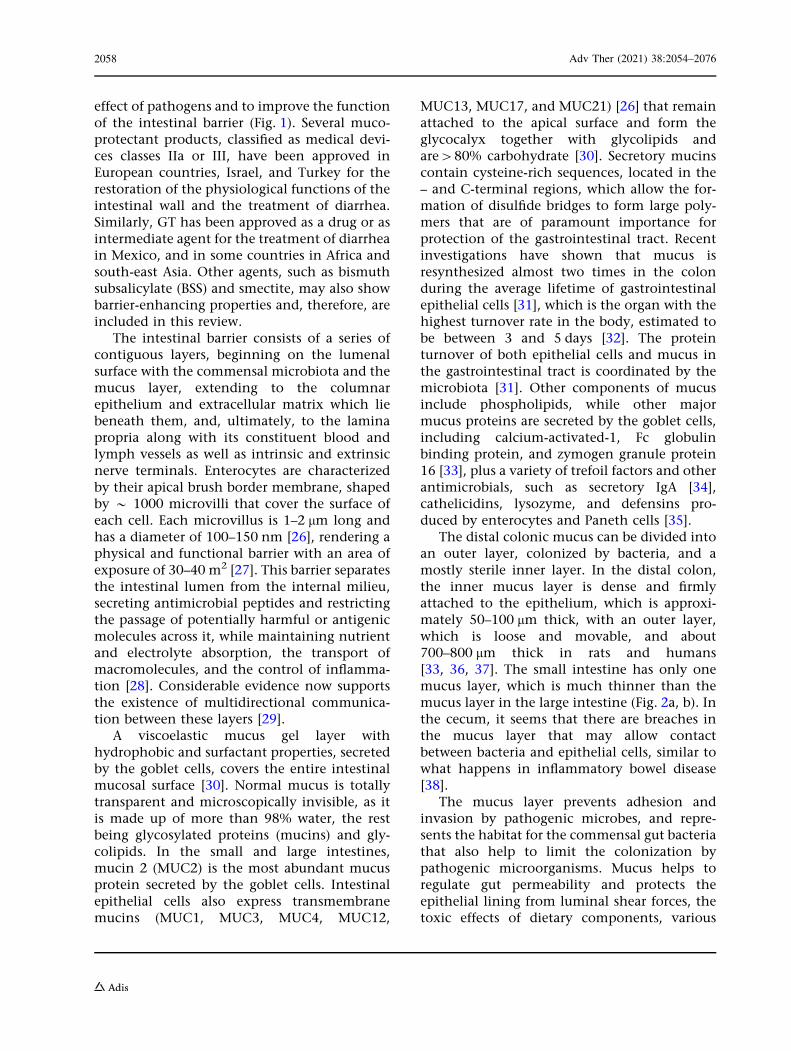
Fig. 1 Mechanism of action of mucoprotectants. Whenthe mucus layer is damaged, access by pathogens, toxins,allergens, and irritants across the intestinal barrier isgranted, which may enhance intestinal epithelial perme-ability and inflammatory and immune responses ofresident immunocytes within the lamina propria. Thisresponse, in turn, may lead to further distortion ofintestinal permeability and perpetuation of mucosal low-grade inflammation, increasing apposition/communicationbetween immune cells, such as mast cells and plasma cells,and nerve endings, neuronal plasticity, and regeneration
affecting the enteric nervous system (ENS) and afferentroutes to the central nervous system (CNS). Mucopro-tectans like xyloglucan and gelatin tannate share mucoad-hesive properties and the ability of creating a film-formingbarrier over the intestinal mucosa or protect the mucuslayer, helping to preserve intestinal permeability and avoidor decrease mucosal inflammation, reducing the effect ofnoxious agents on the intestinal barrier. Other molecules,such as bismuth subsalicylate or smectite, may protect themucus layer via complex mechanisms
Adv Ther (2021) 38:2054–2076 2057
effect of pathogens and to improve the functionof the intestinal barrier (Fig. 1). Several muco-protectant products, classified as medical devi-ces classes IIa or III, have been approved inEuropean countries, Israel, and Turkey for therestoration of the physiological functions of theintestinal wall and the treatment of diarrhea.Similarly, GT has been approved as a drug or asintermediate agent for the treatment of diarrheain Mexico, and in some countries in Africa andsouth-east Asia. Other agents, such as bismuthsubsalicylate (BSS) and smectite, may also showbarrier-enhancing properties and, therefore, areincluded in this review.
The intestinal barrier consists of a series ofcontiguous layers, beginning on the lumenalsurface with the commensal microbiota and themucus layer, extending to the columnarepithelium and extracellular matrix which liebeneath them, and, ultimately, to the laminapropria along with its constituent blood andlymph vessels as well as intrinsic and extrinsicnerve terminals. Enterocytes are characterizedby their apical brush border membrane, shapedby * 1000 microvilli that cover the surface ofeach cell. Each microvillus is 1–2 lm long andhas a diameter of 100–150 nm [26], rendering aphysical and functional barrier with an area ofexposure of 30–40 m2 [27]. This barrier separatesthe intestinal lumen from the internal milieu,secreting antimicrobial peptides and restrictingthe passage of potentially harmful or antigenicmolecules across it, while maintaining nutrientand electrolyte absorption, the transport ofmacromolecules, and the control of inflamma-tion [28]. Considerable evidence now supportsthe existence of multidirectional communica-tion between these layers [29].
A viscoelastic mucus gel layer withhydrophobic and surfactant properties, secretedby the goblet cells, covers the entire intestinalmucosal surface [30]. Normal mucus is totallytransparent and microscopically invisible, as itis made up of more than 98% water, the restbeing glycosylated proteins (mucins) and gly-colipids. In the small and large intestines,mucin 2 (MUC2) is the most abundant mucusprotein secreted by the goblet cells. Intestinalepithelial cells also express transmembranemucins (MUC1, MUC3, MUC4, MUC12,
MUC13, MUC17, and MUC21) [26] that remainattached to the apical surface and form theglycocalyx together with glycolipids andare[80% carbohydrate [30]. Secretory mucinscontain cysteine-rich sequences, located in the– and C-terminal regions, which allow the for-mation of disulfide bridges to form large poly-mers that are of paramount importance forprotection of the gastrointestinal tract. Recentinvestigations have shown that mucus isresynthesized almost two times in the colonduring the average lifetime of gastrointestinalepithelial cells [31], which is the organ with thehighest turnover rate in the body, estimated tobe between 3 and 5 days [32]. The proteinturnover of both epithelial cells and mucus inthe gastrointestinal tract is coordinated by themicrobiota [31]. Other components of mucusinclude phospholipids, while other majormucus proteins are secreted by the goblet cells,including calcium-activated-1, Fc globulinbinding protein, and zymogen granule protein16 [33], plus a variety of trefoil factors and otherantimicrobials, such as secretory IgA [34],cathelicidins, lysozyme, and defensins pro-duced by enterocytes and Paneth cells [35].
The distal colonic mucus can be divided intoan outer layer, colonized by bacteria, and amostly sterile inner layer. In the distal colon,the inner mucus layer is dense and firmlyattached to the epithelium, which is approxi-mately 50–100 lm thick, with an outer layer,which is loose and movable, and about700–800 lm thick in rats and humans[33, 36, 37]. The small intestine has only onemucus layer, which is much thinner than themucus layer in the large intestine (Fig. 2a, b). Inthe cecum, it seems that there are breaches inthe mucus layer that may allow contactbetween bacteria and epithelial cells, similar towhat happens in inflammatory bowel disease[38].
The mucus layer prevents adhesion andinvasion by pathogenic microbes, and repre-sents the habitat for the commensal gut bacteriathat also help to limit the colonization bypathogenic microorganisms. Mucus helps toregulate gut permeability and protects theepithelial lining from luminal shear forces, thetoxic effects of dietary components, various
2058 Adv Ther (2021) 38:2054–2076

chemicals, and radiation, as well as the impactof antigens present in the intestinal lumen[39, 40]. The mucus layer also contributes to theretention of mucosal secretions containingdigestive enzymes and helps to sustain epithe-lial hydration [41]. Mucus seems to enhanceoral tolerance by imprinting dendritic cells withanti-inflammatory properties [42], participatesin epithelial renewal, differentiation, andintegrity, and also interacts with other biologi-cal processes [43]. The importance of the mucuslayer is reflected in studies performed in MUC2knockout mice, in which bacteria are in directcontact with the epithelium leading toincreased intestinal permeability and the spon-taneous and aggravated chemically-induceddevelopment of colonic macroscopic
inflammation [44–46]. Similarly, in patientswith active ulcerative colitis, the inner mucuslayer is highly penetrable to bacteria [45, 46].The small intestine is more exposed to theintestinal bacteria, as the mucus layer is unat-tached and permeable. However, fewermicrobes reside in the small intestine [47] dueto the fast transit time (0.5–5 h) and a highconcentration of antimicrobial peptides.
Just beneath the mucus layer, epithelial cellsremain tightly sealed at the basolateral surface,the paracellular space, by means of the apicaljunctional complex [48]. This complex is com-posed of tight junctions (TJs), adherens junc-tions (AJs), and desmosomes. Threetransmembrane proteins are common to all TJs:claudins, MARVEL domain proteins, and
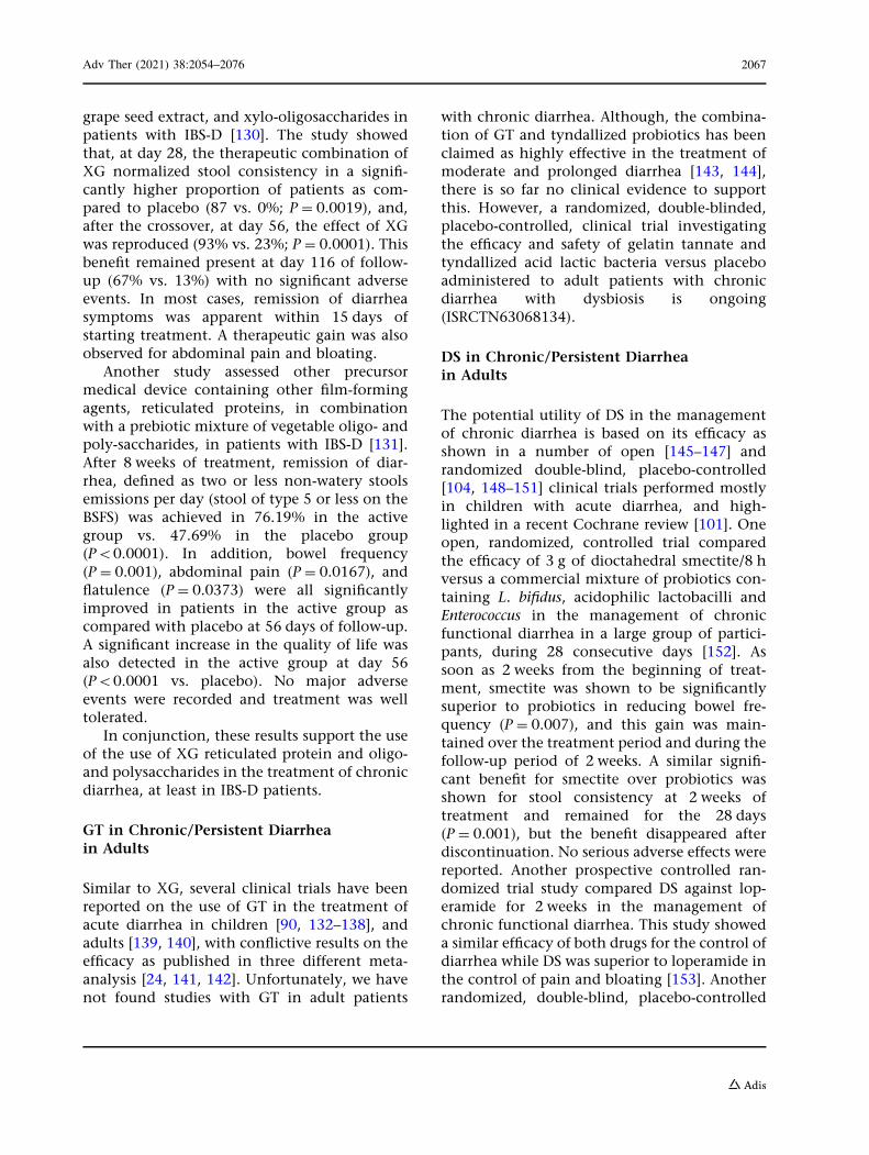
Fig. 2 Ultrastructural images of a normal mucus layer inthe rat ileum and colon. a Representative electron scanningmicrograph aspect of the mucus layer of the terminal ileumof an adult Wistar rat (magnification 9397). (Courtesy
Dr. Maria Vicario.) b Representative electron scanningmicrograph aspect of the mucus layer of the colon of anadult Wistar rat (magnification 9500). (Courtesy Dr.Maria Vicario)
Adv Ther (2021) 38:2054–2076 2059

junctional adhesion molecules (JAMs) [49]. Theclaudin family consists of 26 members whichregulate paracellular permeability in the gas-trointestinal tract. Among claudins, claudin 2and interleukin (IL)-13 regulate the pore path-way to form size (4–5 A at the villus tip; 20 A atthe base) and charge selective ion channels withhigh capacity of transport [50]. Claudin-2expression results in increased paracellularNa? and K? conductance and water flux with-out any effect on Cl- conductance or paracel-lular flux of larger solutes, including mannitol,lactulose, and 4 kD dextran[51]. The tight-junction-associated marvel proteins occludin,tricellulin, and marvelD3 are tetra-membranespanning proteins that regulate the recruitmentof signaling complex proteins to TJs, andcooperate in the development and regulation ofmacromolecular flux through the leak pathwayalong with zonula occludens (ZO)-1, ZO-2, andZO-3, and cingulin [46, 52]. JAM-A, -B, and -Care similar to immunoglobulin-G and may playimportant roles in barrier formation and sig-naling to circulating cells. AJs are located belowTJs and are mainly composed of e-cadherin,catenin, and actin filaments. Alterations inintestinal permeability have been linked to thedisappearance of key structural proteins of theintestinal epithelial barrier, and to be charac-teristic features of several chronic inflammatorydisorders, including inflammatory bowel dis-ease, celiac disease, intestinal graft versus hostdisease, critically ill patients, enteric infections,and infestations, human immunodeficiencyvirus infection, and acquired immune defi-ciency syndrome, IBS-D, asthma, autism,Parkinson’s disease, multiple sclerosis, eczema,psoriasis, eosinophilic esophagitis, environ-mental enteropathy, kwashiorkor, fibromyalgia,depression, chronic fatigue syndrome, multi-organ failure syndrome (shock, burns, trauma),non-alcoholic fatty liver disease, alcoholic cir-rhosis, obesity, metabolic syndrome, pancreati-tis, and rheumatoid arthritis, among others[53].
When understanding the concept of low-grade mucosal inflammation associated withdisorders of chronic diarrhea, it is important toagain consider the histological structure of thegut wall. The deepest layer of the intestinal
barrier is the lamina propria that containseffector cells of both adaptive and innateimmune systems, T and B lymphocytes, IgA-secreting plasma cells, mast cells, dendritic cells,and macrophages. The loss of epithelial integ-rity facilitates antigen, chemical, and toxinpenetration into the lamina propria, whichtriggers immunological responses that, in turn,increase epithelial permeability to luminalcontent, thereby promoting inflammation.Indeed, several common gastrointestinal andsystemic disorders associated with chronicdiarrhea share alterations in the gut epithelialbarrier, leading to abnormal intestinal perme-ability, detachment of mucous layer, intestinaldysbiosis, and, ultimately, low-grade mucosalinflammation [33, 54]. Numerous studies haveprovided evidence of increased numbers ofimmunocytes in the lamina propria of diarrhealdiseases (mainly mast cells, eosinophils, and Tcells), such as IBS-D, ulcerative colitis, ormicroscopic colitis [55–58].
In addition to infectious agents, there areseveral predisposing factors for mucus damageand intestinal leakiness that are commonlyinvolved in the development of chronic diar-rhea and mucosal inflammation. Among these,environmental stress, pregnancy, enduranceexercise, drugs and antibiotics, genetic suscep-tibility, alcohol, and western diet, particularlydietary emulsifiers and surfactants in foodadditives, should be considered when evaluat-ing and treating patients with chronic diarrhea[33, 59–64].
Therefore, agents such as mucoprotectans,due to their mucoadhesive and film-formingbarrier characteristics, may offer advantages forthe prevention of barrier abnormalities andrestoration of the mucus layer and alteredintestinal permeability to reduce mucosalinflammation and gut mucosal homeostasis.
MUCOPROTECTANTS
Bismuth Subsalicylate
BSS is an insoluble salt of salicylic acid andtrivalent bismuth that was first FDA-approvedin 1939 and can be considered as a mucosal
2060 Adv Ther (2021) 38:2054–2076

protector with approved indications for thetreatment of diarrhea, heartburn, indigestions,nausea, and stomach upset [65]. The mecha-nism of action is complex and partly unknown,and involves the gastric hydrolysis into bis-muth, and salicylic acid [66]. The salicylatecompound is almost completely absorbed intothe bloodstream, while bismuth remains in thelumen of the gastrointestinal tract to form otherbismuth salts [67]. These bismuth salts showbactericidal and antimicrobial activity [66, 68],prevent bacteria from binding and growing onthe mucosal cells, inhibit intestinal secretions,promote fluid, sodium, and chloride absorption[69], reduce inflammation via cyclooxygenaseinhibition [70, 71], and decrease proliferativeactions of non-amidated gastrins in the rectalmucosa of Sprague–Dawley rats and mice [72],playing a major role in combating diarrhea.
Xyloglugan
XG is a non-ionic, water-soluble, high molecu-lar weight branched polysaccharide hemicellu-lose (MW: 1331 Kda) [73] that carries xylose andgalactosyl–xylose substituents, extracted from
the most abundant source of XG and solublefiber in nature, the seeds of the tamarind tree(Tamarindus indica) [74, 75] (Fig. 3). XG is non-toxic, edible, biocompatible, bioavailable, withversatile use in foods, and resistant to digestiveenzymes, reaching the colon unaltered, where itis partially broken down to oligosaccharides bybacterial endo-ß-glucanases, followed by bacte-rial fermentation of oligosaccharides [76–78].The ‘mucin-like’ molecular structure of XG isknown to possess mucomimetic, mucoadhesive,and pseudo-plastic properties [73, 79]. In thegut, it acts as a film-forming barrier over theintestinal mucosa, helping to reduce perme-ability changes and invasion by pathogens likeE. coli and to decrease cholera toxin-inducedintestinal secretion in Caco2/goblet cells[80–82], preserving tight junctions [75], andbinding consistently to MUC1 in moleculardocking studies and decreasing the expressionof MUC1 and MUC2, as shown in mice treatedwith dextran sodium sulfate (DSS) [73]. BothXG and GT pretreatment reduced the severity oflipopolysaccharide (LPS)/induced mucosalinflammation and jejunal hyperpermeability inmale Wistar rats, although they did not preventLPS-induced occludin and JAM-A down-regula-tion. Further, GT and XG limited bacterialmucus layer invasion and contact betweenbacteria and intestinal epithelium [83]. XG isoften combined with gelatin or gelose to pro-long its availability within the intestine, butshowing similar protective effects as XG aloneon barrier function and intestinal inflammationin rats after LPS administration [84] and Sal-monella enterica and Enterococcus hirae infections[85]. In preliminary results, a single intracolonicadministration combination of XG with Bifi-dobacterium animalis was found to be effective ininducing mucosal healing in patients withulcerative colitis [86]. The combination of XG,pea proteins, and tannins from grape xylo-oligosaccharides also offered protection againststress-induced visceral hypersensitivity andintestinal hyperpermeability in rats [87].
Fig. 3 Basic molecular structure of hemicellulose. Xyloglu-can from tamarind seeds consists of four types ofoligosaccharides as repeating units, commonly as heptasac-charides [155]. The monomer unit contains three types ofsugars: xylose, galactose,? and glucose. The configurationof this polysaccharide gives the product a ‘‘mucin-like’’molecular structure, thus conferring optimal mucoadhesiveproperties [75]
Adv Ther (2021) 38:2054–2076 2061

Gelatin Tannate
GT is a complex of tannic acid (penta-m-digal-lolyl glucose) and gelatin which forms electro-static bonds with mucin creating a protein-based biofilm on the intestinal mucosa [88, 89].Gelatin is a collagen derivate, which is ingestedas a powder that is insoluble at gastric acidic pH,and which becomes a gelatin with the increaseof pH over 5.5 [90]. This complex enters theintestine unaltered, increasing the epithelialresistance against E. coli infection [91], helpingto restore the normal physiology of barrierfunction, also reducing inflammation inresponse to lipopolysaccharide administrationin rats [92]. GT also helps to restore the mucuslayer and to modulate the intestinal microbiotain the DSS-induced model of murine colitis [93],and in Caco cells [94], where it acts in part bypreventing the release of intercellular adhesionmolecule-1, IL-8, and tumor necrosis factor-ainduced by LPS [95]. Furthermore, the astrin-gent properties of tannins allow the precipita-tion of pro-inflammatory molecules from theintestinal mucus and their fecal elimination[96, 97]. Together, these effects may explain, atleast in part, the protective effect of GT onintestinal barrier function.
Dioctahedral Smectite or Diosmectite
Diosmectite (DS) is a medicinal clay and a pro-duct frequently recommended over-the-counterin Eastern European countries [98], France [99],and China [100] as an adjuvant therapy inchildren and adults with acute diarrhea. It isadministered to reduce stool output, providingsymptomatic relief, and possibly preventingdehydration [101]. It is formed from sheets ofaluminum and magnesium silicate. The mech-anism of action is thought to be the result of:anti-inflammatory activity; modifications of therheological characteristics of the gastrointesti-nal mucus barrier to reduce penetration of tox-ins and adsorptive properties; reduction ofintestinal permeability and apoptosis; andimproved intestinal epithelial cells prolifera-tion, via modulation of IL-8, transforminggrowth factor, extracellular signal-regulated
kinase �, and protein kinase B signaling path-way, and MUC2 expression [102, 103], therebyreducing stool output and stress-induced vis-ceral hypersensitivity. These mechanisms havebeen replicated, mainly in vitro, in Caco-2 andHT-29 cell lines, and in vivo in rodent andpiglets animal models [104–109], and the resultsmay be improved by combination with Lacto-bacillus acidophilus [110].
METHODS
We searched MEDLINE and EMBASE via OVID,from 1977 to January 2021 using a combinationof MeSH terms, EMTREE terms, and keywordsdeveloped for each database. We also conducteda search for all English language articles, sys-tematic reviews, meta-analysis, conference pro-ceedings, and abstracts in relevant scientificmeetings, on the epidemiology, etiology, phys-iopathology, and management of chronic andpersistent diarrhea in immunocompetent indi-viduals using the search terms: persistent diar-rhea, chronic diarrhea, infectious diarrhea, entericinfection, epidemiology, treatment, management,guidelines, adults, mucus, intestinal permeability,xyloglucan, gelatin tannate, bismuth subsalicylate,diosmectite, smectite, and mucoprotectans. Bibli-ographies of review and meta-analysis articleswere used to identify additional sources. Web-sites for the US Centers for Disease Control andPrevention, US Food and Drug Administration,and World Health Organization were alsoaccessed for any additional information relatedto this topic.
All studies were reviewed and summarized bytwo independent reviewers to determine theireligibility. Only primary studies conducted onhuman adult subjects (18 years and older) pre-senting with chronic diarrhea with observedparameters directly related to diarrhea wereincluded.
This article is based on previously conductedstudies and does not contain any new studieswith human participants or animals performedby any of the authors.
2062 Adv Ther (2021) 38:2054–2076

RESULTS
Available studies were read and summarized,and the study design, population, parametersobserved, and outcomes documented (Table 1).
BSS in Chronic/Persistent Diarrheain Adults
Chronic/persistent diarrhea occurs in approxi-mately 3% of individuals traveling to develop-ing countries, and in more than 10% of patientssuffering acute infectious enteritis [111]. Themicrobiologic causes include parasitic (e.g.,Giardia, Cryptosporidium, Schistosoma mansoni)and bacterial (e.g., enteroaggregative E. coli,Shigella, Campylobacter, Salmonella) pathogens[21]. BSS has demonstrated effectiveness in theprevention of traveler’s diarrhea [112, 113] andin the treatment of acute diarrhea [114]. Fortraveler’s diarrhea in adult patients with mildsymptoms [115, 116], BSS has been shown todecrease stool frequency, time to symptomrelief, need for intravenous rehydration, andwork absenteeism in comparison to placebo orantibiotics. However, there are no studies onthe effect of BSS on chronic/persistent diarrheaafter acute infectious enteritis. Considering theefficacy of BSS in the management of acutediarrhea and the increasing prevalence ofpostinfective diarrhea and antibiotic resistanceamong diarrheal pathogens [117], it would bewise to perform additional studies to evaluatethe efficacy and safety of BSS in the manage-ment of postinfective diarrhea.
Both IBS-D and microscopic colitis (MC) arecommon causes of chronic diarrhea in adults.One open-label study showed that the combi-nation of BSS and spasmolytics during 3 weeksimproved bowel symptoms in IBS-D, includingdiarrhea in a small group of patients (n = 20)[118]. BSS is recommended by the 2016 AGAguideline as a second-line therapy for MC whenbudesonide is unable to be used, either due tocost or adverse effects [119]. This is based ontwo small studies that found that treatmentwith BSS for 8 weeks reduced the frequency andweight of bowel movements, improved stoolconsistency, and decreased tissue inflammation
in patients with MC [120, 121]. A retrospectivestudy showed complete response in 53% ofpatients and partial response in 28% of patientstaking three tablets (262 mg each) of bismuthsalicylate three times a day [122]. Chronicdiarrhea is a common manifestation of a varietyof cancers that can be attributed to adverseeffects of treatments, radiotherapy, surgery, andinfection. One prospective pilot study revealedthat the duration of diarrhea experienced bylymphoma patients receiving melphalanchemotherapy was decreased as compared tothe placebo group, while this did not happen inmultiple myeloma patients irrespective oftreatment [123]. However, this article has beenretracted by the journal because major findingscould not be replicated upon reanalysis [124].Though anecdotical, a recent case reportshowed a good temporal response of diarrhea ina COVID-19-positive Crohn’s disease patienttreated with BSS [125].
Limitation to its use in chronic diarrhearelate to the number of daily tablets needed totreat, to the compromised absorption of othercompounds and to the restricted use in patientswith renal impairment [126]. BSS is safe, rela-tively cheap, and has limited side effects, yettinnitus, blackened tongue and dark feces arenot unusual in short-term therapy and make itsuse undesirable for some patients [124].Although quite rare, the most concerningadverse effect of BSS is salicylate and bismuthnon-eurotoxicity that primarily occurs inpatients who have taken bismuth subsalicylateinappropriately, whether through an overdoseor for extended periods of time [127].
XG in Chronic/Persistent Diarrheain Adults
Several clinical trials have been reported on theefficacy of XG in the treatment of acute diarrheain children [128, 129] and adults [81]. However,there are limited data regarding its use inchronic diarrhea. One recent multicenter, dou-ble-blind, placebo-controlled, randomized,crossover clinical trial evaluated the efficacyand safety of a commercially available combi-nation of XG, pea protein, and tannins from
Adv Ther (2021) 38:2054–2076 2063

Table
1Studiesinvolvingmucoprotectansin
themanagem
entof
chronicdiarrhea
inadults
Autho
r,year,
[reference]
Cou
ntry
Intervention
Pop
ulation
Stud
ydesign
nparticipants
Outcomes
Follo
w-up
Observation
s
Adu
lts
Age,years
TG
CG
Bismuthsalicylate
Iakovenko
EP,
2008
[118]
Russia
Bismuth120mg/
8h/
?spasmolytic
Aluminum
phosphate?
spasmolytic
IBS-D
N/A
Open prospective
2010
Abdom
inalpain,m
eteorism
,diarrhea,b
acterialgrow
thin
smallintestine,changes
offecalmicroflora,
histologicalsignsof
mucosalinflammation
3weeks
Abdom
inalpain
was
elim
inated
in90%
and
60%
(TG
vs.C
G),
meteorism
was
absent
in80%
and40%
(TG
vs.
CG),diarrhea
in75%and
50%
(TG
vs.C
G),
excessivebacterialgrow
thin
smallintestinein
75%
and30%
(TG
vs.C
G),
changesof
fecal
microflora
persistedin
20%
and70%
(TG
vs.
CG),histologicalsignsof
mucosalinflammation
remainedin
40%
and
85.7%
(TG
vs.C
G)#
Fine
KD,
1998
[120]
USA
Bismuth262-mgchew
able
tabletsof
bism
uth
subsalicylate(8/day)
MC
35–7
2Open prospective
13–
Diarrhea(frequency
ofbowelmovem
entsdaily)
andhistologicalresponse
8weeks
Elevenpatientshada
resolution
ofdiarrhea
and
areductionin
fecal
weight.The
averagetime
torespondwas
2weeks.
Subepithelialcollagen
thickening
disappeared.
Follow-upfor
7–28
monthsshow
edthat
nine
patients
remainedwellwitha
norm
albowelhabit
Fine
K,
1999
[121]
USA
Bismuth262-mgchew
able
tabletsof
bism
uth
subsalicylate(8–9
/day)
MC
(9CC
?5
LC)
35–7
8Randomized
double-
blind,
placebo-
controlled
77-
Diarrhea(frequencyof
bowelmovem
entsdaily)
andhistologicalresponse
8weeks
Allsevenpatientsin
the
intervention
arm
had
clinicalresponse
vs.n
one
ofthepatientsin
the
placeboarm.P
atients
treatedwithbism
uth
salicylatehadathreefold,
albeitnotstatistically
significant,likelihoodof
achievingaconcom
itant
histologicresponse
2064 Adv Ther (2021) 38:2054–2076

Table
1continued
Autho
r,year,
[reference]
Cou
ntry
Intervention
Pop
ulation
Stud
ydesign
nparticipants
Outcomes
Follo
w-up
Observation
s
Adu
lts
Age,years
TG
CG
Gentile
NM,2
015
[122]
USA
Bismuth262-mgchew
able
tabletsof
bism
uth
subsalicylate(6–9
/day)
MC
(31
CC
?33
LC)
31–8
6Retrospective
study
64-
Com
pleteresponse
was
defin
edas
resolution
ofdiarrhea,w
hereas
partial
responsewasdefin
edasat
least50%
improvem
ent
ofdiarrhea
at8weeks
±2weeks
with
BSS
6–51
weeks
33(52%
)hadcomplete
response,1
8(28%
)had
partialresponse,and
13(20%
)hadno
response.
Ofthe32
remaining
completeresponders,2
3(72%
)recurred.T
hemediantimeto
recurrence
was
4.9weeks.
LC
muchmorelikelyto
have
acompleteresponse
than
CC
(70%
vs.3
2%,
P=
0.001)
Diosm
ectite
Yao-Zong
Y,2
004
[152]
China
3gdioctahedralsm
ectite/
8hvs.B
ifico
210mg/
12h(L.b
ifidus,
acidophilic
lactobacilli
andEnterococcus)for
28days
Functional
diarrhea
43.8
±13.9
Open, rand
omized,
controlled
trial
208
202
Changein
daily
frequencyof
bowelmovem
entsand
stoolconsistency
6weeks
Decreasein
stoolnu
mber
was
significant
withboth
treatm
ents,b
utmore
importantwithsm
ectite
atweek2,
andremained
significant
throughout
the
treatm
entperiod.S
tool
consistency,also
improved
significantly
overthetreatm
entperiod,
ascomparedto
baselin
e(z
=3.310,
P=
0.001)
Dum
itrascu
DL,[153]
Rom
ania
3gdioctahedralsm
ectite/
12hvs.loperam
ide/12
hfor2weeks
Functional
diarrhea
47±
11Prospective
controlled
rand
omized
trial
2525
Symptom
scores
for
diarrhea,p
ainand
bloating
and
psychologicaldistress
2weeks
Symptom
scorefordiarrhea
was
reducedfrom
10.5
±5.7to
2.6±
1.2,
P\
0.001by
diosmectite
andfrom
8.5±
4.2to
1.2±
0.6,P\
0.0001
byloperamide.Diosm
ectite
hadastronger
effect
than
loperamideon
accompanyingpain
(P\
0.05)andbloating
(P\
0.01).(from
12.7
±3.8to
7.7±
2.1
vs.1
1.8±
3.3to
9.05
±2.2,
P\
0.05)
Adv Ther (2021) 38:2054–2076 2065

Table
1continued
Autho
r,year,
[reference]
Cou
ntry
Intervention
Pop
ulation
Stud
ydesign
nparticipants
Outcomes
Follo
w-up
Observation
s
Adu
lts
Age,years
TG
CG
Chang
F-Y,
2007
[154]
Taiwan
3gdiosmectite/8
hvs.
placebo/8h(0.8
ghydrated
glucose,1.1g
corn
starch,0
.008
gsaccharinsodium
,0.192
gtalcum
power,1
.11g
maltose
dextrins,0
.006
gcaramelcoloring
[E150],
and0.004gvanilla)
IBS-D
48.6–5
9.0
Randomized,
double-
blind,
placebo-
controlled
trial
5252
The
prim
aryefficacy
endpoint
was
thechanges
ofVASscoreof
IBS
overalld
isorderandpain/
discom
fort-related
symptom
s.Other
second
aryoutcom
emeasuresincluded
changesin
bowel
movem
entdisordersand
bloating
8weeks
Onday56,d
iosm
ectite
reducedVASscoreof
IBS
overalldisorder
(P=
0.0167)and
abdominalpain
(P\
0.05)as
compared
toplacebo.
Both
treatm
entseffectively
diminishedbowel
movem
entfrequency,but
groupdifferenceswere
notfoun
d,whileonly
diosmectite
improved
abdominalbloating
ateach
visit
Xyloglucan
AlexeaO,
2016
[131]
Spain, Rom
ania
Amixtureof
vegetableoligo-
andpolysaccharides:
750mg;reticulated
protein:
250mg;andthe
excipientscorscarm
ellose
sodium
:133mg;and
magnesium
stearate:17mg;or
placebo(cornstarch,
croscarm
ellose
sodium
andmagnesium
stearate)
four
tablets/day(two
before
breakfastandtwo
beforedinn
er)for56
days
IBS-D
Activegroup:
48.8
±14;
Placebo
group:47.7
±14.2
Multicenter,
rand
omized,
placebo-
controlled,
double-
blind,
parallel
group
6365
Stoolconsistency,bowel
frequency,abdominal
pain,b
loating,qualityof
lifeandgeneralhealth
56days
Rem
ission
ofdiarrhea
and
improvem
entof
abdominalpain,
flatulence,bowel
frequencyandqualityof
lifewasshow
nin
thosein
theactive
groupas
comparedwithplacebo
TrifanA,
2019
[130]
Rom
ania
Xyloglucan?
peaprotein
andtann
ins?
xylo-
oligosaccharides/placebo,
1capsuleb.i.d
for
28days,followed
bycrossoverto
thealternate
treatm
entfor28
days
IBS-D
Activegroup:
35.0
±7.8;
Placebo
group:34.5.±
8.1
Multicenter,
rand
omized
double-
blind,
placebo-
controlled,
crossover
3030
Stoolconsistency,abdom
inal
pain,b
loating,qualityof
lifeandgeneralhealth
116days
Xyloglucancombination
show
nto
besafe
and
efficacious
toim
prove
stoolconsistency,
abdominalpain,b
loating,
qualityof
lifeandgeneral
health.L
ong-lasting
clinicalbenefitsduring
follow-upafterfin
ishing
treatm
entwerealso
show
n
BSFSBristolstoolchart,C
Ccollagenous
colitis,C
Gcontrolgroup,IBS-Dirritablebowelsynd
romeanddiarrhea,L
Clymphocyticcolitis,M
Cmicroscopiccolitis,N
/Anotavailable,TGtreatm
entgroup,t.i.dthreetimesa
day,#statisticalcomparisons
notavailable
2066 Adv Ther (2021) 38:2054–2076
grape seed extract, and xylo-oligosaccharides inpatients with IBS-D [130]. The study showedthat, at day 28, the therapeutic combination ofXG normalized stool consistency in a signifi-cantly higher proportion of patients as com-pared to placebo (87 vs. 0%; P = 0.0019), and,after the crossover, at day 56, the effect of XGwas reproduced (93% vs. 23%; P = 0.0001). Thisbenefit remained present at day 116 of follow-up (67% vs. 13%) with no significant adverseevents. In most cases, remission of diarrheasymptoms was apparent within 15 days ofstarting treatment. A therapeutic gain was alsoobserved for abdominal pain and bloating.
Another study assessed other precursormedical device containing other film-formingagents, reticulated proteins, in combinationwith a prebiotic mixture of vegetable oligo- andpoly-saccharides, in patients with IBS-D [131].After 8 weeks of treatment, remission of diar-rhea, defined as two or less non-watery stoolsemissions per day (stool of type 5 or less on theBSFS) was achieved in 76.19% in the activegroup vs. 47.69% in the placebo group(P\0.0001). In addition, bowel frequency(P = 0.001), abdominal pain (P = 0.0167), andflatulence (P = 0.0373) were all significantlyimproved in patients in the active group ascompared with placebo at 56 days of follow-up.A significant increase in the quality of life wasalso detected in the active group at day 56(P\0.0001 vs. placebo). No major adverseevents were recorded and treatment was welltolerated.
In conjunction, these results support the useof the use of XG reticulated protein and oligo-and polysaccharides in the treatment of chronicdiarrhea, at least in IBS-D patients.
GT in Chronic/Persistent Diarrheain Adults
Similar to XG, several clinical trials have beenreported on the use of GT in the treatment ofacute diarrhea in children [90, 132–138], andadults [139, 140], with conflictive results on theefficacy as published in three different meta-analysis [24, 141, 142]. Unfortunately, we havenot found studies with GT in adult patients
with chronic diarrhea. Although, the combina-tion of GT and tyndallized probiotics has beenclaimed as highly effective in the treatment ofmoderate and prolonged diarrhea [143, 144],there is so far no clinical evidence to supportthis. However, a randomized, double-blinded,placebo-controlled, clinical trial investigatingthe efficacy and safety of gelatin tannate andtyndallized acid lactic bacteria versus placeboadministered to adult patients with chronicdiarrhea with dysbiosis is ongoing(ISRCTN63068134).
DS in Chronic/Persistent Diarrheain Adults
The potential utility of DS in the managementof chronic diarrhea is based on its efficacy asshown in a number of open [145–147] andrandomized double-blind, placebo-controlled[104, 148–151] clinical trials performed mostlyin children with acute diarrhea, and high-lighted in a recent Cochrane review [101]. Oneopen, randomized, controlled trial comparedthe efficacy of 3 g of dioctahedral smectite/8 hversus a commercial mixture of probiotics con-taining L. bifidus, acidophilic lactobacilli andEnterococcus in the management of chronicfunctional diarrhea in a large group of partici-pants, during 28 consecutive days [152]. Assoon as 2 weeks from the beginning of treat-ment, smectite was shown to be significantlysuperior to probiotics in reducing bowel fre-quency (P = 0.007), and this gain was main-tained over the treatment period and during thefollow-up period of 2 weeks. A similar signifi-cant benefit for smectite over probiotics wasshown for stool consistency at 2 weeks oftreatment and remained for the 28 days(P = 0.001), but the benefit disappeared afterdiscontinuation. No serious adverse effects werereported. Another prospective controlled ran-domized trial study compared DS against lop-eramide for 2 weeks in the management ofchronic functional diarrhea. This study showeda similar efficacy of both drugs for the control ofdiarrhea while DS was superior to loperamide inthe control of pain and bloating [153]. Anotherrandomized, double-blind, placebo-controlled
Adv Ther (2021) 38:2054–2076 2067
trial, showed no benefit of DS over placebo inimproving bowel frequency, consistency,urgency and mucus discharge in IBS-D after8 weeks of treatment [154]. However, DS sig-nificantly improved abdominal pain, bloating,and the overall visual analogue scale score ofIBS. No serious adverse effects were reported.However, three patients were hospitalized dur-ing the trial (two in the placebo group becauseof cellulitis and acute appendicitis; one in theDS group because of renal stone). Constipationwas the most common effect related to DStreatment, but its occurrence was not differentfrom placebo. Other recorded adverse effectswere similar in both groups: nausea, abdominalpain, and dyspepsia.
SUMMARY AND CONCLUSIONS
There are many available drugs for the treat-ment of chronic diarrhea. The majority of themtarget specific mechanisms/pathways involvedin the origin of diarrhea. However, it is com-mon for many disorders associated with chronicdiarrhea, particularly (but not only) for the veryprevalent IBS-D and functional diarrhea, thattheir development involves multiple orunidentified mechanisms. Among these mech-anisms, the impairment of the intestinal barrierwith changes in epithelial permeability, mucuslayer, and immune activation deserves specialfocus because they have been increasinglyimplicated in the initiation and perpetuation ofa variety of diseases, thereby justifying theemerging interest in the advent of new phar-macological/non-pharmacological approachesfor the restoration of barrier function.
Much of this work relates to the use ofmucosal protectors, as a new alternative orcomplementary therapy for a more efficient andsafe control of symptoms in disorders associatedwith chronic diarrhea, mostly in IBS-D. Theobjective of mucosal protection is to create anartificial mechanical barrier over the mucosa, toreduce contact/access between noxious aller-gens, irritants, toxins, pathogens, and theirvirulence factors and the mucosal immune sys-tem, to prevent mucus damage and preserveintestinal permeability. The need of these
barrier enhancers, some marketed as medicaldevices, is also supported in the current contextof high levels of antimicrobial resistance and toavoid long-lasting pharmacological treatments,their adverse events, and frequent elevatedcosts. In adults with chronic diarrhea, thestudies available to date suggest that thesemucoprotectants can be helpful, improvingstool frequency and consistency, and showingbeneficial effects on other symptoms such asabdominal pain, bloating, and flatulence.Importantly, they appear to be safe, with fewadverse events, although some caution isadvised on the chronic use of BSS and tanninsand on their potential interference with themechanism of action of other drugs. In addi-tion, it is currently unclear whether the use ofmucoprotectans is cost-effective, partly becausesome of them (xyloglucan, gelatin tannate, anddisomectite) are sold over the counter and notcovered by health insurance or public healthsystems, and partly because the lack of high-quality evidence. However, there are a numberof limitations to the available data. There is apaucity of studies and several of them have beencriticized because they were reported only asabstracts or posters, and many were observa-tional in design and did not include a controlgroup, rendering a low quality of evidence dueto imprecision, inconsistency, and risk of biaswhen defining diarrhea characteristics acrossstudies, yet this criticism may be limited due toheterogeneity in some outcomes [142]. Inaddition, the evidence provided relates mostlyto IBS-D, which may not be applicable to otherdisorders with chronic diarrhea. Finally, it isimportant to note here that, while some ofthese products are marketed as mucoprotectans,the mechanism by which they protect themucosa is not well established, just as it is notwell established that mucoprotection is themechanism by which chronic diarrhea is ame-liorated. In conclusion, although mucopro-tectans are promising, there is a clear need foradditional randomized controlled trials in largeand controlled populations assessing clinicallyrelevant outcomes to further explore theireffects and confirm their usefulness in thetreatment of chronic diarrhea. Microbiologicalanalysis of fecal and mucosal samples would
2068 Adv Ther (2021) 38:2054–2076

also provide useful information about theireffects on intestinal microbiota, particularly inpatients with dysbiosis. In addition, clinical,functional, and laboratory evaluation of theireffects on intestinal permeability and mucusintegrity is also warranted to ascertain theirin vivo ability to restore these functions and toextend their use in the management of a varietyof gastrointestinal diseases associated with‘leaky gut.’
ACKNOWLEDGEMENTS
Funding. Supported in part by Fondo Eur-opeo de Desarrollo Regional (FEDER), Fondo deInvestigacion Sanitaria and Centro de Investi-gacion Biomedica en Red de EnfermedadesHepaticas y Digestivas (CIBEREHD), Instituto deSalud Carlos III, Subdireccion General deInvestigacion Sanitaria, Ministerio de Economıay Competitividad, Ajuts per a la contractacio depersonal investigador FI—Agencia de Gestiod’Ajuts Universitaris i de Recerca (AGAUR),Generalitat de Catalunya, The Swedish ResearchCouncil and The European Commision: FISLOBO 2019? (BL); FI20/00256 (MA-B). PI17/0190 (JS); PI15/00301 (CA-C); CIBERehd CB06/04/0021 (JS & CA-C); dnr 2019–00653 (J-PGM);2020FI_B1 00127, 2019FI_B 00817 (MA-B); GANo:848228 (JS, CA-C, EE, AMG-C, BL). Nofunding or sponsorship was received for thepublication of this article.
Authorship. All named authors meet theInternational Committee of Medical JournalEditors (ICMJE) criteria for authorship for thisarticle, take responsibility for the integrity ofthe work as a whole, and have given theirapproval for this version to be published.
Authorship Contributions. MA-G, BL, AMG-C, MA-B, J-PGM, EE and CA-C reviewed scientificliterature and collected data; CA-C, BL & JS wrotethe paper; MA-G, BL, MA-G, AMG-C, MA-B, EEand J-PGM prepared the tables and figures; Allauthors critically reviewed and edited themanuscript in its final version. All authorsapproved the final draft of the manuscript.
Disclosures. Mar Abril-Gil, Beatriz Lobo,Merce Albert-Bayo, John Peter Ganda-Mall, AnaMarıa Gonzalez-Castro have nothing to dis-close. Carmen Alonso-Cotoner discloses pastscientific collaborations with Noventure S.L.Javier Santos has served as consultant forNoventure and discloses present and past recentscientific collaborations with Salvat, Norgine,Alfa-Sigma, Cosmo, Adare, Devintecpharma,Pileje and Danone.
Compliance with Ethics Guidelines. Thisarticle is based on previously conducted studiesand does not contain any new studies withhuman participants or animals performed byany of the authors.
Open Access. This article is licensed under aCreative Commons Attribution-NonCommer-cial 4.0 International License, which permitsany non-commercial use, sharing, adaptation,distribution and reproduction in any mediumor format, as long as you give appropriate creditto the original author(s) and the source, providea link to the Creative Commons licence, andindicate if changes were made. The images orother third party material in this article areincluded in the article’s Creative Commonslicence, unless indicated otherwise in a creditline to the material. If material is not includedin the article’s Creative Commons licence andyour intended use is not permitted by statutoryregulation or exceeds the permitted use, youwill need to obtain permission directly from thecopyright holder. To view a copy of this licence,visit http://creativecommons.org/licenses/by-nc/4.0/.
REFERENCES
1. Schiller LR, Pardi DS, Spiller R, et al. Gastro 2013APDW/WCOG Shanghai working party report:chronic diarrhea: definition, classification, diagno-sis. J Gastroenterol Hepatol. 2014;29:6–25.
2. Schiller LR. Evaluation of chronic diarrhea andirritable bowel syndrome with diarrhea in adults inthe era of precision medicine. Am J Gastroenterol.2018;113:660–9.
Adv Ther (2021) 38:2054–2076 2069

3. Sandler RS, Everhart JE, Donowitz M, et al. Theburden of selected digestive diseases in the UnitedStates. Gastroenterology. 2002;122:1500–11.
4. Peery AF, Dellon ES, Lund J, et al. Burden of gas-trointestinal disease in the United States: 2012update. Gastroenterology. 2012;143(1179–1187):e3.
5. GBD 2019 Diseases and Injuries Collaborators.Global burden of 369 diseases and injuries in 204countries and territories, 1990–2019: a systematicanalysis for the Global Burden of Disease Study2019. Lancet. 2020;396(10258):1204–22.
6. Wenzl HH, Fine KD, Schiller LR, et al. Determinantsof decreased fecal consistency in patients withdiarrhea. Gastroenterology. 1995;108:1729–38.
7. Fine KD, Schiller L. AGA technical review on theevaluation and management of chronic diarrhea.Gastroenterology. 1999;116:1464–86.
8. Shane AL, Mody RK, Crump JA, et al. 2017 Infec-tious Diseases Society of America Clinical PracticeGuidelines for the Diagnosis and Management ofInfectious Diarrhea. Clin Infect Dis. 2017;65:e45–80.
9. Descoteaux-Friday GJ, Shrimanker I. Chronic Diar-rhea. [Updated 2020 Nov 17]. In: StatPearls [Inter-net]. Treasure Island (FL): StatPearls Publishing;2020. https://www.ncbi.nlm.nih.gov/books/NBK544337/. Accessed 13 Mar 2021.
10. Lewis SJ, Heaton KW. Stool form scale as a usefulguide to intestinal transit time. Scand J Gastroen-terol. 1997;32:920–4.
11. O’Donnell LJ, Virjee J, Heaton KW. Detection ofpseudodiarrhoea by simple clinical assessment ofintestinal transit rate. BMJ. 1990;300:439–40.
12. Arasaradnam RP, Brown S, Forbes A, et al. Guideli-nes for the investigation of chronic diarrhoea inadults: British Society of Gastroenterology, 3rdedition. Gut. 2018;67:1380–99.
13. Talley NJ, Weaver AL, Zinsmeister AR, et al. Onsetand disappearance of gastrointestinal symptomsand functional gastrointestinal disorders. Am JEpidemiol. 1992;136:165–77.
14. Zhao Y-F, Guo X-J, Zhang Z-S, et al. Epidemiology offunctional diarrhea and comparison with diarrhea-predominant irritable bowel syndrome: a popula-tion-based survey in China. PLoS One. 2012;7:e43749.
15. Fosnes GS, Lydersen S, Farup PG. Constipation anddiarrhea-common adverse drug reactions? A crosssectional study in the general population. BMC ClinPharmacol. 2011;11:2.
16. Thompson WG, Irvine EJ, Pare P, et al. Functionalgastrointestinal disorders in Canada: first popula-tion-based survey using Rome II criteria with sug-gestions for improving the questionnaire. Dig DisSci. 2002;47:225–35.
17. Singh P, Mitsuhashi S, Ballou S, et al. Demographicand Dietary Associations of Chronic Diarrhea in aRepresentative Sample of Adults in the UnitedStates. Am J Gastroenterol. 2018;113:593–600.
18. Sperber AD, Bangdiwala SI, Drossman DA, et al.Worldwide prevalence and burden of functionalgastrointestinal disorders, results of Rome Founda-tion Global Study. Gastroenterology. 2021;160(1):99-114.e3.
19. Thomas PD, Forbes A, Green J, et al. Guidelines forthe investigation of chronic diarrhoea, 2nd edition.Gut. 2003;52(Suppl 5):v1–15.
20. Fernandez-Banares F, Accarino A, Balboa A, et al.Diarrea cronica: definicion, clasificacion y diag-nostico [Chronic diarrhoea: Definition, classifica-tion and diagnosis]. Gastroenterol Hepatol.2016;39:535–59.
21. DuPont HL. Persistent diarrhea: a clinical review.JAMA. 2016;315:271223.
22. Schiller LR, Pardi DS, Sellin JH. Chronic diarrhea:diagnosis and management. Clin GastroenterolHepatol. 2017;15(182–193):e3.
23. Schiller LR. Antidiarrheal drug therapy. Curr Gas-troenterol Rep. 2017;19:18.
24. Aloi M, Mennini M. Efficacy of gelatin tannate foracute diarrhea in children: a systematic review andmeta-analysis. J Comp Eff Res. 2019;8:91–102.
25. Eutamene H, Beaufrand C, Harkat C, et al. The roleof mucoprotectants in the management of gas-trointestinal disorders. Expert Rev GastroenterolHepatol. 2018;12:83–90.
26. Pelaseyed T, Hansson GC. Membrane mucins of theintestine at a glance. J Cell Sci. 2020;133(5):jcs240929.
27. Helander HF, Fandriks L. Surface area of the diges-tive tract-revisited. Scand J Gastroenterol. 2014;49:681–9.
28. Wood JD. Neuropathophysiology of functionalgastrointestinal disorders. World J Gastroenterol.2007;13:1313–32.
29. Sansonetti PJ. War and peace at mucosal surfaces.Nat Rev Immunol. 2004;4:953–64.
2070 Adv Ther (2021) 38:2054–2076

30. Johansson ME, Sjovall H, Hansson GC. The gas-trointestinal mucus system in health and disease.Nat Rev Gastroenterol Hepatol. 2013;10:352–61.
31. Arike L, Seiman A, van der Post S, et al. Proteinturnover in epithelial cells and mucus along thegastrointestinal tract is coordinated by the spatiallocation and microbiota. Cell Rep.2020;30(1077–1087):e3.
32. Creamer B, Shorter RG, Bamforth J. The turnoverand shedding of epithelial cells. I. The turnover inthe gastro-intestinal tract. Gut. 1961;2:110–8.
33. Camilleri M. Leaky gut: mechanisms, measurementand clinical implications in humans. Gut. 2019;68:1516–26.
34. Brandtzaeg P. Molecular and cellular aspects of thesecretory immunoglobulin system. APMIS.1995;103:1–19.
35. Bevins CL, Salzman NH. Paneth cells, antimicrobialpeptides and maintenance of intestinal homeosta-sis. Nat Rev Microbiol. 2011;9:356–68.
36. Atuma C, Strugala V, Allen A, et al. The adherentgastrointestinal mucus gel layer: thickness andphysical state in vivo. Am J Physiol GastrointestLiver Physiol. 2001;280:G922–9.
37. Johansson ME, Phillipson M, Petersson J, et al. Theinner of the two Muc2 mucin-dependent mucuslayers in colon is devoid of bacteria. Proc Natl AcadSci USA. 2008;105:15064–9.
38. Furter M, Sellin ME, Hansson GC, et al. Mucusarchitecture and near-surface swimming affect dis-tinct salmonella typhimurium infection patternsalong the murine intestinal tract. Cell Rep.2019;27(2665–2678):e3.
39. Gibson P, Rosella O, Nov R, et al. Colonic epithe-lium is diffusely abnormal in ulcerative colitis andcolorectal cancer. Gut. 1995;36:857–63.
40. Qin X, Caputo FJ, Xu DZ, et al. Hydrophobicity ofmucosal surface and its relationship to gut barrierfunction. Shock. 2008;29:372–6.
41. Lievin-Le Moal V, Servin AL. The front line ofenteric host defense against unwelcome intrusionof harmful microorganisms: mucins, antimicrobialpeptides, and microbiota. Clin Microbiol Rev.2006;19:315–37.
42. Shan M, Gentile M, Yeiser JR, et al. Mucus enhancesgut homeostasis and oral tolerance by deliveringimmunoregulatory signals. Science. 2013;342:44753.
43. Corfield AP, Carroll D, Myerscough N, et al. Mucinsin the gastrointestinal tract in health and disease.Front Biosci. 2001;6:D1321–57.
44. Van der Sluis M, De Koning BA, De Bruijn AC, et al.Muc2-deficient mice spontaneously develop colitis,indicating that MUC2 is critical for colonic protec-tion. Gastroenterology. 2006;131:117–29.
45. Johansson ME, Gustafsson JK, Holmen-Larsson J,et al. Bacteria penetrate the normally impenetrableinner colon mucus layer in both murine colitismodels and patients with ulcerative colitis. Gut.2014;63:281–91.
46. Wenzel UA, Magnusson MK, Rydstrom A, et al.Spontaneous colitis in Muc2-deficient mice reflectsclinical and cellular features of active ulcerativecolitis. PLoS One. 2014;9:e100217.
47. O’Hara AM, Shanahan F. The gut flora as a forgottenorgan. EMBO Rep. 2006;7(7):688–93.
48. Farquhar MG, Palade GE. Junctional complexes invarious epithelia. J Cell Biol. 1963;17:375–412.
49. Zihni C, Mills C, Matter K, et al. Tight junctions:from simple barriers to multifunctional moleculargates. Nat Rev Mol Cell Biol. 2016;17:564–80.
50. Lingaraju A, Long TM, Wang Y, et al. Conceptualbarriers to understanding physical barriers. SeminCell Dev Biol. 2015;42:13–21.
51. Rosenthal R, Gunzel D, Theune D, et al. Waterchannels and barriers formed by claudins. Ann N YAcad Sci. 2017;1397:100–9.
52. Cording J, Berg J, Kading N, et al. In tight junctions,claudins regulate the interactions between occlu-din, tricellulin and marvelD3, which, inversely,modulate claudin oligomerization. J Cell Sci.2013;126(Pt 2):554–64.
53. Arrieta MC, Bistritz L, Meddings JB. Alterations inintestinal permeability. Gut. 2006;55:1512–20.
54. Thaiss CA, Levy M, Grosheva I, et al. Hyperglycemiadrives intestinal barrier dysfunction and risk forenteric infection. Science. 2018;359(6382):13761383.
55. Gonzalez-Castro AM, Martınez C, Salvo-Romero E,et al. Mucosal pathobiology and molecular signa-ture of epithelial barrier dysfunction in the smallintestine in irritable bowel syndrome. J Gastroen-terol Hepatol. 2017;32:5363.
56. Cremon C, Gargano L, Morselli-Labate AM, et al.Mucosal immune activation in irritable bowel syn-drome: gender-dependence and association with
Adv Ther (2021) 38:2054–2076 2071

digestive symptoms. Am J Gastroenterol. 2009;104:392–400.
57. Bashashati M, Moossavi S, Cremon C, et al. Colonicimmune cells in irritable bowel syndrome: a sys-tematic review and meta-analysis. Neurogastroen-terol Motil. 2018;30(1):e13192.
58. Wouters MM, Vicario M, Santos J. The role of mastcells in functional GI disorders. Gut. 2016;65:155–68.
59. He J, Guo H, Zheng W, et al. Effects of stress on themucus-microbial interactions in the gut. Curr Pro-tein Pept Sci. 2019;20:155–63.
60. Da Silva S, Robbe-Masselot C, Ait-Belgnaoui A, et al.Stress disrupts intestinal mucus barrier in rats viamucin O-glycosylation shift: prevention by a pro-biotic treatment. Am J Physiol Gastrointest LiverPhysiol. 2014;307:G420–9.
61. Alonso C, Guilarte M, Vicario M, et al. Maladaptiveintestinal epithelial responses to life stress maypredispose healthy women to gut mucosal inflam-mation. Gastroenterology. 2008;135(163–172):e1.
62. Pals KL, Chang RT, Ryan AJ, et al. Effect of runningintensity on intestinal permeability. J Appl Physiol(1985). 1997;82:571–6.
63. Partridge D, Lloyd KA, Rhodes JM, et al. Foodadditives: assessing the impact of exposure to per-mitted emulsifiers on bowel and metabolic health-introducing the FADiets study. Nutr Bull. 2019;44:329–49.
64. Chelakkot C, Ghim J, Ryu SH. Mechanisms regu-lating intestinal barrier integrity and its pathologi-cal implications. Exp Mol Med. 2018;50:103.
65. Budisak P, Abbas M. Bismuth Subsalicylate. 2020Nov 26. In: StatPearls [Internet]. Treasure Island(FL): StatPearls Publishing; 2020. PMID: 32809532.
66. Pitz AM, Park GW, Lee D, et al. Antimicrobialactivity of bismuth subsalicylate on Clostridium dif-ficile, Escherichia coli O157:H7, norovirus, andother common enteric pathogens. Gut Microbes.2015;6:93–100.
67. Nwokolo CU, Mistry P, Pounder RE. The absorptionof bismuth and salicylate from oral doses of Pepto-Bismol (bismuth salicylate). Aliment PharmacolTher 1990;4:163–9.
68. Manhart MD. In vitro antimicrobial activity of bis-muth subsalicylate and other bismuth salts. RevInfect Dis. 1990;12(Suppl 1):S11–5.
69. Gorbach SL. Bismuth therapy in gastrointestinaldiseases. Gastroenterology. 1990;99:863–75.
70. National Center for Biotechnology Information(2021). PubChem Compound Summary for CID16682734, Bismuth subsalicylate. Retrieved January5, 2021 from https://pubchem.ncbi.nlm.nih.gov/compound/Bismuth-subsalicylate. Accessed 13 Mar2021.
71. Sheele J, Cartowski J, Dart A, et al. Saccharomycesboulardii and bismuth subsalicylate as low-costinterventions to reduce the duration and severity ofcholera. Pathog Glob Health. 2015;109:275–82.
72. Kovac S, Loh SW, Lachal S, et al. Bismuth ionsinhibit the biological activity of non-amidatedgastrins in vivo. Biochem Pharmacol. 2012;83:524–30.
73. Periasamy S, Lin CH, Nagarajan B, et al. Mucoad-hesive role of tamarind xyloglucan on inflamma-tion attenuates ulcerative colitis. J Funct Foods.2018;47:1–10.
74. Kozioł A, Cybulska J, Pieczywek PM, et al. Evalua-tion of structure and assembly of xyloglucan fromtamarind seed (Tamarindus indica L.) with atomicforce microscopy. Food Biophys. 2015;10:396–402.
75. Pique N, Gomez-Guillen MDC, Montero MP.Xyloglucan, a plant polymer with barrier protectiveproperties over the mucous membranes: an over-view. Int J Mol Sci. 2018;19:673.
76. Misrha A, Malhotra AV. Tamarind xyloglucan: apolysaccharide with versatile application potential.J Mater Chem. 2009;19:85288536.
77. Sone Y, Makino C, Misaki A. Inhibitory effect ofoligosaccharides derived from plant xyloglucan onintestinal glucose absorption in rat. J Nutr SciVitaminol (Tokyo). 1992;38:391–5.
78. Hartemink R, Van Laere KMJ, Mertens AKC, et al.Fermentation of xyloglucan by intestinal bacteria.Anaerobe. 1996;2:223–30.
79. Mannucci LL, Fregona I, Di Gennaro A. Use of a newlachrymal substitute (T S Polysaccharide) in con-tactology. J Med Contactology Low Vis. 2000;1:6–9.
80. Bueno L, Theodoru V, Sekkal S. Xyloglucan: a newagent to protect the intestinal mucosa and to pre-vent bacterially-mediated alteration of tight junc-tion permeability. United Eur Gastroenterol J.2014;2(Suppl 1):A592(P1675).
81. Gnessi L, Bacarea V, Marusteri M, et al. Xyloglucanfor the treatment of acute diarrhea: results of arandomized, controlled, open-label, parallel group,multicentre, national clinical trial. BMC Gastroen-terol. 2015;15:153.
2072 Adv Ther (2021) 38:2054–2076

82. de Servi B, Ranzini F, Pique N. Effect of Utipro(�)(containing gelatin-xyloglucan) against Escherichiacoli invasion of intestinal epithelial cells: results ofan in vitro study. Future Microbiol. 2016;11:651–8.
83. Eutamene H. Undissociated gelatine tannate andxyloglucan prevent gut leakiness and mucosalinflammation induced by LPS: insights in themechanism of action. United Eur Gastroenterol J.2016;4(5S):A651(P1449).
84. Eutamene H, Harkat C, Theodoru V. Comparativeeffect of xyloglycan associations with compoundsfrom animal or algae origin on LPS-induced enteri-tis in rats. United Eur Gastroenterol J. 2017;5(5S):A311(P0419).
85. Esposito E, Campolo M, Casili G, et al. Protectiveeffects of xyloglucan in association with thepolysaccharide gelose in an experimental model ofgastroenteritis and urinary tract infections. Int JMol Sci. 2018;19:1844.
86. Bozkurt HS, Kara B. A new treatment for ulcerativecolitis: Intracolonic Bifidobacterium and xyloglu-can application. Eur J Inflamm. 2020;18:1–7.
87. Eutamene H, Placide F, Tondereau V, et al. Protec-tive effect of mucoprotectants and prebiotic com-bination on gut barrier impairment and visceralhypersensitivity induced by an acute stress in rat.Gastroenterology. 2018;154(Suppl 1):S916:(TU1254).
88. Freli V, da Silva RM, Pescio P. New insights into themechanism of action of gelatine tannate for acutediarrhoea. Part 1: film-forming effect [Abstract].Arch Pediatr. 2013;20:549.
89. Lopetuso LR, Scaldaferri F, Bruno G, et al. Thetherapeutic management of gut barrier leaking: theemerging role for mucosal barrier protectors. EurRev Med Pharmacol Sci. 2015;19:1068–76.
90. Esteban Carretero J, Durban Reguera F, Lopez-Ar-gueta Alvarez S, et al. A comparative analysis ofresponse to vs. ORS ? gelatin tannate pediatricpatients with acute diarrhea. Rev Esp Enferm Dig.2009;101:41–8.
91. Servi B, Ranzini F. Protective efficacy of antidiar-rheal agents in a permeability model of Escherichiacoli-infected CacoGoblet� cells. Future Microbiol.2017;12:1449–55.
92. Bueno L, Sekkal S, Theodorou V, et al. Undissoci-ated gelatin tannate reduces intestinal leakiness andmucosa inflammation by forming a protective bio-film: results from in-vitro and in-vivo studies. Uni-ted Eur Gastroenterol J. 2013;1(1 suppl):A75–6(OP249).
93. Scaldaferri F, Lopetuso LR, Petito V, et al. Gelatintannate ameliorates acute colitis in mice by rein-forcing mucus layer and modulating gut microbiotacomposition: emerging role for ‘gut barrier protec-tors’ in IBD? United Eur Gastroenterol J. 2014;2:113–22.
94. de Servi B, Moreira Da Silva R, Meloni M. Newinsights into the mechanism of action of gelatinetannate for acute diarrhoea. Part 2: antibacterialactivity [Abstract]. In: 33rd Congress of the GroupeFrancophone d’Hepato-Gastroenterologie et Nutri-tion Pediatriques; 2012; Nantes, France. www.tasectan.ie. Accessed 13 Mar 2021.
95. Frasca G, Cardile V, Puglia C, et al. Gelatin tannatereduces the proinflammatory effects oflipopolysaccharide in human intestinal epithelialcells. Clin Exp Gastroenterol. 2012;5:61–7.
96. Bheemachari JAK, Joshi NH, Suresh DK, et al.Antidiarrheal evaluation of Ficus racemose Linn.latex. Acta Pharm Sci. 2007;49:133–8.
97. Souza SM, Aquino LC, Milach AC Jr, et al. Antiin-flammatory and antiulcer properties of tanninsfrom Myracrodruon urundeuva Allemao (Anacar-diaceae) in rodents. Phytother Res. 2007;21:220–5.
98. Szajewska H, Hoekstra JH, Sandhu B. Managementof acute gastroenteritis in Europe and the impact ofthe new recommendations: a multicenter study.The Working Group on Acute Diarrhoea of theEuropean Society for Paediatric Gastroenterology,Hepatology, and Nutrition. J Pediatr GastroenterolNutr. 2000;30(5):522–7.
99. Uhlen S, Toursel F, Gottrand F. Association fran-caise de pediatrie ambulatoire. Treatment of acutediarrhea: prescription patterns by private practicepediatricians [Traitement des diarrhees aigues: leshabitudes de prescription des pediatres liberaux].Archives de pediatrie: organe officiel de la Societefrancaise de pediatrie. 2004;11:903–7.
100. Hou FQ, Wang Y, Li J, et al. Management of acutediarrhea in adults in China: a cross-sectional survey.BMC Public Health. 2013;13:41.
101. Perez-Gaxiola G, Cuello-Garcıa CA, Florez ID, et al.Smectite for acute infectious diarrhoea in children.Cochrane Database Syst Rev. 2018;4:CD011526102.
102. Gonzalez R, de Medina FS, Martınez-Augustin O,et al. Anti-inflammatory effect of diosmectite inhapten-induced colitis in the rat. Br J Pharmacol.2004;141:951–60.
103. Song ZH, Ke YL, Xiao K, et al. Diosmectite-zincoxide composite improves intestinal barrierrestoration and modulates TGF-b1, ERK1/2, and Akt
Adv Ther (2021) 38:2054–2076 2073

in piglets after acetic acid challenge. J Anim Sci.2015;93:1599–607.
104. Dupont C, Foo JL, Garnier P, et al. Oral diosmectitereduces stool output and diarrhea duration in chil-dren with acute watery diarrhea. Clin GastroenterolHepatol. 2009;7:456–62.
105. Mahraoui L, Heyman M, Plique O, et al. Apicaleffect of diosmectite on damage to the intestinalbarrier induced by basal tumour necrosis factor-al-pha. Gut. 1997;40:339–43.
106. Buccigrossi V, Russo C, Guarino A, de Freitas MB,Guarino A. Mechanisms of antidiarrhoeal effects bydiosmectite in human intestinal cells. Gut Pathog.2017;9:23.
107. Eutamene H, Beaufrand C, Mathieux-Fortunet H,et al. Diosmectite chronic treatment suppresses gutvisceral hypersensitivity and intestinal transitacceleration induced by chronic stress in rat. UnitedEuropean Gastroenterol J. 2017;5(5S):A332(P0475).
108. Theodorou V, Fioramonti J, Droy-Lefaix MT, et al.Protective action of diosmectite treatment ondigestive disturbances induced by intestinal ana-phylaxis in the guinea-pig. Aliment PharmacolTher. 1994;8:295–9.
109. Kobyliak N, Abenavoli L, Falalyeyeva T, et al. Effi-cacy of probiotics and smectite in rats with non-alcoholic fatty liver disease. Ann Hepatol. 2018;17:153–61.
110. Cao S, Wang L, Jiao L, et al. Effects of diosmectite-Lactobacillus acidophilus on growth performance,intestine microbiota, mucosal architecture ofweaned pigs. Anim Feed Sci Technol. 2016;220:180–6.
111. Klem F, Wadhwa A, Prokop LJ, et al. Prevalence, riskfactors, and outcomes of irritable bowel syndromeafter infectious enteritis: a systematic review andmeta-analysis. Gastroenterology.2017;152(1042–1054):e1.
112. DuPont HL, Sullivan P, Pickering LK, et al. Symp-tomatic treatment of diarrhea with bismuth sub-salicylate among students attending a Mexicanuniversity. Gastroenterology. 1977;73(4 Pt 1):715–8.
113. Ericsson CD. Travellers’ diarrhoea. Int J AntimicrobAgents. 2003;21:11624.
114. Bowen A, Agboatwalla M, Pitz A, et al. Effect ofbismuth subsalicylate vs. placebo on use of antibi-otics among adult outpatients with diarrhea inPakistan: a randomized clinical trial. JAMA NetwOpen. 2019;2(8):e199441.
115. Rendi-Wagner P, Kollaritsch H. Drug prophylaxisfor travelers’ diarrhea. Clin Infect Dis. 2002;34:628–33.
116. Steffen R. Worldwide efficacy of bismuth subsali-cylate in the treatment of travelers’ diarrhea. RevInfect Dis. 1990;12(Suppl 1):S80–6.
117. World Health Organization. Global Action Plan onAntimicrobial Resistance. Geneva, Switzerland:World Health Organization; 2015. https://apps.who.int/iris/bitstream/handle/10665/193736/9789241509763_eng.pdf?sequence=1. Accessed 3Jan 2020.
118. Iakovenko EP, Agafonova NA, Pokhal’skaia OI, et al.The use of bismuth tripotassium dicitrate (De-Nol),a promising line of pathogenetic therapy for irri-tated bowel syndrome with diarrhea. Klin Med(Mosk). 2008;86:47–52.
119. Nguyen GC, Smalley WE, Vege SS, Carrasco-LabraA. Clinical Guidelines Committee. American Gas-troenterological Association Institute Guideline onthe Medical Management of Microscopic Colitis.Gastroenterology. 2016;150:242–6 (quiz e17-8).
120. Fine KD, Lee EL. Efficacy of open-label bismuthsubsalicylate for the treatment of microscopic coli-tis. Gastroenterology. 1998;114:29–36.
121. Fine K, Lee E, Lafon G. Randomized double-blind,placebo-controlled trial of bismuth subsalicylate formicroscopic colitis. Gastroenterology. 1999;116:A880.
122. Gentile NM, Khanna S, Loftus EV, Tremaine WJ,Kammer PP, Pardi DS. outcomes of patients withmicroscopic colitis treated with bismuth subsalicy-late. Gastroenterology. 2015;148(4):Su1353(S483).
123. Hansen PB, Penkowa M. Bismuth adjuvant amelio-rates adverse effects of high-dose chemotherapy inpatients with multiple myeloma and malignantlymphoma undergoing autologous stem cell trans-plantation: a randomised, double-blind, prospectivepilot study. Support Care Cancer. 2017;25:1279–89.
124. Hansen PB, Penkowa M. Retraction Note to: Bis-muth adjuvant ameliorates adverse effects of high-dose chemotherapy in patients with multiple mye-loma and malignant lymphoma undergoing autol-ogous stem cell transplantation: a randomised,double-blind, prospective pilot study. Support CareCancer. 2021;29:533–4.
125. Wolf DC, Wolf CH, Rubin DT. Temporal improve-ment of a COVID-19-Positive Crohn’s diseasepatient treated with bismuth subsalicylate. Am JGastroenterol. 2020;11:1298.
2074 Adv Ther (2021) 38:2054–2076

126. Casburn-Jones AC, Farthing MJG. Management ofinfectious diarrhoea. Gut. 2004;53:296–305.
127. Lambert JR. Pharmacology of bismuth-containingcompounds. Rev Infect Dis. 1991;13(Suppl 8):S691–5.
128. Condratovici CP, Bacarea V, Pique N. Xyloglucanfor the treatment of acute gastroenteritis in chil-dren: results of a randomized, controlled, clinicaltrial. Gastroenterol Res Pract. 2016;2016:6874207.
129. Santos J, Musta V, Luca CM, Belei OA, Cambrea SC.Randomized, placebo-controlled trial of xyloglucanand gelose for the treatment of acute diarrhea inchildren. Expert Rev Gastroenterol Hepatol.2020;19:1–7.
130. Trifan A, Burta O, Tiuca N, Petrisor DC, Lenghel A,Santos J. Efficacy and safety of Gelsectan for diar-rhoea-predominant irritable bowel syndrome: arandomised, crossover clinical trial. United EurGastroenterol J. 2019;7:1093–101.
131. Alexea O, Bacarea V, Pique N. The combination ofoligo- and polysaccharides and reticulated proteinfor the control of symptoms of patients with irri-table bowel syndrome: Results of a randomized,placebo-controlled, double-blind, parallel group,multicentre clinical trial. United Eur GastroenterolJ. 2016;4:455–65.
132. Durban Reguera F, Lopez-Argueta Alvarez S, LopezMontes J, et al. Prospective observational study oninfants and children with acute diarrhoea treatedwith gelatin tannate� [Abstract and poster]. Sem-ana de las Enfermedades Digestivas Congress; 2007bJun; Madrid, Spain. www.tasectan.ie/index.php/healthcareprofessionals. Accessed 13 Mar 2021.
133. Aloi M, Pofi F, Tolone C, et al. Gelatin tannate astreatment for acute diarrhea in children: aprospective, randomized, parallel study. Dig LiverDis. 2013;45(Suppl 4):e279.
134. Mennini M, Tolone C, Frassanito A, et al. Gelatintannate for acute childhood gastroenteritis: a ran-domized, single-blind controlled trial. PaediatrDrugs. 2017;19:131–7.
135. Cagan E, Ceylan S, Mengi S, et al. Evaluation ofgelatin tannate against symptoms of acute diarrheain pediatric patients. Med Sci Monit. 2017;23:20292034.
136. Kara SS, Volkan B, Erten I. The therapeutic effect ofgelatin tannate in acute diarrhea in children. Turk JPediatr. 2017;59:531–6.
137. Serban ED, Manolache M. Gelatin tannate versusother antidiarrheal medication in children with
acute gastroenteritis: a retrospective, observationalstudy. J Comp Eff Res. 2019;8:187–94.
138. Kołodziej M, Bebenek D, Konarska Z, et al. Gelatinetannate in the management of acute gastroenteritisin children: a randomised controlled trial. BMJOpen. 2018;8(5):e020205.
139. Durban Reguera F, Lopez-Argueta Alvarez S, LopezMontes J, et al. Prospective observational study onadults with acute diarrhoea treated with gelatintannate� [Abstract and poster]. In: Semana de lasEnfermedades Digestivas Congress; 2007a Jun;Madrid, Spain. Available from: www.tasectan.ie/index.php/healthcare-professionals. Accessed 13Mar 2021.
140. Allegrini A, Costantini M. Gelatine tannate for thetreatment of acute diarrhoea in adults. J GastrointDig Syst. 2012;2:3.
141. Ruszczynski M, Urbanska M, Szajewska H. Gelatintannate for treating acute gastroenteritis: a system-atic review. Ann Gastroenterol. 2014;27:121–4.
142. Florez ID, Sierra JM, Nino-Serna LF. Gelatin tannatefor acute diarrhoea and gastroenteritis in children: asystematic review and meta-analysis. Arch DisChild. 2020;105:141–6.
143. Lopetuso L, Graziani C, Guarino A, et al. Gelatintannate and tyndallized probiotics: a novelapproach for treatment of diarrhea. Eur Rev MedPharmacol Sci. 2017;21:873–83.
144. Pique N, Berlanga M, Minana-Galbis D. Healthbenefits of heat-killed (Tyndallized) probiotics: anoverview. Int J Mol Sci. 2019;20:2534.
145. Vivatvakin B, Jongpipatvanich S, Harikul S, et al.Control study of oral rehydration solution (ORS)/ORS ? dioctahedral smectite in hospitalized Thaiinfants with acute secretory diarrhea. SoutheastAsian J Trop Med Public Health. 1992;23:414–9.
146. Lexomboon U, Harikul S, Lortholary O. Controlrandomized study of rehydration/rehydration withdioctahedral smectite in ambulatory Thai infantswith acute diarrhea. Southeast Asian J Trop MedPublic Health. 1994;25:157–62.
147. Narkeviciute I, Rudzeviciene O, Leviniene G, et al.Management of Lithuanian children’s acute diar-rhoea with Gastrolit solution and dioctahedralsmectite. Eur J Gastroenterol Hepatol. 2002;14:419–24.
148. Mujawar QM, Naganoor R, Ali MD, et al. Efficacy ofdioctahedral smectite in acute watery diarrhea inIndian children: a randomized clinical trial. J TropPediatr. 2012;58:63–7.
Adv Ther (2021) 38:2054–2076 2075

149. Dupont C, Moreno JL, Barau E, et al. Effect ofdiosmectite on intestinal permeability changes inacute diarrhea: a double-blind placebo-controlledtrial. J Pediatr Gastroenterol Nutr. 1992;14:413–9.
150. Madkour AA, Madina EM, El-Azzouni OE, et al.Smectite in acute diarrhea in children: a double-blind placebo-controlled clinical trial. J PediatrGastroenterol Nutr. 1993;17:176–81.
151. Guarino A, Bisceglia M, Castellucci G, Italian Soci-ety of Pediatric Gastroenterology and HepatologyStudy Group for Smectite in Acute Diarrhea, et al.Smectite in the treatment of acute diarrhea: anationwide randomized controlled study of theItalian Society of Pediatric Gastroenterology andHepatology (SIGEP) in collaboration with primarycare pediatricians. SIGEP Study Group for Smectitein Acute Diarrhea. J Pediatr Gastroenterol Nutr.2001;32:71–5.
152. Yao-Zong Y, Shi-Rong L, Delvaux M. Comparativeefficacy of dioctahedral smectite (Smecta) and aprobiotic preparation in chronic functional diar-rhoea. Dig Liver Dis. 2004;36:824–8.
153. Dumitrascu DL, Stanculete M, Mitrea I, et al. Theeffect of two antidiarrhoeal drugs on the psy-chosocial adjustment to illness in chronic func-tional diarrhoea. Rom J Intern Med. 2004;42:191–7.
154. Chang FY, Lu CL, Chen CY, et al. Efficacy of dioc-tahedral smectite in treating patients of diarrhea-predominant irritable bowel syndrome. J Gastroen-terol Hepatol. 2007;22:2266–72.
155. https://commons.wikimedia.org/wiki/File:Hemicellulose.png. Accessed 13 Mar 2021.
2076 Adv Ther (2021) 38:2054–2076