Embed Size (px)
Citation preview
Clinical Psychological Science2015, Vol. 3(3) 460 –482© The Author(s) 2015Reprints and permissions: sagepub.com/journalsPermissions.navDOI: 10.1177/2167702614566809cpx.sagepub.com
Special Series: Computational Psychiatry
The orbitofrontal cortex (OFC), located in the ventral sur-face of the prefrontal cortex (PFC), plays a key role in emotion, reinforcement learning, and certain forms of behavioral flexibility (Kringelbach & Rolls, 2004; Noonan, Kolling, Walton, & Rushworth, 2012; Schoenbaum, Roesch, Stalnaker, & Takahashi, 2009; Wilson, Takahashi, Schoenbaum, & Niv, 2014; Zald & Kim, 1996). Among the neurochemical influences that affect OFC function, sero-tonin (5-hydroxytryptamine; 5-HT) plays a significant role. Indeed, the OFC is substantially innervated by the raphe nuclei; it receives dense projections from the dor-sal raphe nucleus (DRN) and fewer projections from the median raphe nucleus (MRN; Roberts, 2011). Virtually all behavioral functions dependent on the OFC are strongly modulated by 5-HT (Roberts, 2011).
Obsessive-compulsive disorder (OCD) is character-ized by intrusive and persistent ego-dystonic thoughts or feelings (obsessions) and repetitive behaviors (compul-sions) typically aimed at reducing the anxiety or discom-fort associated with those thoughts or feelings (American Psychiatric Association, 2013). Multiple lines of evidence
have suggested a central role for the OFC and associated striatal regions (particularly the caudate) in OCD (Ahmari et al., 2013; Burguière, Monteiro, Feng, & Graybiel, 2013; Evans, Lewis, & Iobst, 2004; Maia, Cooney, & Peterson, 2008; Marsh, Maia, & Peterson, 2009; Menzies et al., 2008; Rotge et al., 2008; Saxena, Brody, Schwartz, & Baxter, 1998). For example, the OFC and caudate are hyperactive at rest and during symptom provocation in patients with OCD (Evans et al., 2004; Maia et al., 2008; Menzies et al., 2008; Rotge et al., 2008; Saxena et al., 1998). The pharmacological treatment of OCD typically involves serotonin reuptake inhibitors (SRIs), that is, drugs that block the 5-HT transporter (5-HTT or SERT): either clomipramine, an older tricyclic antidepressant that has potent SERT inhibitory effects, or selective
566809 CPXXXX10.1177/2167702614566809Maia, Cano-ColinoSerotonin, Orbitofrontal Function, and Obsessive-Compulsive Disorderresearch-article2015
Corresponding Author:Tiago V. Maia, Instituto de Medicina Molecular, Faculdade de Medicina, Universidade de Lisboa, Ave. Prof. Egas Moniz, 1649-028 Lisboa, Portugal E-mail: [email protected]
The Role of Serotonin in Orbitofrontal Function and Obsessive-Compulsive Disorder
Tiago V. Maia1,2 and Maria Cano-Colino1
1Instituto de Medicina Molecular, Faculdade de Medicina, Universidade de Lisboa, and 2Department of Psychiatry, Columbia University
AbstractSerotonin is crucial for orbitofrontal cortex function and for the treatment of obsessive-compulsive disorder. Using a neurocomputational model of the role of serotonin in orbitofrontal function, we show that (a) low serotonin leads to perseverative neuronal activity, with the network getting “stuck” in specific states; (b) low serotonin leads to an increased tendency both to develop obsessions—strong attractors to which the network activity tends and which are difficult to escape—and to fall into existing obsessions; (c) excessive glutamatergic activity, which may occur in obsessive-compulsive disorder, also leads to an increased tendency to develop obsessions and fall into existing obsessions; (d) increasing serotonin decreases these pathological tendencies, even if they are caused by excessive glutamatergic activity; and (e) the different effects of 5-HT1A and 5-HT2A serotonin receptors on neuronal activity explain the differential effects of drugs that target these receptors.
Keywordsobsessive-compulsive disorder, serotonin, orbitofrontal cortex, perseveration, computational model
Received 11/26/14; Revision accepted 12/9/14
at Association for Psychological Science on July 8, 2015cpx.sagepub.comDownloaded from
Serotonin, Orbitofrontal Function, and Obsessive-Compulsive Disorder 461
serotonin reuptake inhibitors (SSRIs), such as fluoxetine, fluvoxamine, paroxetine, and sertraline (Goddard, Shekhar, Whiteman, & McDougle, 2008; Kaplan & Hollander, 2003; Koran & Simpson, 2013). Therapeutic response to these drugs in OCD is associated with a reduction in the OFC and caudate hyperactivity that is found in the untreated state (Saxena et al., 1999), which suggests that SRI treatment may work by modulating OFC and caudate function.
In the present study, we use a biophysically realistic neurocomputational model of the role of 5-HT in orbito-frontal function to explore the effects of 5-HT on neural activity, perseverative and compulsive behaviors, and OCD and its pharmacological treatment. Given the evi-dence that has suggested possible glutamatergic dysfunc-tion in OCD (Pittenger, Krystal, & Coric, 2006; Wu, Hanna, Rosenberg, & Arnold, 2012), we also explore the effects of excessive glutamate on the development and mainte-nance of OCD, and we investigate the possibility that 5-HT can be useful to treat OCD even if the underlying cause is excessive glutamatergic activity rather than an abnormality in the 5-HT system per se.
5-HT Modulation of Neuronal Activity in the OFC
Review
Electrophysiological studies have demonstrated that, in general, 5-HT has an inhibitory effect on prefrontal neu-rons (Hajós, 2003; Puig, Artigas, & Celada, 2005; Puig & Gulledge, 2011). This inhibitory effect is mediated largely by the 5-HT1A receptor (Araneda & Andrade, 1991; Goodfellow, Benekareddy, Vaidya, & Lambe, 2009; Puig et al., 2005; Puig & Gulledge, 2011), which is abundantly expressed in pyramidal neurons (Santana, Bortolozzi, Serrats, Mengod, & Artigas, 2004). The 5-HT1A receptor couples to a K+ channel through a G protein of the Gi/o type, thereby producing membrane hyperpolarization and a concomitant decrease in neuronal activity (Andrade, Malenka, & Nicoll, 1986; Araneda & Andrade, 1991; Lüscher, Jan, Stoffel, Malenka, & Nicoll, 1997). When the DRN and MRN are stimulated, the activation of 5-HT1A receptors in PFC inhibits the firing of pyramidal neurons (Hajós, 2003; Puig et al., 2005). Moreover, iontophoretical application of 5-HT1A agonists in PFC produces inhibitory effects (Ashby, Edwards, & Wang, 1994).
Although most prefrontal neurons are inhibited by 5-HT, some prefrontal neurons are excited by 5-HT, and others exhibit a biphasic response with inhibition fol-lowed by excitation (Araneda & Andrade, 1991; Puig et al., 2005; Puig & Gulledge, 2011). The excitatory effects of 5-HT on prefrontal neurons are mediated, at least in part, by the 5-HT2A receptor (Araneda & Andrade, 1991;
Puig & Gulledge, 2011). Indeed, 5-HT2A receptor activa-tion on pyramidal neurons by 5-HT and selective agonists generally has a depolarizing effect (Araneda & Andrade, 1991; Marek & Aghajanian, 1999; Puig & Gulledge, 2011). Studies in which the DRN and MRN are stimulated in vivo show that activation of 5-HT2A receptors excites prefrontal pyramidal neurons (Amargós-Bosch et al., 2004; Puig, Celada, Díaz-Mataix, & Artigas, 2003). Stimulation of 5-HT2A receptors in PFC also increases spontaneous excit-atory postsynaptic currents in pyramidal neurons (Aghajanian & Marek, 1997; Béïque, Imad, Mladenovic, Gingrich, & Andrade, 2007; Zhou & Hablitz, 1999). The 5-HT2A receptor acts via G proteins of the Gq type, thereby inducing a complex cascade of incompletely understood physiological effects (see Fig. S1 in the Supplemental Material available online). In pyramidal neurons, 5-HT2A receptors reduce afterhyperpolarizations by inhibiting a Ca2+-dependent K+ current (Villalobos, Beique, Gingrich, & Andrade, 2005; Zhang & Arsenault, 2005) and induce slow afterdepolarizations, likely by increasing a Ca2+-dependent nonselective cationic current (Araneda & Andrade, 1991; Puig & Gulledge, 2011; Zhang & Arsenault, 2005). Stimulation of 5-HT2A receptors also leads to an increase in intracellular Ca2+ (Raymond et al., 2006), which in turn modulates the mentioned currents.
The 5-HT1A and 5-HT2A receptors are present in not only cortical pyramidal neurons but also cortical γ-aminobutyric acid–producing (GABAergic) interneurons (Jakab & Goldman-Rakic, 2000; Puig, Watakabe, Ushimaru, Yamamori, & Kawaguchi, 2010; Santana et al., 2004; Weber & Andrade, 2010; Willins, Deutch, & Roth, 1997), which leads to both direct and indirect effects on pyramidal neu-rons. For example, 5-HT2A receptors in GABAergic inter-neurons increase the neuronal excitability of those neurons (Puig et al., 2010; Puig & Gulledge, 2011; Weber & Andrade, 2010), possibly by inhibiting a K+ current (Deng & Lei, 2008); the increased excitation of the GABAergic neurons, in turn, may lead to inhibition of pyramidal neurons (Puig & Gulledge, 2011; Zhou & Hablitz, 1999).
Starting from a recently published model of the effects of 5-HT in PFC that takes into account the complex bio-physical effects of the 5-HT1A and 5-HT2A receptors on membrane currents (Cano-Colino, Almeida, & Compte, 2013; Cano-Colino, Almeida, Gomez-Cabrero, Artigas, & Compte, 2014), we investigate the role of 5-HT in an OFC network of pyramidal neurons and interneurons. Methodological details of the model and of all simulations are provided in the Model and Simulations sections, respec-tively, of the Supplemental Material.
Simulations
We use the model that includes the effects via the 5-HT1A and 5-HT2A receptors to explore how changes in 5-HT
at Association for Psychological Science on July 8, 2015cpx.sagepub.comDownloaded from

462 Maia, Cano-Colino
Low-ratestable state
High-ratestable state
PL-H
PL-L
PH-H
PH-L
–6 –4 –2 0 +2 +4 +60
0.01
0.02
0.03
0.04
Change relative to physiol. [5-HT] (%)
Prob
abili
ty o
f sta
te c
hang
e
Probability Low to High (PL-H)Probability High to Low (PH-L)
0
0.2
0.4
0.6
0.8
1
–6 –4 –2 0 +2 +4 +6Change relative to physiol. [5-HT] (%)
Prob
abili
ty o
f bei
ng in
the
low
-rat
e st
ate
(PL)
a
db
–30 –25 –20 –15 –10 –5 00
0.2
0.4
0.6
0.8
1
Change relative to physiological [5-HT] (%)
Prop
ortio
n of
tria
ls
SwitchPerseverationMemory loss
Neur
onNe
uron
0 1 2 3 4 5Time (s)
Neur
on
fe
0 50 100 150 200 250 3000
2
4
6
8
10
12 Hz
Time (s)
Neur
on
75 76 77 78 79 80Time (s)
Neur
on
275 276 277 278 279 280Time (s)
Neur
on
c
Low-ratestable state
High-ratestable state
Fig. 1. Effect of 5-hydroxytryptamine (5-HT) on firing-rate states and perseverative neural activity. The graph in (a) illustrates low- and high-rate states in the network. This 300-s example simulation shows the activity of the 1,024 excitatory neurons of the model (one line per neuron; warmer colors represent higher firing rates). The network has periods of low firing rates across all neurons (indicated by blue in the upper bar) and of high
(continued)
at Association for Psychological Science on July 8, 2015cpx.sagepub.comDownloaded from

Serotonin, Orbitofrontal Function, and Obsessive-Compulsive Disorder 463
levels affect the system at a circuit level. The network exhibits two stable states, one with a low and another with a high firing rate (see Fig. 1a and 1b). In the high-rate stable state, the network exhibits a localized “bump” of persistent neuronal activity (a cluster of neighboring cells with persistent increased activity) that supports itself through the strong connections between neighboring excitatory cells (bump attractor). In the low-rate stable state, all neurons fire at a low firing rate in an unstruc-tured pattern. The network can transition between these states as a result of the noise present in the system (see Fig. 1a) or if external activity is applied. The stability of the states depends on the excitability of the neurons and on the structure and strength of the synaptic connections (Cano-Colino & Compte, 2012; Cano-Colino et al., 2014; Compte, Brunel, Goldman-Rakic, & Wang, 2000). Given that, as already explained, 5-HT modulates neuronal excitability, it can be expected to affect the stability of these states.
To assess the effects of 5-HT on the stability of the states, we ran long simulations (300 s) and measured, in each time window, the state of the network (low-rate, L, or high-rate, H, stable state). We could then quantify the probability of remaining in the same state (PL-L and PH-H), the probability of changing from one state to the other (PL-H and PH-L), and the probability of being in each state (PL and PH). In normal, physiological conditions (value 0 in the plots), the network spends approximately the same amount of time in the low-rate (blue) and high-rate (red) states (see Fig. 1c). In this condition, there is also the same probability of changing from the low- to the high-rate state as vice versa (see Fig. 1d). As shown previously (Cano-Colino et al., 2014), a change of 5-HT modifies the excitability of the neurons and, as a consequence, the stability of the states. Therefore, we expected that it
would also change the probabilities of staying in and changing between states. We observed that for low 5-HT levels, the high-rate state is much more stable: There is a higher probability of being in (PH = 1 – PL), remaining in (PH-H = 1 – PH-L), and transitioning into (PL-H) the high-rate state (see Fig. 1c and 1d). For high levels of 5-HT, the network exhibits the opposite behavior: The low-rate state becomes much more stable; thus, there is a higher probability of being in (PL), remaining in (PL-L = 1 – PL-H), and transitioning into (PH-L) the low-rate state (see Fig. 1c and 1d).
The understability or overstability of attractors have been studied in similar network models and can explain some of the symptoms in psychiatric disorders, including schizophrenia (Cano-Colino & Compte, 2012; Loh, Rolls, & Deco, 2007; Rolls, Loh, Deco, & Winterer, 2008) and OCD (Rolls, Loh, & Deco, 2008; Rolls, 2012). Rolls et al. (2008) showed that by increasing excitatory synap-tic weights—via increases in N-methyl-d-aspartate (NMDA) conductance, α-amino-3-hydroxy-5-methyl-4-isoxazolepropionic acid (AMPA) conductance, or both—the basins of attraction for attractors become deeper, thereby leading to an increase in the stability of high-rate states. They proposed that if such overstability takes place in high-order motor areas, it could result in inca-pacity to move out from certain motor patterns or increased difficulty switching to another action. Such an effect could produce, for example, repeated movements or actions, or deficits in response inhibition, all of which are characteristic of patients with OCD (Chamberlain, Fineberg, Blackwell, Robbins, & Sahakian, 2006; Menzies et al., 2008). They proposed that if the overstability occurs in the lateral PFC, it could lead to deficits in shifting attention or planning, also found in OCD (Menzies et al., 2008). Finally, they suggested that the hyperactivity in
firing rates in a cluster of neighboring neurons (indicated by red in the upper bar). Because these periods are relatively stable (i.e., extend over some time), we call them the low-rate stable state and high-rate stable state, respectively. The arrows indicate the time of the transitions between states. Two 5-s windows show insets of raster plots (in which each dot represents a spike) at the bottom. The left inset shows a low-rate state; the right inset shows a high-rate state with a bump of activity (cluster of neighboring neurons with higher activity) where the attractor is located. The schematic in (b) shows the network states and the transitions between them. The network can be in either the low-rate (L; blue) or the high-rate (H; red) stable state and has certain probabilities of transitioning between (PL-H yellow and PH-L purple arrows) and within (PL-L and PH-H black arrows) these states. The graph in (c) shows the effects of 5-HT on the probabilities of being in each state. The probability of the network being in the low-rate state, PL (black line and blue area), for different levels of 5-HT, is compared with the “normal” or “physiological” conditions (value 0). Higher levels of 5-HT increase the probability that the network will be in the low-rate state. The red area indicates the probability of being in the high-rate state (PH = 1 – PL). The graph in (d) shows the effects of 5-HT on the probabilities of transitioning between states. Higher levels of 5-HT increase the probability of transitions from a high- to a low-rate state and decrease the probability of transitions from a low- to a high-rate state. The raster plots in (e) illustrate trial types that occur when one cluster of neurons is stimulated and, after a delay, a second cluster of neurons is stimulated. The raster plots show example simulations in which after 750 ms, a cluster of neighboring neurons is stimulated (for 250 ms) and after another 2 s, another cluster of neurons is stimulated (also for 250 ms). Generally, the stimulation of the first cluster of neurons starts a bump of neural activity in those neurons. Trials can be categorized into one of three types depending on what occurs with the stimulation of the second cluster: switch trials, in which activity changes to the second cluster of neurons (top plot); perseveration trials, in which activity persists in the first cluster (middle plot); and memory-loss trials, in which the network does not maintain activity in either the first or the second cluster but, rather, transitions to the low-rate state (bottom plot). The graph in (f) shows effects of levels of 5-HT on the proportions of the different trial types. Under physiological conditions (value of 0 on the x-axis), the network generally switches activity to the new cluster. Decreases in 5-HT lead to a marked increase in the proportion of perseverative trials (with little effect on the proportion of memory-loss trials).
Fig. 1. (continued)
at Association for Psychological Science on July 8, 2015cpx.sagepub.comDownloaded from
464 Maia, Cano-Colino
OFC and anterior cingulate cortex observed in patients with OCD may be a consequence of the overstability of attractors in these regions, which could cause emotion-related deficits. In our model, a decrease in 5-HT causes the high-rate attractors to become too stable (see Fig. 1a–1d), an effect similar to the one found by Rolls et al. with increases in NMDA conductance, AMPA conduc-tance, or both. Our results provide a potential direct link to the involvement of the serotonergic system in OCD and to the effects of treatment of OCD with serotonergic agents—topics that we address in the remainder of this article.
The effect of the serotonergic system in OFC circuits has been widely studied at the behavioral level, fre-quently linked with OCD. To address this in a more spe-cific manner, in the following section, we use the model to explore the effects of serotonergic changes on behav-iors that are known to involve the OFC and that are implicated in OCD—specifically, perseverative and com-pulsive behaviors.
5-HT Modulation of Perseverative and Compulsive Behaviors
Review
As we described earlier, the serotonergic system modu-lates OFC function, so it would be expected to modu-late OFC-dependent behaviors. Probably the main behavioral deficit associated with OFC dysfunction is impaired reversal learning, which involves the adapta-tion of behavior to changes in stimulus-action-reinforce-ment contingencies (Butter, 1969; Dias, Robbins, & Roberts, 1996; Fellows & Farah, 2003, 2005; Kim & Ragozzino, 2005; Maia & McClelland, 2005; McAlonan & Brown, 2003; Schoenbaum, Saddoris, & Stalnaker, 2007). In these tasks, the subject usually must first learn to make a discrimination and then has to learn to reverse the choice.
Multiple studies have demonstrated that 5-HT plays an important role in reversal learning. Reductions in 5-HT induced by 5,7-dihydroxytryptamine (5,7-DHT, a selec-tive neurotoxin) within the OFC in marmosets produced pronounced perseverative impairments on a reversal-learning task (Clarke, Dalley, Crofts, Robbins, & Roberts, 2004; Clarke et al., 2005; Clarke, Walker, Dalley, Robbins, & Roberts, 2007; Rygula et al., 2014). These studies also demonstrated that the perseverative deficits induced by 5-HT depletion in reversal learning are not due to a fail-ure to reengage with the previously unrewarded stimu-lus; instead, the deficits are due to persistent responding to the previously rewarded stimulus (Clarke et al., 2007). Consistent with these findings, reduction of 5-HT by sys-temic administration in rats of para-chloroamphetamine
and para-chlorophenylalanine induces reversal-learning deficits (Lapiz-Bluhm, Soto-Piña, Hensler, & Morilak, 2009; Masaki et al., 2006; Wallace, Pehrson, Sánchez, & Morilak, 2014). A study in humans showed that acute citalopram administration impaired probabilistic learn-ing, including the stages with reversals, but had no effect on response inhibition (Chamberlain, Müller, et al., 2006). This result might seem contradictory with the ones described previously because citalopram is an SSRI and, thus, might be expected to raise 5-HT. However, acute SSRI administration may decrease 5-HT, through presyn-aptic autoreceptor feedback effects (El Mansari & Blier, 2005), in which case this finding in humans becomes compatible with the findings in animals.
Additional evidence for a role of 5-HT in reversal learning comes from studies that have related variation in the gene for the 5-HTT and performance in reversal-learning tasks. The lack of the transporter in 5-HTT knock-out mice and rats produces an increase in 5-HT availability, and those animals are better than are control animals in reversal-learning tasks (Brigman et al., 2010; Nonkes et al., 2012). A polymorphism in the 5-HTT-linked polymorphic region (HTTLPR) has also been asso-ciated with performance in reversal learning. The short (s) allele of the HTTLPR, compared with the long (l) allele, decreases the efficiency of transcription of the 5-HTT gene promoter, which results in decreased 5-HTT expression and 5-HT uptake (Lesch et al., 1996) and, in turn, leads to higher extracellular 5-HT levels. A study in rhesus monkeys showed that reversal learning is better in s-allele than in l-allele carriers ( Jedema et al., 2010). All of these genetic studies suggest that increased 5-HT is associated with better performance in reversal-learning tasks.
A smaller number of studies have investigated the role of specific 5-HT receptor subtypes in reversal learning. Reversal-learning deficits induced by phencyclidine (an NMDA antagonist) in rats are attenuated both by 5-HT1A receptor agonists (Depoortère et al., 2010; McLean, Woolley, Thomas, & Neill, 2009) and by 5-HT2A antago-nists (Baker, Thompson, Sweeney, & Ragozzino, 2011; Idris et al., 2010). Other studies, however, have shown that 5-HT2A antagonists impair, rather than facilitate, reversal learning (Boulougouris, Glennon, & Robbins, 2008). The 5-HT2C receptor has also been associated with perseverative behaviors in reversal learning and in other tasks and manipulations proposed to model OCD in ani-mals (Boulougouris et al., 2008; Boulougouris & Robbins, 2010; Flaisher-Grinberg, Klavir, & Joel, 2008; Graf, Kantor, Anheuer, Modos, & Bagdy, 2003; Papakosta et al., 2013).
Deficits in reversal learning are one form of persevera-tive responding. Not all forms of perseverative responding depend on the OFC or 5-HT. In particular, both OFC lesions and 5-HT depletion impair reversal learning but
at Association for Psychological Science on July 8, 2015cpx.sagepub.comDownloaded from

Serotonin, Orbitofrontal Function, and Obsessive-Compulsive Disorder 465
not set shifting (Bissonette & Powell, 2012; Clarke et al., 2005; Dias et al., 1996; Lapiz-Bluhm et al., 2009). Other forms of repetitive, “compulsive” responding, however, also depend on the OFC and 5-HT. In a recent study in mice, repeated stimulation of the OFC glutamatergic neu-rons projecting to the ventromedial striatum (VMS) gener-ated a progressive increase in grooming that was reversed by chronic treatment with fluoxetine, an SSRI (Ahmari et al., 2013). In the signal-attenuation rat model of OCD, OFC lesions lead to increased compulsive responding that is reversed by SSRI treatment ( Joel, Doljansky, Roz, & Rehavi, 2005). These effects have considerable anatomical and pharmacological specificity: Lesions of other regions do not produce the increase in compulsive responding (Joel, Doljansky, & Schiller, 2005), and pharmacological treatments that do not act on 5-HT do not ameliorate such responding (Joel, Ben-Amir, Doljansky, & Flaisher, 2004).
Simulations
As described earlier, one of the most consistent conse-quences of 5-HT depletion in OFC is an increase in per-severative, compulsive behaviors. We hypothesize that these perseverative behaviors are the result of persevera-tive neural activity in the OFC when 5-HT is depleted; thus, we tested in our network simulations whether a decrease in 5-HT levels causes perseverative neuronal activity. For this purpose, we ran simulation trials in which, at the beginning, a cluster of neighboring neurons is stimulated. This stimulation induces a network change from the low- to the high-rate stable state, thereby creat-inga bump of activity in the cluster of neurons that was stimulated that persists after the stimulation ends (see Fig. 1e). After a short delay, we stimulated a different cluster of neurons. Typically, this second stimulation moves the bump of activity to the newly stimulated clus-ter of neurons (switch trials; see the top plot in Fig. 1e). In some trials, however, the activity of the network per-severates in the neuronal population that was first stimu-lated (perseveration trials; see the middle plot in Fig. 1e). A third case is that after the second stimulation, none of the bumps are maintained, and the network goes to the low-rate state, in which it exhibits unstructured activity (memory-loss trials; see the bottom plot in Fig. 1e). Under normal physiological conditions, most trials elicit switches of activity to the new cluster of neurons (see value 0, slate blue bar segment in Fig. 1f). As expected on the basis of the literature reviewed earlier, reductions in 5-HT levels produce a marked increase in perseverative trials (see brown bar segments in Fig. 1f) and, in consequence, a decrease in switch trials. The effect of reducing 5-HT is specifically on perseveration; the number of memory-loss trials is largely unaffected (see green bar segments in Fig. 1f).
As could be expected, increasing 5-HT produces a decrease in the number of perseverative trials. However, it also leads to a marked increase in the number of mem-ory-loss trials (see Fig. S2 in the Supplemental Material). This effect occurs because increases in 5-HT destabilize the high-rate states (bumps), which renders the network unable to maintain high-rate activity around the location of either the first or the second stimulation and therefore makes the network move to the low-rate unstructured state (Cano-Colino et al., 2014).
5-HT in OCD
Review
As mentioned earlier, the strongest evidence that impli-cates 5-HT in OCD comes from the therapeutic efficacy of SRIs in the treatment of this disorder. Given the promi-nent role of 5-HT in the pharmacotherapy of OCD, sev-eral studies have investigated candidate genes related to the serotonergic system in OCD. Although there is still some controversy (Di Bella, Erzegovesi, Cavallini, & Bellodi, 2002), as is typically the case in candidate-gene studies in psychiatry, some studies reported a higher prevalence of two copies of the l-allele form (l/l) of the HTTLPR in patients with OCD (Bengel et al., 1999; McDougle, Epperson, Price, & Gelernter, 1998). As previ-ously discussed, the l/l variant is associated with increased 5-HTT expression, which would presumably lead to reduced 5-HT levels in patients with OCD. A meta-analy-sis reported no overall association between this HTTLPR polymorphism and OCD, but stratified meta-analyses indicated a significant association between the l allele and OCD in children and Caucasians (Bloch et al., 2008). A more recent meta-analysis confirmed an association between the HTTLPR and OCD (Taylor, 2013). Another study has shown that the HTTLPR is triallelic: There are two forms of the l allele, lA and lG, depending on a single-nucleotide variant (A or G, respectively; Hu, Lipsky, Zhu, & Akhtar, 2006). The lG variant leads to comparable levels of expression of the 5-HTT as the s variant does; it is specifically the lA variant that is associated with gain of function. Of particular relevance to our purposes here, the same study reported that it is specifically the lA/lA genotype that is linked with OCD. In preceding studies, researchers had combined both forms of the l allele, which may have partially obscured the association between this polymorphism and OCD.
In addition to the HTTLPR, the only other polymor-phism related with the 5-HT system that proved signifi-cant in a comprehensive meta-analysis that included 230 polymorphisms from 113 association studies was the 1438G/A polymorphism for the 5-HT2A gene (Taylor, 2013). Specifically, the 1438A allele is associated with
at Association for Psychological Science on July 8, 2015cpx.sagepub.comDownloaded from
466 Maia, Cano-Colino
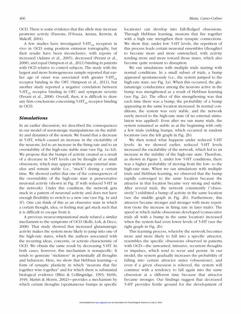
OCD. There is some evidence that this allele may increase promoter activity (Parsons, D’Souza, Arranz, Kerwin, & Makoff, 2004).
A few studies have investigated 5-HT2A receptors in vivo in OCD using positron emission tomography, but their results have been inconclusive, with reports of increased (Adams et al., 2005), decreased (Perani et al., 2008), and equal (Simpson et al., 2011) binding in patients with OCD relative to control subjects. The study with the largest and more homogeneous sample reported that ear-lier age of onset was associated with greater 5-HT2A receptor binding in the OFC (Simpson et al., 2011), but another study reported a negative correlation between 5-HT2A receptor binding in OFC and symptom severity (Perani et al., 2008). Overall, then, it is difficult to draw any firm conclusions concerning 5-HT2A receptor binding in OCD.
Simulations
In an earlier discussion, we described the consequences in our model of serotonergic manipulations on the stabil-ity and dynamics of the system. We found that a decrease in 5-HT, which caused an increase in the excitability of the neurons, led to an increase in the firing rate and to an overstability of the high-rate stable state (see Fig. 1a–1d). We propose that the bumps of activity created as a result of a decrease in 5-HT levels can be thought of as small obsessions, which may appear without any external stim-ulus and remain stable (i.e., “stuck”) during a certain time. We showed earlier that one of the consequences of the overstability of the high-rate state is perseverative neuronal activity (shown in Fig. 1f with reduced 5-HT in the network). Under this condition, the network gets stuck in a pattern of neuronal activity and does not have enough flexibility to switch to a new one (see Fig. 1e and 1f). One can think of this as an obsessive state in which a certain thought, idea, or feeling may get stuck such that it is difficult to escape from it.
A previous neurocomputational study related a similar mechanism to the symptoms of OCD (Rolls, Loh, & Deco, 2008). That study showed that increased glutamatergic activity makes the system more likely to jump into one of the high-rate states, which the authors associated with the recurring ideas, concerns, or actions characteristic of OCD. We obtain the same result by decreasing 5-HT. In both cases, however, this mechanism is nonspecific: It tends to generate “stickiness” in potentially all thoughts and behaviors. Here, we show that Hebbian learning—a form of synaptic plasticity in which “neurons that fire together wire together” and for which there is substantial biological evidence (Bliss & Collingridge, 1993; Hebb, 1949; Martin & Morris, 2002)—provides a mechanism by which certain thoughts (spontaneous bumps in specific
locations) can develop into full-fledged obsessions. Through Hebbian learning, neurons that fire together with a high rate strengthen their synaptic connections. We show that, under low 5-HT levels, the repetition of this process leads certain neuronal ensembles (thoughts) to become more and more entrenched, with activity tending more and more toward those states, which also become quite resistant to disruption.
We ran simulations with multiple trials starting with normal conditions. In a small subset of trials, a bump appeared spontaneously (i.e., the system jumped to the high-rate state; see Fig. 1a). When this occurred, the glu-tamatergic conductance among the neurons active in the bump was strengthened as a result of Hebbian learning (see Fig. 2a). The effect of this strengthening was that each time there was a bump, the probability of a bump appearing in the same location increased. In normal con-ditions, the system was very stable, and the network rarely moved to the high-rate state (if no external stimu-lation was applied). Even after we ran many trials, the system remained as stable as at the beginning with only a few trials yielding bumps, which occurred in random locations (see the left graph in Fig. 2b).
We then tested what happens under reduced 5-HT levels. As we showed earlier, reduced 5-HT levels increased the excitability of the network, which led to an increase in the stability of the high-rate state. Therefore, as shown in Figure 1, under low 5-HT conditions, there was a higher probability of moving from the low- to the high-rate state. When we ran simulations with multiple trials and Hebbian learning, we observed that the bump rapidly converged to the same location because the attractor in that location became very strong and stable. After several trials, the network consistently (“obses-sively”) exhibited a bump of activity in the same location (see the middle graph in Fig. 2b). Furthermore, this attractor became stronger and stronger with more repeti-tion (note the increase in firing rate in later trials). The speed at which stable obsessions developed (consecutive trials all with a bump in the same location) increased when the system had even lower levels of 5-HT (see the right graph in Fig. 2b).
This learning process, whereby the network becomes more and more likely to fall into a specific attractor, resembles the specific obsessions observed in patients with OCD—the unwanted, intrusive, recurrent thoughts or impulses, which tend to recur and persist. In our model, the system gradually increases the probability of falling into certain attractor states (obsessions), and even if a given obsession is relieved, the system will continue with a tendency to fall again into the same obsession at a different time because that attractor became stronger. Our findings suggest that decreased 5-HT provides fertile ground for the development of
at Association for Psychological Science on July 8, 2015cpx.sagepub.comDownloaded from

Serotonin, Orbitofrontal Function, and Obsessive-Compulsive Disorder 467
0
1000500
0
10
20
Neuron
Firin
g ra
te
0 1030 40
60 70
0
1000500
0
10
20
TrialNeuron
Firin
g ra
te
2050
0 1030 40
60 70
0
1000500
0
10
20
TrialNeuron20
50
Firin
g ra
te
35 5-HT ↓ 10%
Neur
on
0 1 2 3 4 5Time (s)
Neur
on
0 1 2 3 4 5Time (s)
0 1 2 3 4 5Time (s)
0 1 2 3 4 5Time (s)
0 1 2 3 4 5Time (s)
a
bNormal conditions
5-HT ↓ 5%
Lower 5-HT level in all neurons
30
0 1030 40
60 70
Trial2050
0 1 2 30
0.2
0.4
0.6
0.8
1
Normal5-HT ↓10%
5-HT ↓20%
Change in glutamatergic synapticstrength in a cluster of neurons (%)
Prop
ortio
n of
“ob
sess
ions
”d
Prop
ortio
n of
sim
ulat
ions
with
abu
mp
in e
ach
grou
p of
neu
rons
10.8
0.20.40.6
01
0.8
0.20.40.6
0Neuron Neuron
NormalGlu level
↑Glu 2% ↑Glu 3%
↑Glu 1%
↑Glutamatergic synapticstrength in a cluster of neurons
Normal
5-HT ↓ 20%in all neurons
c
Fig. 2. Effects of low 5-hydroxytryptamine (5-HT) on the propensity to develop obsessions (through Hebbian learning) and to fall into existing obsessions. The raster plots in (a) illustrate how Hebbian learning leads to the entrenchment of previously repeated patterns of neuronal activity. When a bump appears (second raster plot), the glutamatergic conductance between the neurons of the bump is strengthened in the following trials (orange band) as a result of Hebbian learning (“neurons that fire together wire together”). The repetition of this process (subsequent raster plots) can lead to the entrenchment of certain patterns of neuronal activity, which will tend to fall into the specific attractors that have developed (orange bands). The more a bump occurs in a given clus-ter, the more likely it is that it will occur again in that cluster (see last three raster plots). The graphs in (b) show that low 5-HT leads to the development of obsessions (very strong attractors). In normal conditions (left graph), there is a low probability of developing a very strong attractor (obsession). Under low 5-HT (middle graph), the network tends to develop a very strong attractor (an obsession). In the simulation, that pattern starts to develop at around Trial 30, and in later trials (60–70) tends to recur every time. With even lower levels of 5-HT (right graph), the development of obsessions is even faster. Note also that in addition to recurring more and more often, obsessions become stronger and stronger (i.e., exhibit increasing firing rates; note the change in scale of the y-axis). This process of obsessions becoming both more prevalent and stronger mimics well the phenomenology of obsessive-compulsive disorder. The graphs in (c) show that low 5-HT increases the tendency to fall into existing obsessions. In these simulations, we modeled existing obsessions (attractors) of different strengths by increasing glutamatergic synaptic strengths by different amounts (normal, 1%, 2%, and 3%) in a given cluster of neurons (gray area). The stronger the glutamatergic synaptic strength in the obsession (gray area), the higher the probability of falling into the obsession (compare the different graphs). We then studied the effects of low 5-HT in the tendency to fall into these attractors. Low serotonin (20% decrease of 5-HT in all neurons) further increases the tendency to fall into the obsession for all obsession strengths (the yellow bar is always above the black bar in the gray area in each graph; Glu = glutamate). The graph in (d) illustrates the dose-response relation between the decrease in levels of 5-HT and the tendency to fall into existing obsessions. The graph replots, in a different manner, the results shown in (a) and adds another level of 5-HT to demonstrate that the lower the 5-HT, the greater the probability of falling into existing obsessions.
at Association for Psychological Science on July 8, 2015cpx.sagepub.comDownloaded from

468 Maia, Cano-Colino
obsessions. Thus, even if the subject can overcome a specific obsession, under low 5-HT, the system has a natural tendency to create new ones. Such an effect can be seen in patients with OCD, in whom the obsessions need not always be the same but rather can change or evolve over time.
In addition to increasing the tendency to create new obsessions, low 5-HT may increase the tendency to fall into existing obsessions. To investigate this possibility, we increased the strength of the synaptic connections in a given cluster of neurons, thereby creating an attractor (an “obsession”). We increased this synaptic strength by three different amounts, thereby mimicking “learned obses-sions” at different levels, to analyze whether there is an interaction between low levels of 5-HT and the tendency to fall into obsessions of different strengths (see the graphs in Fig. 2c and 2d). We ran simulations with three different 5-HT levels: normal conditions and 10% and 20% decreases in 5-HT. As expected, greater obsession strengths (i.e., greater connection strengths in the cluster of neurons) led to a greater tendency to fall into the obsession. Furthermore, low 5-HT increased the ten-dency to fall into existing obsessions for all obsession strengths (see Fig. 2c and 2d). This effect followed a dose-response relationship: Greater reductions in 5-HT led to a greater tendency to fall into existing obsessions (for all obsession strengths).
Glutamate in OCD
Review
As noted earlier, OCD seems to involve primarily the orbitofrontal-striatal circuit (Maia et al., 2008; Menzies et al., 2008). Multiple brain-imaging studies have shown that the OFC and the regions of the striatum to which it projects (primarily the head of the caudate nucleus) are hyperactive at rest and during symptom provocation in patients with OCD (Maia et al., 2008). Hyperactivity in these regions in OCD has been confirmed meta-analyti-cally (Whiteside, Port, & Abramowitz, 2004). Furthermore, clinical improvements in OCD are associated with a reduction in this hyperactivity (Baxter et al., 1992; Benkelfat et al., 1990; Nakao et al., 2005; Saxena et al., 1999). These findings are consistent with the aforemen-tioned findings that repeated stimulation of the projec-tions from the OFC to the VMS in mice triggers excessive (compulsive) grooming, which is accompanied by increased evoked activity in the VMS (Ahmari et al., 2013). Consistent with the reduction in striatal hyperac-tivity following successful treatment of OCD (Baxter et al., 1992; Benkelfat et al., 1990; Saxena et al., 1999), results from this study in mice showed that chronic SSRI
treatment that reversed the excessive grooming led to a reduction in the evoked striatal activity.
One possible explanation for the often-replicated find-ings of hyperactivity in the OFC and in its striatal target regions is that OCD may involve a dysregulation in the glutamatergic system (Pittenger et al., 2006; Wu et al., 2012). Specifically, OCD might involve increased gluta-matergic neurotransmission in the OFC and in the projec-tions from the OFC to the striatum. In fact, one magnetic resonance spectroscopy (MRS) study has reported increased levels of glutamate (more precisely, of a com-bined measure of glutamate and glutamine) in the orbi-tofrontal region in patients with OCD, which correlated with symptom severity (Whiteside, Port, Deacon, & Abramowitz, 2006). Another study, however, did not rep-licate these findings (Bédard & Chantal, 2011). Some MRS studies have also reported increased glutamate in the caudate of patients with OCD, which normalized with treatment or correlated with symptom severity, although these findings have not always been replicated (Pittenger, Bloch, & Williams, 2011). In fact, MRS studies of gluta-mate in OCD have tended to generate a fairly large num-ber of null findings in all brain regions considered (Brennan, Rauch, Jensen, & Pope, 2013). Two studies have reported increased glutamate concentrations in the cerebrospinal fluid of patients with OCD (Bhattacharyya et al., 2009; Chakrabarty, Bhattacharyya, Christopher, & Khanna, 2005), which lends further support to the hypothesis of increased glutamatergic neurotransmission in OCD. However, in light of our results showing that reduced 5-HT leads to an increased tendency to be in a high-firing-rate state (see Figs. 1a–1d and 2b), it is diffi-cult to ascertain whether a possible increase in glutama-tergic neurotransmission, if indeed it exists, is due to a primary abnormality in glutamatergic signaling itself or is instead secondary to, for example, low 5-HT.
Despite the limitations with attributing a causal effect to a potential glutamatergic dysfunction on the basis of studies that have shown increased glutamate in patients with OCD, other preclinical and clinical evidence has suggested the possible involvement of a primary distur-bance in glutamatergic signaling in OCD (Pittenger et al., 2006; Wu et al., 2012). For example, an association between the neuronal glutamate transporter gene (SLC1A1) and OCD has been replicated in several studies (Pittenger et al., 2011). The specific alleles that have been shown to be associated with OCD in these studies have varied (Stewart et al., 2013), which makes it difficult to ascertain their functional effects. However, in at least some cases, it seems that variants associated with OCD may lead to decreased expression of the glutamate trans-porter, which could give rise to increased glutamatergic transmission (Pittenger et al., 2011).
at Association for Psychological Science on July 8, 2015cpx.sagepub.comDownloaded from
Serotonin, Orbitofrontal Function, and Obsessive-Compulsive Disorder 469
Simulations
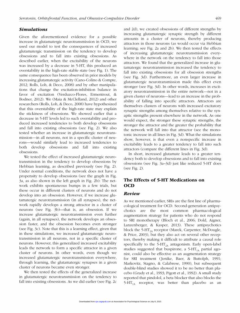
Given the aforementioned evidence for a possible increase in glutamatergic neurotransmission in OCD, we used our model to test the consequences of increased glutamatergic transmission on the tendency to develop obsessions and to fall into existing obsessions. As described earlier, when the excitability of the neurons was increased by a decrease in 5-HT, this produced an overstability in the high-rate stable state (see Fig. 1). The same consequence has been observed in prior models by increasing glutamatergic activity (Cano-Colino & Compte, 2012; Rolls, Loh, & Deco, 2008) and by other manipula-tions that change the excitation-inhibition balance in favor of excitation (Verduzco-Flores, Ermentrout, & Bodner, 2012). We (Maia & McClelland, 2012) and other researchers (Rolls, Loh, & Deco, 2008) have hypothesized that this overstability of the high-rate state may explain the stickiness of obsessions. We showed earlier that a decrease in 5-HT levels led to such overstability and pro-duced increased tendencies to both develop obsessions and fall into existing obsessions (see Fig. 2). We also tested whether an increase in glutamatergic neurotrans-mission—in all neurons, not in a specific cluster of neu-rons—would similarly lead to increased tendencies to both develop obsessions and fall into existing obsessions.
We tested the effect of increased glutamatergic neuro-transmission in the tendency to develop obsessions by Hebbian learning, as described previously (see Fig. 2). Under normal conditions, the network does not have a propensity to develop obsessions (see the graph in Fig. 3a, as also shown in the left graph in Fig. 2b): The net-work exhibits spontaneous bumps in a few trials, but these occur in different clusters of neurons and do not develop into an obsession. However, if we increase glu-tamatergic neurotransmission (in all synapses), the net-work rapidly develops a strong attractor in a cluster of neurons (see Fig. 3b)—that is, an obsession. If we increase glutamatergic neurotransmission even further (again, in all synapses), the network develops an obses-sion faster, and the obsession becomes even stronger (see Fig. 3c). Note that this is a learning effect, given that in these simulations, we increased glutamatergic neuro-transmission in all neurons, not in a specific cluster of neurons. However, this generalized increased excitability leads the network to form a specific attractor in a given cluster of neurons. In other words, even though we increased glutamatergic neurotransmission everywhere, through learning, the glutamatergic synapses in a given cluster of neurons became even stronger.
We then tested the effects of the generalized increase in glutamatergic neurotransmission on the tendency to fall into existing obsessions. As we did earlier (see Fig. 2c
and 2d), we created obsessions of different strengths by increasing glutamatergic synaptic strength by different amounts in a cluster of neurons, thereby producing attractors in those neurons (as would occur via Hebbian learning; see Fig. 2a and 2b). We then tested the effects of increasing glutamatergic neurotransmission every-where in the network on the tendency to fall into those attractors. We found that the generalized increase in glu-tamatergic neurotransmission increased the tendency to fall into existing obsessions for all obsession strengths (see Fig. 3d). Furthermore, an even larger increase in glutamatergic neurotransmission made this effect even stronger (see Fig. 3d). In other words, increases in excit-atory neurotransmission in the entire network—not in a given cluster of neurons—lead to an increase in the prob-ability of falling into specific attractors. Attractors are themselves clusters of neurons with increased excitatory synaptic strengths among themselves relative to the syn-aptic strengths present elsewhere in the network. As one would expect, the stronger these synaptic strengths, the stronger the attractor and the greater the probability that the network will fall into that attractor (see the mono-tonic increase in all lines in Fig. 3d). What the simulations show, however, is that even a nonspecific increase in excitability leads to a greater tendency to fall into such attractors (compare the different lines in Fig. 3d).
In short, increased glutamate leads to a greater ten-dency both to develop obsessions and to fall into existing obsessions (see Fig. 3a–3d) just like reduced 5-HT does (see Fig. 2).
The Effects of 5-HT Medications on OCD
Review
As we mentioned earlier, SRIs are the first line of pharma-cological treatment for OCD. Second-generation antipsy-chotics are the most common pharmacological augmentation strategy for patients who do not respond to SRI monotherapy (Bloch et al., 2006; Dold, Aigner, Lanzenberger, & Kasper, 2013). These antipsychotics block the 5-HT2A receptor (Marek, Carpenter, McDougle, & Price, 2003), but they also act on several other recep-tors, thereby making it difficult to attribute a causal role specifically to the 5-HT2A antagonism. Early open-label studies suggested that buspirone, a 5-HT1A partial ago-nist, could also be effective as an augmentation strategy for SRI treatment ( Jenike, Baer, & Buttolph, 1991; Markovitz, Stagno, & Calabrese, 1990), but subsequent double-blind studies showed it to be no better than pla-cebo (Grady et al., 1993; Pigott et al., 1992). A small study reported that pindolol, a beta blocker that also blocks the 5-HT1A receptor, was better than placebo as an
at Association for Psychological Science on July 8, 2015cpx.sagepub.comDownloaded from

470 Maia, Cano-Colino
0 1030 40
60 70
0
1000
500
0
10
20
TrialNeuron
Firin
g ra
te
2050
Glu ↑ 1%
0
1000
500
0
10
20
Neuron
Firin
g ra
te
Normal conditionsHigher glutamatergic synaptic
strength in all neurons
a b
0
35
Firin
g ra
te
Glu ↑ 2%
0 1030 40
60 70
0
1000
500
TrialNeuron20
50
10
20
30
0 1030 40
60 70
Trial2050
0 20 40 60 80 100
0
1000
500
0
10
20
TrialNeuron
Firin
g ra
te
0 1 2 30
0.2
0.4
0.6
0.8
1
Normal Glu ↑ 4%Glu ↑ 4% + 5-HT ↑ 15%
Change in glutamatergic synapticstrength in a cluster of neurons (%)
Prop
ortio
n of
“ob
sess
ions
”
f
Glu ↑ 4% + 5-HT ↑ 25%
e
Higher glutamatergic synaptic strengthand higher 5-HT level in all neurons
Glu ↑ 4% & 5-HT ↑ 15%
0 1 2 30
0.2
0.4
0.6
0.8
1Normal Glu ↑ 2% Glu ↑ 4%
Change in glutamatergic synapticstrength in a cluster of neurons (%)
Prop
ortio
n of
“ob
sess
ions
”
dHigher glutamatergic synaptic
strength in all neurons
c
Fig. 3. Effects of increased glutamatergic synaptic strength in all neurons on the propensity to develop obsessions and to fall into existing obses-sions and reversal of those effects by increasing 5-hydroxytryptamine (5-HT). Graphs that compare (a) normal conditions and increased glutamater-gic synaptic strength of (b) 1% and (c) 2% in all neurons show that increased glutamatergic synaptic strength in all neurons leads to an increased propensity to develop obsessions. These graphs are similar to those shown in Figure 2b but demonstrate the effects of increased glutamatergic activity in all neurons (not in a specific cluster of neurons), rather than the effects of decreased 5-HT levels in all neurons, on the propensity to develop obsessions. High glutamatergic activity, like low 5-HT (see Fig. 2b), leads to an increased propensity to develop obsessions. The greater the glutamatergic hyperactivity, the earlier the obsessions develop and the stronger they become, as illustrated in the differences between (b) and (c),
(continued)
at Association for Psychological Science on July 8, 2015cpx.sagepub.comDownloaded from

Serotonin, Orbitofrontal Function, and Obsessive-Compulsive Disorder 471
augmentation to paroxetine treatment (Dannon et al., 2000), but another study reported that it was no better than placebo as an augmentation to fluvoxamine (Mundo, Guglielmo, & Bellodi, 1998). Interpretation of the find-ings concerning these 5-HT1A agents is difficult because they act both presynaptically in autoreceptors, where 5-HT1A receptor stimulation has the effect of decreasing serotonin release, and postsynaptically. The interpreta-tion of the effects of buspirone is further complicated by the fact that it is a partial, not a full, 5-HT1A agonist; thus,
it may either increase or decrease the effects of 5-HT1A stimulation depending on the amount of 5-HT present.
Simulations
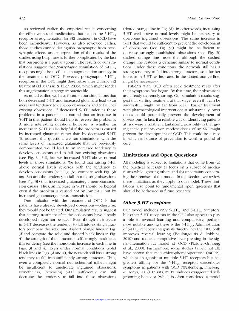
As we discussed earlier, the main evidence for a role of 5-HT in OCD comes from the use of SRIs as the first line of pharmacological treatment for this disorder. We have shown earlier that reduced 5-HT leads to a greater ten-dency to develop obsessions and to fall into existing obses-sions (see Fig. 2); it therefore stands to reason that if patients with OCD have reduced 5-HT, normalization of 5-HT levels with an SRI should be beneficial. We also tested whether increasing 5-HT further could be useful to reduce the tendency to fall into existing obsessions, given that patients do, of course, already present with obsessions. We found that, indeed, increasing 5-HT reduces the tendency to fall into existing obsessions (see Fig. 4; see also Fig. 2d).
Given the aforementioned use of second-generation antipsychotics, which block the 5-HT2A receptor, to aug-ment SRI treatment in OCD, and also given the evidence reviewed earlier for a possible association between abnor-malities in the 5-HT2A gene and OCD, we also used our model to test the effects of combining 5-HT2A antagonism with an increase in 5-HT. Consistent with the pharmacologi-cal effects found empirically, our results showed that adding a 5-HT2A blocker to an SRI further decreased the network’s tendency to fall into existing obsessions (see Fig. 4).
We also used our model to test the effects of combin-ing 5-HT1A agonism with an increase in 5-HT because of the early studies, reviewed earlier, that suggested that medications that act on the 5-HT1A receptor might be use-ful as augmentation therapies for OCD. We found that adding a 5-HT1A agonist to an SRI was also effective at further decreasing the network’s tendency to fall into existing obsessions (see Fig. 4). These findings can be interpreted in terms of the excitability of the network: 5-HT1A receptors have a predominantly inhibitory effect on the network; thus, 5-HT1A agonism decreases network excitability, thereby ameliorating the tendency to fall into obsessions.
noting the change in axis. The graph in (d) illustrates the dose-response relation between levels of glutamatergic synaptic strength in all neurons and the tendency to fall into existing obsessions. This graph is similar to the graph in Figure 2d but shows the effects of different levels of increased glutamatergic activity in all neurons, rather than different levels of decreased 5-HT, on the propensity to fall into existing obsessions. The greater the glutamatergic hyperactivity in all neurons, the greater the probability of falling into existing obsessions (clusters of neurons with specifically increased glutamatergic activity relative to the other neurons in the network). The graph in (e) shows that an increase in 5-HT decrease the tendency to develop obsessions, even in the presence of glutamatergic hyperactivity. Whereas the graphs in (b) and (c) show that glutamatergic hyperactiv-ity (in all neurons) increases the tendency to develop obsessions, the graph in (e) shows that an increase in 5-HT decreases this tendency, even if that tendency is caused by glutamatergic hyperactivity. The graph in (f) shows that increases in 5-HT decrease the tendency to fall into existing obsessions, even in the presence of glutamatergic hyperactivity. Whereas the graph in (d) shows that glutamatergic hyperactivity (in all neurons) increases the tendency to fall into existing obsessions, an effect that is shown again in this graph (solid orange line vs. black line), this graph shows that increasing 5-HT decrease this tendency, even in the presence of the glutamatergic hyperactivity (dashed and dotted orange lines). This effect of 5-HT follows a dose-response relationship wherein greater increases in 5-HT lead to greater reductions in the tendency to fall into obsessions (dotted vs. dashed orange line). Glu = glutamate.
Fig. 3. (continued)
0 1 2 30
0.2
0.4
0.6
0.8
1
Change in glutamatergic synapticstrength in a cluster of neurons (%)
Prop
ortio
n of
“ob
sess
ions
”
NormalNormal + 5-HT ↑ 5%Normal + 5-HT ↑ 5% + 5-HT2A ↓ 10%Normal + 5-HT ↑ 5% + 5-HT1A ↑ 5%
Fig. 4. Effects of 5-Hydroxytryptamine (5-HT) medications on the ten-dency to fall into obsessions. Increases in 5-HT (e.g., through a selec-tive serotonin reuptake inhibitor) decrease the tendency to fall into obsessions (black dashed line vs. black solid line; Fig. 2d shows the reverse effect wherein decreases in 5-HT increase the tendency to fall into obsessions). The addition of either a 5-HT2A antagonist (dashed blue line) or a 5-HT1A agonist (dashed purple line) to a medication that increases 5-HT further reduces the tendency to fall into obsessions.
at Association for Psychological Science on July 8, 2015cpx.sagepub.comDownloaded from
472 Maia, Cano-Colino
As reviewed earlier, the empirical results concerning the effectiveness of medications that act on the 5-HT1A receptor as augmentation for SRI treatment in OCD have been inconclusive. However, as also reviewed earlier, those studies cannot distinguish presynaptic from post-synaptic effects, and interpretation of the results of the studies using buspirone is further complicated by the fact that buspirone is a partial agonist. The results of our sim-ulations suggest that postsynaptic stimulation of 5-HT1A receptors might be useful as an augmentation strategy in the treatment of OCD. However, postsynaptic 5-HT1A receptors in the OFC might desensitize after chronic SRI treatment (El Mansari & Blier, 2005), which might render this augmentation strategy impracticable.
As noted earlier, we have found in our simulations that both decreased 5-HT and increased glutamate lead to an increased tendency to develop obsessions and to fall into existing obsessions. If decreased 5-HT underlies these problems in a patient, it is natural that an increase in 5-HT in that patient should help to reverse the problems. A more interesting question, however, is whether an increase in 5-HT is also helpful if the problem is caused by increased glutamate rather than by decreased 5-HT. To address this question, we ran simulations with the same levels of increased glutamate that we previously demonstrated would lead to an increased tendency to develop obsessions and to fall into existing obsessions (see Fig. 3a–3d), but we increased 5-HT above normal levels in those simulations. We found that raising 5-HT above normal levels reverses both the tendency to develop obsessions (see Fig. 3e; compare with Fig. 3b and 3c) and the tendency to fall into existing obsessions (see Fig. 3f) that increased glutamatergic neurotransmis-sion causes. Thus, an increase in 5-HT should be helpful even if the problem is caused not by low 5-HT but by increased glutamatergic neurotransmission.
One limitation with the treatment of OCD is that patients have already developed obsessions—otherwise, they would not be treated. Our simulation results suggest that starting treatment after the obsessions have already developed might not be ideal: Even though an increase in 5-HT decreases the tendency to fall into existing attrac-tors (compare the solid and dashed orange lines in Fig. 3f and compare the solid and dashed black lines in Fig. 4), the strength of the attractors itself strongly modulates this tendency (see the monotonic increase in each line in Figs. 3f and 4). Even under normal conditions (solid black lines in Figs. 3f and 4), the network still has a strong tendency to fall into sufficiently strong attractors. Thus, even a completely normal neurochemical milieu might be insufficient to ameliorate ingrained obsessions. Nonetheless, increasing 5-HT sufficiently can still decrease the tendency to fall into these obsessions
(dotted orange line in Fig. 3f). In other words, increasing 5-HT well above normal levels might be necessary to overcome ingrained obsessions. The same increase in 5-HT that would be sufficient to prevent the development of obsessions (see Fig. 3e) might be insufficient to decrease strongly established obsessions (see Fig. 3f, dashed orange line—note that although the dashed orange line restores a dynamic similar to normal condi-tions, under those conditions, the network still has a strong tendency to fall into strong attractors, so a further increase in 5-HT, as indicated in the dotted orange line, might be necessary).
Patients with OCD often seek treatment years after their symptoms first began. By that time, their obsessions are already extremely strong. Our simulation results sug-gest that starting treatment at that stage, even if it can be successful, might be far from ideal. Earlier treatment with pharmacological interventions at substantially lower doses could potentially prevent the development of obsessions. In fact, if a reliable way of identifying patients at risk were available, a tantalizing possibility is that giv-ing these patients even modest doses of an SRI might prevent the development of OCD. This could be a case in which an ounce of prevention is worth a pound of cure.
Limitations and Open Questions
All modeling is subject to limitations that come from (a) the practical necessity to focus on a subset of mecha-nisms while ignoring others and (b) uncertainty concern-ing the premises of the model. In this section, we review these limitations as they apply to our model. These limi-tations also point to fundamental open questions that should be addressed in future research.
Other 5-HT receptors
Our model includes only 5-HT1A and 5-HT2A receptors, but other 5-HT receptors in the OFC also appear to play a role in reversal learning and compulsivity; perhaps most notable among these is the 5-HT2C. Administration of 5-HT2C receptor antagonists directly into the OFC both improves reversal learning (Boulougouris & Robbins, 2010) and reduces compulsive lever pressing in the sig-nal-attenuation rat model of OCD (Flaisher-Grinberg et al., 2008). Furthermore, some studies (albeit not all) have shown that meta-chlorophenylpiperazine (mCPP), which is an agonist at multiple 5-HT receptors but has greatest affinity for the 5-HT2C receptor, exacerbates symptoms in patients with OCD (Westenberg, Fineberg, & Denys, 2007). In rats, mCPP induces exaggerated self-grooming behavior (which is often considered a model
at Association for Psychological Science on July 8, 2015cpx.sagepub.comDownloaded from

Serotonin, Orbitofrontal Function, and Obsessive-Compulsive Disorder 473
for OCD-like behaviors), and this effect appears to be mediated by the 5-HT2C receptor (Graf et al., 2003). This set of findings implicating activation of the 5-HT2C recep-tor in OCD-related behaviors and blockade of the 5-HT2C receptor in the amelioration of such behaviors raises the possibility that 5-HT2C antagonists might be useful to treat OCD. Both empirical work aimed at further testing this possibility and computational work akin to the one we present here but focusing on the 5-HT2C receptor would therefore be of great value.
Other studies have highlighted possible roles for other serotonin receptors—for example, the 5-HT1D—in OCD (Aouizerate, Guehl, & Cuny, 2005). A more detailed understanding of the specific receptors involved may allow for more targeted treatments, given that inhibiting the reuptake of 5-HT is a somewhat crude, nonspecific manipulation.
Other behaviors modulated by 5-HT
A long-standing idea is that 5-HT is involved in behavioral inhibition (Soubrié, 1986). Behavioral inhibition is not a unitary concept but, rather, encompasses several distinct processes that are mediated by partially distinct neural structures and neuromodulatory systems (Bari & Robbins, 2013). The prototypical case of behavioral inhibition is response inhibition, which itself can be subdivided into three subtypes: (a) the inhibition of responses whose exe-cution has already started, typically assessed using the stop-signal task; (b) the inhibition of prepotent responses before they start, typically assessed using the go/no-go task; and (c) the inhibition of responses until they are appropriate, so as to prevent premature responding, often assessed using the five-choice serial reaction time task (Dalley & Roiser, 2012; Eagle, Bari, & Robbins, 2008). Manipulations of the serotonergic system have no effect on the ability to inhibit responses in the stop-signal task, which is instead modulated by norepinephrine (Chamberlain, Müller, et al., 2006; Clark et al., 2005; Eagle et al., 2008, 2009; Nandam et al., 2011). However, manipu-lations of the serotonergic system do affect the ability to inhibit responses on go/no-go tasks (Eagle et al., 2008; Harrison, Everitt, & Robbins, 1999) and to inhibit prema-ture responses on serial reaction time tasks (Dalley & Roiser, 2012; Miyazaki, Miyazaki, & Doya, 2012; Worbe, Savulich, Voon, Fernandez-Egea, & Robbins, 2014).
The extent to which these serotonergic effects act through the OFC is unclear. Response inhibition in go/no-go and stop-signal tasks is typically thought to be medi-ated mostly not by the OFC but by the inferior frontal gyrus, supplementary and presupplementary motor areas, and subthalamic nucleus, with possible involvements of the insula and medial frontal gyrus (Aron, Robbins, & Poldrack, 2004; Bari & Robbins, 2013; Simmonds, Pekar, &
Mostofsky, 2008; Swick, Ashley, & Turken, 2011). Conversely, several neuroimaging studies have shown that serotonergic manipulations affect OFC activation during response inhibition in go/no-go tasks (Anderson, McKie, Elliott, Williams, & Deakin, 2008; Del-Ben et al., 2005; Rubia et al., 2005; Völlm et al., 2006). Premature responses on the five-choice serial reaction time task might also depend on the OFC: Lesions of the OFC induce both per-severative responding and increases in premature respond-ing in this task (Chudasama et al., 2003).
Another prominent idea is that 5-HT is involved in aversive processing (Boureau & Dayan, 2011; Daw, Kakade, & Dayan, 2002; Dayan & Huys, 2009). In rela-tion to the role of 5-HT in the OFC, this idea dovetails nicely with the emphasis on the involvement of the OFC in the representation of value, outcome expectan-cies, and reinforcement learning (Frank & Claus, 2006; Jones et al., 2012; O’Doherty, 2007; Schoenbaum & Esber, 2010; Schoenbaum et al., 2009; Schultz, Tremblay, & Hollerman, 2000). The possible role of 5-HT in aver-sive processes might potentially be reconciled with its role in inhibition because aversion is often linked with inhibition. Some evidence has suggested that 5-HT might be involved specifically in punishment-induced inhibition (Crockett, Clark, & Robbins, 2009), and com-putational considerations have suggested a rationale for a tightly interwoven involvement of 5-HT in both aversion and inhibition (Cools, Nakamura, & Daw, 2011).
It is clear that additional work will be necessary to integrate the perspective that we have articulated in this article with these other theories and findings. We can speculate, for example, that the role of 5-HT in disrupting attractors, which we have shown herein, could be useful to prevent prepotent responses in go/no-go tasks by dis-rupting the neural activity corresponding to the prepo-tent response. Such suggestions, however, are at present at best vague, so it is clear that much additional theoreti-cal work would be necessary to seek to integrate these various perspectives.
One difference between our focus in this study and the focus of prior computational perspectives on 5-HT is that here we investigated the effects of changes in tonic 5-HT levels on the target site (in our case, the OFC); thus, our focus was on modeling these effects with a suf-ficient level of biophysical detail. Other computational theories of the role of 5-HT have been concerned pri-marily with understanding, at an algorithmic level, the role of the firing of 5-HT neurons (Daw et al., 2002; Dayan & Huys, 2008, 2009)—thereby seeking, in essence, to obtain an algorithmic understanding of 5-HT akin to the one that has been obtained for the phasic firing of dopamine neurons with the prediction-error hypothesis (Glimcher, 2011; Maia, 2009). Those theories therefore
at Association for Psychological Science on July 8, 2015cpx.sagepub.comDownloaded from
474 Maia, Cano-Colino
focus on the circumstances under which 5-HT neurons would be expected to fire more or fire less, not on explaining in detail how 5-HT affects target sites. Of course, an integrated understanding of the role of 5-HT will have to integrate both aspects: the “presynaptic” side (i.e., under which circumstances 5-HT neurons fire and how much) and the “postsynaptic” side (i.e., what effect 5-HT has on the neuronal circuits in target regions). Developing such an integrated understanding should be an important focus for future computational efforts to understand the role of 5-HT in cognition and behavior. This integration would ideally also occur between levels of analysis. Although it is undoubtedly valuable to use different levels of modeling (e.g., algorithmic vs. bio-physical) to address phenomena at different levels of analysis, it will be important and interesting to seek to integrate these different levels of analysis (Frank, in press).
Another function of 5-HT that we did not include in our model but that may be relevant for the learning (and possibly the unlearning) of obsessions is the mod-ulation of synaptic plasticity. We have shown that by modulating network excitability, 5-HT modulates the tendency to develop obsessions through Hebbian learning. However, 5-HT also has direct effects on syn-aptic plasticity that would be expected to modulate such learning (Lesch & Waider, 2012). It would be inter-esting to incorporate these effects in the model to investigate how they affect the tendency to develop obsessions.
Effects of 5-HT2A receptors on neural activity in the OFC
In our model, 5-HT2A receptors increase excitability both in pyramidal neurons and in GABAergic interneurons, as seems to occur in vitro and in vivo (Puig & Gulledge, 2011). Thus, at the circuit level, stimulation of 5-HT2A receptors has both a direct excitatory effect and an indi-rect inhibitory effect. Nonetheless, also consistent with the empirical evidence concerning the PFC (Puig et al., 2003; Puig & Gulledge, 2011), the overall effect is more excitatory than inhibitory. Some studies, however, have reported that directly applying 2,5-dimethoxy-4-iodoam-phetamine (DOI)—a 5-HT2A, 5-HT2B, and 5-HT2C agonist (Canal & Morgan, 2012)—into the OFC predominantly inhibits neuron activity (El Mansari & Blier, 1997, 2005). Similar findings have sometimes also been reported with microiontophoresis of DOI into the medial PFC, although the effects seem to be dose dependent: At low ejection doses, DOI increases glutamate-evoked excitation, whereas at higher doses, it has an inhibitory effect (Ashby, Edwards, Harkins, & Wang, 1989; Ashby et al., 1994; Ashby, Jiang, Kasser, & Wang, 1990). Furthermore,
the inhibitory effect of DOI in the OFC is blunted by local application of a 5-HT2A antagonist (Rueter, Tecott, & Blier, 2000), thereby suggesting that this inhibitory effect might be mediated at least in part by 5-HT2A receptors. Other areas of PFC also have subpopulations in which 5-HT2A receptor stimulation leads to inhibition, and this effect is hypothesized to result from the increased excitability in GABAergic interneurons (Puig et al., 2003; Puig & Gulledge, 2011). One possibility is therefore that in OFC, the overall effects of 5-HT2A receptor stimulation are due more to the excitation of GABAergic interneurons than to direct effects on pyramidal neurons themselves. Weighing against this possibility, however, is the finding that microiontophoretic application of a GABA antagonist does not blunt the inhibitory effect of DOI in the OFC (Bergqvist, Dong, & Blier, 1999) or in medial PFC (Ashby et al., 1990), thus suggesting that this inhibitory effect is not mediated by GABAergic interneurons. To complicate matters further, other findings have led the authors of these studies to consider the receptor or receptors medi-ating the inhibitory effects of DOI in the OFC “atypical” and pharmacologically distinct from 5-HT2 receptors elsewhere in cortex (Bergqvist et al., 1999; Blier, Habib, & Flament, 2006; El Mansari & Blier, 2005, 2006; Rueter et al., 2000). Additional empirical work characterizing the effects of 5-HT2A receptors in the OFC would clearly be of value. A substantial proportion of the existing work on this issue has used drugs such as DOI and mCPP that are not selective for just one of the 5-HT2 receptors, which makes it difficult to disentangle the contributions of the different 5-HT2 receptors (especially the 5-HT2A vs. 5-HT2C receptors). Agents that are more selective are now avail-able (Glennon & Dukat, 2013) and should shed impor-tant light on the specific roles of 5-HT2A and 5-HT2C receptors in the OFC.
If the biophysical and pharmacological characteristics of 5-HT2A receptors in OFC differ substantially from those implemented in our model, our simulations of the augmentation of SRI treatment with 5-HT2A antagonists (see Fig. 4, blue dashed line) would be based on invalid assumptions. In particular, if 5-HT2A receptors in OFC are predominantly inhibitory, then the results of these simulations would be invalid: To a large extent, in our model, blocking 5-HT2A receptors improves SRI treat-ment because it further decreases the excitability of the network. The results of the rest of the simulations, how-ever, would be unaffected, given that 5-HT itself would still have a predominantly inhibitory effect, which is what drives the findings in the simulations that involve 5-HT manipulations. In fact, the inhibitory effect of 5-HT would be even stronger if 5-HT2A receptor stimulation was also inhibitory; thus, the results of the simulations that involve 5-HT manipulations would be even more pronounced.
at Association for Psychological Science on July 8, 2015cpx.sagepub.comDownloaded from

Serotonin, Orbitofrontal Function, and Obsessive-Compulsive Disorder 475
Modeling the relation between neuronal activity and behavior
In our simulations of perseverative responding in rever-sal-learning and other tasks, we equated perseverative neuronal responding with perseverative errors, but we did not model neuronal activity during the tasks them-selves. Other researchers have developed neurocomputa-tional (Frank & Claus, 2006) and algorithmic (Wilson et al., 2014) models of the role of the OFC in reversal-learning and related tasks, and it would be of interest to seek to merge the perspective articulated here concern-ing modulation of the OFC by 5-HT with those models that more explicitly link the details of OFC function with performance in these tasks.
Relation to dopamine
In a nutshell, the mechanism behind our findings is that decreases in 5-HT lead to overstability of attractors, and increases in 5-HT can compensate for overstable attrac-tors. Biophysical models of the effects of dopamine on cortical circuits suggest that dopamine also modulates attractor stability: Specifically, these models suggest that D1 and D2 receptor stimulation lead, respectively, to an increase and a decrease in the stability of attractors (Durstewitz, 2010). It is interesting that some evidence from animal models has suggested that stimulation of cortical and limbic D1 receptors leads to OCD-like behav-iors (Campbell et al., 1999). In addition, compulsive lever pressing in the signal-attenuation model of OCD is dimin-ished by D1 blockade (although this study involved sys-temic drug administration, so it is not possible to know whether the effect was due to D1 blockade in cortex, striatum, or elsewhere; Joel & Doljansky, 2003). These findings are consistent with our account: Excessive attrac-tor stability, whether the result of reduced 5-HT or increased glutamate, as in our simulations, or of increased D1 receptor stimulation, may lead to OCD and related behaviors. Nonetheless, the parallel between the effects on neural circuit dynamics that we describe for 5-HT and those that have been described previously for dopamine (Durstewitz, 2010) raises a more fundamental theoretical question: What are the differences (if indeed there are any) between the neurodynamic regimes that can be achieved by serotonergic versus dopaminergic manipula-tions? Answering this question will no doubt require additional modeling (and empirical) work.
Conclusions
There has been substantial interest recently in the bud-ding field of computational psychiatry: The use of com-putational techniques to understand psychiatric disorders
and their neural underpinnings (Huys, 2013; Maia & Frank, 2011; Montague, Dolan, Friston, & Dayan, 2012). Prior neurocomputational work has suggested that increased glutamatergic activity in OCD might create excessively strong attractors toward which activity tends and that are difficult to escape (Maia & McClelland, 2012; Rolls, Loh, & Deco, 2008). We showed here that decreased levels of 5-HT have the same effect through a related mechanism: 5-HT has a predominantly inhibitory effect; thus, decreased levels of 5-HT lead to increased excit-ability, which produces excessively strong attractors. We also showed how this effect could, via Hebbian learning, lead to the development of specific, strong, and persis-tent obsessions. Reduced levels of 5-HT might therefore contribute to the development and maintenance of OCD, as well as to other forms of perseverative and compulsive behaviors typically studied in the laboratory.
Despite some evidence that has suggested possible primary abnormalities in the serotonergic system in OCD (reviewed earlier), the strongest evidence implicating 5-HT in OCD comes from the use of SRIs in treatment. The fact that SRIs are helpful in treating OCD, however, does not itself prove that there is a primary abnormality in the 5-HT system; SRIs could also be compensating for another abnormality (van Dijk, Klompmakers, & Denys, 2010). Our results illustrate this possibility by showing that increases in 5-HT help to reduce the tendency to develop obsessions and to fall into existing obsessions, even if those tendencies are caused by excessive gluta-matergic activity, not by low levels of 5-HT.
Our model provides a unified account of a large body of clinical and preclinical results and offers mechanistic insights into the role of 5-HT in OCD and other related perseverative and compulsive behaviors. The model, if correct, also has important clinical implications. If our account of the development of obsessions based on Hebbian learning is realistic, a sobering implication is that having obsessions further strengthens those obses-sions, thereby making early intervention—or even pre-vention—extremely important. Furthermore, in our model, increasing 5-HT decreases the tendency to develop new obsessions and to fall into existing obses-sions, but it does not by itself lead to the unlearning of existing obsessions. (Some such unlearning might occur via more complex Hebbian mechanisms when the fre-quency of obsessions is reduced, but this is not imple-mented in the current version of the model.) This behavior of the model might help to explain why SRIs typically do not produce full remission and especially why, in many cases, they have to be taken long term, possibly indefinitely. Stopping SRI treatment could rap-idly lead to a return of the old obsessions, as well as providing fertile ground for the development of new obsessions.
at Association for Psychological Science on July 8, 2015cpx.sagepub.comDownloaded from

476 Maia, Cano-Colino
Author Contributions
Both authors contributed equally to this work. T. V. Maia devel-oped the study concept. M. Cano-Colino ran the simulations and conducted the analyses. Both authors contributed to the design of the study, interpreted the results, drafted the manu-script, and approved the final version of the manuscript for submission.
Declaration of Conflicting Interests
The authors declared that they had no conflicts of interest with respect to their authorship or the publication of this article.
Funding
This work was funded in part by a grant from the Tourette Syndrome Association to T. V. Maia.
Supplemental Material
Additional supporting information may be found at http://cpx.sagepub.com/content/by/supplemental-data
References
Adams, K. H., Hansen, E. S., Pinborg, L. H., Hasselbalch, S. G., Svarer, C., Holm, S., . . . Knudsen, G. M. (2005). Patients with obsessive-compulsive disorder have increased 5-HT2A receptor binding in the caudate nuclei. International Journal of Neuropsychopharmacology, 8, 391–401.
Aghajanian, G. K., & Marek, G. J. (1997). Serotonin induces excitatory postsynaptic potentials in apical dendrites of neo-cortical pyramidal cells. Neuropharmacology, 36, 589–599.
Ahmari, S. E., Spellman, T., Douglass, N. L., Kheirbek, M. A., Simpson, H. B., Deisseroth, K., . . . Hen, R. (2013). Repeated cortico-striatal stimulation generates persistent OCD-like behavior. Science, 340, 1234–1239. doi:10.1126/science.1234733
Amargós-Bosch, M., Bortolozzi, A., Puig, M. V., Serrats, J., Adell, A., Celada, P., . . . Artigas, F. (2004). Co-expression and in vivo interaction of serotonin1A and serotonin2A receptors in pyramidal neurons of prefrontal cortex. Cerebral Cortex, 14, 281–299.
American Psychiatric Association. (2013). Diagnostic and statis-tical manual of mental disorders (5th ed.). Arlington, VA: American Psychiatric Publishing.
Anderson, I. M., McKie, S., Elliott, R., Williams, S. R., & Deakin, J. F. W. (2008). Assessing human 5-HT function in vivo with pharmacoMRI. Neuropharmacology, 55, 1029–1037. doi:10.1016/j.neuropharm.2008.06.029
Andrade, R., Malenka, R. C., & Nicoll, R. A. (1986). A G protein couples serotonin and GABAB receptors to the same chan-nels in hippocampus. Science, 234, 1261–1265. doi:10.1126/science.2430334
Aouizerate, B., Guehl, D., & Cuny, E. (2005). Updated over-view of the putative role of the serotoninergic system in obsessive-compulsive disorder. Neuropsychiatric Disease and Treatment, 1, 231–243.
Araneda, R., & Andrade, R. (1991). 5-Hydroxytryptamine2 and 5-hydroxytryptamine1A receptors mediate opposing
responses on membrane excitability in rat associa-tion cortex. Neuroscience, 40, 399–412. doi:10.1016/ 0306-4522(91)90128-B
Aron, A. R., Robbins, T. W., & Poldrack, R. A. (2004). Inhibition and the right inferior frontal cortex. Trends in Cognitive Sciences, 8, 170–177.
Ashby, C. R., Edwards, E., Harkins, K., & Wang, R. Y. (1989). Effects of (+-)-DOI on medial prefrontal cortical cells: A microiontophoretic study. Brain Research, 498, 393–396.
Ashby, C. R., Edwards, E., & Wang, R. Y. (1994). Electro-physiological evidence for a functional interaction between 5-HT1A and 5-HT2A receptors in the rat medial prefrontal cortex: An iontophoretic study. Synapse, 17, 173–181.
Ashby, C. R., Jiang, L. H., Kasser, R. J., & Wang, R. Y. (1990). Electrophysiological characterization of 5-hydroxytrypta-mine2 receptors in the rat medial prefrontal cortex. Journal of Pharmacology and Experimental Therapeutics, 252, 171–178.
Baker, P. M., Thompson, J. L., Sweeney, J. A., & Ragozzino, M. E. (2011). Differential effects of 5-HT(2A) and 5-HT(2C) receptor blockade on strategy-switching. Behavioural Brain Research, 219, 123–131.
Bari, A., & Robbins, T. W. (2013). Inhibition and impulsiv-ity: Behavioral and neural basis of response control. Progress in Neurobiology, 108, 44–79. doi:10.1016/j.pneu-robio.2013.06.005
Baxter, L. R., Schwartz, J. M., Bergman, K. S., Szuba, M. P., Guze, B. H., Mazziotta, J. C., . . . Munford, P. (1992). Caudate glucose metabolic rate changes with both drug and behavior therapy for obsessive-compulsive disorder. Archives of General Psychiatry, 49, 681–689.
Bédard, M.-J., & Chantal, S. (2011). Brain magnetic resonance spectroscopy in obsessive-compulsive disorder: The impor-tance of considering subclinical symptoms of anxiety and depression. Psychiatry Research, 192, 45–54. doi:10.1016/j .pscychresns.2010.10.008
Béïque, J.-C., Imad, M., Mladenovic, L., Gingrich, J. A., & Andrade, R. (2007). Mechanism of the 5-hydroxytrypta-mine 2A receptor-mediated facilitation of synaptic activ-ity in prefrontal cortex. Proceedings of the National Academy of Sciences, USA, 104, 9870–9875. doi:10.1073/pnas.0700436104
Bengel, D., Greenberg, B. D., Corá-Locatelli, G., Altemus, M., Heils, A., Li, Q., & Murphy, D. L. (1999). Association of the serotonin transporter promoter regulatory region poly-morphism and obsessive-compulsive disorder. Molecular Psychiatry, 4, 463–466.
Benkelfat, C., Nordahl, T. E., Semple, W. E., King, A. C., Murphy, D. L., & Cohen, R. M. (1990). Local cerebral glu-cose metabolic rates in obsessive-compulsive disorder: Patients treated with clomipramine. Archives of General Psychiatry, 47, 840–848.
Bergqvist, P. B. F., Dong, J., & Blier, P. (1999). Effect of atypi-cal antipsychotic drugs on 5-HT 2 receptors in the rat orbito-frontal cortex: An in vivo electrophysiological study. Psychopharmacology, 143, 89–96.
Bhattacharyya, S., Khanna, S., Chakrabarty, K., Mahadevan, A., Christopher, R., & Shankar, S. K. (2009). Anti-brain autoantibodies and altered excitatory neurotransmitters in
at Association for Psychological Science on July 8, 2015cpx.sagepub.comDownloaded from
Serotonin, Orbitofrontal Function, and Obsessive-Compulsive Disorder 477
obsessive-compulsive disorder. Neuropsychopharmacology, 34, 2489–2496.
Bissonette, G. B., & Powell, E. M. (2012). Reversal learning and attentional set-shifting in mice. Neuropharmacology, 62, 1168–1174.
Blier, P., Habib, R., & Flament, M. (2006). Pharmacotherapies in the management of obsessive-compulsive disorder. Canadian Journal of Psychiatry, 51, 417–429.
Bliss, T. V, & Collingridge, G. L. (1993). A synaptic model of memory: Long-term potentiation in the hippocampus. Nature, 361, 31–39.
Bloch, M. H., Landeros-Weisenberger, A., Kelmendi, B., Coric, V., Bracken, M. B., & Leckman, J. F. (2006). A systematic review: Antipsychotic augmentation with treatment refrac-tory obsessive-compulsive disorder. Molecular Psychiatry, 11, 622–632.
Bloch, M. H., Landeros-Weisenberger, A., Sen, S., Dombrowski, P., Kelmendi, B., Coric, V., . . . Leckman, J. F. (2008). Association of the serotonin transporter polymorphism and obsessive-compulsive disorder: Systematic review. American Journal of Medical Genetics Part B: Neuropsychiatric Genetics, 147B, 850–858. doi:10.1002/ajmg.b.30699
Boulougouris, V., Glennon, J. C., & Robbins, T. W. (2008). Dissociable effects of selective 5-HT2A and 5-HT2C recep-tor antagonists on serial spatial reversal learning in rats. Neuropsychopharmacology, 33, 2007–2019. doi:10.1038/sj.npp.1301584
Boulougouris, V., & Robbins, T. W. (2010). Enhancement of spatial reversal learning by 5-HT2C receptor antagonism is neuroanatomically specific. Journal of Neuroscience, 30, 930–938. doi:10.1523/JNEUROSCI.4312-09.2010
Boureau, Y.-L., & Dayan, P. (2011). Opponency revisited: Competition and cooperation between dopamine and sero-tonin. Neuropsychopharmacology, 36, 74–97. doi:10.1038/npp.2010.151
Brennan, B. P., Rauch, S. L., Jensen, J. E., & Pope, H. G. (2013). A critical review of magnetic resonance spectroscopy stud-ies of obsessive-compulsive disorder. Biological Psychiatry, 73, 24–31. doi:10.1016/j.biopsych.2012.06.023
Brigman, J. L., Mathur, P., Harvey-White, J., Izquierdo, A., Saksida, L. M., Bussey, T. J., . . . Holmes, A. (2010). Pharmacological or genetic inactivation of the serotonin transporter improves reversal learning in mice. Cerebral Cortex, 20, 1955–1963.
Burguière, E., Monteiro, P., Feng, G., & Graybiel, A. M. (2013). Optogenetic stimulation of lateral orbitofronto-striatal path-way suppresses compulsive behaviors. Science, 340, 1243–1246. doi:10.1126/science.1232380
Butter, C. M. (1969). Perseveration in extinction and in discrimi-nation reversal tasks following selective frontal ablations in Macaca mulatta. Physiology & Behavior, 4, 163–171.
Campbell, K. M., de Lecea, L., Severynse, D. M., Caron, M. G., McGrath, M. J., Sparber, S. B., . . . Burton, F. H. (1999). OCD-like behaviors caused by a neuropotentiating trans-gene targeted to cortical and limbic D1+ neurons. Journal of Neuroscience, 19, 5044–5053.
Canal, C. E., & Morgan, D. (2012). Head-twitch response in rodents induced by the hallucinogen 2,5-dimethoxy-4-io-doamphetamine: A comprehensive history, a re-evaluation
of mechanisms, and its utility as a model. Drug Testing and Analysis, 4, 556–576.
Cano-Colino, M., Almeida, R., & Compte, A. (2013). Serotonergic modulation of spatial working memory: Predictions from a computational network model. Frontiers in Integrative Neuroscience, 7, 71. Retrieved from http://journal.frontiersin .org/Journal/10.3389/fnint.2013.00071/full
Cano-Colino, M., Almeida, R., Gomez-Cabrero, D., Artigas, F., & Compte, A. (2014). Serotonin regulates performance nonmonotonically in a spatial working memory network. Cerebral Cortex, 24, 2449–2463. doi:10.1093/cercor/bht096
Cano-Colino, M., & Compte, A. (2012). A computational model for spatial working memory deficits in schizo-phrenia. Pharmacopsychiatry, 45(Suppl. 1), S49–S56. doi:10.1055/s-0032-1306314
Chakrabarty, K., Bhattacharyya, S., Christopher, R., & Khanna, S. (2005). Glutamatergic dysfunction in OCD. Neuropsychopharmacology, 30, 1735–1740.
Chamberlain, S. R., Fineberg, N. A., Blackwell, A. D., Robbins, T. W., & Sahakian, B. J. (2006). Motor inhibition and cognitive flexibility in obsessive-compulsive disorder and trichotil-lomania. American Journal of Psychiatry, 163, 1282–1284. doi:10.1176/appi.ajp.163.7.1282
Chamberlain, S. R., Müller, U., Blackwell, A. D., Clark, L., Robbins, T. W., & Sahakian, B. J. (2006). Neurochemical modulation of response inhibition and probabilistic learning in humans. Science, 311, 861–863. doi:10.1126/ science.1121218
Chudasama, Y., Passetti, F., Rhodes, S. E. V., Lopian, D., Desai, A., & Robbins, T. W. (2003). Dissociable aspects of per-formance on the 5-choice serial reaction time task follow-ing lesions of the dorsal anterior cingulate, infralimbic and orbitofrontal cortex in the rat: Differential effects on selec-tivity, impulsivity and compulsivity. Behavioural Brain Research, 146, 105–119.
Clarke, H. F., Dalley, J. W., Crofts, H. S., Robbins, T. W., & Roberts, A. C. (2004). Cognitive inflexibility after prefrontal serotonin depletion. Science, 304, 878–880. doi:10.1126/science.1094987
Clarke, H. F., Walker, S. C., Crofts, H. S., Dalley, J. W., Robbins, T. W., & Roberts, A. C. (2005). Prefrontal serotonin deple-tion affects reversal learning but not attentional set shift-ing. Journal of Neuroscience, 25, 532–538. doi:10.1523/JNEUROSCI.3690-04.2005
Clarke, H. F., Walker, S. C., Dalley, J. W., Robbins, T. W., & Roberts, A. C. (2007). Cognitive inflexibility after prefrontal serotonin depletion is behaviorally and neurochemically spe-cific. Cerebral Cortex, 17, 18–27. doi:10.1093/cercor/bhj120
Compte, A., Brunel, N., Goldman-Rakic, P. S., & Wang, X. J. (2000). Synaptic mechanisms and network dynamics under-lying spatial working memory in a cortical network model. Cerebral Cortex, 10, 910–923.
Cools, R., Nakamura, K., & Daw, N. D. (2011). Serotonin and dopamine: Unifying affective, activational, and deci-sion functions. Neuropsychopharmacology, 36, 98–113. doi:10.1038/npp.2010.121
Crockett, M. J., Clark, L., & Robbins, T. W. (2009). Reconciling the role of serotonin in behavioral inhibition and aversion:
at Association for Psychological Science on July 8, 2015cpx.sagepub.comDownloaded from

478 Maia, Cano-Colino
Acute tryptophan depletion abolishes punishment-induced inhibition in humans. Journal of Neuroscience, 29, 11993–11999. doi:10.1523/JNEUROSCI.2513-09.2009
Dalley, J. W., & Roiser, J. P. (2012). Dopamine, serotonin and impulsivity. Neuroscience, 215, 42–58. doi:10.1016/j.neuro-science.2012.03.065
Dannon, P. N., Sasson, Y., Hirschmann, S., Iancu, I., Grunhaus, L. J., & Zohar, J. (2000). Pindolol augmentation in treatment-resistant obsessive compulsive disorder: A double-blind pla-cebo controlled trial. European Neuropsychopharmacology, 10, 165–169.
Daw, N. D., Kakade, S., & Dayan, P. (2002). Opponent interac-tions between serotonin and dopamine. Neural Networks, 15, 603–616. doi:10.1016/S0893-6080(02)00052-7
Dayan, P., & Huys, Q. J. M. (2008). Serotonin, inhibition, and negative mood. PLoS Computational Biology, 4(2), e4. Retrieved from http://www.ploscompbiol.org/article/info%3Adoi%2F10.1371%2Fjournal.pcbi.0040004
Dayan, P., & Huys, Q. J. M. (2009). Serotonin in affective control. Annual Review of Neuroscience, 32, 95–126. doi:10.1146/annurev.neuro.051508.135607
Del-Ben, C. M., Deakin, J. F. W., McKie, S., Delvai, N. A., Williams, S. R., Elliott, R., . . . Anderson, I. M. (2005). The effect of citalopram pretreatment on neuronal responses to neuropsychological tasks in normal volunteers: An fMRI study. Neuropsychopharmacology, 30, 1724–1734.
Deng, P.-Y., & Lei, S. (2008). Serotonin increases GABA release in rat entorhinal cortex by inhibiting interneuron TASK-3 K+ channels. Molecular and Cellular Neurosciences, 39, 273–284. doi:10.1016/j.mcn.2008.07.005
Depoortère, R., Auclair, A. L., Bardin, L., Colpaert, F. C., Vacher, B., & Newman-Tancredi, A. (2010). F15599, a preferential post-synaptic 5-HT1A receptor agonist: Activity in models of cognition in comparison with reference 5-HT1A receptor agonists. European Neuropsychopharmacology, 20, 641–654.
Dias, R., Robbins, T. W., & Roberts, A. C. (1996). Dissociation in prefrontal cortex of affective and attentional shifts. Nature, 380, 69–72. doi:10.1038/380069a0
Di Bella, D., Erzegovesi, S., Cavallini, M. C., & Bellodi, L. (2002). Obsessive-compulsive disorder, 5-HTTLPR polymorphism and treatment response. The Pharmacogenomics Journal, 2, 176–181.
Dold, M., Aigner, M., Lanzenberger, R., & Kasper, S. (2013). Antipsychotic augmentation of serotonin reuptake inhibitors in treatment-resistant obsessive-compulsive disorder: A meta-analysis of double-blind, random-ized, placebo-controlled trials. International Journal of Neuropsychopharmacology, 16, 557–574.
Durstewitz, D. (2010). Neurocomputational analysis of dopa-mine function. In L. Iversen, S. Iversen, S. Dunnett, & A. Bjorklund (Eds.), Dopamine handbook (pp. 261–275). New York, NY: Oxford University Press.
Eagle, D. M., Bari, A., & Robbins, T. W. (2008). The neuropsycho-pharmacology of action inhibition: Cross-species translation of the stop-signal and go/no-go tasks. Psychopharmacology, 199, 439–456. doi:10.1007/s00213-008-1127-6
Eagle, D. M., Lehmann, O., Theobald, D. E. H., Pena, Y., Zakaria, R., Ghosh, R., . . . Robbins, T. W. (2009). Serotonin
depletion impairs waiting but not stop-signal reaction time in rats: implications for theories of the role of 5-HT in behav-ioral inhibition. Neuropsychopharmacology, 34, 1311–1321.
El Mansari, M., & Blier, P. (1997). In vivo electrophysiologi-cal characterization of 5-HT receptors in the guinea pig head of caudate nucleus and orbitofrontal cortex. Neuropharmacology, 36, 577–588. doi:10.1016/S0028-3908(97)00035-X
El Mansari, M., & Blier, P. (2005). Responsiveness of 5-HT(1A) and 5-HT2 receptors in the rat orbitofrontal cortex after long-term serotonin reuptake inhibition. Journal of Psychiatry & Neuroscience, 30, 268–274.
El Mansari, M., & Blier, P. (2006). Mechanisms of action of cur-rent and potential pharmacotherapies of obsessive-com-pulsive disorder. Progress in Neuro-Psychopharmacology & Biological Psychiatry, 30, 362–373. doi:10.1016/j .pnpbp.2005.11.005
Evans, D. W., Lewis, M. D., & Iobst, E. (2004). The role of the orbitofrontal cortex in normally developing compulsive-like behaviors and obsessive-compulsive disorder. Brain and Cognition, 55, 220–234. doi:10.1016/S0278-2626(03)00274-4
Fellows, L. K., & Farah, M. J. (2003). Ventromedial frontal cor-tex mediates affective shifting in humans: Evidence from a reversal learning paradigm. Brain, 126, 1830–1837.
Fellows, L. K., & Farah, M. J. (2005). Different underlying impairments in decision-making following ventromedial and dorsolateral frontal lobe damage in humans. Cerebral Cortex, 15, 58–63.
Flaisher-Grinberg, S., Klavir, O., & Joel, D. (2008). The role of 5-HT2A and 5-HT2C receptors in the signal attenuation rat model of obsessive-compulsive disorder. International Journal of Neuropsychopharmacology, 11, 811–825.
Frank, M. J. (in press). Linking levels of computation in model-based cognitive neuroscience. In B. U. Forstmann & E. Wagenmakers (Eds.), An introduction to model-based cog-nitive neuroscience. New York, NY: Springer.
Frank, M. J., & Claus, E. D. (2006). Anatomy of a decision: Striato-orbitofrontal interactions in reinforcement learning, decision making, and reversal. Psychological Review, 113, 300–326. doi:10.1037/0033-295X.113.2.300
Glennon, R. A., & Dukat, M. (2013). Serotonin receptors and drugs affecting serotonergic neurotransmission. In T. L. Lemke, D. A. Williams, V. F. Roche, & S. W. Zito (Eds.), Foye’s principles of medicinal chemistry (7th ed., pp. 365–398). Baltimore, MD: Lippincott Williams & Wilkins.
Glimcher, P. W. (2011). Understanding dopamine and rein-forcement learning: The dopamine reward prediction error hypothesis. Proceedings of the National Academy of Sciences, USA, 108(Suppl. 3), 15647–15654.
Goddard, A. W., Shekhar, A., Whiteman, A. F., & McDougle, C. J. (2008). Serotoninergic mechanisms in the treatment of obsessive-compulsive disorder. Drug Discovery Today, 13, 325–332. doi:10.1016/j.drudis.2007.12.009
Goodfellow, N. M., Benekareddy, M., Vaidya, V. A., & Lambe, E. K. (2009). Layer II/III of the prefrontal cortex: Inhibition by the serotonin 5-HT1A receptor in development and stress. Journal of Neuroscience, 29, 10094–10103. doi:10.1523/JNEUROSCI.1960-09.2009
at Association for Psychological Science on July 8, 2015cpx.sagepub.comDownloaded from

Serotonin, Orbitofrontal Function, and Obsessive-Compulsive Disorder 479
Grady, T. A., Pigott, T. A., L’Heureux, F., Hill, J. L., Bernstein, S. E., & Murphy, D. L. (1993). Double-blind study of adjuvant bus-pirone for fluoxetine-treated patients with obsessive-compul-sive disorder. American Journal of Psychiatry, 150, 819–821.
Graf, M., Kantor, S., Anheuer, Z. E., Modos, E. A., & Bagdy, G. (2003). m-CPP-induced self-grooming is mediated by 5-HT2C receptors. Behavioural Brain Research, 142, 175–179.
Hajós, M. (2003). In vivo inhibition of neuronal activity in the rat ventromedial prefrontal cortex by midbrain-raphe nuclei: Role of 5-HT1A receptors. Neuropharmacology, 45, 72–81.
Harrison, A. A., Everitt, B. J., & Robbins, T. W. (1999). Central serotonin depletion impairs both the acquisition and per-formance of a symmetrically reinforced go/no-go condi-tional visual discrimination. Behavioural Brain Research, 100, 99–112.
Hebb, D. O. (1949). The organization of behavior. New York, NY: Wiley & Sons.
Hu, X., Lipsky, R., Zhu, G., & Akhtar, L. (2006). Serotonin trans-porter promoter gain-of-function genotypes are linked to obsessive-compulsive disorder. American Journal of Human Genetics, 78, 815–826.
Huys, Q. J. M. (2013). Computational psychiatry. In D. Jaeger & R. Jung (Eds.), Encyclopædia of computational neurosci-ence (pp.). Berlin, Germany: Springer.
Idris, N., Neill, J., Grayson, B., Bang-Andersen, B., Witten, L. M., Brennum, L. T., & Arnt, J. (2010). Sertindole improves sub-chronic PCP-induced reversal learning and episodic memory deficits in rodents: Involvement of 5-HT(6) and 5-HT (2A) receptor mechanisms. Psychopharmacology, 208, 23–36.
Jakab, R. L., & Goldman-Rakic, P. S. (2000). Segregation of serotonin 5-HT2A and 5-HT3 receptors in inhibitory cir-cuits of the primate cerebral cortex. Journal of Comparative Neurology, 417, 337–348.
Jedema, H. P., Gianaros, P. J., Greer, P. J., Kerr, D. D., Liu, S., Higley, J. D., . . . Bradberry, C. W. (2010). Cognitive impact of genetic variation of the serotonin transporter in primates is associated with differences in brain morphology rather than serotonin neurotransmission. Molecular Psychiatry, 15, 512–522.
Jenike, M. A., Baer, L., & Buttolph, L. (1991). Buspirone aug-mentation of fluoxetine in patients with obsessive compul-sive disorder. Journal of Clinical Psychiatry, 52, 13–14.
Joel, D., Ben-Amir, E., Doljansky, J., & Flaisher, S. (2004). “Compulsive” lever-pressing in rats is attenuated by the sero-tonin re-uptake inhibitors paroxetine and fluvoxamine but not by the tricyclic antidepressant desipramine or the anx-iolytic diazepam. Behavioural Pharmacology, 15, 241–252.
Joel, D., & Doljansky, J. (2003). Selective alleviation of com-pulsive lever-pressing in rats by D1, but not D2, blockade: Possible implications for the involvement of D1 receptors in obsessive-compulsive disorder. Neuropsychopharmacology, 28, 77–85.
Joel, D., Doljansky, J., Roz, N., & Rehavi, M. (2005). Role of the orbital cortex and of the serotonergic system in a rat model of obsessive compulsive disorder. Neuroscience, 130, 25–36.
Joel, D., Doljansky, J., & Schiller, D. (2005). “Compulsive” lever pressing in rats is enhanced following lesions to the orbital cortex, but not to the basolateral nucleus of the amygdala or to the dorsal medial prefrontal cortex. European Journal of Neuroscience, 21, 2252–2262.
Jones, J. L., Esber, G. R., McDannald, M. A., Gruber, A. J., Hernandez, A., Mirenzi, A., & Schoenbaum, G. (2012). Orbitofrontal cortex supports behavior and learning using inferred but not cached values. Science, 338, 953–956. doi:10.1126/science.1227489
Kaplan, A., & Hollander, E. (2003). A review of pharmacologic treatments for obsessive-compulsive disorder. Psychiatric Services, 54, 1111–1118.
Kim, J., & Ragozzino, M. E. (2005). The involvement of the orbi-tofrontal cortex in learning under changing task contingen-cies. Neurobiology of Learning and Memory, 83, 125–133.
Koran, L. M., & Simpson, H. B. (2013). Guideline watch (March 2013): Practice guideline for the treatment of patients with obsessive-compulsive disorder. Arlington, VA: American Psychiatric Association.
Kringelbach, M. L., & Rolls, E. T. (2004). The functional neu-roanatomy of the human orbitofrontal cortex: Evidence from neuroimaging and neuropsychology. Progress in Neurobiology, 72, 341–372. doi:10.1016/j.pneurobio .2004.03.006
Lapiz-Bluhm, M. D. S., Soto-Piña, A. E., Hensler, J. G., & Morilak, D. A. (2009). Chronic intermittent cold stress and serotonin depletion induce deficits of reversal learning in an attentional set-shifting test in rats. Psychopharmacology, 202, 329–341.
Lesch, K.-P., Bengel, D., Heils, A., Sabol, S. Z., Greenberg, B. D., Petri, S., . . . Murphy, D. L. (1996). Association of anxiety-related traits with a polymorphism in the serotonin trans-porter gene regulatory region. Science, 274, 1527–1531.
Lesch, K.-P., & Waider, J. (2012). Serotonin in the modulation of neural plasticity and networks: Implications for neurode-velopmental disorders. Neuron, 76, 175–191. doi:10.1016/j .neuron.2012.09.013
Loh, M., Rolls, E. T., & Deco, G. (2007). A dynamical systems hypothesis of schizophrenia. PLoS Computational Biology, 3(11), e228. Retrieved from http://www.ploscompbiol.org/article/info%3Adoi%2F10.1371%2Fjournal.pcbi.0030228
Lüscher, C., Jan, L., Stoffel, M., Malenka, R., & Nicoll, R. (1997). G protein-coupled inwardly rectifying K+ channels (GIRKs) mediate postsynaptic but not presynaptic transmitter actions in hippocampal neurons. Neuron, 19, 687–695.
Maia, T. V., (2009). Reinforcement learning, conditioning, and the brain: Successes and challenges. Cognitive, Affective & Behavioral Neuroscience, 9, 343–364. doi:10.3758/CABN.9.4.343
Maia, T. V., Cooney, R. E., & Peterson, B. S. (2008). The neu-ral bases of obsessive-compulsive disorder in children and adults. Development and Psychopathology, 20, 1251–1283. doi:10.1017/S0954579408000606
Maia, T. V., & Frank, M. J. (2011). From reinforcement learning models to psychiatric and neurological disorders. Nature Neuroscience, 14, 154–162. doi:10.1038/nn.2723
at Association for Psychological Science on July 8, 2015cpx.sagepub.comDownloaded from

480 Maia, Cano-Colino
Maia, T. V., & McClelland, J. L. (2005). The somatic marker hypothesis: Still many questions but no answers. Trends in Cognitive Sciences, 9, 162–164. doi:10.1016/j .tics.2005.02.006
Maia, T. V., & McClelland, J. L. (2012). A neurocomputational approach to obsessive-compulsive disorder. Trends in Cognitive Sciences, 16, 14–15. doi:10.1016/j.tics.2011.11.011
Marek, G. J., & Aghajanian, G. K. (1999). 5-HT2A receptor or α1-adrenoceptor activation induces excitatory postsynaptic currents in layer V pyramidal cells of the medial prefrontal cortex. European Journal of Pharmacology, 367, 197–206.
Marek, G. J., Carpenter, L. L., McDougle, C. J., & Price, L. H. (2003). Synergistic action of 5-HT2A antagonists and selec-tive serotonin reuptake inhibitors in neuropsychiatric disor-ders. Neuropsychopharmacology, 28, 402–412. doi:10.1038/sj.npp.1300057
Markovitz, P. J., Stagno, S. J., & Calabrese, J. R. (1990). Buspirone augmentation of fluoxetine in obsessive-compulsive disor-der. American Journal of Psychiatry, 147, 798–800.
Marsh, R., Maia, T. V., & Peterson, B. S. (2009). Functional dis-turbances within frontostriatal circuits across multiple child-hood psychopathologies. American Journal of Psychiatry, 166, 664–674. doi:10.1176/appi.ajp.2009.08091354
Martin, S. J., & Morris, R. G. M. (2002). New life in an old idea: The synaptic plasticity and memory hypothesis revisited. Hippocampus, 12, 609–636.
Masaki, D., Yokoyama, C., Kinoshita, S., Tsuchida, H., Nakatomi, Y., Yoshimoto, K., & Fukui, K. (2006). Relationship between limbic and cortical 5-HT neurotransmission and acquisition and reversal learning in a go/no-go task in rats. Psychopharmacology, 189, 249–258. doi:10.1007/s00213-006-0559-0
McAlonan, K., & Brown, V. J. (2003). Orbital prefrontal cortex mediates reversal learning and not attentional set shifting in the rat. Behavioural Brain Research, 146, 97–103.
McDougle, C. J., Epperson, C. N., Price, L. H., & Gelernter, J. (1998). Evidence for linkage disequilibrium between sero-tonin transporter protein gene (SLC6A4) and obsessive compulsive disorder. Molecular Psychiatry, 3, 270–273.
McLean, S. L., Woolley, M. L., Thomas, D., & Neill, J. C. (2009). Role of 5-HT receptor mechanisms in sub-chronic PCP-induced reversal learning deficits in the rat. Psychopharmacology, 206, 403–414.
Menzies, L., Chamberlain, S. R., Laird, A. R., Thelen, S. M., Sahakian, B. J., & Bullmore, E. T. (2008). Integrating evi-dence from neuroimaging and neuropsychological studies of obsessive-compulsive disorder: The orbitofronto-striatal model revisited. Neuroscience & Biobehavioral Reviews, 32, 525–549. doi:10.1016/j.neubiorev.2007.09.005
Miyazaki, K., Miyazaki, K. W., & Doya, K. (2012). The role of sero-tonin in the regulation of patience and impulsivity. Molecular Neurobiology, 45, 213–224. doi:10.1007/s12035-012-8232-6
Montague, P. R., Dolan, R. J., Friston, K. J., & Dayan, P. (2012). Computational psychiatry. Trends in Cognitive Sciences, 16, 72–80.
Mundo, E., Guglielmo, E., & Bellodi, L. (1998). Effect of adjuvant pindolol on the antiobsessional response to fluvoxamine: A double-blind, placebo-controlled study. International Clinical Psychopharmacology, 13, 219–224.
Nakao, T., Nakagawa, A., Yoshiura, T., Nakatani, E., Nabeyama, M., Yoshizato, C., . . . Kanba, S. (2005). Brain activation of patients with obsessive-compulsive disorder during neuro-psychological and symptom provocation tasks before and after symptom improvement: A functional magnetic reso-nance imaging study. Biological Psychiatry, 57, 901–910.
Nandam, L. S., Hester, R., Wagner, J., Cummins, T. D. R., Garner, K., Dean, A. J., . . . Bellgrove, M. A. (2011). Methylphenidate but not atomoxetine or citalopram modulates inhibitory control and response time variability. Biological Psychiatry, 69, 902–904.
Nonkes, L. J. P., van de Vondervoort, I. I. G. M., de Leeuw, M. J. C., Wijlaars, L. P., Maes, J. H. R., & Homberg, J. R. (2012). Serotonin transporter knockout rats show improved strat-egy set-shifting and reduced latent inhibition. Learning & Memory, 19, 190–193.
Noonan, M. P., Kolling, N., Walton, M. E., & Rushworth, M. F. S. (2012). Re-evaluating the role of the orbitofrontal cortex in reward and reinforcement. European Journal of Neuroscience, 35, 997–1010. doi:10.1111/j.1460-9568.2012.08023.x
O’Doherty, J. P. (2007). Lights, camembert, action! The role of human orbitofrontal cortex in encoding stimuli, rewards, and choices. Annals of the New York Academy of Sciences, 1121, 254–272. doi:10.1196/annals.1401.036
Papakosta, V.-M., Kalogerakou, S., Kontis, D., Anyfandi, E., Theochari, E., Boulougouris, V., . . . Tsaltas, E. (2013). 5-HT2C receptor involvement in the control of persistence in the reinforced spatial alternation animal model of obses-sive-compulsive disorder. Behavioural Brain Research, 243, 176–183. doi:10.1016/j.bbr.2013.01.005
Parsons, M. J., D’Souza, U. M., Arranz, M.-J., Kerwin, R. W., & Makoff, A. J. (2004). The -1438A/G polymorphism in the 5-hydroxytryptamine type 2A receptor gene affects pro-moter activity. Biological Psychiatry, 56, 406–410.
Perani, D., Garibotto, V., Gorini, A., Moresco, R. M., Henin, M., Panzacchi, A., . . . Fazio, F. (2008). In vivo PET study of 5HT(2A) serotonin and D(2) dopamine dysfunction in drug-naive obsessive-compulsive disorder. NeuroImage, 42, 306–314.
Pigott, T. A., L’Heureux, F., Hill, J. L., Bihari, K., Bernstein, S. E., & Murphy, D. L. (1992). A double-blind study of adjuvant buspirone hydrochloride in clomipramine-treated patients with obsessive-compulsive disorder. Journal of Clinical Psychopharmacology, 12, 11–18.
Pittenger, C., Bloch, M. H., & Williams, K. (2011). Glutamate abnormalities in obsessive compulsive dis-order: Neurobiology, pathophysiology, and treat-ment. Pharmacology and Therapeutics, 132, 314–332. doi:10.1016/j.pharmthera.2011.09.006
Pittenger, C., Krystal, J. H., & Coric, V. (2006). Glutamate-modulating drugs as novel pharmacotherapeutic agents in the treatment of obsessive-compulsive disorder. NeuroRX, 3, 69–81.
Puig, M. V., Artigas, F., & Celada, P. (2005). Modulation of the activity of pyramidal neurons in rat prefrontal cortex by raphe stimulation in vivo: Involvement of serotonin and GABA. Cerebral Cortex, 15, 1–14. doi:10.1093/cercor/bhh104
at Association for Psychological Science on July 8, 2015cpx.sagepub.comDownloaded from
Serotonin, Orbitofrontal Function, and Obsessive-Compulsive Disorder 481
Puig, M. V., Celada, P., Díaz-Mataix, L., & Artigas, F. (2003). In vivo modulation of the activity of pyramidal neurons in the rat medial prefrontal cortex by 5-HT2A receptors: Relationship to thalamocortical afferents. Cerebral Cortex, 13, 870–882.
Puig, M. V., & Gulledge, A. T. (2011). Serotonin and prefrontal cortex function: Neurons, networks, and circuits. Molecular Neurobiology, 44, 449–464. doi:10.1007/s12035-011-8214-0
Puig, M. V., Watakabe, A., Ushimaru, M., Yamamori, T., & Kawaguchi, Y. (2010). Serotonin modulates fast-spik-ing interneuron and synchronous activity in the rat pre-frontal cortex through 5-HT1A and 5-HT2A receptors. Journal of Neuroscience, 30, 2211–2222. doi:10.1523/JNEUROSCI.3335-09.2010
Raymond, J. R., Turner, J. H., Gelasco, A. K., Ayiku, H. B., Coaxum, S. D., Arthur, J. M., & Garnovskaya, M. N. (2006). 5-HT receptor signal transduction pathways. In B. L. Roth (Ed.), The serotonin receptors: From molecular pharma-cology to human therapeutics (pp. 143–206). Totowa, NJ: Humana Press.
Roberts, A. C. (2011). The importance of serotonin for orbi-tofrontal function. Biological Psychiatry, 69, 1185–1191. doi:10.1016/j.biopsych.2010.12.037
Rolls, E. T. (2012). Glutamate, obsessive-compulsive disorder, schizophrenia, and the stability of cortical attractor neuro-nal networks. Pharmacology Biochemistry and Behavior, 100, 736–751. doi:10.1016/j.pbb.2011.06.017
Rolls, E. T., Loh, M., & Deco, G. (2008). An attractor hypoth-esis of obsessive-compulsive disorder. European Journal of Neuroscience, 28, 782–793. doi:10.1111/j.1460-9568.2008.06379.x
Rolls, E. T., Loh, M., Deco, G., & Winterer, G. (2008). Computational models of schizophrenia and dopamine modulation in the prefrontal cortex. Nature Reviews Neuroscience, 9, 696–709. doi:10.1038/nrn2462
Rotge, J.-Y., Guehl, D., Dilharreguy, B., Cuny, E., Tignol, J., Bioulac, B., . . . Aouizerate, B. (2008). Provocation of obsessive-compulsive symptoms: A quantitative voxel-based meta-analysis of functional neuroimaging studies. Journal of Psychiatry & Neuroscience, 33, 405–412.
Rubia, K., Lee, F., Cleare, A. J., Tunstall, N., Fu, C. H. Y., Brammer, M., & McGuire, P. (2005). Tryptophan depletion reduces right inferior prefrontal activation during response inhibition in fast, event-related fMRI. Psychopharmacology, 179, 791–803.
Rueter, L. E., Tecott, L. H., & Blier, P. (2000). In vivo electro-physiological examination of 5-HT2 responses in 5-HT2C receptor mutant mice. Naunyn-Schmiedeberg’s Archives of Pharmacology, 361, 484–491.
Rygula, R., Clarke, H. F., Cardinal, R. N., Cockcroft, G. J., Xia, J., Dalley, J. W., . . . Roberts, A. C. (2014). Role of central serotonin in anticipation of rewarding and punishing out-comes: Effects of selective amygdala or orbitofrontal 5-HT depletion. Cerebral Cortex. Advance online publication. doi:10.1093/cercor/bhu102
Santana, N., Bortolozzi, A., Serrats, J., Mengod, G., & Artigas, F. (2004). Expression of serotonin1A and serotonin2A receptors in pyramidal and GABAergic neurons of the
rat prefrontal cortex. Cerebral Cortex, 14, 1100–1109. doi:10.1093/cercor/bhh070
Saxena, S., Brody, A. L., Maidment, K. M., Dunkin, J. J., Colgan, M., Alborzian, S., . . . Baxter, L. R. (1999). Localized orbi-tofrontal and subcortical metabolic changes and predictors of response to paroxetine treatment in obsessive-compul-sive disorder. Neuropsychopharmacology, 21, 683–693. doi:10.1016/S0893-133X(99)00082-2
Saxena, S., Brody, A. L., Schwartz, J. M., & Baxter, L. R. (1998). Neuroimaging and frontal-subcortical circuitry in obses-sive-compulsive disorder. British Journal of Psychiatry, 35, 26–37.
Schoenbaum, G., & Esber, G. R. (2010). How do you (estimate you will) like them apples? Integration as a defining trait of orbitofrontal function. Current Opinion in Neurobiology, 20, 205–211. doi:10.1016/j.conb.2010.01.009
Schoenbaum, G., Roesch, M. R., Stalnaker, T. A., & Takahashi, Y. K. (2009). A new perspective on the role of the orbi-tofrontal cortex in adaptive behaviour. Nature Reviews Neuroscience, 10, 885–892. doi:10.1038/nrn2753
Schoenbaum, G., Saddoris, M. P., & Stalnaker, T. A. (2007). Reconciling the roles of orbitofrontal cortex in reversal learning and the encoding of outcome expectancies. Annals of the New York Academy of Sciences, 1121, 320–335.
Schultz, W., Tremblay, L., & Hollerman, J. R. (2000). Reward processing in primate orbitofrontal cortex and basal gan-glia. Cerebral Cortex, 10, 272–284.
Simmonds, D. J., Pekar, J. J., & Mostofsky, S. H. (2008). Meta-analysis of go/no-go tasks demonstrating that fMRI activa-tion associated with response inhibition is task-dependent. Neuropsychologia, 46, 224–232.
Simpson, H., Slifstein, M., Bender, J., Jr., Xu, X., Hackett, E., Maher, M. J., & Abi-Dargham, A. (2011). Serotonin 2A receptors in obsessive-compulsive disorder: A positron emission tomogra-phy study with [11C]MDL 100907. Biological Psychiatry, 70, 897–904. doi:10.1016/j.biopsych.2011.06.023.Serotonin
Soubrié, P. (1986). Reconciling the role of central serotonin neurons in human and animal behavior. Behavioral & Brain Sciences, 9, 319–335.
Stewart, S. E., Mayerfeld, C., Arnold, P. D., Crane, J. R., O’Dushlaine, C., Fagerness, J. A., . . . Mathews, C. A. (2013). Meta-analysis of association between obsessive-compulsive disorder and the 3’ region of neuronal glutamate transporter gene SLC1A1. American Journal of Medical Genetics Part B: Neuropsychiatric Genetics, 162B, 367–379. doi:10.1002/ajmg.b.32137
Swick, D., Ashley, V., & Turken, U. (2011). Are the neural corre-lates of stopping and not going identical? Quantitative meta-analysis of two response inhibition tasks. NeuroImage, 56, 1655–1665.
Taylor, S. (2013). Molecular genetics of obsessive-compulsive disorder: A comprehensive meta-analysis of genetic asso-ciation studies. Molecular Psychiatry, 18, 799–805.
van Dijk, A., Klompmakers, A., & Denys, D. (2010). The sero-tonergic system in obsessive-compulsive disorder. In C. P. Muller & B. L. Jacobs (Eds.), Handbook of the behavioral neurobiology of serotonin (pp. 547–564). London, England: Academic Press.
at Association for Psychological Science on July 8, 2015cpx.sagepub.comDownloaded from
482 Maia, Cano-Colino
Verduzco-Flores, S., Ermentrout, B., & Bodner, M. (2012). Modeling neuropathologies as disruption of nor-mal sequence generation in working memory net-works. Neural Networks, 27, 21–31. doi:10.1016/j.neunet.2011.09.007
Villalobos, C., Beique, J.-C., Gingrich, J. A., & Andrade, R. (2005). Serotonergic regulation of calcium-activated potas-sium currents in rodent prefrontal cortex. European Journal of Neuroscience, 22, 1120–1126. doi:10.1111/j.1460-9568.2005.04307.x
Völlm, B., Richardson, P., McKie, S., Elliott, R., Deakin, J. F. W., & Anderson, I. M. (2006). Serotonergic modulation of neuronal responses to behavioural inhibition and reinforc-ing stimuli: An fMRI study in healthy volunteers. European Journal of Neuroscience, 23, 552–560.
Wallace, A., Pehrson, A. L., Sánchez, C., & Morilak, D. A. (2014). Vortioxetine restores reversal learning impaired by 5-HT depletion or chronic intermittent cold stress in rats. International Journal of Neuropsychopharmacology, 17, 1695–1706.
Weber, E. T., & Andrade, R. (2010). Htr2a gene and 5-HT2A receptor expression in the cerebral cortex studied using genetically modified mice. Frontiers in Neuroscience, 4. Retrieved from http://journal.frontiersin.org/Journal/ 10.3389/fnins.2010.00036/full
Westenberg, H. G., Fineberg, N. A., & Denys, D. (2007). Neurobiology of obsessive-compulsive disorder: Serotonin and beyond. CNS Spectrums, 12(2, Suppl. 3), 14–27.
Whiteside, S. P., Port, J. D., & Abramowitz, J. S. (2004). A meta-analysis of functional neuroimaging in obsessive-compul-sive disorder. Psychiatry Research, 132, 69–79.
Whiteside, S. P., Port, J. D., Deacon, B. J., & Abramowitz, J. S. (2006). A magnetic resonance spectroscopy investigation of obsessive-compulsive disorder and anxiety. Psychiatry Research, 146, 137–147.
Willins, D. L., Deutch, A. Y., & Roth, B. L. (1997). Serotonin 5-HT2A receptors are expressed on pyramidal cells and interneurons in the rat cortex. Synapse, 2, 79–82.
Wilson, R. C., Takahashi, Y. K., Schoenbaum, G., & Niv, Y. (2014). Orbitofrontal cortex as a cognitive map of task space. Neuron, 81, 267–279. doi:10.1016/j.neuron.2013.11.005
Worbe, Y., Savulich, G., Voon, V., Fernandez-Egea, E., & Robbins, T. W. (2014). Serotonin depletion induces “wait-ing impulsivity”on the human four-choice serial reac-tion time task: Cross-species translational significance. Neuropsychopharmacology, 39, 1519–1526. doi:10.1038/npp.2013.351
Wu, K., Hanna, G. L., Rosenberg, D. R., & Arnold, P. D. (2012). The role of glutamate signaling in the pathogenesis and treatment of obsessive-compulsive disorder. Pharmacology Biochemistry and Behavior, 100, 726–735.
Zald, D. H., & Kim, S. W. (1996). Anatomy and function of the orbital frontal cortex, II: Function and relevance to obses-sive-compulsive disorder. Journal of Neuropsychiatry and Clinical Neurosciences, 8, 249–261.
Zhang, Z., & Arsenault, D. (2005). Gain modulation by sero-tonin in pyramidal neurones of the rat prefrontal cor-tex. Journal of Physiology, 566, 379–394. doi:10.1113/jphysiol.2005.086066
Zhou, F. M., & Hablitz, J. J. (1999). Activation of serotonin recep-tors modulates synaptic transmission in rat cerebral cortex. Journal of Neurophysiology, 82, 2989–2999.
at Association for Psychological Science on July 8, 2015cpx.sagepub.comDownloaded from