Embed Size (px)
Citation preview
i
The role of substance P in the
progression and complications of
secondary brain tumours
Kate Lewis, BHlthSc (Hons)
Discipline of Anatomy and Pathology, School of Medical Sciences
The University of Adelaide
August 2012
Thesis submitted to The University of Adelaide in partial fulfilment of the
requirements for the degree of Doctor of Philosophy

ii
Declaration
This work contains no material which has been accepted for the award of any other
degree or diploma in any university or other tertiary institution to Kate Lewis and, to
the best of my knowledge and belief, contains no material previously published or
written by another person, except where due reference has been made in the text.
I give consent to this copy of my thesis, when deposited in the University Library,
being made available for loan and photocopying, subject to the provisions of the
Copyright Act 1968.
I also give permission for the digital version of my thesis to be made available on the
web, via the University's digital research repository, the Library catalogue, the
Australasian Digital Theses Program (ADTP) and also through web search engines,
unless permission has been granted by the University to restrict access for a period of
time.
Kate Lewis
Date:

iii
Publications
The following articles have been published or accepted for publication during the
period of my PhD candidature, and sections of these articles are included in the
present thesis.
Lewis KM, Harford-Wright E, Vink R, Ghabriel MN. (2012) Targeting classical but
not neurogenic inflammation reduces peritumoral oedema in secondary brain tumours.
Journal of Neuroimmunology; 250: 59-65.
Lewis KM, Harford-Wright E, Vink R, Nimmo AJ, Ghabriel MN. (2012) Walker 256
tumour cells increase Substance P immunoreactivity locally and modify the properties
of the blood-brain barrier during extravasation and brain invasion. Clinical and
Experimental Metastases. Paper accepted on 8th May 2012, DOI: 10.1007/s10585-
012-9487-z
Harford-Wright E, Lewis KM, Vink R. (2011) Towards drug discovery for brain
tumours: interaction of kinins and tumours at the blood brain barrier interface. Recent
Patents in CNS Drug Discovery; 6: 31-40.
Submitted manuscripts:
Lewis KM, Harford-Wright E, Vink R, Ghabriel MN. NK1 receptor antagonists and
dexamethasone as anticancer agents in vitro and in a model of brain tumours
secondary to breast cancer. Anti-Cancer Drugs.

iv
Lewis KM, Harford-Wright E, Vink R, Ghabriel MN. Tumorigenicity of Walker 256
breast carcinoma cells from two tumour cell banks as assessed using two models of
secondary brain tumours. Cancer Cell International.

v
Acknowlegements
This PhD project was made possible by the support and expertise of many people who
I express my gratitude to here.
In particular, I would like to thank my supervisors for their valuable guidance,
Associate Professor Mounir Ghabriel and Professor Robert Vink.
I would also like to acknowledge the assistance, advice and support of my colleagues
within the laboratory, past and present. This includes but is not limited to Elizabeth
Harford-Wright, Christine Barry, Anna Leonard, Frances Corrigan, Emma Thornton,
Renee Turner, Jenna Ziebell, Stephen Helps, Corrina Van Den Heuvel, Adam Wells,
Levon Gabrielian, Tim Kleinig and Naiomi Cook.
Valued technical assistance was provided by Jim Manivas and the helpful staff in the
Neurological Diseases Laboratory in the IMVS. Animal care and support were
provided by Brian Lewis and the animal house staff of the IMVS.
This project was generously supported by the Neurosurgical Research Foundation.
Special thanks to my friends and family for all of their loving support and especially
my parents for their tolerance and enthusiasm.

vi
Abbreviations
ºC Degrees Celsius
µL Micro Litres
µm Micro Metres
AQP-4 Aquaporin-4
AQP-1 Aquaporin-1
BBB Blood-Brain Barrier
CCA Common Carotid Artery
CNS Central Nervous System
CPP Cerebral Perfusion Pressure
CSF Cerebrospinal Fluid
d Days
DAB 3,3-diaminobenzidine
Dex Dexamethasone
ECA External Carotid Artery
GFAP Glial Fibrillary Acidic Protein
H & E Haematoxylin and Eosin
h Hours
IBA1 Ionized Calcium Binding Adaptor Molecule 1
ICA Internal Carotid Artery
ICP Intra-cranial Pressure

vii
IFN-γ Interferon Gamma
IL-6 Interleukin-6
IL-11 Interleukin-11
iNOS Inducible Nitric Oxide Synthase
IU International Units
MAP Mean Arterial Pressure
mg Milligrams
mL Millilitres
mm Millimetres
MRI Magnetic Resonance Imaging
n Number
NAT n-acetyl L-tryptophan
NHS Normal Horse Serum
OA Ophthalmic Artery
PPT-A Pre Protachykinin-A
RPM Revolutions per Minute
SEM Standard Error of the Mean
SP Substance P
STA Superior Thyroid Artery
TJ Tight Junction
TNF-α Tumour Necrosis Factor Alpha

viii
VEGF Vascular Endothelial Growth Factor
wk Weeks

ix
Table of Contents
DECLARATION ........................................................................................................ II
PUBLICATIONS ....................................................................................................... III
ACKNOWLEGEMENTS ...........................................................................................V
ABBREVIATIONS ................................................................................................... VI
TABLE OF CONTENTS .......................................................................................... IX
LIST OF TABLES AND FIGURES: .................................................................... XIII
ABSTRACT .............................................................................................................. XV
1 INTRODUCTION ............................................................................................... 1
1.1 EPIDEMIOLOGY OF BRAIN METASTASES .......................................................... 1
1.1.1 Incidence ................................................................................................... 1 1.1.2 Organ of origin .......................................................................................... 2
1.2 LOCATION OF METASTATIC TUMOUR .............................................................. 3 1.3 CLINICAL RELEVANCE .................................................................................... 3 1.4 TUMOUR ASSOCIATED MORBIDITY.................................................................. 4
1.4.1 Seizures ...................................................................................................... 4 1.5 CURRENT TREATMENTS FOR METASTATIC BRAIN TUMOURS ........................... 5
1.5.1 Chemotherapy ........................................................................................... 5 1.5.2 Radiation therapy ...................................................................................... 7 1.5.3 Dexamethasone ......................................................................................... 7
1.6 BLOOD BRAIN BARRIER ................................................................................ 10 1.6.1 Cerebral capillary endothelial cells ........................................................ 10 1.6.2 Tight junctions ......................................................................................... 10
1.6.3 Basement membrane ................................................................................ 11 1.6.4 Astrocytes ................................................................................................ 11 1.6.5 Pericytes .................................................................................................. 11
1.7 FORMATION OF BRAIN METASTASES ............................................................. 12 1.7.1 Immune system interactions .................................................................... 14
1.8 EXPERIMENTAL MODELS OF BRAIN METASTASES .......................................... 16
1.8.1 Tumour injection into the internal carotid artery ................................... 16 1.8.2 Syngeneic model ...................................................................................... 17 1.8.3 Walker 256 breast carcinoma ................................................................. 17
1.9 FEATURES OF METASTATIC BRAIN TUMOURS ................................................ 19 1.9.1 Tumour morphology ................................................................................ 19
1.9.2 Tumour border ........................................................................................ 19 1.9.3 Blood-tumour barrier .............................................................................. 19 1.9.4 Angiogenesis ............................................................................................ 21
1.9.5 Peri-tumoral environment ....................................................................... 22 1.10 PERITUMORAL OEDEMA ................................................................................ 22
1.10.1 Tumour size ......................................................................................... 23 1.10.2 Complications of cerebral oedema ...................................................... 23
1.10.3 Clearance of oedema ........................................................................... 24 1.11 SUBSTANCE P ............................................................................................... 24
1.11.1 Immunoreactivity in the brain ............................................................. 25
1.11.2 Substance P effects on the blood-brain barrier ................................... 26

x
1.11.3 Substance P and oedema formation .................................................... 26
1.11.4 Substance P and NK1 expression in cancer cells ................................ 28 1.11.5 Role of substance P and NK1 receptors on cancer growth ................. 33 1.11.6 Substance P effects on angiogenesis ................................................... 40 1.11.7 Substance P interactions with radiotherapy of cancer ....................... 40 1.11.8 Potential effects of Substance P on tumour cell extravasation into the
brain 41 1.12 CONCLUSION ................................................................................................ 41
2 MATERIALS AND METHODS ...................................................................... 42
2.1 CELL CULTURE ............................................................................................. 42 2.1.1 Walker 256 cells from American Type Culture Collection ..................... 42 2.1.2 Walker 256 cells from Cell Resource Centre at Tohoku University ....... 42 2.1.3 Cell viability assay .................................................................................. 43
2.2 ANIMALS ...................................................................................................... 43
2.3 INTERNAL CAROTID ARTERY INJECTION ........................................................ 44 2.4 DIRECT INTRACEREBRAL INOCULATION ....................................................... 47 2.5 DRUG TREATMENTS ...................................................................................... 47
2.5.1 Emend ...................................................................................................... 47 2.5.2 NAT .......................................................................................................... 48 2.5.3 Dexamethasone ....................................................................................... 48
2.6 ASSESSMENT OF BRAIN HISTOLOGY .............................................................. 48 2.7 TUMOUR VOLUME ........................................................................................ 49 2.8 IMMUNOHISTOCHEMISTRY ............................................................................ 49 2.9 BRAIN WATER CONTENT ............................................................................... 50 2.10 EVANS BLUE EXTRAVASATION ..................................................................... 50 2.11 STATISTICAL ANALYSIS ................................................................................ 51
3 TUMORIGENICITY OF WALKER 256 BREAST CARCINOMA CELLS
FROM TWO DIFFERENT TUMOUR CELL BANKS AS ASSESSED USING
TWO MODELS OF BRAIN METASTASES......................................................... 52
3.1 ABSTRACT .................................................................................................... 52
3.2 INTRODUCTION ............................................................................................. 53 3.3 METHOD ....................................................................................................... 54
3.3.1 Cell Culture ............................................................................................. 54 3.3.2 Animals .................................................................................................... 54 3.3.3 Internal Carotid Artery Injection ............................................................ 54
3.3.4 Direct Inoculation ................................................................................... 55 3.3.5 Tumour Volume ....................................................................................... 55 3.3.6 Immunohistochemistry ............................................................................. 55
3.3.7 Statistical Analysis .................................................................................. 56 3.4 RESULTS ....................................................................................................... 57
3.4.1 Cell Morphology ...................................................................................... 57 3.4.2 Tumorigenicity ......................................................................................... 58
3.4.3 Tumour Interactions with the BBB .......................................................... 63 3.4.4 Brain Microenvironment ......................................................................... 65
3.5 DISCUSSION .................................................................................................. 69
4 WALKER 256 TUMOUR CELLS INCREASE SUBSTANCE P
IMMUNOREACTIVITY LOCALLY AND MODIFY THE PROPERTIES OF

xi
THE BLOOD-BRAIN BARRIER DURING EXTRAVASATION AND BRAIN
INVASION ................................................................................................................. 74
4.1 ABSTRACT .................................................................................................... 74 4.2 INTRODUCTION ............................................................................................. 75 4.3 METHODS ..................................................................................................... 78
4.3.1 Animals .................................................................................................... 78 4.3.2 Cell Culture ............................................................................................. 78 4.3.3 Internal Carotid Artery Inoculation ........................................................ 78 4.3.4 Tumour Volume ....................................................................................... 78 4.3.5 Immunolabelling ...................................................................................... 79 4.3.6 Immunolabelling analysis ........................................................................ 79
4.4 RESULTS ....................................................................................................... 80 4.5 DISCUSSION .................................................................................................. 89
5 NK1 ANTAGONIST TREATMENT IS NOT SUFFICIENT TO PREVENT
WALKER 256 BREAST CARCINOMA EXTRAVASATION AND
METASTATIC BRAIN TUMOUR DEVELOPMENT ......................................... 94
5.1 ABSTRACT .................................................................................................... 94 5.2 INTRODUCTION ............................................................................................. 95 5.3 METHOD ....................................................................................................... 97
5.3.1 Animals .................................................................................................... 97 5.3.2 Cell culture .............................................................................................. 97 5.3.3 Internal carotid artery inoculation .......................................................... 97 5.3.4 Treatment ................................................................................................. 97 5.3.5 Immunostaining ....................................................................................... 98 5.3.6 Tumour volume ........................................................................................ 98 5.3.7 Statistical analysis ................................................................................... 98
5.4 RESULTS ....................................................................................................... 99
5.5 DISCUSSION ................................................................................................ 103
6 TARGETING CLASSICAL BUT NOT NEUROGENIC INFLAMMATION
REDUCES PERITUMORAL OEDEMA IN SECONDARY BRAIN TUMOURS
106
6.1 ABSTRACT .................................................................................................. 106 6.2 INTRODUCTION ........................................................................................... 107 6.3 METHODS ................................................................................................... 110
6.3.1 Animals .................................................................................................. 110
6.3.2 Cell culture ............................................................................................ 110 6.3.3 Tumour inoculation ............................................................................... 110 6.3.4 Treatment ............................................................................................... 110
6.3.5 Immunostaining ..................................................................................... 111
6.3.6 Brain Water Content ............................................................................. 111 6.3.7 Evans blue extravasation ....................................................................... 111 6.3.8 Statistical Analysis ................................................................................ 112
6.4 RESULTS ..................................................................................................... 113 6.4.1 SP immunoreactivity .............................................................................. 113 6.4.2 Brain water content ............................................................................... 115 6.4.3 Blood-brain barrier permeability .......................................................... 115
6.5 DISCUSSION ................................................................................................ 120
xii
7 NK1 RECEPTOR ANTAGONISTS AND DEXAMETHASONE AS
ANTICANCER AGENTS IN VITRO AND IN A MODEL OF BRAIN
TUMOURS SECONDARY TO BREAT CANCER ............................................. 124
7.1 ABSTRACT .................................................................................................. 124 7.2 INTRODUCTION ........................................................................................... 125 7.3 MATERIALS AND METHODS ........................................................................ 128
7.3.1 Cell Viability Assay ............................................................................... 128 7.3.2 Cell Culture for Inoculation .................................................................. 128 7.3.3 Animals .................................................................................................. 128 7.3.4 Tumour Inoculation ............................................................................... 128 7.3.5 Treatment ............................................................................................... 128 7.3.6 Tumour Volume ..................................................................................... 129 7.3.7 Immunostaining ..................................................................................... 129 7.3.8 Analysis of NK1 receptor, GFAP and IBA1 immunostained sections ... 129
7.3.9 Tumour cell replication, density and apoptosis .................................... 130 7.3.10 Statistical Analysis ............................................................................ 130
7.4 RESULTS ..................................................................................................... 131 7.4.1 Cell Viability Assay ............................................................................... 131 7.4.2 NK1 receptor expression ....................................................................... 133 7.4.3 Tumour Growth ..................................................................................... 134 7.4.4 Brain microenvironment ........................................................................ 138
7.5 DISCUSSION ................................................................................................ 143
8 GENERAL DISCUSSION .............................................................................. 149
8.1 PURPOSE ..................................................................................................... 149 8.2 MODELS USED ............................................................................................ 150 8.3 PRINCIPAL FINDINGS ................................................................................... 151 8.4 FURTHER RESEARCH ................................................................................... 159
8.5 CONCLUSION .............................................................................................. 161
9 REFERENCE LIST ........................................................................................ 162
xiii
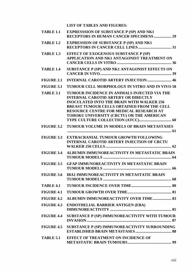
LIST OF TABLES AND FIGURES:
TABLE 1.1 EXPRESSSION OF SUBSTANCE P (SP) AND NK1
RECEPTORS IN HUMAN CANCER SPECIMENS .................. 29
TABLE 1.2 EXPRESSION OF SUBSTANCE P (SP) AND NK1
RECEPTORS IN CANCER CELL LINES .................................. 31
TABLE 1.3 EFFECT OF EXOGENOUS SUBSTANCE P (SP)
APPLICATION AND NK1 ANTAGONIST TREATMENT ON
CANCER CELLS IN VITRO ........................................................ 36
TABLE 1.4 SUBSTANCE P (SP) AND NK1 ANTAGONIST EFFECTS ON
CANCER IN VIVO ......................................................................... 39
FIGURE 2.1 INTERNAL CAROTID ARTERY INJECTION ......................... 46
FIGURE 3.1 TUMOUR CELL MORPHOLOGY IN VITRO AND IN VIVO 58
TABLE 3.1 TUMOUR INCIDENCE IN ANIMALS INJECTED VIA THE
INTERNAL CAROTID ARTERY OR DIRECTLY
INOCULATED INTO THE BRAIN WITH WALKER 256
BREAST TUMOUR CELLS OBTAINED FROM THE CELL
RESOURCE CENTRE FOR MEDICAL RESEARCH AT
TOHOKU UNIVERSITY (CRCTU) OR THE AMERICAN
TYPE CULTURE COLLECTION (ATCC) ................................. 60
FIGURE 3.2 TUMOUR VOLUME IN MODELS OF BRAIN METASTASES
........................................................................................................... 61
FIGURE 3.3 EXTRACRANIAL TUMOUR GROWTH FOLLOWING
INTERNAL CAROTID ARTERY INJECTION OF CRCTU
WALKER 256 CELLS .................................................................... 62
FIGURE 3.4 ALBUMIN IMMUNOREACTIVITY IN METASTATIC BRAIN
TUMOUR MODELS ...................................................................... 64
FIGURE 3.5 GFAP IMMUNOREACTIVITY IN METASTATIC BRAIN
TUMOUR MODELS ...................................................................... 66
FIGURE 3.6 IBA1 IMMUNOREACTIVITY IN METASTATIC BRAIN
TUMOUR MODELS ...................................................................... 68
TABLE 4.1 TUMOUR INCIDENCE OVER TIME ......................................... 80
FIGURE 4.1 TUMOUR GROWTH OVER TIME ............................................. 81
FIGURE 4.2 ALBUMIN IMMUNOREACTIVITY OVER TIME ................... 83
FIGURE 4.3 ENDOTHELIAL BARRIER ANTIGEN (EBA)
IMMUNOREACTIVITY ............................................................... 85
FIGURE 4.4 SUBSTANCE P (SP) IMMUNOREACTIVITY WITH TUMOUR
INVASION ....................................................................................... 87
FIGURE 4.5 SUBSTANCE P (SP) IMMUNOREACTIVITY SURROUNDING
ESTABLISHED BRAIN METASTASES ..................................... 88
TABLE 5.1 EFFECT OF TREATMENT ON INCIDENCE OF
METASTATIC BRAIN TUMOURS ............................................. 99

xiv
FIGURE 5.1 EFFECT OF TREATMENT ON TUMOUR VOLUME IN MM3
SHOWING NO SIGNIFICANT DIFFERENCE IN TUMOUR
VOLUME AMONG THE GROUPS ........................................... 100
FIGURE 5.2 ALBUMIN AND SUBSTANCE P (SP) IMMUNOREACTIVITY
......................................................................................................... 101
FIGURE 5.3 ANIMAL WEIGHT CHANGE FOLLOWING TUMOUR CELL
INOCULATION AND TREATMENT *P<0.05 ......................... 102
FIGURE 6.1 SUBSTANCE P (SP) IMMUNOREACTIVITY WITH
METASTATIC BRAIN TUMOUR GROWTH ......................... 114
FIGURE 6.2 BRAIN WATER CONTENT ....................................................... 115
FIGURE 6.3 EVANS BLUE EXTRAVASATION ........................................... 117
FIGURE 6.4 ALBUMIN IMMUNOREACTIVITY WITH TUMOUR
INOCULATION AND TREATMENT ....................................... 118
FIGURE 7.1 CELL VIABILITY ASSAY .......................................................... 132
FIGURE 7.2 NK1 RECEPTOR IMMUNOREACTIVITY ............................. 134
FIGURE 7.3 TUMOUR GROWTH ................................................................... 136
FIGURE 7.4 TUMOUR GROWTH CHARACTERISTICS ........................... 138
FIGURE 7.5 TUMOUR AND PERITUMORAL GLIAL REACTION ......... 140
FIGURE 7.6 BRAIN MICROENVIRONMENT REACTION TO TUMOUR
GROWTH ...................................................................................... 142

xv
Abstract
Secondary brain tumours occur when cancer cells enter the circulation from their
primary site and colonise the brain, previously shown to occur across the blood-brain
barrier (BBB). Substance P (SP), a neurogenic inflammatory mediator, acting
predominantly through NK1 receptors plays a role in opening the BBB and in the
formation of oedema following stroke and brain trauma. It is hypothesised that SP
may also promote the extravasation of tumour cells through the BBB, formation of
peritumoral oedema and progression of secondary brain tumours.
Walker 256 rat breast carcinoma cells obtained from the Centre for Medical Research,
Tohoku University had superior tumorigenic properties compared to cells from the
American Type Culture Collection, and were therefore subsequently used in two
albino Wistar rat models of tumorigenesis.
Firstly, internal carotid artery tumour cell injection was used to establish the effect of
tumour cell extravasation across the BBB on brain albumin, endothelial barrier
antigen (EBA) and SP immunoreactivity. I then determined if NK1 receptor
antagonists could prevent tumour cell extravasation, by evaluating tumour incidence
and volume.
Secondly, a stereotaxic direct inoculation model was used to investigate the effect of
NK1 receptor antagonists on brain tumour growth and peritumoral oedema, compared
with dexamethasone treatment. Evan’s blue extravasation and albumin
immunoreactivity were used to assess BBB permeability, and brain water content to
evaluate cerebral oedema. Tumour volume, Ki67 immunoreactivity, caspase-3
immunoreactivity and tumour cell density were used as measures of tumour growth.
Furthermore, cell viability and cell death assays determined if NK1 antagonists or
dexamethasone treatment cause alterations in tumour cell growth in vitro.
In the carotid model, SP and albumin immunoreactivity increased in the brain during
the extravasation of tumour cells, and in the peritumoral area of established tumours.
The invaded blood vessels lacked EBA immunoreactivity, indicating loss of BBB
properties. However, NK1 antagonists administered in the first three days following
tumour cell injection failed to reduce tumour incidence or volume, suggesting that

xvi
extravasation may be a multifactorial process, and that NK1 receptor antagonism
alone is not sufficient to prevent tumour extravasation and growth.
In the direct inoculation model, NK1 receptor antagonists did not reduce peritumoral
oedema or decrease tumour growth when used to treat established brain metastases. In
contrast, dexamethasone, the standard treatment for peritumoral oedema, caused a
reduction in brain water content and decreased tumour volume, but not tumour
growth. The decrease in tumour volume with dexamethasone reflects reduced fluid
content, as there was increased tumour cell density with no change in
immunoreactivity to Ki67 (marker for proliferation) or caspase-3 (marker for
apoptosis). Furthermore, in vitro studies showed no effect for dexamethasone on
tumour cell viability. These results suggest that peritumoral oedema is driven by
classical inflammation rather than neurogenic inflammation in the direct inoculation
model.
In conclusion, in these models of secondary brain tumours, SP does not appear to play
a role sufficient to promote NK1 receptor antagonism as an appropriate preventative
treatment for brain metastasis, as an anticancer agent, or as an alternative to
dexamethasone for the management of peritumoral oedema.

1
1 Introduction
Metastatic brain tumours are an end stage of disease for patients suffering from breast
cancer, whose incidence is steadily increasing. In part, this is due to longer survival
times on account of improved methods of treatment for the causative primary
tumours. However, metastatic brain tumours are particularly difficult to treat,
reflecting the need for more research into the mechanisms of cancer invasion of the
brain and the complications associated with tumour growth. A common complication
of brain metastases is peritumoral oedema, which occurs because of increased
permeability of the blood vessels that grow within the metastatic brain tumours
compared to the normal blood-brain barrier (BBB) vessels. Previous studies have
shown that to develop metastatic brain tumours, cancer cells arrest within the cerebral
vasculature and then form cytoplasmic protrusions which penetrate the BBB during
the extravasation process (Lorger 2010). The mechanisms that allow tumour cells to
penetrate the BBB and metastasise to the brain are unclear. Substance P (SP) is a
mediator of neurogenic inflammation and is known to increase the permeability of the
BBB. It is therefore proposed that SP is secreted by tumour cells, or released by BBB
endothelial cells and/or perivascular nerve terminals of primary sensory neurons
through an interaction with the tumour cells. This would result in an increase in the
permeability of the BBB, which may allow the tumour cells to extravasate into the
brain parenchyma. Furthermore, SP release has been implicated in cerebral oedema
associated with stroke and traumatic brain injury and thus may also be responsible, in
part, for the formation of oedema surrounding metastatic brain tumours.
1.1 Epidemiology of brain metastases
1.1.1 Incidence
Incidence data for secondary brain tumours varies greatly within the literature.
Approximately 20% of cancer patients have a secondary brain tumour found at
autopsy, whereas clinical studies generally report an incidence of about 10% (Posner
1978; Cifuentes 1979; Soffietti 2002; Gavrilovic 2005; Lin 2008a). Studies of autopsy
data generally report a much higher incidence because asymptomatic tumours are also
included in the data. The incidence of metastatic brain tumours has increased in recent
2
times (Dobec-Meic 2006; Pelletier 2008), presumably because of improved treatment
for the primary cancer leading to increased survival time of cancer patients, thus
leaving more time for the metastatic process to take place. Furthermore, superior
diagnostic techniques may have resulted in better diagnosis of secondary brain
tumours.
1.1.2 Organ of origin
In many cases of brain metastases, the organ of primary tumour origin cannot be
identified (Potts 1980; Clark 1989; Becher 2006). Where it has been possible, the
most common primary sites responsible for metastatic brain tumours are lung, breast,
renal, colorectal and skin (melanoma) (Schouten 2002; Barnholtz-Sloan 2004; Villa
2011). These cancer types are responsible for many cancer deaths and have short
predicted survival times comparable to that of glioblastoma multiforme, commonly
regarded as an incurable cancer type (Tran 2010). Approximately 25% of people with
lung cancer will develop a metastatic brain tumour detectable at autopsy, making lung
cancer patients responsible for 30-60% of all metastatic brain tumours (O'Neill 1994;
Sen 1998; Tabaka 2006). Between 22-30% of patients diagnosed with breast cancer
will also be diagnosed with a metastatic brain tumour (Schuette 2004; Hines 2008).
Melanoma is responsible for about 1% of the total cancer burden, but 75% of these
cases will result in a metastatic brain tumour (Salgado 2007).
The vast majority of breast cancer brain metastases occur in the late stages of the
disease (Lorger 2010; Kim 2012) and despite improved treatments for breast cancer,
the survival time for patients with brain metastatic disease remains in the order of
months (Sperduto 2012). Nearly 50% of patients with metastatic triple negative breast
cancer will develop a metastatic brain tumour, with a median survival time of 4.9
months from diagnosis (Lin 2008b). There is a predilection for metastatic brain
tumours and CNS recurrence amongst node positive, estrogen receptor-negative,
young patients with high grade breast cancer, with no evidence that there is any
benefit from an early diagnosis and treatment for their intracranial malignancy
(Pestalozzi 2006). Furthermore, Her-2 positivity has been linked with poor prognosis
3
in a cohort of breast cancer patients from 1996-2010 (Berghoff 2012a). Therefore
there is pressing need for improved treatment and prevention strategies for brain
metastatic breast cancer.
Cancer patients who develop cerebral secondary tumours have a much poorer
prognosis (Tsimberidou 2011). Brain metastases are a major cause of cancer
morbidity and mortality and are often the first site of relapse in patients with systemic
tumours (Atahan 2008). Brain metastatic disease progresses quickly, with one study
reporting that 41% of patients undergoing gamma knife surgery for secondary brain
tumours had additional lesions identified on the day of surgery (Patel 2012).
Approximately 80% of patients with secondary brain tumours will have multiple
intracranial lesions (Miabi 2011), substantially reducing survival time (Elaimy 2011).
1.2 Location of metastatic tumour
The most common location of metastatic brain tumour growth is in the frontal lobe,
followed by the cerebellum, parietal lobe, temporal lobe, occipital lobe, deep brain
nuclei and the brain stem (Potts 1980; Ghia 2007).
1.3 Clinical relevance
Metastatic brain tumours are an important area of research due to the large number of
people affected and the lack of effective treatment strategies available. It is also
pertinent that the mechanisms of tumour cell extravasation into the brain across the
BBB be elucidated because this understanding may lead to the development of a novel
prevention strategy.
Much of the current research into CNS malignancies is focused on primary tumours
of the brain, despite the fact that brain metastases are ten times more common (Landis
1998). In many studies involving cancer research, CNS neoplasms are excluded,

4
because the BBB is a complicating factor and novel treatments are often not tested on
brain metastases.
Previously, many cancer related deaths were as a result of either the primary
neoplasm or systemic metastases. The isolated nature of the brain means that it is
often not the preferred site of metastatic tumour formation for most tumour cell types
and is generally the end stage of disease. However, with increasingly effective
chemotherapeutic strategies, many patients are able to combat their primary
malignancies and systemic metastases. This has unmasked a population of patients
suffering and unfortunately dying from brain metastases due to inadequate treatment
options. Thus, brain metastases are becoming an increasing burden on our health
system and reducing the quality of life that can be expected for cancer sufferers.
1.4 Tumour associated morbidity
The presence of a metastatic tumour in the brain parenchyma can cause compression
of structures within the brain and peritumoral oedema. This can result in neurological
symptoms including motor weakness, aphasia, visual deficits and epilepsy (Shuto
2008). Peritumoral oedema will be considered in more detail later in this chapter after
the structure of the BBB has been discussed.
1.4.1 Seizures
Often seizures are the initial symptom that leads to the diagnosis of the causative
intracranial neoplasm (Shamji 2009). Furthermore, seizures often begin or return
when a brain tumour is surgically excised (Beaumont 2000). The pathogenesis of
brain metastases associated epilepsy is thought to be related to the tumour-associated
alterations to the brain environment including oedema formation, insufficient blood
supply, and release of inflammatory mediators and metabolically active substances
(Shamji 2009). Changes in the pH of the peritumoral environment and loss of BBB
integrity have both been linked to tumour associated epilepsy (Beaumont 2000).

5
When mannitol was used to compromise the function of the BBB in order to aid
delivery of chemotherapy to primary CNS lymphoma, this procedure caused focal
motor seizures in one quarter of the cases (Marchi 2007). In contrast, no seizures
occurred when the chemotherapeutic agent was administered alone.
1.5 Current treatments for metastatic brain tumours
Functional impairment resulting from brain metastases is complex, as there are many
factors that may contribute to morbidity. The tumour itself may cause neurological
deficits, there may be systemic cancer symptoms as a result of the primary tumour and
the prescribed treatments may impair the patient whilst combating the tumour.
The indications for different treatment options for metastatic brain tumours are
complex. The most common first line treatment for metastatic brain tumours is
surgery and chemotherapy, whilst whole brain radiotherapy and chemotherapy are
more regularly used as second line treatment (Fabi 2011). Generally, the best results
are seen when radiosurgery is combined with whole-brain radiotherapy (Rades 2012).
Surgical intervention is most effective when there is only a single metastatic brain
tumour, although the tumour location dictates wheather tumour excision is possible.
Furthermore, more than 80% of surgically removed malignancies recur at the
boundary of the excision (Veiseh 2007).
1.5.1 Chemotherapy
Treatment for the primary tumours that commonly spread to the brain has improved in
recent times with the development of targeted chemotherapeutic agents. This may be
the reason why the time from cancer diagnosis to brain metastases development has
increased over time, alternatively primary tumours may be being diagnosed earlier
(Nieder 2009). However there has not been any significant increase in survival time
post-metastatic brain tumour diagnosis since 1983 (Nieder 2009), suggesting that the

6
improved treatment options for systemic cancer are not effective in treating the
secondary brain tumours that result from these malignancies.
Chemotherapeutic agents have variable ability in crossing the BBB and reaching
therapeutic concentrations within metastatic brain tumours (Lien 1991). Some
lipophilic agents are able to cross the BBB in order to treat secondary brain tumours
although often these agents are not best suited to target the type of tumour that has
invaded the brain. Although brain metastases disrupt the BBB, increasing its
permeability, this may not be sufficient to transfer targeted chemotherapeutic agents
into the brain. Treatment of micrometastases in the brain with chemotherapy may be
inadequate. Larger metastatic brain tumours are less protected by the BBB, as tumour
angiogenesis disrupts the BBB in the tumour area (Zhang 1992). However, interstitial
pressure within the tumour mass may preclude the entry of chemotherapeutic agents.
In a human study, 66% of brain metastatic lung tumours showed significantly
decreased uptake of a labelled chemotherapeutic agent when compared to the primary
lung tumour (Front 1987).
Chemically induced increased BBB permeability, for improved delivery of
chemotherapeutic agents is a popular area of investigation. The administration of
hyperosmolar solutions cause dehydration of cerebral endothelial cells and
cytoskeleton contraction resulting in cell shrinkage and increased TJ spacing between
the cells; this can be used to increase the blood to tissue transfer in the brain (Hiesiger
1986; Kozler 2003). Furthermore, a temporary nitric oxide (NO) donor has also been
used in a rat glioma model to compromise the BBB for more effective chemotherapy
delivery (Weyerbrock 2003). However, the disruption of the BBB for chemotherapy
delivery may cause increased toxicity in the healthy brain tissue. Therefore, agents
that selectively increase the permeability of blood vessels within the tumour mass are
currently under investigation. Bradykinin B2 receptor agonists are showing promise
in this area, with hydrophilic agent uptake into rat implanted glioma increased after
B2 agonist treatment (Cote 2010). The increased density of B2 receptors on tumour
vasculature is thought to be the mechanism behind this drug selectivity (Wu 2002).
7
Tumoral blood flow may also influence the effectiveness of chemotherapeutic agents
through delivery to the target tissue. In a study where experimental Walker 256 breast
carcinoma metastatic brain tumour blood flow was measured, the range varied
markedly and was correlated with tumour size and morphological features including
necrosis and cyst formation (Blasberg 1984b).
1.5.2 Radiation therapy
Radiation therapy is a foundation treatment for both primary and secondary brain
tumours. Indeed, patients who suffer from small cell lung carcinoma (SCLC) and are
thought to be in complete remission, are often treated with cranial irradiation because
of the high rate of metastatic brain tumour recurrence (Tarnawski 2011). However,
acute cranial irradiation causes a breakdown of the BBB through the induction of
proteolytic enzymes disrupting the basal lamina of cerebral blood vessels, such as
matrix metalloproteinases (MMP) and plasminogen activators (Adair 1999).
Similarly, in one study two of three patients suffering from brain metastatic lesions
with breast cancer origins showed an increase in oedema in a scan following radiation
therapy (Hyman 1978). This was in contrast to the overall 53% of patients with brain
metastases that showed improvement following cranial irradiation (Hyman 1978).
1.5.3 Dexamethasone
Dexamethasone is currently used clinically to treat peritumoral oedema, with
significant improvements to patient outcomes since it began being used in 1962
(Jelsma 1967). It is recommended that patients suffering from metastatic brain
tumours, with symptoms related to the mass effect of the neoplasm, be treated with 4-
16mg/day of dexamethasone depending on the severity of symptoms and not the size
of the tumour (Ryken 2010). 75% of patients report neurological benefits within 48-
72 hours after treatment initiation (Soffietti 2006). Approximately 70% of metastatic
brain tumour patients receive dexamethasone treatment concomitantly with
radiotherapy (Hempen 2002). Limitations of dexamethasone use include many
harmful side-effects including insomnia, immune suppression (Lesniak 2004),
hyperglycemia (McGirt 2008) and occasional psychosis (Alpert 1986).
8
In humans, several magnetic resonance imaging (MRI) studies have shown that
dexamethasone reduced peritumoral oedema resulting from primary glial tumours
(Armitage 2007) and metastatic brain tumours, but not meningiomas (Andersen
1994a; b). However a variable response to dexamethasone treatment has been
reported between different brain metastases with the same organ of origin, meaning
that tumours of the same type may have an inconsistent oedema response (Wolfson
1994).
The exact mechanism of dexamethasone action to reduce cerebral oedema remains
controversial, although it is thought to be through suppression of classical
inflammation and actions on glucocorticoid receptors, resulting in reduction of brain
microvessel permeability (Andersen 1994a; Heiss 1996; Andersen 1998; Sinha 2004).
Classical inflammation is characterised by accumulation and proliferation of
microglia along with perivascular macrophages (Graeber 2011). Animal models of
peritumoral oedema have been used in an attempt to elucidate the precise molecular
actions of dexamethasone on BBB permeability. Models used are predominantly
intracerebral implantation of rat C6 or 9L glioma cells, with dexamethasone treatment
causing a decrease in Evans blue or radiolabelled serum albumin extravasation into
the neuropil (Nakagawa 1987; Guerin 1992; Gu 2009a; Gu 2009b) and decreased
MRI measures of oedema (Nakagawa 1987; Ewing 2008).
Dexamethasone induced decrease in oedema and BBB permeability was also
associated with decreased vascular endothelial growth factor (VEGF) (Heiss 1996),
elevated GLUT1 (Guerin 1992), increased occludin and increased calcium activated
potassium channel expression (Gu 2009a) by cerebral vasculature. These studies
indicate possible pathways by which dexamethasone counteracts tumour-initiated
increased BBB permeability. Furthermore, actions of dexamethasone on cerebral
vasculature include decreased plasma vascular volume and decreased total vascular
density (Nakagawa 1987; Badruddoja 2003). However conflicting results exist for the
density and volume of brain microvessels in response to dexamethasone treatment,
although different methods of detection were used for these experiments (Wolff 1993;

9
Badruddoja 2003). These results suggest a possible angiogenic modulation effect of
dexamethasone along with its established effects on BBB permeability and oedema.
Despite the positive effects of dexamethasone on peritumoral oedema, some debate
exists about its use in conjunction with chemotherapeutic agents. In vitro studies have
shown inhibition of TRAIL, taxol, doxorubicine, gemcitabinetemozolomide, cisplatin
and 5-fluorouracil chemotherapy induced tumour cell apoptosis with dexamethasone
treatment (Kim 2004; Sur 2005; Zhang 2006). These experiments were fairly
consistent showing the protective effect of dexamethasone on chemotherapy induced
bone (MG63, HOS, SAOS, HT1080), brain (HS683, H4, A172, U-373-MG, TE671,
T98G), breast (BT-474, BT-20, Colo-824, MCF7, MDAMB-436), cervix (P5,
CASKI, MRH-215, MRH-186, MRH-196), melanoma (HS695T, WM1341,
WM98.1), lung (A549) and neuroblastoma (IMR32, KELLY, SKN-SH, SHEP,
IMR5) cancer cell line apoptosis, although the opposite effect was seen in primary
lymphoid cells (Kim 2004; Sur 2005; Zhang 2006). Dexamethasone treatment
enhances chemotherapy induced T cell leukaemia cell apoptosis (Zhang 2006).
However, interaction of dexamethasone with chemotherapeutic agents in vitro is a
phenomenon that has not been confirmed in animal models of brain tumours.
Dexamethasone is well known to cause significant toxicity to human patients and to
animals used in oedema studies, as evidenced by animal weight loss and necrosis at
the site of administration (Villeneuve 2008; Moroz 2011). When administered alone,
several studies have found that dexamethasone has an anti-proliferative, pro-apoptotic
effect on cancer cells in vitro (Bavaresco 2007; Piette 2009; Tazik 2009).
Furthermore, dexamethasone caused a decrease in tumour volume in numerous animal
studies (Guerin 1992; Wolff 1993; Badruddoja 2003; Villeneuve 2008), although this
is likelydue to reduced oedematous fluid within the tumour mass. Furthermore,
dexamethasone treatment has not extended animal survival with cerebral tumour
implantation (Moroz 2011).
10
1.6 Blood brain barrier
The BBB is the interface between the blood and the brain, separating the brain
parenchyma from the blood within cerebral capillaries and involves the interactions
between endothelial cells, astrocytes, pericytes and the capillary basement membrane.
Often cells, but not solutes, are able to move through a supposedly non-permeable and
undamaged BBB (Perry 1997). An example of this is when no leakage of Evan’s blue
was evident surrounding capillaries within a metastatic brain tumour, indicating an
intact BBB, although macrophages were able to infiltrate the brain parenchyma
(Schackert 1988b).
1.6.1 Cerebral capillary endothelial cells
Cerebral capillary endothelial cells are simple squamous cells of uniform thickness,
with few pinocytotic vesicles (Schackert 1988b). These cells rest on the basal lamina
and lack fenestrations (Freed 2002; Petty 2002). There is an increase in the number of
mitochondria in the endothelial cells of the BBB when compared to the capillary
endothelial cells found systemically, as there is increased demand for active transport
across those in the brain. This is because cerebral capillary endothelial cells have tight
junctions (TJ) between them, which prevent many substances from entering the brain
parenchyma via the intercellular space. These blood vessels enter the brain
parenchyma through the peri-vascular spaces that are lined by the pia mater. Thus
there is also a pia-glial membrane present between the brain parenchyma and the
endothelial cells of the BBB (Saito 2008).
1.6.2 Tight junctions
TJ are located on the most apical region of the cleft between cerebral capillary
endothelial cells and form a seal to prevent substances from passing between them
(Petty 2002). Claudins are involved in the primary makeup or backbone of TJs,
forming dimers which interact with opposing claudin molecules and form the primary
seal of the TJ (Petty 2002; Persidsky 2006). In the brain, claudin 5 is predominant
(Petty 2002). Junctional adhesion molecule (JAM) is an immunoglobulin with a
11
single transmembrane segment, which mediates the initial cell to cell attachment and
is able to mediate permeability through this avenue (Persidsky 2006). Occludin has
four transmembrane segments and is present in higher concentractions in endothelial
cells of the BBB than in those in systemic capillary endothelial cells. It induces high
membrane resistance, which is indicative of low ion permeability (Joo 1996;
Persidsky 2006). Occludin interacts with the cytoskeleton of BBB endothelial cells
through zona occludens-1 (ZO1), ZO2 and ZO3 molecules, which form the sub-
membranous plaque of the TJ (Petty 2002; Persidsky 2006).
1.6.3 Basement membrane
The basement membrane of the BBB provides another obstacle to prevent the entry of
unwanted substances into the brain. It is made up of proteins found within the
extracellular matrix including collagens, vitronectin, fibronectin, tenascin and
proteoglycans (Baumann 2009). These components provide stability to the structure
of the blood vessels and a surface upon which cerebral capillary endothelial cells can
rest.
1.6.4 Astrocytes
Astrocyte end feet surround 99% of BBB endothelial cells and act to support and
enhance the TJs between them (Persidsky 2006; Bundgaard 2008). Gap junctions
between astrocytes allows for quick transfer of substances and information (Escartin
2008). Astrocytes also mediate the connection between neurones and endothelial cells
(Kim 2006). Astrocytes become activated in response to pathological stimuli which
results in the hypertrophy of the astrocytic processes and over-expression of
intermediate filaments, namely glial fibrillary acidic protein (GFAP) (Escartin 2008).
1.6.5 Pericytes
Pericytes have a stellate appearance and cytoplasmic processes. They cover 22-32%
of the capillary cell surfaces (Fisher 2009). Gap Junctions (GJ) between pericytes and
12
cerebral capillary endothelial cells allow communication to occur (Persidsky 2006).
The main function of pericytes seems to be in blood flow regulation particularly in the
pre-capillary arterioles that supply the brain with blood (Joo 1993). The structure of
pericytes makes them ideal for this function, as they are contractile and express the
smooth muscle actin isoform (Fisher 2009). Collagen type IV glycosaminoglycans
and laminin are also synthesised in pericytes to be used in formation of the basement
membrane (Fisher 2009). They have the ability to regulate endothelial cell
proliferation, survival, migration and differentiation (Persidsky 2006).
1.7 Formation of brain metastases
In order for metastatic tumour cells to reach the brain through the blood stream, they
must first have the ability to leave the site of the primary systemic tumour. By
migrating through the basement membrane of vessels in the primary tumour, some
cells are able to reach the systemic circulation. This process is called intravasation and
will involve the tumour cells either passing between or through the endothelial cells
of the blood vessels. The tumour cells must be able to resist programmed cell death,
due to the loss of cellular contact with the primary tumour, in order to form viable
metastases (Khanna 2005). Similarly, the host immune system must be evaded.
Mechanical forces associated with narrowing blood vessels allow tumour cell
attachment to endothelial cells at a distant site (Marchetti 2003). Firstly, weak
carbohydrate-carbohydrate locking reactions allow tumour cells to interact with
endothelial cells of the microcirculation of the brain (Petty 2002). Following this, the
bond between tumour cells and endothelial cells of the BBB are strengthened with the
interaction of adhesion molecules (Petty 2002).
The metastatic pathway continues with extravasation, where the tumour cells must
pass through the BBB (Marchetti 2003). This means that the tumour cells must pass
through both the endothelial barrier and the basement membrane (Petty 2002). The
mechanism for this is yet to be elucidated.
13
Many tumour cells that are able to break away from the primary tumour will not
survive in the circulation to form metastatic tumours at distant sites. For example, in a
study using radiolabeled B16 melanoma cells, less than 1% of the tumour cells
survived the first 24 hours in the circulation and less than 0.1% of the cells went on to
form metastatic tumours (Fidler 1970). In addition, when an in vivo MRI was
performed on a nude mouse that had human breast cancer cells injected into its left
ventricle, 81% of the tumour cells evident at day 0 were no longer detectable at day 3
(Heyn 2006).
Tumour cells have variable propensity for metastases to the brain, based on the
properties of the tumour cell that aid its progression through the metastatic process. In
a study where eight different melanoma tumour cell lines were injected both
intracerebrally and into the carotid artery of nude mice, all eight cell lines were able to
grow tumours in the brain as a result of their intracerebral inoculation (Schackert
1990). However, only seven of the cell lines resulted in any metastatic melanomas in
the brain through intra-carotid delivery (Schackert 1990). The TXM-31-3 cell line that
was derived from a lymph node metastatic melanoma in a human patient did not enter
the brain after intra-carotid inoculation, whereas other cell lines derived from brain
metastatic melanoma tumours had already acquired the characteristics needed to pass
through the BBB into the brain parenchyma (Schackert 1990). It has been shown that
cancer cells round up inside the cerebral capillaries before cytoplasmic processes
extend through the BBB (Lorger 2010).
Specific qualities are required of tumour cells in order to metastasise to the brain. It
has been established that melanoma cells derived from central nervous system
metastases are more likely to form experimental metastases in the brain, than tumour
cells derived from other areas of the body (Schackert 1988a; Fidler 1990; Kusters
2001). For example, tumour cell lines that were derived from human CNS metastatic
melanoma tumours (cell lines TXM-13 and TXM-40) were more likely to form brain
parenchyma lesions when injected into the carotid artery of nude mice, whereas

14
tumour cells that were derived from human melanoma lymph node metastases (cell
lines A375-SM, DM-4, TXM-1 and TXM-31-4) and subcutaneous metastases (DX-3)
preferentially formed meningeal or ventricle metastases when inoculated in the same
way (Schackert 1990).
1.7.1 Immune system interactions
Microglia are the macrophage like cells of the CNS (He 2006). The population of
microglia in the brain is maintained by local replication, although some infiltration of
macrophages originating from outside the CNS is evident when the brain is injured
(Perry 1997). When microglia are activated, usually by neuronal degeneration or
decreased interactions with neuronal terminals, they become scavenger cells that
repair neurons and promote regeneration through alerting the wider immune system
through signalling cascades (Kreutzberg 1996). Surrounding metastatic brain tumours,
microglia have been found to be enlarged with thicker processes, which are thought to
be involved in the activation of astrocytes (Zhang 1995). The activated microglia also
proliferate, forming a barrier like pattern surrounding brain metastatic lung cancer (He
2006). Some, but not all of these microglia showed expression of inducible nitric
oxide synthase (iNOS) and Tumour necrosis factor alpha (TNF-α) (He 2006).
Injection of SP into the brainstem induces microglial activation as indicated by an
increase in the expression of class II MHC proteins (McCluskey 2001).
Leukocytes may migrate into the brain from the blood through the choroid plexus and
into the CSF, as choroidal endothelium is fenestrated with no tight junctions between
the cells (Ransohoff 2003). There are approximately 3,000 leukocytes present in 1ml
of CSF from a normal healthy individual (Ransohoff 2003). Leukocytes may also
migrate through the vasculature into the subarachnoid or Virchow-Robin spaces,
although it is thought that during pathological events leukocytes travel directly
through the BBB and into the perivascular spaces, although the mechanism for this is
unclear (Ransohoff 2003). It is possible that inflammatory mediators increase the
permeability of the BBB to allow leukocyte migration into the brain.

15
Metastatic brain tumours may also be infiltrated by macrophages that are of blood
monocyte origin, even with an intact BBB (Schackert 1988b). This has been shown to
occur even in the absence of a major T-lymphocyte response (Schackert 1988b). One
study showed that all six metastatic brain tumours from various primary tumours were
positive for macrophage infiltration using anti-Leu-M3 monoclonal antibody, in the
brain tumour itself and in the surrounding oedematous tissue (Shinonaga 1988).
Activated T-lymphocytes have also been known to infiltrate the brain across an intact
BBB in the absence of inflammation as part of immune surveillance (Perry 1997).
Increased number of regulatory T cells (CD4+CD25+) in glioma infiltrates, are
thought to play an important role in suppressing the immune response of CD8+ T-cell
infiltration of tumour cells expressing self antigens in the CNS (El Andaloussi 2006;
Grauer 2007). This was shown when animals with glioma cells implanted in their
brains had increased survival times when regulatory T cells were depleted (El
Andaloussi 2006).
Tumour xenografts implanted into the brains of non-immunologically compromised
rodents show a delayed rejection when compared to those same tumours implanted
into the skin. This may be because in contrast to the skin, the brain responds to
noxious stimuli with microglial activation and then delayed monocyte infiltration and
no neutrophil response (Ransohoff 2003). Biphasic tumour growth at the primary site
for the 4T1 mammary tumour, which occurred at week three to four in animals with a
normally functioning immune system, did not occur in immune compromised mice
(Tao 2008). Therefore it is likely that the immune system was responsible for this
tumour regression, which was associated with tumour necrosis and leukocyte
infiltration (Tao 2008).

16
TNF-α and interferon gamma (IFN-γ) are cytokines that are released by immune cells
and have the ability to destroy tumour cells directly (Lampson 2003). However,
toxicity can become a problem if doses sufficient to kill tumour cells are
administered. Lower doses are able to be used to initiate an indirect pathway of
tumour cell death through the immune enhancing effects of these cytokines (Lampson
2003). Mice that were immunised with peptide pulsed dendritic cells and systemic
application of interleukin 2 (IL-2) caused a significant reduction in the size of
metastatic brain tumours and increased the survival time of the animals (Prins 2008).
The immune system plays an important role in fighting metastatic brain tumours, but
the location of the tumour within the brain may dictate how effective this system of
defence is. For example, when tumour cells were implanted directly into the cerebral
ventricles, the survival time was increased when compared to the same volume of
cells injected into the brain parenchyma (Thomas 2008). Similarly, when a larger
volume of cells was infused into the same intra-parenchymal site, the cells leaked into
the ventricles, and thus caused an immune reaction similar to that seen for the direct
implantation of tumour cells into the ventricles (Thomas 2008). This also resulted in
an increase in animal survival time when compared to the smaller intra-parenchymal
inoculation (Thomas 2008). Activated T-cells will cross the BBB and accumulate
within an intra-parenchymal tumour mass, although the environment in the brain
parenchyma is more hostile to inflammatory cells than that in the ventricles (Thomas
2008).
1.8 Experimental models of brain metastases
1.8.1 Tumour injection into the internal carotid artery
Injection of tumour cells into the internal carotid artery accurately simulates
haematogenous spread of metastatic tumour cells to the brain, allowing the
extravasation process to be investigated. Injection of tumour cells into the internal
carotid artery of the animal is very effective in producing brain metastases, as the
brain contains the first capillary bed that the tumour cells encounter following their
injection (Khanna 2005). In addition, when tumour cells are injected into the right
17
internal carotid artery, metastases preferentially form in the right hemisphere of the
brain (Zhang 1992). The pattern of tumour growth as a result of carotid artery
injection has been shown to include both angiocentric collars of neoplastic cells where
the pia-glial membrane was intact, along with invasive proliferations of neoplastic
cells with fragments of collagen fibers from the breakdown of the pia-glial membrane
(Saito 2008). When tumour cells are injected into the internal carotid artery, only the
last steps of the metastatic process are able to be examined (Fidler 1990). However,
this model is suitable for investigation of tumour cell extravasation.
1.8.2 Syngeneic model
Syngeneic brain metastases models, where the animals have the same genetic
background as the tumour cell line used, allow the normal interactions between the
animal and the tumour cell of the metastatic pathway to take place (Khanna 2005).
However, the cells used in these models may lack the complexity seen in human
tumour cells, both genetically and in the formation of mutations. This is because
tumour cells used in syngeneic models are usually derived from inbred mice or rats
(Rangarajan 2004; Khanna 2005). An example of a syngeneic murine model of brain
metastases is the Walker 256 carcinoma cells, which have previously been injected
into the internal carotid artery of Wistar rats (Hasegawa 1979).
1.8.3 Walker 256 breast carcinoma
The Walker 256 carcinoma cell line is a rat tumour that originally occurred
spontaneously in the mammary gland of a pregnant albino rat and grew with the
morphology of a carcinosarcoma (Buffon 2007). The Walker 256 cell line has been
used previously to establish experimental brain metastases through an internal carotid
artery injection and direct implantation into the cerebral cortex (Hasegawa 1979;
Hasegawa 1983; Blasberg 1984a; b; Blasberg 1986; Hiesiger 1986; Jamshidi 1992).
18
Walker 256 cells are highly immunogenic (Ferreira 2004), with a regression in tumour
growth seen following oxidative burst, involving the release of reactive oxygen
species and hydrolytic enzymes, from granulocytes that infiltrate tumours implanted
into the hind leg of rats (Jaganjac 2008). The infiltration of an implanted Walker 256
tumour with granulocytes occurred in just five hours (Jaganjac 2008). The Walker 256
carcinoma is also sensitive to cisplatin and chlorambucil (Simpkins 1991). An
angiogenic substance has been purified from the Walker 256 carcinoma (Fenselau
1981). This substance causes proliferation of fetal bovine aortic endothelial cells in
vitro and similarly acts to stimulate blood vessel growth when applied in vivo
(Fenselau 1981). It is possible for this angiogenic factor to be detected in the serum of
a Walker 256 bearing animal and therefore to determine if animals are in a
neurovascular diseased state through detection of this factor (Fenselau 1981).
Walker 256 tumours have the ability to cause changes in oxidative metabolism in
tumour-free tissues (Freitas 2001). For instance, when a Walker 256 tumour cell
suspension was injected into the right flank of male Wistar rats, there was evidence of
oxidative stress in the brain tissue (Freitas 2001). This is possible because of an
increase in the production of compounds that favour oxidation and decrease the
activities of antioxidants, which ordinarily would combat oxidative stress (Freitas
2001). The possible results of this oxidative stress placed on the brain in Walker 256
tumour bearing rats is that the hypothalamic responses to starving are impaired
through the increase in compounds such as vasopressin, IL-1, prostaglandin E2 and
TNF-α. This may be the mechanism by which cachexia is induced in Walker 256
bearing rats, which is one of the characteristic symptoms of this tumour cell type.
When 107 Walker 256 cells were injected sub-cutaneously into the left flank of male
albino wistar rats, the first apparent signs of cachexia were seen 7 days after tumour
cell inoculation (Ferreira 2004). Furthermore, these animals died as a result 15-16
days post tumour cell inoculation (Ferreira 2004). In contrast to this, when 106 cells
were injected into the right common carotid artery of female Wistar rats, the cancer
related weight loss began at approximately day 28 post inoculation (Hasegawa 1979).

19
1.9 Features of metastatic brain tumours
1.9.1 Tumour morphology
Metastatic brain tumours vary greatly in morphology, as a result of their wide range of
cell types associated with their organs of origin. Their features can range from yellow
and soft to abundantly necrotic, with dark coloured tumours metastasising to the brain
from melanomas (Potts 1980; Subramanian 2002).
1.9.2 Tumour border
Many CNS metastases invade the brain parenchyma via tumour expansion,
compressing the surrounding structures, rather than tumour cells infiltrating the
surrounding tissues as is the case for primary brain tumours like gliomas (Hasegawa
1983). However, there is variation in method of expansion between different organs
of origin, for example SCLC brain metastases have a less defined border than seen in
other metastatic brain tumours, although this type does not grow particularly quickly
(Tabaka 2006).
1.9.3 Blood-tumour barrier
The capillaries within metastatic brain tumours are characteristic of the organ of the
primary tumour (Cornford 1992; Shuto 2008), as indicated by the expression of
aquaporin 1 (AQP-1). AQP-1 is not normally present in brain parenchyma, but is
expressed in the endothelial cells of the blood vessels within many metastatic brain
tumours (Papadopoulos 2004). Endothelial cells are also able to be recruited from the
brain tissue surrounding the tumour due to the highly vascular nature of the brain
(Strugar 1994). This process is known as vessel co-option, where the cerebral
capillary endothelial cells may de-differentiate in response to lack of interaction with
astrocytes and other signalling from the tumour cells themselves (Leenders 2003;
Papadopoulos 2004). Therefore the co-opted vessels do not retain the properties of the
20
BBB and are often dilated, irregular and permeable to proteins normally found in the
blood plasma (Leenders 2003).
If a metastatic brain tumour exceeds approximately 2mm in diameter, then
angiogenesis must occur to ensure adequate blood supply (Blasberg 1984a; Fidler
1990; Zhang 1992). These newly formed blood vessels make up the blood tumour
barrier, which may have variable barrier properties (Front 1984). Some common
features of the blood tumour barrier include short, elongated or open endothelial
junctions, a non-continuous basement membrane of varying width that has lost its 3
layered appearance and fenestrated membranes that lead to pores involving a fused
basal and luminal plasma membrane when endothelial cells become thinner (Hirano
1975; Shibata 1989; Strugar 1994). Furthermore, there are increased pinocytotic
vesicles and vacuoles when compared to the brain endothelial cells, along with
increased perivascular space between the basement membrane and the tumour cells
(Hirano 1975; Shibata 1989). Interestingly, one study showed that pertechnetate
uptake was correlated to the absence of TJ, but not to the extent of endothelium
fenestration (Bar-Sella 1979). Brain metastases from five bronchiogenic carcinomas
studied showed significantly elevated extravasation of tracer and capillary transit
times when compared to normal brain tissue (Koh 2006).
The structural integrity of blood vessels surrounding metastatic brain tumours is
controversial. Only the blood vessels within a metastatic brain tumour mass are
angiogenic and thus distinct from the normal BBB. However many believe that the
mass effect of tumour growth and associated oedema may result in disruption of the
BBB in the brain parenchyma surrounding metastatic brain tumours. Ultrastructural
studies report that the features of the blood-tumour barrier are not shared by their
counterparts outside of the tumour mass in oedematous tissue (Long 1979). Similarly
the pathological changes in GLUT1 expression on blood vessels within metastatic
brain tumours are not evident in the peritumoral environment (Zhang 1996b).
However, the peritumoral area of some metastatic adenocarcinomas have been shown
to be devoid of the tight junctional protein occluden, meaning that tight junctions both

21
outside and inside the metastatic brain tumour mass are disrupted (Papadopoulos
2001) . Therefore further investigaton is reqired to determine the extent to which the
BBB is disrupted in the peritumoral area.
1.9.4 Angiogenesis
Malignant brain tumours cause more angiogenesis than systemic malignancies (Ito
1993). Vasoactive substances secreted by metastatic brain tumours may induce
angiogenesis. Tumour cell expression of VEGF is directly correlated with rate of
angiogenesis (Yano 2000). Furthermore, there is evidence to suggest that SP may be
involved with metastatic brain tumour associated angiogenesis. When brain
metastases from malignant melanomas, lung carcinomas and breast carcinomas were
compared, the mean number of blood vessels in brain metastatic breast carcinomas
was significantly elevated when compared to the CNS metastases of melanomas
(Maniotis 1999).
Angiogenesis is not the only way in which metastatic brain tumours supply
themselves with sufficient blood to be able to grow. Light microscopy has revealed
that 45% of the brain metastases from malignant melanomas showed evidence of
hollow channels containing blood that had a basement membrane and were not lined
by endothelial cells, but tumour cells instead, without signs of necrosis or
inflammation (Maniotis 1999; Zhang 2007). This phenomenon of tumours forming
their own blood supply without the need for angiogenesis is known as vascular
mimicry. This process is not unique to melanoma tumours and has been identified in
many other tumour types including inflammatory breast cancer and astrocytomas
(Shirakawa 2002; Yue 2005). In one study the presence of vascular mimicry in
primary invasive ductal carcinomas was linked to a more invasive and metastatic
phenotype (Shirakawa 2001). In an oxygen-deprived environment, B16 melanoma
cells showed increased incidence of vascular mimicry and expression of MMP-2,
MMP-9 and VEGF when compared to the same tumour in non-ischemic limbs (Sun
2007). However, it seems that vascular mimicry is more prevalent in the early stages
of tumour growth, progressing to an intermediate stage where a combination of
22
tumour and endothelial lining is present lining the vessels within the tumour, called
mosaic blood vessels (Zhang 2007).
1.9.5 Peri-tumoral environment
In the area around metastatic brain tumours there is often white matter tract
disruptions indicated by β-amyloid precursor protein accumulation (Zhang 1996a),
neuronal degradation and cytoskeleton breakdown (Toh 2007). The reasons for this
could include cytotoxic products secreted by the metastatic brain tumour or
disturbances in the microcirculation (Toh 2007). When compared to meningiomas,
brain metastases were found to contain more extracellular water and white matter tract
disorganisation in the peri-tumoral environment (Toh 2007).
A rim of reactive glial cells is often evident surrounding metastatic brain tumours, one
function of which may be to act as a barrier to the flow of oedematous fluid (Zhang
1997). There is often an increase in reactive astrocytes in the peri-tumoral area, which
may proliferate and become activated in response to contact with serum proteins, such
as albumin, present in oedematous fluid that accumulates around the tumour
(Jamshidi 1992). The reactive astrocytes surrounding metastatic brain tumour samples
from 73 autopsy cases that originated from many different organs showed endothelin
expression, which may be partially responsible for peri-tumoral gliosis (Zhang 1995).
1.10 Peritumoral oedema
Oedema in the brain is most commonly vasogenic in nature and results from a
compromised BBB (Zhang 1997). Vasogenic oedema is characterised by the
abnormal increase in the volume of interstitial fluid and may be caused by increased
blood pressure in the capillaries, increased capillary permeability and decreased
plasma protein concentration within the capillaries (Zhang 1997). Due to oncotic
forces, plasma protein leakage into the tissue is followed by fluid, which results in
oedema. It is thought that this inflammatory vasogenic oedema may provide a
23
favourable environment for the proliferation of malignant cells (Van Den Brenk
1974a; Van Den Brenk 1974b; Zhang 1997). It is thought that the secretion of
substances that act on vascular permeability, like neurogenic inflammatory mediators,
may play a role in the development of oedema surrounding metastatic brain tumours
(Zhang 1997).
1.10.1 Tumour size
Tumour size and subsequent angiogenesis is thought to determine the extent of
peritumoral oedema formation through effects on overall vascular permeability.
Metastases larger than 0.2mm2 have the ability to decrease the integrity of the BBB as
indicated by the leakage of sodium fluorescein out of the cerebral blood vessels and
into the brain parenchyma (Zhang 1992). The leakage of sodium fluorescein was
directly correlated with the size of the tumour, but not with the anatomical location of
the tumour in the brain or the origin of the primary tumour (Zhang 1992).
Paradoxically, no leakage of sodium fluorescein was seen in another study using a
similar protocol, although the size of the tumours was not reported (Schackert 1988b).
Therefore it is possible that the experimental metastases in that study were too small
to disrupt the BBB. However, the capillaries within the tumours were enlarged and
irregular with regions of astrocytosis and oedema, although the BBB was intact
(Schackert 1988b). The plasma transfer constant, which takes into account the
capillary blood flow, surface area and permeability, for small tumours in the brain
were very similar to that of the normal brain tissue, but in the same study the value
was increased 5-20 fold for tumours larger than 0.6mm2 (Blasberg 1984a).
1.10.2 Complications of cerebral oedema
Approximately 80% of the brain is made up of water (Tait 2008). Inside the skull,
brain tissue is made up of approximately 1.1 L of water in the intracellular
compartment and 100ml in the interstitium. Furthermore, the intravascular contents
and CSF each make up approximately 100ml of the intracerebral volume
(Lukaszewicz 2011). However, when the BBB is disrupted, then fluid is able to move
from the intravascular compartment into the interstitium, thus increasing the volume

24
taken up by the brain tissue within the skull. There is limited capacity for blood or
cerebrospinal fluid (CSF) to counteract the chages in intracranial pressure (ICP). This
compensation is responsible for the initial plateau the ICP / volume curve, which
becomes exponential once compensatory mechanisms are exhausted (Smith 2008)
Raised ICP results in alterations to cerebral blood flow, because when the ICP is
raised, the cerebral perfusion pressure (CPP) is reduced. With a reduced CPP, the
brain may become deprived of oxygen, resulting in vasodilation of cerebral blood
vessels. This further exacerbates the problem as it results in further increase in ICP.
Complications of raised ICP can include compression of cerebral vasculature leading
to localized ischaemia (Ayata 2002). Since the brain is divided into discrete
compartments by membranous structures, local oedema may result in herniation of the
brain tissue from one compartment into the other (Zhang 1997).
1.10.3 Clearance of oedema
Oedematous fluid spreads via bulk flow, as particles of different diffusion coefficients
move at the same rate through the brain in the oedematous fluid (Reulen 1977; Ito
1988; Stummer 2007). Oedema may be absorbed into the CSF or astrocytes may
resorb proteins in order to reduce the oncotic pressure that drives fluid out of the
capillaries (Stummer 2007). Furthermore, oedema may be cleared through the
trancellular pathway, for example through AQP-4 water channels. AQP-4 is expressed
by activated astrocytes that surround metastatic brain tumours, which is postulated to
aid in the removal of peritumoral oedema (Saadoun 2002).
1.11 Substance P
Substance P (SP) is an excitatory tachykinin that is predominantly released from
primary sensory nerve endings in the central nervous system, but is also localized in
brain endothelial cells (Cioni 1998; Ribeiro-da-Silva 2000; Lazarczyk 2007). SP
preferentially binds to NK1 receptors, although there is some cross reactivity with the
other tachykinin receptors, NK2 and NK3 (Harrison 2001). The pre-protackykinin A
(PPT-A) gene encodes for SP (Bruno 2003; Chappa 2007). The transcription of this
gene is influenced by nerve growth factor, interleukin-11 (IL-11) and interleukin-6

25
(IL-6) and is increased in response to noxious chemical stimulation of tissue (Noguchi
1988; Bruno 2003). Throughout the body, SP acts to increase microvascular leakage
(Nimmo 2004).
When SP acts on NK1 receptors, which normally reside in the cell membrane of
primary sensory neurons, it is internalized (Hokfelt 2001). NK1 is a G-protein
coupled receptor (Lazarczyk 2007). SP inserts itself in the hydrophobic ligand
binding pocket located between the transmembrane domain of the NK1 receptor and
the extracellular surface (Harrison 2001).
1.11.1 Immunoreactivity in the brain
SP is the most abundant neuropeptide in the central nervous system (Black 2002).
Human CSF is positive for SP and therefore SP can come into contact with the brain
tissue surface (Zubrzycka 2002).
The areas of the brain where SP is present include the rhinencephalon, telencephalon,
basal ganglia, hippocampus, amygdala, septal areas, diencephalon, hypothalamus,
mesencephalon, pons, myelencephalon and spinal cord (Harrison 2001). NK1
receptors are expressed in the highest concentration in the caudate, putamen and
superior colliculus, which are areas of the brain that modulate behaviour and affect
stress responses (Harrison 2001; Black 2002). In the rat brain, SP is predominantly
expressed in the olfactory tubercle, amygdala, nucleus caudatus, septum,
hypothalamus, habenula, posterior pituitary, thalamic nuclei, globus pallidus,
substantia nigra, peri-aqueductal central gray, locus coeruleus, nuclei parabranchiales,
medulla oblongata, dorsal and ventral horns of the spinal cord (Severini 2002).
26
1.11.2 Substance P effects on the blood-brain barrier
Virtually all blood vessels are surrounded by primary sensory nerve fibres that secrete
SP and the cerebral blood vessels are particularly well innervated. In brain endothelial
cells the normal resting level of free Ca2+ is 100nM (Revest 1991). SP causes
calcium responses in the endothelial cells of the BBB of approximately 1000nM and
hence increased Ca2+ levels lead to increased BBB permeability through cell
contraction (Revest 1991; Paemeleire 1999). In conjunction with this, SP treatment of
cerebral capillary endothelial cells co-cultured with astrocytes, has been shown to
decrease the concentration of ZO-1 and claudin-5 tight junctional proteins, resulting
in increased permeability of the simulated BBB (Lu 2008).
SP is present in cerebral capillary endothelial cells and its secretion by these cells can
be increased through treatment with high doses of cytokines, including IL-1β and
TNFα (Cioni 1998; Annunziata 2002). This increase in SP released from brain
endothelial cells was found to be associated with an increase in the expression of b-
preprotachykinin mRNA, a precursor for SP, inside the cells (Cioni 1998). Spantide, a
NK1 antagonist, reversed this increase in SP release from endothelial cells and the
subsequent increased permeability of the BBB in a dose-dependent fashion
(Annunziata 2002). Through the use of electron microscopy, it was discovered that
the morphological changes associated with SP interactions with endothelial cells were
also neutralized (Annunziata 2002).
1.11.3 Substance P and oedema formation
Substance P is involved in neurogenic inflammation and subsequent cerebral oedema
formation. Arterial infusion with SP causes blood vessels to dilate along with
extravasation of plasma proteins and fluid (Severini 2002). Similarly, inoculation of
the skin with SP causes a dose dependant oedema as a result of the increase in
vascular permeability and vasodilation (Black 2002; Severini 2002).

27
Traumatic brain injury is associated with significant oedema formation, proposed to
be mediated by the SP and thus neurogenic inflammation. In the human condition, SP
immunoreactivity is increased following traumatic brain injury (Zacest 2010).
Similarly, perivascular SP immunoreactivity was increased in a rat model of brain
trauma, which was closely associated with increased Evans blue leakage into the
neuropil (Donkin 2009). Supporting this, capsaicin depletion of SP resulted in
decreased BBB disruption and vasogenic oedema formation in a rodent model of
diffuse traumatic brain injury (Nimmo 2004). Likewise, NK1 antagonist treatment has
been shown to reduce BBB permeability and cerebral oedema, and to improve
functional outcome in this model (Vink 2004; Donkin 2009). Therefore, there may be
a link between the increased perilesional neurogenic inflammatory mediators and
increased BBB permeability leading to cerebral oedema formation following both
experimental and clinical traumatic brain injury.
Ischemic stroke is also the cause of substantial vasogenic cerebral oedema, with SP
thought to play a similar role in its pathogenesis to that seen in traumatic brain injury.
The expression of SP is increased by cerebral ischemia and may exacerbate the
damage caused (Bruno 2003). This can be partially reversed by NK1 antagonist
administration, preventing the increase in vascular permeability normally associated
with SP release (Souza 2002). Similarly, when SP was significantly elevated in a
model of cerebral ischaemia, treatment with the NK1 antagonist SR-14033 resulted in
a reduction in the area of the infarct (Yu 1997). Another model of brain ischemia,
using a middle cerebral artery occlusion, showed increased SP expression and
associated oedema at 24 hours after the reperfusion of the ischaemic site (Turner
2006). In this case, treatment with an NK1 antagonist 4 hours after the occlusion and
reperfusion of the middle cerebral artery was able to reduce the amount of oedema
evident (Turner 2011). It is proposed that SP may play a similar role in the
pathogenesis of oedema formation surrounding metastatic brain tumours.
28
1.11.4 Substance P and NK1 expression in cancer cells
It is well documented that NK1 receptors are expressed in many human cancer cell
types, particularly breast carcinomas, melanoma and astrocytomas (Table 1). In many
of these studies, NK1 receptor levels in cancerous tissue have been compared to the
equivalent non-cancerous tissue, with the non-neoplastic tissues exhibiting
significantly reduced NK1 receptor expression (Khare 1998; Singh 2000; Friess
2003). NK1 receptors have also been identified on peritumoral blood vessels and
adjacent epithelial tissue (Hennig 1995; Gonzalez-Moles 2009), although there are
few studies that have investigated NK1 receptos in this location, but have rather been
focused on the tumour cell expression. Some human tumours have also been found to
be positive for SP expression (Table 1), although this has been studied less frequently
than NK1 receptors. Furthermore, SP has been found to be upregulated in the
peritumoral area of several human tumours (Tarkkanen 1983; Gonzalez-Moles 2009).
SP and NK1 receptor expression has been studied extensively in cell culture
conditions along with many human tumour cell lines. Similar to the experiments on
human surgical specimens, many human tumour cell lines express NK1 receptors,
although less studies were performed regarding SP secretion (Table 2). There were
few studies that identified human cell lines where NK1 receptors were not expressed
(Palma 1999). Furthermore, it was rare that murine cell lines were investigated, with
only one showing an absence of NK1 receptor mRNA using RNA blots (Fukuhara
1996). One study showed that with increased invasiveness of breast cancer cell lines,
there was an associated increase in both SP and NK1 using ELISA and RT-PCR
respectively (Castro 2005). Similar to the studies done in human tissue, when
cancerous cell lines were compared to normal epithelial cell lines, there was increased
SP and NK1 receptors in the neoplastic cells (Singh 2000; Ramkissoon 2007).

29
Table 1.1 Expresssion of substance P (SP) and NK1 receptors within cancer cells
in human cancer specimens
Reference Cancer Type SP NK1
(Singh 2000) Breast carcinoma Yes yes
(Hennig 1995) Breast carcinoma - yes
(Huang 2010) Breast carcinoma - yes
(Schulz 2006) Breast carcinoma - yes
(Khare 1998) Melanoma Yes -
(Allen 1985) Astrocytoma Yes -
(Hennig 1995) Astrocytoma - yes
(Schulz 2006) Glioblastoma - yes
(Hennig 1995) Glioblastoma - yes
(Schulz 2006) Carcinoid meningioma - no
(Allen 1985) Meningioma Yes -
(Schulz 2006) Insulinoma meningioma - no
(Hennig 1995) Ganglioneuroblastomas - yes
(Nowicki 2002) Neuroblastoma bone marrow metastases Yes -
(Hennig 1995) Medullary thyroid carcinomas - yes
(Rosso 2008) Gastric adenocarcinoma - yes
(Rosso 2008) Colon adenocarcinoma - yes
(Schulz 2006) Colorectal carcinoma - no
(Schulz 2006) Ovarian carcinoma - yes
(Schulz 2006) Prostate carcinoma - no
(Friess 2003) Pancreatic carcinoma - yes
(Schulz 2006) Pancreatic carcinoma - yes
(Zhu 1995) Pituitary adenoma Yes -
(Schulz 2006) Pituitary adenomas - yes
(Schulz 2006) Pheochromocytoma - no
(Schulz 2006) Thyroid carcinoma - yes
(Gonzalez Moles 2008) Odontogenic tumours Yes yes
(Gonzalez-Moles 2009) Oral carcinomas Yes yes
(Brener 2009) Oral squamous cell carcinoma Yes yes
(Esteban 2009) Human laryngeal cancer Yes yes

30
(Munoz 2007) Retinoblastoma Yes yes
(Tarkkanen 1983) Retinoblastoma No -

31
Table 1.2 Expression of substance P (SP) and NK1 receptors in cancer cell lines
Reference Cancer Type Cell line SP NK1
(Singh 2000) Human breast
cancer
ZR-75–30, BT-474, T-
47D, MDA-MB-330,
184B5, CP-96 345-1,
DU4475, BT 483
yes yes
(Aalto 1998) Human breast
cancer
MDA-MB-231, MDA-
MB-231 Dox
yes -
(Castro 2005) Breast cancer MCF7, LCC1, LCC2 yes yes
(Ramkissoon 2007) Human breast
cancer
ZR-75-30, BT-474, T47D,
DU4475, BT 483, MDA-
MB-330, SK-BR-3
yes yes
(Bigioni 2005) Human breast
carcinoma
MDA-MB-231 - yes
(Huang 2010) Human breast
carcinoma
T47D - yes
(Munoz 2010b) Human melanoma COLO 858, MEL HO,
COLO 679
- yes
(Palma 1999) Human glioma SNB-19, DBTRG-05 MG,
U373 MG
- yes
(Palma 1999) Human glioma U138 MG, MOG-G-GCM - no
(Munoz 2005a) Human
neuroblastoma
SKN-BE(2) - yes
(Munoz 2005a) Human glioma GAMG - yes
(Fowler 1994) Human
astrocytoma
UC11MG - yes
(Johnson 1992) Human
astrocytoma
UC11 - yes
(Eistetter 1992) Human
astrocytoma
U373MG - yes
(Akazawa 2009) Human
glioblastoma
U373 - yes
(Fukuhara 1996) Murine C1300 - no

32
neuroblastoma
(Munoz 2007) Human
retinoblastoma
WERI-Rb-1, Y-79 - yes
(Munoz 2008) Human laryngeal
cancer
HEp-2 - yes
(Rosso 2008) Human gastric
adenocarcinoma
23132/87 - yes
(Rosso 2008) Human colon
adenocarcinoma
SW-403 - yes
(Friess 2003) Human pancreatic
cancer
PANC-1, MIA-PaCa-2,
ASPC-1, CAPAN-1,
T3M4
- yes
(Munoz 2012) Human acute
lymphoblastic
leukaemia
T-ALL BE-13, B-ALL
SD-1
- yes

33
1.11.5 Role of substance P and NK1 receptors on cancer growth
In vitro, the predominant action of SP on cancer cells is to cause replication and often
induce tumour cell migration (Table 3). Similarly, NK1 antagonist treatments has
been fairly consistently shown to inhibit tumour cell growth and cause apoptosis
(Table 3). However, several studies showed opposing effects (Ogasawara 1997;
Nagakawa 1998; Korcum 2009) or were not able to detect any effect of SP on tumour
cells in vitro (Palma 1999; Nagakawa 2001). Interestingly two of the cell lines in
which SP was not beneficial for cancer growth, were the only two murine cell lines
described in this literature (Ogasawara 1997; Korcum 2009). This may be indicative
of differential treatment responsive properties between species, as it is generally
accepted that human and murine tumour development is fundamentally different
(Rangarajan 2004). In contrast, studies that used NK1 antagonist treatment in vitro
consistently showed inhibition of tumour cell growth or induction of apoptosis (Table
3).
The effects of NK1 antagonists are proposed to be cancer specific, with one study
demonstrating no effect on non-cancerous epithelial cell lines when compared to the
growth inhibition seen in human breast cancer cell lines (Singh 2000). NK1
antagonists reported to be effective in inhibition of tumour growth were effective at
concentrations on the order of micromoles and acted in a dose dependant manner,
whereas SP was reported to cause tumour cell mitogenesis at nanomolar
concentrations (Munoz 2004b; Munoz 2005a; Munoz 2005b; Munoz 2007); (Palma
1999; Friess 2003; Munoz 2004a; Munoz 2010a; Munoz 2010b). The most common
agents investigated in cancer in vitro studies were L-733,060, L-732,138, MEN
11467, SR140333 and aprepitant (Bigioni 2005; Munoz 2007; Rosso 2008; Huang
2010). Similar to these studies are experiments performed on cancer cells in vitro
where SP was inhibited using antibodies, also resulting in breast, prostate and colon
carcinoma apoptosis using multiple cell lines (Mayordomo 2011).
34
Far fewer animal studies have investigated the role of SP in cancer (Table 3); these
need to be performed to confirm the results seen in the numerous in vitro studies
(Table 4). In contrast to the cell culture studies, which are fairly consistent in the
conclusions of the stimulatory effect of SP on cancer, experiments in vivo are much
more inconclusive. The marginally predominant finding of the in vivo studies is that
SP is beneficial for the treatment of cancer, rather than stimulating the growth of
cancer cells as seen in cell culture studies (Harris 2003; Erin 2004; Manske 2005; Erin
2006). Interestingly, all animal models that showed a positive effect of SP on tumour
inhibition were syngenic or carcinogen induced tumour models and therefore did not
use immune compromised animals. A possible explanation of this is that the
stimulatory effect of SP on the immune system aids the natural response to the
presence of neoplastic cells. Manske and colleagues showed that in a subcutaneous
inoculation model of melanoma, SP delivered by an osmotic pump caused delay in
tumour growth, but only in the presence of natural killer and T cells (Manske 2005).
Similarly, Harris et al (2003) showed that the incidence of side stream cigarette
exposure induced lung carcinomas were reduced along with increased survival and
immune activation when treated with aerosolised SP. The possible role of T
lymphocytes in SP mediated inhibition of cancer makes nude mice, lacking functional
T cells, an imperfect host for models evaluating the role of SP or NK1 receptors in
cancer progression.
In contrast, treatment with the NK1 antagonists MEN 11467 and MEN 11149 has
been shown to reduce tumour volume when U373 MG astrocytoma grade III cells
were inoculated subcutaneously into the right flank of female nude mice. The fact that
nude mice were used in the study may explain why blocking the actions of SP were
beneficial in this model. Similar studies, where A2780 ovarian carcinoma and MDA-
MB-231 breast carcinoma cells were also subcutaneously injected into the right flank
of female nude mice showed no effect of MEN 11467 on tumour volume (Palma
2000; Bigioni 2005). Therefore, SP does not play a sufficient stimulatory role on
tumour growth in these models for NK1 antagonist treatment to be effective in
decreasing tumour burden. Furthermore, it has been reported that subcutaneous
xenografted tumours are poor predictors of human response to anticancer treatments

35
because they do not reflect normal tumour host interactions and rarely metastasise
(Cespedes 2006). Therefore, the role of SP on the growth of cancer in vivo remains
controversial.

36
Table 1.3 Effect of exogenous substance P (SP) application and NK1 antagonist
treatment on cancer cells in vitro
Reference Cancer type Cell line Exogenous
SP
NK1
antagonist
(Munoz
2005b)
Human
retinoblastoma
WERI-Rb-1, Y-79 Mitogenesis Inhibited
growth
(Munoz
2007)
Human
retinoblastoma
WERI-Rb-1, Y-79 Mitogenesis Apoptosis
(Munoz
2004b)
Human
melanoma
COLO 858, MEL
H0, COLO 679
Mitogenesis Cytotoxic
(Munoz
2005a)
Human
neuroblastoma
SKN-BE(2) Mitogenesis Apoptosis
(Munoz
2005a)
Human glioma GAMG Mitogenesis Apoptosis
(Palma 1999) Human glioma SNB-19, DBTRG-
05 MG and U373
MG
IL-6
secretion,
mitogenesis
Inhibited
growth
(Palma 1999) Human glioma U138 MG and
MOG-G-GCM
No effect -
(Friess 2003) Human
pancreatic
cancer
ASPC-1, CAPAN-1 Mitogenesis Inhibited
growth
(Luo 1996) Human
astrocytoma
U-373MG Mitogenesis -
(Munoz
2008)
Human
laryngeal cancer
HEp-2 Mitogenesis Apoptosis
(Rosso 2008) Human
gastrointestinal
adenocarcinoma
23132/87, SW-403 Mitogenesis Inhibited
growth
(Bigioni
2005)
Human breast
carcinoma
MDA-MB-231 Mitogenesis Inhibited
growth
(Akazawa
2009)
Human
glioblastoma
U373 Reduce
apoptosis
Apoptosis

37
(Huang
2010)
Human breast
cancer
T47D Mitogenesis Apoptosis
(Lang 2004) Human breast
carcinoma
MDA-MB-468 Migration -
(Lang 2004) Human prostate
carcinoma
PC-3 Migration -
(Drell 2003) Human breast
carcinoma
MDA-MB-468 Migration -
(Korcum
2009)
Murine
melanoma
B16F10,
B16LNAD
Inhibit
growth
-
(Nagakawa
1998)
Human prostate
cancer
PC-3 Inhibit
migration
-
(Nagakawa
2001)
Human prostate
cancer
DU-145 No effect on
migration
-
(Ogasawara
1997)
Murine colon
adenocarcinoma
26L.5 Inhibit
migration
-
(Palma 1998) Human
astrocytoma
U373 MG, SNB-
19, DBTRG-05 MG
Increased
cytokine
secretion
Blocked
cytokine
secretion
(Ruff 1985) Human lung
carcinoma
Calu-3 , SK-MES-
1, NCI-H69, A549
Chemotaxis -
(Munoz
2004a)
Human
neuroblastoma
SK-N-BE(2) - Inhibited
growth
(Munoz
2004a)
Human glioma GAMG - Inhibited
growth
(Munoz
2010b)
Human
melanoma
COLO 858 MEL
HO and COLO 679
- Apoptosis
(Singh 2000) Human breast
cancer
ZR-75–30, BT-474,
T-47D, MDA-MB-
330,184B5, CP-96
345-1, DU4475, BT
483
- Inhibited
growth
(Munoz Human acute T-ALL BE-13, - Apoptosis
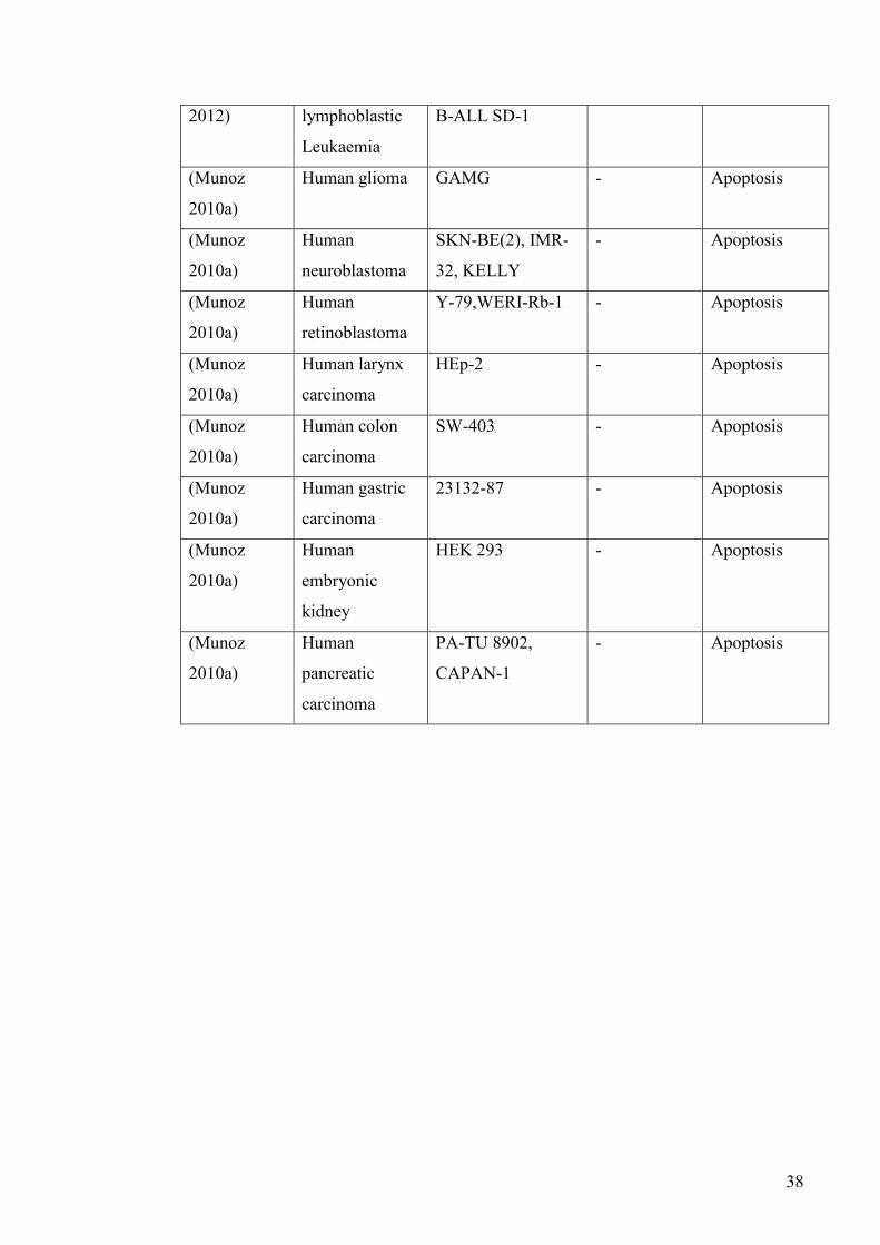
38
2012) lymphoblastic
Leukaemia
B-ALL SD-1
(Munoz
2010a)
Human glioma GAMG - Apoptosis
(Munoz
2010a)
Human
neuroblastoma
SKN-BE(2), IMR-
32, KELLY
- Apoptosis
(Munoz
2010a)
Human
retinoblastoma
Y-79,WERI-Rb-1 - Apoptosis
(Munoz
2010a)
Human larynx
carcinoma
HEp-2 - Apoptosis
(Munoz
2010a)
Human colon
carcinoma
SW-403 - Apoptosis
(Munoz
2010a)
Human gastric
carcinoma
23132-87 - Apoptosis
(Munoz
2010a)
Human
embryonic
kidney
HEK 293 - Apoptosis
(Munoz
2010a)
Human
pancreatic
carcinoma
PA-TU 8902,
CAPAN-1
- Apoptosis

39
Table 1.4 Substance P (SP) and NK1 antagonist effects on cancer in vivo
Reference Cancer
type
Cell line Species SP
beneficial
NK1
antagonist
beneficial
(Korcum
2009)
Melanoma B16F10 C5BL/6
mice
No -
(Palma
2000)
Astrocytom
a grade III
U373 MG Female
nude mice
No Yes
(Palma
2000)
Ovarian
carcinoma
A2780 Female
nude mice
- No
(Bigioni
2005)
Human
breast
carcinoma
MDA-MB-231 Female
nude mice
- No
(Pagan
2010)
Colon
carcinoma
intracolonic, then
systemic,
administration of
trinitrobenzene
sulfonic acid
Sprague-
Dawley
rats
- -
(Manske
2005)
Melanoma K1735 Mice Yes -
(Harris
2003)
Lung
carcinoma
exposure to side
stream cigarette
smoke
Inbred
C57BL
mice
Yes -
(Erin
2008)
Breast
carcinoma
4THMpc Balb-c
mice
No -
(Erin
2006)
Breast
carcinoma
4T1 cells,
4THMpc
Female
Balb-c
mice
Yes -
(Erin
2004)
Breast
carcinoma
Syngeneic 4T1 Adult
mice
Yes -
40
1.11.6 Substance P effects on angiogenesis
The stimulatory effect of SP on cell growth and the presence of NK1 receptors on
many capillary endothelial cells have made NK1 antagonism an attractive target for
inhibition of cancer-associated angiogenesis. SP has been shown to cause endothelial
cell migration and proliferation in cell culture studies (Ziche 1990; Wang 2009). SP
injected into the synovium of rat knees stimulated endothelial cell proliferation when
compared to their saline injected counterparts (Seegers 2003). Growth of new
capillaries was also seen in a rabbit avascular cornea with the application of a 1-5
microgram pellet of SP (Ziche 1990). Furthermore, NK1 antagonist treatment
inhibited the angiogenesis seen in this model (Ziche 1990). Similarly, NK1
antagonism at least partially ameliorated the increased blood vessel growth seen when
capsaicin injection induced angiogenesis in rat knees (Mapp 2012). Therefore it is
possible that SP mediates tumour-initiated angiogenesis and that NK1 antagonist
treatment may be effective in reducing tumour growth by decreasing available blood
supply, although more studies are required to confirm this in tumour models.
1.11.7 Substance P interactions with radiotherapy of cancer
Radiation therapy for the treatment of cancer may cause alterations in SP expression
in and around tumours, although the mechanism and implications of this is yet to be
determined. In vitro, irradiation caused an increase in SP secretion by a subpopulation
of SP immunostained MDA-MB-231 breast carcinoma cells (Aalto 1998). In contrast
to this study, B16F10 melanoma cells implanted into C5BL/6 mice showed delayed
tumour growth by three weeks, when treated with ionising radiation. In conjunction,
the radiation reduced SP expression in and surrounding the skin lesions, whilst
improving animal survival (Korcum 2009). Thus the role of SP in relation to
radiotherapy is unclear.

41
1.11.8 Potential effects of Substance P on tumour cell extravasation into the
brain
Since SP has the ability to increase the permeability of the BBB, it is a potential
candidate involved in tumour cell extravasation into the brain parenchyma to form
secondary brain tumours. It is therefore proposed that tumour cells may secrete SP or
cause a release of SP from cerebral capillary endothelial cells or the primary sensory
neurons that surround the blood vessels in the brain. This would result in a decrease in
the barrier properties of the BBB and may allow tumour cells to metastasise to the
brain.
1.12 Conclusion
The mechanism of tumour cell extravasation from the cerebral circulation into the
brain parenchyma through the BBB is not yet understood. However, it is known that
SP acts to increase the permeability of the BBB in other pathologies and therefore it
may play a role in the metastatic progression of tumour cells to the brain. If this is the
case, treatment with NK1 receptor antagonists may be able to prevent the formation of
metastatic tumours in the brain. Furthermore, SP had previously been shown to cause
an increase in oedema and a worse clinical outcome in animal models of stroke and
traumatic brain injury. Therefore it is proposed that SP may play a role in the
increased permeability of the BBB that leads to oedema formation surrounding
metastatic brain tumours. This will be investigated in the hope that novel treatments
for peritumoral oedema in the brain may be developed in order to prevent the
complications that often arise as a result of cerebral oedema.
A brief introduction will precede each experimental investigation, along with a
summary of the experimental protocol; these are outlined in detail in Chapter 2.
While each chapter reports results specific to that chapter, many of the results have
implications not only for the immediate point being considered, but often for other
aspects raised in the thesis. For this reason, there will be some overlap in
interpretation and discussion. A concluding general discussion will thereafter
integrate the major conclusions drawn from each chapter.
42
2 Materials and Methods
2.1 Cell culture
Walker 256 rat breast carcinoma cells were obtained from two cell banks, the
American Type Culture Collection (ATCC), and the Cell Resource Centre for
Medical Research at Tohoku University, Japan (CRCTU). Both cell populations were
cultured according to the instructions from the respective cell banks. These cells were
not synchronised before experiments, as this may impair cell replication. In each
experiment treatment groups were performed concurrently where possible to
compensate for different tumour cell passage number.
2.1.1 Walker 256 cells from American Type Culture Collection
Walker 256 cells from the ATCC were cultured in growth medium made up of Sigma
199 M4530 culture medium containing 5% sterile normal horse serum and 1%
penicillin and streptomycin (Sigma 10,000 units of penicillin and 10 mg of
streptomycin/mL). Culture flasks of 150cm2 were used to grow the cells and once
>90% confluence was reach the cells were detached with addition of 3.5 mL of 1%
trypsin (Sigma). The cells were spun down in a centrifuge (5 minutes at 1500 RPM)
and then resuspended in serum free culture medium. The number of cells was
calculated using a haemocytometer and then diluted, so that the correct number of
cells suspended in serum free culture medium were ready for inoculation. The cell
suspension was mixed prior to inoculation to maintain tumour cell concentration
throughout the entire volume. The specific concentrations used are described in the
relevant results chapters.
2.1.2 Walker 256 cells from Cell Resource Centre at Tohoku University
Walker 256 cells from the CRCTU were cultured in growth medium made up of
Sigma RPMI-1640 culture medium containing 10% sterile fetal bovine serum and 1
mL of penicillin and streptomycin (Sigma 10,000 units penicillin and 10 mg of
streptomycin/mL) for each 100 mL volume. Culture flasks of 150 cm2 were used to
grow the cells and once >90% confluence was reached the cells were detached with

43
addition of 3.5 mL of 0.02% EDTA. The cells were spun down in a centrifuge (5
minutes at 1500 RPM) and then resuspended in serum free culture medium. The
number of cells was calculated using a haemocytometer and then diluted, so that the
correct number of cells suspended in serum free culture medium were ready for
inoculation. The cell suspension was mixed prior to inoculation to maintain tumour
cell concentration throughout the entire volume. The specific concentrations used are
described in the relevant results chapters.
2.1.3 Cell viability assay
The cell viability assay was performed on Walker 256 cells from the CRCTU and as
such culture was performed in complete culture medium consisting of Sigma RPMI-
1640 containing 10% sterile foetal bovine serum and 1% penicillin and streptomycin
(Sigma 10,000 units penicillin and 10 mg of streptomycin / mL). Fosaprepitant
dimeglumine (EmendR; MERCK & CO), n-acetyl L-tryptophan (NAT) and
dexamethasone sodium phosphate (DBL) were used as treatments in this assay. To
assess the response of Walker 256 tumour cells to differing doses of NK1 receptor
antagonists (Emend and NAT) and dexamethasone, a trypan blue cell viability assay
was used. 105 cells were seeded into each well of a 12 well tissue culture plate with 2
mL of complete culture medium. Cells were allowed to grow for 24 hours, after which
the drugs of interest or saline as a vehicle control were added for a further 24 hours.
Each treatment was applied to 3 wells on 4 different occasions (n=12), with three
concentrations, 10, 100 and 1000µg / mL for each of the three agents. Cells were
detached using 0.02% EDTA and transferred into tubes that were subsequently
centrifuged for 5 minutes at 1500 RPM. The cells were then resuspended in 1 mL of
fresh medium with 1 mL of 0.4% trypan blue. Cells capable of excluding the trypan
blue dye were counted as viable cells. The percentage of viable Walker 256 cells was
calculated in relation to the total cell count using a haemocytometer.
2.2 Animals
The experimental procedures described throughout this project were performed within
the National Health and Medical Research Council (NHMRC) guidelines and were
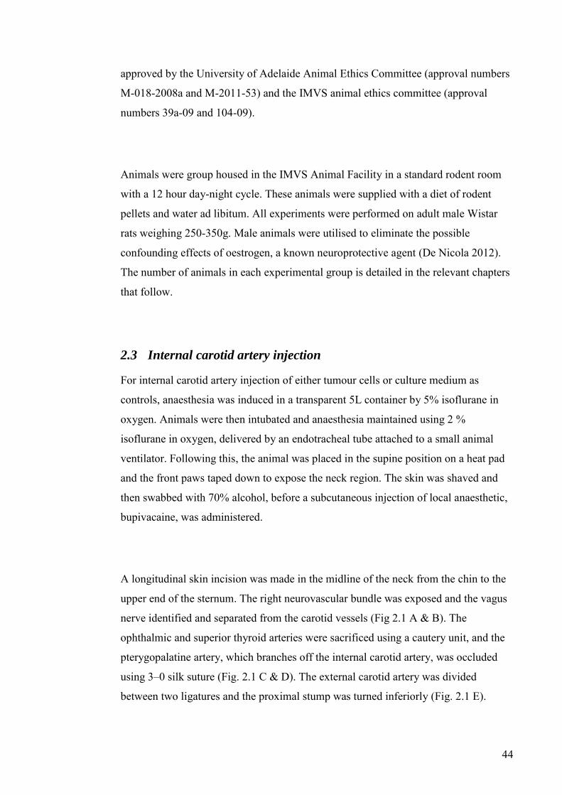
44
approved by the University of Adelaide Animal Ethics Committee (approval numbers
M-018-2008a and M-2011-53) and the IMVS animal ethics committee (approval
numbers 39a-09 and 104-09).
Animals were group housed in the IMVS Animal Facility in a standard rodent room
with a 12 hour day-night cycle. These animals were supplied with a diet of rodent
pellets and water ad libitum. All experiments were performed on adult male Wistar
rats weighing 250-350g. Male animals were utilised to eliminate the possible
confounding effects of oestrogen, a known neuroprotective agent (De Nicola 2012).
The number of animals in each experimental group is detailed in the relevant chapters
that follow.
2.3 Internal carotid artery injection
For internal carotid artery injection of either tumour cells or culture medium as
controls, anaesthesia was induced in a transparent 5L container by 5% isoflurane in
oxygen. Animals were then intubated and anaesthesia maintained using 2 %
isoflurane in oxygen, delivered by an endotracheal tube attached to a small animal
ventilator. Following this, the animal was placed in the supine position on a heat pad
and the front paws taped down to expose the neck region. The skin was shaved and
then swabbed with 70% alcohol, before a subcutaneous injection of local anaesthetic,
bupivacaine, was administered.
A longitudinal skin incision was made in the midline of the neck from the chin to the
upper end of the sternum. The right neurovascular bundle was exposed and the vagus
nerve identified and separated from the carotid vessels (Fig 2.1 A & B). The
ophthalmic and superior thyroid arteries were sacrificed using a cautery unit, and the
pterygopalatine artery, which branches off the internal carotid artery, was occluded
using 3–0 silk suture (Fig. 2.1 C & D). The external carotid artery was divided
between two ligatures and the proximal stump was turned inferiorly (Fig. 2.1 E).

45
The common carotid artery was temporarily occluded using a silk sling (Fig. 2.1 D &
E). Using micro-scissors, a small hole was cut in the proximal external carotid stump
through which a cannula was threaded into the internal carotid artery and tied in place.
Tumour cell suspension or culture medium (0.2 mL) was slowly injected into the
internal carotid artery, after which the blood flow through the common carotid artery
was established by removal of the sling. The wound was closed with sutures and
animals allowed to recover.
46
Figure 2.1 Internal Carotid Artery injection
(A) Diagram of the surgical field showing the location of the carotid bifurcation (B) Photograph of the surgical field (C) Close up of image B showing the common carotid artery (CCA), superior thyroid artery (STA), external carotid artery (ECA), ophthalmic artery (OA) and internal carotid artery (ICA) (D) Diagram of the blood vessels shown in image C demonstrating the location of sutures (blue lines) and sites of cauterisation (red starburst) (E) Diagram of carotid bifurcation during internal carotid artery cannulation, small black arrow indicates the cannulation point and direction in which the tube was threaded.
47
2.4 Direct intracerebral inoculation
For the direct inoculation model of tumour cells, anaesthesia was induced with 5%
isoflurane in oxygen in a transparent 5L container, and maintained at 3% via a nose
cone. The animal was placed in a stereotaxic frame, local anaesthetic bupivacaine
injected subcutaneously in the scalp and a midline scalp incision made to expose the
skull. A 0.7 mm burr hole was performed at stereotaxic coordinates, 0.5 mm anterior
and 3 mm lateral to the bregma, on the right half of the skull. A 30-gauge needle was
inserted and lowered 5 mm ventral to bregma using a micrometre device. Between 105
and 106 Walker 256 carcinoma cells in 8 μL of sterile culture medium were injected
into the striatum over 10 min. Control animals had only culture medium injected. The
needle remained in place for 5 min, then withdrawn. The hole was sealed with bone
wax and the wound sutured.
2.5 Drug treatments
In treatment studies animals were randomly assigned to receive fosaprepitant
dimeglumine (EmendR; MERCK & CO), n-acetyl L-tryptophan (NAT),
dexamethasone sodium phosphate (DBL) or equal volumes of vehicle saline. The
treatment regimen for each study is described in the relevant results chapters. All
agents were dissolved in 0.9% saline solution and delivered via intraperitoneal
injection with a 30gauge ½ inch needle attached to a 1mL syringe.
2.5.1 Emend
The NK1 receptor antagonist Emend was used at a dose of 3 mg/kg/day in all
treatment studies described, although the duration and timing varied. This dose was
determined based on the dose recommended clinically and that shown to previously
elicit a central effect in rodents (Watanabe 2008). Emend was prepared by mixing
15mg of the drug with 5ml of 0.9% saline until dissolved and was stored at 4ºC for a
maximum of 4 days.

48
2.5.2 NAT
The NK1 receptor antagonist, NAT, was used in all treatment studies at a dose of 7.5
mg/kg/day. Dosage was determined based on previous studies from our laboratory
describing the dose response required to reverse the increased BBB permeability
evident following traumatic brain injury (Donkin 2009). NAT was prepared by
mixing 150mg of the drug with 20 mL of 0.9% saline, with the addition of 3 drops of
5N NaOH to aid in dissolving the NAT powder. Once the solution was clear, the pH
was adjusted to between 7.3 and 7.6. This solution was refrigerated at 4ºC and used
within 4 days of preparation.
2.5.3 Dexamethasone
Dexamethasone, a synthetic glucocorticoid, was obtained as a 4mg/mL solution in
2mL ampoules from the Royal Adelaide Hospital pharmacy and stored at 4ºC. A high
dose of 8 mg/kg/day dexamethasone was chosen as determined by a previous study
that had successfully used dexamethasone to treat tumour-associated oedema (Gu
2007a).
2.6 Assessment of brain histology
For histological analysis, animals were transcardially perfused with 10% formalin
under terminal anaesthesia induced by intraperitoneal administration of
pentobarbitone sodium (60 mg/kg). Animals were decapitated 1 hour following
perfusion and the brains submerged in 10% normal buffered formalin for 24 hours.
Once the cerebellum and olfactory bulb have been removed, the remaining tissue was
placed into a cassette and exposed to increasing concentrations of ethanol for 20
minutes each (50%, 70%, 80%, 90%, 95%, 100%, 100%). Following this, the tissue
was placed in two baths of xyline each of 90 minutes duration and then bathed in
paraffin for increasing periods of time (30, 60, 60 and 90 minutes).

49
Brains were subsequently embedded in paraffin wax and 5 µm coronal sections were
cut. This was done every 2mm in preliminary studies and then every 400µm in
subsequent experiments. The brain sections were then mounted on glass slides
(Menzel-Glaser, Superfrost Plus) and stored in slide boxes in a 37ºC humidified oven
until required for staining.
2.7 Tumour volume
Haematoxylin and eosin stained slides were scanned using a Nanozoomer
(Hammamatsu, Hammamatsu City, Japan) and images used to calculate tumour
volume. This was achieved by determining the area of tumour in each section using
the NDP viewer programme and multiplying the area by the distance between
sections.
2.8 Immunohistochemistry
Immunostaining for albumin (ICN Pharmaceuticals, 1:20,000 anti goat), SP (Santa
Cruz Biotechnology, 1:2000 anti goat), NK1 receptors (Biocore, 1:8000 anti rabbit),
endothelial barrier antigen (EBA Chemicon, 1:5000 anti mouse), glial fibrillary acidic
protein (GFAP Dako 1:40,000 anti rabbit) and ionized calcium binding adaptor
molecule 1 (IBA1 Dako 1:50,000 anti rabbit) was performed. The specific stains used
in each study are described in the relevant results chapters. Tumour cells were also
grown onto cover slips in vitro to be immunostained for cytokeratin18 (Gene Tex
1:3000 anti mouse).
Immunohistochemistry was performed using the standard streptavidin procedure used
regularly in our laboratory with 3,3`-diaminobenzidine (DAB) for visualization and
haematoxylin counterstaining. All immunostained slides were scanned using the
Nanozoomer. Analysis of immunostaining for each antibody is described in the
relevant results chapters.
50
2.9 Brain water content
The wet weight–dry weight method was used to calculate brain water content in order
to quantify the effect of treatment on peritumoral oedema. Animals were
anaesthetised and rapidly decapitated. The brain was quickly removed from the skull
and the cerebellum and olfactory bulbs discarded. The cerebrum was placed in a quick
seal jar that had been previously weighed using a fine balance, and then weighed
again containing the wet brain. The lid of the jar was then removed and the jar was
placed in a 100 °C oven for 24 h. The dry brain was then weighed using a fine
balance. The % of brain water was calculated using the following equation:
% of brain water = ((wet weight−dry weight) / wet weight) x100
2.10 Evans blue extravasation
Evans blue was used as an indicator of BBB permeability, since it binds to serum
albumin and is only able to leak into the brain tissue from the vasculature under
pathological conditions that increase the permeability of the BBB. Animals were
injected intravenously with 0.8 mL of 4% Evans blue (MW 69,000; Sigma, E-2129)
30 min before they were perfused transcardially with saline under general anaesthesia
induced by pentobarbitone sodium (60 mg/kg). 10 minutes before perfusion, animals
were administered an intraperitoneal injection of 1000 IU of heparin in 0.5mL using a
30 gauge ½ inch needle.
The brain was quickly removed from the skull and dissected to remove the cerebellum
and olfactory bulbs. The cerebrum was placed into vials, weighed and then
homogenised in 7.5 mL of phosphate buffered saline. After adding 2.5 mL of
trichloroacetic acid (Sigma, T-0699) the samples were vortexed for 2 min, stored
overnight at 4 °C and then centrifuged at 1000 g over 30 min. A UV/Vis
spectrophotometer was used to measure the Evans blue absorbance in the supernatant
51
at 610 nm. The quantity of Evans blue, expressed as μg/g of brain tissue was
calculated using an Evans blue standard curve that had been previously determined.
2.11 Statistical analysis
Data are expressed as mean±SEM throughout. To determine statistical significance,
an unpaired two tailed t test (for two groups) or one-way analysis of variance
followed by a Bonferroni post test (for more than two groups) was performed as
applicable, with p<0.05 designated as significant.

52
3 Tumorigenicity of Walker 256 breast carcinoma cells
from two different tumour cell banks as assessed using
two models of brain metastases
3.1 Abstract
Metastatic brain tumours are a common end stage of breast cancer progression, with
significant associated morbidity and high mortality. Walker 256 is a rat breast
carcinoma cell line syngeneic to Wistar rats and commonly used to induce metastatic
brain tumours. In this study internal carotid artery injection and direct cerebral
inoculation models of metastatic brain tumour were used to determine the
tumorigenicity of Walker 256 cells obtained from the American Type Culture
Collection (ATCC), and the Cell Resource Centre for Medical Research at Tohoku
University (CRCTU). Tumour incidence and volume, plus immunoreactivity to
albumin, IBA1 and GFAP, were used as indicators of tumorigenicity and tumour
interaction with the host brain microenvironment. CRCTU Walker 256 cells showed
greater incidence, larger tumour volume, pronounced blood-brain barrier (BBB)
disruption and prominent glial response when compared to ATCC cell line. These
findings indicate that immortalised cancer cell lines obtained from different cell banks
may have diverse characteristics and behaviour in vivo.

53
3.2 Introduction
Cancer research has received much attention and funding over the past decades,
reflecting its increased incidence and significance as a public health problem.
Carcinogenesis is a multifaceted and complex disease process, making malignancies
inherently difficult to treat, while at the same time presenting multiple pathways for
investigation as management options. Novel treatments targeting these different
pathways can then be assessed, although tumours in the brain have been excluded
from many clinical trials due to the restrictive nature of the BBB, often making brain
metastases not accessible to novel treatments (Puduvalli 2001; Harford-Wright 2011).
Metastatic brain tumours are present in 22-30% of patients diagnosed with breast
cancer (Schuette 2004; Heyn 2006; Hines 2008), therefore making animal models of
brain metastases important tools to explore adequate treatment options for this aspect
of the disease.
The process of brain metastases involves cells from a primary tumour entering blood
vessels, avoiding death signals in the circulation, then undergoing extravasation
through the BBB (Marchetti 2003). The BBB is a dynamic interface between the
cerebral circulation and brain tissue, and acts to protect the brain microenvironment
(Hawkins 2005). While investigating metastases, many scientists using cell culture
presume that tumour cell lines will behave indefinitely in a uniform manner, although
several studies have demonstrated that this is not the case. Changes exhibited with
extended in vitro growth time, high passage number and cross contamination with
other cell lines have been frequently described in the literature (Sacchi 1984; Chang-
Liu 1997; Buehring 2004; Liscovitch 2007), particularly when cancer cell lines are
obtained from sources other than reputable major cell libraries (Reid 2011). There is
the assumption that well characterised cell lines available from cancer cell repositories
are verified and maintained at a high standard, meaning that researchers do not need
to authenticate these cell lines before commencing their experiments (Cree 2011).
However, the current publication reports differential characteristics of the same cancer
cell line obtained from two different reputable cell banks, suggesting that researchers
cannot assume that cells obtained from reputable cancer cell repositories will all
behave identically.
54
3.3 Method
3.3.1 Cell Culture
Walker 256 breast tumour cells (rat) were obtained from two cell banks, the American
Type Culture Collection (ATCC), and the Cell Resource Centre for Medical Research
at Tohoku University (CRCTU). Both cell populations were cultured according to the
instructions from the respective cell bank. This has been described previously in
sections 2.1.1 and 2.1.2 on page 42.
Culture flasks of 150cm2 were used to grow the cells and once >90% confluence was
reached, the cells were detached by the addition of 3.5 mL of 1% trypsin (Sigma) or
3.5 mL of 0.02% EDTA for ATCC and CRCTU Walker 256 cells, respectively. The
cells were spun down in a centrifuge (5 minutes at 1500 RPM) and then resuspended
in serum free culture medium. The number of cells was calculated using a
haemocytometer and then diluted, so that there was between 105 and106 cells in every
0.2 mL of cell suspension for internal carotid artery injection, or the same number of
cells in 8 µL for direct inoculation into the brain.
3.3.2 Animals
The experimental procedures were performed as described in section 2.2 on page 43.
Animals were randomly selected for either the internal carotid injection procedure or
the direct inoculation procedure and then were further divided into culture medium
only control group, Walker 256 tumour CRCTU group and Walker 256 tumour
ATCC group.
3.3.3 Internal Carotid Artery Injection
Animals allocated to the internal carotid injection procedure were sacrificed at 24 h
(early, n=5), 6 days (intermediate, n=5) and 9 days (late, n=9) for the CRCTU Walker
256 cells, and at 24 h (early, n=5), 4 weeks (intermediate, n=5) and 10 weeks (late,
n=9) for the ATCC Walker 256 cells. The selected late time points were determined

55
after a pilot study of tumour burden and animal weight loss for both cell lines. The
method for internal carotid artery injection of tumour cells to induce metastatic brain
tumour growth has been previously described in detail in section 2.3 on page 44.
3.3.4 Direct Inoculation
Animals that received direct intraparenchymal inoculation were sacrificed at 7 days
and 4 weeks for the CRCTU and ATCC Walker 256 cells, respectively (n=6/group).
Direct stereotaxic inoculation of tumour cells into the right striatum for induction of
metastatic brain tumour has been previously described in detail in section 2.4 on page
47.
3.3.5 Tumour Volume
For histological analysis, animals were transcardially perfused with 10% formalin
under terminal anaesthesia induced by intraperitoneal administration of
pentobarbitone sodium (60 mg/kg) as described in section 2.6 page 48. Brains were
embedded in paraffin wax and sequential 5 µm coronal sections were cut from blocks
2mm thick in a rostro-caudal direction, to be used for haematoxylin and eosin staining
and immunohistochemistry. The haematoxylin and eosin stained slides were scanned
using a Nanozoomer (Hammamatsu, Hammamatsu City, Japan) and images used to
calculate tumour volume. This was performed by determining the area of tumour in
each section using the NDP viewer programme and multiplying the area by the
distance between sections as previously described in section 2.7 page 49 (Corrigan
2012).
3.3.6 Immunohistochemistry
Slides from each model were stained for albumin (ICN Pharmaceuticals, 1:20,000),
GFAP (Dako 1:40,000) and IBA1 (Dako 1:50,000). Tumour cells were also grown on
cover-slips in vitro to be immunostained for cytokeratin18 (Gene Tex 1:3,000).
Immunohistochemistry was performed as described in section 2.8 page 49. Slides

56
were scanned using the Nanozoomer. Albumin immunostaining, expressed as the
weighted %DAB in each coronal section, was estimated using colour deconvolution
techniques, as described previously (Harford-Wright 2010; Helps 2012). For GFAP
and IBA1 immunoreactivtiy, 4 fields of view were taken from the cortex and striatum
for the internal carotid artery injection model and the direct inoculation model. The
immunolabelled cells in these images were counted and the mean number calculated
for all images from each brain.
3.3.7 Statistical Analysis
Results were expressed as mean±SEM and an unpaired two tailed t test (for two
groups) or a one-way analysis of variance followed by a Bonferroni post test (for
more than two groups) performed. Values of p<0.05 were designated as significant.

57
3.4 Results
3.4.1 Cell Morphology
In cell culture, both the Walker 256 cell populations received from the CRCTU and
the ATCC grew very effectively, although with very different cell morphology. The
cells from the CRCTU were small and spicular in appearance with deeply stained
nuclei (Fig. 3.1A), whereas the cytoplasm of the ATCC cells was abundant and the
cells had a larger, flatter appearance with open-face lighter stained nuclei (Fig. 3.1B).
Nuclei of the two cell populations were comparable in size (Fig. 1a and b). Both the
CRCTU and ATCC Walker 256 cell populations stained positively for cytokeratin 18,
a marker of breast cancer cells (Fig. 3.1A and B).
When the CRCTU and ATCC Walker 256 breast carcinoma cells were delivered to
the brain through internal carotid artery injection, the resultant tumours also showed
differential cell morphology. The tumours from CRCTU Walker 256 cells that grew 9
days following internal carotid artery injection showed cells with large nuclei and
scanty cytoplasm (Fig. 3.1C). In contrast, the single tumour that grew 10 weeks
following internal carotid artery injection of ATCC Walker 256 cells showed spindle-
shaped cells with smaller nuclei and a larger cytoplasmic component (Fig. 3.1D).
Furthermore, there was no evidence of mitotic figures in the ATCC tumour, whereas
CRCTU tumours exhibited several cells undergoing replication (Fig 3.1C).

58
Figure 3.1 Tumour cell morphology in vitro and in vivo
(A) CRCTU Walker 256 cells in culture stained for cytokeratin 18, showing spicular appearance with deeply-stained nuclei. (B) ATCC Walker 256 cell in culture stained for cytokeratin 18 showing flattened cells with a large cytoplasmic component and lightly stained nuclei and mitotic figures (arrows) (C) CRCTU Walker 256 tumour cells in vivo, 9 days following internal carotid artery injection, stained with haematoxylin and eosin showing large nuclei and scanty cytoplasm with many mitotic figures (arrows) (D) ATCC Walker 256 tumour cell in vivo, 10 weeks following internal carotid artery injection, showing smaller nuclei and abundant elongated cytoplasmic component, giving the section an eosinic appearance
3.4.2 Tumorigenicity
The CRCTU Walker 256 cells grew much more aggressively in vivo than the ATCC
population as indicated by the earlier sacrifice time required for the CRCTU injected
animals in both models. Following internal carotid artery injection, only one animal of
9 injected with ATCC cells developed a metastatic brain tumour at the 10 week time
point, whereas 8 out of the 9 animals injected with the CRCTU cells showed tumours
at the late time point of 9 days (Table 3.1). Furthermore, the CRCTU internal carotid
artery injected animals also showed metastatic brain tumours in one out of the 5
animals killed at the intermediate time point of 6 days following surgery (Table 3.1).
Neither the CRCTU nor the ATCC Walker 256 injected animals showed any evidence
59
of tumour growth at the early time point of 24 hours post internal carotid artery
injection (Table 3.1). The single tumour that resulted from inoculation with ATCC
Walker 256 cells was located in the striatum. In contrast, the masses in the CRCTU
Walker 256 inoculated animals were predominantly found in the lateral ventricles.
Similar to the internal carotid artery injection model, the CRCTU cells were effective
in producing metastatic brain tumours when inoculated directly into the brain, whilst
the ATCC cells were not (Table 3.1). Direct inoculation of CRCTU tumour cells into
the striatum resulted in development of large neoplastic masses in the brain tissue of
100% of the animals, whereas none of the ATCC Walker 256 inoculated animals
showed any evidence of tumour growth (Table 3.1). Comparison of the two models
used in this study revealed that direct injection of CRCTU Walker 256 cells into the
brain resulted in larger and more consistent location of tumour growth in the striatum
with a mean volume of 55.28 mm3, compared with an average tumour volume of
36.61mm3 following internal carotid artery injection of the same CRCTU Walker 256
cells (Fig. 3.2A and B).
All the animals that developed metastatic brain tumours in the 9 day CRCTU group
showed a concurrent growth of a tumour in the right eye (Fig. 3.3A). Also 44.4% of
these animals had small tumour nodules in the right temporalis muscle, and 33.3%
developed lung tumours (Fig. 3.3B and C). None of these features were seen in the
animals injected with ATCC Walker 256 cells, or with animals inoculated directly
into the striatum with the CRCTU Walker 256 cells.

60
Table 3.1 Tumour incidence in animals injected via the internal carotid artery or
directly inoculated into the brain with Walker 256 breast tumour cells obtained
from the Cell Resource Centre for Medical Research at Tohoku University
(CRCTU) or the American Type Culture Collection (ATCC)
Internal Carotid Artery
injection
CRCTU ATCC
Early time point (n=5) 24 hours 0% (0/5) 24 hours 0% (0/5)
Intermediate time point (n=5) 6 days 20% (1/5) 4 weeks 0% (0/5)
Late time point (n=9) 9 days 89% (8/9) 10 weeks 11% (1/9)
Direct inoculation CRCTU ATCC
(n=6) 7 days 100% (6/6) 4 weeks 0% (0/6)
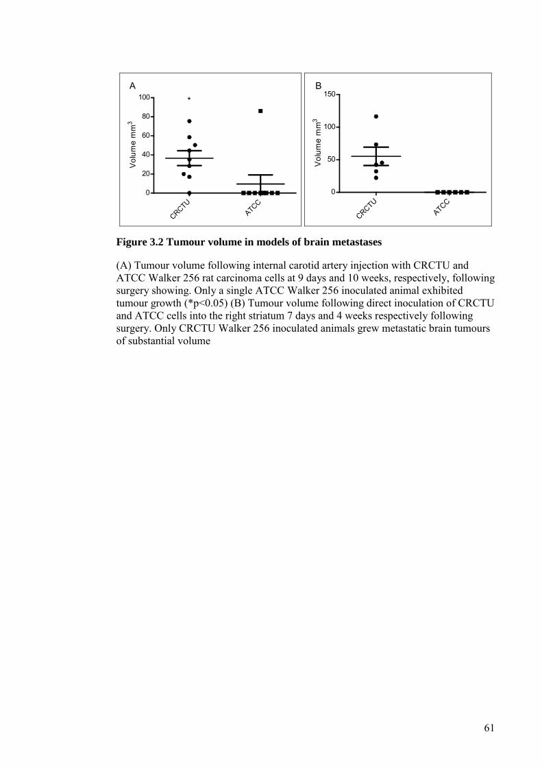
61
CRCTU
ATCC
0
20
40
60
80
100 *A
Vol
ume
mm
3
CRCTUATCC
0
50
100
150B
Vol
ume
mm
3
Figure 3.2 Tumour volume in models of brain metastases
(A) Tumour volume following internal carotid artery injection with CRCTU and ATCC Walker 256 rat carcinoma cells at 9 days and 10 weeks, respectively, following surgery showing. Only a single ATCC Walker 256 inoculated animal exhibited tumour growth (*p<0.05) (B) Tumour volume following direct inoculation of CRCTU and ATCC cells into the right striatum 7 days and 4 weeks respectively following surgery. Only CRCTU Walker 256 inoculated animals grew metastatic brain tumours of substantial volume

62
Figure 3.3 Extracranial tumour growth following internal carotid artery
injection of CRCTU Walker 256 cells
(A) CRCTU Walker 256 tumour growth (arrows) in the eye 9 days following internal carotid artery injection stained with haematoxylin and eosin (B) CRCTU Walker 256 tumour growth (arrows) invading the temporalis muscle 9 days post internal carotid artery injection, stained with haematoxylin and eosin (C) Lung section stained with haematoxylin and eosin, showing a large tumour mass (arrows) 9 days following CRCTU Walker 256 cells carotid injection
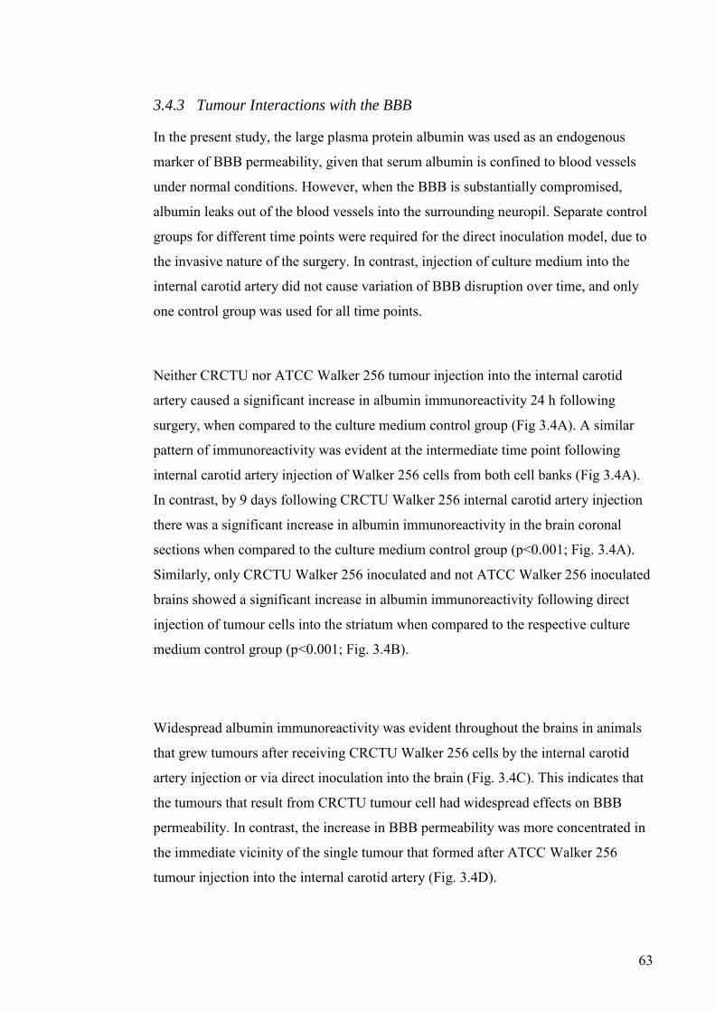
63
3.4.3 Tumour Interactions with the BBB
In the present study, the large plasma protein albumin was used as an endogenous
marker of BBB permeability, given that serum albumin is confined to blood vessels
under normal conditions. However, when the BBB is substantially compromised,
albumin leaks out of the blood vessels into the surrounding neuropil. Separate control
groups for different time points were required for the direct inoculation model, due to
the invasive nature of the surgery. In contrast, injection of culture medium into the
internal carotid artery did not cause variation of BBB disruption over time, and only
one control group was used for all time points.
Neither CRCTU nor ATCC Walker 256 tumour injection into the internal carotid
artery caused a significant increase in albumin immunoreactivity 24 h following
surgery, when compared to the culture medium control group (Fig 3.4A). A similar
pattern of immunoreactivity was evident at the intermediate time point following
internal carotid artery injection of Walker 256 cells from both cell banks (Fig 3.4A).
In contrast, by 9 days following CRCTU Walker 256 internal carotid artery injection
there was a significant increase in albumin immunoreactivity in the brain coronal
sections when compared to the culture medium control group (p<0.001; Fig. 3.4A).
Similarly, only CRCTU Walker 256 inoculated and not ATCC Walker 256 inoculated
brains showed a significant increase in albumin immunoreactivity following direct
injection of tumour cells into the striatum when compared to the respective culture
medium control group (p<0.001; Fig. 3.4B).
Widespread albumin immunoreactivity was evident throughout the brains in animals
that grew tumours after receiving CRCTU Walker 256 cells by the internal carotid
artery injection or via direct inoculation into the brain (Fig. 3.4C). This indicates that
the tumours that result from CRCTU tumour cell had widespread effects on BBB
permeability. In contrast, the increase in BBB permeability was more concentrated in
the immediate vicinity of the single tumour that formed after ATCC Walker 256
tumour injection into the internal carotid artery (Fig. 3.4D).
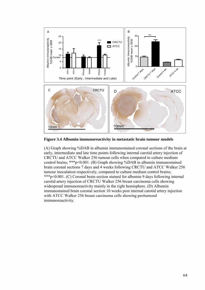
64
0
5
10
15
20
25
CRCTUATCC
***
24hr
24hr
6day
s
4wks
9day
s
10w
ks
Time point (Early , Intermediate and Late)
Control
A
Albu
min
imm
unor
eact
ivity
%D
AB m
ean
± SE
M
Contro
l 7 da
ysCRCTU 7
days
Contro
l 4 w
k
ATCC 4 wk
0
10
20
30 ***B
Alb
umin
Imm
unor
eact
ivity
%D
AB
mea
n±
SE
M
Figure 3.4 Albumin immunoreactivity in metastatic brain tumour models
(A) Graph showing %DAB in albumin immunostained coronal sections of the brain at early, intermediate and late time points following internal carotid artery injection of CRCTU and ATCC Walker 256 tumour cells when compared to culture medium control brains; ***p<0.001. (B) Graph showing %DAB in albumin immunostained brain coronal sections 7 days and 4 weeks following CRCTU and ATCC Walker 256 tumour inoculation respectively, compared to culture medium control brains; ***p<0.001. (C) Coronal brain section stained for albumin 9 days following internal carotid artery injection of CRCTU Walker 256 breast carcinoma cells showing widespread immunoreactivity mainly in the right hemisphere. (D) Albumin immunostained brain coronal section 10 weeks post internal carotid artery injection with ATCC Walker 256 breast carcinoma cells showing peritumoral immunoreactivity.

65
3.4.4 Brain Microenvironment
Both models of metastatic tumour induction caused changes in the brain
microenvironment when CRCTU Walker 256 breast carcinoma cells were utilised
(Fig. 3.5 and 3.6). There was a significant increase in the number of GFAP positive
cells in the cortex of animals 9 days following internal carotid artery injection of
CRCTU Walker 256 cells when compared to the culture medium control group
(p<0.01; Fig. 3.5A). Correspondingly, there was a significant increase in the number
of astrocytes immunostained for GFAP in the striatum surrounding the tumour mass 7
days following direct injection of CRCTU Walker 256 cells (p<0.01; Fig. 3.5B). In
contrast, ATCC Walker 256 cells administered via either the internal carotid artery
injection or direct inoculation into the striatum did not significantly alter the number
of GFAP labelled cells when compared to the same location in culture medium
inoculated brains (Fig. 3.5A, B).
GFAP immunoreactivity was absent within the tumour masses for both the CRCTU
and ATCC Walker 256 internal carotid artery models indicating the absence of
astrocytes within the tumours (Fig. 3.5C and D). However, the single tumour that
grew 10 weeks following internal carotid artery injection of ATCC Walker 256
tumour cells showed an increase in GFAP labelled cells in the peritumoral area (Fig.
3.5D) and some infiltrating labelled cells within the periphery of the tumour. The
astrocytes surrounding the tumour mass exhibited short, blunt, thickened processes,
with the flattened cells creating a limiting rim (Fig. 3.5D). The tumours that grew 9
days following internal carotid artery injection of CRCTU Walker 256 cells within the
lateral ventricles, had limited contact with the neuropil and lacked the GFAP positive
astrocytic border that was evident around the ATCC Walker 256 tumour (Fig. 3.5C).

66
Contro
l 4 w
k
CRCTU 9 da
ys
ATCC 10 w
k
0
5
10
15
20
**
A
*N
umbe
r of G
FAP
pos
itive
cel
lsm
ean
± S
EM
Contro
l 7 da
ysCRCTU 7
days
Contro
l 4 w
k
ATCC 4 wk
0
5
10
15
20
**
B
Num
ber o
f GFA
P p
ositi
ve c
ells
mea
n±
SE
M
Figure 3.5 GFAP immunoreactivity in metastatic brain tumour models
(A) Graph showing the average number of GFAP positive cells in 4 areas of the cortex (0.0678 mm2) in animals injected with culture medium, or injected with CRCTU or ATCC Walker 256 cells into the internal carotid artery; **p<0.01; *p<0.05 (B) Graph showing the average number of GFAP positive cells in 4 areas of the striatum (0.0678 mm2) following direct inoculation of culture medium, CRCTU or ATCC Walker 256 cells into the brain; **p<0.01 (C) GFAP immunostained section 9 days following CRCTU Walker 256 internal carotid artery injection, showing absence of staining within the tumour, but showing GFAP labelled cell in peritumoral area (arrows) (D) Brain section stained for GFAP 10 weeks following carotid ATCC Walker 256 tumour cell injection. Labelled astrocytes (arrows) are seen in the peritumoral area and in the periphery of the tumour mass between the tumour cells
Tumour cell inoculation caused an increase in the number of microglia, as indicated
by IBA1 labelling, in the cortex of brains 9 days following internal carotid artery
injection of CRCTU Walker 256 cells in comparison to control brains (Fig. 3.6A).
Similarly, 7 days after direct injection of CRCTU Walker 256 cells into the brain,
there was a significant increase in IBA1 positive cells in the striatum surrounding the

67
tumour mass, when compared to the same location in the culture medium control
group (p<0.01; Fig. 3.6B). However, the increase in microglia seen with CRCTU
Walker 256 cell inoculation, was not replicated by ATCC Walker 256 cells when
injected into the internal carotid artery or inoculated directly into the striatum (Fig.
3.6A and B).
Examination of brain sections immunolabelled for IBA1 showed a distinct pattern of
staining for each Walker 256 cell type. The CRCTU tumours showed sparse but
specific discrete labelling of infiltration by microglia (Fig. 3.6C). In contrast, the
ATCC tumour showed more widespread ill-defined labelling throughout the tumour
mass (Fig. 3.6D). Furthermore, there was a halo of IBA1 labelled cells surrounding
the tumour mass following internal carotid artery inoculation of ATCC Walker 256
cells, a feature that was not present surrounding the CRCTU Walker 256 induced
tumours (Fig. 3.6C and D).
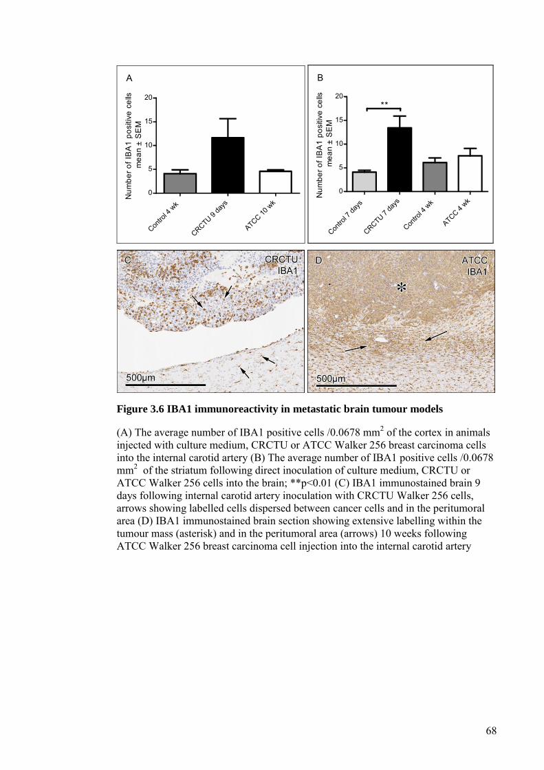
68
Contro
l 4 w
k
CRCTU 9 da
ys
ATCC 10 w
k0
5
10
15
20
A
Num
ber o
f IB
A1
posi
tive
cells
mea
n±
SE
M
Contro
l 7 da
ysCRCTU 7
days
Contro
l 4 w
k
ATCC 4 wk
0
5
10
15
20**
B
Num
ber o
f IB
A1
posi
tive
cells
mea
n±
SE
M
Figure 3.6 IBA1 immunoreactivity in metastatic brain tumour models
(A) The average number of IBA1 positive cells /0.0678 mm2 of the cortex in animals injected with culture medium, CRCTU or ATCC Walker 256 breast carcinoma cells into the internal carotid artery (B) The average number of IBA1 positive cells /0.0678 mm2 of the striatum following direct inoculation of culture medium, CRCTU or ATCC Walker 256 cells into the brain; **p<0.01 (C) IBA1 immunostained brain 9 days following internal carotid artery inoculation with CRCTU Walker 256 cells, arrows showing labelled cells dispersed between cancer cells and in the peritumoral area (D) IBA1 immunostained brain section showing extensive labelling within the tumour mass (asterisk) and in the peritumoral area (arrows) 10 weeks following ATCC Walker 256 breast carcinoma cell injection into the internal carotid artery

69
3.5 Discussion
In the current study, Walker 256 cells obtained from the CRCTU had potent
tumorigenic properties when compared to the ATCC Walker 256 breast carcinoma
cells. Evidence of this includes the substantially increased incidence of tumour growth
and tumour volume after CRCTU Walker 256 inoculation in the two tumour models
used in this study, as well as the fact that only CRCTU Walker 256 internal carotid
artery injected animals developed tumours in the eye, temporalis muscle and lung. It
has been shown in previous studies that different tumour cell lines cloned from the
same neoplasm may have different tumerogenic properties when implanted in vivo
(Kripke 1978; van Lamsweerde 1983). However, cell lines developed from a single
mouse mammary tumour that showed differing culture morphology and growth
characteristics in vitro, resulted in tumours that displayed similar histology to each
other and comparable tumerigenicity when injected into syngeneic hosts (Dexter
1978).
Despite the fact that both populations of Walker 256 breast carcinoma cells were
obtained from reputable tumour cell banks that described the Walker 256 cell line as
tumorigenic in Wistar rats, there was considerable variability in both their growth
behaviour in vivo and morphology in vitro. ATCC has been instrumental in the push
to develop a standard method of cell line verification involving short tandem repeat
profiling along with the development of a database of short tandem repeat profiles for
commonly used cell lines (Barallon 2010; A.T.C.C.S.D. Organisation 2010).
Control of cancer cell tumorigenicity has been extensively studied, predominantly in
relation to genetic control of cancer growth in vivo. For example, p75 has been linked
to reduced neuroblastoma tumorigenicity (Schulte 2009). However, characteristics of
tumour cells in culture have also been investigated, with shorter doubling time,
reduced monolayer density, poor motility and lower incidence of focus formation in
vitro linked to decreased tumorigenicity of cell lines when used in vivo (Reynolds
1987; Gildea 2000), although these experiments were generally comparing different
70
cell lines. In contrast, the current study aimed to determine the differences between
the same cell line obtained from two different sources.
The CRCTU Walker 256 breast carcinoma cells, found to be more tumorigenic than
their ATCC counterparts, showed darker nuclear staining and increased nucleus to
cytoplasm ratio when compared to the flatter more eosinophilic ATCC Walker 256
cells. There have been few previous studies to determine the relationship between cell
morphology and cancer cell tumorigenicity. Further investigation is required to
determine if the characteristics observed in this experiment are related to the
tumorigenicity of the cells described. Furthermore, previous studies have suggested
that behaviour of cancer cell lines in vitro is poorly correlated with tumorigenicity in
vivo (Reynolds 1987). Despite this, in the current study morphological features seen
in vitro for Walker 256 cells from both the CRCTU and ATCC were closely
associated with the morphology evident in vivo.
There are many plausible explanations for the differential characteristics evident for
CRCTU and ATCC Walker 256 breast carcinoma cells in this study. It is possible that
variations in storage methods, extended culture times and high passage number may
have contributed to the differences seen in the same cell line obtained from the
CRCTU and the ATCC. Immortalised tumour cell lines evolve over time in animal
models where malignancies are induced by inoculation with a homogenous population
of tumour cells (Poste 1982b). Conversely, human neoplastic tissue is not a uniform
entity. Within a tumour mass, there exists various heterogeneous subpopulations of
tumour cells with different metastatic potential and diverse propensity to metastasise
to various organs (Fidler 1978; Poste 1982a).
Tumour cells harvested from a neoplasm in vivo have been known to develop
characteristics over time in vitro that are distinct from those evident in the original
cancerous tissue (van Lamsweerde 1983). The proposed reason for this phenotypic
change is that more aggressive or mitotic properties are favoured by clonal selection
71
in vitro, with highly metastatic varieties more phenotypically stable (Chambers 1981;
Hiraiwa 1997). Long term passage of Walker 256 cells has previously been shown to
alter chemotactic behaviour in vitro (Oda 1984).
Walker 256 carcinoma is rat mammary tumour cell line that originally occurred
spontaneously in a pregnant albino Wistar rat (Buffon 2007). The Walker 256 cell line
has been used previously to establish experimental brain metastases through an
internal carotid artery injection and direct implantation into the cerebral cortex
(Blasberg 1984a; b; Blasberg 1986; Hiesiger 1986; Jamshidi 1992).
Tumour growth evident following both inoculation methods of CRCTU Walker 256
cells showed larger tumour volume in a shorter period of time, when compared to
previous experiments described in the literature using the Walker 256 cell line,
although the incidence was comparable (Hasegawa 1979; Hasegawa 1983; Morreale
1993; Felix 2012). In contrast, the ATCC Walker 256 cells showed a much lower
incidence and longer incubation period required to form only a single tumour when
compared to these previous studies. Therefore, neither the CRCTU, nor the ATCC
Walker 256 breast carcinoma cells behaved exactly as previously studies have
described, although the CRCTU population were more analogous to the literature.
Despite the consistency of the direct injection model of tumour induction, the ATCC
Walker 256 cells did not grow any tumours through the use of this method. Thus the
extravasation process through the BBB is not the limiting factor for ATCC Walker
256 tumour growth in the brain. Furthermore, 11% of animals grew metastatic brain
tumours 10 weeks following ATCC Walker 256 inoculation into the internal carotid
artery, meaning that at least some of the tumour cells were able to complete the
extravasation process.

72
The CRCTU Walker 256 inoculated animals for both the internal carotid artery and
the direct inoculation model showed a significant increase in albumin
immunoreactivity when compared to the culture medium group. It is likely that
albumin immunoreactivity was increased in response to the substantial tumour growth
evident in the CRCTU Walker 256 tumour inoculated groups and subsequent
angiogenesis. Angiogenic blood vessels that grow within brain metastases of breast
cancer are more permeable than BBB microvessels, as they are characteristic of the
breast tissue origin of the tumour cells (Cornford 1992; Shuto 2008). The process of
angiogenesis has been shown to take place once tumours have exceeded 2mm in
diameter (Blasberg 1984a; Fidler 1990; Zhang 1992).
The ATCC tumour cell inoculated animals only grew one tumour in either model of
metastatic brain tumour induction, which was not sufficient to cause a significant
difference in albumin immunoreactivity from vehicle level and thus did not increase
the permeability of the BBB. This shows that the presence of tumour cells with low
tumorigenicity in the brain microcirculation do not cause an inflammatory reaction
disrupts the normal function of the BBB. Furthermore, ATCC Walker 256 localisation
in the neuropil of the striatum did not cause long term damage to the brain sufficient
to increase the permeability of the BBB 4 weeks following direct injection.
A rim of reactive glial cells is often evident surrounding metastatic brain tumours in
human surgical tissue (Zhang 1995), as was also apparent surrounding tumours grown
in this study. The pattern of glial cell reaction was different surrounding CRCTU and
ATCC Walker 256 tumours that grew following internal carotid artery inoculation.
The location of CRCTU tumours within the lateral ventricles may be the cause of
these differences, as the mass is in less direct contact with the neuropil. In contrast,
the single tumour that grew 10 weeks following internal carotid artery inoculation of
ATCC Walker 256 cells, showed much more extensive microglial infiltration along
with increased microglia and astrocytes surrounding the tumour. Astrocytes and
microglia may proliferate and become activated in response to contact with serum
73
proteins, such as albumin which are present in oedematous fluid that accumulates
around the tumour (Jamshidi 1992; Hooper 2009).
The low tumorigenicity of ATCC Walker 256 cells may be the reason that these cells
did not show the same influence on the brain microenvironment as CRCTU Walker
256 growth. This is demonstrated by the significant increase in IBA1 and GFAP
labelled cells following both internal carotid artery and direct injection of CRCTU
Walker 256 tumour cells when compared to the culture medium injected groups.
However this phenomenon was not evident following ATCC Walker 256 tumour
inoculation for either model used in this study. Thus, the presence of low
tumorigenicity cancer cells in the brain microcirculation or the neuropil, did not show
significant interaction with the host microenvironment.
In conclusion, this study has demonstrated that the Walker 256 tumour cells obtained
from two reputable sources have different tumorigenicity, growth characteristics and
interactions with the host brain. Such variability should be considered when
comparing studies using the same cell line obtained from different sources.

74
4 Walker 256 tumour cells increase Substance P
immunoreactivity locally and modify the properties of the
blood-brain barrier during extravasation and brain
invasion
4.1 Abstract
It is not yet known how tumour cells traverse the blood-brain barrier (BBB) to form
brain metastases. Substance P (SP) release is a key component of neurogenic
inflammation which has been recently shown to increase the permeability of the BBB
following CNS insults, making it a possible candidate as a mediator of tumour cell
extravasation into the brain. This study investigated the properties of the BBB in the
early stages of tumour cell invasion into the brain, and the possible involvement of
SP. Male Wistar rats were injected with Walker 256 breast carcinoma cells via the
internal carotid artery and euthanised at 1, 3, 6 and 9 days post tumour inoculation.
Culture medium-injected animals served as controls at 1 and 9 days. Evidence of
tumour cell extravasation across the BBB was first observed at three days post
inoculation, which corresponded with significantly increased albumin (p<0.05) and
SP immunoreactivity (p<0.01) and significantly reduced endothelial barrier antigen
labelling of microvessels when compared to culture medium control animals
(p<0.001). By day 9 after tumour cell inoculation, 100% of animals developed large
intracranial neoplasms that had significantly increased albumin in the peri-tumoral
area (p<0.001). The increased SP immunoreactivity and altered BBB properties at
three days post inoculation that coincided with early tumour invasion may be
indicative of a mechanism for tumour cell extravasation into the brain. Thus,
extravasation of tumour cells into the brain to form cerebral metastases may be a SP-
mediated process.
75
4.2 Introduction
It is estimated that cancer is responsible for more than 6 million deaths each year
(WHO 2002). Approximately 20-40% of patients suffering from systemic cancer will
also have a metastatic brain tumour at autopsy, whereas clinical studies generally
report an incidence of about 10-20% (Posner 1978; Cifuentes 1979; Soffietti 2002;
Gavrilovic 2005; Lin 2008a). Breast cancer is one of the most common malignancies
to metastasise to the brain, occurring in late stages of the disease (Lorger 2010).
Nearly 50% of patients with metastatic triple-negative breast cancer, which do not
express estrogen receptors, progesterone receptors or human epidermal growth factor
receptor 2, will develop a metastatic brain tumour, with a median survival time of 4.9
months from diagnosis (Lin 2008b). There is a predilection for metastatic brain
tumours and central nervous system (CNS) recurrence amongst node positive,
estrogen receptor negative, young patients with high-grade breast cancer, with no
evidence that there is any benefit from an early diagnosis and treatment for their
intracranial malignancy (Pestalozzi 2006). Therefore there is a pressing need for
improved treatment and prevention strategies for brain metastatic breast cancer.
Metastatic brain tumours are caused by cancerous cells that detach from their primary
site, migrate through the blood stream, attach to cerebral endothelium and cross the
BBB to colonise the brain (Marchetti 2003). Despite the high incidence and clinical
impact of metastatic brain tumours, the exact mechanism of tumour cell extravasation
across the BBB has not been elucidated. This information is vital for the development
of treatments that aim to prevent invasion of the CNS by cancer cells.
The BBB is a dynamic structure composed of endothelial cells joined by tight
junctions, encompassed by pericytes and supported by astrocytic end feet (Hawkins
2005). This arrangement is able to be modified and respond to external stimuli or
disease states, allowing for differential barrier properties. For example, macrophages
are able to infiltrate the brain neuropil through an intact BBB, which remains
impermeable to serum proteins (Schackert 1988b).
76
Extravasation of tumour cells through the BBB is most likely a multifactorial process,
and few studies have investigated substances proposed to be involved in metastatic
spread to the brain. Vascular endothelial growth factor (VEGF) has been considered
as a driving factor for tumour cell extravasation, using the VEGF receptor antagonist
cediranib AZD2171 (Juanyin 2009). Other substances that are under investigation are
serine proteases released by melanoma cells, which disrupt tight junctions between
cerebral capillary endothelial cells (Fazakas 2011) and matrix metalloproteinase 2
(MMP2), as breast cancer cells transfected with tissue inhibitor of MMP2 have been
shown to have reduced propensity for metastases to the brain (Mendes 2007).
Substance P (SP) is an excitatory tachykinin that is predominantly released from
primary sensory nerve endings in the CNS, but is also localized in brain endothelial
cells (Cioni 1998; Ribeiro-da-Silva 2000). It is a potent mediator of neurogenic
inflammation and preferentially binds to NK1 receptors to induce microvascular
leakage in the brain and throughout the body (Harrison 2001; Nimmo 2004).
Neurogenic inflammation is characterized by the release of vasoactive peptides from
neurons, driving vasogenic oedema in the CNS through increased BBB permeability.
In vitro, SP treatment of rat cortical capillary endothelial cells causes an increase in
intracellular free calcium ions and is postulated to cause modulation of the BBB by
endothelial cell contraction (Paemeleire 1999). Furthermore, cytokine stimulated
endothelial cells caused release of SP, stimulating a NK1 receptor dependent increase
in BBB permeability (Cioni 1998; Annunziata 2002). Moreover, SP alters expression
of tight junction proteins by a NK1 receptor mediated mechanism (Annunziata 1998;
Lu 2008).
SP has also been implicated in cancer cell migration (Drell 2003; Lang 2004), chemo-
attraction (Ruff 1985), DNA synthesis, replication and cytokine secretion (Palma
1999). NK1 receptors are expressed by many tumour cell lines (Prasad 2007; Munoz
2008; Huang 2010). Antagonism of the actions of SP on tumour cells in vitro has been
an active area of research in recent times. NK1 receptor antagonists have been found
77
to inhibit tumour cell mitogenesis and induce apoptosis of human melanoma, breast
cancer, glioma, neuroblastoma, colon cancer and prostate cancer cell lines in cell
culture studies (Munoz 2004b; Munoz 2005a; Huang 2010; Munoz 2010b;
Mayordomo 2011). Many tumour cells release SP in culture, acting in an autocrine
fashion to promote survival (Mayordomo 2011). However, because these studies are
performed in vitro, the effect of SP secretion on the peri-tumoral tissue in vivo is yet
to be investigated. Furthermore, studies comparing in vitro brain metastatic invasion
models to their in vivo counterparts demonstrated that there was a poor correlation
between the two (Lorger 2011).
Endothelial barrier antigen (EBA) is a protein triplet used as an immunological
marker of the BBB in rats. Although its role in the structure and function of the BBB
is not entirely understood, it has been shown that its immunological deactivation by
intravenous injection of a monoclonal antibody to EBA, results in increased
permeability of the BBB to horseradish peroxidase through the widening of junctional
complexes and an increase in cytoplasmic vesicles (Ghabriel 2000; Ghabriel 2002).
Therefore EBA is integral to the normal functioning of the BBB in rats. Furthermore,
EBA expression in rat cerebral capillary endothelial cells is altered in models of
several pathological conditions that are known to disrupt the BBB, such as traumatic
brain injury, stroke and glioma (Lin 2001; Chekhonin 2007; Park 2010).
The actions of SP on both cancer cells and the BBB make it a possible candidate for
involvement in the mechanism of tumour cell extravasation into the brain. The aim of
this study was to investigate the early interactions of Walker 256 carcinoma cells with
the brain vasculature and to elucidate the role of SP in the modification of the BBB in
response to tumour invasion.

78
4.3 Methods
4.3.1 Animals
This project was performed as described in section 2.2 on page 43. Animals were
randomly selected for carotid inoculation with Walker 256 cells or injection with
culture medium only as controls. Tumour-inoculated animals were sacrificed at 1, 3, 6
and 9 days (n=5 per group). Culture medium-injected animals were sacrificed at 1 and
9 days (n=5 per group).
4.3.2 Cell Culture
Walker 256 rat breast tumour cells were obtained from the Cell Resource Centre for
Medical Research at Tohoku University and cultured as described previously 2.1.2 on
page 42. The number of cells was calculated using a haemocytometer and then
diluted, so that in every 0.2 mL of cell suspension, there was between 105 and106 cells
ready for inoculation.
4.3.3 Internal Carotid Artery Inoculation
The internal carotid artery inoculation procedure has been detailed in section 2.3 page
44.
4.3.4 Tumour Volume
For histological study of the brain, animals were perfused via the heart at 1, 3, 6 and 9
days with 10% formalin under general anaesthesia, then brains were removed and
embedded in paraffin wax. The brain was divided into 2 mm coronal blocks in a
cranio-caudal sequence. 5 µm sequential sections were cut from each block and used
for haematoxylin and eosin (H&E) staining and immunolabelling. The H&E stained
sections were scanned (Nanozoomer, Hamamatsu, Hamamatsu City, Japan) and
tumour volume calculated as described in section 2.7 page 49.

79
4.3.5 Immunolabelling
Brain sections were stained for albumin (ICN Pharmaceuticals, polyclonal, 1:20,000),
SP (Santa Cruz Biotechnology N-18:9758, polyclonal, 1:2000), NK1-receptor
(Biocore Pty Limited AB-N-33AP, polyclonal, 1:8000), and EBA ( Chemicon SMI-
71R-100, monoclonal, 1:5000). Immunolabelling was done as described in section 2.8
page 49. Objective assessment of the immunolabelling was achieved through colour
deconvolution techniques, to reveal the % of DAB in the scanned slides.
Immunolabelling was performed on slides from the brain corresponding to a position
at 0.8 mm posterior to bregma (Paxinos 1998), the location showing maximal blood
vessel invasion by tumour cells at 3 days after tumour cell inoculation.
4.3.6 Immunolabelling analysis
Virtual dissection was completed for all scanned immunolabelled slides. Albumin
immunoreactivity was analysed by using stained whole coronal sections of the brain,
whereas analysis of substance P and NK1 receptor immunolabelling required images
to be taken from the cortex, striatum, the tumours and in the peri-tumoral areas due to
the specificity and localised nature of these stains. These exported files were run
through colour deconvolution software and expressed as DAB wt % total, using a
technique previously described in detail (Harford-Wright 2010; Helps 2012). This
process involves the removal of background stining intensity variations (Helps 2012).
EBA immunoreactivity was evaluated by counting the blood vessels that were
negative for EBA immunoreactivity as a percentage of the total number of blood
vessels in the virtually dissected areas. Data were expressed as mean ± SEM.
Statistical differences were determined using an unpaired t-test (for 2 groups) or one
way analysis of variance (ANOVA) followed by a Bonferroni post test (for more than
2 groups), as applicable.
80
4.4 Results
Invasion of microvessels by tumour cells was first evident in 60% of animals at 3
days after tumour inoculation (Table 4.1). Thus, tumour cells begin passing from the
circulation, through the BBB and into the brain tissue between 1 and 3 days after
inoculation in this model of secondary brain tumours. Tumour cell invasion across the
BBB occurred in the right cerebral hemisphere, ipsilateral to the injected carotid
artery, mainly in the brain segment located at a coordinate 0.8mm posterior to
bregma.
In this study, a tumour mass is defined as the formation of more than 3 layers of
tumour cells around the circumference of microvessels. This was initially seen by 6
days post tumour inoculation in 20% of animals, increasing to 100% by day 9 (Table
4.1). Tumour growth was not allowed to progress past this time point because
extensive tumour burden was evident with a mean tumour volume of 36.61 mm3 (Fig.
4.1A). Although, the most common location of tumour cell invasion of brain
microvessels was within the cortex (Fig. 4.1B), large tumour masses were seen within
the lateral ventricles (Fig. 4.1C) and in the striatum.
Table 4.1 Tumour Incidence over Time
Time post tumour
inoculation
Percentage of animals showing
invasion of microvessels
Percentage of animals
showing mass lesion
1 day 0% 0%
3 days 60% 0%
6 days 80% 20%
9 days 100% 100%

81
T6d T9d
0
10
20
30
40
50 *A
Time Post Tumour Inoculation
Tum
our V
olum
e m
m3
Figure 4.1 Tumour growth over time
(A) Tumour volume in mm3 over time following tumour (T) inoculation at 6 and 9 days, * p<0.05. (B) H&E stained section of cortex 3 days post tumour inoculation (T3d) showing tumour cells around a microvessel (arrow). (C) H&E stained coronal section of rat brain 9 days after tumour cell inoculation (T9d) showing a large tumour mass within the lateral ventricles.
Albumin immunoreactivity was not observed in any of the brains of culture medium-
injected control animals at 1 and 9 days post-inoculation (Fig. 4.2A and B), indicating
that the injection procedure did not cause any long-term disruption of the BBB.
Sections from 9 day control animals were subsequently used in the quantitative
analysis. In control animals, albumin immunoreactivity was only seen in the choroid
plexus and the meninges (Fig. 4.2B). In tumour-inoculated animals, albumin
immunoreactivity in the brain was significantly increased compared to vehicle control
levels with widespread staining at 3 and 9 days post tumour inoculation (P<0.05 and

82
p<0.001, respectively; Figs. 4.2A, C and E). Six days following tumour inoculation,
weaker immunolabelling for albumin was seen where tumour cells had invaded across
microvessels (Fig. 4.2D).

83
Contro
lT1d T3d T6d T9d
0
5
10
15
20
25
*
***
Time Post Tumour Inoculation
A
Alb
umin
(DA
Bw
t % to
tal M
ean
± S
EM
)
Figure 4.2 Albumin immunoreactivity over time
(A) Albumin immunoreactivity over time post tumour (T) inoculation at 1, 3, 6 and 9 days post-inoculation, compared to culture medium-injected animals, *p<0.05, **p<0.01 (B) Brain coronal section from a culture medium- injected control animal 9 days following surgery (C9d), showing minimal albumin immunoreactivity in the meninges and ependymal lining of the ventricles (C) Brain coronal section 3 days post tumour inoculation (T3d) showing widespread albumin immunoreactivity, appearing as dark brown reaction product, indicating breakdown of the BBB (D) Albumin
84
immunolabelled section of cortex 6 days post tumour inoculation (T6d) showing focal albumin immunoreactivity (E) Coronal section of rat brain, 9 days post-inoculation (T9d) stained for albumin showing extensive labelling surrounding an intra-ventricular tumour
EBA staining at 3 days was absent in tumour-invaded blood vessels when compared
with non-invaded blood vessels within the same brain p<0.001 (Fig.4.3). Absence of
EBA staining is indicative of compromised BBB function. Almost all microvessels in
culture medium-injected animals were labeled for EBA. In well-developed tumours,
EBA immunolabelling was absent in 100% of blood vessels within the tumour,
indicating that these blood vessels no longer retain the characteristics of the BBB.
EBA labelled blood vessels were present in the peri-tumoral area, with similar
incidence of labeled vessels in the same location in culture medium control brains but
significantly greater unlabelled vessels in the tumours (Fig. 4.3C and D).
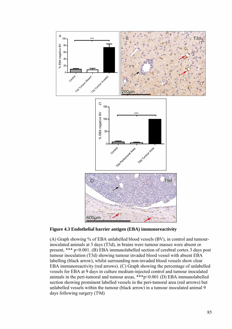
85
Contro
lT3d
Tumou
r Abs
ent
T3d Tum
our In
vade
d
0
20
40
60
80
100 ***A
% E
BA n
egat
ive
BV
Contro
lT9d
Peritum
oral A
reaT9d
Tumou
r Area
0
50
100
150
***
C%
EBA
neg
ativ
e BV
Figure 4.3 Endothelial barrier antigen (EBA) immunoreactivity
(A) Graph showing % of EBA unlabelled blood vessels (BV), in control and tumour-inoculated animals at 3 days (T3d), in brains were tumour masses were absent or present, *** p<0.001. (B) EBA immunolabelled section of cerebral cortex 3 days post tumour inoculation (T3d) showing tumour invaded blood vessel with absent EBA labelling (black arrow), whilst surrounding non-invaded blood vessels show clear EBA immunoreactivity (red arrows). (C) Graph showing the percentage of unlabelled vessels for EBA at 9 days in culture medium-injected control and tumour inoculated animals in the peri-tumoral and tumour areas, ***p<0.001 (D) EBA immunolabelled section showing prominent labelled vessels in the peri-tumoral area (red arrows) but unlabelled vessels within the tumour (black arrow) in a tumour inoculated animal 9 days following surgery (T9d)
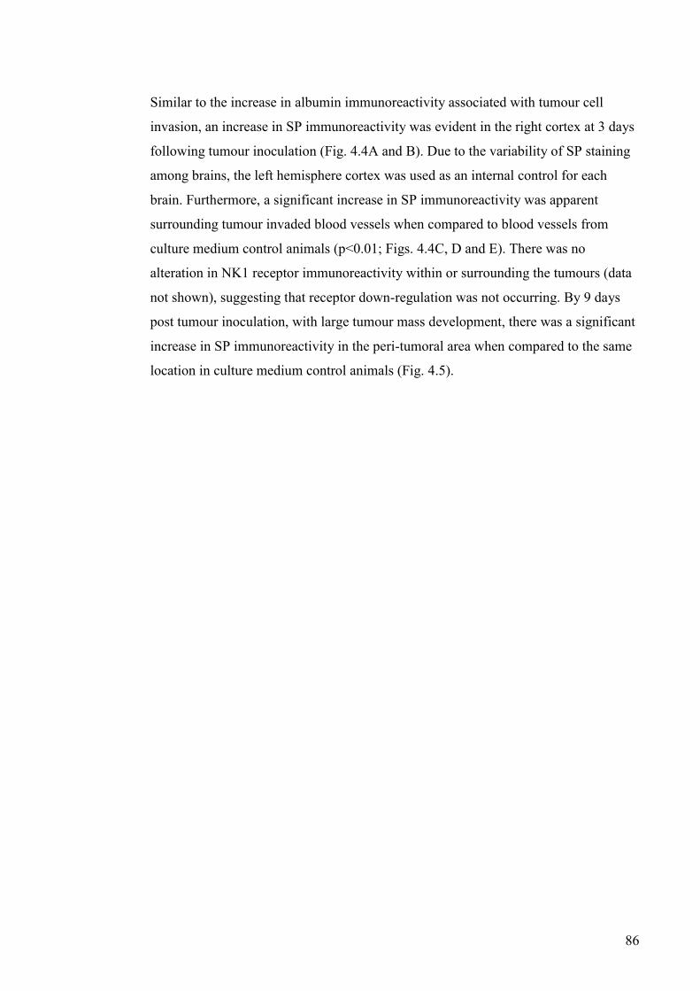
86
Similar to the increase in albumin immunoreactivity associated with tumour cell
invasion, an increase in SP immunoreactivity was evident in the right cortex at 3 days
following tumour inoculation (Fig. 4.4A and B). Due to the variability of SP staining
among brains, the left hemisphere cortex was used as an internal control for each
brain. Furthermore, a significant increase in SP immunoreactivity was apparent
surrounding tumour invaded blood vessels when compared to blood vessels from
culture medium control animals (p<0.01; Figs. 4.4C, D and E). There was no
alteration in NK1 receptor immunoreactivity within or surrounding the tumours (data
not shown), suggesting that receptor down-regulation was not occurring. By 9 days
post tumour inoculation, with large tumour mass development, there was a significant
increase in SP immunoreactivity in the peri-tumoral area when compared to the same
location in culture medium control animals (Fig. 4.5).

87
Contro
l
T1d T3d T6d T9d
0
50
100
150
200
A
Time Post Tumour Inoculation
Subs
tanc
e P
right
cor
tex
as %
of l
eft c
orte
x(D
ABw
t % to
tal M
ean
± SE
M)
Contro
lTum
our N
on-in
vade
d
Tumou
r-inva
ded
0
5
10
15
20 **C
*
Subs
tanc
e P
(DAB
wt %
tota
l Mea
n±
SEM
)
Figure 4.4 Substance P (SP) immunoreactivity with tumour invasion
(A) SP Immunoreactivity over time post tumour inoculation, right cortex as a percentage of the left cortex (B) SP immunolabelled coronal section 3 days post tumour inoculation showing increased immunoreactivity in the right cortex (arrow) (C) Graph showing SP immunoreactivity 3 and 6 days post tumour inoculation in areas of tumour-invaded compared with non-invaded blood vessels from the same animal and culture medium-injected control animals, *p<0.05, ** p<0.01 (D) Brain section from culture medium control animal 9 days following surgery (C9d), showing faint SP immunoreactivity around cortical blood vessels (E) Brain section from a tumour inoculated animal 3 days after inoculation (T3d) showing increased SP immunoreactivity in the neuropil around cortical microvessels.
88
Contro
lT9d
0
5
10
15 *A
Sub
stan
ce P
(DA
Bw
t % to
tal M
ean
± S
EM
)
Figure 4.5 Substance P (SP) immunoreactivity surrounding established brain
metastases
(A) Graph showing SP Immunoreactivity in the peri-tumoral area 9 days following tumour inoculation compared to control animals, *p<0.05 (B) SP immunolabelled section showing the striatum 9 days post culture medium injection (C9d), showing no increase in SP immunoreactivity (C) SP immunostained section showing a tumour and the increased SP staining in the peri-tumoral area 9 days post tumour inoculation (T9d)
89
4.5 Discussion
The model of brain metastases used in this study produced tumour invasion across the
BBB by day 3 following internal carotid artery inoculation. This time frame is
consistent with other studies using either internal or common carotid artery injection
methods of tumour induction (Ballinger 1979; Kienast 2010; Lorger 2011). Despite
the fact that previous studies have shown that tumour cells reach the brain
microvasulature within 15 minutes of internal carotid artery inoculation, tumour cells
still take at least 3 days to invade the BBB (Lorger 2010). Thus, tumour cells appear
to remain arrested in the vasculature when metastasising to the brain compared to
other organs like the liver or adrenals, which have been shown to have a tumour
extravasation time of 6 hours following intravenous inoculation of lung cancer cells in
nude mice (Paku 2000). In that study, initial tumour cell replication was located along
the inside of blood vessel walls which has been well established in the literature as the
method of tumour growth, regardless of tumour cell type or method of inoculation
(Carbonell 2009).
The fate and behavior of tumour cells within the circulation prior to forming brain
metastases has been the subject of several investigations. Some studies showed that
tumour cells replicate within the brain microvasculature before invading the brain
across the BBB, with one study reporting arrested non-proliferating breast cancer cells
by 33 days post intra-cardiac inoculation that had not been removed from the
circulation (Heyn 2006; Lorger 2011). On the other hand, it has been shown that
human lung and melanoma tumour cells that do not cross the BBB before day 3
following tumour inoculation do not go on to form brain metastases in an
experimental setting (Kienast 2010). The presence of intravascular tumour cells was
not investigated in the present study as all animals were fixed by intracardiac
perfusion before tissue processing.
The intra-ventricular location of the tumours growth in the current study is not
uncommon, with several other studies reporting tumour growth within the lateral

90
ventricles along with the more common malignant growth within the cortex
(Schackert 1988b; Schackert 1990). The preference for growth in the choroid plexus
may be a specific feature of the Walker 256 cell line, although the extravasation of
tumour cells also occurred across the cortical microvessels. Other investigators
reported that 58.3% of animals inoculated with Walker 256 cells into the internal
carotid artery demonstrated choroid plexus and meningeal tumours (Hasegawa 1983).
Coinciding with the first evidence of Walker 256 tumour cell invasion across the BBB
at 3 days post tumour inoculation, there was widespread albumin immunoreactivity
and an increase in SP immunoreactivity. The invaded microvessels showed
significantly increased perivascular SP immunoreactivity along with a significantly
reduced number of EBA labelled blood vessels. This indicates that tumour cell
extravasation across the BBB causes a modification to the properties of the BBB. It is
possible that the altered BBB permeability seen in the current study is induced by the
increased SP levels and the process of neurogenic inflammation. Alternatively,
tumour cells may preferentially invade brain microvessels that lack EBA
immunoreactivity and thus are more susceptible to barrier disruption.
The role of neurogenic inflammation and SP in tumour cell extravasation across the
BBB has not been investigated previously. However, the effects of neurogenic
inflammation in other BBB altering pathologies has been well characterised. SP is
known to be associated with BBB dysfunction leading to oedema in acute brain injury
(Donkin 2011; Turner 2011). The effects of SP on BBB permeability may be a
mechanism by which tumour cells modify the BBB to allow for extravasation. The
increase in SP seen in this study may have originated from cerebral endothelial cells,
being triggered to secrete SP upon interaction with tumour cells, or it may have been
secreted by the tumour cells themselves (Cioni 1998; Mayordomo 2011).
The increased SP immunoreactivity evident with tumour cell extravasation supports
further investigation into the possible use of NK1 antagonists as a preventative

91
treatment for brain metastatic breast cancer. The effects of SP on increased tumour
cell mitogenesis (Luo 1996; Friess 2003; Munoz 2004b; Munoz 2005b; Munoz 2007;
Munoz 2008; Rosso 2008; Huang 2010), migration (Ruff 1985; Drell 2003) and
angiogenic initiation (Ziche 1990; Wang 2009) have been well characterized, all
effects that lead to increased propensity for metastases (Munoz 2011a). Therefore, it
is not only possible that NK1 receptor antagonist treatment is able to reduce BBB
permeability and prevent tumour cell extravasation into the brain, but may also
provide additional protection from SP mediated initiation of a tumour cell phenotype
with increased metastatic potential.
This is the first study to report alterations in EBA labelling of brain microvessels in
response to tumour cell extravasation across the BBB. At early stages of tumour
invasion, angiogenesis is yet to occur, thus the lack of EBA staining reflects a
modification of the existing BBB rather than the growth of new blood vessels with
different properties to the BBB.
Albumin immunoreactivity decreased at 6 days following tumour cell inoculation
compared to 3 days post inoculation, but then increased again by day 9. This partial
resolution of increased BBB permeability at day 6 implies that the mechanisms of
BBB disruption are different at days 3 and 9 post tumour inoculation. It is likely that
the increase in BBB permeability, observed at 9 days, was caused by angiogenic
blood vessels within the tumour mass. Therefore, the absence of EBA labelling on the
angiogenic vessels that develop within the Walker 256 brain metastases at 9 days was
to be expected, as secondary brain tumours grow blood vessels that are characteristic
of their organ of origin rather than having the BBB characteristics of the host vessels.
Previous studies have gathered substantial evidence supporting the theory that blood
vessels within metastatic brain tumours are more permeable than the normal cerebral
vasculature (Greig 1983; Zhang 1992; Andersen 1998; Lockman 2010).

92
In contrast, the increase in albumin immunoreactivity at day 3 following tumour
inoculation, indicates increased permeability of the host BBB, and is likely to be due
to the interaction of tumour cells with endothelial cells of the BBB. This may be
consistent with the presumption that tumour cell extravasation into the brain occurs
through the paracellular pathway (Kienast 2010; Lorger 2010), and the increased
permeability of the BBB may aid tumour cell passage between cerebral capillary
endothelial cells. Pranlukast, a leukotriene receptor antagonist, has been successful in
reducing brain metastatic colon cancer development in other studies, but only when
the BBB was pre-treated with arachidonic acid causing increased BBB permeability
(Nozaki 2010). Therefore the modification of the BBB leading to increased
permeability may play a role in metastatic brain tumour extravasation in humans.
However, no studies have yet investigated preventative treatments for metastatic brain
tumours that impede tumour cell extravasation across the BBB under physiological
conditions in vivo.
With maximal tumour growth seen at 9 days post tumour inoculation, SP
immunoreactivity was increased in the peri-tumoral area. The compressive nature of
metastatic brain tumours means that damage to the host microenvironment commonly
occurs (Zhang 1997). The increased SP expression surrounding brain metastases may
be implicated in peri-tumoral oedema. SP has been linked to vasogenic oedema in the
brain and throughout the body following acute injury (Alves 1999; Donkin 2009), and
the use of NK1 receptor antagonists ameliorate this effect. A similar mechanism may
drive vasogenic oedema formation surrounding metastatic brain tumours. There has
been limited experimentation on the in vivo effect of NK1 receptor antagonists on
cancer, aiming to inhibit malignant growth and progression. Human glioma cells
injected subcutaneously into the flank of nude mice showed a decrease in tumour
volume when treated with a NK1 receptor antagonist (Palma 2000). However, this
study does not accurately replicate human gliomas as the tumour was grown in a non-
neural environment, thus preventing the study of the interactions of the NK1 receptor
antagonists with the BBB or the brain microenvironment.

93
In conclusion, the present study has demonstrated that the properties of the BBB are
altered during early stages of tumour cell extravasation, which presents a potential
window for therapeutic intervention to prevent the formation of metastatic brain
tumours. The increase in SP expression surrounding brain vessels associated with
tumour cells, combined with its known effects of increasing BBB permeability,
warrants further investigation into the role of SP in the formation of secondary brain
tumours.
94
5 NK1 antagonist treatment is not sufficient to prevent
Walker 256 breast carcinoma extravasation and
metastatic brain tumour development
5.1 Abstract
Metastatic brain tumours are increasing in incidence, however the exact mechanism
by which tumour cells traverse the blood-brain barrier (BBB) remains to be
elucidated. It is currently unclear why the BBB is unable to prevent the entry of
tumour cells into the brain. Substance P (SP) is an excitatory tachykinin that acts
preferentially on NK1 receptors and has been shown to increase the permeability of
the BBB. Previous studies have shown that SP is increased in the perivascular area of
Walker 256 breast carcinoma invaded microvessels, along with an increase in albumin
immunoreactivity. This suggests that SP may be involved in the tumour extravasation
process and thus NK1 antagonists may be a promising treatment for the prevention of
metastatic brain tumour formation. As such, the NK1 antagonists Emend and NAT
were tested in a haematogenic model of breast cancer-induced brain metastasis.
Walker 256 breast carcinoma cells were injected into the internal carotid artery and
Emend, NAT or saline vehicle treatment were administered on days 0-3 following
surgery. Tumour incidence and volume were used to determine the efficacy of the
NK1 antagonist treatment. There was no significant difference in tumour incidence or
volume with either NK1 antagonist treatment when compared to the vehicle treated
group. Therefore, the increase in SP with tumour invasion of brain microvessels
reported in previous studies is not the predominate factor driving brain colonisation
by cancer cells. It is likely that the extravasation process is multifactorial and that
blocking only a single factor is not sufficient to prevent tumour cells from crossing
the BBB.

95
5.2 Introduction
Secondary brain tumours occur in up to 30% of breast cancer cases (Schuette 2004;
Hines 2008). The development of increasingly effective targeted therapies for primary
breast cancer has resulted in increased survival time following initial diagnosis and
consequently an increase in the incidence of metastatic brain tumour. However, even
with the best available multimodal treatments, the prognosis remains poor. This is
because the location of metastases in the brain and the restricting function of the BBB,
making these neoplasms inherently difficult to treat.
Many factors contribute of metastatic progression of breast cancer to the brain,
including migration, angiogenesis and extravasation potential of cancer cells. Several
studies have shown that specific cancer types have a predilection for metastatic brain
tumour formation, for example melanoma, lung cancer and breast cancer (Barnholtz-
Sloan 2004; Villa 2011). Furthermore, cancer cells may acquire characteristics, which
increase propensity for brain invasion as they develop (Schackert 1988a; Fidler 1990;
Schackert 1990; Kusters 2001).
The development of metastasis in the brain is inherently dissimilar to metastases to
other organs because of the presence of the BBB. Extravasation into the brain requires
cancer cells to pass through the tight junctions between cerebral capillary endothelial
cells, and then through the basement membrane surrounding endothelial cells (Petty
2002). The exact mechanism of tumour cell extravasation into the brain is yet to be
elucidated and may lead to the development of treatment strategies to prevent brain
metastasis.
SP is a potent neurogenic inflammatory mediator, a process which is characterised by
passage of plasma proteins accompanied by fluid from the vasculature into
surrounding tissue (Harford-Wright 2011). Acting preferentially on NK1 receptors,
SP is able to increase the permeability of the BBB via endothelial cell contraction
96
along with decreased expression of tight junction proteins (Annunziata 1998;
Paemeleire 1999; Lu 2008).
Studies of a haematogenous model of brain metastasis have demonstrated increased
SP immunoreactivity surrounding tumour invaded microvessels in correlation with
albumin immunoreactivity in the neuropil, indicative of increased BBB permeability
(Lewis 2012b). These results, along with the known actions of SP on the BBB, make
SP a likely mediator of tumour cell extravasation into the brain. Therefore the current
study aims to test the efficacy of NK1 antagonists to prevent breast cancer
extravasation through the BBB and thus, metastatic brain tumour development.

97
5.3 Method
5.3.1 Animals
The current study was performed as detailed in section 2.2 page 43. They were
randomly selected for culture medium injection, tumour inoculation and treatment
groups, with all animal sacrificed 9 days following surgery (n=6 per group).
5.3.2 Cell culture
The Walker 256 breast carcinoma cell line was obtained from the Cell Resource
Centre for Medical Research at Tohoku University and cultured as detailed in section
2.1.2 page 42. Using a haemocytometer, the cell suspension was diluted so that 105-
106 walker 256 cells/0.2 mL of serum free media was ready for tumour inoculation
5.3.3 Internal carotid artery inoculation
The internal carotid inoculation procedure was previously described in section 2.3
page 44.
5.3.4 Treatment
The NK1 antagonists fosaprepitant dimeglumine (Emend, MERCK & CO) 3
mg/kg/day and n-acetyl L-tryptophan (NAT) 7.5 mg/kg/day were dissolved in 0.9%
saline solution and administered IP on days 0-3 following tumour inoculation. Equal
volumes of saline were used as a vehicle control. The treatment concentration of NAT
was determined based on a previous dose response, in which the dose required to
elicit maximal decrease in BBB permeability following diffuse traumatic brain injury
was evaluated (Donkin 2009). The dose for Emend was determined based on that used
clinically and previously shown to have a central effect in rodents (Watanabe 2008).

98
5.3.5 Immunostaining
Animals were euthanized and brains processed as detailed in 2.6 page 48. Slides from
each group were stained for SP (Santa Cruz Biotechnology, 1:2000) and albumin
immunoreactivity (ICN Pharmaceuticals, 1:20,000) using the method described in
section 2.8 page 49. These slides were scanned using a Hammamatsu nanozoomer
(Hammamatsu City, Japan) and then run through the colour deconvolution software.
This was done to estimate the %DAB in coronal sections of the brain for albumin
immunostaining and in four 0.0678 mm2 areas in the right cortex for SP
immunostaining. The use of colour deconvolution software has been described
previously (Helps 2012).
5.3.6 Tumour volume
Slides from each group were stained with haematoxylin and eosin and scanned using
a Hammamatsu nanozoomer so that the tumour area could be determined in each slide
using NDP viewer software as detailed in section 2.7 page 49.
5.3.7 Statistical analysis
A one-way analysis of variance followed by a Bonferroni post test performed to
determine statistical significance, with p values of <0.05 designated significant. All
data are expressed as mean ± SEM.
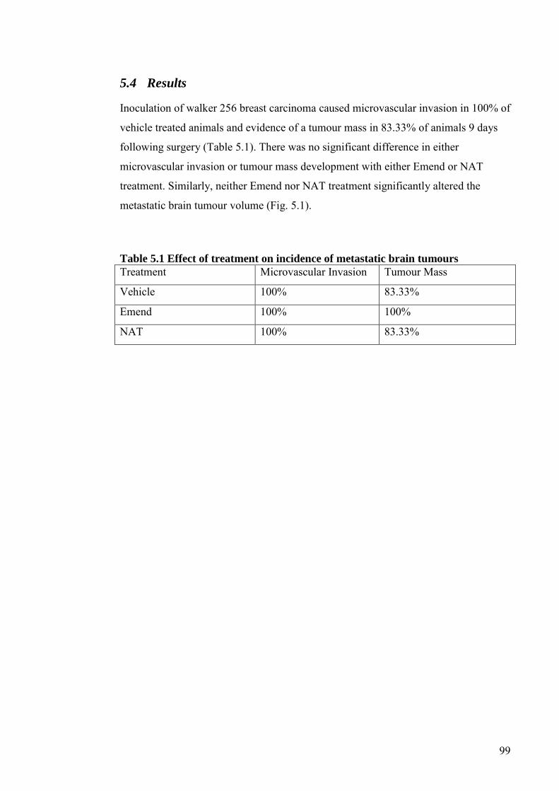
99
5.4 Results
Inoculation of walker 256 breast carcinoma caused microvascular invasion in 100% of
vehicle treated animals and evidence of a tumour mass in 83.33% of animals 9 days
following surgery (Table 5.1). There was no significant difference in either
microvascular invasion or tumour mass development with either Emend or NAT
treatment. Similarly, neither Emend nor NAT treatment significantly altered the
metastatic brain tumour volume (Fig. 5.1).
Table 5.1 Effect of treatment on incidence of metastatic brain tumours
Treatment Microvascular Invasion Tumour Mass
Vehicle 100% 83.33%
Emend 100% 100%
NAT 100% 83.33%
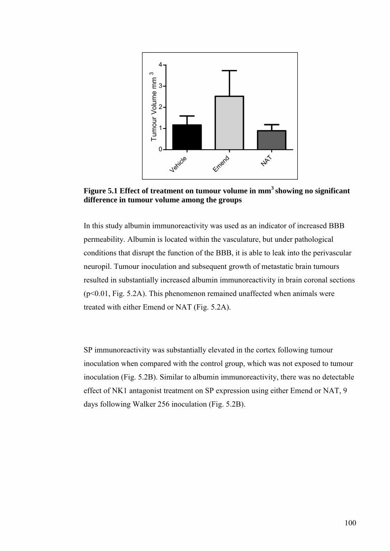
100
Vehicle
Emend
NAT
0
1
2
3
4
Tum
our V
olum
e m
m3
Figure 5.1 Effect of treatment on tumour volume in mm3
showing no significant
difference in tumour volume among the groups
In this study albumin immunoreactivity was used as an indicator of increased BBB
permeability. Albumin is located within the vasculature, but under pathological
conditions that disrupt the function of the BBB, it is able to leak into the perivascular
neuropil. Tumour inoculation and subsequent growth of metastatic brain tumours
resulted in substantially increased albumin immunoreactivity in brain coronal sections
(p<0.01, Fig. 5.2A). This phenomenon remained unaffected when animals were
treated with either Emend or NAT (Fig. 5.2A).
SP immunoreactivity was substantially elevated in the cortex following tumour
inoculation when compared with the control group, which was not exposed to tumour
inoculation (Fig. 5.2B). Similar to albumin immunoreactivity, there was no detectable
effect of NK1 antagonist treatment on SP expression using either Emend or NAT, 9
days following Walker 256 inoculation (Fig. 5.2B).

101
Contro
lTum
our +
Vehicl
eTum
our +
Emend
Tumou
r + N
AT
0
5
10
15
20
25
A
** *****
Albu
min
imm
unor
eact
ivity
%D
AB in
car
onal
sec
tion
Mea
n±
SEM
Contro
lTum
our +
Vehicle
Tumou
r + Emen
dTum
our +
NAT
0
5
10
15
20B
*
SP
imm
unor
eact
ivity
%D
AB
Mea
n±
SE
M
Figure 5.2 Albumin and substance P (SP) immunoreactivity
(a) Effect of tumour inoculation and treatment on %DAB representing albumin immunoreactivity in brain coronal sections when compared to the control group, **p<0.01, ***p<0.001 (b) Average Substance P (SP) immunoreactivity in four areas of 0.0678 mm2 in the right cortex, expressed as %DAB following tumour inoculation and NK1 antagonist treatment *p<0.05
On average, animals injected with culture medium alone as controls showed 19.9 g of
weight gain 9 days following surgery (Fig. 5.3). In contrast, tumour inoculation
caused a mean weight loss of 0.92 g during the same time period (Fig. 5.3). Treatment
with both NK1 antagonists reversed this, such that the animals gained approximately
8 g 9 days following surgery (Fig. 5.3).
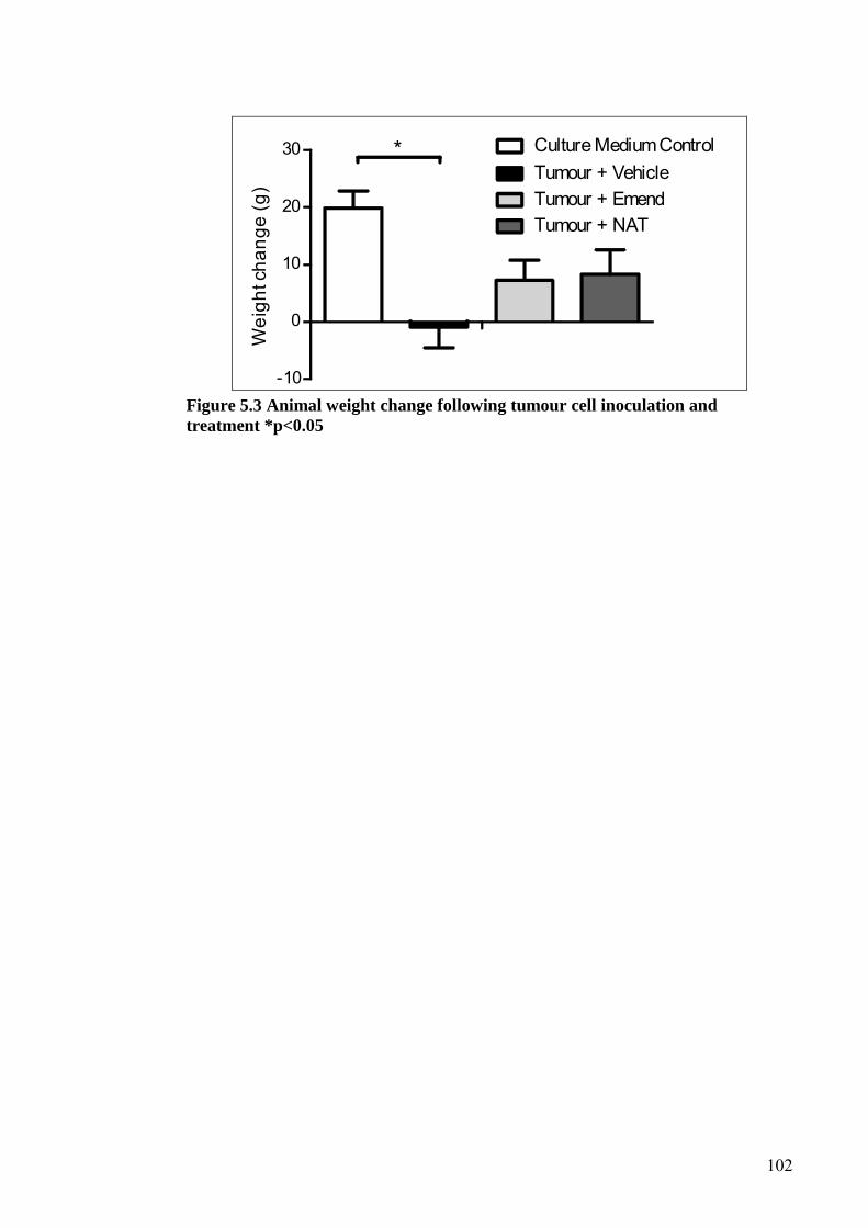
102
-10
0
10
20
30Tumour + VehicleTumour + EmendTumour + NAT
Culture Medium Control*
Wei
ght c
hang
e (g
)
Figure 5.3 Animal weight change following tumour cell inoculation and
treatment *p<0.05

103
5.5 Discussion
In this study using an internal carotid artery model of Walker 256 breast carcinoma
inoculation, treatment with NK1 antagonists did not prevent metastatic brain tumour
growth. This was despite the previous findings that SP immunoreactivity was
increased locally surrounding tumour invaded cerebral microvessels, which was
associated with increased BBB permeability as indicated by albumin
immunoreactivity in the neuropil (Lewis 2012b). Therefore the SP immunoreactivity
in conjunction with tumour cell extravasation is not the primary mediator of BBB
compromise that permits cancer cells to invade the brain microenvironment.
SP has previously been implicated in many processes involved in cancer growth and
metastatic progression. For example, the well-characterised effects of SP on tumour
cells in vitro, such as induction of tumour cell mitogenesis, migration, chemotactic
behaviour and cytokine secretion (Ruff 1985; Palma 1998; Lang 2004; Bigioni 2005;
Munoz 2005a). Furthermore, NK1 antagonist treatment has resulted in decreased
oedema in rodent models of traumatic brain injury and stroke through resolution of
BBB disruption (Donkin 2009; Turner 2011).
In contrast, the results of the current study suggest that extravasation is a
multifactorial process, in which the role of SP is not of sufficient magnitude for its
blockade by NK1 antagonist treatment to result in prevention of metastatic brain
tumour formation. Tumour progression involves the expression of multiple adhesion
molecules such as integrins and selectins, and matrix metalloproteases (Li 1993; Lu
2001; Mendes 2005; Mendes 2007). Furthermore, vasoactive inflammatory
substances may be required for extravasation into the brain to take place. It is
plausible that blocking any one of these activities would be ineffective for metastatic
brain tumour prevention, rather multiple pathways may need to be targeted.

104
Although this study deals solely with the neurogenic inflammatory peptide SP, there
are many other vasoactive substances that may contribute to increased BBB
permeability along with tumour cell extravasation into the brain microenvironment
(Lewis 2012b). Classical inflammation, characterised by signals for accumulation and
proliferation of microglia and perivascular macrophages, has been implicated in BBB
disruption that results in oedema development associated with metastatic brain
tumours (Lewis 2012a), making bradykinin a possible candidate for involvement in
the passage of tumour cells between cerebral capillary endothelial cells.
Similarly, vascular endothelial growth factor has also been proposed to play a role in
the progression of cancer to the brain, due to the evidence of its involvement in
angiogenesis and oedema development in animal models of brain metastases (Wang
1996; Batchelor 2007). However, blockade of vascular endothelial growth factor also
failed to reduce the incidence of secondary brain tumours (Juanyin 2009). It is
possible that multiple vasoactive inflammatory mediators are involved in brain
metastases development and as such, agents that act through multiple pathways may
be more effective as prophylactic agents for secondary brain tumour progression.
In the current study, albumin immunoreactivity, which was elevated with tumour
inoculation, was not altered with either Emend or NAT treatment. This is likely a
reflection of the similar tumour volume evident in the treatment groups, as the link
between tumour volumes and vascular permeability is well documented (Zhang
1992). This result also reflects that SP alone is not responsible for the increased BBB
permeability evident with metastatic brain tumour growth in this model.
By 9 days following walker 256 breast carcinoma inoculation into the internal carotid
artery, animals lost weight, contrasting sharply with the culture medium inoculated
animals and also both NK1 antagonist treatment groups which gained weight
following surgery. The reversal of tumour associated weight loss by NK1 antagonist
treatment has not been reported previously. However, both weight loss and weight

105
gain have been reported as a rare side effect of Emend treatment when co-
administered with other agents for the treatment of chemotherapy induced emesis
(Kramer 2004).
In conclusion, NK1 antagonist treatment was not sufficient to prevent metastatic brain
tumour formation in this model. Therefore, this study demonstrates that SP does not
mediate Walker 256 breast cancer extravasation into the brain following internal
carotid artery inoculation. This is despite the increased SP expression evident in
association with tumour invasion of brain microvessels and the associated increase in
BBB permeability to albumin. Thus, it is plausible that extravasation of Walker 256
tumour cells across the BBB is a multifactorial process, the inhibition of which would
require multiple pathways to be targeted.
106
6 Targeting classical but not neurogenic inflammation
reduces peritumoral oedema in secondary brain tumours
6.1 Abstract
Cerebral metastases are associated with the development of substantial vasogenic
peritumoral oedema formation, which is the source of many neurological
complications associated with secondary brain malignancies. The current standard
treatment for peritumoral brain oedema is dexamethasone, which inhibits classical
inflammation, however it is associated with numerous adverse side effects.
Neurogenic inflammation has been implicated in vasogenic oedema formation
following animal models of reperfusion ischemic stroke and traumatic brain injury
through the actions of substance P (SP). SP acts preferentially on NK1 receptors, the
blockade of which may provide an alternative treatment to dexamethasone for
peritumoral oedema. Thus, this study investigates the efficacy of NK1 receptor
antagonist treatment, in comparison with dexamethasone treatment, for the
management of peritumoral brain oedema in a rat model of intrastriatal inoculation of
Walker 256 rat breast carcinoma cells. Brain water content, albumin
immunoreactivity and Evan’s blue extravasation were evaluated at 7 days following
tumour inoculation with culture medium injected animals used as controls.
Dexamethasone was effective in reducing brain water content and caused a reduction
in Evan’s blue extravasation, whilst NK1 receptor antagonist treatment groups had no
effect on oedema formation. Thus, we conclude that classical inflammation rather
than neurogenic inflammation drives peritumoral oedema in this model.

107
6.2 Introduction
Cancer results in the death of more than 6 million people each year (WHO 2002). At
autopsy 20% of cancer patients will be found to have suffered from a metastatic brain
tumour, whilst clinical studies commonly describe an incidence of approximately
10% (Posner 1978; Cifuentes 1979; Gavrilovic 2005). It is estimated that 170 000
people per year in America suffer from at least one metastatic brain tumour, with
metastatic brain tumours being 10 times more common than primary cerebral
malignancies (Landis 1998).
Cerebral oedema is a common complication of metastatic brain tumours, which
results in an increase in the water content within the skull and subsequent increase in
intracranial pressure. This may lead to localized ischemia and brain herniation
through compression adjacent structures. The mass effect of tumour and the addition
of oedematous fluid often cause many neurological symptoms. These may include
blindness, cognitive deficit, weakness, headaches and aphasia (Mukand 2001; Ayata
2002; Shinoura 2010).
Angiogenic blood vessels that grow within metastatic brain tumours have the same
characteristics of their tissue of origin (Cornford 1992; Shuto 2008). Thus, these
blood vessels exhibit different properties from those of the blood-brain barrier (BBB).
It is widely accepted that the angiogenic vessels, which form the blood-tumour barrier
(BTB) are more permeable to serum proteins, facilitating the development of
peritumoral oedema (Front 1984; Zhang 1997). As such, peritumoral oedema is
related to the size of the neoplasm and the extent of angiogenesis (Zhang 1992). Both
the BBB and BTB are dynamic structures with barrier properties that may vary in
response to external stimuli, including vasoactive substances (Zhang 1997; Turner
2007). Consequently, vasoactive substances that are able to decrease the permeability
of capillaries within the brain could result in reduced peritumoral brain oedema.
108
Dexamethasone is the current standard treatment for brain tumour-associated cerebral
oedema. Since the introduction of dexamethasone in 1962, there has been a significant
decrease in deaths related to this pathology (Jelsma 1967). Approximately 70% of
metastatic brain tumour patients receive dexamethasone treatment whilst they undergo
radiotherapy (Hempen 2002). The effects of dexamethasone on oedema have been
extensively studied, but contradictory results have prevented the exact mechanism of
action being fully elucidated. It is thought that dexamethasone blocks classical
inflammation, acting via glucocorticoid receptors to decrease the permeability of brain
and tumoral microvessels, thus leading to a reduction in cerebral oedema (Andersen
1994a; Heiss 1996; Andersen 1998; Sinha 2004). Unfortunately, dexamethasone
treatment is associated with many harmful side-effects including insomnia, immune
suppression (Lesniak 2004), hyperglycemia (McGirt 2008)and occasional psychosis
(Alpert 1986)..
Neurogenic inflammation is characterised by vasoactive neuropeptide release causing
vasodilatation, plasma extravasation and subsequent oedema formation (Woie 1993).
Substance P (SP) is a potent neurogenic inflammatory mediator, causing increased
BBB permeability when it acts on NK1 receptors expressed throughout the central
nervous system (Cioni 1998; Paemeleire 1999; Annunziata 2002; Lu 2008). The
modulatory effect of SP on the BBB is involved in the pathogenesis of peri-lesional
cerebral oedema in ischemic stroke and traumatic brain injury. Treatment with NK1
receptor antagonists in these conditions results in reduction of water content and
improved functional outcome (Nimmo 2004; Vink 2004; Turner 2006; Turner 2007;
Donkin 2009; Harford-Wright 2010).
The neurally induced release of SP and its effect on the BBB and oedema formation in
other brain pathologies makes it a likely candidate as a mediator of peritumoral
oedema formation in metastatic brain tumours, especially given recent reports of
increased SP in and around CNS tumours (Palma 2000) . The aim of the current study
was to use an intracerebral injection model to produce secondary neoplasms of
consistent size and location, and to elucidate the role of neurogenic inflammation in

109
the pathogenesis and development of cerebral oedema associated with these
secondary brain tumours.
110
6.3 Methods
6.3.1 Animals
Animal procedures were performed as described in section 2.2 on page 43. Six
animals for each treatment group were randomly selected for the immunostaining
study, brain water content study and Evans blue extravasation study.
6.3.2 Cell culture
Walker 256 rat breast carcinoma cells were obtained from the Cell Resource Centre
for Medical Research at Tohoku University, Japan and cultured according to the
description in section 2.1.2 page 42. The cells were counted using a hemocytometer
and then diluted, so that between 105 and 106 cells/8 µL of serum free RPMI-1640
media were used for direct injection into the brain.
6.3.3 Tumour inoculation
The direct inoculation of tumour cells has been described in section 2.4 page 47.
6.3.4 Treatment
All compounds were dissolved in 0.9% saline solution, which was also used as
vehicle control. On days 4-6 following tumour inoculation, groups of animals were
given a daily intraperitoneal injection with one of the following agents: the NK1
antagonist fosaprepitant dimeglumine (EmendR, MERCK & CO) 3mg/kg/day, the
NK1 antagonist n-acetyl L-tryptophan (NAT) 7.5 mg/kg/day, DBL dexamethasone
sodium phosphate 8 mg/kg/day, or saline vehicle (as controls). All animals were
sacrificed on day 7 following tumour inoculation. The dose of 8 mg/kg/day over three
days for dexamethasone was used because this had previously been effective in
ameliorating cerebral oedema in a rat model of primary brain tumour (Gu 2007a). The
concentration of NAT was determined from a previous study where a dose response
was performed and 2.5 mg/kg of intravenously administered NAT caused maximal
resolution of BBB permeability following traumatic brain injury (Donkin 2009). This
111
dose was tripled to allow for intraperitoneal administration, as used for
dexamethasone treatment. Emend was given at three times the dose recommended
clinically for IV administration that has also been used previously with central effects
in animal models (Watanabe 2008).
6.3.5 Immunostaining
Brains were analysed for histology as described in section 2.6 page 48. Slides from
each treatment group were stained for substance P (Santa Cruz Biotechnology,
1:2,000) and albumin (ICN Pharmaceuticals, 1:20,000) as detailed in section 2.8 page
49. Immunostained slides were scanned using the nanozoomer (Hammamatsu,
Hammamatsu City, Japan) and objective assessment of the immunocytochemical
staining was achieved through colour deconvolution techniques, to reveal the %DAB
in the scanned slides. The colour deconvolution technique has been described
previously (Harford-Wright 2010; Helps 2012). Whole coronal sections for albumin-
stained brain sections, and peritumoral areas (0.0678mm2) from SP immunostained
sections were taken and run through the colour deconvolution software to
automatically estimate the % of brown stain in the selected area and through this
process removes background staining variations.
6.3.6 Brain Water Content
The wet weight-dry weight method was used to calculate brain water content in order
to quantify the effect of treatment on peritumoral oedema as described in section 2.9
on page 50.
6.3.7 Evans blue extravasation
Animals were injected intravenously with 0.8 ml of 4% Evans blue (MW 69,000;
Sigma, E-2129) 30 minutes before they were perfused transcardially with saline under
general anaesthesia induced by pentobarbitone sodium (60mg/kg). This has been
described in detail in section 2.10 page 50.

112
6.3.8 Statistical Analysis
Data were expressed as mean ± SEM. To determine statistical significance, an
unpaired two tailed t test (for two groups) or one-way analysis of variance followed
by a Bonferroni post test (for more than two groups) was performed as applicable,
with p<0.05 designated as significant.

113
6.4 Results
Tumour inoculation produced large consistent tumours by day 7. All animals were
sacrificed at this time point with 100% of animals showing evidence of tumour
burden upon histological analysis. Walker 256 implantation models of secondary
brain tumours have been described previously in the literature (Yamada 1983;
Jamshidi 1992; Morreale 1993). However tumours in the current study grew quicker
than those reported previously. No adverse effects for any treatment were seen, and
tumour inoculated animals showed maximum weight loss of 19.1% of their body
weight. There was no animal mortality associated with this model, 100% of animals
survived until the designated euthanasia date. Treatment over 3 days was pre-
determined based on the previous use of dexamethasone to treat brain tumour
associated oedema in the literature (Gu 2007a). Days 4, 5 and 6 following tumour
inoculation were chosen for treatment so that tumours were well established prior to
its commencement.
6.4.1 SP immunoreactivity
Tumour inoculation caused a significant increase in SP immunoreactivity evident in
the peritumoral area, when compared to the same location of control brains injected
with culture medium (**p<0.01) (Fig. 1A - C). Within the tumour mass SP
immunoreactivity was significantly lower than both the peritumoral area of the same
animals (***p<0.001) and the striatum of control animals (*p<0.05) (Fig. 1A - C).
This increase in SP immunoreactivity in the peri-tumoral area was not altered by any
of the treatment regimes used in this study (data not shown).
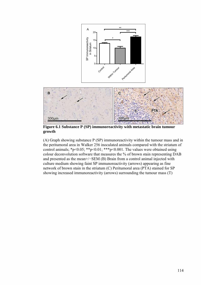
114
Contro
l
Within
Tumou
rPeri
tumora
l Area
0
5
10
15
20
*
*****
A
SP Im
mun
orea
ctiv
ityin
Stri
atum
Figure 6.1 Substance P (SP) immunoreactivity with metastatic brain tumour
growth
(A) Graph showing substance P (SP) immunoreactivity within the tumour mass and in the peritumoral area in Walker 256 inoculated animals compared with the striatum of control animals; *p<0.05; **p<0.01; ***p<0.001. The values were obtained using colour deconvolution software that measures the % of brown stain representing DAB and presented as the mean+/−SEM (B) Brain from a control animal injected with culture medium showing faint SP immunoreactivity (arrows) appearing as fine network of brown stain in the striatum (C) Peritumoral area (PTA) stained for SP showing increased immunoreactivity (arrows) surrounding the tumour mass (T)

115
6.4.2 Brain water content
Tumour inoculation caused a significant increase in brain water content when
compared to injection of culture medium alone (p<0.05, Fig. 2). This was not resolved
following treatment with Emend or NAT, but dexamethasone treatment reduced brain
water content to the level of the culture medium control group (Fig. 2).
Contro
lTum
our +
Vehicle
Tumou
r + Emen
dTum
our +
NAT
Tumou
r + D
ex
78
79
80
81
82
****
Treatment
Bra
in W
ater
Con
tent
(%
)
Figure 6.2 Brain water content
Graph showing brain water content as a percentage (%) of brain weight in tumour-inoculates and control animals. NAT, n-acetyl L-tryptophan; Dex, dexamethasone. The values represent the mean+/−SEM; *p<0.05; ***p<0.001
6.4.3 Blood-brain barrier permeability
Evans blue was used as an indicator of BBB permeability along with albumin
immunoreactivity. Albumin is located within the blood vessels and not in the neuropil
of the brain under normal conditions. However pathological conditions that increase
the permeability of the BBB allow albumin to leak out of the cerebral vasculature and
into the brain tissue. Evans blue binds to serum albumin and is thus used as an
exogenous indicator of BBB permeability at the time of tracer application. Albumin
immunoreactivity was used as an endogenous indicator of BBB permeability over a
longer period of time. Evans blue extravasation was increased in all tumour-injected
and treated groups when compared to the culture medium control group (Fig. 3).
However, there was a small non-significant decrease in Evans blue extravasation in

116
the dexamethasone treated group compared to other treatments (Fig. 3). Albumin
immunoreactivity was significantly elevated in all tumour-inoculated groups injected
with the vehicle (p<0.001), Emend (p<0.001), NAT (p<0.001) and dexamethasone
(p<0.05), when compared with the control animals injected with culture medium
alone (Fig. 4A-F). Similar to Evans blue, there was a slight decrease in albumin
immunoreactivity in the dexamethasone treated brains compared to other treatments
(Fig. 4A). Figure 4E and 4D show more extensive peritumoral albumin
immunoreactivity in the NAT treated brain when compared to the Emend treated
brain, likely due to the variation in tumour size. However, there was no significant
difference seen between these groups using colour deconvolution of albumin
immunoreactivity.

117
Contro
lTum
our +
Veh
icleTum
our +
Emen
dTum
our +
NAT
Tumou
r + D
ex
0
1
2
3
Treatment
Evan
s Bl
uemg
/g B
rain
Tiss
ue
Figure 6.3 Evans blue extravasation
Graph showing Evans blue concentration in brain tissue (μg/g) of controls injected with culture medium only and tumour-inoculated animals injected with saline vehicle, Emend, n-acetyl L-tryptophan (NAT) and dexamethasone (Dex). The values represent the mean+/−SEM
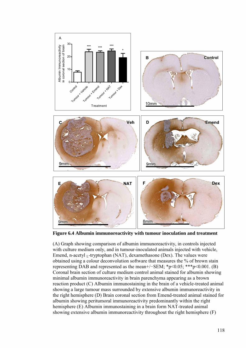
118
Contro
lTum
our +
Vehicle
Tumou
r + Emen
dTum
our +
NAT
Tumou
r + D
ex
0
10
20
30*** *** ***
*
Treatment
Alb
umin
Imm
unor
eact
ivity
in c
oron
al s
ectio
n of
bra
in
A
Figure 6.4 Albumin immunoreactivity with tumour inoculation and treatment
(A) Graph showing comparison of albumin immunoreactivity, in controls injected with culture medium only, and in tumour-inoculated animals injected with vehicle, Emend, n-acetyl L-tryptophan (NAT), dexamethasone (Dex). The values were obtained using a colour deconvolution software that measures the % of brown stain representing DAB and represented as the mean+/−SEM; *p<0.05; ***p<0.001. (B) Coronal brain section of culture medium control animal stained for albumin showing minimal albumin immunoreactivity in brain parenchyma appearing as a brown reaction product (C) Albumin immunostaining in the brain of a vehicle-treated animal showing a large tumour mass surrounded by extensive albumin immunoreactivity in the right hemisphere (D) Brain coronal section from Emend-treated animal stained for albumin showing peritumoral immunoreactivity predominantly within the right hemisphere (E) Albumin immunostaining in a brain form NAT-treated animal showing extensive albumin immunoreactivity throughout the right hemisphere (F)

119
Coronal brain section from a dexamethasone-treated animal stained for albumin showing a small tumour with widespread immunoreactivity in the peritumoral area

120
6.5 Discussion
This study showed that inoculated tumour cells produced large, consistent tumour
masses that increased brain water content and barrier permeability. For this reason,
the model used in this study was suited to the investigation of potential therapeutic
benefits of NK1 antagonism on peritumoral cerebral oedema. Direct injection models
of secondary brain tumours have been frequently used previously to test treatments
for tumour associated cerebral oedema (Yamada 1983; Jamshidi 1992; Engelhorn
2009). The requirement of the model was that enough oedema be produced by the
tumour growth, such that a therapeutic intervention could be effective and that the
results be measurable. This is evident in the current study by the significant decrease
in brain water content seen in the dexamethasone treated group when compared to the
vehicle treated group. Direct injection of tumour cells into the brain bypasses the
usual route of tumour cell invasion through the BBB in human metastases. Models of
internal carotid artery or intra-cardiac injection of tumour cells to produce metastatic
brain tumours allow for the study of extravasation through the BBB, although these
models often produce multiple secondary brain tumours without predictable location
(Ushio 1977; Hasegawa 1983; Song 2011; Budde 2012).
BBB permeability was measured by Evans blue extravasation and albumin
immunoreactivity in the brain parenchyma. By seven days following tumour
inoculation, brain water content was 0.8% above the control level of 79.1%. While
significant, this elevation in brain water content is less than that seen in some other
CNS pathologies reporting vasogenic oedema. For example, water content increases
of 3.6%, 3.2% and 2.2% have been reported for rat models of ischaemic reperfusion
stroke, intracerebral haemorrhage and traumatic brain injury respectively (Nimmo
2004; Turner 2006; Li 2009). However, the brain water content increase seen in the
current study is comparable to the 0.87% elevation observed in a rat model of
subarachnoid haemorrhage (Barry 2011). The substantially larger percentage change
in brain water content for models of trauma and reperfusion ischemic stroke may be
indicative of a different pathogenesis associated with oedema formation compared to
that seen in the current study and in models of subarachnoid haemorrhage. Thus, the
extent of increase in brain water content may be linked to the degree to which

121
neurogenic inflammation contributes to breakdown of the BBB in different models of
neurological diseases.
NK1 receptor antagonist treatment did not change the brain water content, albumin
immunoreactivity or Evans blue extravasation when compared to vehicle treated
controls. These results were seen despite the increase in SP immunoreactivity evident
in the peritumoral area. Therefore it is possible that the increase in SP expression was
not of sufficient magnitude to be the primary mediator of cerebral oedema formation
in this model. These results contrast with other studies using NK1 receptor
antagonists to block neurogenic inflammation and treat vasogenic oedema. NAT has
previously been used to decrease brain oedema and improve functional motor
outcome after experimental traumatic brain injury and ischemic reperfusion stroke
(Donkin 2009; Donkin 2011; Turner 2011). In contrast, NK1 antagonists have not
been shown to be effective in reducing vasogenic oedema associated with
subarachnoid haemorrhage (Barry 2011). This suggests that the mechanisms of
peritumoral vasogenic oedema formation in the current study and that observed in
subarachnoid haemorrhage are likely to be non-neurogenic, and distinctly different
from the vasogenic oedema following stroke and traumatic brain injury, in both of
which NK1 receptor antagonists appear to be more effective as treatment.
Both classical inflammation and neurogenic inflammation involve increased
permeability of the BBB, thus both have the potential to mediate cerebral oedema in
many neurological pathologies. Neurogenic inflammation typically involves the
release of SP and calcitonin gene related peptide from primary sensory nerve endings
resulting in vasodilatation and plasma extravasation (Nimmo 2009). Thus in this
study, the failure of NK1 receptor antagonist treatment to ameliorate peritumoral
oedema indicates that its formation is not mediated by SP-driven neurogenic
inflammation.
122
Dexamethasone is a classical anti-inflammatory agent, having previously been shown
to decrease bradykinin and prostaglandin E2 production by white blood cells in cattle
(Myers 2010). In the CNS, classical inflammation is characterised by accumulation
and proliferation of microglia along with perivascular macrophages (Graeber 2011).
This leads to blood vessel alterations driven by classical inflammatory mediators like
bradykinin (Donkin 2010). The well-documented effects of dexamethasone in treating
peritumoral oedema, also seen in the current study, suggest that classical
inflammation is the mechanism behind peritumoral oedema formation.
In the current study, treatment with dexamethasone was used as a positive control to
determine if the model of brain metastases used here produces enough peritumoral
oedema for a treatment intervention to have an effect. Treatment of animals with
dexamethasone has previously been used effectively in models of vasogenic oedema
to reduce brain water content, BBB permeability and other measures of cerebral
oedema (Betz 1990; Guerin 1992). The outcomes associated with dexamethasone
treatment are thought to be through its actions on glucocorticoid receptors, with
modulation of VEGF (Heiss 1996; Kim 2008) and occludin (Forster 2006; Gu 2009a)
proposed to play a role in its activity. Despite the undefined mechanism of action,
dexamethasone has been shown to decrease transendothelial fluid movement and
extravascular fluid volume (Nakagawa 1987; Andersen 1998). However, the benefits
of improved fluid homeostasis were not sufficient to improve survival when animals
bearing U87 or C6 intracranial gliomas were treated with dexamethasone (Moroz
2011).
The decrease in brain water content in the dexamethasone-treated group was
comparable to that seen in the control group injected with culture medium only. In
contrast, the decrease in Evans blue and albumin immunoreactivity with
dexamethasone treatment was non-significant and was elevated above that seen in the
control groups. These data suggest that the mechanisms of extravasation of water and
proteins components of oedema are different, and that the mechanism of
dexamethasone-induced resolution of peritumoral oedema is only partially mediated

123
by decreasing the permeability of the BBB. Therefore, further investigation is needed
to elucidate the specific mechanistic effects of dexamethasone on peritumoral
oedema.
In conclusion, the results of this study demonstrate that dexamethasone is more
effective in treating peritumoral oedema than the NK1 receptor antagonists. This
suggests that the pathogenesis of peritumoral oedema may be more related to classical
inflammation, rather than neurogenic inflammation driven by substance P.

124
7 NK1 receptor antagonists and dexamethasone as
anticancer agents in vitro and in a model of brain
tumours secondary to breat cancer
7.1 Abstract
Emend, a NK1 antagonist, and dexamethasone are used to treat complications
associated with metastatic brain tumours and their treatment. It has been suggested
that these agents have anti-cancer effects apart from their current use. Effects of the
NK1 antagonists, Emend and n-acetyl L-tryptophan (NAT), and dexamethasone on
tumour growth were investigated in vitro and in vivo at clinically relevant doses. For
animal experiments, a stereotaxic injection model of Walker 256 rat breast carcinoma
cells into the striatum of Wistar rats was used. Emend treatment caused a decrease in
tumour cell viability in vitro, although this effect was not replicated by NAT.
Dexamethasone did not decrease tumour cell viability in vitro but decreased tumour
volume in vivo, likely to be through a reduction in tumour oedema, as indicated by
the increase in tumour cell density. None of the agents investigated altered tumour
cell replication or apoptosis in vivo. Inoculated animals showed increased GFAP and
IBA1 immunoreactivity indicative of astrocytes and microglia in the peritumoral area,
while treatment with Emend and dexamethasone reduced the labelling for both glial
cells. These results do not support the hypothesis that NK1 antagonists or
dexamethasone have cytotoxic action on tumour cells, although these conclusions
may be specific to this model and cell line.
125
7.2 Introduction
Malignancies of the CNS have shown an increased incidence in recent years, although
mortality rates have plateaued (Bray 2010). Breast cancer is the most prevalent cancer
type in women and the second most common cancer type to cause metastatic brain
tumours after lung cancer, accounting for approximately 20% of all secondary brain
tumours (Schouten 2002; AIHW 2010; Villa 2011) and causing a significant patient
morbidity and mortality, with survival time commonly in the order of months
(Sperduto 2012).
While curative treatments for metastatic brain tumours remain elusive, anti-
inflammatory agents are commonly prescribed to patients with secondary brain
tumours, to control symptoms associated with tumour complications and treatment
side effects. Dexamethasone is a synthetic glucocorticoid administered to patients
with brain metastases to reduce neurological symptoms related to the mass effect of
peri-tumoral oedema. Since it was first used in 1962 it contributed to a significant
reduction in mortality in brain tumour patients (Jelsma 1967). However, the beneficial
effects of dexamethasone treatment are limited by its associated side effects, including
suppression of the immune system (Lesniak 2004), hyperglycaemia (McGirt 2008)
andoccasionallypsychosis (Alpert 1986).
Aprepitant, also known as L-754,030 and its intravenous prodrug fosaprepitant
diglutemide also termed L-758,298 (Emend), is an NK1 receptor antagonist used as
an antiemetic, to control chemotherapy induced nausea in many cancer patients; it is
the only NK1 antagonist that has been approved for use in humans (Hesketh 2003;
Herrstedt 2005; Warr 2005; Ruhlmann 2012). The mechanism of chemotherapy-
induced nausea is thought to be through neurotransmitter release in the
gastrointestinal tract and in the central nervous system, with the vomiting centre and
chemoreceptor trigger zone in the medulla oblongata being particularly affected
(Navari 2004a). It is thought that NK1 antagonism has inhibitory activity on this
process both centrally and peripherally (Navari 2004b). Emend is commonly co-
126
administered with dexamethasone, and maximum benefit is seen when combined with
5-HT3 receptor-antagonist (Hesketh 2006). Despite the common use of
dexamethasone and Emend in cancer patients, their effect on tumour growth remains
controversial.
It has been suggested that dexamethasone may also act to control cancer growth
(Villeneuve 2008; Moroz 2011). Dexamethasone has been shown to reduce brain
tumour volume in vivo in murine models of brain tumours (Guerin 1992; Wolff 1993;
Badruddoja 2003; Villeneuve 2008; Moroz 2011), although it remains unclear if this
results from decreased oedematous fluid or decreased tumour cell viability and
proliferation (Guerin 1992; Wolff 1993; Badruddoja 2003; Villeneuve 2008). Indeed,
the effect of dexamethasone on tumour cell proliferation and apoptosis in vivo has not
been investigated. Several in vitro studies have found that dexamethasone has an anti-
proliferative, pro-apoptotic effect on cancer cells (Bavaresco 2007; Piette 2009; Tazik
2009), while in other apparently conflicting in vitro studies dexamethasone was not
always able to inhibit tumour cell growth (Wolff 1993; Villeneuve 2008). Despite the
consistent use of dexamethasone as the standard treatment for peritumoral brain
oedema for many years, its exact mechanism of action remains unclear. Furthermore,
dexamethasone treatment is often investigated in conjunction with other
chemotherapy treatments, meaning that its mechanism of action alone remains unclear
(Kim 2004; Sur 2005; Zhang 2006; Moroz 2011).
Several studies have demonstrated an increase in NK1 receptor expression in human
surgical specimens of astrocytoma and brain metastases from breast carcinomas and
melanoma (Allen 1985; Hennig 1995; Khare 1998; Singh 2000; Schulz 2006; Huang
2010). Substance P (SP) is a pro-inflammatory tachykinin that acts preferentially on
NK1 receptors. Recent studies have implicated SP in the proliferation and progression
of many cancer types (Munoz 2011b). In cell culture studies, SP has consistently been
shown to induce tumour cell mitogenesis, with NK1 antagonists causing apoptosis
(Friess 2003; Munoz 2004a; Bigioni 2005; Munoz 2005a; Munoz 2005b; Munoz
2007; Rosso 2008; Akazawa 2009; Huang 2010) and decreased mitogenesis,

127
particularly on NK1 receptor-expressing human cancer cell lines, while non-
neoplastic cell lines did not exhibit these effects (Singh 2000). Thus, it has been
suggested that the NK1 receptor antagonist Emend may not only be useful in the
treatment of chemotherapy-induced emesis, but may also inhibit SP-induced tumour
cell proliferation. Therefore, Emend may aid in the treatment of cancer itself, as well
as the side effects of chemotherapeutic agents (Kast 2009; Munoz 2010b; Harford-
Wright 2011). However, the literature shows conflicting results. While some studies
showed that NK1 antagonists or inhibition of SP decrease tumour growth (Palma
2000; Erin 2008; Korcum 2009), nearly an equal number of studies demonstrated that
SP inhibits tumour growth through its stimulatory effects on the immune system
(Harris 2003; Erin 2004; Manske 2005; Erin 2006). The current study was undertaken
to determine the effects of NK1 antagonism and dexamethasone on breast carcinoma
cell growth in vitro and in vivo.
128
7.3 Materials and Methods
7.3.1 Cell Viability Assay
Walker 256 rat breast carcinoma cells were obtained from the Cell Resource Centre
for Medical Research at Tohoku University were cultured as described in section
2.1.2 page 42. A cell viability assay was then performed as detailed in section 2.1.3 on
page 43.
7.3.2 Cell Culture for Inoculation
Cells growing in 150 cm2 culture flasks were passaged once >90% confluence was
reached using 3.5 mL of 0.02% EDTA. The cells were spun down in a centrifuge (5
minutes at 1500 RPM) and resuspended in serum free media. The cells were counted
using a haemocytometer and then diluted, so that between 105 and 106 cells / 8 µL
used for direct injection into the brain.
7.3.3 Animals
Animal procedures were performed as detailed in section 2.2 page 43. Six male albino
Wistar rats between 250-350 g were randomly selected for each treatment group.
7.3.4 Tumour Inoculation
The direct inoculation of tumour cells into the striatum has been described previously
in section 2.4 page 47.
7.3.5 Treatment
All drugs used were dissolved in 0.9% sodium chloride solution, and this was also
used as a vehicle control. In each study, each animal group was treated with an IP
injection of either the NK1 antagonist EmendR (3 mg/kg/day), the NK1 antagonist
NAT (7.5 mg/kg/day), DBL dexamethasone sodium phosphate (8 mg/kg/day) or equal

129
volume saline on days 4-6 following tumour inoculation. All animals were sacrificed
on day 7 following tumour inoculations. The dose used for dexamethasone treatment
was determined by that previously reported in the literature for use on murine models
of brain tumours (Gu 2007a; Ewing 2008). The Emend dose was based on that
recommended for intravenous injection clinically and previously shown to have a
central effect in animal models (Watanabe 2008), while the NAT dose was
determined based on that previously used to alter the permeability of the blood-brain
barrier (BBB) (Donkin 2009).
7.3.6 Tumour Volume
To determine tumour volume, animals were transcardially perfused with 10%
formalin as described in 2.6 and 2.7 on pages 48 and 49. The same technique was
performed to determine the volume of haemorrhage and necrosis within the tumour
mass.
7.3.7 Immunostaining
Slides from each treatment group were stained for NK1 receptors (Biocore 1:8000),
Ki67 (Abcam 1:2000), caspase 3 (Bio Vision 1:400), GFAP (Dako 1:40,000) and
IBA1 (Dako 1:50,000) as described in 2.8 page 49.
7.3.8 Analysis of NK1 receptor, GFAP and IBA1 immunostained sections
Images were exported from the Nanozoomer files for all immunostained sections.
From each slide, four images were taken from each of the following areas: the
tumour, the peritumoral area and the striatum. Non-subjective estimation of the
immunocytochemical staining was achieved through colour deconvolution techniques
to reveal the %DAB in the scanned slides as described previously (Harford-Wright
2010; Helps 2012). The %DAB from the four fields of view were averaged to
determine the mean immunoreactivity in each area for each stain used. For GFAP and
IBA1 immunostained slides, in addition to colour deconvolution, labelled cells in the
130
images were counted to determine the effect of treatment on the number of GFAP and
IBA1 labelled cells.
7.3.9 Tumour cell replication, density and apoptosis
Ki67 labelled cells were counted in four fields of view, each equalling 0.0678 mm2 as
representative for the tumour and the percentage of labelled cells in relation to the
total number of tumour cells in the tumour mass was calculated. The same method
was used to determine the percentage of caspase 3 labelled cells indicative of
apoptotic tumour cells. Similarly, the density of tumour cells was determined by
counting tumour cells within six fields of view 0.0678 mm2 each within the tumour,
from haematoxylin and eosin stained slides for each brain.
7.3.10 Statistical Analysis
Data were expressed as mean ± SEM. To determine statistical significance either an
unpaired two tailed t-test (2 groups) or a one-way analysis of variance (more than two
groups) followed by Bonferroni post tests was performed as appropriate. A value of
p<0.05 was considered significant.

131
7.4 Results
7.4.1 Cell Viability Assay
Treatment with NK1 antagonists in vitro showed inconsistent results on Walker 256
cell viability. While Emend treatment at both 100 and 1000 µg/mL caused a
significant reduction in viable cells that excluded trypan blue dye (p<0.001; Fig. 1),
NAT treatment had no effect on the percentage of viable tumour cells at any
concentration (Fig. 1). Similarly, dexamethasone did not alter the percentage of viable
tumour cells after 24 hours of treatment at any of the concentrations used (Fig. 1).

132
10.0
100.0
1000
.00
20
40
60
80
100
EmendNATDex
Saline
*** ***
Treatment concentration µg/mL
% v
iabl
e ce
lls
Figure 7.1 Cell viability assay
Walker 256 cell viability as indicated by trypan blue exclusion assay. NAT, n-acetyl L-tryptophan; Dex, Dexamethasone; ***p<0.001

133
7.4.2 NK1 receptor expression
The induced Walker 256 tumours expressed NK1 receptors in vivo, as evidenced by
the significant increase in NK1 receptor immunoreactivity within the tumour mass
when compared to the peritumoral area of tumour inoculated brains 7 days following
surgery (p<0.05; Fig 2A & B). The immunostaining was localised within the tumour
cell cytoplasm (Fig 2B).

134
Peritum
oral A
rea
Tumou
r
0
10
20
30 *A
NK
1 im
mun
orea
ctiv
ity%
DA
B m
ean
± S
EM
Figure 7.2 NK1 receptor immunoreactivity
(A) Graph showing NK1 receptor immunoreactivity in the peritumoral area and within the tumour mass in tumour inoculated animals 7 days following surgery, *p<0.05 (B) NK1 receptor immunostained brain section showing tumour cells labelled brown, 7 days following tumour inoculation; TM, tumour mass; PTA, peritumoral area
7.4.3 Tumour Growth
Following Walker 256 tumour cell inoculation, treatment with the NK1 receptor
antagonists Emend or NAT, did not cause a significant difference in tumour volume
when compared to vehicle treated animals (Fig. 3A, C, D & E). Conversely,

135
dexamethasone treatment resulted in a significant decrease in tumour volume when
compared to the NAT treated group (p<0.05; Fig. 3A, E & F). Furthermore,
dexamethasone treatment also caused a significant reduction in necrosis within the
tumour mass when compared to the NAT treated group (p<0.05; Fig. 3B). NK1
antagonists did not have any effect on the percentage of necrosis or haemorrhage
within the tumours compared to vehicle control (Fig.3B).
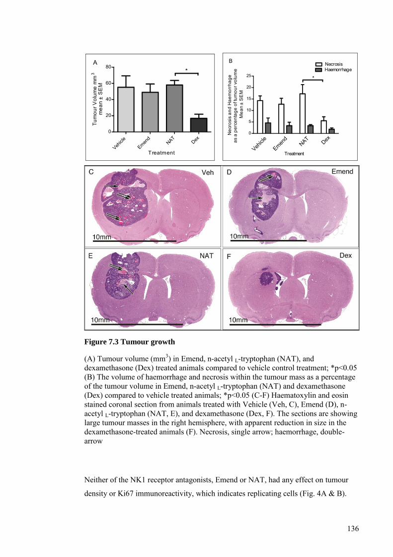
136
Vehicle
Emend
NATDex
0
20
40
60
80*
A
Treatment
Tum
our V
olum
e m
m3
mea
n±
SE
M
0
5
10
15
20
25
NecrosisHaemorrhage
Vehicle NAT
Emend
Dex
*
B
Treatment
Nec
rosi
s an
d H
aem
orrh
age
as a
per
cent
age
of tu
mou
r vol
ume
Mea
n±
SE
M
Figure 7.3 Tumour growth
(A) Tumour volume (mm3) in Emend, n-acetyl L-tryptophan (NAT), and dexamethasone (Dex) treated animals compared to vehicle control treatment; *p<0.05 (B) The volume of haemorrhage and necrosis within the tumour mass as a percentage of the tumour volume in Emend, n-acetyl L-tryptophan (NAT) and dexamethasone (Dex) compared to vehicle treated animals; *p<0.05 (C-F) Haematoxylin and eosin stained coronal section from animals treated with Vehicle (Veh, C), Emend (D), n-acetyl L-tryptophan (NAT, E), and dexamethasone (Dex, F). The sections are showing large tumour masses in the right hemisphere, with apparent reduction in size in the dexamethasone-treated animals (F). Necrosis, single arrow; haemorrhage, double-arrow
Neither of the NK1 receptor antagonists, Emend or NAT, had any effect on tumour
density or Ki67 immunoreactivity, which indicates replicating cells (Fig. 4A & B).

137
Similarly, neither Emend, nor NAT altered the percentage of caspase 3 positive cells,
which indicates apoptotic tumour cells (Fig. 4C). Dexamethasone treatment caused a
significant increase in the density of tumour cells within the tumour mass when
compared to the vehicle treated group (p<0.01; Fig. 4A). However, dexamethasone
treatment did not alter the percentage of Ki67 positive tumour cells or caspase 3
positive cells (Fig. 4B & C). Therefore, the dexamethasone treated group exhibited
tumour masses with cancer cells more tightly packed together, but with no change in
replication or apoptosis.

138
Vehicle
Emend
NAT
Dex
200
250
300
350
400
**
A
Treatment
Ave
rage
num
ber o
f tum
our c
ells
Mea
n±
SE
M
Vehicle
Emend
NATDex
0
20
40
60
80B
Treatment
% K
i67
Pos
itive
cel
lsM
ean
± S
EM
Vehicle
Emend
NAT
Dex
0.0
0.5
1.0
1.5C
Treatment
% C
aspa
se 3
pos
itive
cel
lsM
ean
± SE
M
Figure 7.4 Tumour growth characteristics
(A) The effect of treatment on tumour cell density. The average number of cells was obtained from four areas within the tumours in animals treated with Vehicle, Emend, n-acetyl L-tryptophan (NAT), and dexamethasone (Dex); **p<0.01 (B) The percentage of replicating tumour cells as indicated by Ki67 immunoreactivity in animals treated with vehicle, Emend, n-acetyl L-tryptophan (NAT) and dexamethasone (Dex) (C) The percentage of caspase 3 immunopositive cells, indicative of apoptosis, in tumour masses from animals treated with vehicle, Emend, n-acetyl L-tryptophan (NAT) and dexamethasone (Dex)
7.4.4 Brain microenvironment
GFAP and IBA1 immunoreactivity were used as indicators of astrocytic and
microglial response, respectively, and thus represented the interaction of the tumour
cells and treatment agents with these components of the brain microenvironment.
Inoculation with Walker 256 breast carcinoma cells and subsequent tumour growth,
caused a significant increase in GFAP and IBA1 immunoreactivity in the peritumoral

139
area when compared to the same location in culture medium control animals (p<0.001
and *p<0.05, respectively; Fig. 5A-E). GFAP immunoreactivity was not present
within the tumour, and thus showed significantly reduced %DAB when compared to
the peritumoral area (p<0.001; Fig. 5A & B). Furthermore, the tumour mass also
showed less GFAP immunoreactivity than the same location in brains of control
animals, injected with culture medium without Walker 256 tumour cells (Fig. 5A).
However, IBA1 immunoreactivity was significantly elevated within the tumour mass,
when compared with that evident in the striatum of brains from the culture medium
control group (p<0.01; Fig. 5C, D & E).

140
Contro
l Stria
tum
Tumou
rPeri
tumora
l Area
0
5
10
15
20
25***
A
GFA
P im
mun
orea
ctiv
ity%
DA
B m
ean
± S
EM
Contro
l Stria
tumTum
our
Peritum
oral A
rea
0
5
10
15
20
***
C
IBA
1 im
mun
orea
ctiv
ity%
DA
B m
ean
± S
EM
Figure 7.5 Tumour and peritumoral glial reaction
(A) GFAP immunoreactivity in the striatum of culture medium control animals compared with the tumour mass and the peritumoral area of the right hemisphere of tumour inoculated brains 7 days following surgery, ***p<0.001 (B) Coronal section of GFAP immunostained brain 7 days following tumour inoculation showing increased immunoreactivity in the peritumoral area when compared to similar location in the contralateral hemisphere (C) IBA1 immunoreactivity in the striatum of the culture medium control group compared with the tumour mass and in the peritumoral
141
area of the right hemisphere of tumour inoculated brains 7 days following surgery, **p<0.01, *p<0.05 (D) Section from the right striatum of a culture medium control brain stained for IBA1 antibody, showing minimal microglial immunoreactivity (E) IBA1 immunostained brain 7 days following tumour inoculation showing increased immunoreactivity in the peritumoral area (PTA) and tumour mass (TM)
In conjunction with the color deconvolution results, GFAP and IBA1 immunolabelled
cells were also counted to determine the number of astrocytes and microglia
respectively. The growth of Walker 256 breast carcinoma tumours caused an increase
in the number of astrocytes and microglia in the striatum surrounding tumour masses,
when compared to similar brain locations in the culture medium control group,
although this difference was only significant for IBA1 (p<0.05; Fig. 6A & B).
Treatment with Emend caused a reduction in the number of GFAP and IBA1 positive
cells to levels comparable to the culture medium injected control group (Fig. 6A &
B). Dexamethasone caused a similar phenomenon to Emend, particularly in regards to
the number of IBA1 positive cells (Fig. 6A & B). Furthermore, there was a small
reduction in the number of astrocytes and microglia present in the peritumoral area of
NAT treated animals (Fig. 6A & B).

142
Contro
lTum
our +
Vehicle
Tumou
r + Emen
dTum
our +
NAT
Tumou
r + D
ex
0
5
10
15A
Treatment
GFA
P po
sitiv
e ce
llsin
0.0
678m
m2
in th
e st
riatu
mM
ean
± SE
M
Contro
lTum
our +
Veh
icleTum
our +
Emen
dTum
our +
NAT
Tumou
r + D
ex
0
5
10
15
20*
B
Treatment
IBA1
pos
itive
cel
lsin
0.0
678m
m2
in th
e st
riatu
mM
ean
± SE
M
Figure 7.6 Brain microenvironment reaction to tumour growth
(A) The effect of treatment on the number of GFAP immunolabelled cells in the striatum of culture medium control animals and the striatum surrounding the tumours in inoculated animals 7 days following surgery; NAT, n-acetyl L-tryptophan; Dex, Dexamethasone (B) The effect of treatment on number of IBA1 immunolabelled cells within the striatum of culture medium control animals and the striatum surrounding the tumours in inoculated animals, 7 days following surgery; NAT, n-acetyl L-tryptophan, Dex, Dexamethasone; *p<0.05.

143
7.5 Discussion
This model of direct stereotaxic inoculation of Walker 256 tumour into the striatum of
male albino Wistar rats caused consistent growth of large, spherical-shaped secondary
brain tumours. These tumours showed a prominent cystic component and extensive
central necrosis and haemorrhage, as described previously in the literature (Lewis
2012a). The model used in the current study is advantageous in that it is used in
immune-competent animals, thus allowing the study of the interaction of tumour cells
with the host microenvironment and immune system.
The Walker 256 rat breast carcinoma cells expressed NK1 receptors in vivo when
injected into the striatum of male Wistar rats. It has previously been reported that
many human tumour specimens and their derived tumour cell lines express NK1
receptors (Eistetter 1992; Johnson 1992; Friess 2003; Rosso 2008; Akazawa 2009;
Munoz 2010b; Munoz 2012). Some cell lines have been reported to express up to 40,
000 NK1 receptors per cell (Fowler 1994), with the level of NK1 receptor expression
being linked to invasiveness of the cancer cell lines (Castro 2005). Moreover, non-
cancerous epithelial cell lines did not show evidence of NK1 receptor expression
(Singh 2000; Ramkissoon 2007). However, NK1 receptors are by no means present
on all cancer cell types, with one study demonstrating NK1 expression on SNB-19,
DBTRG-05 MG and U373 MG human glioma cell lines, but not on the U138 MG or
MOG-G-GCM human glioma cells (Palma 1999). Furthermore, the murine
neuroblastoma cell line C1300 did not show evidence of NK1 receptor expression
(Fukuhara 1996).
Despite the fact that the Walker 256 cells express NK1 receptors, only Emend was
able to cause a reduction of cell viability and not NAT. A possible explanation for the
differential response of Walker 256 cells to Emend and NAT is that Emend has an
increased affinity for the NK1 receptor and a greater ability to penetrate the tumour
cell than NAT, as; NAT is not membrane permeable unless modified to N-acetyl-L-

144
tryptophan 3, 5-bis (trifluoromethyl) benzyl ester, also known as L-732-138 (Donkin
2011).
Previous studies to determine the effects of NK1 antagonist treatment on tumour cells
in vitro have reported an inhibition of tumour growth, initiation of apoptosis,
decreased migration and reduced cytokine secretion by tumour cells (Palma 1998;
Palma 1999; Bigioni 2005; Munoz 2007; Rosso 2008; Huang 2010; Munoz 2010a;
Munoz 2010b; Munoz 2012).The predominant effects of exogenous SP on tumour
cells in vitro is mitogenesis, migration, cytokine secretion and chemotaxis (Ruff 1985;
Luo 1996; Drell 2003; Friess 2003; Lang 2004; Munoz 2004b; Munoz 2008;
Akazawa 2009). These effects are thought to be NK1 receptor dependent, as tumour
cell lines with differential expression of NK1 receptors, show that the cell lines
lacking NK1 receptors do not respond to NK1 antagonist treatment (Palma 1999).
Alternatively, several studies have shown evidence of inhibitory effects for SP on
growth and migration of cancer cells (Ogasawara 1997; Nagakawa 1998; Korcum
2009). However, the NK1 receptor expression status of these cell lines has not been
determined. In contrast, the Walker 256 cells used in the current study are positive for
NK1 receptor expression, but only responded to Emend treatment and not NAT
treatment in vitro. Aside from Emend having a much greater binding capacity for the
NK1 receptor because of its lipophilicity, a plausible explanation for this may be that
this cell line does not secrete SP into the culture medium in vitro and thus is unable to
have a stimulatory autocrine effect on the NK1 expressing cells. It has previously
been reported that Walker 256 breast carcinoma cells do not express SP in this model
when directly injected into the brain (Lewis 2012a). However, tumours induced from
this cell line do show increased SP immunoreactivity in the peritumoral neuropil in
two different models of metastatic brain tumours (Lewis 2012a; Lewis 2012b). Thus
it is plausible that exogenous SP from the brain microenvironment may have a
stimulatory effect on these tumour cells in vivo.
145
Unlike previous experiments showing cytotoxic effects of dexamethasone on tumour
cells (Bavaresco 2007; Piette 2009; Tazik 2009), the current study did not show any
evidence of dexamethasone toxicity on Walker 256 breast carcinoma cells as
measured by the trypan blue exclusion cell viability assay. Dexamethasone effects on
Walker 256 cell viability have not previously been investigated in culture, although it
has been shown to decrease parathyroid hormone secretion in a dose dependent
manner (Schilling 1996). Previous in vitro studies have shown dexamethasone to
inhibit mitogenesis and cell viability of C6 glioma cells along with decreasing
migration and invasive properties of U373 MG human glioblastoma cells (Bavaresco
2007; Piette 2009). However, if dexamethasone is toxic to tumour cells in vivo, then it
is also likely that non-neoplastic brain cells would also become apoptotic also (Tazik
2009).
NK1 antagonist treatment of metastatic brain tumours grown in this study did not alter
tumour cell volume or the volume of necrotic haemorrhagic tissue within the tumour
mass. Furthermore, levels of tumour cell replication and apoptosis remained
unaffected by both NK1 antagonist treatments used. There have been few previous
studies investigating the effects of NK1 antagonism on cancer growth in animal
models, despite the fact that Emend is already approved for use in cancer patients for
nausea associated with chemotherapy. The limited number of experiments that have
been performed in vivo have been inconsistent in their findings. One published study
demonstrated a beneficial effect of the NK1 antagonist MEN 11467 on
subcutaneously inoculated human U373 MG astrocytoma grade III cells by arresting
tumour growth, but not when used on human A2780 ovarian carcinoma lacking NK1
receptors, also inoculated into the right flank of female nude mice (Palma 2000).
Another study also employed MEN 11467 on the MDA-MB-231 human breast cancer
cell line, which express NK1 receptors, injected subcutaneously into the right flank of
female nude mice (Bigioni 2005). This experiment showed no effect of MEN 11467
on final tumour volume when administered alone, although caused a reduction when
co-administered with the NK2 antagonist nepadutant (Bigioni 2005).

146
In contrast, studies that have been performed in immune competent animals have
suggested that SP may be beneficial in the treatment of cancer patients through its
immune stimulating properties. For example, aerosolised SP caused a reduction in the
incidence of lung carcinoma in C57BL mice exposed to side stream cigarette smoke
(Harris 2003). Similarly, capsaicin-induced depletion of SP secreting sensory
neurones, resulted in the induction of a more aggressive phenotype, including
increased cardiac and lung metastases from orthotropically implanted 4T1 breast
carcinoma cells, although primary tumour growth remained unaffected (Erin 2004;
Erin 2006). Furthermore, K1735 melanoma cells injected subcutaneously into nude
mice responded to exogenous SP with a delay in tumour growth, but only with the
addition of natural killer and T cells, normally absent in immune compromised
animals (Manske 2005). Therefore, the role of SP in cancer growth in vivo remains
controversial, with more research required to elucidate the factors that determine if
NK1 antagonist treatment is effective in the inhibition of tumour growth in animal
models.
Dexamethasone treatment resulted in decreased brain tumour volume when compared
to the vehicle treated group. This is likely to be due to impaired accumulation of fluid,
as seen in previous studies (Lewis 2012a), rather than a decrease in the growth of the
tumour cells. Evidence of this is the absence of change in tumour cell replication and
apoptosis with dexamethasone treatment. Moreover, this is supported by the
significantly increased tumour cell density, evident in the dexamethasone treated
group when compared to vehicle-treated animals. Thus, the dexamethasone treated
group may have the same number of tumour cells, packed more closely together,
reducing the tumour volume. Also the decreased necrotic tissue within the
dexamethasone-treated tumours indicates that although the tumour volume is reduced
in this group, it is possible that the tumours had viable tumour cells comparable to the
number of cells in the vehicle treated tumours.
Reduction in tumour volume has been repeatedly shown with dexamethasone
treatment in the literature (Guerin 1992; Wolff 1993; Badruddoja 2003; Villeneuve

147
2008). These data have been used with dexamethasone’s toxicity in vitro to suggest an
anticancer effect for dexamethasone. However, dexamethasone has also consistently
been shown to reduce BBB permeability and oedema (Nakagawa 1987; Guerin 1992;
Wolff 1993; Heiss 1996; Badruddoja 2003; Ewing 2008; Gu 2009a; Gu 2009b),
which may account for the volume decreases seen. Another proposed mechanism of
dexamethasone inhibition of tumour growth is through manipulation of the host
microenvironment. Dexamethasone causes a blockade of classical inflammation,
normally favourable for tumour invasion and growth (Van Den Brenk 1974a; Van
Den Brenk 1974b; Zhang 1997; Lewis 2012a).
In the current study dexamethasone treatment was accompanied by a reduction in the
volume of necrotic tissue and haemorrhage in the tumours. A possible reason for the
decreased necrotic tissue is the decreased distance between tumour cells and blood
vessels, as a result of reduced oedema. Accumulation of tumour interstitial fluid in the
vehicle treated group may cause tumour cells to become increasingly remote from
blood vessels, with reduced availability of nutrients. This model of secondary brain
tumour has previously been shown to cause an increase in brain water content, which
is significantly reduced with dexamethasone treatment (Lewis 2012a). Furthermore, it
has previously been reported that dexamethasone treatment decreased blood vessel
density and volume within experimental gliomas (Wolff 1993; Badruddoja 2003).
In the current study, tumour growth caused an increase in both astrocytes and
microglia, particularly in the peritumoral environment. This phenomenon has been
well documented in many metastatic brain tumour models (Zhang 1995; Lorger
2010). Emend treatment decreased this glial response to tumour growth, showing that
the dose used in this study was sufficient to elicit a central effect. NAT was less
effective than Emend in this capacity, possibly due to its inability to cross the BBB
(Donkin 2011). Emend is a BBB penetrating drug able to occupy cerebral NK1
receptors (Bergstrom 2004), therefore it is logical that it would initiate a greater effect
on the brain microenvironment. Dexamethasone also reduced glial numbers to a level
similar to Emend treatment, although it is unclear if this effect was due to inhibition
148
of tumour initiated glial recruitment, activation and proliferation, or through toxic
effects on astrocytes and microglia. It has been postulated that microglia secrete
growth stimulatory substances that aid metastatic brain tumour growth, as evidenced
by the increase in MDA-MB-231 human breast cancer cells when co-cultured with
microglia in vitro (Fitzgerald 2008). Other tumour cells have been shown to
differentially activate microglia so that subpopulations express inducible nitric oxide
synthase and tumour necrosis factor-alpha (He 2006). Thus with more investigation,
glial regulation may provide a possible target for manipulation of metastatic brain
tumour growth.
In conclusion, neither NK1 antagonist treatment acted as an anticancer agent in this
animal model of metastatic brain tumours, despite the expression of NK1 receptors on
Walker 256 tumour cells and the fact that Emend reduced cell viability in vitro. In
conjunction, dexamethasone treatment also did not affect tumour growth,
notwithstanding evident reduction of tumour volume. Therefore neither Emend nor
dexamethasone is effective as anticancer agents in this model of brain metastatic
breast cancer. However, further research is required using different models and cancer
cells to definitively determine the exact effect of NK1 antagonists and dexamethasone
treatments on tumour growth.

149
8 General Discussion
8.1 Purpose
The aim of this thesis was to determine the role of SP in the development, growth and
complications of metastatic brain tumours caused by breast cancer, particularly in
relation to the interactions between SP and the BBB. The BBB is integral to many
aspects of metastatic brain tumour development and makes these types of neoplasms
distinct from cancer in other organs. The progression of cancer to the brain relies on
the ability of tumour cells to undergo extravasation through the BBB. Similarly,
vasogenic cerebral oedema is a common complication of metastatic brain tumour
growth and involves a compromised BBB function so that plasma proteins and fluid
are able to pass from the vasculature into the brain microenvironment.
Aspects of metastatic brain tumour growth are also affected by the presence of the
BBB as angiogenesis and often vessel co-option allow the nutritional needs of the
growing neoplasm to be met (Ito 1993; Maniotis 1999; Bugyik 2011), although these
new blood vessels have different properties from those of the normal BBB (Front
1984). Furthermore, the treatment of metastatic brain tumours is inherently difficult
because the BBB excludes many substances from the brain microenvironment (Lien
1991), such that agents that disrupt the BBB have been investigated to improve
delivery of chemotherapeutic agents to CNS tumours (Cote 2010). In conjunction,
whole brain radiotherapy has been shown to cause the BBB to become more
permeable (Adair 1999), which may exacerbate cerebral oedema and may lead to
increased development of complications ( Shamji 2009).
The effects of SP on the BBB in other neurological diseases have been well
established and the blockade of its actions led to novel treatment strategies (Donkin
2009; Turner 2011). Therefore the role of SP on the BBB in association with
metastatic brain tumour progression, growth and complications is of great interest.
Therefore, the effects of NK1 antagonists on these parameters were investigated here

150
with regard to their possible benefits for patients suffering from brain metastatic
breast cancer.
8.2 Models used
The Walker 256 cells used in the experiments detailed in this thesis were obtained
from two different reputable cell banks. These two populations of Walker 256 cells
were evaluated for their tumorigenicity and the more tumorigenic CRCTU Walker
256 cells were chosen for the remainder of the studies. CRCTU Walker 256 cells
produced more reliable metastatic brain tumours in both the internal carotid artery and
direct intracerebral inoculation models. Therefore using these cells required less
animals in order to determine the effects of NK1 antagonist treatment. Furthermore,
the consistency in growth characteristics using the CRCTU Walker 256 cells made
reproducible results more likely.
Differential incidence of tumour growth was apparent, not only between the tumour
cells from different cell banks, but also between the two models of inoculation. The
direct injection model consistently produced a single large tumour mass in the
striatum of the brain when CRCTU Walker 256 cells were utilised. In contrast, the
internal carotid artery inoculation model of CRCTU Walker 256 breast carcinoma
frequently resulted in multiple brain metastases, predominantly located within the
lateral ventricles. These patterns of growth have been observed previously in studies
comparing modes of metastatic brain tumour induction using the Lewis lung
carcinoma cell line (Saito 2008).
The direct injection had the advantage of more consistent tumour size and predictable
location of tumour burden, although the internal carotid artery inoculation model
allows for the BBB to be investigated during the extravasation process. The internal
carotid artery CRCTU Walker 256 inoculation model also elicited moderate
extracranial tumour growth, although this did not lead to any increase in mortality
before the euthanasia time point designated by metastatic brain tumour growth. This

151
is typical of haematogenously spread cancer metastases, which commonly affect
multiple organs (Harrell 2012), and once again demonstrates the suitability of the
internal carotid artery model of tumour induction as a model for the human metastatic
spread of cancer cells to the brain.
8.3 Principal findings
In vivo, the Walker 256 breast carcinoma cells from CRCTU were positive for NK1
receptor immunoreactivity, but did not exhibit SP expression. It has previously been
shown that NK1 receptor expression is necessary for SP to induce replication in
culture (Palma 1999). In vitro, this cell line did not respond to NAT, but treatment
with Emend reduced cell viability. Both Emend and NAT are NK1 receptor
antagonists, although Emend is more lipophilic and has greater binding affinity for
NK1 receptors (Bergstrom 2004; Donkin 2009). This result may indicate that
autocrine SP secretion by tumour cells is required for NK1 antagonists to be
consistently effective anticancer agents in vitro.
In the current study both the internal carotid artery injection and direct inoculation
model of metastatic brain tumour induction showed increased SP immunoreactivity in
the peritumoral area, despite being largely absent within the tumour mass. In previous
cell culture studies, exogenous SP added to tumour cells has been shown to cause
increased mitogenesis, migration, cytokine secretion and chemotactic behaviour (Ruff
1985; Luo 1996; Palma 1998; Drell 2003; Friess 2003; Lang 2004; Munoz 2004b;
Bigioni 2005; Munoz 2005a; Munoz 2005b; Munoz 2007; Munoz 2008; Rosso 2008;
Akazawa 2009; Huang 2010). Therefore the author hypothesised that the increased SP
expression surrounding metastatic brain tumour growth seen in the current study
would result in a similar stimulatory effect on implanted cancer cells in the striatum.
However, NK1 antagonist treatment did not alter any indicators of tumour growth,
meaning that the peritumoral SP did not have a sufficient stimulatory effect on cancer
cells in the direct injection model of Walker 256 brain metastases for SP blockade to
152
inhibit tumour growth. The current study therefore showed that the presence of NK1
receptors on tumour cell membranes is not sufficient in iteslf for NK1 antagonists to
be beneficial for the treatment of cancer. It is possible that tumour cells need to be of
a type that secretes SP for a treatment effect to be evident. There may, however, be a
subset of cancer types that are more responsive to NK1 receptor antagonist treatment,
although further investigation is required to clarify this potential.
Many human cancer specimens have been shown to express SP, although there is
significant variations both between and within cancer types (Allen 1985; Zhu 1995;
Khare 1998; Singh 2000; Nowicki 2002; Gonzalez Moles 2008; Brener 2009; Esteban
2009; Gonzalez-Moles 2009). Furthermore, tumours that are postitive for SP often do
not express it ubiquitously and rather have subpopulations of immunoreactive cells
(Munoz 2007). Thus the effectiveness of NK1 antagonist treatment may be limited,
even for tumours that do express SP.
Following internal carotid artery inoculation of Walker 256 cells, there was an
increase in perivascular SP immunoreactivity associated with tumour cell invasion of
brain microvessels. At the same time point, there was also increased albumin
immunoreactivity indicative of increased BBB permeability. It was therefore
postulated that contact of tumour cells with luminal membranes of brain endothelial
cells caused the release of SP, which resulted in increased BBB permeability,
allowing tumour cell extravasation into the brain. However, treatment with NK1
antagonists was ineffective in preventing metastatic brain tumour growth. Therefore it
was concluded that SP was not the primary mediator of Walker 256 extravasation
through the BBB in this model.
The variable CNS tropism of different cancer types implies that there are inherent
molecular characteristics of tumour cells that are beneficial for metastatic progression
to the brain. It has been postulated that only specific cancer types, or even
subpopulations of tumour cells within a neoplastic mass, have the propensity for
153
metastases to the brain, in particular extravasation through the BBB. Despite the
largely negative results presented in this thesis, the possibility cannot be ruled out that
SP may play a role in brain targeting by other cancer types, particularly if a
subpopulation of their cells secrete high levels of SP, unlike the cell line used in the
current study.
Increased SP immunoreactivity surrounding established brain metastases led to the
hypothesis that SP caused increased permeability of blood vessels surrounding the
neoplasms, which in turn allowed plasma proteins and fluid to leak into the neuropil.
However, in the current study neither BBB permeability nor brain water content was
affected by NK1 antagonist treatment. These findings contrast with previous studies,
where NK1 antagonist treatment has been used to decrease the disruption of the BBB
following traumatic brain injury and stroke (Donkin 2009; Turner 2011). This
suggests that SP plays a more prominent role in the pathogenesis of these acute injury
conditions as opposed to oedema formation in the metastatic brain tumour models
used in the current study. The mechanism of oedema development secondary to
metastatic brain tumours is therefore distinct from that driving oedema formation
following stroke and traumatic brain injury, despite both being vasogenic in nature.
In contrast to NK1 antagonist treatment, dexamethasone decreased brain water
content, indicative of odedma. This result was not surprising given that
dexamethasone is the current standard clinical treatment for peritumoral oedema and
was thus used as a positive control for the oedema study in this thesis. Interestingly,
dexamethasone treatment also caused increased tumour density along with decreased
tumour volume, when compared to the vehicle control group of animals. This
indicates that a substantial proportion of the oedematous fluid associated with
metastatic brain tumour growth is localised within the tumour mass.
Despite the consistency of dexamethasone effects on tumour-associated oedema, the
exact mechanism of its action remains controversial. It is thought that dexamethasone

154
predominantly acts by decreasing the permeability of the BBB (Nakagawa 1987;
Guerin 1992; Gu 2009b). Previous studies have shown that dexamethasone modulates
vasoactive molecules and substances involved in tight junction structure between
cerebral endothelial cells of the BBB. For example, dexamethasone treatment has
been shown to decrease tumour cell secretion of VEGF in vitro and reduce the action
of conditioned media from the tumour cells on vascular permeability in vivo (Heiss
1996; Das 2008). Furthermore, dexamethasone treatment of brain tumours has
resulted in increased expression of the tight junction protein occluden within the
tumour (Gu 2009a).
Despite previous studies showing the modulatory effect of dexamethasone on the
BBB, only a small BBB response was evident in the current study, with Evans blue
concentration in the neuropil still elevated in the dexamethasone treated group
compared to control level. Despite this, dexamethasone caused a reduction in brain
water content to a level equivalent to brains that were not exposed to tumour growth.
Therefore it is possible that modulation of BBB permeability is only partially
responsible for the actions of dexamethasone on cerebral oedema. Also suggesting
that permeability to water and Evans blue bound to albumin are driven by different
mechanisms.
An alternative mechanism of dexamethasone action is modulation of water channels
in the tumour cells or the surrounding neuropil. Under normal conditions, AQP-4 is
expressed by brain astrocytes and AQP-1 is present in the choroid plexus and is
involved with CSF production (Lukaszewicz 2011). Previous studies have shown that
dexamethasone modulates brain AQP-4 expression following intracerebral
haemorrhage and that these alterations in AQP-4 were correlated with oedema
clearance achieved by dexamethasone treatment (Gu 2007b). A similar mechanism
may be involved in the action of dexamethasone treatment of peritumoral oedema
following direct injection of Walker 256 cells into the striatum. In conjunction,
dexamethasone has demonstrated ability to induce AQP-1 expression in gliosarcoma
cells in vitro (Hayashi 2007), although AQP-1 has been linked to increased migration

155
and metastatic potential of cancer (Hu 2006). Thus, in the current study,
dexamethasone treatment may have acted by modulation of water channel proteins in
both the tumour cells and the peritumoral environment to effect the clearance of
cerebral oedema and tumour invasiveness. This requires further characterisation.
NK1 antagonist treatment did not alter the permeability of the BBB in either the
internal carotid artery or direct inoculation models used in these studies. This suggests
that NK1 antagonists are not suitable as preventative agents against BBB permeability
or to treat the complications associated with metastatic brain tumours. However, it is
possible that SP had a small contribution as one of many factors involved in the
pathogenesis of tumour associated oedema in the direct inoculation model. Similarly,
prevention of cancer cell extravasation across the BBB may still involve, but not
require, the activity of SP. Therefore agents that act via multiple pathways, like
dexamethasone, may be more suitable to target the BBB in metastatic brain tumour
progression and subsequent complications.
Along with other possible pathways already discussed, there is some evidence of SP
modulation by glucocorticoids, like dexamethasone. For example, topical
glucocorticoid treatment inhibits oedema caused by intradermal SP injection
(Ahluwalia 1995). Similarly, glucocorticoids inhibit plasma extravasation following
tachykinin release from sensory neurones in tracheal mucosa, presumably by
upregulation of proteins involved in the cleavage of SP (Piedimonte 1991).
Furthermore, glucocorticoid treatment reduces capsaicin induced plasma
extravasation in the trachea (Piedimonte 1990). Therefore if SP does have a minor
role in the increased BBB permeability evident in the studies described in this thesis,
its inhibition may be one of the multiple pathways by which dexamethasone
successfully reduces cerebral oedema. However, the high brain water content evident
in the NK1 antagonist treated group clearly shows that this is not the sole mechanism.

156
IBA1and GFAP immunoreactivity were used as indicators of brain tumour interaction
with microglia and astrocytes, respectively. The number of astrocytes and microglia
were substantially increased surrounding metastatic brain tumour growth in both
models used. Furthermore, this study showed that microglia infiltrated the entirety of
the tumour mass, but that astrocytes were only evident at the tumour border. This
pattern of IBA1 and GFAP immunoreactivity has been observed in numerous studies
where immunostained human specimens were analysed. These clinical studies
demonstrated substantial labelling of microglia and astrocytes in the peritumoral area,
whereas only microglia were evident within the human brain metastases, and
astrocytes were apparent in septa between tumour cells at the border of the neoplasm
(Zhang 1995; Berghoff 2012b). This pattern of staining occurs regardless of tumour
malignancy or type (Strik 2004). However, the extent of microglial infiltration has
been reported to vary from a few microglial cells to a 1:1 ratio of microglial cells to
tumour cells (Pukrop 2010).
It is well established that the presence of tumour growth within the brain results in
microglial and astrocytic activation through factors released from the tumour cells. In
turn, the glial cells also release factors that act upon the neoplastic tissue. Therefore
the relationship between neoplastic growth and the brain microenvironment is
bidirectional. Previous studies have shown that co-culture of breast cancer cells with
microglia caused microglial activation, whilst the activated microglia simultaneously
enhanced tumour cell invasiveness (Hagemann 2004; Pukrop 2006; Pukrop 2010).
Glial activation by tumour cells is achieved through release of pro-inflammatory
factors, as evident when human lung cancer cells activate astrocytes in vitro through
secretion of macrophage migration inhibitory factor, interleukin-8 and plasminogen
activator inhibitor-1 (Seike 2011). Several studies have also shown that serum
albumin activates microglia, functioning to clear albumin through phagocytosis
(Hooper 2005; Hooper 2009; Alonso 2011). This is pertinent, as it is widely accepted
that within and surrounding metastatic brain tumours, blood vessel permeability is
substantially increased. This was also demonstrated in the current thesis through the
157
increased albumin immunoreactivity in the neuropil, associated with metastatic brain
tumour growth in both models used. Therefore, this leakage of albumin from the
vasculature may have caused glial activation and the accumulation of these cells
evident in the peritumoral area.
The function of tumour infiltration of microglia and peritumoral accumulation of glia
is debatable. Both factors that promote growth and tumoricidal factors may be
released from glia to act on neoplastic tissue. However, glial cell manipulation of
tumour growth properties is likely dependant on activation of the glia by the tumour
cells themselves. This was evident in previous studies when control microglia did not
have any effect on tumour cell viability, whereas once activated, caused tumour cell
lysis in vitro (Brantley 2010). Furthermore, activated microglia potently act on cancer
cells but non-neoplastic cells remained unaffected (Brantley 2010). Moreover, one
study showed that the supernatant from lipopolysaccharide activated microglia caused
dose dependant apoptosis of human non-small cell lung cancer cells (He 2006). Thus,
factors with tumoricidal properties are secreted by the microglia and do not require
cell-to-cell contact.
In contrast, co-culture of MDA-MB-231 human breast cells with mixed glial cells
caused tumour cell mitogenesis to increase 5 fold (Fitzgerald 2008). More
specifically, activation of astrocytes by tumour cells causes production of
inflammatory mediators, such as interleukin-6, tumour necrosis factor-alpha and
interleukin-1 beta, which induce tumour cell proliferation (Langley 2009; Seike
2011). Furthermore, co-culture of human melanoma, lung cancer and breast cancer
cells with murine astrocytes protected cancer cells from chemotherapy induced
apoptosis, a process that was dependent on gap junctions (Lin 2010; Kim 2011).
These studies, along with the glial staining patterns evident in the current thesis,
suggest that targeting specific activation pathways of microglia may be a useful
therapeutic target for metastatic brain tumour treatment. In contrast, astrocyte
158
activation promotes tumour growth. Therefore, development of treatment options that
enhance microglial activation whilst inhibiting activation of astrocytes may be
beneficial for patients suffering from neoplastic growth in the CNS.
Treatment with Emend largely reduced the glial reaction evident with neoplastic
growth in the brain, although NAT was not as effective in this capacity. This is likely
the result of superior BBB penetration and NK1 receptor binding affinity of Emend
when compared to NAT (Bergstrom 2004; Donkin 2009). Previous studies have
shown that NK1 antagonist treatment partially reverses the increase in GFAP and
IBA1 immunoreactive cells evident after 6-OHDA induced Parkinson’s disease in
rodents (Thornton 2012). Furthermore, NAT treatment has been found to inhibit
microglial proliferation following traumatic brain injury (Carthew 2012). Therefore in
the current study the mechanism behind the decrease in microglia with NK1
antagonist treatment is likely through reduced proliferation of these cells rather than
drug toxicity. Similar pathways may also be responsible for the reduction in
astrocytes surrounding metastatic brain tumours as a result of NK1 antagonist
treatment.
Dexamethasone also reduced glial reaction to metastatic brain tumour growth, similar
to Emend. This effect was potentially due to the anti-inflammatory immune
suppression properties of dexamethasone or alternatively may have been a result of
widespread cytotoxicity due to the high dose used in this study. In vitro,
dexamethasone has previously been shown to inhibit astrocyte proliferation, and a
similar phenomenon was observed in vivo when excess adrenocorticotropic hormone
decreased astrocyte numbers in the frontal cortex, although this was evident under
non-pathological conditions (Unemura 2012).
When gram-negative bacteria derived lipopolysaccharide is applied to mixed glial
cells in culture, its effects to decrease functionality of astrocytes, increase microglial
proliferation, nitric oxide and reactive oxygen species production are ameliorated with
159
dexaemthasone treatment (Hinkerohe 2010; Huo 2011). Similarly, dexamethasone
reduced microglial activation in a cell culture system made up of astrocytes with 30%
microglia, representing a pathological condition of microglial overrepresentation
(Hinkerohe 2011). Dexamethasone has also been shown to decrease inflammatory
mediator mRNA expression by microglia in culture (Graber 2012). Therefore,
dexamethasone has repeatedly been shown to reduce the pathological alterations in
astrocytes and microglia commonly evident with many neurological diseases as was
evident in the current study following dexamethasone treatment of metastatic brain
tumours. However the exact mechanism and possible implications of these actions are
yet to be fully elucidated.
8.4 Further research
The current thesis has demonstrated that NK1 antagonist treatment was ineffective for
prevention of tumour cell extravasation, tumour growth inhibition and resolution of
peritumoral oedema. SP therefore does not play a prominent role in the progression,
growth or complications of the Walker 256 internal carotid artery injection or direct
inoculation models of metastatic brain tumour. These results were apparent despite
the NK1 receptor expression on the Walker 256 breast carcinoma cells. However, it is
possible that SP secretion by tumour cells is required for NK1 antagonist treatment to
modulate the BBB in animal models of metastatic brain tumours. In further studies it
would be pertinent to investigate the effect of NK1 antagonist treatment on the BBB
in models of metastatic brain tumours that employ tumour cells that secrete high
levels of SP. Thus, the additional effect of the combination of tumour-secreted and
peritumoral SP could be investigated, rather than only the tumour initiated release of
SP from the brain microenvironment, as was the case in the current thesis.
Similarly, future research should employ both human and murine tumour cell lines.
Moreover, more than one species should be utilised for animal models of brain
metasatatic disease. Human tumour cells develop more complex mutations over a
longer period of time, although their use in animal models of cancer requires immune
compromised animals (Rangarajan 2004). Therefore, employing human tumour cells

160
more closely replicates the human disease process, but not the interaction with host
microenvironment evident in human pathology.
Interaction with the host immune system is particularly important for determining the
role of SP in metastatic brain tumour pathogenesis. This is because SP is an
inflammatory mediator that has a modulatory effect on the immune system and has
previously been shown to inhibit cancer growth, but only in the presence of natural
killer and T cells (Manske 2005). Therefore experiments investigating NK1 receptor
antagonist effects on cancer that are performed in immune compromised animals may
show positive results that may not be replicated when used in the human condition.
In the current thesis, dexamethasone effectively resolved tumour induced cerebral
oedema, although the exact mechanism of its action is yet to be elucidated.
Dexamethasone has been found to modulate many different pathways including, but
not limited to, aquaporin water channels, VEGF, SP, bradykinin and tight junction
proteins. The multiple mechanisms of dexamethasone action may be the reason for its
effectiveness in treating peritumoral oedema clinically, as it is likely that tumour
progression and complications also occurs via a combination of multiple pathways.
Unfortunately, its use is associated with many harmful side effects, which limit its
use.
Subsequent studies should aim to elucidate the pathways of dexamethasone action on
oedema and determine those actions which cause the unwanted side effects.
Therefore, if the beneficial and detrimental pathways do not overlap, scientists may
endeavour to develop more targeted therapies that are able to replicate the effects of
dexamethasone on cerebral oedema without the side effects currently experienced by
patients. This may allow for more prolonged high dose treatment of oedema, which
remains a serious complication of metastatic brain tumours.

161
8.5 Conclusion
Despite elevated SP levels surrounding tumour-invaded blood vessels and in well
established metastatic brain tumours, NK1 antagonist treatment did not alter tumour
incidence, tumour volume or brain water content. This was in spite of NK1 receptor
expression by the Walker 256 cells used and the effective reduction of tumour cell
viability with Emend treatment in vitro. The results in this thesis suggest that tumour
cell secretion of high levels of SP are required for NK1 to be an effective anticancer
agent or to resolve tumour induced increases in BBB permeability.

162
9 Reference List Aalto, Y., S. Forsgren, U. Kjorell, J. Bergh, L. Franzen and R. Henriksson (1998).
Enhanced expression of neuropeptides in human breast cancer cell lines following irradiation. Peptides 19(2): 231-239.
Adair, J. C., N. Baldwin, M. Kornfeld and G. A. Rosenberg (1999). Radiation-induced blood-brain barrier damage in astrocytoma: relation to elevated gelatinase B and urokinase. J Neurooncol 44(3): 283-289.
Ahluwalia, A., P. Newbold, S. D. Brain and R. J. Flower (1995). Topical glucocorticoids inhibit neurogenic inflammation: involvement of lipocortin 1. Eur J Pharmacol 283(1-3): 193-198.
AIHW and AACR (2010). Cancer in Australia: an overview, 2010. Cancer series no.
60(Cat. no. CAN 56). Akazawa, T., S. G. Kwatra, L. E. Goldsmith, M. D. Richardson, E. A. Cox, J. H.
Sampson and M. M. Kwatra (2009). A constitutively active form of neurokinin 1 receptor and neurokinin 1 receptor-mediated apoptosis in glioblastomas. J Neurochem 109(4): 1079-1086.
Allen, J. M., N. R. Hoyle, J. C. Yeats, M. A. Ghatei, D. G. Thomas and S. R. Bloom (1985). Neuropeptides in neurological tumours. J Neurooncol 3(3): 197-202.
Alonso, A., E. Reinz, M. Fatar, M. G. Hennerici and S. Meairs (2011). Clearance of albumin following ultrasound-induced blood-brain barrier opening is mediated by glial but not neuronal cells. Brain Res 1411: 9-16.
Alpert, E. and C. Seigerman (1986). Steroid withdrawal psychosis in a patient with closed head injury. Arch Phys Med Rehabil 67(10): 766-769.
Alves, R. V., M. M. Campos, A. R. Santos and J. B. Calixto (1999). Receptor subtypes involved in tachykinin-mediated edema formation. Peptides 20(8): 921-927.
Andersen, C., J. Astrup and C. Gyldensted (1994a). Quantitation of peritumoural oedema and the effect of steroids using NMR-relaxation time imaging and blood-brain barrier analysis. Acta Neurochir Suppl (Wien) 60: 413-415.
Andersen, C., J. Astrup and C. Gyldensted (1994b). Quantitative MR analysis of glucocorticoid effects on peritumoral edema associated with intracranial meningiomas and metastases. J Comput Assist Tomogr 18(4): 509-518.
Andersen, C. and F. T. Jensen (1998). Differences in blood-tumour-barrier leakage of human intracranial tumours: quantitative monitoring of vasogenic oedema and its response to glucocorticoid treatment. Acta Neurochir (Wien) 140(9): 919-924.
Annunziata, P., C. Cioni, R. Santonini and E. Paccagnini (2002). Substance P antagonist blocks leakage and reduces activation of cytokine-stimulated rat brain endothelium. J Neuroimmunol 131(1-2): 41-49.
Annunziata, P., C. Cioni, S. Toneatto and E. Paccagnini (1998). HIV-1 gp120 increases the permeability of rat brain endothelium cultures by a mechanism involving substance P. Aids 12(18): 2377-2385.
Armitage, P. A., C. Schwindack, M. E. Bastin and I. R. Whittle (2007). Quantitative assessment of intracranial tumor response to dexamethasone using diffusion, perfusion and permeability magnetic resonance imaging. Magn Reson Imaging 25(3): 303-310.
Atahan, I. L., G. Ozyigit, F. Yildiz, M. Gurkaynak, U. Selek, S. Sari and M. Hayran (2008). Percent positive axillary involvement predicts for the development of

163
brain metastasis in high-risk patients with nonmetastatic breast cancer receiving post-mastectomy radiotherapy. Breast J 14(3): 245-249.
A. T. C. C. S. D. Organization (2010). Cell line misidentification: the beginning of the end. Nat Rev Cancer 10(6): 441-448.
Ayata, C. and A. H. Ropper (2002). Ischaemic brain oedema. J Clin Neurosci 9(2): 113-124.
Badruddoja, M. A., H. G. Krouwer, S. D. Rand, K. J. Rebro, A. P. Pathak and K. M. Schmainda (2003). Antiangiogenic effects of dexamethasone in 9L gliosarcoma assessed by MRI cerebral blood volume maps. Neuro Oncol 5(4): 235-243.
Ballinger, W. E., Jr. and R. D. Schimpff (1979). An experimental model for cerebral metastasis: preliminary light and ultrastructural studies. J Neuropathol Exp Neurol 38(1): 19-34.
Bar-Sella, P., D. Front, R. Hardoff, E. Peyser, B. Borovich and I. Nir (1979). Ultrastructural basis for different pertechnetate uptake patterns by various human brain tumours. J Neurol Neurosurg Psychiatry 42(10): 924-930.
Barallon, R., S. R. Bauer, J. Butler, A. Capes-Davis, W. G. Dirks, E. Elmore, M. Furtado, M. C. Kline, A. Kohara, G. V. Los, R. A. MacLeod, J. R. Masters, M. Nardone, R. M. Nardone, R. W. Nims, P. J. Price, Y. A. Reid, J. Shewale, G. Sykes, A. F. Steuer, D. R. Storts, J. Thomson, Z. Taraporewala, C. Alston-Roberts and L. Kerrigan (2010). Recommendation of short tandem repeat profiling for authenticating human cell lines, stem cells, and tissues. In Vitro Cell Dev Biol Anim 46(9): 727-732.
Barnholtz-Sloan, J. S., A. E. Sloan, F. G. Davis, F. D. Vigneau, P. Lai and R. E. Sawaya (2004). Incidence proportions of brain metastases in patients diagnosed (1973 to 2001) in the Metropolitan Detroit Cancer Surveillance System. J Clin Oncol 22(14): 2865-2872.
Barry, C. M., S. C. Helps, C. den Heuvel and R. Vink (2011). Characterizing the role of the neuropeptide substance P in experimental subarachnoid hemorrhage. Brain Res 1389: 143-151.
Batchelor, T. T., A. G. Sorensen, E. di Tomaso, W. T. Zhang, D. G. Duda, K. S. Cohen, K. R. Kozak, D. P. Cahill, P. J. Chen, M. Zhu, M. Ancukiewicz, M. M. Mrugala, S. Plotkin, J. Drappatz, D. N. Louis, P. Ivy, D. T. Scadden, T. Benner, J. S. Loeffler, P. Y. Wen and R. K. Jain (2007). AZD2171, a pan-VEGF receptor tyrosine kinase inhibitor, normalizes tumor vasculature and alleviates edema in glioblastoma patients. Cancer Cell 11(1): 83-95.
Baumann, E., E. Preston, J. Slinn and D. Stanimirovic (2009). Post-ischemic hypothermia attenuates loss of the vascular basement membrane proteins, agrin and SPARC, and the blood-brain barrier disruption after global cerebral ischemia. Brain Res 1269: 185-197.
Bavaresco, L., A. Bernardi, E. Braganhol, M. R. Wink and A. M. Battastini (2007). Dexamethasone inhibits proliferation and stimulates ecto-5'-nucleotidase/CD73 activity in C6 rat glioma cell line. J Neurooncol 84(1): 1-8.
Beaumont, A. and I. R. Whittle (2000). The pathogenesis of tumour associated epilepsy. Acta Neurochir (Wien) 142(1): 1-15.
Becher, M. W., T. W. Abel, R. C. Thompson, K. D. Weaver and L. E. Davis (2006). Immunohistochemical analysis of metastatic neoplasms of the central nervous system. J Neuropathol Exp Neurol 65(10): 935-944.
Berghoff, A., Z. Bago-Horvath, C. De Vries, P. Dubsky, U. Pluschnig, M. Rudas, A. Rottenfusser, M. Knauer, H. Eiter, F. Fitzal, K. Dieckmann, R. M. Mader, M.

164
Gnant, C. C. Zielinski, G. G. Steger, M. Preusser and R. Bartsch (2012a). Brain metastases free survival differs between breast cancer subtypes. Br J Cancer 106(3): 440-446.
Berghoff, A. S., H. Lassmann, M. Preusser and R. Hoftberger (2012b). Characterization of the inflammatory response to solid cancer metastases in the human brain. Clin Exp Metastasis.
Bergstrom, M., R. J. Hargreaves, H. D. Burns, M. R. Goldberg, D. Sciberras, S. A. Reines, K. J. Petty, M. Ogren, G. Antoni, B. Langstrom, O. Eskola, M. Scheinin, O. Solin, A. K. Majumdar, M. L. Constanzer, W. P. Battisti, T. E. Bradstreet, C. Gargano and J. Hietala (2004). Human positron emission tomography studies of brain neurokinin 1 receptor occupancy by aprepitant. Biol Psychiatry 55(10): 1007-1012.
Betz, A. L. and H. C. Coester (1990). Effect of steroids on edema and sodium uptake of the brain during focal ischemia in rats. Stroke 21(8): 1199-1204.
Bigioni, M., A. Benzo, C. Irrissuto, C. A. Maggi and C. Goso (2005). Role of NK-1 and NK-2 tachykinin receptor antagonism on the growth of human breast carcinoma cell line MDA-MB-231. Anticancer Drugs 16(10): 1083-1089.
Black, P. H. (2002). Stress and the inflammatory response: a review of neurogenic inflammation. Brain Behav Immun 16(6): 622-653.
Blasberg, R. G., W. R. Shapiro, P. Molnar, C. S. Patlak and J. D. Fenstermacher (1984a). Local blood-to-tissue transport in Walker 256 metastatic brain tumors. J Neurooncol 2(3): 205-218.
Blasberg, R. G., W. R. Shapiro, P. Molnar, C. S. Patlak and J. D. Fenstermacher (1984b). Local blood flow in Walker 256 metastatic brain tumors. J Neurooncol 2(3): 195-204.
Blasberg, R. G., M. Shinohara, W. R. Shapiro, C. S. Patlak, K. D. Pettigrew and J. D. Fenstermacher (1986). Apparent glucose utilization in Walker 256 metastatic brain tumors. J Neurooncol 4(1): 5-16.
Brantley, E. C., L. Guo, C. Zhang, Q. Lin, K. Yokoi, R. R. Langley, E. Kruzel, M. Maya, S. W. Kim, S. J. Kim, D. Fan and I. J. Fidler (2010). Nitric oxide-mediated tumoricidal activity of murine microglial cells. Transl Oncol 3(6): 380-388.
Bray, F., G. Engholm, T. Hakulinen, M. Gislum, L. Tryggvadottir, H. H. Storm and A. Klint (2010). Trends in survival of patients diagnosed with cancers of the brain and nervous system, thyroid, eye, bone, and soft tissues in the Nordic countries 1964-2003 followed up until the end of 2006. Acta Oncol 49(5): 673-693.
Brener, S., M. A. Gonzalez-Moles, D. Tostes, F. Esteban, J. A. Gil-Montoya, I. Ruiz-Avila, M. Bravo and M. Munoz (2009). A role for the substance P/NK-1 receptor complex in cell proliferation in oral squamous cell carcinoma. Anticancer Res 29(6): 2323-2329.
Bruno, G., F. Tega, A. Bruno, U. Graf, F. Corelli, R. Molfetta and M. Barucco (2003). The role of substance P in cerebral ischemia. Int J Immunopathol Pharmacol 16(1): 67-72.
Budde, M. D., E. Gold, E. K. Jordan, M. Smith-Brown and J. A. Frank (2012). Phase contrast MRI is an early marker of micrometastatic breast cancer development in the rat brain. NMR Biomed 25(5): 726-736.
Buehring, G. C., E. A. Eby and M. J. Eby (2004). Cell line cross-contamination: how aware are Mammalian cell culturists of the problem and how to monitor it? In Vitro Cell Dev Biol Anim 40(7): 211-215.

165
Buffon, A., V. B. Ribeiro, M. R. Wink, E. A. Casali and J. J. Sarkis (2007). Nucleotide metabolizing ecto-enzymes in Walker 256 tumor cells: molecular identification, kinetic characterization and biochemical properties. Life Sci 80(10): 950-958.
Bugyik, E., K. Dezso, L. Reiniger, V. Laszlo, J. Tovari, J. Timar, P. Nagy, W. Klepetko, B. Dome and S. Paku (2011). Lack of Angiogenesis in Experimental Brain Metastases. J Neuropathol Exp Neurol.
Bundgaard, M. and N. J. Abbott (2008). All vertebrates started out with a glial blood-brain barrier 4-500 million years ago. Glia.
Carbonell, W. S., O. Ansorge, N. Sibson and R. Muschel (2009). The vascular basement membrane as "soil" in brain metastasis. PLoS One 4(6): e5857.
Carthew, H. L., J. M. Ziebell and R. Vink (2012). Substance P-induced changes in cell genesis following diffuse traumatic brain injury. Neuroscience 214: 78-83.
Castro, T. A., M. C. Cohen and P. Rameshwar (2005). The expression of neurokinin-1 and preprotachykinin-1 in breast cancer cells depends on the relative degree of invasive and metastatic potential. Clin Exp Metastasis 22(8): 621-628.
Cespedes, M. V., I. Casanova, M. Parreno and R. Mangues (2006). Mouse models in oncogenesis and cancer therapy. Clin Transl Oncol 8(5): 318-329.
Chambers, A. F., R. P. Hill and V. Ling (1981). Tumor heterogeneity and stability of the metastatic phenotype of mouse KHT sarcoma cells. Cancer Res 41(4): 1368-1372.
Chang-Liu, C. M. and G. E. Woloschak (1997). Effect of passage number on cellular response to DNA-damaging agents: cell survival and gene expression. Cancer Lett 113(1-2): 77-86.
Chappa, A. K., J. D. Cooper, K. L. Audus and S. M. Lunte (2007). Investigation of the metabolism of substance P at the blood-brain barrier using LC-MS/MS. J Pharm Biomed Anal 43(4): 1409-1415.
Chekhonin, V. P., V. P. Baklaushev, G. M. Yusubalieva, K. A. Pavlov, O. V. Ukhova and O. I. Gurina (2007). Modeling and immunohistochemical analysis of C6 glioma in vivo. Bull Exp Biol Med 143(4): 501-509.
Cifuentes, N. and J. W. Pickren (1979). Metastases from carcinoma of mammary gland: an autopsy study. J Surg Oncol 11(3): 193-205.
Cioni, C., D. Renzi, A. Calabro and P. Annunziata (1998). Enhanced secretion of substance P by cytokine-stimulated rat brain endothelium cultures. J Neuroimmunol 84(1): 76-85.
Clark, W. C., J. A. Nicoll and H. Coakham (1989). Monoclonal antibody immunophenotyping of solitary cerebral metastases with unknown primary sites. Br J Neurosurg 3(5): 591-595.
Cornford, E. M., D. Young, J. W. Paxton, G. J. Finlay, W. R. Wilson and W. M. Pardridge (1992). Melphalan penetration of the blood-brain barrier via the neutral amino acid transporter in tumor-bearing brain. Cancer Res 52(1): 138-143.
Corrigan, F., R. Vink, P. C. Blumbergs, C. L. Masters, R. Cappai and C. van den Heuvel (2012). sAPPalpha rescues deficits in amyloid precursor protein knockout mice following focal traumatic brain injury. J Neurochem 122(1): 208-220.
Cote, J., M. Savard, V. Bovenzi, C. Dubuc, L. Tremblay, A. M. Tsanaclis, D. Fortin, M. Lepage and F. Gobeil, Jr. (2010). Selective tumor blood-brain barrier opening with the kinin B2 receptor agonist [Phe(8)psi(CH(2)NH)Arg(9)]-BK in a F98 glioma rat model: an MRI study. Neuropeptides 44(2): 177-185.

166
Cree, I. A. (2011). Principles of cancer cell culture. Methods Mol Biol 731: 13-26. Das, A., N. L. Banik and S. K. Ray (2008). Modulatory effects of acetazolomide and
dexamethasone on temozolomide-mediated apoptosis in human glioblastoma T98G and U87MG cells. Cancer Invest 26(4): 352-358.
De Nicola, A. F., M. E. Brocca, L. Pietranera and L. M. Garcia-Segura (2012). Neuroprotection and Sex Steroid Hormones: Evidence of Estradiol-Mediated Protection in Hypertensive Encephalopathy. Mini Rev Med Chem.
Dexter, D. L., H. M. Kowalski, B. A. Blazar, Z. Fligiel, R. Vogel and G. H. Heppner (1978). Heterogeneity of tumor cells from a single mouse mammary tumor. Cancer Res 38(10): 3174-3181.
Dobec-Meic, B., S. Pikija, D. Cvetko, V. Trkulja, L. Pazanin, N. Kudelic, K. Rotim, I. Pavlicek and A. R. Kostanjevec (2006). Intracranial tumors in adult population of the Varazdin County (Croatia) 1996-2004: a population-based retrospective incidence study. J Neurooncol 78(3): 303-310.
Donkin, J. J., I. Cernak, P. C. Blumbergs and R. Vink (2011). A substance p antagonist reduces axonal injury and improves neurologic outcome when administered up to 12 hours after traumatic brain injury. J Neurotrauma 28(2): 217-224.
Donkin, J. J., A. J. Nimmo, I. Cernak, P. C. Blumbergs and R. Vink (2009). Substance P is associated with the development of brain edema and functional deficits after traumatic brain injury. J Cereb Blood Flow Metab 29(8): 1388-1398.
Donkin, J. J. and R. Vink (2010). Mechanisms of cerebral edema in traumatic brain injury: therapeutic developments. Curr Opin Neurol 23(3): 293-299.
Drell, T. L. t., J. Joseph, K. Lang, B. Niggemann, K. S. Zaenker and F. Entschladen (2003). Effects of neurotransmitters on the chemokinesis and chemotaxis of MDA-MB-468 human breast carcinoma cells. Breast Cancer Res Treat 80(1): 63-70.
Eistetter, H. R., A. Mills, R. Brewster, S. Alouani, C. Rambosson and E. Kawashima (1992). Functional characterization of neurokinin-1 receptors on human U373MG astrocytoma cells. Glia 6(2): 89-95.
El Andaloussi, A., Y. Han and M. S. Lesniak (2006). Prolongation of survival following depletion of CD4+CD25+ regulatory T cells in mice with experimental brain tumors. J Neurosurg 105(3): 430-437.
Elaimy, A. L., A. R. Mackay, W. T. Lamoreaux, R. K. Fairbanks, J. J. Demakas, B. S. Cooke, B. J. Peressini, J. T. Holbrook and C. M. Lee (2011). Multimodality treatment of brain metastases: an institutional survival analysis of 275 patients. World J Surg Oncol 9: 69.
Engelhorn, T., N. E. Savaskan, M. A. Schwarz, J. Kreutzer, E. P. Meyer, E. Hahnen, O. Ganslandt, A. Dorfler, C. Nimsky, M. Buchfelder and I. Y. Eyupoglu (2009). Cellular characterization of the peritumoral edema zone in malignant brain tumors. Cancer Sci 100(10): 1856-1862.
Erin, N., G. Akdas Barkan, J. F. Harms and G. A. Clawson (2008). Vagotomy enhances experimental metastases of 4THMpc breast cancer cells and alters substance P level. Regul Pept 151(1-3): 35-42.
Erin, N., P. J. Boyer, R. H. Bonneau, G. A. Clawson and D. R. Welch (2004). Capsaicin-mediated denervation of sensory neurons promotes mammary tumor metastasis to lung and heart. Anticancer Res 24(2B): 1003-1009.
Erin, N., W. Zhao, J. Bylander, G. Chase and G. Clawson (2006). Capsaicin-induced inactivation of sensory neurons promotes a more aggressive gene expression phenotype in breast cancer cells. Breast Cancer Res Treat 99(3): 351-364.

167
Escartin, C. and G. Bonvento (2008). Targeted activation of astrocytes: a potential neuroprotective strategy. Mol Neurobiol 38(3): 231-241.
Esteban, F., M. A. Gonzalez-Moles, D. Castro, M. Martin-Jaen Mdel, M. Redondo, I. Ruiz-Avila and M. Munoz (2009). Expression of substance P and neurokinin-1-receptor in laryngeal cancer: linking chronic inflammation to cancer promotion and progression. Histopathology 54(2): 258-260.
Ewing, J. R., S. L. Brown, T. N. Nagaraja, H. Bagher-Ebadian, R. Paudyal, S. Panda, R. A. Knight, G. Ding, Q. Jiang, M. Lu and J. D. Fenstermacher (2008). MRI measurement of change in vascular parameters in the 9L rat cerebral tumor after dexamethasone administration. J Magn Reson Imaging 27(6): 1430-1438.
Fabi, A., A. Felici, G. Metro, A. Mirri, E. Bria, S. Telera, L. Moscetti, M. Russillo, G. Lanzetta, G. Mansueto, A. Pace, M. Maschio, A. Vidiri, I. Sperduti, F. Cognetti and C. M. Carapella (2011). Brain metastases from solid tumors: disease outcome according to type of treatment and therapeutic resources of the treating center. J Exp Clin Cancer Res 30: 10.
Fazakas, C., I. Wilhelm, P. Nagyoszi, A. E. Farkas, J. Hasko, J. Molnar, H. Bauer, H. C. Bauer, F. Ayaydin, N. T. Dung, L. Siklos and I. A. Krizbai (2011). Transmigration of melanoma cells through the blood-brain barrier: role of endothelial tight junctions and melanoma-released serine proteases. PLoS One 6(6): e20758.
Felix, F. H., J. B. Fontenele, M. G. Teles, J. E. Bezerra Neto, M. H. Santiago, R. L. Picanco Filho, D. B. Menezes, G. S. Viana and M. O. Moraes (2012). Cyclosporin safety in a simplified rat brain tumor implantation model. Arq Neuropsiquiatr 70(1): 52-58.
Fenselau, A., S. Watt and R. J. Mello (1981). Tumor angiogenic factor. Purification from the Walker 256 rat tumor. J Biol Chem 256(18): 9605-9611.
Ferreira, A. C., E. Martins, Jr., S. C. Afeche, J. Cipolla-Neto and L. F. Costa Rosa (2004). The profile of melatonin production in tumour-bearing rats. Life Sci 75(19): 2291-2302.
Fidler, I. J. (1970). Metastasis: guantitative analysis of distribution and fate of tumor embolilabeled with 125 I-5-iodo-2'-deoxyuridine. J Natl Cancer Inst 45(4): 773-782.
Fidler, I. J. (1978). Tumor heterogeneity and the biology of cancer invasion and metastasis. Cancer Res 38(9): 2651-2660.
Fidler, I. J. (1990). Critical factors in the biology of human cancer metastasis: twenty-eighth G.H.A. Clowes memorial award lecture. Cancer Res 50(19): 6130-6138.
Fisher, M. (2009). Pericyte signaling in the neurovascular unit. Stroke 40(3 Suppl): S13-15.
Fitzgerald, D. P., D. Palmieri, E. Hua, E. Hargrave, J. M. Herring, Y. Qian, E. Vega-Valle, R. J. Weil, A. M. Stark, A. O. Vortmeyer and P. S. Steeg (2008). Reactive glia are recruited by highly proliferative brain metastases of breast cancer and promote tumor cell colonization. Clin Exp Metastasis 25(7): 799-810.
Forster, C., J. Waschke, M. Burek, J. Leers and D. Drenckhahn (2006). Glucocorticoid effects on mouse microvascular endothelial barrier permeability are brain specific. J Physiol 573(Pt 2): 413-425.
Fowler, C. J. and G. Brannstrom (1994). Substance P enhances forskolin-stimulated cyclic AMP production in human UC11MG astrocytoma cells. Methods Find Exp Clin Pharmacol 16(1): 21-28.
168
Freed, A. L., K. L. Audus and S. M. Lunte (2002). Investigation of substance P transport across the blood-brain barrier. Peptides 23(1): 157-165.
Freitas, J. J., C. Pompeia, C. K. Miyasaka and R. Curi (2001). Walker-256 tumor growth causes oxidative stress in rat brain. J Neurochem 77(2): 655-663.
Friess, H., Z. Zhu, V. Liard, X. Shi, S. V. Shrikhande, L. Wang, K. Lieb, M. Korc, C. Palma, A. Zimmermann, J. C. Reubi and M. W. Buchler (2003). Neurokinin-1 receptor expression and its potential effects on tumor growth in human pancreatic cancer. Lab Invest 83(5): 731-742.
Front, D., E. Even-Sapir, G. Iosilevsky, O. Israel, A. Frenkel, G. M. Kolodny and M. Feinsud (1987). Monitoring of 57Co-bleomycin delivery to brain metastases and their tumors of origin. J Neurosurg 67(4): 506-510.
Front, D., O. Israel, S. Kohn and I. Nir (1984). The blood-tissue barrier of human brain tumors: correlation of scintigraphic and ultrastructural findings: concise communication. J Nucl Med 25(4): 461-465.
Fukuhara, S., H. Mukai, K. Kako, K. Nakayama and E. Munekata (1996). Further identification of neurokinin receptor types and mechanisms of calcium signaling evoked by neurokinins in the murine neuroblastoma C1300 cell line. J Neurochem 67(3): 1282-1292.
Gavrilovic, I. T. and J. B. Posner (2005). Brain metastases: epidemiology and pathophysiology. J Neurooncol 75(1): 5-14.
Ghabriel, M. N., C. Zhu, G. Hermanis and G. Allt (2000). Immunological targeting of the endothelial barrier antigen (EBA) in vivo leads to opening of the blood-brain barrier. Brain Res 878(1-2): 127-135.
Ghabriel, M. N., C. Zhu and C. Leigh (2002). Electron microscope study of blood-brain barrier opening induced by immunological targeting of the endothelial barrier antigen. Brain Res 934(2): 140-151.
Ghia, A., W. A. Tome, S. Thomas, G. Cannon, D. Khuntia, J. S. Kuo and M. P. Mehta (2007). Distribution of brain metastases in relation to the hippocampus: implications for neurocognitive functional preservation. Int J Radiat Oncol Biol Phys 68(4): 971-977.
Gildea, J. J., W. L. Golden, M. A. Harding and D. Theodorescu (2000). Genetic and phenotypic changes associated with the acquisition of tumorigenicity in human bladder cancer. Genes Chromosomes Cancer 27(3): 252-263.
Gonzalez-Moles, M. A., S. Brener, I. Ruiz-Avila, J. A. Gil-Montoya, D. Tostes, M. Bravo and F. Esteban (2009). Substance P and NK-1R expression in oral precancerous epithelium. Oncol Rep 22(6): 1325-1331.
Gonzalez Moles, M. A., A. Mosqueda-Taylor, F. Esteban, J. A. Gil-Montoya, M. A. Diaz-Franco, M. Delgado and M. Munoz (2008). Cell proliferation associated with actions of the substance P/NK-1 receptor complex in keratocystic odontogenic tumours. Oral Oncol 44(12): 1127-1133.
Graber, D. J., W. F. Hickey, E. W. Stommel and B. T. Harris (2012). Anti-inflammatory efficacy of dexamethasone and Nrf2 activators in the CNS using brain slices as a model of acute injury. J Neuroimmune Pharmacol 7(1): 266-278.
Graeber, M. B., W. Li and M. L. Rodriguez (2011). Role of microglia in CNS inflammation. FEBS Lett 585(23): 3798-3805.
Grauer, O. M., S. Nierkens, E. Bennink, L. W. Toonen, L. Boon, P. Wesseling, R. P. Sutmuller and G. J. Adema (2007). CD4+FoxP3+ regulatory T cells gradually accumulate in gliomas during tumor growth and efficiently suppress antiglioma immune responses in vivo. Int J Cancer 121(1): 95-105.

169
Greig, N. H., H. B. Jones and J. B. Cavanagh (1983). Blood-brain barrier integrity and host responses in experimental metastatic brain tumours. Clin Exp Metastasis 1(3): 229-246.
Gu, Y. T., L. J. Qin, X. Qin and F. Xu (2009a). The molecular mechanism of dexamethasone-mediated effect on the blood-brain tumor barrier permeability in a rat brain tumor model. Neurosci Lett 452(2): 114-118.
Gu, Y. T., Y. X. Xue, P. Wang, H. Zhang and L. J. Qin (2009b). Dexamethasone Enhances Calcium-activated Potassium Channel Expression in Blood-brain Tumor Barrier in a Rat Brain Tumor Model. Brain Res 1259: 1-6.
Gu, Y. T., H. Zhang and Y. X. Xue (2007a). Dexamethasone enhances adenosine 5'-triphosphate-sensitive potassium channel expression in the blood-brain tumor barrier in a rat brain tumor model. Brain Res 1162: 1-8.
Gu, Y. T., H. Zhang and Y. X. Xue (2007b). Dexamethasone treatment modulates aquaporin-4 expression after intracerebral hemorrhage in rats. Neurosci Lett 413(2): 126-131.
Guerin, C., J. E. Wolff, J. Laterra, L. R. Drewes, H. Brem and G. W. Goldstein (1992). Vascular differentiation and glucose transporter expression in rat gliomas: effects of steroids. Ann Neurol 31(5): 481-487.
Hagemann, T., S. C. Robinson, M. Schulz, L. Trumper, F. R. Balkwill and C. Binder (2004). Enhanced invasiveness of breast cancer cell lines upon co-cultivation with macrophages is due to TNF-alpha dependent up-regulation of matrix metalloproteases. Carcinogenesis 25(8): 1543-1549.
Harford-Wright, E., K. M. Lewis and R. Vink (2011). Towards drug discovery for brain tumours: interaction of kinins and tumours at the blood brain barrier interface. Recent Pat CNS Drug Discov 6(1): 31-40.
Harford-Wright, E., E. Thornton and R. Vink (2010). Angiotensin-converting enzyme (ACE) inhibitors exacerbate histological damage and motor deficits after experimental traumatic brain injury. Neurosci Lett 481(1): 26-29.
Harrell, J. C., A. Prat, J. S. Parker, C. Fan, X. He, L. Carey, C. Anders, M. Ewend and C. M. Perou (2012). Genomic analysis identifies unique signatures predictive of brain, lung, and liver relapse. Breast Cancer Res Treat 132(2): 523-535.
Harris, D. T. and M. Witten (2003). Aerosolized substance P protects against cigarette-induced lung damage and tumor development. Cell Mol Biol (Noisy-le-grand) 49(2): 151-157.
Harrison, S. and P. Geppetti (2001). Substance p. Int J Biochem Cell Biol 33(6): 555-576.
Hasegawa, H., W. R. Shapiro and J. B. Posner (1979). Chemotherapy of experimental metastatic brain tumors in female Wistar rats. Cancer Res 39(7 Pt 1): 2691-2697.
Hasegawa, H., Y. Ushio, T. Hayakawa, K. Yamada and H. Mogami (1983). Changes of the blood-brain barrier in experimental metastatic brain tumors. J Neurosurg 59(2): 304-310.
Hawkins, B. T. and T. P. Davis (2005). The blood-brain barrier/neurovascular unit in health and disease. Pharmacol Rev 57(2): 173-185.
Hayashi, Y., N. A. Edwards, M. A. Proescholdt, E. H. Oldfield and M. J. Merrill (2007). Regulation and function of aquaporin-1 in glioma cells. Neoplasia 9(9): 777-787.
He, B. P., J. J. Wang, X. Zhang, Y. Wu, M. Wang, B. H. Bay and A. Y. Chang (2006). Differential reactions of microglia to brain metastasis of lung cancer. Mol Med 12(7-8): 161-170.

170
Heiss, J. D., E. Papavassiliou, M. J. Merrill, L. Nieman, J. J. Knightly, S. Walbridge, N. A. Edwards and E. H. Oldfield (1996). Mechanism of dexamethasone suppression of brain tumor-associated vascular permeability in rats. Involvement of the glucocorticoid receptor and vascular permeability factor. J Clin Invest 98(6): 1400-1408.
Helps, S. C., E. Thornton, T. J. Kleinig, J. Manavis and R. Vink (2012). Automatic nonsubjective estimation of antigen content visualized by immunohistochemistry using color deconvolution. Appl Immunohistochem Mol Morphol 20(1): 82-90.
Hempen, C., E. Weiss and C. F. Hess (2002). Dexamethasone treatment in patients with brain metastases and primary brain tumors: do the benefits outweigh the side-effects? Support Care Cancer 10(4): 322-328.
Hennig, I. M., J. A. Laissue, U. Horisberger and J. C. Reubi (1995). Substance-P receptors in human primary neoplasms: tumoral and vascular localization. Int J Cancer 61(6): 786-792.
Herrstedt, J., H. B. Muss, D. G. Warr, P. J. Hesketh, P. D. Eisenberg, H. Raftopoulos, S. M. Grunberg, M. Gabriel, A. Rodgers, C. M. Hustad, K. J. Horgan and F. Skobieranda (2005). Efficacy and tolerability of aprepitant for the prevention of chemotherapy-induced nausea and emesis over multiple cycles of moderately emetogenic chemotherapy. Cancer 104(7): 1548-1555.
Hesketh, P. J., S. M. Grunberg, R. J. Gralla, D. G. Warr, F. Roila, R. de Wit, S. P. Chawla, A. D. Carides, J. Ianus, M. E. Elmer, J. K. Evans, K. Beck, S. Reines and K. J. Horgan (2003). The oral neurokinin-1 antagonist aprepitant for the prevention of chemotherapy-induced nausea and vomiting: a multinational, randomized, double-blind, placebo-controlled trial in patients receiving high-dose cisplatin--the Aprepitant Protocol 052 Study Group. J Clin Oncol 21(22): 4112-4119.
Hesketh, P. J., S. M. Grunberg, J. Herrstedt, R. de Wit, R. J. Gralla, A. D. Carides, A. Taylor, J. K. Evans and K. J. Horgan (2006). Combined data from two phase III trials of the NK1 antagonist aprepitant plus a 5HT 3 antagonist and a corticosteroid for prevention of chemotherapy-induced nausea and vomiting: effect of gender on treatment response. Support Care Cancer 14(4): 354-360.
Heyn, C., J. A. Ronald, S. S. Ramadan, J. A. Snir, A. M. Barry, L. T. MacKenzie, D. J. Mikulis, D. Palmieri, J. L. Bronder, P. S. Steeg, T. Yoneda, I. C. MacDonald, A. F. Chambers, B. K. Rutt and P. J. Foster (2006). In vivo MRI of cancer cell fate at the single-cell level in a mouse model of breast cancer metastasis to the brain. Magn Reson Med 56(5): 1001-1010.
Hiesiger, E. M., R. M. Voorhies, G. A. Basler, L. E. Lipschutz, J. B. Posner and W. R. Shapiro (1986). Opening the blood-brain and blood-tumor barriers in experimental rat brain tumors: the effect of intracarotid hyperosmolar mannitol on capillary permeability and blood flow. Ann Neurol 19(1): 50-59.
Hines, S. L., L. A. Vallow, W. W. Tan, R. B. McNeil, E. A. Perez and A. Jain (2008). Clinical outcomes after a diagnosis of brain metastases in patients with estrogen- and/or human epidermal growth factor receptor 2-positive versus triple-negative breast cancer. Ann Oncol 19(9): 1561-1565.
Hinkerohe, D., D. Smikalla, A. Schoebel, A. Haghikia, G. Zoidl, C. G. Haase, U. Schlegel and P. M. Faustmann (2010). Dexamethasone prevents LPS-induced microglial activation and astroglial impairment in an experimental bacterial meningitis co-culture model. Brain Res 1329: 45-54.

171
Hinkerohe, D., D. Wolfkuhler, A. Haghikia, C. Meier, P. M. Faustmann and U. Schlegel (2011). Dexamethasone differentially regulates functional membrane properties in glioma cell lines and primary astrocytes in vitro. J Neurooncol 103(3): 479-489.
Hiraiwa, H., M. Hamazaki, A. Takata, H. Kikuchi and J. Hata (1997). A neuroblastoma cell line derived from a case detected through a mass screening system in Japan: a case report including the biologic and phenotypic characteristics of the cell line. Cancer 79(10): 2036-2044.
Hirano, A. and T. Matsui (1975). Vascular structures in brain tumors. Hum Pathol 6(5): 611-621.
Hokfelt, T., B. Pernow and J. Wahren (2001). Substance P: a pioneer amongst neuropeptides. J Intern Med 249(1): 27-40.
Hooper, C., F. Pinteaux-Jones, V. A. Fry, I. G. Sevastou, D. Baker, S. J. Heales and J. M. Pocock (2009). Differential effects of albumin on microglia and macrophages; implications for neurodegeneration following blood-brain barrier damage. J Neurochem 109(3): 694-705.
Hooper, C., D. L. Taylor and J. M. Pocock (2005). Pure albumin is a potent trigger of calcium signalling and proliferation in microglia but not macrophages or astrocytes. J Neurochem 92(6): 1363-1376.
Hu, J. and A. S. Verkman (2006). Increased migration and metastatic potential of tumor cells expressing aquaporin water channels. Faseb J 20(11): 1892-1894.
Huang, W. Q., J. G. Wang, L. Chen, H. J. Wei and H. Chen (2010). SR140333 counteracts NK-1 mediated cell proliferation in human breast cancer cell line T47D. J Exp Clin Cancer Res 29(1): 55-61.
Huo, Y., P. Rangarajan, E. A. Ling and S. T. Dheen (2011). Dexamethasone inhibits the Nox-dependent ROS production via suppression of MKP-1-dependent MAPK pathways in activated microglia. BMC Neurosci 12: 49.
Hyman, R. A., M. F. Loring, A. L. Liebeskind, J. B. Naidich and H. L. Stein (1978). Computed tomographic evaluation of therapeutically induced changes in primary and secondary brain tumors. Neuroradiology 14(5): 213-218.
Ito, T., H. Kitamura, N. Nakamura, Y. Kameda and M. Kanisawa (1993). A comparative study of vascular proliferation in brain metastasis of lung carcinomas. Virchows Arch A Pathol Anat Histopathol 423(1): 13-17.
Ito, U., H. J. Reulen, H. Tomita, J. Ikeda, J. Saito and T. Maehara (1988). Formation and propagation of brain oedema fluid around human brain metastases. A CT Study. Acta Neurochir (Wien) 90(1-2): 35-41.
Jaganjac, M., M. Poljak-Blazi, K. Zarkovic, R. J. Schaur and N. Zarkovic (2008). The involvement of granulocytes in spontaneous regression of Walker 256 carcinoma. Cancer Lett 260(1-2): 180-186.
Jamshidi, J., T. Yoshimine, Y. Ushio and T. Hayakawa (1992). Effects of glucocorticoid and chemotherapy on the peritumoral edema and astrocytic reaction in experimental brain tumor. J Neurooncol 12(3): 197-204.
Jelsma, R. and P. C. Bucy (1967). The treatment of glioblastoma multiforme of the brain. J Neurosurg 27(5): 388-400.
Johnson, C. L. and C. G. Johnson (1992). Characterization of receptors for substance P in human astrocytoma cells: radioligand binding and inositol phosphate formation. J Neurochem 58(2): 471-477.
Joo, F. (1993). The blood-brain barrier in vitro: the second decade. Neurochem Int 23(6): 499-521.
172
Joo, F. (1996). Endothelial cells of the brain and other organ systems: some similarities and differences. Prog Neurobiol 48(3): 255-273.
Juanyin, J., K. Tracy, L. Zhang, J. Munasinghe, E. Shapiro, A. Koretsky and K. Kelly (2009). Noninvasive imaging of the functional effects of anti-VEGF therapy on tumor cell extravasation and regional blood volume in an experimental brain metastasis model. Clin Exp Metastasis 26(5): 403-414.
Kast, R. E. (2009). Why cerebellar glioblastoma is rare and how that indicates adjunctive use of the FDA-approved anti-emetic aprepitant might retard cerebral glioblastoma growth: a new hypothesis to an old question. Clin Transl Oncol 11(7): 408-410.
Khanna, C. and K. Hunter (2005). Modeling metastasis in vivo. Carcinogenesis 26(3): 513-523.
Khare, V. K., A. P. Albino and J. A. Reed (1998). The neuropeptide/mast cell secretagogue substance P is expressed in cutaneous melanocytic lesions. J Cutan Pathol 25(1): 2-10.
Kienast, Y., L. von Baumgarten, M. Fuhrmann, W. E. Klinkert, R. Goldbrunner, J. Herms and F. Winkler (2010). Real-time imaging reveals the single steps of brain metastasis formation. Nat Med 16(1): 116-122.
Kim, H., J. M. Lee, J. S. Park, S. A. Jo, Y. O. Kim, C. W. Kim and I. Jo (2008). Dexamethasone coordinately regulates angiopoietin-1 and VEGF: a mechanism of glucocorticoid-induced stabilization of blood-brain barrier. Biochem Biophys Res Commun 372(1): 243-248.
Kim, H. J., S. A. Im, B. Keam, Y. J. Kim, S. W. Han, T. M. Kim, D. Y. Oh, J. H. Kim, S. H. Lee, E. K. Chie, W. Han, D. W. Kim, T. Y. Kim, D. Y. Noh, D. S. Heo, I. A. Park, Y. J. Bang and S. W. Ha (2012). Clinical outcome of central nervous system metastases from breast cancer: differences in survival depending on systemic treatment. J Neurooncol 106(2): 303-313.
Kim, J. H., J. A. Park, S. W. Lee, W. J. Kim, Y. S. Yu and K. W. Kim (2006). Blood-neural barrier: intercellular communication at glio-vascular interface. J Biochem Mol Biol 39(4): 339-345.
Kim, S. J., J. S. Kim, E. S. Park, J. S. Lee, Q. Lin, R. R. Langley, M. Maya, J. He, S. W. Kim, Z. Weihua, K. Balasubramanian, D. Fan, G. B. Mills, M. C. Hung and I. J. Fidler (2011). Astrocytes upregulate survival genes in tumor cells and induce protection from chemotherapy. Neoplasia 13(3): 286-298.
Kim, Y. S., J. S. Park, Y. K. Jee and K. Y. Lee (2004). Dexamethasone inhibits TRAIL- and anti-cancer drugs-induced cell death in A549 cells through inducing NF-kappaB-independent cIAP2 expression. Cancer Res Treat 36(5): 330-337.
Koh, T. S., L. H. Cheong, C. K. Tan and C. C. Lim (2006). A distributed parameter model of cerebral blood-tissue exchange with account of capillary transit time distribution. Neuroimage 30(2): 426-435.
Korcum, A. F., S. Sanlioglu, G. Aksu, N. Tuncel and N. Erin (2009). Radiotherapy-induced decreases in substance P levels may potentiate melanoma growth. Mol Med Report 2(2): 319-326.
Kozler, P. and J. Pokorny (2003). Altered blood-brain barrier permeability and its effect on the distribution of Evans blue and sodium fluorescein in the rat brain applied by intracarotid injection. Physiol Res 52(5): 607-614.
Kramer, M. S., A. Winokur, J. Kelsey, S. H. Preskorn, A. J. Rothschild, D. Snavely, K. Ghosh, W. A. Ball, S. A. Reines, D. Munjack, J. T. Apter, L. Cunningham, M. Kling, M. Bari, A. Getson and Y. Lee (2004). Demonstration of the

173
efficacy and safety of a novel substance P (NK1) receptor antagonist in major depression. Neuropsychopharmacology 29(2): 385-392.
Kreutzberg, G. W. (1996). Microglia: a sensor for pathological events in the CNS. Trends Neurosci 19(8): 312-318.
Kripke, M. L., E. Gruys and I. J. Fidler (1978). Metastatic heterogeneity of cells from an ultraviolet light-induced murine fibrosarcoma of recent origin. Cancer Res 38(9): 2962-2967.
Kusters, B., J. R. Westphal, D. Smits, D. J. Ruiter, P. Wesseling, U. Keilholz and R. M. de Waal (2001). The pattern of metastasis of human melanoma to the central nervous system is not influenced by integrin alpha(v)beta(3) expression. Int J Cancer 92(2): 176-180.
Lampson, L. A. (2003). Brain tumor immunotherapy: an immunologist's perspective. J Neurooncol 64(1-2): 3-11.
Landis, S. H., T. Murray, S. Bolden and P. A. Wingo (1998). Cancer statistics, 1998. CA Cancer J Clin 48(1): 6-29.
Lang, K., T. L. t. Drell, A. Lindecke, B. Niggemann, C. Kaltschmidt, K. S. Zaenker and F. Entschladen (2004). Induction of a metastatogenic tumor cell type by neurotransmitters and its pharmacological inhibition by established drugs. Int J Cancer 112(2): 231-238.
Langley, R. R., D. Fan, L. Guo, C. Zhang, Q. Lin, E. C. Brantley, J. H. McCarty and I. J. Fidler (2009). Generation of an immortalized astrocyte cell line from H-2Kb-tsA58 mice to study the role of astrocytes in brain metastasis. Int J Oncol 35(4): 665-672.
Lazarczyk, M., E. Matyja and A. Lipkowski (2007). Substance P and its receptors -- a potential target for novel medicines in malignant brain tumour therapies (mini-review). Folia Neuropathol 45(3): 99-107.
Leenders, W., B. Kusters, J. Pikkemaat, P. Wesseling, D. Ruiter, A. Heerschap, J. Barentsz and R. M. de Waal (2003). Vascular endothelial growth factor-A determines detectability of experimental melanoma brain metastasis in GD-DTPA-enhanced MRI. Int J Cancer 105(4): 437-443.
Lesniak, M. S., P. Gabikian, B. M. Tyler, D. M. Pardoll and H. Brem (2004). Dexamethasone mediated inhibition of local IL-2 immunotherapy is dose dependent in experimental brain tumors. J Neurooncol 70(1): 23-28.
Lewis, K. M., E. Harford-Wright, R. Vink and M. N. Ghabriel (2012a). Targeting classical but not Neurogenic inflammation reduces peritumoral edema in secondary brain tumors. J Neuroimmunol 250(1-2): 59-65.
Lewis, K. M., E. Harford-Wright, R. Vink, A. J. Nimmo and M. N. Ghabriel (2012b). Walker 256 tumour cells increase substance P immunoreactivity locally and modify the properties of the blood-brain barrier during extravasation and brain invasion. Clin Exp Metastasis. 10.1007/s10585-012-9487-z
Li, H., M. F. Hamou, N. de Tribolet, R. Jaufeerally, M. Hofmann, A. C. Diserens and E. G. Van Meir (1993). Variant CD44 adhesion molecules are expressed in human brain metastases but not in glioblastomas. Cancer Res 53(22): 5345-5349.
Li, Z. Q., G. B. Liang, Y. X. Xue and Y. H. Liu (2009). Effects of combination treatment of dexamethasone and melatonin on brain injury in intracerebral hemorrhage model in rats. Brain Res 1264: 98-103.
Lien, E. A., K. Wester, P. E. Lonning, E. Solheim and P. M. Ueland (1991). Distribution of tamoxifen and metabolites into brain tissue and brain metastases in breast cancer patients. Br J Cancer 63(4): 641-645.

174
Lin, B., M. D. Ginsberg, W. Zhao, O. F. Alonso, L. Belayev and R. Busto (2001). Quantitative analysis of microvascular alterations in traumatic brain injury by endothelial barrier antigen immunohistochemistry. J Neurotrauma 18(4): 389-397.
Lin, N. U., L. A. Carey, M. C. Liu, J. Younger, S. E. Come, M. Ewend, G. J. Harris, E. Bullitt, A. D. Van den Abbeele, J. W. Henson, X. Li, R. Gelman, H. J. Burstein, E. Kasparian, D. G. Kirsch, A. Crawford, F. Hochberg and E. P. Winer (2008a). Phase II trial of lapatinib for brain metastases in patients with human epidermal growth factor receptor 2-positive breast cancer. J Clin Oncol 26(12): 1993-1999.
Lin, N. U., E. Claus, J. Sohl, A. R. Razzak, A. Arnaout and E. P. Winer (2008b). Sites of distant recurrence and clinical outcomes in patients with metastatic triple-negative breast cancer: high incidence of central nervous system metastases. Cancer 113(10): 2638-2645.
Lin, Q., K. Balasubramanian, D. Fan, S. J. Kim, L. Guo, H. Wang, M. Bar-Eli, K. D. Aldape and I. J. Fidler (2010). Reactive astrocytes protect melanoma cells from chemotherapy by sequestering intracellular calcium through gap junction communication channels. Neoplasia 12(9): 748-754.
Liscovitch, M. and D. Ravid (2007). A case study in misidentification of cancer cell lines: MCF-7/AdrR cells (re-designated NCI/ADR-RES) are derived from OVCAR-8 human ovarian carcinoma cells. Cancer Lett 245(1-2): 350-352.
Lockman, P. R., R. K. Mittapalli, K. S. Taskar, V. Rudraraju, B. Gril, K. A. Bohn, C. E. Adkins, A. Roberts, H. R. Thorsheim, J. A. Gaasch, S. Huang, D. Palmieri, P. S. Steeg and Q. R. Smith (2010). Heterogeneous blood-tumor barrier permeability determines drug efficacy in experimental brain metastases of breast cancer. Clin Cancer Res 16(23): 5664-5678.
Long, D. M. (1979). Capillary ultrastructure in human metastatic brain tumors. J Neurosurg 51(1): 53-58.
Lorger, M. and B. Felding-Habermann (2010). Capturing changes in the brain microenvironment during initial steps of breast cancer brain metastasis. Am J Pathol 176(6): 2958-2971.
Lorger, M., H. Lee, J. S. Forsyth and B. Felding-Habermann (2011). Comparison of in vitro and in vivo approaches to studying brain colonization by breast cancer cells. J Neurooncol 104(3): 689-696.
Lu, T. S., H. K. Avraham, S. Seng, S. D. Tachado, H. Koziel, A. Makriyannis and S. Avraham (2008). Cannabinoids inhibit HIV-1 Gp120-mediated insults in brain microvascular endothelial cells. J Immunol 181(9): 6406-6416.
Lu, Z., G. Jiang, P. Blume-Jensen and T. Hunter (2001). Epidermal growth factor-induced tumor cell invasion and metastasis initiated by dephosphorylation and downregulation of focal adhesion kinase. Mol Cell Biol 21(12): 4016-4031.
Lukaszewicz, A. C., B. Soyer and D. Payen (2011). Water, water, everywhere: sodium and water balance and the injured brain. Curr Opin Anaesthesiol 24(2): 138-143.
Luo, W., T. R. Sharif and M. Sharif (1996). Substance P-induced mitogenesis in human astrocytoma cells correlates with activation of the mitogen-activated protein kinase signaling pathway. Cancer Res 56(21): 4983-4991.
Maniotis, A. J., R. Folberg, A. Hess, E. A. Seftor, L. M. Gardner, J. Pe'er, J. M. Trent, P. S. Meltzer and M. J. Hendrix (1999). Vascular channel formation by human melanoma cells in vivo and in vitro: vasculogenic mimicry. Am J Pathol 155(3): 739-752.

175
Manske, J. M. and S. E. Hanson (2005). Substance-P-mediated immunomodulation of tumor growth in a murine model. Neuroimmunomodulation 12(4): 201-210.
Mapp, P. I., D. F. McWilliams, M. J. Turley, E. Hargin and D. A. Walsh (2012). A role for the sensory neuropeptide calcitonin gene-related peptide in endothelial cell proliferation in vivo. Br J Pharmacol.
Marchetti, D., Y. Denkins, J. Reiland, A. Greiter-Wilke, J. Galjour, B. Murry, J. Blust and M. Roy (2003). Brain-metastatic melanoma: a neurotrophic perspective. Pathol Oncol Res 9(3): 147-158.
Marchi, N., L. Angelov, T. Masaryk, V. Fazio, T. Granata, N. Hernandez, K. Hallene, T. Diglaw, L. Franic, I. Najm and D. Janigro (2007). Seizure-promoting effect of blood-brain barrier disruption. Epilepsia 48(4): 732-742.
Mayordomo, C., S. Garcia-Recio, E. Ametller, P. Fernandez-Nogueira, E. M. Pastor-Arroyo, L. Vinyals, I. Casas, P. Gascon and V. Almendro (2011). Targeting of Substance P induces cancer cell death and decreases the steady state of EGFR and Her2. J Cell Physiol 227(4): 1358-1366.
McCluskey, L. P. and L. A. Lampson (2001). Local immune regulation in the central nervous system by substance P vs. glutamate. J Neuroimmunol 116(2): 136-146.
McGirt, M. J., K. L. Chaichana, M. Gathinji, F. Attenello, K. Than, A. J. Ruiz, A. Olivi and A. Quinones-Hinojosa (2008). Persistent outpatient hyperglycemia is independently associated with decreased survival after primary resection of malignant brain astrocytomas. Neurosurgery 63(2): 286-291; discussion 291.
Mendes, O., H. T. Kim, G. Lungu and G. Stoica (2007). MMP2 role in breast cancer brain metastasis development and its regulation by TIMP2 and ERK1/2. Clin Exp Metastasis 24(5): 341-351.
Mendes, O., H. T. Kim and G. Stoica (2005). Expression of MMP2, MMP9 and MMP3 in breast cancer brain metastasis in a rat model. Clin Exp Metastasis 22(3): 237-246.
Miabi, Z. (2011). Metastatic brain tumors: a retrospective review in East Azarbyjan (Tabriz). Acta Med Iran 49(2): 115-117.
Moroz, M. A., R. Huang, T. Kochetkov, W. Shi, H. Thaler, E. de Stanchina, I. Gamez, R. P. Ryan and R. G. Blasberg (2011). Comparison of corticotropin-releasing factor, dexamethasone, and temozolomide: treatment efficacy and toxicity in U87 and C6 intracranial gliomas. Clin Cancer Res 17(10): 3282-3292.
Morreale, V. M., B. H. Herman, V. Der-Minassian, M. Palkovits, P. Klubes, D. Perry, A. Csiffary and A. P. Lee (1993). A brain-tumor model utilizing stereotactic implantation of a permanent cannula. J Neurosurg 78(6): 959-965.
Mukand, J. A., D. D. Blackinton, M. G. Crincoli, J. J. Lee and B. B. Santos (2001). Incidence of neurologic deficits and rehabilitation of patients with brain tumors. Am J Phys Med Rehabil 80(5): 346-350.
Munoz, M. and R. Covenas (2011a). NK-1 receptor antagonists: a new paradigm in pharmacological therapy. Curr Med Chem 18(12): 1820-1831.
Munoz, M., A. Gonzalez-Ortega and R. Covenas (2012). The NK-1 receptor is expressed in human leukemia and is involved in the antitumor action of aprepitant and other NK-1 receptor antagonists on acute lymphoblastic leukemia cell lines. Invest New Drugs 30(2): 529-540.
Munoz, M., A. Perez, R. Covenas, M. Rosso and E. Castro (2004a). Antitumoural action of L-733,060 on neuroblastoma and glioma cell lines. Arch Ital Biol 142(2): 105-112.
176
Munoz, M., A. Perez, M. Rosso, C. Zamarriego and R. Rosso (2004b). Antitumoral action of the neurokinin-1 receptor antagonist L-733 060 on human melanoma cell lines. Melanoma Res 14(3): 183-188.
Munoz, M. and M. Rosso (2010a). The NK-1 receptor antagonist aprepitant as a broad spectrum antitumor drug. Invest New Drugs 28(2): 187-193.
Munoz, M., M. Rosso, F. J. Aguilar, M. A. Gonzalez-Moles, M. Redondo and F. Esteban (2008). NK-1 receptor antagonists induce apoptosis and counteract substance P-related mitogenesis in human laryngeal cancer cell line HEp-2. Invest New Drugs 26(2): 111-118.
Munoz, M., M. Rosso and R. Covenas (2011b). The NK-1 receptor: a new target in cancer therapy. Curr Drug Targets 12(6): 909-921.
Munoz, M., M. Rosso, R. Covenas, I. Montero, M. A. Gonzalez-Moles and M. J. Robles (2007). Neurokinin-1 receptors located in human retinoblastoma cell lines: antitumor action of its antagonist, L-732,138. Invest Ophthalmol Vis Sci 48(6): 2775-2781.
Munoz, M., M. Rosso, A. Perez, R. Covenas, R. Rosso, C. Zamarriego and J. I. Piruat (2005a). The NK1 receptor is involved in the antitumoural action of L-733,060 and in the mitogenic action of substance P on neuroblastoma and glioma cell lines. Neuropeptides 39(4): 427-432.
Munoz, M., M. Rosso, A. Perez, R. Covenas, R. Rosso, C. Zamarriego, J. A. Soult and I. Montero (2005b). Antitumoral action of the neurokinin-1-receptor antagonist L-733,060 and mitogenic action of substance P on human retinoblastoma cell lines. Invest Ophthalmol Vis Sci 46(7): 2567-2570.
Munoz, M., M. Rosso, M. J. Robles-Frias, M. V. Salinas-Martin, R. Rosso, A. Gonzalez-Ortega and R. Covenas (2010b). The NK-1 receptor is expressed in human melanoma and is involved in the antitumor action of the NK-1 receptor antagonist aprepitant on melanoma cell lines. Lab Invest 90(8): 1259-1269.
Myers, M. J., M. L. Scott, C. M. Deaver, D. E. Farrell and H. F. Yancy (2010). Biomarkers of inflammation in cattle determining the effectiveness of anti-inflammatory drugs. J Vet Pharmacol Ther 33(1): 1-8.
Nagakawa, O., M. Ogasawara, H. Fujii, K. Murakami, J. Murata, H. Fuse and I. Saiki (1998). Effect of prostatic neuropeptides on invasion and migration of PC-3 prostate cancer cells. Cancer Lett 133(1): 27-33.
Nagakawa, O., M. Ogasawara, J. Murata, H. Fuse and I. Saiki (2001). Effect of prostatic neuropeptides on migration of prostate cancer cell lines. Int J Urol 8(2): 65-70.
Nakagawa, H., D. R. Groothuis, E. S. Owens, J. D. Fenstermacher, C. S. Patlak and R. G. Blasberg (1987). Dexamethasone effects on [125I]albumin distribution in experimental RG-2 gliomas and adjacent brain. J Cereb Blood Flow Metab 7(6): 687-701.
Navari, R. M. (2004a). Aprepitant: a neurokinin-1 receptor antagonist for the treatment of chemotherapy-induced nausea and vomiting. Expert Rev Anticancer Ther 4(5): 715-724.
Navari, R. M. (2004b). Inhibiting substance p pathway for prevention of chemotherapy-induced emesis: preclinical data, clinical trials of neurokinin-1 receptor antagonists. Support Cancer Ther 1(2): 89-96.
Nieder, C., A. Pawinski and L. Balteskard (2009). Colorectal cancer metastatic to the brain: time trends in presentation and outcome. Oncology 76(5): 369-374.
Nimmo, A. J., I. Cernak, D. L. Heath, X. Hu, C. J. Bennett and R. Vink (2004). Neurogenic inflammation is associated with development of edema and

177
functional deficits following traumatic brain injury in rats. Neuropeptides 38(1): 40-47.
Nimmo, A. J. and R. Vink (2009). Recent patents in CNS drug discovery: the management of inflammation in the central nervous system. Recent Pat CNS Drug Discov 4(2): 86-95.
Noguchi, K., Y. Morita, H. Kiyama, K. Ono and M. Tohyama (1988). A noxious stimulus induces the preprotachykinin-A gene expression in the rat dorsal root ganglion: a quantitative study using in situ hybridization histochemistry. Brain Res 464(1): 31-35.
Nowicki, M. and B. Miskowiak (2002). Comparison of the cell immunophenotype of metastatic and primary foci in stage IV-S neuroblastoma. Folia Histochem Cytobiol 40(3): 297-303.
Nozaki, M., M. Yoshikawa, K. Ishitani, H. Kobayashi, K. Houkin, K. Imai, Y. Ito and T. Muraki (2010). Cysteinyl leukotriene receptor antagonists inhibit tumor metastasis by inhibiting capillary permeability. Keio J Med 59(1): 10-18.
O'Neill, B. P., J. C. Buckner, R. J. Coffey, R. P. Dinapoli and E. G. Shaw (1994). Brain metastatic lesions. Mayo Clin Proc 69(11): 1062-1068.
Oda, D. and F. W. Orr (1984). Effects of passage, growth phase, and heterogeneity of a tumor cell population on tumor cell chemotaxis. Invasion Metastasis 4(4): 189-197.
Ogasawara, M., J. Murata, K. Ayukawa and I. Saimi (1997). Differential effect of intestinal neuropeptides on invasion and migration of colon carcinoma cells in vitro. Cancer Lett 116(1): 111-116.
Paemeleire, K., A. de Hemptinne and L. Leybaert (1999). Chemically, mechanically, and hyperosmolarity-induced calcium responses of rat cortical capillary endothelial cells in culture. Exp Brain Res 126(4): 473-481.
Pagan, B., A. A. Isidro, D. Coppola, Z. Chen, Y. Ren, J. Wu and C. B. Appleyard (2010). Effect of a neurokinin-1 receptor antagonist in a rat model of colitis-associated colon cancer. Anticancer Res 30(9): 3345-3353.
Paku, S., B. Dome, R. Toth and J. Timar (2000). Organ-specificity of the extravasation process: an ultrastructural study. Clin Exp Metastasis 18(6): 481-492.
Palma, C., M. Bigioni, C. Irrissuto, F. Nardelli, C. A. Maggi and S. Manzini (2000). Anti-tumour activity of tachykinin NK1 receptor antagonists on human glioma U373 MG xenograft. Br J Cancer 82(2): 480-487.
Palma, C. and S. Manzini (1998). Substance P induces secretion of immunomodulatory cytokines by human astrocytoma cells. J Neuroimmunol 81(1-2): 127-137.
Palma, C., F. Nardelli, S. Manzini and C. A. Maggi (1999). Substance P activates responses correlated with tumour growth in human glioma cell lines bearing tachykinin NK1 receptors. Br J Cancer 79(2): 236-243.
Papadopoulos, M. C., S. Saadoun, et al. (2001). Occludin expression in microvessels of neoplastic and non-neoplastic human brain. Neuropathol Appl Neurobiol 27(5): 384-395.
Papadopoulos, M. C., S. Saadoun, D. K. Binder, G. T. Manley, S. Krishna and A. S. Verkman (2004). Molecular mechanisms of brain tumor edema. Neuroscience 129(4): 1011-1020.
Park, J. W., H. J. Kim, G. S. Song and H. S. Han (2010). Blood-brain barrier experiments with clinical magnetic resonance imaging and an immunohistochemical study. J Korean Neurosurg Soc 47(3): 203-209.
178
Patel, T. R., A. K. Ozturk, J. P. Knisely and V. L. Chiang (2012). Implications of Identifying Additional Cerebral Metastases during Gamma Knife Radiosurgery. Int J Surg Oncol 2012: 748284.
Paxinos, G. and C. Watson (1998). The rat brain in stereotaxic coordinates. San Diego, Academic Press.
Pelletier, E. M., B. Shim, S. Goodman and M. M. Amonkar (2008). Epidemiology and economic burden of brain metastases among patients with primary breast cancer: results from a US claims data analysis. Breast Cancer Res Treat 108(2): 297-305.
Perry, V. H., D. C. Anthony, S. J. Bolton and H. C. Brown (1997). The blood-brain barrier and the inflammatory response. Mol Med Today 3(8): 335-341.
Persidsky, Y., S. H. Ramirez, J. Haorah and G. D. Kanmogne (2006). Blood-brain barrier: structural components and function under physiologic and pathologic conditions. J Neuroimmune Pharmacol 1(3): 223-236.
Pestalozzi, B. C., D. Zahrieh, K. N. Price, S. B. Holmberg, J. Lindtner, J. Collins, D. Crivellari, M. F. Fey, E. Murray, O. Pagani, E. Simoncini, M. Castiglione-Gertsch, R. D. Gelber, A. S. Coates and A. Goldhirsch (2006). Identifying breast cancer patients at risk for Central Nervous System (CNS) metastases in trials of the International Breast Cancer Study Group (IBCSG). Ann Oncol 17(6): 935-944.
Petty, M. A. and E. H. Lo (2002). Junctional complexes of the blood-brain barrier: permeability changes in neuroinflammation. Prog Neurobiol 68(5): 311-323.
Piedimonte, G., D. M. McDonald and J. A. Nadel (1990). Glucocorticoids inhibit neurogenic plasma extravasation and prevent virus-potentiated extravasation in the rat trachea. J Clin Invest 86(5): 1409-1415.
Piedimonte, G., D. M. McDonald and J. A. Nadel (1991). Neutral endopeptidase and kininase II mediate glucocorticoid inhibition of neurogenic inflammation in the rat trachea. J Clin Invest 88(1): 40-44.
Piette, C., M. Deprez, T. Roger, A. Noel, J. M. Foidart and C. Munaut (2009). The dexamethasone-induced inhibition of proliferation, migration, and invasion in glioma cell lines is antagonized by macrophage migration inhibitory factor (MIF) and can be enhanced by specific MIF inhibitors. J Biol Chem 284(47): 32483-32492.
Posner, J. B. and N. L. Chernik (1978). Intracranial metastases from systemic cancer. Adv Neurol 19: 579-592.
Poste, G., J. Doll, A. E. Brown, J. Tzeng and I. Zeidman (1982a). Comparison of the metastatic properties of B16 melanoma clones isolated from cultured cell lines, subcutaneous tumors, and individual lung metastases. Cancer Res 42(7): 2770-2778.
Poste, G., J. Tzeng, J. Doll, R. Greig, D. Rieman and I. Zeidman (1982b). Evolution of tumor cell heterogeneity during progressive growth of individual lung metastases. Proc Natl Acad Sci U S A 79(21): 6574-6578.
Potts, D. G., G. F. Abbott and J. V. von Sneidern (1980). National Cancer Institute study: evaluation of computed tomography in the diagnosis of intracranial neoplasms. III. Metastatic tumors. Radiology 136(3): 657-664.
Prasad, S., A. Mathur, M. Jaggi, A. T. Singh and R. Mukherjee (2007). Substance P analogs containing alpha,alpha-dialkylated amino acids with potent anticancer activity. J Pept Sci 13(8): 544-548.
Prins, R. M., C. J. Shu, C. G. Radu, D. D. Vo, H. Khan-Farooqi, H. Soto, M. Y. Yang, M. S. Lin, S. Shelly, O. N. Witte, A. Ribas and L. M. Liau (2008). Anti-tumor

179
activity and trafficking of self, tumor-specific T cells against tumors located in the brain. Cancer Immunol Immunother 57(9): 1279-1289.
Puduvalli, V. K. (2001). Brain metastases: biology and the role of the brain microenvironment. Curr Oncol Rep 3(6): 467-475.
Pukrop, T., F. Dehghani, H. N. Chuang, R. Lohaus, K. Bayanga, S. Heermann, T. Regen, D. Van Rossum, F. Klemm, M. Schulz, L. Siam, A. Hoffmann, L. Trumper, C. Stadelmann, I. Bechmann, U. K. Hanisch and C. Binder (2010). Microglia promote colonization of brain tissue by breast cancer cells in a Wnt-dependent way. Glia 58(12): 1477-1489.
Pukrop, T., F. Klemm, T. Hagemann, D. Gradl, M. Schulz, S. Siemes, L. Trumper and C. Binder (2006). Wnt 5a signaling is critical for macrophage-induced invasion of breast cancer cell lines. Proc Natl Acad Sci U S A 103(14): 5454-5459.
Rades, D., T. Veninga, D. Hornung, O. Wittkugel, S. E. Schild and J. Gliemroth (2012). Single brain metastasis: whole-brain irradiation plus either radiosurgery or neurosurgical resection. Cancer 118(4): 1138-1144.
Ramkissoon, S. H., P. S. Patel, M. Taborga and P. Rameshwar (2007). Nuclear factor-kappaB is central to the expression of truncated neurokinin-1 receptor in breast cancer: implication for breast cancer cell quiescence within bone marrow stroma. Cancer Res 67(4): 1653-1659.
Rangarajan, A., S. J. Hong, A. Gifford and R. A. Weinberg (2004). Species- and cell type-specific requirements for cellular transformation. Cancer Cell 6(2): 171-183.
Ransohoff, R. M., P. Kivisakk and G. Kidd (2003). Three or more routes for leukocyte migration into the central nervous system. Nat Rev Immunol 3(7): 569-581.
Reid, Y. A. (2011). Characterization and authentication of cancer cell lines: an overview. Methods Mol Biol 731: 35-43.
Reulen, H. J., R. Graham, M. Spatz and I. Klatzo (1977). Role of pressure gradients and bulk flow in dynamics of vasogenic brain edema. J Neurosurg 46(1): 24-35.
Revest, P. A., N. J. Abbott and J. I. Gillespie (1991). Receptor-mediated changes in intracellular [Ca2+] in cultured rat brain capillary endothelial cells. Brain Res 549(1): 159-161.
Reynolds, V. L., M. DiPietro, R. M. Lebovitz and M. W. Lieberman (1987). Inherent tumorigenic and metastatic properties of rat-1 and rat-2 cells. Cancer Res 47(23): 6384-6387.
Ribeiro-da-Silva, A. and T. Hokfelt (2000). Neuroanatomical localisation of Substance P in the CNS and sensory neurons. Neuropeptides 34(5): 256-271.
Rosso, M., M. J. Robles-Frias, R. Covenas, M. V. Salinas-Martin and M. Munoz (2008). The NK-1 receptor is expressed in human primary gastric and colon adenocarcinomas and is involved in the antitumor action of L-733,060 and the mitogenic action of substance P on human gastrointestinal cancer cell lines. Tumour Biol 29(4): 245-254.
Ruff, M., E. Schiffmann, V. Terranova and C. B. Pert (1985). Neuropeptides are chemoattractants for human tumor cells and monocytes: a possible mechanism for metastasis. Clin Immunol Immunopathol 37(3): 387-396.
Ruhlmann, C. H. and J. Herrstedt (2012). Fosaprepitant for the prevention of chemotherapy-induced nausea and vomiting. Expert Rev Anticancer Ther 12(2): 139-150.
180
Ryken, T. C., M. McDermott, P. D. Robinson, M. Ammirati, D. W. Andrews, A. L. Asher, S. H. Burri, C. S. Cobbs, L. E. Gaspar, D. Kondziolka, M. E. Linskey, J. S. Loeffler, M. P. Mehta, T. Mikkelsen, J. J. Olson, N. A. Paleologos, R. A. Patchell and S. N. Kalkanis (2010). The role of steroids in the management of brain metastases: a systematic review and evidence-based clinical practice guideline. J Neurooncol 96(1): 103-114.
Saadoun, S., M. C. Papadopoulos, et al. (2002). "Aquaporin-4 expression is increased in oedematous human brain tumours." J Neurol Neurosurg Psychiatry 72(2): 262-265.
Sacchi, A., F. Mauro and G. Zupi (1984). Changes of phenotypic characteristics of variants derived from Lewis lung carcinoma during long-term in vitro growth. Clin Exp Metastasis 2(2): 171-178.
Saito, N., T. Hatori, N. Murata, Z. A. Zhang, H. Nonaka, K. Aoki, S. Iwabuchi and M. Ueda (2008). Comparison of metastatic brain tumour models using three different methods: the morphological role of the pia mater. Int J Exp Pathol 89(1): 38-44.
Salgado, K. B., N. V. Toscani, L. L. Silva, A. Hilbig and L. M. Barbosa-Coutinho (2007). Immunoexpression of endoglin in brain metastasis secondary to malignant melanoma: evaluation of angiogenesis and comparison with brain metastasis secondary to breast and lung carcinomas. Clin Exp Metastasis 24(6): 403-410.
Schackert, G. and I. J. Fidler (1988a). Site-specific metastasis of mouse melanomas and a fibrosarcoma in the brain or meninges of syngeneic animals. Cancer Res 48(12): 3478-3484.
Schackert, G., J. E. Price, R. D. Zhang, C. D. Bucana, K. Itoh and I. J. Fidler (1990). Regional growth of different human melanomas as metastases in the brain of nude mice. Am J Pathol 136(1): 95-102.
Schackert, G., R. D. Simmons, T. M. Buzbee, D. A. Hume and I. J. Fidler (1988b). Macrophage infiltration into experimental brain metastases: occurrence through an intact blood-brain barrier. J Natl Cancer Inst 80(13): 1027-1034.
Schilling, T., M. Pecherstorfer, E. Blind, B. Kohl, H. Wagner, R. Ziegler and F. Raue (1996). Glucocorticoids decrease the production of parathyroid hormone-related protein in vitro but not in vivo in the Walker carcinosarcoma 256 rat model. Bone 18(4): 315-319.
Schouten, L. J., J. Rutten, H. A. Huveneers and A. Twijnstra (2002). Incidence of brain metastases in a cohort of patients with carcinoma of the breast, colon, kidney, and lung and melanoma. Cancer 94(10): 2698-2705.
Schuette, W. (2004). Treatment of brain metastases from lung cancer: chemotherapy. Lung Cancer 45 Suppl 2: S253-257.
Schulte, J. H., F. Pentek, W. Hartmann, A. Schramm, N. Friedrichs, I. Ora, J. Koster, R. Versteeg, J. Kirfel, R. Buettner and A. Eggert (2009). The low-affinity neurotrophin receptor, p75, is upregulated in ganglioneuroblastoma/ganglioneuroma and reduces tumorigenicity of neuroblastoma cells in vivo. Int J Cancer 124(10): 2488-2494.
Schulz, S., R. Stumm, C. Rocken and C. Mawrin (2006). Immunolocalization of full-length NK1 tachykinin receptors in human tumors. J Histochem Cytochem 54(9): 1015-1020.
Seegers, H. C., V. C. Hood, B. L. Kidd, S. C. Cruwys and D. A. Walsh (2003). Enhancement of angiogenesis by endogenous substance P release and

181
neurokinin-1 receptors during neurogenic inflammation. J Pharmacol Exp Ther 306(1): 8-12.
Seike, T., K. Fujita, Y. Yamakawa, M. A. Kido, S. Takiguchi, N. Teramoto, H. Iguchi and M. Noda (2011). Interaction between lung cancer cells and astrocytes via specific inflammatory cytokines in the microenvironment of brain metastasis. Clin Exp Metastasis 28(1): 13-25.
Sen, M., A. S. Demiral, R. Cetingoz, H. Alanyali, F. Akman, D. Senturk and M. Kinay (1998). Prognostic factors in lung cancer with brain metastasis. Radiother Oncol 46(1): 33-38.
Severini, C., G. Improta, G. Falconieri-Erspamer, S. Salvadori and V. Erspamer (2002). The tachykinin peptide family. Pharmacol Rev 54(2): 285-322.
Shamji, M. F., E. C. Fric-Shamji and B. G. Benoit (2009). Brain tumors and epilepsy: pathophysiology of peritumoral changes. Neurosurg Rev.
Shibata, S. (1989). Ultrastructure of capillary walls in human brain tumors. Acta Neuropathol 78(6): 561-571.
Shinonaga, M., C. C. Chang, N. Suzuki, M. Sato and T. Kuwabara (1988). Immunohistological evaluation of macrophage infiltrates in brain tumors. Correlation with peritumoral edema. J Neurosurg 68(2): 259-265.
Shinoura, N., Y. Suzuki, R. Yamada, Y. Tabei, K. Saito and K. Yagi (2010). Relationships between brain tumor and optic tract or calcarine fissure are involved in visual field deficits after surgery for brain tumor. Acta Neurochir (Wien) 152(4): 637-642.
Shirakawa, K., H. Kobayashi, Y. Heike, S. Kawamoto, M. W. Brechbiel, F. Kasumi, T. Iwanaga, F. Konishi, M. Terada and H. Wakasugi (2002). Hemodynamics in vasculogenic mimicry and angiogenesis of inflammatory breast cancer xenograft. Cancer Res 62(2): 560-566.
Shirakawa, K., H. Tsuda, Y. Heike, K. Kato, R. Asada, M. Inomata, H. Sasaki, F. Kasumi, M. Yoshimoto, T. Iwanaga, F. Konishi, M. Terada and H. Wakasugi (2001). Absence of endothelial cells, central necrosis, and fibrosis are associated with aggressive inflammatory breast cancer. Cancer Res 61(2): 445-451.
Shuto, T., S. Matusnaga, S. Inomori and H. Fujino (2008). Efficacy of gamma knife surgery for control of peritumoral edema associated with metastatic brain tumors. J Neurol Neurosurg Psychiatry 79(9): 1061-1065.
Simpkins, H., J. M. Lehman, J. E. Mazurkiewicz and B. H. Davis (1991). A morphological and phenotypic analysis of Walker 256 cells. Cancer Res 51(4): 1334-1338.
Singh, D., D. D. Joshi, M. Hameed, J. Qian, P. Gascon, P. B. Maloof, A. Mosenthal and P. Rameshwar (2000). Increased expression of preprotachykinin-I and neurokinin receptors in human breast cancer cells: implications for bone marrow metastasis. Proc Natl Acad Sci U S A 97(1): 388-393.
Sinha, S., M. E. Bastin, J. M. Wardlaw, P. A. Armitage and I. R. Whittle (2004). Effects of dexamethasone on peritumoural oedematous brain: a DT-MRI study. J Neurol Neurosurg Psychiatry 75(11): 1632-1635.
Smith, M. (2008). Monitoring intracranial pressure in traumatic brain injury. Anesth Analg 106(1): 240-248.
Soffietti, R., P. Cornu, J. Y. Delattre, R. Grant, F. Graus, W. Grisold, J. Heimans, J. Hildebrand, P. Hoskin, M. Kalljo, P. Krauseneck, C. Marosi, T. Siegal and C. Vecht (2006). EFNS Guidelines on diagnosis and treatment of brain metastases: report of an EFNS Task Force. Eur J Neurol 13(7): 674-681.
182
Soffietti, R., R. Ruda and R. Mutani (2002). Management of brain metastases. J Neurol 249(10): 1357-1369.
Song, H. T., E. K. Jordan, B. K. Lewis, E. Gold, W. Liu and J. A. Frank (2011). Quantitative T2* imaging of metastatic human breast cancer to brain in the nude rat at 3 T. NMR Biomed 24(3): 325-334.
Souza, D. G., V. A. Mendonca, A. C. M. S. de, S. Poole and M. M. Teixeira (2002). Role of tachykinin NK receptors on the local and remote injuries following ischaemia and reperfusion of the superior mesenteric artery in the rat. Br J Pharmacol 135(2): 303-312.
Sperduto, P. W., N. Kased, D. Roberge, Z. Xu, R. Shanley, X. Luo, P. K. Sneed, S. T. Chao, R. J. Weil, J. Suh, A. Bhatt, A. W. Jensen, P. D. Brown, H. A. Shih, J. Kirkpatrick, L. E. Gaspar, J. B. Fiveash, V. Chiang, J. P. Knisely, C. M. Sperduto, N. Lin and M. Mehta (2012). Effect of tumor subtype on survival and the graded prognostic assessment for patients with breast cancer and brain metastases. Int J Radiat Oncol Biol Phys 82(5): 2111-2117.
Strik, H. M., M. Stoll and R. Meyermann (2004). Immune cell infiltration of intrinsic and metastatic intracranial tumours. Anticancer Res 24(1): 37-42.
Strugar, J., D. Rothbart, W. Harrington and G. R. Criscuolo (1994). Vascular permeability factor in brain metastases: correlation with vasogenic brain edema and tumor angiogenesis. J Neurosurg 81(4): 560-566.
Stummer, W. (2007). Mechanisms of tumor-related brain edema. Neurosurg Focus 22(5): E8.
Subramanian, A., A. Harris, K. Piggott, C. Shieff and R. Bradford (2002). Metastasis to and from the central nervous system--the 'relatively protected site'. Lancet Oncol 3(8): 498-507.
Sun, B., D. Zhang, S. Zhang, W. Zhang, H. Guo and X. Zhao (2007). Hypoxia influences vasculogenic mimicry channel formation and tumor invasion-related protein expression in melanoma. Cancer Lett 249(2): 188-197.
Sur, P., E. A. Sribnick, S. J. Patel, S. K. Ray and N. L. Banik (2005). Dexamethasone decreases temozolomide-induced apoptosis in human gliobastoma T98G cells. Glia 50(2): 160-167.
Tabaka, J., P. Nowacki and J. Pankowski (2006). The interaction between lung cancer metastases to the brain and their surroundings. Folia Neuropathol 44(1): 42-49.
Tait, M. J., S. Saadoun, et al. (2008). Water movements in the brain: role of aquaporins. Trends Neurosci 31(1): 37-43.
Tao, K., M. Fang, J. Alroy and G. G. Sahagian (2008). Imagable 4T1 model for the study of late stage breast cancer. BMC Cancer 8: 228.
Tarkkanen, A., T. Tervo, K. Tervo, L. Eranko, O. Eranko and A. C. Cuello (1983). Substance P immunoreactivity in normal human retina and in retinoblastoma. Ophthalmic Res 15(6): 300-306.
Tarnawski, R., L. Michalecki, S. Blamek, L. Hawrylewicz, T. Piotrowski, K. Slosarek, R. Kulik and B. Bobek-Billewicz (2011). Feasibility of reducing the irradiation dose in regions of active neurogenesis for prophylactic cranial irradiation in patients with small-cell lung cancer. Neoplasma 58(6): 507-515.
Tazik, S., S. Johnson, D. Lu, C. Johnson, M. B. Youdim, C. A. Stockmeier and X. M. Ou (2009). Comparative neuroprotective effects of rasagiline and aminoindan with selegiline on dexamethasone-induced brain cell apoptosis. Neurotox Res 15(3): 284-290.
183
Thomas, D. L., D. M. Kranz and E. J. Roy (2008). Experimental manipulations of afferent immune responses influence efferent immune responses to brain tumors. Cancer Immunol Immunother 57(9): 1323-1333.
Thornton, E. and R. Vink (2012). Treatment with a substance p receptor antagonist is neuroprotective in the intrastriatal 6-hydroxydopamine model of early Parkinson's disease. PLoS One 7(4): e34138.
Toh, C. H., A. M. Wong, K. C. Wei, S. H. Ng, H. F. Wong and Y. L. Wan (2007). Peritumoral edema of meningiomas and metastatic brain tumors: differences in diffusion characteristics evaluated with diffusion-tensor MR imaging. Neuroradiology 49(6): 489-494.
Tran, B. and M. A. Rosenthal (2010). Survival comparison between glioblastoma multiforme and other incurable cancers. J Clin Neurosci 17(4): 417-421.
Tsimberidou, A. M., K. Letourneau, S. Wen, J. Wheler, D. Hong, A. Naing, N. G. Iskander, C. Uehara and R. Kurzrock (2011). Phase I clinical trial outcomes in 93 patients with brain metastases: the MD anderson cancer center experience. Clin Cancer Res 17(12): 4110-4118.
Turner, R. and R. Vink (2007). Inhibition of neurogenic inflammation as a novel treatment for ischemic stroke. Timely Top Med Cardiovasc Dis 11: E24.
Turner, R. J., P. C. Blumbergs, N. R. Sims, S. C. Helps, K. M. Rodgers and R. Vink (2006). Increased substance P immunoreactivity and edema formation following reversible ischemic stroke. Acta Neurochir Suppl 96: 263-266.
Turner, R. J., S. C. Helps, E. Thornton and R. Vink (2011). A substance P antagonist improves outcome when administered 4 h after onset of ischaemic stroke. Brain Res 1393: 84-90.
Unemura, K., T. Kume, M. Kondo, Y. Maeda, Y. Izumi and A. Akaike (2012). Glucocorticoids decrease astrocyte numbers by reducing glucocorticoid receptor expression in vitro and in vivo. J Pharmacol Sci 119(1): 30-39.
Ushio, Y., N. L. Chernik, W. R. Shapiro and J. B. Posner (1977). Metastic tumor of the brain: development of an experimental model. Ann Neurol 2(1): 20-29.
Van Den Brenk, H. A., H. Kelly and C. Orton (1974a). Reduction by anti-inflammatory corticosteroids of clonogenic growth of allogeneic tumour cells in normal and irradiated tissues of the rat. Br J Cancer 29(5): 365-372.
Van Den Brenk, H. A., M. Stone, H. Kelly, C. Orton and C. Sharpington (1974b). Promotion of growth of tumour cells in acutely inflamed tissues. Br J Cancer 30(3): 246-260.
van Lamsweerde, A. L., N. Henry and G. Vaes (1983). Metastatic heterogeneity of cells from Lewis lung carcinoma. Cancer Res 43(11): 5314-5320.
Veiseh, M., P. Gabikian, S. B. Bahrami, O. Veiseh, M. Zhang, R. C. Hackman, A. C. Ravanpay, M. R. Stroud, Y. Kusuma, S. J. Hansen, D. Kwok, N. M. Munoz, R. W. Sze, W. M. Grady, N. M. Greenberg, R. G. Ellenbogen and J. M. Olson (2007). Tumor paint: a chlorotoxin:Cy5.5 bioconjugate for intraoperative visualization of cancer foci. Cancer Res 67(14): 6882-6888.
Villa, S., D. C. Weber, C. Moretones, A. Manes, C. Combescure, J. Jove, P. Puyalto, P. Cuadras, J. Bruna, E. Verger, C. Balana and F. Graus (2011). Validation of the new Graded Prognostic Assessment scale for brain metastases: a multicenter prospective study. Radiat Oncol 6: 23.
Villeneuve, J., H. Galarneau, M. J. Beaudet, P. Tremblay, A. Chernomoretz and L. Vallieres (2008). Reduced glioma growth following dexamethasone or anti-angiopoietin 2 treatment. Brain Pathol 18(3): 401-414.

184
Vink, R., J. J. Donkin, M. I. Cruz, A. J. Nimmo and I. Cernak (2004). A substance P antagonist increases brain intracellular free magnesium concentration after diffuse traumatic brain injury in rats. J Am Coll Nutr 23(5): 538S-540S.
Wang, Q., L. A. Muffley, K. Hall, M. Chase and N. S. Gibran (2009). Elevated glucose and fatty acid levels impair substance P-induced dermal microvascular endothelial cell migration and proliferation in an agarose gel model system. Shock 32(5): 491-497.
Wang, W., M. J. Merrill and R. T. Borchardt (1996). Vascular endothelial growth factor affects permeability of brain microvessel endothelial cells in vitro. Am J Physiol 271(6 Pt 1): C1973-1980.
Warr, D. G., P. J. Hesketh, R. J. Gralla, H. B. Muss, J. Herrstedt, P. D. Eisenberg, H. Raftopoulos, S. M. Grunberg, M. Gabriel, A. Rodgers, N. Bohidar, G. Klinger, C. M. Hustad, K. J. Horgan and F. Skobieranda (2005). Efficacy and tolerability of aprepitant for the prevention of chemotherapy-induced nausea and vomiting in patients with breast cancer after moderately emetogenic chemotherapy. J Clin Oncol 23(12): 2822-2830.
Watanabe, Y., H. Asai, T. Ishii, S. Kiuchi, M. Okamoto, H. Taniguchi, M. Nagasaki and A. Saito (2008). Pharmacological characterization of T-2328, 2-fluoro-4'-methoxy-3'-[[[(2S,3S)-2-phenyl-3-piperidinyl]amino]methyl]-[1, 1'-biphenyl]-4-carbonitrile dihydrochloride, as a brain-penetrating antagonist of tachykinin NK1 receptor. J Pharmacol Sci 106(1): 121-127.
Weyerbrock, A., S. Walbridge, R. M. Pluta, J. E. Saavedra, L. K. Keefer and E. H. Oldfield (2003). Selective opening of the blood-tumor barrier by a nitric oxide donor and long-term survival in rats with C6 gliomas. J Neurosurg 99(4): 728-737.
WHO (2002). National Cancer Control Programmes: Policies and Managerial Guidelines. Geneva, Switzerland, World Health Organisation.
Woie, K., M. E. Koller, K. J. Heyeraas and R. K. Reed (1993). Neurogenic inflammation in rat trachea is accompanied by increased negativity of interstitial fluid pressure. Circ Res 73(5): 839-845.
Wolff, J. E., C. Guerin, J. Laterra, J. Bressler, R. R. Indurti, H. Brem and G. W. Goldstein (1993). Dexamethasone reduces vascular density and plasminogen activator activity in 9L rat brain tumors. Brain Res 604(1-2): 79-85.
Wolfson, A. H., S. M. Snodgrass, J. G. Schwade, A. M. Markoe, H. Landy, L. G. Feun, K. S. Sridhar, A. H. Brandon, M. Rodriguez and P. V. Houdek (1994). The role of steroids in the management of metastatic carcinoma to the brain. A pilot prospective trial. Am J Clin Oncol 17(3): 234-238.
Wu, J., T. Akaike, K. Hayashida, Y. Miyamoto, T. Nakagawa, K. Miyakawa, W. Muller-Esterl and H. Maeda (2002). Identification of bradykinin receptors in clinical cancer specimens and murine tumor tissues. Int J Cancer 98(1): 29-35.
Yamada, K., Y. Ushio, T. Hayakawa, N. Arita, N. Yamada and H. Mogami (1983). Effects of methylprednisolone on peritumoral brain edema. A quantitative autoradiographic study. J Neurosurg 59(4): 612-619.
Yano, S., H. Shinohara, R. S. Herbst, H. Kuniyasu, C. D. Bucana, L. M. Ellis, D. W. Davis, D. J. McConkey and I. J. Fidler (2000). Expression of vascular endothelial growth factor is necessary but not sufficient for production and growth of brain metastasis. Cancer Res 60(17): 4959-4967.
Yu, Z., G. Cheng, X. Huang, K. Li and X. Cao (1997). Neurokinin-1 receptor antagonist SR140333: a novel type of drug to treat cerebral ischemia. Neuroreport 8(9-10): 2117-2119.

185
Yue, W. Y. and Z. P. Chen (2005). Does vasculogenic mimicry exist in astrocytoma? J Histochem Cytochem 53(8): 997-1002.
Zacest, A. C., R. Vink, J. Manavis, G. T. Sarvestani and P. C. Blumbergs (2010). Substance P immunoreactivity increases following human traumatic brain injury. Acta Neurochir Suppl 106: 211-216.
Zhang, C., B. Beckermann, G. Kallifatidis, Z. Liu, W. Rittgen, L. Edler, P. Buchler, K. M. Debatin, M. W. Buchler, H. Friess and I. Herr (2006). Corticosteroids induce chemotherapy resistance in the majority of tumour cells from bone, brain, breast, cervix, melanoma and neuroblastoma. Int J Oncol 29(5): 1295-1301.
Zhang, M. and Y. Olsson (1995). Reactions of astrocytes and microglial cells around hematogenous metastases of the human brain. Expression of endothelin-like immunoreactivity in reactive astrocytes and activation of microglial cells. J Neurol Sci 134(1-2): 26-32.
Zhang, M. and Y. Olsson (1996a). Beta-amyloid precursor protein accumulates in axons around hematogenous metastases of the human brain: immunohistochemical observations. Clin Neuropathol 15(2): 74-78.
Zhang, M. and Y. Olsson (1996b). Vascular expression of glucose transporter in and around hematogenous metastases of the human brain. Immunohistochemical observations. Apmis 104(4): 293-301.
Zhang, M. and Y. Olsson (1997). Hematogenous metastases of the human brain--characteristics of peritumoral brain changes: a review. J Neurooncol 35(1): 81-89.
Zhang, R. D., J. E. Price, T. Fujimaki, C. D. Bucana and I. J. Fidler (1992). Differential permeability of the blood-brain barrier in experimental brain metastases produced by human neoplasms implanted into nude mice. Am J Pathol 141(5): 1115-1124.
Zhang, S., D. Zhang and B. Sun (2007). Vasculogenic mimicry: current status and future prospects. Cancer Lett 254(2): 157-164.
Zhu, X., J. T. Robertson, H. S. Sacks, F. C. Dohan, Jr., J. L. Tseng and D. M. Desiderio (1995). Opioid and tachykinin neuropeptides in prolactin-secreting human pituitary adenomas. Peptides 16(6): 1097-1107.
Ziche, M., L. Morbidelli, M. Pacini, P. Geppetti, G. Alessandri and C. A. Maggi (1990). Substance P stimulates neovascularization in vivo and proliferation of cultured endothelial cells. Microvasc Res 40(2): 264-278.
Zubrzycka, M. and A. Janecka (2002). Substance P content in the cerebrospinal fluid and fluid perfusing cerebral ventricles during elicitation and inhibition of trigemino-hypoglossal reflex in rats. Brain Res 941(1-2): 29-33.


![Improvement of Hepatic and Extrahepatic Complications from ... · disease compared with patients without HCV [10], while substance abuse contributes to viral exposure due to usage](https://img.pdfslide.net/doc/110x75/5f0ab53a7e708231d42cf29a/improvement-of-hepatic-and-extrahepatic-complications-from-disease-compared.jpg)