Embed Size (px)
Citation preview
PHYSIOLOGICAL RESEARCH • ISSN 0862-8408 (print) • ISSN 1802-9973 (online) 2018 Institute of Physiology of the Czech Academy of Sciences, Prague, Czech Republic Fax +420 241 062 164, e-mail: [email protected], www.biomed.cas.cz/physiolres
Physiol. Res. 67: 1-11, 2018
REVIEW
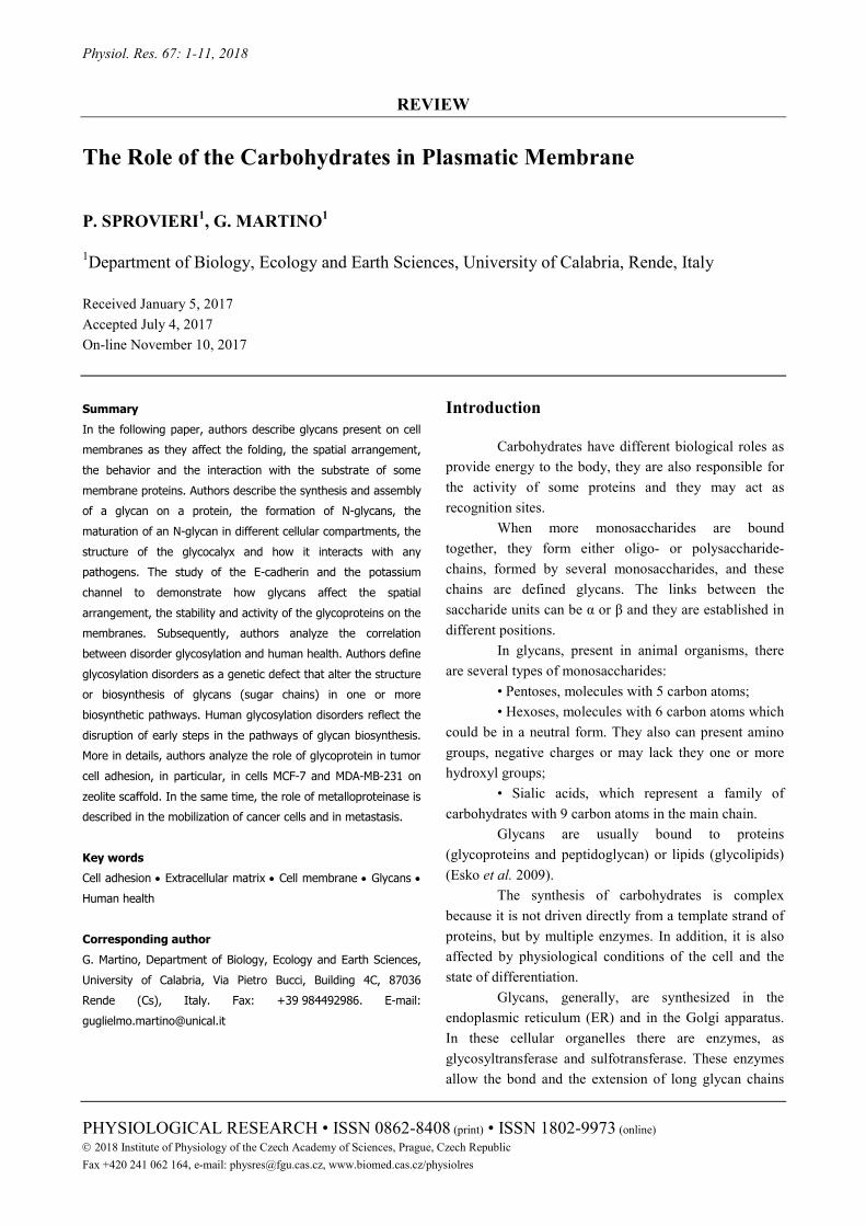
The Role of the Carbohydrates in Plasmatic Membrane P. SPROVIERI1, G. MARTINO1 1Department of Biology, Ecology and Earth Sciences, University of Calabria, Rende, Italy
Received January 5, 2017 Accepted July 4, 2017 On-line November 10, 2017 Summary In the following paper, authors describe glycans present on cell membranes as they affect the folding, the spatial arrangement, the behavior and the interaction with the substrate of some membrane proteins. Authors describe the synthesis and assembly of a glycan on a protein, the formation of N-glycans, the maturation of an N-glycan in different cellular compartments, the structure of the glycocalyx and how it interacts with any pathogens. The study of the E-cadherin and the potassium channel to demonstrate how glycans affect the spatial arrangement, the stability and activity of the glycoproteins on the membranes. Subsequently, authors analyze the correlation between disorder glycosylation and human health. Authors define glycosylation disorders as a genetic defect that alter the structure or biosynthesis of glycans (sugar chains) in one or more biosynthetic pathways. Human glycosylation disorders reflect the disruption of early steps in the pathways of glycan biosynthesis. More in details, authors analyze the role of glycoprotein in tumor cell adhesion, in particular, in cells MCF-7 and MDA-MB-231 on zeolite scaffold. In the same time, the role of metalloproteinase is described in the mobilization of cancer cells and in metastasis. Key words Cell adhesion • Extracellular matrix • Cell membrane • Glycans • Human health Corresponding author G. Martino, Department of Biology, Ecology and Earth Sciences, University of Calabria, Via Pietro Bucci, Building 4C, 87036 Rende (Cs), Italy. Fax: +39 984492986. E-mail: [email protected]
Introduction
Carbohydrates have different biological roles as provide energy to the body, they are also responsible for the activity of some proteins and they may act as recognition sites.
When more monosaccharides are bound together, they form either oligo- or polysaccharide-chains, formed by several monosaccharides, and these chains are defined glycans. The links between the saccharide units can be α or β and they are established in different positions.
In glycans, present in animal organisms, there are several types of monosaccharides:
• Pentoses, molecules with 5 carbon atoms; • Hexoses, molecules with 6 carbon atoms which
could be in a neutral form. They also can present amino groups, negative charges or may lack they one or more hydroxyl groups;
• Sialic acids, which represent a family of carbohydrates with 9 carbon atoms in the main chain.
Glycans are usually bound to proteins (glycoproteins and peptidoglycan) or lipids (glycolipids) (Esko et al. 2009).
The synthesis of carbohydrates is complex because it is not driven directly from a template strand of proteins, but by multiple enzymes. In addition, it is also affected by physiological conditions of the cell and the state of differentiation.
Glycans, generally, are synthesized in the endoplasmic reticulum (ER) and in the Golgi apparatus. In these cellular organelles there are enzymes, as glycosyltransferase and sulfotransferase. These enzymes allow the bond and the extension of long glycan chains
2 Sprovieri and Martino Vol. 67 on proteins, leading to modifications in their activities according to the structure of the linked glycan (Esko et al. 2009).
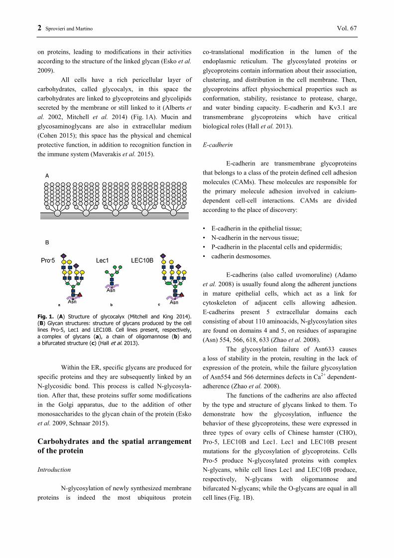
All cells have a rich pericellular layer of carbohydrates, called glycocalyx, in this space the carbohydrates are linked to glycoproteins and glycolipids secreted by the membrane or still linked to it (Alberts et al. 2002, Mitchell et al. 2014) (Fig. 1A). Mucin and glycosaminoglycans are also in extracellular medium (Cohen 2015); this space has the physical and chemical protective function, in addition to recognition function in the immune system (Maverakis et al. 2015).
Fig. 1. (A) Structure of glycocalyx (Mitchell and King 2014). (B) Glycan structures: structure of glycans produced by the cell lines Pro-5, Lec1 and LEC10B. Cell lines present, respectively, a complex of glycans (a), a chain of oligomannose (b) and a bifurcated structure (c) (Hall et al. 2013).
Within the ER, specific glycans are produced for specific proteins and they are subsequently linked by an N-glycosidic bond. This process is called N-glycosyla-tion. After that, these proteins suffer some modifications in the Golgi apparatus, due to the addition of other monosaccharides to the glycan chain of the protein (Esko et al. 2009, Schnaar 2015). Carbohydrates and the spatial arrangement of the protein Introduction
N-glycosylation of newly synthesized membrane proteins is indeed the most ubiquitous protein
co-translational modification in the lumen of the endoplasmic reticulum. The glycosylated proteins or glycoproteins contain information about their association, clustering, and distribution in the cell membrane. Then, glycoproteins affect physiochemical properties such as conformation, stability, resistance to protease, charge, and water binding capacity. E-cadherin and Kv3.1 are transmembrane glycoproteins which have critical biological roles (Hall et al. 2013). E-cadherin
E-cadherin are transmembrane glycoproteins that belongs to a class of the protein defined cell adhesion molecules (CAMs). These molecules are responsible for the primary molecule adhesion involved in calcium-dependent cell-cell interactions. CAMs are divided according to the place of discovery: • E-cadherin in the epithelial tissue; • N-cadherin in the nervous tissue; • P-cadherin in the placental cells and epidermidis; • cadherin desmosomes.
E-cadherins (also called uvomoruline) (Adamo et al. 2008) is usually found along the adherent junctions in mature epithelial cells, which act as a link for cytoskeleton of adjacent cells allowing adhesion. E-cadherins present 5 extracellular domains each consisting of about 110 aminoacids, N-glycosylation sites are found on domains 4 and 5, on residues of asparagine (Asn) 554, 566, 618, 633 (Zhao et al. 2008).
The glycosylation failure of Asn633 causes a loss of stability in the protein, resulting in the lack of expression of the protein, while the failure glycosylation of Asn554 and 566 determines defects in Ca2+ dependent-adherence (Zhao et al. 2008).
The functions of the cadherins are also affected by the type and structure of glycans linked to them. To demonstrate how the glycosylation, influence the behavior of these glycoproteins, these were expressed in three types of ovary cells of Chinese hamster (CHO), Pro-5, LEC10B and Lec1. Lec1 and LEC10B present mutations for the glycosylation of glycoproteins. Cells Pro-5 produce N-glycosylated proteins with complex N-glycans, while cell lines Lec1 and LEC10B produce, respectively, N-glycans with oligomannose and bifurcated N-glycans; while the O-glycans are equal in all cell lines (Fig. 1B).

2018 Carbohydrates in Cell Membranes 3
The cells are then observed by fluorescence microscopy (TIRF). The membranes are isolated and treated with various glycosidases and subsequently Western Blot is carried out. From the treatment of the membranes with PNGase F (which removes the complexes and oligomannose), neuraminidase (which removes sialic acid residues) and Endo H (which removes
oligomannose) and by analysis by Western Blot, in all three cell lines treated with PNGase F, there was an increase in electrophoretic mobility. Moreover, the mobility also increases following treatment with Endo H in the cells Lec1, however there was a minimum migration in cell lines LEC10B and Pro 5 following treatment with neuraminidase (Fig. 2A).
Fig. 2. (A) Western Blot of E-cadherins: western blot analysis of E-cadherins expressed from three different cell lines treated with different glycosidase. The sign (+) indicates the treatment with the enzyme quoted to the left (Hall et al. 2013). (B) Available E-cadherins: electron microscope images in fluorescence (TIRF) representing the arrangement of E-cadherins at the plasma membrane of the three cell lines Lec1 compared to cell lines Pro-5, while the smaller amount is found in the cells LEC10B (Hall et al. 2013).
From the images (Fig. 2B) in fluorescence microscopy can be seen a strong fluorescence of cell-cell interface in the cells Lec1 and Pro-5, and a weak fluorescence in the cytoplasm and in other areas of the membrane. Instead, cell line LEC10B presents a weaker signal in the cell-cell interface, but it is more accentuated in the cytoplasm.
From the pictures it seems that the amount of cell-cell glycoproteins interface is higher in cell lines Lec1 compared to cell lines Pro-5, while the smaller amount is found in the cells LEC10B (Hall et al. 2013).
N-glycan also contributes to clustering of E-cadherins. By treating of cells Pro-5 with PNGase F, before and after the link cell-cell, there are a change of glycoproteins distribution on the membrane.
From the images of the fluorescence microscope,
the signal at the cell to cell interface is strong, but it changes in the other aerial according to the treatment. In cells treated with PNGase F, the signal weakens to cell-cell interface and increases in the areas of the free membrane.
When the cells are treated with PNGase F, it is noted a reduction of proteins cell-cell interface, very strong (43 %) in the treated cells before making the contact and to a lesser extent (24 %) in the cells after they have made contact. This reduces, therefore, the adhesive force, established by E-cadherins between cells (Hall et al. 2014). Anyway, it cannot have glycoforms of the protein without treatment with PNGase F, in fact the protein isn’t stable and isn’t incorporated with the plasma membrane (Zhao et al. 2008). After treatment with PNGase F, there is a reduction of N-glycan present in

4 Sprovieri and Martino Vol. 67 proteins, but there are not proteins totally free glycan on the cell. However, E-cadherins from cell line Lec1, show a stronger adhesive force than cell lines Pro-5, while E-cadherins from cell line LEC10B have a weak force (Hall et al. 2013). Kv3.1 channels
Voltage-gated K+ channel (Kv3.1) plays a fundamental role in neuronal excitability. Upon stimulation, the voltage-dependent gate opens and
potassium ions flow out of the cells, inducing negative intracellular voltage, and termination of excitation. These channels have six transmembrane segments (S1-S6) and cytoplasmic N- and C-terminal (Fig. 3A). The segments between S1-S2 and S3-S4 are extracytoplasmic loops, and those between S2-S3 and S4-S5 are cytoplasmic loops.
This protein contains two sites of N-glycosylation between S1 and S2, on residues Asn220 and 229 (Brooks et al. 2006).
Fig. 3. (A) Topological model of a Kv3.1 channel: available on the plasma membrane of transmembrane segments, the glycosylation of sites and the servings of N- and C-terminal. Western Blot of channel Kv3.1: potassium channel wild type (B) (Brooks et al. 2006) in the different cell lines, and potassium channels N220Q/N229Q (C) (Hall et al. 2013.)
The voltage-gated K+ channels (Kv3.1), according to their state of glycosylation, change their conduction, threshold and activation speed. To determine how the glycosylation changes the properties of the channels are compared with wild-type channels of mutant forms N220Q, N229Q and N220Q/N229Q. These forms have a mutation on the residue in position 220 and 229, which have a residue of glutamine (Q) instead of
an asparagine residue (N). Cells of rat brain and Sf9 cells were used to demonstrate that glycosylation is not important for the transport of the protein to the plasma membrane. In fact, the expression of variant N220Q⁄N229Q produces a protein devoid of N-glycosyla-tion sites. Then, the expression of wild-type channel shows a protein free of N-glycosylation in Sf9 cells treated with tunicamycin, an antibiotic which inhibits the

2018 Carbohydrates in Cell Membranes 5
enzyme oligosaccariltransferase, an important enzyme to N-glycosylation. In the channels N220Q/N229Q, the time activation is higher compared to wild type channels, in both cases, however, the time activation decreases as the applied potential increased. In addition, these channels require a higher depolarization compared to wild type. This condition is also valid for channels partially glycosylated such as N220Q and N229Q. However, not all the channels open, since under equal conditions open fewer not-glycosylated channels than the glycosylated channels. These conditions are the same for the channels partially glycosylated, which behave similarly to totally non-glycosylated channels. This suggests that N-glycans are essential on these glycoproteins for the voltage dependence of the protein, while they are not essential for folding and targeting of the protein (Brooks et al. 2006). Available in the membrane
Glycosylation of Kv3.1 also influences the layout of the channels on the membrane, thus affecting the grouping and the position of the channels. This is determined using the ovary cells of the Chinese hamster (CHO) from three different lines, cells Pro-5, LEC10B and Lec1, the last two have defects in glycosylation producing respectively bifurcated and oligomannose
glycans, (Fig 1B). They are observed under a fluorescence microscope and then Western Blot is executed on them.
From these cell lines some membranes are isolated. The membranes are treated with PNGase F, neuraminidase and Endo H, then the Western Blot analysis is made on treated samples either with or without enzymes, of all types of cell lines, in all variants of the glycoprotein. From the pictures of the Western Blot (Fig. 3) it can be observed that the size of glycoprotein varies depending on the treatment and variant cell and protein, this implies that the glycans have a different sizes and kinds on the protein. In cell lines Pro-5 with wild type channels, not treated with any enzyme, electrophoresis is very slow compared to other cell lines, and is quick in Lec1 (Fig. 3B). From these data it is clear that the glycoproteins in cells Pro-5 have a “higher" size than LEC10B and Lec1. Treatment with enzymes confirms the type of glycan bound to the protein as the electrophoretic run changes.
Totally different is the size of the proteins N220Q/N229Q, that instead maintain an electrophoretic run similar for all cell lines. The migration does not vary even following treatment with the enzymes PNGase F, neuraminidase and Endo H (Fig. 3C).
Fig. 4. Available potassium channel: electron microscope images in fluorescence (TIRF) representing the arrangement of the voltage- dependent potassium channel (Kv3.1) glycosylated and non-glycosylated on the membrane of cells Pro-5 (A) and in the other cell lines (Hall et al. 2013) (B). Selectin and ligand: selectins and their respective ligands in the epithelial cells and leukocytes. To the left, at the top, there is the membrane of the epithelial cells that present selectins for binding with the glycoconjugates present on the leukocyte membrane to the left (C). In addition, lower left, there is the epithelial cell membrane having the PNAd that are related by L-selectin present on leukocytes, right (D) (McEver and Zhu 2010).

6 Sprovieri and Martino Vol. 67
All this is due to lack of sites of N-glycosylation. From the pictures under a fluorescence microscope, it can be observed that in cells Pro-5 with glycosylated channels, the signal strength is strongest in the area of cell-cell interface and along the membrane, while the signal of the channels not glycosylated is more distributed (Fig. 4A).
In cell lines Pro-5 and LEC10B, it can be seen how the channels are located at the cell-cell interface, while in Lec1 cell line the signal is distributed within the plasma membrane (Fig. 4B). This demonstrated that glycans contribute, as with the E-cadherins, also in the channels Kv3.1 to the provision on the plasma membrane.
Therefore, the amount of proteins changes along the area of contact between the two cells compared to other areas of the membrane, depending on the glycan type. In fact, channels that contain sialic acid (Pro-5) are very concentrated along the area of contact with the other cell, while this concentration decreases in the cells LEC10B and then in the cells Lec1. The not glycosylated channels not focused on cell-cell interface, but they remain scattered along the membrane in all cell lines (Hall et al. 2013). Interaction between glycan and protein
The presence of the glycocalyx, glycoproteins and glycoconjugates on the membrane allows some proteins to take advantage of structures rich in saccharide residues for different functions, such as adhesion to a tissue, recognition by the immune system and they have an important role in the functionality of glycoproteins (Maverakis et al. 2015). In fact, these saccharide residues allow a correct folding, a right arrangement on the membrane and also a correct activity.
White blood cells migrate into the tissues thanks to the interaction of some proteins with glycans. These proteins are the selectins, called lectins Ca2+-dependent. These proteins bind to the particular structures defined sialyl-Lewis X (sLex), this structure consists of sialic acid, galactose, fucose and N-acetylglucosamine. The L-selectins are expressed on the leukocyte membrane, while the E- and P-selectins are expressed on the membrane of the endothelial cells.
Leukocytes can migrate through the interaction between L-selectin and GlyCAM-1 (cell adhesion molecules glycosylated) (McEver and Zhu 2010).
GlyCAM-1 are glycoproteins containing sulfur.
They are similar to mucin and the majority of their weight is given by the carbohydrate chains linked to it, the carbohydrates are linked to the protein by O-glycosyl bonds to either serine or threonine residues. These glycoproteins, which are found on the endothelium of the peripheral and mesenteric lymphnodes, are very important for the transport of leukocytes to these organs (Dowbenko et al. 1993). In addition, with GlyCAM-1 on the endothelium of the lymph nodes and some sites of inflammation, there are other glycoproteins CD34 and podocalixine. These glycoproteins are called peripheral node addressins (PNAd) (McEver and Zhu 2010) (Fig. 4D).
Leukocytes have glycoproteins and glycolipids on their membrane. These glycans are recognized by selectins present in some tissues. Indeed, leukocytes are attracted to a site of infection due to the presence of E- and P-selectins on affected cells. E-and P-selectins bind to sugars found on glycoproteins and glycolipids of leukocytes (Cohen 2015). In fact, three glycoproteins are on leukocytes and they are called PSGL-1, CD44 and ESL-1 (Cohen 2015). PSGL-1 is transmembrane glycoprotein that acts as a ligand for P- selectin. PSGL-1 is located on both leukocytes and on some endothelial cells. It possesses saccharide chains linked by O-glycosyl bond residues of serine and threonine, as GlyCAM-1 (Fig. 4C) (McEver and Zhu 2010).
PSGL-1 is the most important ligand for L- and P-selectin, but are also linked by E-selectin. These mediate leukocyte displacements. The bond with the P-selectins is optimized by the presence of sulfated tyrosine residues near the N-terminal.
CD44 is a transmembrane glycoprotein expressed in most vertebrate cells, including hematopoietic stem cells, monocytes, lymphocytes, neutrophils and endothelial cells. These glycoproteins are linked by L- and E-selectin on leukocytes and act as a ligand for E-selectin. Altered forms of the protein can cause pathological conditions. They may be expressed in different isoforms of the protein, which indicate the tissue of belonging and the differentiation state.
ESL-1, also called GLG1, FGM-160 and CFR-1, are encoded by the gene Glg1. They are glycoproteins found on the Golgi membrane and they are also found on the cell membrane but to a lesser extent. These glycoproteins are cysteine-rich proteins with 5 sites of N-glycosylation which are located on rat neutrophils, while are not found on human neutrophils or human lymphocytes. They have a high affinity for E-selectin,

2018 Carbohydrates in Cell Membranes 7
and also occur in abundance in human metastatic prostate cells (Zarbock et al. 2011). Glycosylation and human health
Glycosylation disorder is defined as a genetic defect that alters the structure or biosynthesis of glycans (sugar chains) in one or more biosynthetic pathways. Congenital disorders of glycosylation (CDG) in humans emphasize the importance of the N-glycosylation process in the context of a multicellular organism. One such congenital disorder is caused by mutations in PMM2 gene that provides instructions for making an enzyme called phosphomannomutase 2. This enzyme catalyzes the conversion of mannose 6-phosphate to mannose 1-phosphate. This enzyme provides activation of precursors to at least four different glycosylation pathways as N-linked and O-mannose (Freeze et al. 2014).
N- and O-glycosylation abnormalities occur in glycogen storage disease (MIM 232220), caused by mutations in the genes that encode glucose-6-phosphate translocase and glucose-6-phosphatase (G6PC3). Loss of function of both enzymes has profound effects on neutrophils, leading to neutropenia because of ER stress and subsequent apoptosis (Hayee et al. 2011, Freeze et al. 2014).
Defective glycosylation can also cause congenital myasthenia syndromes (CMSs). These syndromes compromise signal transmission at the neuromuscular synapse. The postsynaptic disorders are caused by mutations in one of the genes encoding the pentameric acetylcholine receptor (AChR) subunits or mutations in enzymes required for subunit glycosylation, both of which inhibit assembly of the complex (Engel et al. 1999, Freeze et al. 2014). Glycosylation of proteins can influence their roles in tumor progression and metastases formation. In fact, high levels of sialic acid, fucose, T and Tn antigens, compared to healthy renal tissue, are characteristic for renal cell carcinoma. Several studies of human kidneys have shown that the binding sites for certain lectins are strictly confined to various parts of the nephron (Ortmann and Vierbuchen 1989, Borzym-Kluczyk et al. 2012). During a tumor process as prostate cancer, some of the cell enzymes can degrade the extracellular matrix and migrate to other body structures. This can cause other cancers in other areas, this process is called metastasis. During this process, following the degradation of the extracellular matrix, the cells undergo
a transformation from epithelial cells to mesenchymal cells, in this way the cells increase cell motility and plasticity which allows them to migrate into the circulatory flow. The cancer cells in the circulatory flow are defined circulating tumor cells (CTC). Since from blood, the cells adhere to endothelium, they enter into the tissues and then go to create a metastatic site, converting from mesenchymal cells in epithelial cells. The endothelium adhesion and "rolling" on it are allowed by ligands of E-selectin or ESL-1, CD44 and PSGL-1. In such cells are present increased expression levels of these glycoconjugates, in particular are very high levels of expression of ESL-1. This causes the activation of RAS, MEK and ERK that induce the expression of certain genes that allow the cells to migrate, invade another organ and develop a tumor in the organ “target” (Yasmin-Karim et al. 2014). The structure of the glycocalyx also interacts with receptors of some pathogenic organism. Haemophilus Influenzae binds to the glycan containing sialic acid and this not allow the cell to be attacked by immune system (Cohen 2015, Maverakis et al. 2015). The Influenzae A virus is an RNA virus and on envelope of this virus are present some glycoproteins as sialic acid binding hemagglutinin (HA) and neuraminidase (NA). HA binds all the sialic acid residues. Mucin present on epithelial cells act as bait for the virus. While, neuraminidase remove sialic acid residues thus allowing the release of the virus and also prevent the possible superinfection caused by the interaction of the virus with the cell, in this way infected cell can be recognized (Huang et al. 2008). Interaction of cell membranes with biomaterials
This topic has recently developed and his purpose is to study normal pathological cells in culture, even if the main clone is adherent to standard substrates, such as polyethylene plate, even with either special chemicals treatment or derivatization. Tavolaro et al. (2016a) demonstrated that the use of either pure zeolite membranes or mixed matrix with different percentages of zeolite crystals (5 %, 35 %, 70 %, 80 %) and polylactic acid polymer, as scaffolds for the adhesion and growth of cancer cells in the carrier membranes for selective adsorption, promote the delivery of antineoplastic drugs and cell adhesion. Authors demonstrated that cell adhesion is always mediated by membrane glycans so that started to investigate cell adhesion both in standard

8 Sprovieri and Martino Vol. 67 culture media and in anti-cancer drug and enriched culture media to better study drug role and in order to assess the various survival cell responses. As these membranes have different physico-chemical characteristics from each one, because while the former are only made of zeolite crystals and interact with molecules exclusively through the siloxane groups, the latter are formed by a polymeric matrix within which the crystals are dispersed possessing different chemical group, which depends on the polymeric materials structure. A difference between Pure Zeolite Membranes and Mixed Matrix Membranes is their different to absorb more drug than the hybrid membranes, carrying it much more, and causing, at the same time, a more drastic reduction of survival following treatment (Tavolaro et al. 2016b). The synthetic auto-supported zeolite membranes provide ideal, economic, reusable, pH and bacterial resistant, undegradable supports for cell adhesion, because many of their physico-chemical features like the point of zero charge (PZC), can be modulated by chemical manipulations in order to obtain, in this way, the best performance e.g. of fibroblast adhesion,
good-spreading and growth (Tavolaro et al. 2016c). In fact, MCF-7 and MDA-MB-231, human breast cancer cell lines, are used as model of tumor cells. The production of metalloproteinase (MMPs), is one of the mechanisms implicated in the lysis of membrane glycoproteins, in the invasion of the extracellular matrix and in cell migration, their increase in cancer cells is correlated to an increase in proteolytic and invasive activity. In Figure 5, the activities of metalloproteinase 2 and 9 are described in cancer cells treated with DOX and untreated. The obtained results in the histograms are obtained by the optical scanning of the collagen-containing gels into agar and detectable either in the presence of DOX or in the absence of the same. The treatment with DOX influences the ability to hydrolyze binding glycoproteins to the matrix. This shows that cancer cells bind to the extracellular matrix and that DOX inhibits engraftment of any type of scaffold, facilitating the detachment of cancer cells from the matrix and allowing the migration of cells to form metastases (Fig. 5) that are not clearly observable on other cell culture substrata without cell adhesion.
Fig. 5. Effects of metalloproteinase 2 and 9: the activity of the metalloproteinase 2 and 9 on tumor cell MCF-7 (A) and MDA-MB-231 (B). The controls (CRL) are cultured on polyphosphatidylserine (poly-PS). The other histogram groups refer to cells grown on different zeolitic scaffolds (Tavolaro et al. 2016c).
2018 Carbohydrates in Cell Membranes 9
This study showed that all the zeolite membranes synthesized are excellent scaffolds because they are very selective materials to support the adhesion and growth of neoplastic cells. Results obtained indicate that all cell adhesions are specific membrane-type and, in particular, that the MCF-7 cells maintain their individuality and tend to interact more with the scaffolds, assuming most of the times a form rather elongated, while cells MDA-MB-231 tend to melt their cell bodies forming irregular masses and they mainly interact with the scaffold by the terminal part of the cytoplasmic pseudopodia, confirming a more aggressiveness and labile adhesion. The understanding of the mechanisms of adhesion and physical translocation is important in order to prevent and avoid metastasis in early cancer lesions, whereas understanding the mechanisms leading to successful colonization may lead to effective therapies in already established metastases. Another important finding was that built zeolite scaffolds interact with human breast cancer cell lines and influence their development in different ways (Tavolaro et al. 2016a). Conclusions
Human glycosylation disease reflect the disruption of steps in the pathways of glycan biosynthesis. The defect in glycan synthesis causes a severe disorder leading and different pathologies.
In particular, authors investigated the response of two invasive phenotypes of human breast cancer cells, MCF-7 and MDA-MB-231, grown on synthesized zeolite scaffolds in order to study the influence of those biomaterials in controlled conditions with and without anti-tumoral drug treatments. The activity of metalloproteinases produced by cancer cells, is correlated to an increase of invasive activity. The results indicate that both drugs inhibit the cell viability of all cell lines grown on all zeolite scaffolds and that all pure zeolite membranes are more responsive with respect to all mixed matrix membranes. In the same time, defects in glycosylation pathways causes other disease as congenital disorders and myasthenia syndromes. Finally, glycocalyx structures interacts with receptors of pathogenic organism as Haemophilus influenza that binds to the glycan containing sialic acid and this not let to the cells to be antagonized by immune system. Conflict of Interest There is no conflict of interest.
Abbreviations AChR – pentameric acetylcholine receptor, Asn554 – E-cadherin mutant missing N-glycans at Asn554 fail to induce cell circle arrest in G1 phase, Asn566 – E-cadherin mutant missing N-glycans at Asn554 fail to induce cell circle arrest in G1 phase, Asn618 – E-cadherin mutant missing N-glycans at Asn554 fail to induce cell cycle arrest in G1 phase, Asn633 – Unglycosylation at Asn633 made extracellular domain of E-cadherin folded incorrectly and arrested in endoplasmatic reticulum, CAMs – cell adhesion molecules, CD34 – hematopoietic progenitor cell antigen, CD44 – antigen involved in cell to cell interaction, CDG – congenital disorders of glycosylation, CFR-1 – also nominated ESL-1, CHO – ovarian cells from Chinese hamster, CMSs – congenital myastenia syndromes, CTC – Circulating tumor cells, DOX – doxorubicin, antineoplastic drug, Endo H – Endoglycosidase H used for research purposes to deglycosylate glycoproteins, ER – endoplasmic reticulum, E-selectin – cell adhesion molecule that activated by cytokines, ESL-1 – E-selectin ligand is a variant of a receptor for fibroblast growth factor, FGM-160 – also nominated ESL-1, G6PC3 – glucose-6-phosphatase, GLG1 – Golgi glycoprotein 1, GlyCAM-1 – glycosylated cell adhesion molecules, HA – hemagglutinin, Kv3.1 – voltage-gated K+ channel, Lec1 – from female Cricetulus lack type N-glycans, Lec10B – ovarian cells Chinese hamster that express N-acetylglucosaminyltrasferase III. There are from activation of gene Mgat-3, L-selectin – cell adhesion molecule found on lymphocytes membrane, MIM 232220 – glycogen storage disease. Mutation of gene that prevents the conversion of glucose-6-phosphate translocase in glucose-6-phosphatase, MCF-7 – human breast cancer cell line as model tumor cell lines, MDA-MB-231 – human breast cancer cell line as model tumor cell lines, NA – neuraminidase, N-cadherin – cell adhesion molecule found in neurons, N – mutation of Kv3.1 protein in position 220/229 in particular in asparagine residue, N220Q – N-glycosylation consensus sequence for the Kv3.1 channel protein, N229Q – N-glycosylation consensus sequence for the Kv3.1 channel protein, PMM2 – genes expressed enzyme that converts Mannitol-6-phosphate in Mannitol-1-phosphate, PNAd – peripheral node adressin, PNGase F – Peptide N-glycosidase F not able to cleave N-linked glycans, P-selectin – cell adhesion molecule that active platelet adhesion, PSGL-1 – P-selectin glycoprotein ligand 1, Pro-5: epithelial cells from Cricetulus griseus, PZG –

10 Sprovieri and Martino Vol. 67 point of zero charge, Q – mutation of Kv3.1 protein in position 220/229 in particular in glutamine residue, Sf9 – isolated by insect Spodoptera frugiperda for the expression of recombinant proteins from baculovirus,
sLex – Sialyl-Lewis X this structure consists of sialic acid, galactose, fucose and N-acetylglucosamine, TIRF – total internal reflection fluorescence microscope, 5-MOP – bergapten, antineoplastic drug.
References ADAMO S, CARINCI P, MOLINARO M, SIRACUSA G, STEFANINI M, ZIPARO E: Istologia di V. Monesi 5
Edizione. PICCIN (ed.), Padova, 2008, 560 p. ALBERTS B, JOHNSON A, LEWIS J: Molecular Biology of the Cell. Garland Science, New York, 2002, 1616 p. BORZYM-KLUCZYK M, RADZIEJEWSKA I, DAREWICZ B: Glycosylation of proteins in healthy and pathological
human renal tissues. Folia Histochem Cytobiol 50: 599-604, 2012. BROOKS NL, COREY MJ, SCHWALBE RA: Characterization of N-glycosylation consensus sequences in the Kv3.1
channel. FEBS J 273: 14, 3287-3300, 2006. COHEN M: Notable aspects of glycan-protein interactions. Biomolecules 5: 2056-2072, 2015. DOWBENKO D, KIKUTA A, FENNIE C, GILLETT N, LASKY LA: Glycosylation-dependent cell adhesion
molecule 1 (GlyCAM 1) mucin is expressed by lactating mammary gland epithelial cells and is present in milk. J Clin Invest 92: 952-960, 1993.
ENGEL AG, OHNO K, SINE SM: Congenital myasthenic syndromes: recent advances. Arch Neurol 56: 163-167, 1999.
ESKO JD, VARKI A, CUMMINGS RD, FREEZE HH, STANLEY P, BERTOZZI CR, HART GW, ETZLER ME: Essentials of Glycobiology. Cold Spring Harbor Laboratory Press, New York, 2009, 784 p.
FREEZE HH, CHONG JX, BAMSHAD MJ, NG BG: Solving glycosylation disorders: fundamental approaches reveal complicated pathways. Am J Hum Genet 94: 161-175, 2014.
HALL MK, WEIDNER DA, CHEN JM, BERNETSKI CJ, SCHWALBE RA: Glycan structures contain information for the spatial arrangement of glycoproteins in the plasma membrane. PLoS One 8: e75013, 2013.
HALL MK, WEIDNER DA, DAYAL S, SCHWALBE RA: Cell surface N-glycans influence the level of functional E-cadherin at the cell-cell border. FEBS Open Bio 4: 892-897, 2014.
HAYEE B, ANTONOPOULOS A, MURPHY EJ, RAHMAN FZ, SEWELL G, SMITH BN, MCCARTNEY S, FURMAN M, HALL G, BLOOM SL, HASLAM SM, ET AL.: G6PC3 mutations are associated with a major defect of glycosylation: a novel mechanism for neutrophil dysfunction. Glycobiology 21: 914-924, 2011.
HUANG IC, LI W, SUI J, MARASCO W, CHOE H, FARZAN M: Influenza A virus neuraminidase limits viral superinfection. J Vir 82: 4834-4843, 2008.
MAVERAKIS E, KIM K, SHIMODA M, GERSHWIN ME, PATEL F, WILKEN R, RAYCHAUDHURI S, RUHAAK LR, LEBRILLA CB: Glycans in the immune system and the altered glycan theory of autoimmunity: a critical review. J Autoimmun 57: 1-13, 2015.
MCEVER RP, ZHU C: Rolling cell adhesion. Ann Rev Cell Dev Biol 26: 363-396, 2010. MITCHELL MJ, KING MR: The role of cell glycocalyx in vascular transport of circulating tumor cells. Am J Physiol
Cell Physiol 306: C89-C97, 2014. ORTMANN M, VIERBUCHEN M: Lectin histochemistry in the kidney and renal tumors. (In German) Verh Dtsch Ges
Pathol 73: 339-349, 1989. SCHNAAR RL: Glycans and glycan-binding proteins in immune regulation: A concise introduction to glycobiology for
the allergist. J All Clin Immun 135: 609-615, 2015. TAVOLARO P, CATALANO S, MARTINO G, TAVOLARO A: Zeolite inorganic scaffolds for novel biomedical
application: Effect of physicochemical characteristic of zeolite membranes on cell adhesion and viability. Appl Surf Sci 380: 135-140, 2016a.
TAVOLARO P, MARTINO G, ANDÒ S, TAVOLARO A: Fabrication and evaluation of novel zeolite membranes to control the neoplastic activity and anti-tumoral drug treatments in human breast cancer cells. Part 1: Synthesis and characterization of pure zeolite membranes and mixed matrix membranes for adhesion and growth of cancer cells. Mater Sci Eng C 69: 894-904, 2016b.
2018 Carbohydrates in Cell Membranes 11
TAVOLARO P, MARTINO G, ANDÒ S, TAVOLARO A: Zeolite scaffolds for cultures of human breast cancer cells. Part II: Effect of pure and hybrid zeolite membranes on neoplastic and metastatic activity control. Mater Sci Eng C 68: 474-481, 2016c.
YASMIN-KARIM S, KING MR, MESSING EM, LEE YF: E-selectin ligand-1 controls circulating prostate cancer cell rolling/adhesion and metastasis. Oncotarget 5: 12097-12110, 2014.
ZHAO H, LIANG Y, XU Z, WANG L, ZHOU F, LI Z, JIN J, YANG Y, FANG Z, HU Y, ZHANG L, SU J, ZHA X: N-glycosylation affects the adhesive function of E-cadherin through modifying the composition of adherens junctions (AJs) in human breast carcinoma cell line MDA-MB-435. J Cell Biochem 104: 162-175, 2008.
ZARBOCK A, LEY K, MCEVER RP, HIDALGO A: Leukocyte ligands for endothelial selectins: specialized glycoconjugates that mediate rolling and signaling under flow. Blood 118: 6743-6751, 2011.