Embed Size (px)
Citation preview

REVIEW ARTICLEpublished: 10 December 2013doi: 10.3389/fnins.2013.00233
The role of the striatum in social behaviorRaymundo Báez-Mendoza* and Wolfram Schultz
Department of Physiology, Development and Neuroscience, University of Cambridge, Cambridge, UK
Edited by:
Steve W. C. Chang, Duke University,USA
Reviewed by:
Jorge Moll, D’Or Institute forResearch and Education, BrazilBrian Lau, Centre de Recherche del’Institut du Cerveau et de la MoelleEpinière, France
*Correspondence:
Raymundo Báez-Mendoza,Department of Physiology,Development and Neuroscience,University of Cambridge, AnatomyBuilding, Downing Site, CB2 3DY,Cambridge, UKe-mail: [email protected]
Where and how does the brain code reward during social behavior? Almost all elements ofthe brain’s reward circuit are modulated during social behavior. The striatum in particular isactivated by rewards in social situations. However, its role in social behavior is still poorlyunderstood. Here, we attempt to review its participation in social behaviors of differentspecies ranging from voles to humans. Human fMRI experiments show that the striatumis reliably active in relation to others’ rewards, to reward inequity and also while learningabout social agents. Social contact and rearing conditions have long-lasting effects onbehavior, striatal anatomy and physiology in rodents and primates. The striatum also playsa critical role in pair-bond formation and maintenance in monogamous voles. We reviewrecent findings from single neuron recordings showing that the striatum contains cellsthat link own reward to self or others’ actions. These signals might be used to solve theagency-credit assignment problem: the question of whose action was responsible for thereward. Activity in the striatum has been hypothesized to integrate actions with rewards.The picture that emerges from this review is that the striatum is a general-purposesubcortical region capable of integrating social information into coding of social actionand reward.
Keywords: social interactions, social neurophysiology, agency, value, human, macaque, vole, rat
INTRODUCTIONThe striatum is necessary for voluntary motor control. Researchon its role in movement planning and execution uncoveredits participation in cognition and reward processes. Rigorousexperimentation demanded social isolation to properly studythis neuronal circuit. However, action, rewards and cognitionalso occur in the company of conspecifics, in a social con-text. Social behaviors, those behaviors that occur in a socialcontext, place an extra demand on cognition since others’behaviors are difficult to predict and they affect our own behav-ior. Therefore, to understand the properties of the striatum itis important to study it while the organism engages in socialbehavior. Recent studies highlight this brain structure duringdifferent social behaviors. Among these studies, we found thatthe striatum contains neurons that signal the social action thatwill result in own reward. We place these new findings withinthe context of previous findings on the known role of thisarea in movement and reward coding in the brain. The ques-tion that guides the review is as follows: “does the striatumserve a social function?” We conclude that the striatum is ageneral-purpose subcortical region capable of integrating andreflecting social information into its better known non-socialfunctions.
ANATOMY AND NEUROPHYSIOLOGY OF THE STRIATUMThe striatum is the input module to the basal ganglia, a neuronalcircuit necessary for voluntary movement control (Hikosakaet al., 2000). The striatum is composed of three nuclei: caudate,putamen, and ventral striatum. The latter contains the nucleusaccumbens (NAcc). The caudate and putamen/ventral striatum
are separated by the internal capsule, a white matter tract betweenbrain cortex and brainstem.
Striatal afferents arrive from three major sources: cortex, mid-brain and thalamus (Selemon and Goldman-Rakic, 1985; Haber,2003). The cortical input from temporal, parietal and frontalis mostly ipsilateral (Künzle, 1975; Vanhoesen et al., 1981) andtopographically arranged in the medio-lateral and dorsal-ventralaxes (Selemon and Goldman-Rakic, 1985; Haber, 2003; Haberand Knutson, 2010). The striatum receives inputs from all ele-ments of the reward circuit (Figure 1, reviewed in Haber andKnutson, 2010): from striato-nigral midbrain cells (Becksteadet al., 1979), amygdala (Russchen et al., 1985; Fudge et al., 2002),orbitofrontal cortex (OFC) (Haber et al., 2006), and anteriorcingulate cortex (ACC) (Selemon and Goldman-Rakic, 1985;Calzavara et al., 2007).
The striatum has two main efferent pathways. The directpathway is formed by axons of medium spiny neuron (MSN)expressing D1 receptors which mainly project to GABAergic neu-rons in the substantia nigra pars reticulata (SNr) (Parent et al.,1984; Gerfen et al., 1990; Kawaguchi et al., 1990; Chuhma et al.,2011). MSN that express D2 receptors mostly target the externalsegment of the globus pallidus (GPe) and form the indirect path-way (Parent et al., 1984; Gerfen et al., 1990; Kawaguchi et al., 1990;Chuhma et al., 2011). GABAeric neurons in GPe project to SNrand the internal segment of the globus pallidus (GPi) (Parent andHazrati, 1995; Wilson, 1998). The SNr and GPi are the outputnuclei of the basal ganglia.
The principal cell type in the striatum is the MSN (Wilson,1998; Tepper and Bolam, 2004). These neurons release γ-aminobutyric acid (GABA) at their synaptic terminals (Wilson, 1998).
www.frontiersin.org December 2013 | Volume 7 | Article 233 | 1

Báez-Mendoza and Schultz Striatum and social behavior
FIGURE 1 | Depiction of the brain’s reward circuit highlighting the role
of the striatum and its anatomical connections. Abbreviations: dACC,dorsal anterior cingulate cortex; DPFC, dorsal prefrontal cortex; vmPFC,ventromedial prefrontal cortex; VP, ventral pallidum; LHb, lateral habenula;Hypo, hypothalamus; STN, subthalamic nucleus; SN, substantia nigra; VTA,ventral tegmental area; PPT, pedunculopontine tegmentum. Based onHaber and Knutson (2010), reproduced with permission.
The striatum contains many other cell types besides MSN, includ-ing cholinergic and fast-firing GABAergic interneurons (Tepperand Bolam, 2004). Cholinergic interneuron activity has a rela-tionship to reward-predicting stimuli and reward and punish-ment (Apicella et al., 1991b; Ravel et al., 2003). These firingproperties suggest that these neurons may play a role in learn-ing (Schulz and Reynolds, 2013). Fast-firing interneurons are alsoinvolved in reward prediction error coding (Stalnaker et al., 2012).However, for brevity we will limit this review to MSN and referto them as striatal neurons. Functionally, striatal neurons showmotor and reward responses (Hikosaka et al., 2000). Functionaland anatomical evidence led to the hypothesis that striatal activ-ity forms a “limbic-motor” interface (Mogenson et al., 1980).Neurons in the striatum integrate information about expectedreward with motor information to guide behavior (Hollermanet al., 1998; Hikosaka et al., 2000; Schultz, 2000; Schultz andDickinson, 2000; Goldstein et al., 2012). We review MSN neuro-physiological responses to action and reward in the next section.
STRIATUM NEUROPHYSIOLOGY: ACTION AND REWARDThe striatum contains neuronal activity related to move-ments, rewards and the conjunction of both movement andreward. Striatal neurons show activity related to the preparation,
initiation and execution of movements (Hollerman et al., 2000).These neurons are also active before overt goal-directed move-ments (Schultz and Romo, 1988; Romo et al., 1992; Figure 2A).Some of these neurons are exclusively active during self-initiated movements, whilst other neurons are only active duringinstructed trials, and some others do not discriminate betweenself-initiated and instructed movements. In addition to this, stri-atal neurons also show reward related activity. Neuronal activityin the striatum is modulated by reward expectation indepen-dent of the movement necessary to obtain it (Hikosaka et al.,1989b; Apicella et al., 1991a, 1992; Schultz et al., 1992). Striatalneurons that discharge after reward delivery do so in two mainmodes: phasic or tonic. Phasic responses usually have short laten-cies (<50 ms) and are relatively short lived—median duration:500 ms (Apicella et al., 1991b; Hollerman et al., 1998; Lau andGlimcher, 2007; Figure 2B). By contrast, tonic responses havelonger latencies and can last as long as the intertrial interval,i.e., up to 3 s (Apicella et al., 1991b; Hollerman et al., 1998;Histed et al., 2009). Furthermore, there are striatal neurons cod-ing which action is associated to reward and which action isnot (Hollerman et al., 1998; Kawagoe et al., 1998; Figure 2C).This coding is independent of the stimuli indicating the actionrequired to obtain reward (Kimchi and Laubach, 2009; Kimchiet al., 2009). Reward-predicting cues modulate the activity of cau-date neurons (Kawagoe et al., 1998; Lauwereyns et al., 2002). Aftersaccade execution up to 50% of neurons encode only the action,while around 20% of recorded neurons encode whether the actionwas rewarded or not and close to 40% of neurons are modulatedby both movement and reward (Kobayashi et al., 2006; Lau andGlimcher, 2007). Together, these data suggest that striatal neuronsresponse is modulated by action and reward. These responses arenot limited to the moment of movement or reward receipt; ratherthey are present during cue and during reward expectation.
Most striatal neurons that respond during task performanceshow higher activity when a reward is expected compared to whenno reward is expected (Hollerman et al., 1998). However, thereare also neurons that are active preferentially after the monkey isinstructed to not move to obtain reward (Hollerman et al., 1998).These data suggest that striatal neurons flexibly encode the typeof action that will produce reward.
An action-value neuron tracks the value of one action, inde-pendent of the performed action. By tracking the value of dif-ferent candidate actions and comparing their values an organismcan decide to exploit the most valuable action or to explore thevalue of other actions. Samejima et al. (2005) were the first groupto show that striatal neurons code action-value (Figure 2D).Neuronal activity tracked over time the value of performing oneaction regardless of the animal’s choice. Later, Lau and Glimcher(2008) trained macaques to perform a matching task. In thistask rewards are distributed probabilistically between two optionsand subjects match the frequency with which they choose oneaction with its reward probability (Herrnstein, 1961). This taskopens the possibility of investigating the presence of action-value and chosen-value (i.e., value of the chosen action) neurons.Indeed, Lau found that caudate neurons code both action-valueand chosen-value. These signals can inform decision makingmechanisms.
Frontiers in Neuroscience | Decision Neuroscience December 2013 | Volume 7 | Article 233 | 2

Báez-Mendoza and Schultz Striatum and social behavior
FIGURE 2 | Action and reward coding by striatal neurons. (A)
Example striatal neuron active before movement (go) and silent beforeno-movement (no-go). Based on Schultz and Romo (1988), reproducedwith permission. (B) Example striatal neurons coding reward. First rowdepicts a neuron with phasic active after juice reward deliveryindependent of the action to obtain reward. Second row depicts aneuron with tonic activity after juice reward delivery. Third row shows aneuron with tonic activity after no reward is delivered. Based onHollerman et al. (1998), reproduced with permission. (C) Examplecaudate neuron coding the conjunction of action and reward. This
neuron is active during the presentation of a cue indicating the saccadenecessary to complete the trial if the trial will be rewarded (rewardeddirection is highlighted by a bulls eye). R, right; U, up; L, left; D, down.Polar plots show the average response for each cue and direction.Based on Kawagoe et al. (1998), reproduced with permission. (D) (Top)Depiction of the probability of larger rewards associated with left orright actions on each condition block. Colored numbers refer to theprobability associated with left-right actions. (Bottom) Example striatalneuron coding right action value. Based on Samejima et al. (2005),reproduced with permission.
In conclusion, the striatum contains neuronal activity relatedto movements, rewards and the conjunction of both movementand reward. These neuronal representations serve many functionslike goal directed movements and decision making.
STRIATAL ACTIVITY DURING SOCIAL BEHAVIORSOCIAL REWARDRewards are events or objects that elicit learning, elicit approachbehavior and produce positive emotions (Schultz, 2004). Socialrewards are just like any other rewards with the particular-ity that they occur in a social context. We propose a simple
classification of social rewards using two axes: who acts and whoreceives reward. For example, observing others is a social reward(Anderson, 1998; Deaner et al., 2005) where the individual acts(observes) and receives reward (the social stimuli). Pro-socialbehavior refers to a preference to increase the welfare of oth-ers (Fehr and Camerer, 2007). Depending on individual socialpreferences these choices can be rewarding by themselves, e.g., incharitable giving (Harbaugh et al., 2007). Vicarious reward refersto the situation when observing someone else receive reward isrewarding in itself (Mobbs et al., 2009). Finally, in several socialrewards the recipient is the individual and the actor is someone
www.frontiersin.org December 2013 | Volume 7 | Article 233 | 3

Báez-Mendoza and Schultz Striatum and social behavior
else. Examples of other’s actions that are rewarding include praiseand pleasant touch (Francis et al., 1999; Olausson et al., 2002;Rolls et al., 2008; Korn et al., 2012). Building a desired repu-tation is also considered a social reward; critically, reputationdepends on other’s perception of the individual, not on the indi-vidual’s perception of herself (Izuma et al., 2008; Izuma, 2012).Receiving gifts or social actions that result in own reward can alsobe considered as other-generated social rewards. Social inclusioncan be considered a social reward and facilitates learning (Egeret al., 2013). Although this classification might further our under-standing of the neuronal underpinnings of social rewards, furtherexperimentation might validate its use.
Observing othersFuelling a brain entails a huge cost, and the ratio of brain sizeto body size is larger in primates than any other Order in the ani-mal kingdom (Laughlin and Sejnowski, 2003; Dunbar and Shultz,2007). The huge cost of fuelling a large brain begs the ques-tion what is the benefit of such large brains? Byrne and Whittensuggest that only a costly primate brain can deal with the com-plexity of primate social living, the so-called social brain hypoth-esis (Dunbar and Shultz, 2007). The primate brain has a greatdeal of specializations to acquire information about conspecifics.Neurons in the ventral visual pathway respond selectively to bio-logical motion, gaze direction, body parts and faces (Perrett et al.,1984, 1985a,b; Gross, 1992; Oram and Perrett, 1996; Tsao et al.,2006). Social information arrives through all senses. For exam-ple, the superior temporal polysensory area contains neurons thatselectively respond to conspecific calls (Perrodin et al., 2011) andlocal field potentials in the temporal lobe are modulated by faceor call familiarity (Báez-Mendoza and Hoffman, 2009). The vol-ume of gray matter correlates with the size of the individual’stroop in mid superior temporal sulcus, inferotemporal cortex,rostral superior temporal sulcus, amygdala—all areas involvedin perceiving individuals—and rostral PFC in macaques (Salletet al., 2011). These findings suggest that the brain has special-ized structures dealing with the acquisition and representation ofinformation about conspecifics.
If the brain has specialized structures for the acquisition andrepresentation of information about conspecifics, then acquir-ing this information must be valuable for the individual. In aclever paradigm Deaner and colleagues measured the value ofacquiring access to observe pictures of conspecifics (Deaner et al.,2005). They pitted a constant amount of juice against a variableamount of juice plus the opportunity to observe the picture of aconspecific. The monkeys made their choices depending on theamount of juice offered along with the picture. If the monkeychose a smaller amount of juice plus the opportunity to watchan image, it strongly indicated that the monkey valued watchingthe image equivalent to the difference between offered juice vol-umes. For example, a monkey that likes watching a high-rankingmonkey will choose watching the image and receiving 0.8 ml ofjuice vs. only receiving 1ml of juice. When the monkey chose withequal probability between the two alternatives then the differencein offered juice volume is the subjective value for observing theimage, the so-called point of subjective equivalence. Researchersusing this method can measure the subjective value of varying
juice magnitudes (fluid value) and that of social images (imagevalue). Another advantage of this method is that it facilitates thecomparison of different goods (Glimcher, 2010), e.g., observingfemale perinea or a subordinate male face. Using this methodDeaner and colleagues reported that male monkeys valued highlylooking at dominant monkeys and the perinea of female monkeyscompared to looking at subordinate monkeys or a non-salientvisual stimulus (Deaner et al., 2005).
Neuronal activity during this task has been measured in dif-ferent brain regions. LIP neuronal activity correlates with bothimage value and fluid value when the monkeys chose to look at theimage (Klein et al., 2008). OFC neurons showed distinct codingof reward magnitude or image value, but not both (Watson andPlatt, 2012). Thus, these results suggest that OFC neurons do notcode reward on a single currency (e.g., in juice volume), rather asdifferent variables, as shown before (O’Neill and Schultz, 2010).Intriguingly, these animals strongly preferred looking at picturesof subordinates, a finding at odds with previously reported strongpreferences for dominant faces in the same paradigm (Deaner andPlatt, 2003; Deaner et al., 2005; Shepherd et al., 2006; Klein et al.,2008); but this result suggests that the encoding of social rewardreflects subjective preferences.
Neurons in the anterior striatum showed an interestingresponse pattern in the same paradigm (Klein and Platt, 2013).The large majority of reward responsive neurons were selectivefor reward type. These neurons also showed a regional pattern:those in the caudate were more strongly modulated by socialreward, conversely, putamen neurons were more strongly modu-lated by liquid reward. This pattern can be alternatively explainedby simple saccade direction coding because caudate neurons aretuned for saccade direction, particularly for contralateral saccades(Hikosaka et al., 1989a).
Humans also value observing other humans; and among dif-ferent targets we value highly observing our romantic partnersand mothers (Bartels and Zeki, 2000, 2004; Aron, 2005; Acevedoet al., 2012). Observing pictures of a partner elicits higher bloodoxygenated level-dependant (BOLD) activity in caudate/putamenand VTA along with cingulate and insular cortex compared toviewing pictures of friends matched for age, gender and length-of-friendship as their partners (Figure 3, green squares). This effectis present either when the relationship is recent (Aron, 2005)or when has been long established (Acevedo et al., 2012). TheseBOLD responses are a neural correlate of the value of observing aloved one.
In summary, acquiring social information, in particular look-ing at conspecifics, is valuable for the individual (Deaner et al.,2005). The primate temporal lobe contains regions whose func-tion includes the processing of social information (Tsao et al.,2006; Perrodin et al., 2011). Both social information and valueconverge in the striatum, opening the possibility of social rewardcoding in this brain region—as shown by Klein and Platt (2013).
Other social rewardsA positive reputation is a social reward as it can elicit learning,approach behavior and positive emotions. This is particularly evi-dent in indirect reciprocity: a donor who helps a recipient inpublic might receive in the future a donation from someone that
Frontiers in Neuroscience | Decision Neuroscience December 2013 | Volume 7 | Article 233 | 4

Báez-Mendoza and Schultz Striatum and social behavior
FIGURE 3 | fMRI studies of social behaviors in which the striatum is
active. Peak activation coordinates in the striatum of the fMRI studiescited in this review color-coded for each section as illustrated in thelegend. Studies using a region of interest analysis strategy were notincluded in this image. These striatal responses are compatible with ageneral activation in response to social behaviors, including social rewards.A functional subdivisions according to types of social rewards need toawait further experiments. Studies aggregated in “Other social rewards”:
(Rilling et al., 2002; Moll et al., 2006; Izuma et al., 2008; Mobbs et al.,2009; Acevedo et al., 2012; Fareri et al., 2012; Korn et al., 2012). Studiesclustered in “Observing others”: (Bartels and Zeki, 2000, 2004; Aron,2005; Acevedo et al., 2012). Studies in “Learning about others”: (Delgadoet al., 2005; King-Casas et al., 2005; Baumgartner et al., 2008; Burkeet al., 2010; Phan et al., 2010; Xiang et al., 2012; Fouragnan et al., 2013).Studies in “Reward inequity”: (Moll et al., 2006; Fliessbach et al., 2007;Hsu et al., 2008; Tricomi et al., 2010).
has observed its “altruistic” behavior (Nowak, 2006). Obtaininga good reputation from others increases BOLD activity in thehuman striatum (Izuma et al., 2008; Korn et al., 2012) (Figure 3,red squares), but not in individuals diagnosed with autism (Izumaet al., 2011). This difference is likely due to insensitivity to socialrewards in autistics (Dawson et al., 1998; Schultz, 2005).
Other social rewards that also increase BOLD activity inthe striatum include charitable donations (Moll et al., 2006;Harbaugh et al., 2007) and observing someone else succeed(Mobbs et al., 2009). Vicarious reward is also modulated by thecloseness of the recipient: there is higher striatal BOLD activ-ity when sharing a monetary gain with close friends comparedto sharing with strangers, and sharing with the latter is associ-ated with higher activations compared to when the “recipient” isa computer (Fareri et al., 2012). This social vs. non-social effecthas also been observed when cooperating with a human partnervs. cooperating with a computer (Rilling et al., 2002). The peakactivations from studies cited in this section are illustrated withred squares in Figure 3. Taken together, these data suggest thatsocial rewards are associated with BOLD activity in the striatumand can be modulated by the social context.
LEARNING ABOUT SOCIAL AGENTSSocial life is rife with opportunities to learn about others. Forexample, we learn to trust or mistrust other people. The trustgame is an economic game that measures how trust is builtbetween two individuals. During the trust game the investorreceives an initial endowment that she can choose to invest ina trustee, the trustee receives three times the investment anddecides how much of the gains to return to the investor. Whenthis game is played iteratively the investor learns to trust (ormistrust) the trustee and vice versa. Thus, both players developa model of the other’s reputation (King-Casas et al., 2005). Tobuild a trust model investors use previous behavior to predictfuture behavior. If there is a deviation from what is predicted—a reward prediction error—then the model is updated. Activity indorsal striatum mirrored prediction errors during the repayment
phase (Figure 3, yellow squares; King-Casas et al., 2005). Whenan investor returned more than what a trustee expected thetrustee reciprocated by increasing her investment. During theinvestment phase activity increased in middle cingulate cor-tex of the investor and also in ACC of the trustee. Activityin both areas correlated with activity in the trustee’s caudate;most importantly the peak of these correlations shifted from therepayment epoch to the investment epoch (King-Casas et al.,2005). These results suggest that generating someone else’s rep-utation engages a reinforcement learning algorithm that usesprediction errors and the latter are reflected in striatal BOLDactivity.
Prior information about someone’s trustworthiness sets theinitial state of the trust model. This initial bias can be overruledby observing someone’s willingness to reciprocate trust (Figure 3,yellow squares; Delgado et al., 2005; Phan et al., 2010; Fouragnanet al., 2013). Prior information diminishes the magnitude of thereward prediction error signal in the striatum during the repay-ment phase (Fouragnan et al., 2013). Following advice to solve atask (a type of prior information) generates an outcome-bonus ina version of the Iowa gambling task (Biele et al., 2011). These stud-ies suggest that prior information not only sets the initial state ofthe trust model, but it has a long lasting effect on its computation.
Depth-of-thought refers to a person’s inference about some-one else’s intention and to how many iterations of this inferencethey perform (Dixit and Skeath, 2004). Players in the trust gamesolve the game with different levels of depth-of-thought (Xianget al., 2012). If the investor makes no inference about the trustee’sintention to reciprocate, then a prediction error occurs whenthe trustee does not reciprocate trust. This prediction error isreflected in increased striatal activity (Figure 3, yellow squares;Xiang et al., 2012). If the investor infers that he plays this gameagainst a trustee that infers what he will offer, then the predic-tion error occurs when the investor submits its investment to thetrustee; again, the striatum reflects this prediction error (Xianget al., 2012). Thus, the computation of prediction errors, duringthe trust game, depends on depth-of-thought.
www.frontiersin.org December 2013 | Volume 7 | Article 233 | 5

Báez-Mendoza and Schultz Striatum and social behavior
Oxytocin, a neuropeptide, also modifies how we updatethe trust model. Intranasal administration of this neuropep-tide increases the rate of trust decisions compared to placebo,even after repeated violations of trust (Kosfeld et al., 2005).Correspondingly, people that received oxytocin showed a smallernegative prediction error signal in the striatum after repeatedviolations of trust (Baumgartner et al., 2008). Although the dis-tribution of oxytocin receptors in the human brain is unknown,one possible locus where oxytocin modifies trust is in the stria-tum (see section “Involvement of the Striatum in Pair-BondFormation and Maintenance” below).
Social life is also rife with opportunities to learn from oth-ers. Observational learning is another social cognitive processthat can be modeled with reinforcement learning. Burke and col-leagues hypothesized that observational learning is composed oftwo prediction errors, an action observation prediction error andan outcome observation prediction error (Burke et al., 2010). Intheir task two individuals took turns to learn which one of twodecks of cards provided a better outcome. In order to disentan-gle individual learning from imitation learning and observationallearning the individuals performed the task in three conditions:other’s actions and outcomes were private, only the other’s out-come was visible and both the partner’s action and outcome wereobservable. Burke and colleagues found a correlate for actionobservation prediction error in dorsolateral prefrontal cortex(DLPFC) and for outcome observation in ventromedial pre-frontal cortex (VMPFC) and ventral striatum (Figure 3, yellowsquares). Specifically, VMPFC activity correlated positively andventral striatum correlated negatively with the outcome observa-tion prediction error (Burke et al., 2010). Thus, they found neuralcorrelates of observational learning in frontal cortex and ventralstriatum.
In conclusion, the neuronal mechanism of learning to trustsomeone else or from someone else is based on a reinforce-ment learning algorithm. This algorithm makes predictions aboutother’s behavior and prediction errors help to update the model.The type of predictions depends on depth-of-thought and priorinformation modifies the rate to which the model is updated.These learning signals are reflected in changes in BOLD activityin the striatum.
INEQUITY AND FAIRNESS CONSIDERATIONSInequity arises from an asymmetric distribution of resourcesbetween two or more conspecifics. Classic economics assumesthat agents always intend to maximize their own benefit regard-less of other’s wellbeing (Von Neumann and Morgenstern, 1947).However, the difference in resource distribution can have a neg-ative impact on the utility and subjective value of an object(Loewenstein et al., 1989; Fehr and Schmidt, 1999). The disu-tility from an unequal outcome depends on who obtains moreresources. When the agent receives more than the conspecific,we speak of advantageous inequity. Conversely, when the agentreceives less than the conspecific we speak of disadvantageousinequity.
Interestingly, humans choose to lower their own payoff so thatinequity is smaller, a so-called pro-social behavior. For exam-ple, when people donate money to charity they diminish their
wealth so that others can be better off (Harbaugh et al., 2007).Disadvantageous inequity, having less than others, can have anegative effect in behavior. For example, progressive taxation isdesigned to reduce income inequality by implementing highertaxes on higher earners (Wilkinson and Pickett, 2010). An influ-ential hypothesis of how people react to inequity (Fehr andSchmidt, 1999) posits that unequal payoffs are aversive, there-fore agents try to minimize them. This theory has its roots onthe idea that one can estimate social utility functions that spec-ify level of satisfaction as a function of outcome to self and other(Loewenstein et al., 1989). Other example theories where socialutility functions help to explain human preferences that devi-ate from pure maximization include “Equity, Reciprocity, andCompetition” by Bolton and Ockenfels (Bolton and Ockenfels,2000) and “Fairness” by Rabin (Rabin, 1993).
One experimental task commonly used to measure advan-tageous inequity aversion is the dictator game (Forsythe et al.,1994). In this task the person playing as dictator receives an ini-tial financial endowment and decides to give an amount of theendowment to a receiver. The neoclassical assumption of ratio-nal behavior predicts that dictators will not give away anythingof their payoff; however, dictators usually give away between 5and 25% of their initial endowment (Forsythe et al., 1994). It isassumed that the proportion of money given to the receiver is ameasure of the disutility for the dictator of having more than theother (Gibbons, 1992; Camerer et al., 2004). To measure disad-vantageous inequity aversion scientists use the ultimatum game(Güth et al., 1982). In this game the proposer receives an endow-ment and proposes a split to the responder, just as in the dictatorgame. The responder then either rejects the split, thereby forgoingall monies, or accepts it. Neoclassical economic models predictthat the responder will accept any split that results in him hav-ing more than nothing. However, responders tend to only acceptsplits where they obtain more than 30% of the initial endowment(Güth et al., 1982). The responder’s minimum acceptable offeris the percentage of the initial endowment that he is willing toaccept 50% of the time (Camerer et al., 2004). This last parameteris directly proportional to the degree of disadvantageous inequityaversion.
When subjects play the dictator game as dictators the ventralstriatum is active when deciding to donate money to a charity(Moll et al., 2006; Harbaugh et al., 2007) and when enactingthe decision on how to distribute a good between two chari-table possibilities (Hsu et al., 2008). The relative wealth of thedonor and the receiver also matter to how the brain respondsto these decisions. After one of two volunteers is made better-off than the other volunteer, the worse-off volunteers rankedreceiving money much more appealing than their better-off coun-terparts (Tricomi et al., 2010). Accordingly, ventral striatum andVMPFC show higher activity during transfers to self than to theother. Better-off volunteers found more appealing that the otherreceived money than themselves. Ventral striatum and VMPFCreflected this preference: both brain regions showed higher activ-ity during transfers to other than to self (Tricomi et al., 2010).In a related experiment, Fliessbach and colleagues paid in differ-ent ratios to pairs of volunteers for correctly completing a simpletask while they were in an MRI scanner (Fliessbach et al., 2007).
Frontiers in Neuroscience | Decision Neuroscience December 2013 | Volume 7 | Article 233 | 6

Báez-Mendoza and Schultz Striatum and social behavior
Ventral striatum activity was positively correlated with the ratioof the payoff regardless of the actual personal monetary payoff.Furthermore, striatal activity was lowest during own errors andhighest during other’s errors. Such a social contrast has been con-firmed, e.g. activity in ventral striatum is higher after winning alottery in public vs. winning the same amount in private (Baultet al., 2011). The peak activations from the fMRI studies citedin this section are illustrated in Figure 3 with pink squares. Thus,these data suggest that the striatum reflects the difference betweenown and other’s rewards.
AGENCY CODING IN STRIATAL NEURONSReciprocal social interactions provide the opportunity to increasefitness through repeated exchanges with a particular individ-ual, although one of its by-products is reward inequality. Forthis interaction to be successful several mental processes needto take place (Axelrod and Hamilton, 1981): both participantsneed to identify their partner, assign agency for the current out-come, decide how to act depending on the series of events andkeep a tally of the recent exchanges. Without partner identifi-cation reciprocity is virtually impossible (unless all interactionstake place with a uniform population) (Dawkins, 2006). Withouta memory trace of the outcomes of the recent exchanges, par-ticipants might see themselves locked onto a “one-way street”reciprocal exchange. Agency assignment allows the individual toassign credit (or blame) for a shared outcome (Wolpert et al.,2003; Tomlin et al., 2006). With precise agency assignment inthe memory of recent exchanges individuals can avoid free rid-ers (Dawkins, 2006). Therefore, agency assignment is a trait thatmight have been favored by evolution in social animals.
Another way to frame the problem of agency assignment isto think of it as the “social” extension of the credit-assignmentproblem (Figure 4A). Let us revise what the credit-assignmentproblem is. In order for an action to be reinforced, it needs tobe selected from various actions made between the operant andthe reinforce. The organism needs to assign credit to the oper-ant, and not assign (or subtract) credit to other non-contingentactions (Sutton and Barto, 1998). This is done by changing theweights of different eligibility traces, or memories of past actions(Sutton and Barto, 1998). The agency credit assignment problemapplies when more than one actor can generate a reward (Tomlinet al., 2006). Thus, the agency credit assignment problem can becast by paraphrasing Sutton and Barto (1998): how do you dis-tribute credit for success among the many actors that may havebeen involved in producing it?
The striatum is well-suited for integrating social action (anaction made in a social context) and reward given its anatom-ical connections and known role in action and reward coding.We recorded striatal neuron’s activity while an animal performeda reward giving task with a conspecific in order to investigatethe interaction of social action and reward (Báez-Mendoza et al.,2013). The reward giving task is an extension of the paradigmdescribed by Hollerman et al. (1998) to encompass several socialdimensions. In the original paradigm the activity of striatal neu-rons was tested for relationships to movement vs. no-movementand reward vs. no-reward. In our task we tested if striatal neu-ron activity was related to own vs. conspecific’s movement and
own and/or conspecific’s reward. During the experiment twomonkeys sat opposite each other across a table with a touch-screen. Both animals took turns to complete the following task:the actor held a resting key with its right arm, the computerpresented two simultaneous cues predicting reward (circle) orno reward (square) separately for each animal (Figure 4B), fol-lowed by a blue go signal eliciting the actor’s arm movement fortouching it (Figure 4B). After a brief delay, the computer deliv-ered reward to the actor and then to the conspecific. We wereable to probe the neuronal correlates of agency and reward cod-ing by varying reward presence and absence for both players andwho performed the task. This simple test allowed us to test theneuronal mechanisms of a complex cognitive process.
Our first concern was whether the monkeys were sensitive tothe social nature of the task. Reaction times and eye fixation anal-ysis suggested that the monkeys were sensitive to reward receivedby themselves and their conspecific. Importantly, the animalswere less likely to move whenever it was the conspecific’s turn,suggesting that they had an understanding of the turn-takingstructure of the task. This is particularly relevant for agency creditassignment because during “own turns” the animal should haveassigned credit to itself for own reward and during “conspecific’sturns” to the conspecific.
Own reward modulated the activity of striatal neurons, as pre-viously observed (Hikosaka et al., 1989b; Apicella et al., 1991a);but few striatal neurons responded to conspecific’s reward.Interestingly, a sub-population of neurons differentiated betweensocial actors, with some neurons firing more strongly duringone of the actor’s turn. Given these types of neuronal modula-tions, we then looked at the neurons’ sensitivity to whose turnit was. A large number of own reward coding neurons reflectedthe social actor: some neurons responded to own reward onlywhen the recorded animal acted (Figure 4C) whereas a differentsub-population responded to own reward when the conspecificacted (Figure 4D). We tested a series of alternative hypothesis forthese data including: eye position, response inhibition, temporaldiscounting and reward cost, none of which were a satisfactoryexplanation of the data.
We also found a collection of neurons that reflected whosetrial it was. These neurons fired more strongly during own trialsthan conspecific’s trials, or vice versa: conspecific > own tri-als. These neurons reflected social action as they differentiatedbetween actors. To test whether these neurons truly reflected a“social” component of the task we measured their activity whilethe animal performed the task with the conspecific or a non-social juice recipient (an empty bucket). If a neuron is modulatedby the social component of the task, then it should stop differ-entiating between actors during the “bucket test.” This test forsocial-specific coding indicated that close to 50% of social actorcoding-neurons were indeed modulated by the social environ-ment. This is, to our knowledge, the first direct test of a neuronalcorrelate of social behavior in single neurons.
These experiments showed that there are multiple signals inthe striatum relevant for social interactions. The data suggests anextension of the known role of the striatum in movement andreward processing into the social domain. Several questions arisefrom these findings.
www.frontiersin.org December 2013 | Volume 7 | Article 233 | 7

Báez-Mendoza and Schultz Striatum and social behavior
FIGURE 4 | Agency credit assignment cartoon and striatal neurons
coding social action and own reward. (A) Once the monkey receives abanana it needs to know which action produced reward to assign credit.The action can be its own (solid lines) or someone else’s (dashed lines).Many actions take place before reward is delivered, therefore looking at amemory of each action or eligibility trace (brown arrows) can solve theagency credit assignment problem. (B) Task sequence for the actor: shapeof conditioned cue predicted absence or presence of reward for eachanimal. Appearance of a subsequent blue go signal was followed by key
release, stimulus touch and reward for actor, and later for conspecific. Afterthe ITI the monkeys switched roles as actor and passive. (C) Single striatalneuron coding own action and own reward. Note the higher neuronalactivity during own action and own reward compared to own rewardabsence and conspecific’s actions. (D) Single striatal neuron coding socialaction and own reward. This neuron is active during conspecific’s actionsthat will result in own reward, a complement to the neuron shown in (A).Monkey picture by smerikal (Flickr), reproduced with permission. Panels(B–D) based on Báez-Mendoza et al. (2013), reproduced with permission.
How are these signals formed? One possible mechanism isas follows: Striatal neurons receive biological motion informa-tion either directly from area STP (Oram and Perrett, 1996) orindirectly via parietal lobe (Cavada and Goldman-Rakic, 1991)while simultaneously receiving reward-related information fromdopaminergic neurons and other reward-related areas (Haberand Knutson, 2010, see also Figure 1). Converging inputs andlocal interactions (Chuhma et al., 2011) are also well-suited tocombine information about other’s actions and own reward.Future experiments will test and measure the formation ofagency and reward conjoint coding in the population of striatalneurons.
Another issue is: how are these signals used? We hypothesizethat this neuronal signal may help assign, and maintain, credit to asocial agent when receiving reward in a social context. Solving thisproblem is necessary for successful interactions. It is possible thestriatum provides a signal to distribute credit for reward amongthe many actors that may have been involved in producing it. Onekey experiment would test the individual-specificity of this signal:is the signal specific for one individual or it only discriminatesbetween own action and “other’s” actions? Such a fine grainedsignal would aid in discriminating who is a better partner andwho is not.
SOCIAL CONTACT AND STRIATAL FUNCTIONThe striatum is involved in other social behaviors besides socialaction, social reward and reward inequity. Social isolation andsocial defeat compromise the normal function of the striatum.These effects highlight the interplay between normal social con-tact and striatal function. Social isolation has long-lasting effectsin behavior, neuronal anatomy and neurochemistry. For example,social deprivation in the first year of life of macaques is relatedto abnormal social behaviors including fearfulness, withdrawal,lack of play, apathy, indifference to external stimuli, deficien-cies in communication and aggression (Martin et al., 1991).Macaques reared in social deprivation show decreased numbersof caudate/putamen neurons reactive to substance P, tyrosinehydroxylase (TH), leucine-enkephaline, and calbindin; in con-trast, the number of somatostatin interneurons did not differ tonormally-reared conspecifics. TH staining was reduced in SNcbut neuron numbers were stable. Other subcortical regions wereunaffected, including the NAcc, amygdala and BNST (Martinet al., 1991). Further characterization of the behavioral, anatomi-cal and neurochemical effects of social isolation have been carriedout in rodents.
Social isolation leaves consistent behavioral effects onrodents. These include hyper-reactivity to novel environments,
Frontiers in Neuroscience | Decision Neuroscience December 2013 | Volume 7 | Article 233 | 8
Báez-Mendoza and Schultz Striatum and social behavior
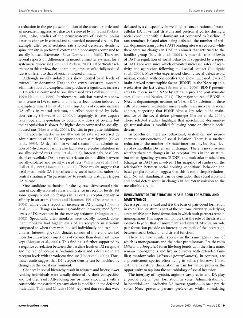
a reduction in the pre-pulse inhibition of the acoustic startle, andan increase in aggressive behavior (reviewed by Fone and Porkess,2008). Also, studies of the neuroanatomy of isolates’ brainsdescribe changes in cortical and subcortical neuronal circuits. Forexample, after social isolation rats showed decreased dendriticspine density in prefrontal cortex and hippocampus compared tosocially-housed littermates (Silva-Gomez et al., 2003). There areseveral reports on differences in neurotransmitter systems, for asystematic review see (Fone and Porkess, 2008). Of particular rel-evance to this review, the dopaminergic system of socially isolatedrats is different to that of socially-housed animals.
Although socially isolated rats show normal basal levels ofextracellular dopamine (DA) in the ventral striatum, systemicadministration of d-amphetamine produces a significant increasein DA release compared to socially-reared rats (Wilkinson et al.,1994; Hall et al., 1999). Furthermore, isolation-reared rats showan increase in DA turnover and in hyper-locomotion induced byd-amphetamine (Hall et al., 1998). Injections of cocaine increaseDA efflux in ventral striatum, an effect potentiated by isola-tion rearing (Howes et al., 2000). Intriguingly, isolates acquirefaster operant responding to obtain low doses of cocaine buttheir acquisition is slower for higher doses compared to socially-housed rats (Howes et al., 2000). Deficits in pre-pulse inhibitionof the acoustic startle in socially-isolated rats are reversed byadministration of the D2 receptor antagonist raclopride (Geyeret al., 1993). DA depletion in ventral striatum after administra-tion of 6-hydroxydopamine also facilitates pre-pulse inhibition insocially-isolated rats (Powell et al., 2003). Interestingly, basal lev-els of extracellular DA in ventral striatum do not differ betweensocially-isolated and socially-reared rats (Wilkinson et al., 1994;Hall et al., 1999; Howes et al., 2000). These results suggest thatbasal mesolimbic DA is unaffected by social isolation, rather theventral striatum is “hypersensitive” to events that naturally triggerDA release.
One candidate mechanism for the hypersensitive ventral stria-tum of socially-isolated rats is a difference in receptor levels. Yetsome groups report no changes in D1 or D2 receptor density oraffinity in striatum (Bardo and Hammer, 1991; Del Arco et al.,2004); while others report an increase in D2 binding (Djoumaet al., 2006). Changes in housing condition, however, modify thelevels of D2 receptors in the monkey striatum (Morgan et al.,2002). Specifically, after monkeys were socially housed, dom-inant monkeys had higher levels of D2 receptors in striatumcompared to when they were housed individually and to subor-dinates. Interestingly, subordinates consumed more and workedmore for intravenous injections of cocaine than dominant mon-keys (Morgan et al., 2002). This finding is further supported bya negative correlation between the baseline levels of D2 receptorsand the rate of cocaine self-administration and a decrease in D2receptor levels with chronic cocaine use (Nader et al., 2006). Thus,these results suggest that D2 receptor density can be modified bychanges in the social environment.
Changes in social hierarchy result in winners and losers: lowerranking individuals were usually defeated by their conspecificsand lost their rank. After losing one or more encounters with aconspecific, mesostriatal transmission is modified in the defeatedindividual. Tidey and Miczek (1996) reported that rats that were
defeated by a conspecific, showed higher concentrations of extra-cellular DA in ventral striatum and prefrontal cortex during asocial encounter with a dominant rat compared to baseline. Ifrats remained isolated after being defeated, the number of stri-atal dopamine transporter (DAT) binding sites was reduced, whilethere were no changes in DAT in animals that returned to thefamiliar group (Isovich et al., 2001). A potential role of levelsof DAT in regulation of social behavior is suggested by a reportof DAT knockout mice which exhibited increased rates of reac-tivity and aggression following mild social contact (Rodriguizet al., 2004). Mice who experienced chronic social defeat avoidmaking contact with conspecifics and show increased levels ofbrain derived neurotrophic factor (BDNF) in the NAcc up to 4weeks after the last defeat (Berton et al., 2006). BDNF potenti-ates DA release in the NAcc by acting in pre- and post-synapticsites (Russo and Nestler, 2013). The major source of BDNF inNAcc is dopaminergic neurons in VTA. BDNF deletion in thesecells of chronically-defeated mice results in an increase in socialcontact, suggesting that BDNF plays a key role in the main-tenance of the social defeat phenotype (Berton et al., 2006).These selected studies highlight that mesolimbic dopaminer-gic transmission is modified following acute or chronic socialdefeats.
In conclusion there are behavioral, anatomical and neuro-chemical consequences of social isolation. There is a markedreduction in the number of striatal interneurons, but basal lev-els of extracellular DA remain unchanged. There is no consensuswhether there are changes in DA receptor levels in the striatum,but other signaling systems (BDNF) and molecular mechanisms(changes in DAT) are involved. This snapshot of studies on therelationship between social housing conditions, behavior andbasal ganglia function suggest that this is not a simple relation-ship. Notwithstanding, it can be concluded that social isolationand social defeat result in changes in neurotransmission to themesolimbic circuit.
INVOLVEMENT OF THE STRIATUM IN PAIR-BOND FORMATION ANDMAINTENANCESex is a primary reward and it is the basis of pair-bond formationin voles. The striatum is part of the neuronal circuitry underlyinga remarkable pair-bond formation in which both partners remainmonogamous. It is important to note that the role of the striatumextends beyond that of movement and reward. Studies on volepair formation provide an interesting example of the interactionbetween social behavior and striatal function.
There are two similar species in the same genus: one ofwhich is monogamous and the other promiscuous. Prairie voles(Microtus ochrogaster) form life-long bonds with their first mate,remain monogamous and live in burrows with extended fam-ilies; meadow voles (Microtus pennsylvanicus), in contrast, area promiscuous species often living in solitary burrows (Insel,2010). This natural dissociation in pair formation provides theopportunity to tap into the neurobiology of social behavior.
The interplay of oxytocin, arginine-vasopressin and DA playa pivotal role in pair formation in voles. Administration ofhaloperidol—an unselective DA inverse agonist—in male prairievoles’ NAcc prevents partner preference, whilst stimulating
www.frontiersin.org December 2013 | Volume 7 | Article 233 | 9

Báez-Mendoza and Schultz Striatum and social behavior
D2-like receptors in caudate-putamen induces partner prefer-ence in the absence of mating (Aragona et al., 2003, 2006).Conversely, DA D1-like receptor activation prevents pair-bondformation (Aragona et al., 2006). This mechanism is similar infemales, since D2-like receptor stimulation induces partner pref-erence whereas administration of a D1-like agonist had no effect(Wang et al., 1999). Vasopressin V1a receptor gene transfer intothe ventral pallidum of polygamous meadow voles is sufficient toinduce pair-bond-like behavior after mating (Lim et al., 2004b).Similarly, overexpression of oxytocin receptor in NAcc facilitatedpartner preference in female prairie voles but has no effect inparental care, nor any effect on female meadow voles (Ross et al.,2009). Prairie voles have a high density of oxytocin-receptors inthe NAcc and of vasopressin V1a receptors in the ventral pallidumcompared to meadow voles (Insel and Shapiro, 1992; Hammockand Young, 2006). Interestingly, oxytocin-receptors are boundby oxytocin, and with lower affinity, vasopressin (Gimpl andFahrenholz, 2001). Interestingly, there are no differences in thedistribution of D1-like and D2-like receptors in the striatumbetween these two species (Lim et al., 2004a). Thus, these resultssuggest that the differential distribution of oxytocin and vaso-pressin receptors is responsible for pair-bond formation. In con-clusion, pair-bond formation is modulated by the interaction ofoxytocin, vasopressin and DA in NAcc neurons as well as thedistribution of oxytocin and vasopressin V1a receptors.
The role of oxytocin and vasopressin in social recognition issupported further by the absence of habituation to conspecifics inoxytocin and V1a-R knockout mice (Ferguson et al., 2000; Bielskyet al., 2004). Oxytocin knockout mice “recover” social habituationafter infusion of oxytocin agonists in central amygdala (Fergusonet al., 2001). Similarly, local infusion of V1a-R antagonists in lat-eral septum of rats inhibits habituation to conspecifics (Everts andKoolhaas, 1999). Thus, both oxytocin and vasopressin regulatesocial recognition.
The endogenous opioid system is another neuronal mecha-nism that may play a role in pair-bond formation. Mu-opioidreceptor (MOR) activation modulates partner preference infemale prairie voles (Burkett et al., 2011). MOR density is striatalregion specific, thus this effect is probably mediated by specificstriatal regions (Resendez et al., 2013). MORs within the dorsalstriatum mediate partner preference formation via impairment ofmating, whereas receptors in NAcc appear to mediate pair bondformation through the positive hedonics associated with mating(Resendez et al., 2013). Interestingly, monogamous voles showhigher MOR density in forebrain including the caudate-putamenand NAcc than the closely-related polygamous voles (Inoue et al.,2013), but see (Insel and Shapiro, 1992). Thus, interspecies dif-ferences in opiate receptor density and pharmacological effectssuggest a role of opiates in social attachment.
A relevant question is how and where these neurotransmit-ter systems interact. Rat NAcc core neurons expressing D1-likereceptors co-express prodynorphin, conversely D2-like express-ing cells co-express proenkephalin (Curran and Watson, 1995).An electron microscope investigation indicates that about halfof neurons in the rat dorsolateral striatum co-express D2 andMORs (Ambrose et al., 2004). These anatomical studies supportthe possibility that oxytocin, vasopressin and D2-like receptors
are present in single striatal cells, yet their interactions remain tobe further investigated.
Little is known about pair-bond formation in primates.However, marmosets, a monogamous new-world monkey, showoxytocin receptor labeling in NAcc among other subcorti-cal structures (Schorscher-Petcu et al., 2009), whereas rhesusmacaques, a polygamous old-world monkey, only show label-ing for this receptor in hypothalamus and the nucleus basalis ofMeynert (Freeman et al., 2012). Titi monkeys are a monogamousspecies that exhibit small, but significant, changes in glucoseintake in the NAcc and ventral pallidum 48 hr. after mating (Baleset al., 2007).
Whereas we have learned about pair-bond formation, the neu-ronal mechanisms of pair-bond maintenance are just starting tobe investigated. For example, monogamous male voles show asignificant increase in D1-like receptors in NAcc after pair-bondformation, and D1-like receptor antagonists diminish aggressivebehavior toward female strangers—a behavioral marker of pairbond formation (Aragona et al., 2006). This is probably the mostexciting open question in pair-bond formation, what are theneuronal mechanisms of pair-bond maintenance?
The striatum might also play a role in mother’s recognitionof offspring. The pregnancy hormones progesterone and oestro-gen prime the brain for the synthesis of oxytocin and its receptor(Keverne and Curley, 2004). Olfaction is the prime sense formaternal offspring recognition in mammals. Oxytocin receptorsexpression increases in central olfactory projections and NAccduring pregnancy (Keverne and Curley, 2004).
Overall, these studies suggest a mechanism for pair-bondingformation in voles. The hypothetical mechanism is centered inthe striatum’s capability to facilitate the association between olfac-tory social cues and reward. A potential mate’s pheromones reachthe vomeronasal organ (VNO), which in turns transmits the indi-vidual’s information to the extended amygdala and the centralamygdala further transmits this information to striatum. VNOlesions in female voles disrupt pair formation (Curtis et al., 2001),a finding that supports this hypothetical mechanism. However,other brain areas may also play a role in pair-bond formation.For example there are marked differences in the distribution ofdopamine, oxytocin and vasopressin receptors in the medial pre-frontal cortex of monogamous and promiscuous voles (Smeltzeret al., 2006). As noted by Wang and Young (Lim et al., 2004b;Young and Wang, 2004), the cellular mechanism might be theco-activation of D2-expressing accumbal neurons by vasopressinand/or oxytocin. Oxytocin is released by the hypothalamus, odorinformation transmitted from the central amygdala and DA isreleased by dopaminergic neurons in VTA. Striatal neurons arewell-suited for detecting the conjunction of sensorimotor infor-mation and reward. In pair-bond formation the role of thestriatum, particularly the NAcc is to facilitate the association ofsocial cues and reward to guarantee reproductive success.
CONCLUSIONSBased on the studies reviewed here, we conclude that the stria-tum plays a role in computations that take place during socialbehavior. These computations revolve around social actions andsocial rewards. fMRI and neurophysiology studies show that
Frontiers in Neuroscience | Decision Neuroscience December 2013 | Volume 7 | Article 233 | 10

Báez-Mendoza and Schultz Striatum and social behavior
neural activity in the striatum is modulated by social rewardsand by learning in a social context (Figure 3). By learning inthis context we refer to: learning about other’s preferences, a newmate, about other’s actions that lead to own reward, or updat-ing our predictions about other’s preferences. We have shownthat neuronal activity in the striatum is also modulated by socialactions and, critically, by the conjunction of social action and ownreward (Figure 4). The computations performed by the stria-tum are critical for successful social interactions. A breakdown insocial interactions leads to compromised striatal function, whichhighlights the interplay between this neuronal circuit and socialbehavior.
Overall, these observations suggest that the striatum does notappear to have a particular “social” specialization; rather its neu-rons are capable of flexibly incorporating social information intotheir computations. Therefore, it is justified to speak of the stria-tum as containing a general purpose neuronal mechanism toassociate actions or events with reward. Importantly, it can alsoassociate—or reflect—other’s actions to the rewards they lead to.Rewards are also coded in the activity of striatal neurons, and associal rewards are a sub-class of rewards, they are processed in thestriatum. Importantly, a functional subdivision based on differenttypes of social behaviors need to await further experimentation.In conclusion, the striatum plays a role in the computation ofsocial behavior.
ACKNOWLEDGMENTSWe thank Fabian Grabenhorst for discussions and comments onthe manuscript. We also thank Kelly Diederen and Charlotte vanCoeverden for critically reading the manuscript. Kelly Diederengenerated the images of Figure 3. Our research is funded by grantsfrom Wellcome Trust and European Research Council.
REFERENCESAcevedo, B. P., Aron, A., Fisher, H. E., and Brown, L. L. (2012). Neural correlates
of long-term intense romantic love. Soc. Cogn. Affect. Neurosci. 7, 145–159. doi:10.1093/scan/nsq092
Ambrose, L. M., Unterwald, E. M., and Van Bockstaele, E. J. (2004). Ultrastructuralevidence for co-localization of dopamine D2 and μ-opioid receptors in the ratdorsolateral striatum. Anat. Rec. A Discov. Mol. Cell. Evol. Biol. 279A, 583–591.doi: 10.1002/ar.a.20054
Anderson, J. R. (1998). Social stimuli and social rewards in primate learning andcognition. Behav. Process. 42, 159–175. doi: 10.1016/S0376-6357(97)00074-0
Apicella, P., Ljungberg, T., Scarnati, E., and Schultz, W. (1991a). Responses toreward in monkey dorsal and ventral striatum. Exp. Brain Res. 85, 491–500. doi:10.1007/BF00231732
Apicella, P., Scarnati, E., and Schultz, W. (1991b). Tonically discharging neurons ofmonkey striatum respond to preparatory and rewarding stimuli. Exp. Brain Res.84, 672–675. doi: 10.1007/BF00230981
Apicella, P., Scarnati, E., Ljungberg, T., and Schultz, W. (1992). Neuronal activity inmonkey striatum related to the expectation of predictable environmental events.J. Neurophysiol. 68, 945–960.
Aragona, B. J., Liu, Y., Curtis, T., Stephan, F. K., and Wang, Z. X. (2003). A criticalrole for nucleus accumbens dopamine in partner-preference formation in maleprairie voles. J. Neurosci. 23, 3483–3490.
Aragona, B. J., Liu, Y., Yu, Y. J., Curtis, J. T., Detwiler, J. M., Insel, T. R., et al.(2006). Nucleus accumbens dopamine differentially mediates the formationand maintenance of monogamous pair bonds. Nat. Neurosci. 9, 133–139. doi:10.1038/nn1613
Aron, A. (2005). Reward, motivation, and emotion systems associated withearly-stage intense romantic love. J. Neurophysiol. 94, 327–337. doi:10.1152/jn.00838.2004
Axelrod, R., and Hamilton, W. D. (1981). The evolution of cooperation. Science211, 1390–1396. doi: 10.1126/science.7466396
Báez-Mendoza, R., Harris, C. J., and Schultz, W. (2013). Activity of striatal neu-rons reflects social action and own reward. Proc. Natl. Acad. Sci. U.S.A. 110,16634–16639. doi: 10.1073/pnas.1211342110
Báez-Mendoza, R., and Hoffman, K. L. (2009). “Object ontology in temporal lobeensembles,” in Cortical Mechanisms of Vision, 1st Edn., eds M. Jenkin and L.Harris (Cambridge: Cambridge University Press), 237–253.
Bales, K. L., Mason, W. A., Catana, C., Cherry, S. R., and Mendoza, S. P. (2007).Neural correlates of pair-bonding in a monogamous primate. Brain Res. 1184,245–253. doi: 10.1016/j.brainres.2007.09.087
Bardo, M. T., and Hammer, R. P. (1991). Autoradiographic localization ofdopamine D1 and D2 receptors in rat nucleus accumbens. Resistance todifferential rearing conditions. Neuroscience 45, 281–290. doi: 10.1016/0306-4522(91)90226-E
Bartels, A., and Zeki, S. (2000). The neural basis of romantic love. Neuroreport 11,3829–3834. doi: 10.1097/00001756-200011270-00046
Bartels, A., and Zeki, S. (2004). The neural correlates of maternal and romanticlove. Neuroimage 21, 1155–1166. doi: 10.1016/j.neuroimage.2003.11.003
Bault, N., Joffily, M., Rustichini, A., and Coricelli, G. (2011). Medial pre-frontal cortex and striatum mediate the influence of social comparison onthe decision process. Proc. Natl. Acad. Sci. U.S.A. 108, 16044–16049. doi:10.1073/pnas.1100892108
Baumgartner, T., Heinrichs, M., Vonlanthen, A., Fischbacher, U., and Fehr, E.(2008). Oxytocin shapes the neural circuitry of trust and trust adaptation inhumans. Neuron 58, 639–650. doi: 10.1016/j.neuron.2008.04.009
Beckstead, R. M., Domesick, V. B., and Nauta, W. J. H. (1979). Efferent connectionsof the substantia nigra and ventral tegmental area in the rat. Brain Res. 175,191–217. doi: 10.1016/0006-8993(79)91001-1
Berton, O., Mcclung, C. A., Dileone, R. J., Krishnan, V., Renthal, W., Russo, S. J.,et al. (2006). Essential role of BDNF in the mesolimbic dopamine pathway insocial defeat stress. Science 311, 864–868. doi: 10.1126/science.1120972
Biele, G., Rieskamp, J., Krugel, L. K., and Heekeren, H. R. (2011). The neural basisof following advice. PLoS Biol. 9:e1001089. doi: 10.1371/journal.pbio.1001089
Bielsky, I. F., Hu, S. B., Szegda, K. L., Westphal, H., and Young, L. J. (2004).Profound impairment in social recognition and reduction in anxiety-like behav-ior in vasopressin V1a receptor knockout mice. Neuropsychopharmacology 29,483–493. doi: 10.1038/sj.npp.1300360
Bolton, G. E., and Ockenfels, A. (2000). ERC: a theory of equity, reciprocity, andcompetition. Am. Econ. Rev. 90, 166–193. doi: 10.1257/aer.90.1.166
Burke, C. J., Tobler, P. N., Baddeley, M., and Schultz, W. (2010). Neural mechanismsof observational learning. Proc. Natl. Acad. Sci. U.S.A. 107, 14431–14436. doi:10.1073/pnas.1003111107
Burkett, J. P., Spiegel, L. L., Inoue, K., Murphy, A. Z., and Young, L. J. (2011).Activation of mu-opioid receptors in the dorsal striatum is necessary for adultsocial attachment in monogamous prairie voles. Neuropsychopharmacology 36,2200–2210. doi: 10.1038/npp.2011.117
Calzavara, R., Mailly, P., and Haber, S. N. (2007). Relationship between the cor-ticostriatal terminals from areas 9 and 46, and those from area 8A, dorsaland rostral premotor cortex and area 24c: an anatomical substrate for cogni-tion to action. Eur. J. Neurosci. 26, 2005–2024. doi: 10.1111/j.1460-9568.2007.05825.x
Camerer, C., Loewenstein, G., and Rabin, M. (2004). Advances in BehavioralEconomics. Princeton, NJ: Russell Sage Foundation; Princeton University Press.
Cavada, C., and Goldman-Rakic, P. S. (1991). Topographic segregation of corticos-triatal projections from posterior parietal subdivisions in the macaque monkey.Neuroscience 42, 683–696. doi: 10.1016/0306-4522(91)90037-O
Chuhma, N., Tanaka, K. F., Hen, R., and Rayport, S. (2011). Functional connec-tome of the striatal medium spiny neuron. J. Neurosci. 31, 1183–1192. doi:10.1523/JNEUROSCI.3833-10.2011
Curran, E. J., and Watson, S. J. (1995). Dopamine receptor mRNA expressionpatterns by opioid peptide cells in the nucleus accumbens of the rat: a dou-ble in situ hybridization study. J. Comp. Neurol. 361, 57–76. doi: 10.1002/cne.903610106
Curtis, J. T., Liu, Y., and Wang, Z. (2001). Lesions of the vomeronasal organ disruptmating-induced pair bonding in female prairie voles (Microtus ochrogaster).Brain Res. 901, 167–174. doi: 10.1016/S0006-8993(01)02343-5
Dawkins, R. (2006). The Selfish Gene: –with a New Introduction by the Author.Oxford: University Press.
www.frontiersin.org December 2013 | Volume 7 | Article 233 | 11
Báez-Mendoza and Schultz Striatum and social behavior
Dawson, G., Meltzoff, A. N., Osterling, J., Rinaldi, J., and Brown, E. (1998).Children with autism fail to orient to naturally occurring social stimuli.J. Autism Dev. Disord. 28, 479–485. doi: 10.1023/A:1026043926488
Deaner, R. O., Khera, A. V., and Platt, M. L. (2005). Monkeys pay per view: adap-tive valuation of social images by rhesus macaques. Curr. Biol. 15, 543–548. doi:10.1016/j.cub.2005.01.044
Deaner, R. O., and Platt, M. L. (2003). Reflexive social attention in monkeys andhumans. Curr. Biol. 13, 1609–1613. doi: 10.1016/j.cub.2003.08.025
Del Arco, A., Zhu, S., Terasmaa, A., Mohammed, A. H., and Fuxe, K. (2004).Hyperactivity to novelty induced by social isolation is not correlated withchanges in D2 receptor function and binding in striatum. Psychopharmacology(Berl.) 171, 148–155. doi: 10.1007/s00213-003-1578-8
Delgado, M. R., Frank, R. H., and Phelps, E. A. (2005). Perceptions of moral charac-ter modulate the neural systems of reward during the trust game. Nat. Neurosci.8, 1611–1618. doi: 10.1038/nn1575
Dixit, A. K., and Skeath, S. (2004). Games of Strategy. New York, NY: W.W. Norton.Djouma, E., Card, K., Lodge, D. J., and Lawrence, A. J. (2006). The CRF1 receptor
antagonist, antalarmin, reverses isolation-induced up-regulation of dopamineD-2 receptors in the amygdala and nucleus accumbens of Fawn-Hooded rats.Eur. J. Neurosci. 23, 3319–3327. doi: 10.1111/j.1460-9568.2006.04864.x
Dunbar, R. I. M., and Shultz, S. (2007). Evolution in the social brain. Science 317,1344–1347. doi: 10.1126/science.1145463
Eger, E., Moretti, L., Dehaene, S., and Sirigu, A. (2013). Decoding the represen-tation of learned social roles in the human brain. Cortex 49, 2484–2493. doi:10.1016/j.cortex.2013.02.008
Everts, H. G. J., and Koolhaas, J. M. (1999). Differential modulation of lateral sep-tal vasopressin receptor blockade in spatial learning, social recognition, andanxiety-related behaviors in rats. Behav. Brain Res. 99, 7–16. doi: 10.1016/S0166-4328(98)00004-7
Fareri, D. S., Niznikiewicz, M. A., Lee, V. K., and Delgado, M. R. (2012). Socialnetwork modulation of reward-related signals. J. Neurosci. 32, 9045–9052. doi:10.1523/JNEUROSCI.0610-12.2012
Fehr, E., and Camerer, C. F. (2007). Social neuroeconomics: the neural circuitry ofsocial preferences. Trends Cogn. Sci. 11, 419–427. doi: 10.1016/j.tics.2007.09.002
Fehr, E., and Schmidt, K. M. (1999). A theory of fairness, competition, andcooperation. Q. J. Econ. 114, 817–868. doi: 10.1162/003355399556151
Ferguson, J. N., Aldag, J. M., Insel, T. R., and Young, L. J. (2001). Oxytocin in themedial amygdala is essential for social recognition in the mouse. J. Neurosci. 21,8278–8285.
Ferguson, J. N., Young, L. J., Hearn, E. F., Matzuk, M. M., Insel, T. R., and Winslow,J. T. (2000). Social amnesia in mice lacking the oxytocin gene. Nat. Genet. 25,284–288. doi: 10.1038/77040
Fliessbach, K., Weber, B., Trautner, P., Dohmen, T., Sunde, U., Elger, C. E., et al.(2007). Social comparison affects reward-related brain activity in the humanventral striatum. Science 318, 1305–1308. doi: 10.1126/science.1145876
Fone, K. C. F., and Porkess, M. V. (2008). Behavioural and neurochemicaleffects of post-weaning social isolation in rodents - Relevance to developmen-tal neuropsychiatric disorders. Neurosci. Biobehav. Rev. 32, 1087–1102. doi:10.1016/j.neubiorev.2008.03.003
Forsythe, R., Horowitz, J. L., Savin, N. E., and Sefton, M. (1994). Fairnessin simple bargaining experiments. Games Econ. Behav. 6, 347–369. doi:10.1006/game.1994.1021
Fouragnan, E., Chierchia, G., Greiner, S., Neveu, R., Avesani, P., and Coricelli, G.(2013). Reputational priors magnify striatal responses to violations of trust.J. Neurosci. 33, 3602–3611. doi: 10.1523/JNEUROSCI.3086-12.2013
Francis, S., Rolls, E. T., Bowtell, R., Mcglone, F., O’doherty, J., Browning, A., et al.(1999). The representation of pleasant touch in the brain and its relationshipwith taste and olfactory areas. Neuroreport 10, 453–459. doi: 10.1097/00001756-199902250-00003
Freeman, S. M., Smith, A. L., Goodman, M. M., and Young, L. J. (2012). In vivoand in vitro methods for localizing the oxytocin receptor in primate tissue. Am.J. Primatol. 74, 71–71.
Fudge, J. L., Kunishio, K., Walsh, P., Richard, C., and Haber, S. N. (2002).Amygdaloid projections to ventromedial striatal subterritories in the primate.Neuroscience 110, 257–275. doi: 10.1016/S0306-4522(01)00546-2
Gerfen, C. R., Engber, T. M., Mahan, L. C., Susel, Z., Chase, T. N., Monsma,F. J., et al. (1990). D1 and D2 dopamine receptor-regulated gene expres-sion of striatonigral and striatopallidal neurons. Science 250, 1429–1432. doi:10.1126/science.2147780
Geyer, M. A., Wilkinson, L. S., Humby, T., and Robbins, T. W. (1993). Isolationrearing of rats produces a deficit in prepulse inhibition of acoustic startlesimilar to that in schizophrenia. Biol. Psychiatry 34, 361–372. doi: 10.1016/0006-3223(93)90180-L
Gibbons, R. (1992). Game Theory for Applied Economists. Princeton, NJ: PrincetonUniversity Press.
Gimpl, G., and Fahrenholz, F. (2001). The oxytocin receptor system: structure,function, and regulation. Physiol. Rev. 81, 629–683.
Glimcher, P. (2010). Foundations of Neuroeconomic Analysis. New York, NY: OxfordUniversity Press. doi: 10.1093/acprof:oso/9780199744251.001.0001
Goldstein, B. L., Barnett, B. R., Vasquez, G., Tobia, S. C., Kashtelyan, V.,Burton, A. C., et al. (2012). Ventral striatum encodes past and predictedvalue independent of motor contingencies. J. Neurosci. 32, 2027–2036. doi:10.1523/JNEUROSCI.5349-11.2012
Gross, C. G. (1992). Representation of visual stimuli in inferior temporal cortex.Philos. Trans. Biol. Sci. 335, 3–10. doi: 10.1098/rstb.1992.0001
Güth, W., Schmittberger, R., and Schwarze, B. (1982). An experimental analysis ofultimatum bargaining. J. Econ. Behav. Organ. 3, 367–388. doi: 10.1016/0167-2681(82)90011-7
Haber, S. N. (2003). The primate basal ganglia: parallel and integrative networks.J. Chem. Neuroanat. 26, 317–330. doi: 10.1016/j.jchemneu.2003.10.003
Haber, S. N., Kim, K. S., Mailly, P., and Calzavara, R. (2006). Reward-related corticalinputs define a large striatal region in primates that interface with associa-tive cortical connections, providing a substrate for incentive-based learning.J. Neurosci. 26, 8368–8376. doi: 10.1523/JNEUROSCI.0271-06.2006
Haber, S. N., and Knutson, B. (2010). The reward circuit: linking primateanatomy and human imaging. Neuropsychopharmacology 35, 4–26. doi:10.1038/npp.2009.129
Hall, F. S., Wilkinson, L. S., Humby, T., Inglis, W., Kendall, D. A., Marsden, C.A., et al. (1998). Isolation rearing in rats: pre- and postsynaptic changes instriatal dopaminergic systems. Pharmacol. Biochem. Behav. 59, 859–872. doi:10.1016/S0091-3057(97)00510-8
Hall, F. S., Wilkinson, L. S., Humby, T., and Robbins, T. W. (1999). Maternaldeprivation of neonatal rats produces enduring changes in dopamine function.Synapse 32, 37–43.
Hammock, E. A. D., and Young, L. J. (2006). Oxytocin, vasopressin and pair bond-ing: implications for autism. Philos. Trans. R. Soc. B Biol. Sci. 361, 2187–2198.doi: 10.1098/rstb.2006.1939
Harbaugh, W. T., Mayr, U., and Burghart, D. R. (2007). Neural responses to taxa-tion and voluntary giving reveal motives for charitable donations. Science 316,1622–1625. doi: 10.1126/science.1140738
Herrnstein, R. J. (1961). Relative and absolute strength of response as a func-tion of frequency of reinforcement. J. Exp. Anal. Behav. 4, 267–272. doi:10.1901/jeab.1961.4-267
Hikosaka, O., Sakamoto, M., and Usui, S. (1989a). Functional properties of monkeycaudate neurons I: activities related to saccadic eye movements. J. Neurophysiol.61, 780–799.
Hikosaka, O., Sakamoto, M., and Usui, S. (1989b). Functional properties of mon-key caudate neurons III: activites related to expectation of target and reward.J. Neurophysiol. 61, 814–833.
Hikosaka, O., Takikawa, Y., and Kawagoe, R. (2000). Role of the basal ganglia in thecontrol of purposive saccadic eye movements. Physiol. Rev. 80, 953–978.
Histed, M. H., Pasupathy, A., and Miller, E. K. (2009). Learning substrates in theprimate prefrontal cortex and striatum: sustained activity related to successfulactions. Neuron 63, 244–253. doi: 10.1016/j.neuron.2009.06.019
Hollerman, J. R., Tremblay, L., and Schultz, W. (1998). Influence of reward expecta-tion on behavior-related neuronal activity in primate striatum. J. Neurophysiol.80, 947–963.
Hollerman, J. R., Tremblay, L., and Schultz, W. (2000). Involvement of basal gan-glia and orbitofrontal cortex in goal-directed behavior. Prog. Brain Res. 126,193–215. doi: 10.1016/S0079-6123(00)26015-9
Howes, S. R., Dalley, J. W., Morrison, C. H., Robbins, T. W., and Everitt, B.J. (2000). Leftward shift in the acquisition of cocaine self-administration inisolation-reared rats: relationship to extracellular levels of dopamine, serotoninand glutamate in the nucleus accumbens and amygdala-striatal FOS expression.Psychopharmacology (Berl.) 151, 55–63. doi: 10.1007/s002130000451
Hsu, M., Anen, C., and Quartz, S. R. (2008). The right and the good: distributivejustice and neural encoding of equity and efficiency. Science 320, 1092–1095.doi: 10.1126/science.1153651
Frontiers in Neuroscience | Decision Neuroscience December 2013 | Volume 7 | Article 233 | 12

Báez-Mendoza and Schultz Striatum and social behavior
Inoue, K., Burkett, J. P., and Young, L. J. (2013). Neuroanatomical distribution ofμ-opioid receptor mRNA and binding in monogamous prairie voles (Microtusochrogaster) and non-monogamous meadow voles (Microtus pennsylvanicus).Neuroscience 244, 122–133. doi: 10.1016/j.neuroscience.2013.03.035
Insel, T. R. (2010). The challenge of translation in social neuroscience: a reviewof oxytocin, vasopressin, and affiliative behavior. Neuron 65, 768–779. doi:10.1016/j.neuron.2010.03.005
Insel, T. R., and Shapiro, L. E. (1992). Oxytocin receptor distribution reflectssocial organization in monogamous and polygamous voles. Proc. Natl. Acad.Sci. U.S.A. 89, 5981–5985. doi: 10.1073/pnas.89.13.5981
Isovich, E., Engelmann, M., Landgraf, R., and Fuchs, E. (2001). Social isolationafter a single defeat reduces striatal dopamine transporter binding in rats. Eur.J. Neurosci. 13, 1254–1256. doi: 10.1046/j.0953-816x.2001.01492.x
Izuma, K. (2012). The social neuroscience of reputation. Neurosci. Res. 72, 283–288.doi: 10.1016/j.neures.2012.01.003
Izuma, K., Matsumoto, K., Camerer, C. F., and Adolphs, R. (2011). Insensitivity tosocial reputation in autism. Proc. Natl. Acad. Sci. U.S.A. 108, 17302–17307. doi:10.1073/pnas.1107038108
Izuma, K., Saito, D. N., and Sadato, N. (2008). Processing of social andmonetary rewards in the human striatum. Neuron 58, 284–294. doi:10.1016/j.neuron.2008.03.020
Kawagoe, R., Takikawa, Y., and Hikosaka, O. (1998). Expectation of reward mod-ulates cognitive signals in the basal ganglia. Nat. Neurosci. 1, 411–416. doi:10.1038/1625
Kawaguchi, Y., Wilson, C. J., and Emson, P. C. (1990). Projection subtypes of ratneostriatal matrix cells revealed by intracellular injection of biocytin. J. Neurosci.10, 3421–3438.
Keverne, E. B., and Curley, J. P. (2004). Vasopressin, oxytocin and social behaviour.Curr. Opin. Neurobiol. 14, 777–783. doi: 10.1016/j.conb.2004.10.006
Kimchi, E. Y., and Laubach, M. (2009). Dynamic encoding of action selection by themedial striatum. J. Neurosci. 29, 3148–3159. doi: 10.1523/JNEUROSCI.5206-08.2009
Kimchi, E. Y., Torregrossa, M. M., Taylor, J. R., and Laubach, M. (2009). Neuronalcorrelates of instrumental learning in the dorsal striatum. J. Neurophysiol. 102,475–489. doi: 10.1152/jn.00262.2009
King-Casas, B., Tomlin, D., Anen, C., Camerer, C. F., Quartz, S. R., and Montague,P. R. (2005). Getting to know you: reputation and trust in a two-personeconomic exchange. Science 308, 78–83. doi: 10.1126/science.1108062
Klein, J. T., Deaner, R. O., and Platt, M. L. (2008). Neural correlates ofsocial target value in macaque parietal cortex. Curr. Biol. 18, 419–424. doi:10.1016/j.cub.2008.02.047
Klein, J. T., and Platt, M. L. (2013). Social information signaling by neurons inprimate striatum. Curr. Biol. 23, 691–696. doi: 10.1016/j.cub.2013.03.022
Kobayashi, S., Kawagoe, R., Takikawa, Y., Koizumi, M., Sakagami, M., andHikosaka, O. (2006). Functional differences between macaque prefrontal cor-tex and caudate nucleus during eye movements with and without reward. Exp.Brain Res. 176, 341–355. doi: 10.1007/s00221-006-0622-4
Korn, C. W., Prehn, K., Park, S. Q., Walter, H., and Heekeren, H. R. (2012).Positively biased processing of self-relevant social feedback. J. Neurosci. 32,16832–16844. doi: 10.1523/JNEUROSCI.3016-12.2012
Kosfeld, M., Heinrichs, M., Zak, P. J., Fischbacher, U., and Fehr, E. (2005). Oxytocinincreases trust in humans. Nature 435, 673–676. doi: 10.1038/nature03701
Künzle, H. (1975). Bilateral projections from precentral motor cortex to the puta-men and other parts of the basal ganglia. An autoradiographic study in MacacaFascicularis. Brain Res. 88, 195–209. doi: 10.1016/0006-8993(75)90384-4
Lau, B., and Glimcher, P. W. (2007). Action and outcome encoding in the primatecaudate nucleus. J. Neurosci. 27, 14502–14514. doi: 10.1523/JNEUROSCI.3060-07.2007
Lau, B., and Glimcher, P. W. (2008). Value representations in the pri-mate striatum during matching behavior. Neuron 58, 451–463. doi:10.1016/j.neuron.2008.02.021
Laughlin, S. B., and Sejnowski, T. J. (2003). Communication in neuronal networks.Science 301, 1870–1874. doi: 10.1126/science.1089662
Lauwereyns, J., Takikawa, Y., Kawagoe, R., Kobayashi, S., Koizumi, M., Coe, B.,et al. (2002). Feature-based anticipation of cues that predict reward in monkeycaudate nucleus. Neuron 33, 463–473. doi: 10.1016/S0896-6273(02)00571-8
Lim, M. M., Murphy, A. Z., and Young, L. J. (2004a). Ventral striatopallidal oxy-tocin and vasopressin V1a receptors in the monogamous prairie vole (Microtusochrogaster). J. Comp. Neurol. 468, 555–570. doi: 10.1002/cne.10973
Lim, M. M., Wang, Z., Olazabal, D. E., Ren, X., Terwilliger, E. F., and Young,L. J. (2004b). Enhanced partner preference in a promiscuous species bymanipulating the expression of a single gene. Nature 429, 754–757. doi:10.1038/nature02539
Loewenstein, G., Thompson, L., and Bazerman, M. H. (1989). Social utility anddecision making in interpersonal contexts. J. Pers. Soc. Psychol. 57, 426–441. doi:10.1037/0022-3514.57.3.426
Martin, L. J., Spicer, D. M., Lewis, M. H., Gluck, J. P., and Cork, L. C. (1991). Socialdeprivation of infant Rhesus monkeys alters the chemoarchitecture of the brain.1. Subcortical regions. J. Neurosci. 11, 3344-3358.
Mobbs, D., Yu, R., Meyer, M., Passamonti, L., Seymour, B., Calder, A. J., et al.(2009). A key role for similarity in vicarious reward. Science 324, 900–900. doi:10.1126/science.1170539
Mogenson, G. J., Jones, D. L., and Yim, C. Y. (1980). From motivation to action—functional interface between the limbic system and the motor system. Prog.Neurobiol. 14, 69–97. doi: 10.1016/0301-0082(80)90018-0
Moll, J., Krueger, F., Zahn, R., Pardini, M., De Oliveira-Souza, R., andGrafman, J. (2006). Human fronto-mesolimbic networks guide decisions aboutcharitable donation. Proc. Natl. Acad. Sci. U.S.A. 103, 15623–15628. doi:10.1073/pnas.0604475103
Morgan, D., Grant, K. A., Gage, H. D., Mach, R. H., Kaplan, J. R., Prioleau,O., et al. (2002). Social dominance in monkeys: dopamine D2 receptorsand cocaine self-administration. Nat. Neurosci. 5, 169–174. doi: 10.1038/nn798
Nader, M. A., Morgan, D., Gage, H. D., Nader, S. H., Calhoun, T. L., Buchheimer,N., et al. (2006). PET imaging of dopamine D2 receptors during chroniccocaine self-administration in monkeys. Nat. Neurosci. 9, 1050–1056. doi:10.1038/nn1737
Nowak, M. A. (2006). Five rules for the evolution of cooperation. Science 314,1560–1563. doi: 10.1126/science.1133755
Olausson, H., Lamarre, Y., Backlund, H., Morin, C., Wallin, B. G., Starck, G., et al.(2002). Unmyelinated tactile afferents signal touch and project to insular cortex.Nat. Neurosci. 5, 900–904. doi: 10.1038/nn896
O’Neill, M., and Schultz, W. (2010). Coding of reward risk by orbitofrontal neu-rons is mostly distinct from coding of reward value. Neuron 68, 789–800. doi:10.1016/j.neuron.2010.09.031
Oram, M. W., and Perrett, D. (1996). Integration of form and motion in theanterior superior temporal polysensory area (STPa) of the macaque monkey.J. Neurophysiol. 76, 109–130.
Parent, A., Bouchard, C., and Smith, Y. (1984). The striatopallidal and striatonigralprojections: two distinct fiber systems in primate. Brain Res. 303, 385–390. doi:10.1016/0006-8993(84)91224-1
Parent, A., and Hazrati, L. N. (1995). Functional anatomy of the basal ganglia: 1.The cortico-basal ganglia-thalamo-cortical loop. Brain Res. Rev. 20, 91–127. doi:10.1016/0165-0173(94)00007-C
Perrett, D., Smith, P. A. J., Potter, D. D., Mistlin, A. J., Head, A. S., Milner, A. D., andJeeves, M.A. (1984). Neurones responsive to faces in the temporal cortex: stud-ies of funcional organization, sensitivity to identity and relation to perception.Hum. Neurobiol. 3, 197–208.
Perrett, D., Smith, P. A. J., Potter, D. D., Mistlin, A. J., Head, A. S., Milner, A.D., and Jeeves, M.A. (1985a). Visual cells in the temporal cortex sensitive toface view and gaze direction. Proc. Biol. Sci. 223, 293–317. doi: 10.1098/rspb.1985.0003
Perrett, D. I., Smith, P. A., Mistlin, A. J., Chitty, A. J., Head, A. S., Potter, D. D., et al.(1985b). Visual analysis of body movements by neurones in the temporal cortexof the macaque monkey: a preliminary report. Behav. Brain Res. 16, 153–170.doi: 10.1016/0166-4328(85)90089-0
Perrodin, C., Kayser, C., Logothetis, N. K., and Petkov, C. I. (2011).Voice cells in the primate temporal lobe. Curr. Biol. 21, 1408–1415. doi:10.1016/j.cub.2011.07.028
Phan, K. L., Sripada, C. S., Angstadt, M., and Mccabe, K. (2010). Reputation forreciprocity engages the brain reward center. Proc. Natl. Acad. Sci. U.S.A. 107,13099–13104. doi: 10.1073/pnas.1008137107
Powell, S. B., Geyer, M. A., Preece, M. A., Pitcher, L. K., Reynolds, G. P., andSwerdlow, N. R. (2003). Dopamine depletion of the nucleus accumbens reversesisolation-induced deficits in prepulse inhibition in rats. Neuroscience 119,233–240. doi: 10.1016/S0306-4522(03)00122-2
Rabin, M. (1993). Incorporating fairness into game theory and economics. Am.Econ. Rev. 83, 1281–1302.
www.frontiersin.org December 2013 | Volume 7 | Article 233 | 13
Báez-Mendoza and Schultz Striatum and social behavior
Ravel, S., Legallet, E., and Apicella, P. (2003). Responses of tonically active neuronsin the monkey striatum discriminate between motivationally opposing stimuli.J. Neurosci. 23, 8489–8497.
Resendez, S. L., Dome, M., Gormley, G., Franco, D., Nevarez, N., Hamid, A. A., et al.(2013). mu-Opioid receptors within subregions of the striatum mediate pairbond formation through parallel yet distinct reward mechanisms. J. Neurosci.33, 9140–9149. doi: 10.1523/JNEUROSCI.4123-12.2013
Rilling, J., Gutman, D., Zeh, T., Pagnoni, G., Berns, G., and Kilts, C. (2002). Aneural basis for social cooperation. Neuron 35, 395–405. doi: 10.1016/S0896-6273(02)00755-9
Rodriguiz, R. M., Chu, R., Caron, M. G., and Wetsel, W. C. (2004). Aberrantresponses in social interaction of dopamine transporter knockout mice. Behav.Brain Res. 148, 185–198. doi: 10.1016/S0166-4328(03)00187-6
Rolls, E. T., Grabenhorst, F., and Parris, B. A. (2008). Warm pleasant feelings in thebrain. Neuroimage 41, 1504–1513. doi: 10.1016/j.neuroimage.2008.03.005
Romo, R., Scarnati, E., and Schultz, W. (1992). Role of primate basal gangliaand frontal cortex in the internal generation of movements. II. Movement-related activity in the anterior striatum. Exp. Brain Res. 91, 385–395. doi:10.1007/BF00227835
Ross, H. E., Freeman, S. M., Spiegel, L. L., Ren, X., Terwilliger, E. F., and Young,L. J. (2009). Variation in oxytocin receptor density in the nucleus accumbenshas differential effects on affiliative behaviors in monogamous and polygamousvoles. J. Neurosci. 29, 1312–1318. doi: 10.1523/JNEUROSCI.5039-08.2009
Russchen, F. T., Bakst, I., Amaral, D. G., and Price, J. L. (1985). The amygdalos-triatal projections in the monkey. An anterograde tracing study. Brain Res. 329,241–257. doi: 10.1016/0006-8993(85)90530-X
Russo, S. J., and Nestler, E. J. (2013). The brain reward circuitry in mood disorders.Nat. Rev. Neurosci. 14, 609–625. doi: 10.1038/nrn3381
Sallet, J., Mars, R. B., Noonan, M. P., Andersson, J. L., O’reilly, J. X., Jbabdi, S.,et al. (2011). Social network size affects neural circuits in macaques. Science 334,697–700. doi: 10.1126/science.1210027
Samejima, K., Ueda, Y., Doya, K., and Kimura, M. (2005). Representation ofaction-specific reward values in the striatum. Science 310, 1337–1340. doi:10.1126/science.1115270
Schorscher-Petcu, A., Dupre, A., and Tribollet, E. (2009). Distribution of vaso-pressin and oxytocin binding sites in the brain and upper spinal cord of the com-mon marmoset. Neurosci. Lett. 461, 217–222. doi: 10.1016/j.neulet.2009.06.016
Schultz, R. T. (2005). Developmental deficits in social perception in autism: the roleof the amygdala and fusiform face area. Int. J. Dev. Neurosci. 23, 125–141. doi:10.1016/j.ijdevneu.2004.12.012
Schultz, W. (2000). Multiple reward signals in the brain. Nat. Rev. Neurosci. 1,199–207. doi: 10.1038/35044563
Schultz, W. (2004). Neural coding of basic reward terms of animal learning theory,game theory, microeconomics and behavioural ecology. Curr. Opin. Neurobiol.14, 139–147. doi: 10.1016/j.conb.2004.03.017
Schultz, W., Apicella, P., Scarnati, E., and Ljungberg, T. (1992). Neuronal activityin monkey ventral striatum related to the expectation of reward. J. Neurosci. 12,4595–4610.
Schultz, W., and Dickinson, A. (2000). Neuronal coding of prediction errors. Annu.Rev. Neurosci. 23, 473–500. doi: 10.1146/annurev.neuro.23.1.473
Schultz, W., and Romo, R. (1988). Neuronal activity in the monkey stria-tum during the initiation of movements. Exp. Brain Res. 71, 431–436. doi:10.1007/BF00247503
Schulz, J. M., and Reynolds, J. N. J. (2013). Pause and rebound: sensory con-trol of cholinergic signaling in the striatum. Trends Neurosci. 36, 41–50. doi:10.1016/j.tins.2012.09.006
Selemon, L. D., and Goldman-Rakic, P. S. (1985). Longitudinal topography andinterdigitation of corticostriatal projections in the rhesus monkey. J. Neurosci.5, 776–794.
Shepherd, S. V., Deaner, R. O., and Platt, M. L. (2006). Social status gates socialattention in monkeys. Curr. Biol. 16, R119–R120. doi: 10.1016/j.cub.2006.02.013
Silva-Gomez, A. B., Rojas, D., Juarez, I., and Flores, G. (2003). Decreased dendriticspine density on prefrontal cortical and hippocampal pyramidal neurons inpostweaning social isolation rats. Brain Res. 983, 128–136. doi: 10.1016/S0006-8993(03)03042-7
Smeltzer, M. D., Curtis, J. T., Aragona, B. J., and Wang, Z. X. (2006). Dopamine,oxytocin, and vasopressin receptor binding in the medial prefrontal cortexof monogamous and promiscuous voles. Neurosci. Lett. 394, 146–151. doi:10.1016/j.neulet.2005.10.019
Stalnaker, T. A., Calhoon, G. G., Ogawa, M., Roesch, M. R., and Schoenbaum, G.(2012). Reward prediction error signaling in posterior dorsomedial striatum isaction specific. J. Neurosci. 32, 10296–10305. doi: 10.1523/JNEUROSCI.0832-12.2012
Sutton, R. S., and Barto, A. G. (1998). Reinforcement Learning: An Introduction.Cambridge, MA: The MIT press.
Tepper, J. M., and Bolam, J. P. (2004). Functional diversity and speci-ficity of neostriatal interneurons. Curr. Opin. Neurobiol. 14, 685–692. doi:10.1016/j.conb.2004.10.003
Tidey, J. W., and Miczek, K. A. (1996). Social defeat stress selectively alters meso-corticolimbic dopamine release: an in vivo microdialysis study. Brain Res. 721,140–149. doi: 10.1016/0006-8993(96)00159-X
Tomlin, D., Kayali, M. A., King-Casas, B., Anen, C., Camerer, C. F., Quartz, S. R.,et al. (2006). Agent-specific responses in the cingulate cortex during economicexchanges. Science 312, 1047–1050. doi: 10.1126/science.1125596
Tricomi, E., Rangel, A., Camerer, C. F., and O’doherty, J. P. (2010). Neural evi-dence for inequality-averse social preferences. Nature 463, 1089–1091. doi:10.1038/nature08785
Tsao, D. Y., Freiwald, W. A., Tootell, R. B. H., and Livingstone, M. S. (2006). Acortical region consisting entirely of face-selective cells. Science 311, 670–674.doi: 10.1126/science.1119983
Vanhoesen, G. W., Yeterian, E. H., and Lavizzomourey, R. (1981). Widespread cor-ticostriate projections from temporal cortex of the Rhesus-monkey. J. Comp.Neurol. 199, 205–219. doi: 10.1002/cne.901990205
Von Neumann, J., and Morgenstern, O. (1947). The Theory of Games and EconomicBehavior. Princeton, NJ: Princeton University Press.
Wang, Z., Yu, G., Cascio, C., Liu, Y., Gingrich, B., and Insel, T. R. (1999). DopamineD2 receptor-mediated regulation of partner preferences in female prairie voles(Microtus ochrogaster): a mechanism for pair bonding? Behav. Neurosci. 113,602–611. doi: 10.1037/0735-7044.113.3.602
Watson, K. K., and Platt, M. L. (2012). Social signals in primate orbitofrontalcortex. Curr. Biol. 22, 2268–2273. doi: 10.1016/j.cub.2012.10.016
Wilkinson, L. S., Killcross, S. S., Humby, T., Hall, F. S., Geyer, M. A., and Robbins, T.W. (1994). Social isolation in the rat produces developmentally specific deficitsin prepulse inhibition of the acoustic startle response without disrupting latentinhibition. Neuropsychopharmacology 10, 61–72. doi: 10.1038/npp.1994.8
Wilkinson, R. G., and Pickett, K. (2010). The Spirit Level: Why Equality is Better forEveryone. London: Penguin Books.
Wilson, C. J. (1998). “Basal ganglia,” in The synaptic organization of the brain, ed G.M. Shepherd (New York, NY: Oxford University Press), 329–375.
Wolpert, D. M., Doya, K., and Kawato, M. (2003). A unifying computational frame-work for motor control and social interaction. Philos. Trans. R. Soc. B Biol. Sci.358, 593–602. doi: 10.1098/rstb.2002.1238
Xiang, T., Ray, D., Lohrenz, T., Dayan, P., and Montague, P. R. (2012).Computational phenotyping of two-person interactions reveals differentialneural response to depth-of-thought. PLoS Comput. Biol. 8:e1002841. doi:10.1371/journal.pcbi.1002841
Young, L. J., and Wang, Z. (2004). The neurobiology of pair bonding. Nat. Neurosci.7, 1048–1054. doi: 10.1038/nn1327
Conflict of Interest Statement: The authors declare that the research was con-ducted in the absence of any commercial or financial relationships that could beconstrued as a potential conflict of interest.
Received: 11 October 2013; paper pending published: 05 November 2013; accepted: 18November 2013; published online: 10 December 2013.Citation: Báez-Mendoza R and Schultz W (2013) The role of the striatum in socialbehavior. Front. Neurosci. 7:233. doi: 10.3389/fnins.2013.00233This article was submitted to Decision Neuroscience, a section of the journal Frontiersin Neuroscience.Copyright © 2013 Báez-Mendoza and Schultz. This is an open-access article dis-tributed under the terms of the Creative Commons Attribution License (CC BY).The use, distribution or reproduction in other forums is permitted, providedthe original author(s) or licensor are credited and that the original publica-tion in this journal is cited, in accordance with accepted academic practice. Nouse, distribution or reproduction is permitted which does not comply with theseterms.
Frontiers in Neuroscience | Decision Neuroscience December 2013 | Volume 7 | Article 233 | 14