Embed Size (px)
Citation preview
Full Paper
The Role of VUV Radiation in the Inactivationof Bacteria with an Atmospheric PressurePlasma Jet
Simon Schneider, Jan-Wilm Lackmann, Dirk Ellerweg, Benjamin Denis,Franz Narberhaus, Julia E. Bandow, Jan Benedikt*
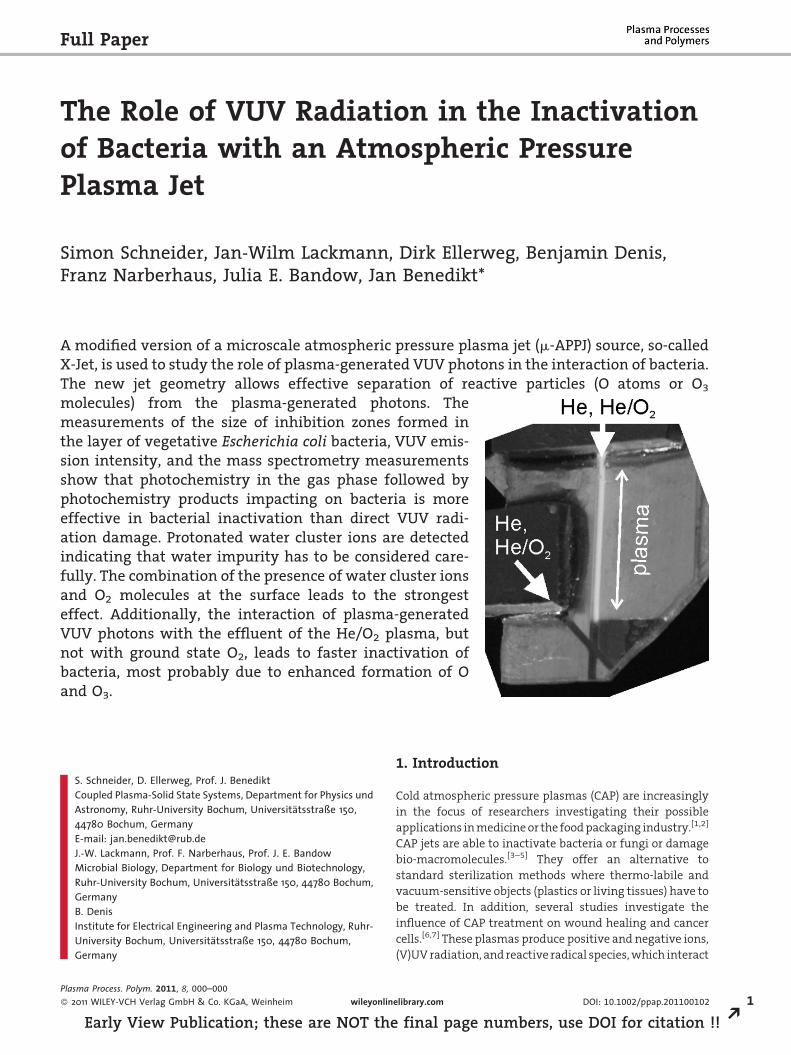
A modified version of a microscale atmospheric pressure plasma jet (m-APPJ) source, so-calledX-Jet, is used to study the role of plasma-generated VUV photons in the interaction of bacteria.The new jet geometry allows effective separation of reactive particles (O atoms or O3
molecules) from the plasma-generated photons. Themeasurements of the size of inhibition zones formed inthe layer of vegetative Escherichia coli bacteria, VUV emis-sion intensity, and the mass spectrometry measurementsshow that photochemistry in the gas phase followed byphotochemistry products impacting on bacteria is moreeffective in bacterial inactivation than direct VUV radi-ation damage. Protonated water cluster ions are detectedindicating that water impurity has to be considered care-fully. The combination of the presence of water cluster ionsand O2 molecules at the surface leads to the strongesteffect. Additionally, the interaction of plasma-generatedVUV photons with the effluent of the He/O2 plasma, butnot with ground state O2, leads to faster inactivation ofbacteria, most probably due to enhanced formation of Oand O3.
S. Schneider, D. Ellerweg, Prof. J. BenediktCoupled Plasma-Solid State Systems, Department for Physics undAstronomy, Ruhr-University Bochum, Universitatsstraße 150,44780 Bochum, GermanyE-mail: [email protected]. Lackmann, Prof. F. Narberhaus, Prof. J. E. BandowMicrobial Biology, Department for Biology und Biotechnology,Ruhr-University Bochum, Universitatsstraße 150, 44780 Bochum,GermanyB. DenisInstitute for Electrical Engineering and Plasma Technology, Ruhr-University Bochum, Universitatsstraße 150, 44780 Bochum,Germany
Plasma Process. Polym. 2011, 8, 000–000
� 2011 WILEY-VCH Verlag GmbH & Co. KGaA, Weinheim wileyonlin
Early View Publication; these are NOT the
1. Introduction
Cold atmospheric pressure plasmas (CAP) are increasingly
in the focus of researchers investigating their possible
applications in medicine or the food packaging industry.[1,2]
CAP jets are able to inactivate bacteria or fungi or damage
bio-macromolecules.[3–5] They offer an alternative to
standard sterilization methods where thermo-labile and
vacuum-sensitive objects (plastics or living tissues) have to
be treated. In addition, several studies investigate the
influence of CAP treatment on wound healing and cancer
cells.[6,7] These plasmas produce positive and negative ions,
(V)UV radiation, and reactive radical species, which interact
elibrary.com DOI: 10.1002/ppap.201100102 1
final page numbers, use DOI for citation !! R
2
REa
S. Schneider et al.
with the treated surface. The effects of different plasma-
generated species on the treated systems are a topic of
current scientific discussion.[1] The role of reactive oxygen
species (ROS) has been stressed by several authors as key
particles affecting vegetative prokaryotic and eukaryotic
cells.[8,9] More recently, the combination of ions and ROS has
also been discussed.[2,10] Capacitively coupled atmospheric
pressure plasma jet (APPJ) sources operated with He with
some addition of O2 (�1%) are known to be efficient sources
of ROS, particularly oxygen atoms, ozone molecules (O3), or
singlet delta oxygen metastables O2(a1Dg). Measurements
and modeling have been reported for a coaxial jet with a
1 cm diameter inner electrode and a 1 mm electrode gap, a
parallel plate jet with 1 mm electrode separation and 1 mm
electrode width, or for sources with an electrode width
larger than 1 mm.[11–15] The plasma dynamics and plasma
chemistry in these discharges have also been modeled by
several authors.[16,17] These works show that densities of
above mentioned ROS are around 1015 cm�3 in the effluent
of these jets and can be tuned by adjusting O2 concentra-
tion, applied power, gas flow, and jet-substrate distance.
This kind of plasma is a promising tool for treatment of
living tissues or for antibacterial treatment of surfaces at
atmospheric pressure. The knowledge of ROS densities and,
therefore, also the fluxes could be used to evaluate
quantitatively the effects of ROS, for example, on vegetative
bacteria. With respect to the surface being treated, these
sources are remote sources, where the plasma is confined
between electrodes and mainly neutral plasma effluent
without short-lived reactive species reaches the surface.
The absence of direct contact of plasma with the treated
surface makes the treatment of bacteria or living tissues
probably less effective compared to sources generating
plasma in direct contact with the substrate. However,
it will be shown that these sources allow the fundamental
study of the effects of different plasma components
on the living cells. An unknown factor in the treatment
is the effect of VUV photons, which are produced in
the plasma in addition to heavy reactive particles.
These photons can propagate unabsorbed through
He atmosphere, irradiate the treated surface, and induce
uncontrolled radiation damage. We report in this article the
results of a study on the role of VUV photons in inactivation
of vegetative Escherichia coli bacteria. This study is
performed with a modified microplasma jet, which
allows well-defined separation of plasma-generated VUV
photons and reactive particles.
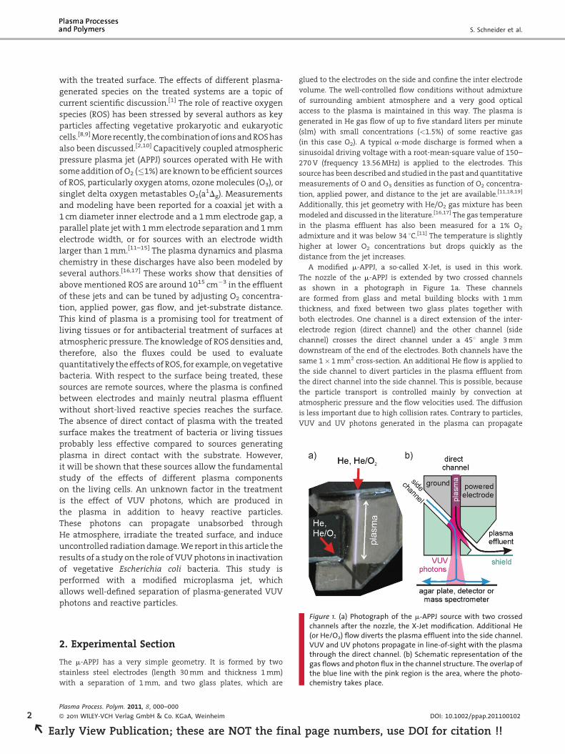
Figure 1. (a) Photograph of the m-APPJ source with two crossedchannels after the nozzle, the X-Jet modification. Additional He(or He/O2) flow diverts the plasma effluent into the side channel.VUV and UV photons propagate in line-of-sight with the plasmathrough the direct channel. (b) Schematic representation of thegas flows and photon flux in the channel structure. The overlap ofthe blue line with the pink region is the area, where the photo-chemistry takes place.
2. Experimental Section
The m-APPJ has a very simple geometry. It is formed by two
stainless steel electrodes (length 30 mm and thickness 1 mm)
with a separation of 1 mm, and two glass plates, which are
Plasma Process. Polym. 2011, 8, 000–000
� 2011 WILEY-VCH Verlag GmbH & Co. KGaA, Weinheim
rly View Publication; these are NOT the fina
glued to the electrodes on the side and confine the inter electrode
volume. The well-controlled flow conditions without admixture
of surrounding ambient atmosphere and a very good optical
access to the plasma is maintained in this way. The plasma is
generated in He gas flow of up to five standard liters per minute
(slm) with small concentrations (<1.5%) of some reactive gas
(in this case O2). A typical a-mode discharge is formed when a
sinusoidal driving voltage with a root-mean-square value of 150–
270 V (frequency 13.56 MHz) is applied to the electrodes. This
source has been described and studied in the past and quantitative
measurements of O and O3 densities as function of O2 concentra-
tion, applied power, and distance to the jet are available.[11,18,19]
Additionally, this jet geometry with He/O2 gas mixture has been
modeled and discussed in the literature.[16,17] The gas temperature
in the plasma effluent has also been measured for a 1% O2
admixture and it was below 34 8C.[11] The temperature is slightly
higher at lower O2 concentrations but drops quickly as the
distance from the jet increases.
A modified m-APPJ, a so-called X-Jet, is used in this work.
The nozzle of the m-APPJ is extended by two crossed channels
as shown in a photograph in Figure 1a. These channels
are formed from glass and metal building blocks with 1 mm
thickness, and fixed between two glass plates together with
both electrodes. One channel is a direct extension of the inter-
electrode region (direct channel) and the other channel (side
channel) crosses the direct channel under a 458 angle 3 mm
downstream of the end of the electrodes. Both channels have the
same 1�1 mm2 cross-section. An additional He flow is applied to
the side channel to divert particles in the plasma effluent from
the direct channel into the side channel. This is possible, because
the particle transport is controlled mainly by convection at
atmospheric pressure and the flow velocities used. The diffusion
is less important due to high collision rates. Contrary to particles,
VUV and UV photons generated in the plasma can propagate
DOI: 10.1002/ppap.201100102
l page numbers, use DOI for citation !!
The Role of VUV Radiation in the Inactivation . . .
further through the direct channel (also filled with He), cf. the
scheme in Figure 1b. Only a very small fraction (<1.5%) of photons is
reflected or scattered into the side channel. Therefore, operation of
the X-Jet with additional He flow through the side channel leads to
effective separation of the plasma-generated reactive particles (e.g.,
O atoms or O3 molecules emanating from the side channel) and
plasma-generated photons, which propagate through the direct
channel. Additionally, the experiments are performed in a chamber
with controlled He atmosphere (volume �8 L) to minimize the
influence of ambient atmosphere. The substrate was always placed
perpendicular to the axis of the corresponding channel used for
treatment at a distance of 4 mm. The details regarding the
separation of reactive particles and photons, the results of the
simulation of the convection/diffusion transport in the X-Jet, and
the testing of the X-Jet performance in etching experiments of
plasma polymer films, emission spectroscopy measurements in the
115–875 nm wavelength range, as well as treatment of bacteria in
their vegetative form can be found elsewhere.[20]
Combined and separate effects of the reactive heavy particles
and the VUV and UV radiation of the plasma effluent on bacteria
or other substrates can now be studied in the following ways:
(i) Photons and reactive particles are applied together: an X-Jet
without additional He flow in the side channel will result in the
transport of both reactive particles and photons through the direct
channel. The gas flow simulation indicates that 20% of the gas
flow exits through the side channel resulting in a slightly longer
residence time of the species in the direct channel after the crossing
and the loss of some O and O3 through this channel. (ii) Reactive
particles only: the same He flow is used in both channels. The
additional flow through the side channel will push the heavy
particles from the plasma effluent into the side channel as
demonstrated in Figure 1b. The flow rates through both
channels after the crossing are the same due to the symmetry of
this geometry. The flux of ROS at the exit of the side channel is
expected to be similar to the ROS flux at the direct channel
in (i), although some differences will occur due to slightly
different residence times as discussed above, due to missing
photodissociation and excitation of the particles in the effluent
after the crossing of both channels (see also Section 2.4 of
E. coli treatment later) and due to slightly asymmetric velocity
fields across both channels in the case (ii). The plasma generated
VUV and UV photons cannot enter directly into side channel due to
geometry constrains, which is corroborated by measurements
presented later in this article. (iii) VUV and UV only: with the
additional He flow, only VUV and UV photons reach the treated
surface under the direct channel. Some ROS species will diffuse into
the direct channel (especially if the gas flows in both channels are
equal), but they will not reach the center of the gas flow in this
channel. Only the species close to the center, where the gas velocity is
the highest, are transported to the vicinity of the surface and can
reach the substrate. We have already discussed[20] that the species at
the periphery of the gas channel will be transported to the side from
the jet axis and not to the surface, the diffusion across the
streamlines is too slow for that. Additionally, a larger He flow in the
side channel can be used to make sure that no ROS from the plasma
diffuse into the direct channel. The He flow of 2 slm in the side
channel (compared to 1.4 slm through the plasma) was, therefore,
used in some investigations presented in this article.
Plasma Process. Polym. 2011, 8, 000–000
� 2011 WILEY-VCH Verlag GmbH & Co. KGaA, Weinheim
Early View Publication; these are NOT the
2.1. Measurement of Emission Intensity
A solar blind VUV and UV detector (PMT-142, effective in the 115–
450 nm wavelength range with a maximum relative efficiency at
around 220 nm) in an evacuated housing with MgF2 window has
been used to measure the wavelength integrated intensity of VUV
and UV emission from the direct channel of the X-Jet.[21] A 1 mm
diameter diaphragm was placed on the MgF2 window and the jet
was always at 4 mm distance from the window to maintain the
same acceptance angle for each measurement. The same setup
has been used in previous measurements to verify that the
photon flux (in this wavelength range) through the side channel is
negligible.[20]
2.2. Molecular Beam Mass Spectrometry
Measurements
A molecular beam mass spectrometry (MS) sampling system is
used to check the presence of O atoms and O3 molecules and to
detect positive ions in the gas phase. The three-stage differentially
pumped MS sampling system described previously is used for
these measurements.[19,22] This system is equipped with a rotating
chopper to modulate the molecular beam and maintain vacuum
in the MS. However, this chopper was not used here for the
detection of ions because a smaller sampling orifice (20 mm
diameter) was installed. The X-Jet is placed into a small chamber
with controlled He atmosphere, which is mounted directly at
the mass spectrometer front plate with the sampling orifice.
Comparable conditions as during the treatment of bacteria are
arranged in this way.
2.3. Preparation of Biological Probes
Vegetative E. coli cells have been used as model substrate in this
study. E. coli K12 liquid cultures (American Type Culture Collection,
Acces. Number 10798) were incubated for 18 h overnight at 37 8C in
LB medium.[23] The cultures were diluted to an optical density of
0.05 at 580 nm and were sprayed onto LB agar plates for 1 s. A low
surface coverage with �2.2�103 � cm�2 cell density was achieved
in this case with well-separated individual cells. This cell density is
the minimal density necessary to obtain homogeneous surface
coverage after the incubation. Even small changes in cell density
due to treatment with the plasma jet were therefore visible. The
low cell density also minimizes the possible effect of one cell
shadowing another, which has to be considered in the case of a
multilayer or clusters of cells at the surface. The plates were
incubated for 1 h at 37 8C before plasma treatment. After plasma
treatment, the sample plates were incubated overnight for 18 h at
37 8C to allow survivors to grow. Untreated plates and controls
without any effect were covered by a homogeneous layer of
bacteria, whereas, zones of inhibition formed where treatment was
lethal. The incubated agar plates were scanned afterwards on a
digital color scanner to document the final effect of treatment on
bacterial survival. Each treatment was repeated in three indepen-
dent experiments. The effect of treatment was reproducible and
representative results for each condition are shown here.
www.plasma-polymers.org 3
final page numbers, use DOI for citation !! R
4
REa
S. Schneider et al.
It was checked that prolonged treatment of the agar plates with
(i) gas flow without plasma or (ii) with RF electromagnetic fields
generated by electrodes without plasma and without gas flows
(plasma does not ignite without He between the electrodes) has no
visible effect on bacteria. The treatment of agar plates with plasma
before application of cells had no effect on bacterial growth.
Furthermore, the pH change of the medium has been checked.
Liquid pH color indicator (Sigma–Aldrich) was either added on top
of the agar plate after plasma treatment or mixed into the warm LB-
agar solution into Petri dishes to test for plasma-mediated changes
of pH values. A pH change would be indicated by a change in color of
the indicator reagent. No color changes were observed even after
30 min of plasma treatment.
We report in the following results of treatment of vegetative
E. coli bacteria with different components of the plasma effluent.
The main focus is on the role of VUV and UV photons in the
inactivation of bacteria.
2.4. Experimental Results and Discussion
The advantage of the X-Jet is that it allows us to separate the effects
of plasma-generated VUV and UV photons from the effects induced
by reactive particles (O3, O, impurities, etc.). The effects of VUV and
UV photons only, reactive particles only, and photons and reactive
particles together have already been studied in E. coli and selected
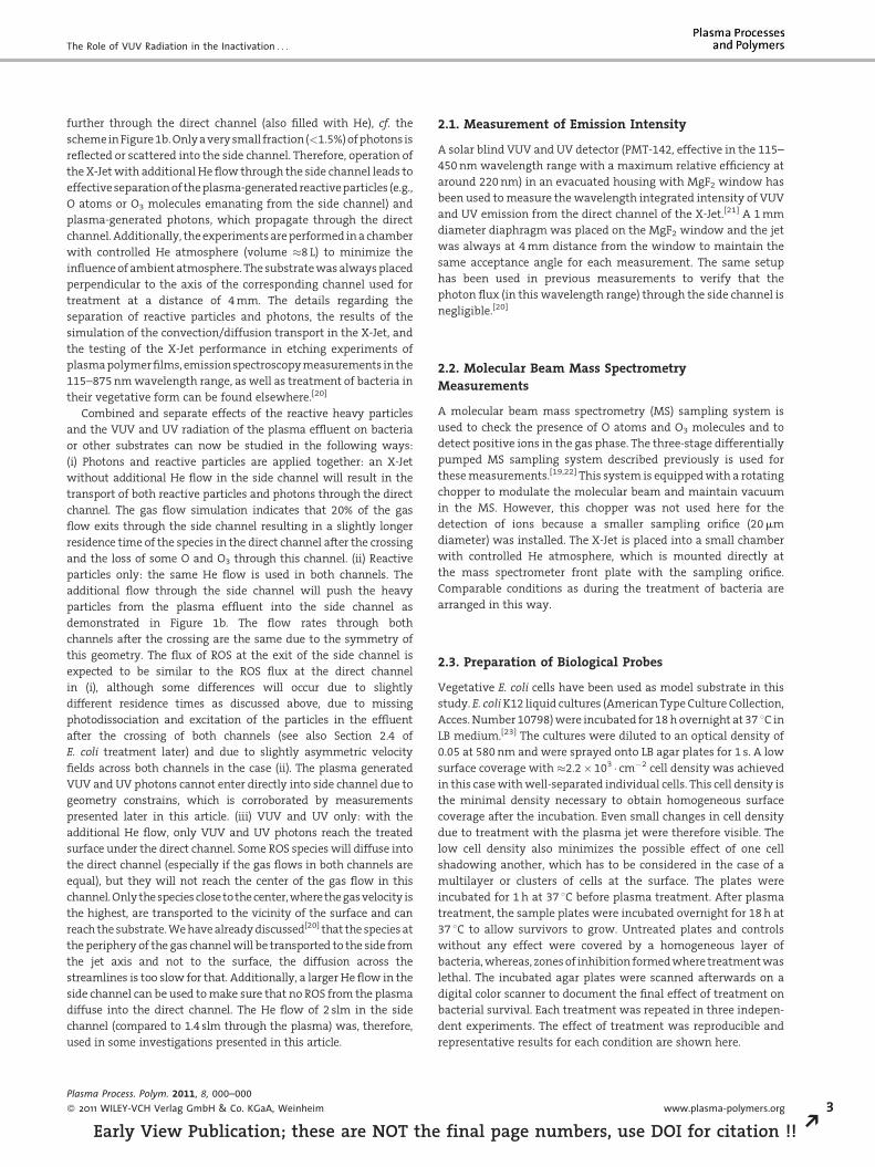
results are shown in Figure 2.[20] The following observations have
been made: (i) The VUV and UV photons generated in plasma had
only a weak effect on E. coli survival. No inactivation was visible in
samples treated with the direct channel for 1 and 3 min. The zone of
Figure 2. 40 mm� 40 mm details of photographs of Petri disheswith zones of inhibition in E. coli monolayers after 1, 3, and 6 minof treatment with reactive particles only, VUV and UV photonsonly, or treatment with both combined. Details marked by �: theentire plate (80 mm diameter) was affected. Adopted fromref.[20]. Plasma conditions: URMS¼ 230 V, He flow through plasma1.4 slm, O2 concentration in the direct channel 0.6%, He flowthrough the side channel 0, or 1.4 slm.
Plasma Process. Polym. 2011, 8, 000–000
� 2011 WILEY-VCH Verlag GmbH & Co. KGaA, Weinheim
rly View Publication; these are NOT the fina
inhibition at the jet axis appeared only after 6 min of treatment
with photons. (ii) The combined treatment and reactive particle-
only treatment showed typical dose–effect relationships.
Elongated treatment times resulted in larger zones of inhibition
and lower numbers of colony forming units. In both cases, the
whole area of the Petri dish (80 mm diameter) was affected after
3 min of treatment. (iii) Inactivation by the combined treatment
was approximately twice as fast as with the reactive particle-only
treatment (cf. Figure 2). The fast inactivation by the reactive
particle-only and by the combined treatment is due to the fact that
the m-APPJ is an effective source of atomic oxygen and ozone. For
example, the densities (concentrations) of O and O3 measured by
molecular beam MS at 4 mm distance from the jet under conditions
used in this work are 7� 1014 cm�3 (�28 ppm) and 5� 1014 cm�3
(�20 ppm), respectively.[19] The concentration of ozone increases
with the distance and reaches �56 ppm at 50 mm. The concentra-
tions of both O and O3 can be expected to be high enough to cause
the observed effects. Ozone is known for its bactericidal activity.
Just 5 min of treatment with 0.2 ppm of ozone in water is lethal for
E. coli, Bacillus cereus, or Bacillus megaterium.[24] Moreover,
experiments in air with ozone have shown that it also effectively
kills bacteria on agar plates. Ozone concentrations below 1 ppm
and treatment times shorter than 100 min have been reported[25] to
be effective in killing Staphylococcus albus, Streptococcus salivarius,
and Bacillus prodigiosus. The higher doses of airborne ozone (300–
1 500 ppm) have been tested and proven to be effective against
E. coli and Staphylococcus aureus with rates on the time scale of few
tens of seconds.[26] Atomic oxygen also has detrimental effects on
bacteria. It can etch biological material or cause oxidative stress
inside the cell. We have shown that the flux of atomic oxygen from
the side channel of the X-Jet etches a BSA protein layer or a model
plasma polymer film of hydrogenated amorphous carbon with a
rate of 30 nm/min and that the area on the surface, which is
affected by atomic oxygen, is limited to a diameter of�10 mm.[20,27]
This limitation is given by the fast loss of O in the gas phase in the
three body reaction:[28]
l pag
Oþ O2 þHe! O3 þ He (1)
Regarding the fact that in treatments with reactive particles
longer than 6 min the whole Petri dish (80 mm diameter) was
affected and taking into account the short lifetime of O atoms, we
conclude that mainly O3 is responsible for the inactivation of
bacteria at large distances from the jet axis. Simultaneous action of
O, O3, O2 metastables, some possible impurities (like OH from
water), and in the case of combined treatment through the direct
channel, the VUV and UV radiation, most likely play a role in
bacterial inactivation near to the jet axis.[20]
An interesting effect, which is further studied and discussed in
this article, is the fact that the combined treatment is twice as fast
as the treatment with reactive particles without photons. More
effective inactivation was expected when reactive particles and
photons were treating the bacteria at the substrate simultaneously.
However, the simultaneous treatment is limited only to an area of
2–3 mm diameter just underneath the direct channel. The rest of
the substrate is shadowed by the channel structure and no
synergistic effects were expected there. However, Figure 2 shows
that the zone of inhibition of the combined treatment had a
DOI: 10.1002/ppap.201100102
e numbers, use DOI for citation !!
The Role of VUV Radiation in the Inactivation . . .
diameter of 20 mm after 1 min of the treatment, whereas, the
treatment with reactive particles only resulted in a 5 mm diameter
after the same time. Additionally, the combined treatment at 3 min
has an effect comparable to the reactive particles-only treatment at
6 min. These observations indicate that the plasma effluent is
changed by the presence of VUV photons. Some photochemistry
reactions take place in the gas phase in the second part of the direct
channel and on the way to the substrate, which are missing in the
reactive particle-only treatment. The photochemistry of VUV
photons in the effluent of the He/O2 jets as an additional source
of O and subsequently O3 have been suggested and tested before
by Reuter et al.[12] and Reuter[13]. It could not be determined,
which reaction was responsible for this production, but the direct
photodissociation of O2 molecules has been proposed as a possible
explanation. The fact that the inactivation is accelerated at greater
distance from the jet axis indicates that additional ozone (and
additional O necessary for ozone formation) is indeed formed in the
effluent. The X-Jet geometry offers a unique opportunity to study
these effects in more detail.
2.5. Study of the Photochemistry in the X-Jet
The interaction of plasma-generated photons with O2 molecules in
the gas phase and its effects on bacteria can be studied by adding O2
at different concentrations into the He flow through the side
channel. Figure 3 shows the comparison of zones of inhibition
induced by photons only (He gas is supplied through the side
channel) and by photons and O2 photochemistry products (He gas
with admixture of O2 at different concentrations was fed through
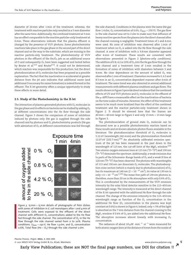
Figure 3. 15 mm� 15 mm details of photographs of Petri disheswith zones of inhibition in E. coli monolayers after 1 and 4 min oftreatment. Cells were exposed to the effluent of the directchannel with different O2 concentrations added to the He flowfed through the side channel. The concentration of O2 in the Heflow through the side channel varied from 0 to 20%. Plasmaconditions: URMS¼ 230 V, He flow 1.4 slm, and O2 concentration0.6%. Total flow (HeþO2) through the side channel 2 slm.
Plasma Process. Polym. 2011, 8, 000–000
� 2011 WILEY-VCH Verlag GmbH & Co. KGaA, Weinheim
Early View Publication; these are NOT the
the side channel). Conditions in the plasma were the same (He gas
flow 1.4 slm, O2 concentration of 0.6%, URMS¼ 230 V). The gas flow
in the side channel was set to 2 slm to make sure that diffusion of
heavy reactive species from the plasma into the direct channel after
the channel crossing is negligible. Treatment times of 1 and 4 min
were used. No zone of inhibition was observed after 1 min of
treatment when no O2 is added into the He flow through the side
channel. A zone of inhibition with a 3.8 mm diameter appeared
after 4 min of treatment. These results are consistent with
observations presented in Figure 2 (photons-only conditions).
The addition of 0.8, 4.2 or 20% of O2 into the He gas flow through the
side channel and a treatment time of 1 min resulted in the
formation of zones of inhibition with diameters between 3 and
4 mm. No clear dependence on the amount of added O2 was
observed after 1 min of treatment. Diameters increased to 5, 6.5 and
8.5 mm in an O2 concentration-dependent manner after 4 min of
treatment. The same trend was also observed in several additional
measurements with different plasma conditions and gas flows. The
results shown in Figure 3 provide direct evidence that the combined
effects of UV and VUV photons and O2 molecules in the effluent of
the m-APPJ have a measurable effect on the inactivation of bacteria
on the time scales of minutes. However, the effect of this treatment
seems to be much more localized than the effect of the combined
treatment and the reactive particles-only treatment shown in
Figure 2. It should be stressed here that the frames are
40 mm� 40 mm large in Figure 2 and only 15 mm�15 mm large
in Figure 3.
The photodissociation of ground state O2 molecule can be
discussed now as a possible photochemistry reaction based on
these results and on known absolute photon fluxes available in the
literature. The photodissociation threshold of O2 molecules is
5.12 eV (wavelength 242.4 nm) and the photoionization threshold
12.07 eV (102.7 nm).[29] An absolutely calibrated emission spec-
trum of the jet has been measured in the past down to the
wavelength of 115 nm, the cut-off limit of the MgF2 window.[30]
Two atomic oxygen emission lines at 115 nm (1D–1Do) and 130 nm
(3P–3So) dominate the spectrum. Additionally, a weak emission due
to parts of the Schumann–Runge bands of O2 and a weak H line at
120 nm (2P–2So) has been observed. The photons with wavelengths
of 115 and 130 nm can dissociate O2 molecules. The photoabsorp-
tion cross-section (which is mainly due to photodissociation) of O2
has its maximum at 140 nm (2� 10�17 cm2), its value at 130 nm is
only �3�10�19 cm2.[29] The mean free path of 130 nm photons is,
therefore, more than 20 cm in He atmosphere with only 0.6% of O2.
This is corroborated by the measurements of the VUV emission
intensity by the solar blind detector sensitive in the 115–450 nm
wavelength range. The intensity is measured at the direct channel
of the X-Jet operated with the additional He flow through the side
channel. The change of the emission intensity in the 115–450 nm
wavelength range as function of the O2 concentration in the
additional He flow (O2 concentration in the plasma was kept
constant at 0.6%) is shown in Figure 4. Indeed, only�3% of the light
is absorbed on the 7 mm distance from the channel crossing to the
MgF2 window if 0.6% of O2 are added into the additional He flow.
The absorption increases almost linearly with increasing O2
concentration.
The radiances of about 10 mW �mm�2 � sr�1 were measured for
both atomic oxygen lines at the distance of 4 mm from the nozzle of
www.plasma-polymers.org 5
final page numbers, use DOI for citation !! R
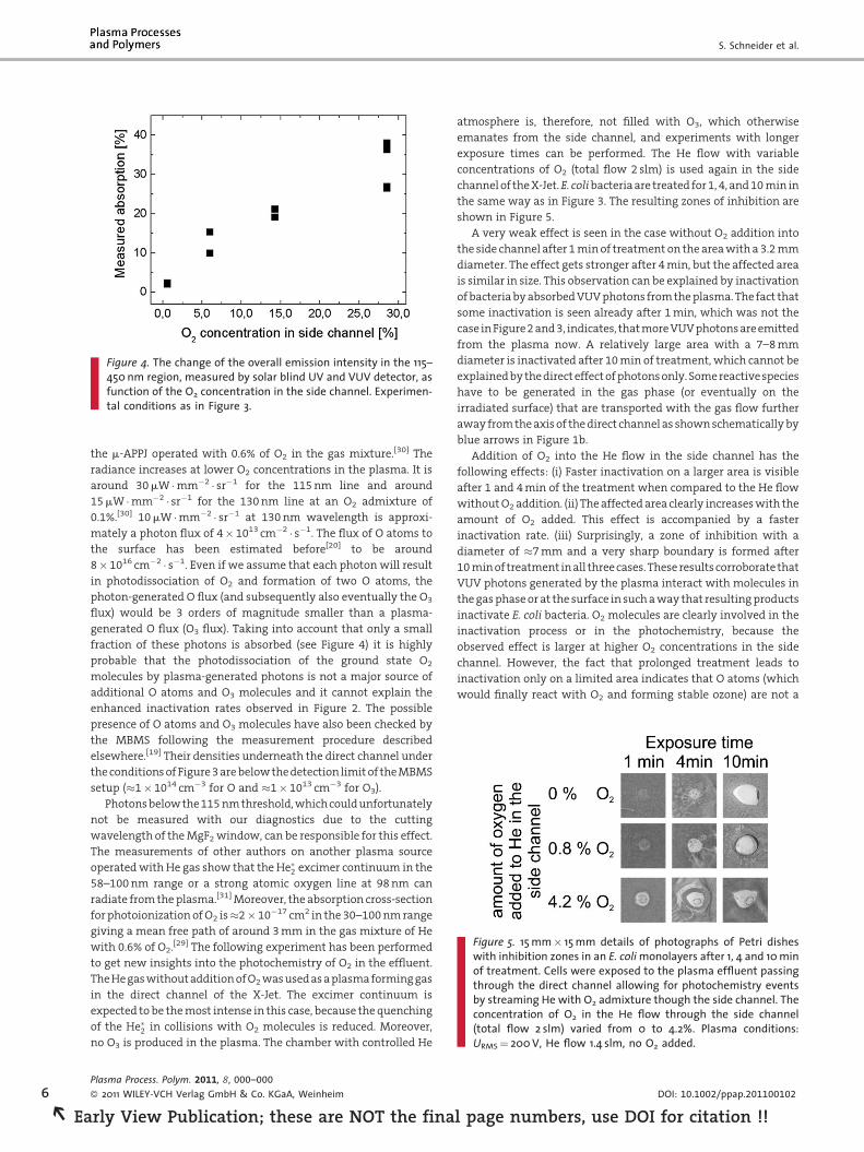
Figure 4. The change of the overall emission intensity in the 115–450 nm region, measured by solar blind UV and VUV detector, asfunction of the O2 concentration in the side channel. Experimen-tal conditions as in Figure 3.
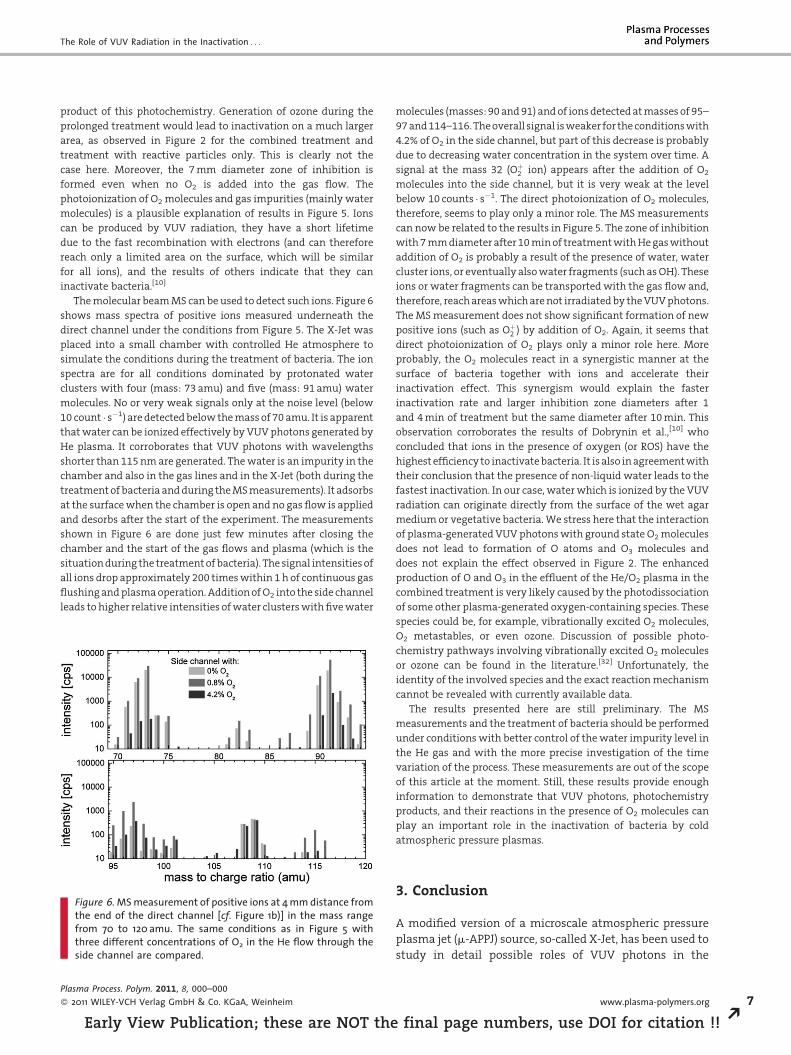
Figure 5. 15 mm� 15 mm details of photographs of Petri disheswith inhibition zones in an E. coli monolayers after 1, 4 and 10 minof treatment. Cells were exposed to the plasma effluent passingthrough the direct channel allowing for photochemistry eventsby streaming He with O2 admixture though the side channel. Theconcentration of O2 in the He flow through the side channel(total flow 2 slm) varied from 0 to 4.2%. Plasma conditions:URMS¼ 200 V, He flow 1.4 slm, no O2 added.
6
REa
S. Schneider et al.
the m-APPJ operated with 0.6% of O2 in the gas mixture.[30] The
radiance increases at lower O2 concentrations in the plasma. It is
around 30 mW �mm�2 � sr�1 for the 115 nm line and around
15 mW �mm�2 � sr�1 for the 130 nm line at an O2 admixture of
0.1%.[30] 10 mW �mm�2 � sr�1 at 130 nm wavelength is approxi-
mately a photon flux of 4� 1013 cm�2 � s�1. The flux of O atoms to
the surface has been estimated before[20] to be around
8�1016 cm�2 � s�1. Even if we assume that each photon will result
in photodissociation of O2 and formation of two O atoms, the
photon-generated O flux (and subsequently also eventually the O3
flux) would be 3 orders of magnitude smaller than a plasma-
generated O flux (O3 flux). Taking into account that only a small
fraction of these photons is absorbed (see Figure 4) it is highly
probable that the photodissociation of the ground state O2
molecules by plasma-generated photons is not a major source of
additional O atoms and O3 molecules and it cannot explain the
enhanced inactivation rates observed in Figure 2. The possible
presence of O atoms and O3 molecules have also been checked by
the MBMS following the measurement procedure described
elsewhere.[19] Their densities underneath the direct channel under
the conditions of Figure 3 are below the detection limit of the MBMS
setup (�1�1014 cm�3 for O and �1�1013 cm�3 for O3).
Photons below the 115 nm threshold, which could unfortunately
not be measured with our diagnostics due to the cutting
wavelength of the MgF2 window, can be responsible for this effect.
The measurements of other authors on another plasma source
operated with He gas show that the He�2 excimer continuum in the
58–100 nm range or a strong atomic oxygen line at 98 nm can
radiate from the plasma.[31] Moreover, the absorption cross-section
for photoionization of O2 is�2�10�17 cm2 in the 30–100 nm range
giving a mean free path of around 3 mm in the gas mixture of He
with 0.6% of O2.[29] The following experiment has been performed
to get new insights into the photochemistry of O2 in the effluent.
The He gas without addition of O2 was used as a plasma forming gas
in the direct channel of the X-Jet. The excimer continuum is
expected to be the most intense in this case, because the quenching
of the He�2 in collisions with O2 molecules is reduced. Moreover,
no O3 is produced in the plasma. The chamber with controlled He
Plasma Process. Polym. 2011, 8, 000–000
� 2011 WILEY-VCH Verlag GmbH & Co. KGaA, Weinheim
rly View Publication; these are NOT the fina
atmosphere is, therefore, not filled with O3, which otherwise
emanates from the side channel, and experiments with longer
exposure times can be performed. The He flow with variable
concentrations of O2 (total flow 2 slm) is used again in the side
channel of the X-Jet. E. coli bacteria are treated for 1, 4, and 10 min in
the same way as in Figure 3. The resulting zones of inhibition are
shown in Figure 5.
A very weak effect is seen in the case without O2 addition into
the side channel after 1 min of treatment on the area with a 3.2 mm
diameter. The effect gets stronger after 4 min, but the affected area
is similar in size. This observation can be explained by inactivation
of bacteria by absorbed VUV photons from the plasma. The fact that
some inactivation is seen already after 1 min, which was not the
case in Figure 2 and 3, indicates, that more VUV photons are emitted
from the plasma now. A relatively large area with a 7–8 mm
diameter is inactivated after 10 min of treatment, which cannot be
explained by the direct effect of photons only. Some reactive species
have to be generated in the gas phase (or eventually on the
irradiated surface) that are transported with the gas flow further
away from the axis of the direct channel as shown schematically by
blue arrows in Figure 1b.
Addition of O2 into the He flow in the side channel has the
following effects: (i) Faster inactivation on a larger area is visible
after 1 and 4 min of the treatment when compared to the He flow
without O2 addition. (ii) The affected area clearly increases with the
amount of O2 added. This effect is accompanied by a faster
inactivation rate. (iii) Surprisingly, a zone of inhibition with a
diameter of �7 mm and a very sharp boundary is formed after
10 min of treatment in all three cases. These results corroborate that
VUV photons generated by the plasma interact with molecules in
the gas phase or at the surface in such a way that resulting products
inactivate E. coli bacteria. O2 molecules are clearly involved in the
inactivation process or in the photochemistry, because the
observed effect is larger at higher O2 concentrations in the side
channel. However, the fact that prolonged treatment leads to
inactivation only on a limited area indicates that O atoms (which
would finally react with O2 and forming stable ozone) are not a
DOI: 10.1002/ppap.201100102
l page numbers, use DOI for citation !!
The Role of VUV Radiation in the Inactivation . . .
product of this photochemistry. Generation of ozone during the
prolonged treatment would lead to inactivation on a much larger
area, as observed in Figure 2 for the combined treatment and
treatment with reactive particles only. This is clearly not the
case here. Moreover, the 7 mm diameter zone of inhibition is
formed even when no O2 is added into the gas flow. The
photoionization of O2 molecules and gas impurities (mainly water
molecules) is a plausible explanation of results in Figure 5. Ions
can be produced by VUV radiation, they have a short lifetime
due to the fast recombination with electrons (and can therefore
reach only a limited area on the surface, which will be similar
for all ions), and the results of others indicate that they can
inactivate bacteria.[10]
The molecular beam MS can be used to detect such ions. Figure 6
shows mass spectra of positive ions measured underneath the
direct channel under the conditions from Figure 5. The X-Jet was
placed into a small chamber with controlled He atmosphere to
simulate the conditions during the treatment of bacteria. The ion
spectra are for all conditions dominated by protonated water
clusters with four (mass: 73 amu) and five (mass: 91 amu) water
molecules. No or very weak signals only at the noise level (below
10 count � s�1) are detected below the mass of 70 amu. It is apparent
that water can be ionized effectively by VUV photons generated by
He plasma. It corroborates that VUV photons with wavelengths
shorter than 115 nm are generated. The water is an impurity in the
chamber and also in the gas lines and in the X-Jet (both during the
treatment of bacteria and during the MS measurements). It adsorbs
at the surface when the chamber is open and no gas flow is applied
and desorbs after the start of the experiment. The measurements
shown in Figure 6 are done just few minutes after closing the
chamber and the start of the gas flows and plasma (which is the
situation during the treatment of bacteria). The signal intensities of
all ions drop approximately 200 times within 1 h of continuous gas
flushing and plasma operation. Addition of O2 into the side channel
leads to higher relative intensities of water clusters with five water
Figure 6. MS measurement of positive ions at 4 mm distance fromthe end of the direct channel [cf. Figure 1b)] in the mass rangefrom 70 to 120 amu. The same conditions as in Figure 5 withthree different concentrations of O2 in the He flow through theside channel are compared.
Plasma Process. Polym. 2011, 8, 000–000
� 2011 WILEY-VCH Verlag GmbH & Co. KGaA, Weinheim
Early View Publication; these are NOT the
molecules (masses: 90 and 91) and of ions detected at masses of 95–
97 and 114–116. The overall signal is weaker for the conditions with
4.2% of O2 in the side channel, but part of this decrease is probably
due to decreasing water concentration in the system over time. A
signal at the mass 32 (Oþ2 ion) appears after the addition of O2
molecules into the side channel, but it is very weak at the level
below 10 counts � s�1. The direct photoionization of O2 molecules,
therefore, seems to play only a minor role. The MS measurements
can now be related to the results in Figure 5. The zone of inhibition
with 7 mm diameter after 10 min of treatment with He gas without
addition of O2 is probably a result of the presence of water, water
cluster ions, or eventually also water fragments (such as OH). These
ions or water fragments can be transported with the gas flow and,
therefore, reach areas which are not irradiated by the VUV photons.
The MS measurement does not show significant formation of new
positive ions (such as Oþ2 ) by addition of O2. Again, it seems that
direct photoionization of O2 plays only a minor role here. More
probably, the O2 molecules react in a synergistic manner at the
surface of bacteria together with ions and accelerate their
inactivation effect. This synergism would explain the faster
inactivation rate and larger inhibition zone diameters after 1
and 4 min of treatment but the same diameter after 10 min. This
observation corroborates the results of Dobrynin et al.,[10] who
concluded that ions in the presence of oxygen (or ROS) have the
highest efficiency to inactivate bacteria. It is also in agreement with
their conclusion that the presence of non-liquid water leads to the
fastest inactivation. In our case, water which is ionized by the VUV
radiation can originate directly from the surface of the wet agar
medium or vegetative bacteria. We stress here that the interaction
of plasma-generated VUV photons with ground state O2 molecules
does not lead to formation of O atoms and O3 molecules and
does not explain the effect observed in Figure 2. The enhanced
production of O and O3 in the effluent of the He/O2 plasma in the
combined treatment is very likely caused by the photodissociation
of some other plasma-generated oxygen-containing species. These
species could be, for example, vibrationally excited O2 molecules,
O2 metastables, or even ozone. Discussion of possible photo-
chemistry pathways involving vibrationally excited O2 molecules
or ozone can be found in the literature.[32] Unfortunately, the
identity of the involved species and the exact reaction mechanism
cannot be revealed with currently available data.
The results presented here are still preliminary. The MS
measurements and the treatment of bacteria should be performed
under conditions with better control of the water impurity level in
the He gas and with the more precise investigation of the time
variation of the process. These measurements are out of the scope
of this article at the moment. Still, these results provide enough
information to demonstrate that VUV photons, photochemistry
products, and their reactions in the presence of O2 molecules can
play an important role in the inactivation of bacteria by cold
atmospheric pressure plasmas.
3. Conclusion
A modified version of a microscale atmospheric pressure
plasma jet (m-APPJ) source, so-called X-Jet, has been used to
study in detail possible roles of VUV photons in the
www.plasma-polymers.org 7
final page numbers, use DOI for citation !! R
8
REa
S. Schneider et al.
inactivation of E. coli bacteria. The X-Jet plasma source
allows effective separation of heavy reactive particles (such
as O atoms or ozone molecules) and the plasma-generated
photons. Additionally, it allows checking the possible
photochemistry reaction pathways and their effects on
bacteria. The results clearly show that plasma-generated
VUV photons play an important role in the inactivation of
bacteria. However, we could show that the main effect of
the VUV radiation on bacteria is not the direct radiation
damage but more the indirect production of additional
reactive species in the gas phase. The rate of inactivation is
doubled when plasma-generated photons interact with the
plasma effluent, most probably by additional production of
O atoms and subsequently also O3 molecules. However, the
ground state O2 molecules are not directly involved in these
photochemistry reactions.
The interaction of the plasma-generated VUV photons
with the He/O2 gas (He and O2 in their ground states) also
leads to localized inactivation on the time scale of minutes.
The molecular beam MS measurements of ions in the gas
phase indicate that photoionization of water clusters by
plasma generated VUV photons and the subsequent
interaction of these ions with bacteria could be involved.
The results also indicate that the simultaneous presence of
ions and ground state O2 molecules at the treated surface
leads to the acceleration of the inactivation process.
Acknowledgements: The authors thank Volker Schulz-von derGathen for fruitful discussions about the operation of the m-APPJsource. This work has been performed with the support of theresearch group FOR1123 (Project C1) approved by the GermanResearch Foundation (DFG). This work has also been supported bythe Research Department Plasmas with Complex Interactions ofthe Ruhr-Universitat Bochum as well as through a stipend toJ.-W. L. from the Ruhr University Research School.
Received: May 23, 2011; Revised: September 15, 2011; Accepted:October 11, 2011; DOI: 10.1002/ppap.201100102
Keywords: atmospheric pressure plasma jet; bacterial inactivation;mass spectrometry; photochemistry; plasma sterilization
[1] M. G. Kong, G. Kroesen, G. Morfill, T. Nosenko, T. Shimizu,J. van Dijk, J. L. Zimmermann, New J. Phys. 2009, 11, 115012.
[2] E. Stoffels, Y. Sakiyama, D. B. Graves, IEEE Trans. Plasma Sci.2008, 36, 1441.
[3] G. Daeschlein, T. von Woedtke, E. Kindel, R. Brandenburg, K.-D.Weltmann, M. Junger, Plasma Process. Polym. 2010, 20107,224.
[4] G. Daeschlein, S. Scholz, T. von Woedtke, M. Niggemeier, E.Kindel, R. Foest, R. Brandenburg, K.-D. Weltmann, M. Junger,IEEE Trans. Plasma Sci. 2011, 39, 81.
Plasma Process. Polym. 2011, 8, 000–000
� 2011 WILEY-VCH Verlag GmbH & Co. KGaA, Weinheim
rly View Publication; these are NOT the fina
[5] X. T. Deng, J. J. Shi, M. G. Kong, J. Appl. Phys. 2007, 101,074701.
[6] G. Fridman, A. Shereshevsky, M. Jost, A. Brooks, A. Fridman, A.Gutsol, V. Vasilets, G. Friedman, Plasma Chem. Plasma Pro-cess. 2007, 27, 163.
[7] M. Vandamme, E. Robert, S. Pesnel, E. Barbosa, S. Dozias,J. Sobilo, S. Lerondel, A. Le Pape, J. M. Pouvesle, Plasma Process.Polym. 2010, 7, 264.
[8] J. Goree, B. Liu, D. Drake, J. Phys. D: Appl. Phys. 2006, 39,3479.
[9] M. Hahnel, T. von Woedtke, K.-D. Weltmann, Plasma Process.Polym. 2010, 7, 244.
[10] D. Dobrynin, G. Fridman, G. Friedman, A. Fridman, New J.Phys. 2009, 11, 115020.
[11] V. Schulz-von der Gathen, V. Buck, T. Gans, N. Knake, K. Niemi,S. Reuter, L. Schaper, J. Winter, Contrib. Plasma Phys. 2007, 47,510.
[12] S. Reuter, K. Niemi, V. Schulz-von der Gathen, H. F. Dobele,Plasma Sources Sci. Technol. 2009, 18, 015006.
[13] S. Reuter, Formation Mechanisms of Atomic Oxygen in anAtmospheric Pressure Plasma Jet Characterised by Spectro-scopic Methods, Cuvillier Verlag Gottingen, Germany 2008.
[14] J. Jeong, S. Babayan, A. Schutze, V. Tu, M. Morajev, G. Selwyn,R. Hicks, Plasma Sources Sci. Technol. 1998, 7, 282.
[15] J. Laimer, H. Stori, Plasma Process. Polym. 2006, 3, 573.[16] J. Waskoenig, K. Niemi, N. Knake, L. M. Graham, S. Reuter, V.
Schulz-von der Gathen, T. Gans, Plasma Sources Sci. Technol.2010, 19, 045018.
[17] D.-X. Liu, M.-Z. Rong, X.-H. Wang, F. Iza, M. G. Kong, P.Bruggeman, Plasma Process. Polym. 2010, 7, 846.
[18] N. Knake, S. Reuter, K. Niemi, V. Schulz-von der Gathen, J.Winter, J. Phys. D: Appl. Phys. 2008, 41, 194006.
[19] D. Ellerweg, J. Benedikt, A. von Keudell, N. Knake, V.Schulz-von der Gathen, New J. Phys. 2010, 12, 013021.
[20] S. Schneider, J.-W. Lackmann, F. Narberhaus, J. E. Bandow,B. Denis, J. Benedikt, J. Phys. D: Appl. Phys. 2011, 44, 295201.
[21] N. K. Bibinov, D. O. Bolshukhin, D. B. Kokh, A. M. Pravilov, I. P.Vinogradov, K. Wiesemann, Meas. Sci. Technol. 1997, 8,773.
[22] J. Benedikt, D. Ellerweg, A. von Keudell, Proceedings of SVC2011 TechCon conference, http://arxiv.org/abs/1105.2687.
[23] J. Sambrook, E. Fritsch, T. Maniatis, Molecular Cloning: ALaboratory Manual, Cold Spring Harbor Laboratory Press,New York 1989.
[24] W. T. Broadwater, R. C. Hoehn, P. H. King, Appl. Microbiol.1973, 26, 391.
[25] W. J. Elford, J. van den Ende, J. Hyg. 1942, 42, 240.[26] W. J. Kowalski, W. P. Bahnfleth, T. S. Whittam, Ozone-Sci. Eng.
1998, 20, 205.[27] J.-W. Lackmann, S. Schneider, F. Narberhaus, J. Benedikt, J. E.
Bandow, Plasma for Bio-Decontamination, Medicine and FoodSecurity, NATO Science for Peace and Security Series – A,scheduled for Jan. 2012.
[28] D. S. Stafford, M. J. Kushner, J. Appl. Phys. 2004, 96, 2451.[29] Y. Itikawa, A. Ichimura, K. Onda, K. Sakimoto, K. Takayanagi,
Y. Hatano, M. Hayashi, H. Nishimura, S. Tsurubuchi, J. Phys.Chem. Ref. Data 1989, 18, 23.
[30] H. Bahre, H. Lange, V. Schulz-von der Gathen, R. Foest, ActaTechnica 2011, 56, T199.
[31] P. Kurunczi, J. Lopez, H. Shah, K. Becker, Int. J. Mass Spectrom.2001, 205, 277.
[32] G. W. Flynn, C. S. Parmenter, A. M. Wodtke, J. Phys. Chem.1996, 100, 12817.
DOI: 10.1002/ppap.201100102
l page numbers, use DOI for citation !!


![The role of VUV radiation in the inactivation of bacteria ... · - 4 - measured for a 1% O2 admixture and it was below 34 C. [11] The temperature is slightly higher at lower O2 concentrations](https://img.pdfslide.net/doc/110x75/5e5281bceefd7c30c14dcea8/the-role-of-vuv-radiation-in-the-inactivation-of-bacteria-4-measured-for.jpg)