Embed Size (px)
Citation preview

九州大学学術情報リポジトリKyushu University Institutional Repository
The Roles of Angiotensin II in StretchedPeriodontal Ligament Cells
Monnouchi, SatoshiDivision of Oral Rehabilitation, Department of Endodontology and Operative Dentistry, Facultyof Dental Science, Kyushu University
Maeda, HidefumiDepartment of Endodontology, Kyushu University Hospital
Fujii, Shinsuke
Tomokiyo, AtsushiDivision of Oral Rehabilitation, Department of Endodontology and Operative Dentistry, Facultyof Dental Science, Kyushu University
他
http://hdl.handle.net/2324/25451
出版情報:Journal of Dental Research. 90 (2), pp.181-185, 2011-02. International & AmericanAssociations for Dental Researchバージョン:権利関係:(C) 2012 by International & American Associations for Dental Research

1 / 26
The roles of angiotensin II in stretched periodontal
ligament cells S. Monnouchi1, H. Maeda2*, S. Fujii2, A. Tomokiyo2, K. Hori1, and A. Akamine1,2 1Division of Oral Rehabilitation, Department of Endodontology and Operative Dentistry, Faculty of Dental Science, Kyushu University, and
2Department of Endodontology, Kyushu University Hospital, 3-1-1 Maidashi, Higashi-ku, Fukuoka 812-8582, Japan; *corresponding author, [email protected]
< Information > 1) short title ; Ang II mediates the signal in stretched PDL. 2) three key words ; HPLF, stretch, angiotensin ll 3) the number of words in the abstract ; 146 words 4) the number of words in the abstract and text ; 2499 words 5) the number of tables and figures ; 4 figures 6) the number of cited references ; 28 references

2 / 26
ABSTRACT
The loading caused by occlusion and mastication plays an important role in
maintaining periodontal ligament (PDL) tissues. We hypothesized that a loading
magnitude would be involved in the production of biological factors that function
in the maintenance of PDL tissues. Here, we identified up-regulated gene
expressions of transforming growth factor-β1 (TGF-β1), alkaline phosphatase
(ALP) and angiotensinogen (AGT) in human PDL fibroblastic cells (HPLF) that
were exposed to 8% stretch loading. Immuno-localization of angiotensin I/II (Ang
I/II), which were converted from AGT, were detected in rat PDL tissues. HPLFs
that were stimulated by Ang II also increased their gene expressions of TGF-β1
and ALP. Furthermore, the antagonist for Ang II type 2 receptor, rather than for
type 1, significantly inhibited gene expressions induced by the stretch loading.
These data suggest that Ang II mediates the loading signal in stretched HPLFs
to induce expressions of TGF-β1 and ALP.
3 / 26
INTRODUCTION
Periodontal ligament (PDL) is a dense specialized connective tissue that lies
between the cementum and the alveolar bone. The homeostasis of PDL tissues
is maintained while continuously being subjected to the mechanical tensile
loading caused by occlusion and mastication (Yamaguchi et al., 2002). Human
PDL cells are known to include osteoblastic properties and to express alkaline
phosphatase (ALP) (Somerman et al., 1988). It has been reported that, while the
basal ALP activity in human PDL cells tends to increase, the cells also can
differentiate into osteoblastic cells and form mineral-like nodules, depending on
various extracellular stimuli (Basdra and Komposch, 1997). On the other hand,
human PDL cells are also known to express receptor activator of nuclear factor
kappa B ligand (RANKL) (Wada et al., 2004), a known regulator of
osteoclastogenesis (Udagawa, 2002). RANKL signaling is inhibited by
osteoprotegerin (OPG), and one of the biological roles of OPG in the PDL may
be the protection of the tooth from attack by osteoclasts activated by various
stimuli (Wada et al., 2001).
Mechanical stress is an essential stimulus for the development, function and
repair of the major elements of the musculoskeletal system, such as bones,

4 / 26
tendons, ligaments and cartilage. Among these stress, stretching loading is
known to be one of the important regulators of ligament and tendon remodeling
(Kim et al., 2002). It has been reported that proper mechanical stimulation is
required for maintaining PDL tissues (Shi et al., 2005).
The renin-angiotensin system (RAS) has been described as a major
regulator of cardiovascular physiology and has been strongly implicated in the
development of several cardiovascular diseases including hypertension and
cardiac hypertrophy (Senbonmatsu et al., 2003). Angiotensin II (Ang II), a
vasoactive octapeptide, is converted from angiotensinogen (AGT) via
angiotensin I (Ang I) (Jeunemaitre et al., 1992) and plays an important role as
the principal mediator of RAS (Tamura et al., 1998). It has also been reported
that Ang II contributes to stretch-induced hypertrophic responses (Yamazaki et
al., 1995). In mammals, Ang II acts via the Ang II type 1 receptor (AT1) and type
2 receptor (AT2). AT1 and AT2 exhibit limited sequence homology (~34% amino
acid sequence identity) (Inagami et al., 1992). In bone tissue, AT2 is expressed
in both osteoblasts and osteoclasts in vivo, and the treatment with AT2 blocker
has increased bone mass through both enhancement of osteoblastic activity and
suppression of osteoclastic activity in vivo (Izu et al., 2008).

5 / 26
In the present study, we report the response of human PDL fibroblastic cells
(HPLF) exposed to stretch loading and the correlating role of Ang II in the
signaling of these cells.
6 / 26
MATERIALS & METHODS
Reagents
Recombinant human Ang II was purchased from Calbiochem
(Darmstadt, Germany). Signaling pathways were investigated using specific
antagonists: candesartan (10 ng/mL, TRC Inc., NY), an antagonist of AT1, and
PD123319 (100 nM, Sigma, St Louis, MO), an antagonist of AT2.
Cell Culture
HPLFs were obtained from healthy third molars that were extracted for
orthodontic reasons and prepared as previously described (Fujii et al., 2006).
Cells isolated from a 30-year-old female and a 39-year-old female were denoted
as HPLF-2D and HPLF-2E, respectively. All cells were cultured in
alpha-minimum essential medium (α-MEM, Gibco-BRL, Grand Island, NY),
supplemented with 50 μg/mL streptomycin and 50 U/mL penicillin (Gibco-BRL)
and containing 10% fetal bovine serum (FBS, Gibco-BRL; 10% FBS/α-MEM), at
37ºC in a 5% CO2 incubator. HPLFs underwent 5 to 6 passages prior to use in
the experiments. All the procedures in this study were performed in compliance
with the regulations of Kyushu University.
7 / 26
Application of Mechanical Stress
Stretch loading was applied to HPLF cultures using STB-140 (STREX, Osaka,
Japan) in a CO2 incubator. HPLFs were pre-cultured in flexible-bottomed culture
chambers coated with type I collagen (Cell matrix I-P, Nitta Gelatin Inc., Osaka,
Japan) until reaching sub-confluence. HPLFs were subjected to stretch loading
(0, 8 and 12% elongation, 0.5 sec stretch and 0.5 sec relaxation per cycle) for 1
h. After loading, HPLFs were subjected to RNA extraction.
Semi-quantitative RT-PCR
Total RNA was extracted using TRIzol Reagent (Invitrogen, Carlsbad, CA)
according to the manufacturer’s instructions. First-strand cDNA synthesis and
PCR were performed using a Thermal Cycler Dice (Takara Bio Inc., Shiga,
Japan) as described previously (Maeda et al., 2005; Tomokiyo et al., 2008).
Each cycle consisted of a heat denaturation at 94ºC for 30 sec, annealing for 30
sec and extension at 72ºC for 30 sec. Annealing temperatures were optimized
for each primer-pair as follows: GAPDH [452bp]: sense,
5’-ACCACAGTCCATGCCATCCAC-3’, antisense,

8 / 26
5’-TCCACCACCCTGTTGCTGTA-3’ , 60ºC; RANKL [196bp]: sense,
5’-ACCAGCATCAAAATCCCAAG-3’, antisense,
5’-CCCCAAAGTATGTTGCATCC-3’, 59ºC; OPG [472bp]: sense,
5’-GTACGTCAAGCAGGAGTGCAATC-3’, antisense,
5’-TTCTTGTGAGCTGTGTTGCCG-3’, 55ºC. GAPDH primers were used as
internal controls. The PCR products were analyzed using picture-imaging
software (NIH Image; National Institutes of Health, Bethesda, MD).
Quantitative RT-PCR
First-strand cDNA was synthesized from 1 μg of total RNA using ExScript RT
Reagent kit (Takara Bio Inc.). Briefly, total RNA was reverse-transcribed with
random 6mers and ExScript RTase for 15 min at 42ºC, and the reaction was
stopped by incubation for 2 min at 99ºC, followed by 5 min at 5ºC. PCR was
performed using SYBR Green I (Takara Bio Inc.) in a Thermal Cycler Dice Real
Time System (Takara Bio Inc.) under the following conditions: 95ºC for 10 sec
(initial denaturation), followed by 40 cycles of 95ºC for 5 sec and 60ºC for 30 sec,
followed by a dissociation program at 95ºC for 15 sec, 60ºC for 30 sec and 95ºC
for 15 sec. A human β-actin primer was used as an internal control. Expressions
9 / 26
of the target genes were calculated from the delta-delta Ct values. Specific
primer sequences for each gene purchased from Takara were as follows: β-actin
[89bp] : sense, 5’-ATTGCCGACAGGATGCAGA-3’, antisense
5’-GAGTACTTGCGCTCAGGAGGA-3’; transforming growth factor-β1 (TGF-β1)
[125bp]: sense, 5’-AGCGACTCGCCAGAGTGGTTA -3’, antisense,
5’-AGTACATGGCGTAACCTCTAGTCA-3’; ALP [118bp]: sense,
5’-GGACCATTCCCACGTCTTCAC-3’, antisense,
5’-CCTTGTAGCCAGGCCCATTG-3’; AGT [182bp]: sense,
5’-AGCTGCCGTTGTTCTGGGTACTA-3’, antisense,
5’-GTGGAGCAGTAGGTGTTACTCTCA-3’; RANKL [174bp]: sense,
5’-TGGATGCCTTGAATAATAAGCAGGA-3’, antisense,
5’-AAGGTGTTCACGGCGTTTAA-3’; OPG [196bp]: sense,
5’-TGGCACCAAAGTAAACGCAGAG -3’, antisense,
5’-CTGTACGATTGGAGTGGAAGCTC-3’.
Detection of Ang II in vivo and in vitro
All procedures were approved by the Animal Research Committee of Kyushu
University.

10 / 26
Five-week-old Sprague-Dawley rats were purchased from Kyudo Co. Ltd.
(Saga, Japan) and were perfused through the left ventricle with 4%
paraformaldehyde (PFA). The mandibles were dissected, decalcified in 10%
EDTA solution and embedded in paraffin. Five μm horizontal sections of the first
molars were prepared. Immunohistochemical analysis was performed using an
anti-Ang I/II antibody (Santa Cruz, CA, USA) as a primary antibody and a
biotinylated anti-goat IgG (Nichirei Biosciences Inc., Tokyo, Japan) as a
secondary antibody. Positive reactions were visualized with Simple Stain DAB
Solution (Nichirei). HPLFs were fixed with 4%PFA and 0.5% dimethyl sulfoxide
(Wako, Osaka, Japan), and the expressions of Ang I/II were also examined
immunocytochemically with the same procedure.
Statistical Analysis
All values are expressed as mean ± SD. Statistical analysis of the results was
performed using the Student’s paired t-test.
11 / 26
RESULTS
Subjection of HPLFs to stretch loading
Gene expressions of RANKL and OPG were examined from HPLF-2E cultures
exposed to stretch loading at a frequency of 60 cycles/min with 0, 8 or 12%
elongation. After 1 h, the expression level of RANKL mRNA was significantly
down-regulated by exposure to 8% stretch loading as compared with
non-loading, whereas the expression was up-regulated by exposure to 12%
stretch loading (P<0.02) (Fig. 1A). The expression level of OPG mRNA was
up-regulated by exposure to both 8% and 12% stretch loading as compared with
non-loading (P<0.02) (Fig. 1B). Because these data suggested that 8%
elongation may exert inhibitory effects on osteoclastogenesis in HPLFs, we used
this magnitude in subsequent experiments. Next, we examined the effects of 8%
stretch loading on gene expression in HPLF-2D and -2E cultures by quantitative
RT-PCR. The expression levels of TGF-β1 and ALP mRNA were up-regulated
by exposure to stretch loading as compared with non-loading (Figs. 1C, 1D).
Interestingly, stretch loading also up-regulated AGT mRNA expression in both
HPLF cultures (Figs. 1C, 1D).

12 / 26
Localization of Ang I/II in rat PDL tissues
The expression of Ang I/II was examined using rat PDL tissues and HPLF-2E
cultures. In rat periodontal tissues, the intense immunoreactivity for an anti-Ang
I/II antibody could be seen in the entire PDL tissue while bone and dentin
matrices showed no positive reactions (Figs. 2A-2C). Ang I/II protein also
strongly expressed in the cytoplasm of HPLF-2Es as compared with the negative
control (Figs. 2D, 2E), and HPLF-2Ds revealed the same results (data not
shown).
The effects of Ang II on gene expression of HPLFs
We next wanted to determine whether Ang II could mimic the effects of stretch
loading on gene expressions in HPLFs. Both TGF-β1 and ALP mRNA
expressions were up-regulated dose-dependently by the 1h-stimulus of
recombinant Ang II in HPLF-2E cultures (Fig. 2F). Additionally, HPLF-2Es
treated with recombinant Ang II for 1 h up-regulated OPG and AGT mRNA
expressions and down-regulated RANKL mRNA expression, similar to that
stimulated by exposure to stretch loading (Fig. 2G). In HPLF-2Ds, Ang II
significantly increased the gene expressions of both TGF-β1 and ALP (Fig. 2H).

13 / 26
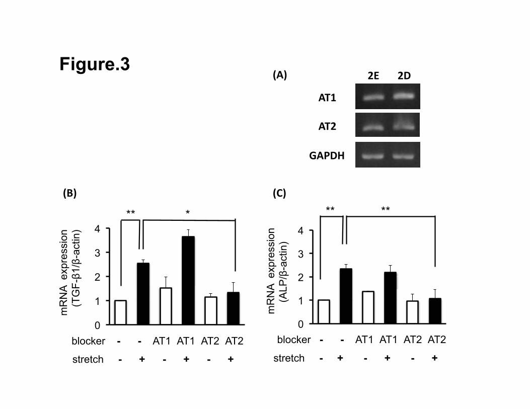
Roles of two Ang II receptors in HPLFs exposed to stretch loading
We examined whether stretch-induced TGF-β1 and ALP mRNA expressions
were modulated through the Ang II receptor in HPLFs. Both HPLF-2D and -2E
cultures exhibited gene expressions of AT1 and AT2 (Fig. 3A). Before exposure
to the stretch loading, HPLF-2Es were pre-incubated with candesartan or
PD123319. After loading, only PD123319 suppressed up-regulation of gene
expressions of both TGF-β1 and ALP by the stretch loading (Figs. 3B, 3C). In
contrast, candesartan had no effects on the stretch-mediated gene expressions
of TGF-β1 and ALP.

14 / 26
DISCUSSION
Stretch loading caused by occlusion and mastication functionally contributes to
the homeostasis of oxytalan fibers in PDL tissue (Tsuruga et al., 2008). However,
excess mechanical loading of PDL tissues has been reported to induce
osteoclastogenesis / cementoclastogenesis that resorbs bone or root via
up-regulated RANKL expression in human PDL cells (Yamaguchi et al., 2006).
Human PDL cells regulate osteoclastogenesis by opposing mechanisms,
including stimulation by RANKL combined with inhibition by OPG (Kanzaki et al.,
2001), and tensile loading that inhibits osteoclastogenesis through OPG
induction (Kanzaki et al., 2006). Therefore, in the present study, we recognized
the possibility to reduce RANKL expression and to induce OPG expression
during stretch loading, and that this mechanism may be necessary for bone
metabolism and maintenance of the PDL tissues. Indeed, our present data
demonstrated that 8% stretch loading down-regulated mRNA expression of
RANKL and up-regulated that of OPG. Thus, we fixed the elongation rate at 8%
for the necessary magnitude of stretch loading to investigate the associated
biological effects.
A recent study showed that mechanical stretch activated AGT mRNA

15 / 26
expression and caused secretion of Ang II in cardiomyocytes (Yamazaki et al.,
1995), and other reports have also demonstrated that Ang II acts as a mediator
of the mechanical stretch signaling in cardiomyocytes (Sadoshima et al., 1993;
Tamura et al., 1998). These reports support our present results, which showed
that 8% stretch loading up-regulated mRNA expression of AGT in HPLFs. Thus,
we hypothesized that RAS, including AGT, may be involved in the signal
transduction in HPLFs that have been exposed to stretch loading. In this study,
we demonstrated that Ang I/II was localized in PDL tissues, and furthermore,
that HPLFs expressed Ang II receptors, AT1 and AT2. Though there are few
reports about the role of Ang II in the PDL tissues, these results suggest that
Ang II plays a role in maintaining PDL tissues in an autocrine or paracrine
manner. Surprisingly, our present data showed that mimicking the stimulus by
exposure to 8% stretch loading, exogenous Ang II significantly up-regulated the
expression of AGT, TGF-β1, ALP and OPG, and down-regulated that of RANKL.
Therefore, we speculate that Ang II is involved in the cellular signaling of stretch
loaded-HPLFs.
Finally, we investigated the signaling pathways in stretched HPLFs using
specific antagonists of Ang II receptors. Pre-incubation of HPLF cultures with an

16 / 26
AT2 antagonist, but not an AT1 antagonist, suppressed up-regulation of gene
expressions of both TGF-β1 and ALP by the stretch loading. However, because
there is little information about the AT2 pathway with regard to stretch loading,
further studies are necessary to elucidate this mechanism.
TGF-β1 is one of the most multifunctional peptides, which is involved in a
wide variety of biological processes (Mehta and Attramadal, 2003). Recent
studies have revealed that in human PDL cells exposed to orthodontic force,
mRNA expression of TGF-β1 was augmented in both the compression side and
tension side (Garlet et al., 2007). Additionally, in cardiomyocytes, Ang II has
been reported to act as a paracrine mediator of stretch-induced TGF-β1 mRNA
expression (van Wamel JET et al., 2002). These reports support our current data.
Other researchers have discussed that TGF-β1, up-regulated by mechanical
stretch, plays a critical role in the healing and remodeling process of the human
anterior cruciate ligament (Kim et al., 2002). Therefore, the percentage of stretch
loading utilized in this study may be suitable to further investigate the
metabolism of the PDL tissues that are exposed to mechanical stress.
A couple of studies have shown that human PDL cells express some
osteoblastic characteristics in vitro (Somerman et al., 1988) and that mechanical
17 / 26
stimulation, including tensile force, can induce the differentiation of human PDL
cells into osteoblast-like cells (Matsuda et al., 1998; Yang et al., 2006).
Short-term stretch loading significantly induced ALP mRNA expression in human
PDL cells cultured in three dimensions (Ku et al., 2009). The experimental
loading system used in our present report resulted in the up-regulation of ALP
expression in HPLFs induced by exposure to stretch loading.
We summarize the current results in Fig. 4. PDL cells expressed Ang I/II, and
stretch loading up-regulated gene expression of AGT, which is potentially
converted into Ang I/II by RAS, in HPLFs. Subsequently, extracellularly-secreted
Ang II may up-regulate mRNA expression of TGF-β1 and ALP in an
autocrine/paracrine manner via the stimulation of the AT2 signaling pathway in
HPLFs. In conclusion, we have obtained evidence that Ang II is involved as a
transducer of the stretch loading signals in HPLFs, which consequently induces
expression of TGF-β1 and ALP.

18 / 26
ACKNOWLEDGMENTS
This work was supported by grants-in-aid (projects 19390486, 20791387,
21390510 and 21791942) for scientific research from the Ministry of Education,
Culture, Sports, Science, and Technology, Japan.

19 / 26
REFERENCES
Basdra EK, Komposch G (1997). Osteoblast-like properties human periodontal
ligament cells: an in vitro analysis. Eur J Orthod 19:615-621.
Jeunemaitre X, Soubrier F, Kotelevtsev YV, Lifton RP, Williams CS, Charru A,
Hunt SC, Hopkins PN, Williams RR, Lalouel JM, Corvol P (1992). Molecular
basis of human hypertension: role of angiotensinogen. Cell 71:169-180.
Fujii S, Maeda H, Wada N, Kano Y, Akamine A (2006). Establishment and
characterizing human periodontal ligament fibroblasts immortalized by
SV40T-antigen and hTERT gene transfer. Cell Tissue Res 324: 117-125.
Garlet TP, Coelho U, Silva JS, Garlet GP (2007). Cytokine expression pattern in
compression and tension sides of periodontal ligament during orthodontic
tooth movement in humans. Eur J Oral Sci 115:355-362.
Inagami T, Iwai N, Sasaki K, Yamamo Y, Bardhan S, Chaki S, Guo DF, Furuta H
(1992). Cloning, expression and regulation of angiotensin II receptors. J
Hypertens 10:713-716.
Izu Y, Mizoguchi F, Kawamata A, Hayata T, Nakamoto T, Nakashima K, Inagami
T, Ezura Y, Noda M (2008). Angiotensin ll type 2 receptor blockade
increases bone mass. J Biol Chem 284:4857-4864.
20 / 26
Kanzaki H, Chiba M, Shimizu Y, Mitani H (2001). Dual regulation of osteoclast
differentiation by periodontal ligament cells through RANKL stimulation and
OPG inhibition. J Dent Res 80:887-891.
Kanzaki H, Chiba M, Sato A, Miyagawa A, Arai K, Nukatsuka S, Mitani H (2006).
Cyclical tensile foce on periodontal ligament cells inhibits osteoclastogenesis
through OPG inhibition. J Dent Res 85:457-462.
Kim SG, Akaike T, Sasagaw T, Atomi Y, Kurosawa H (2002). Gene expression
of type I and type III collagen by mechanical stretch in anterior cruciate
ligament cells. Cell Struct Funct 27:139–144.
Ku SJ, Chang YI, Chae CH, Kim SG, Park Yw, Jung YK, Choi JY (2009). Static
tensional forces increase osteogenic gene expression in three-dimensional
periodontal ligament cell culture. BMB reports 42:427-432.
Maeda H, Wada N, Fujii S, Akamine A (2005). Fibroblastic cells from human
periapical granulation tissue preferentially form calcified matrices in
decalcified boiled rat bone. Cell Tissue Res 320:135-140.
Matsuda N, Morita N, Matsuda K, Watanabe M (1998). Proliferation and
differentiation of human osteoblastic cells associated with differential
activation of MAP kinases in response to epidermal growth factor, hypoxia,

21 / 26
and mechanical stress in vivo. Biophys Res Commun 249:350-354.
Mehta JL, Attramadal H (2003). The TGFβ superfamily in cardiovascular biology.
Cardiovasc Res 74:181-183.
Sadoshima J, Xu Y, Slayter HS, Izumo S (1993). Autocrine release of
Angiotensin II mediates stretch-induced hypertrophy of cardiac myocytes in
vitro. Cell 75:977-984.
Senbonmatsu T, Saito T., Landon E, Watanabe O, Price EJ, Roberts R, Imboden
H, Fitzgerald T, Gaffney F, Inagami T (2003). A novel angiotensin II type 2
receptor signaling pathway: possible role in cardiac hypertrophy. EMBO J
22:6471-6482.
Shi L, Kodama Y, Atsumi Y, Honma S, Wakisaka S (2005). Requirement of
occlusal force for maintenance of the terminal morphology of the periodontal
Ruffini endings. Arch Histol Cytol 68:289-299.
Somerman MJ, Archer SY, Imm GR, Foster RA (1988). A comparative study of
human periodontal ligament cells and gingival fibroblasts in vitro. J Dent Res
67: 66–70.
Tamura K, Umeura S, Nyui N, Hibi K, Ishigami T, Kihara M, Toya Y, Ishii M
(1998). Activation of angiotensinogen gene in cardiac myocytes by

22 / 26
angiotensin II and mechanical stretch. Am J Physiol 275:R1-9.
Tomikiyo A, Maeda H, Fujii S, Wada N, Shima K, Akamine A (2008).
Development of a multipotent clonal human periodontal ligament cell line.
Differentiation 76:337-347.
Tsuruga E, Nakashima K, Ishikawa H, Yajima T, Sawa Y (2008). Stretching
modulates oxytalan fibers in human periodontal ligament cells. J Periodont
Res 10:1111.
Udagawa N (2002). Mechanisms involved in bone resorption. Biogerontology
3:79-83.
van Wamel JET, Ruwhof C, van der Valk-Kokshoorn LJM, Schrier PI, van der
Laarse A (2002). Stretch-induced paracrine hypertrophic stimuli increase
TGF-β1 expression in cardiomyocytes. Mol Cell Biochem 236:147-153.
Wada N, Maeda H, Tanabe K, Tsuda E, Yano K, Nakamuta H, Akamine A
(2001). Periodontal ligament cells secrete the factor that inhibits osteoclastic
differentiation and function: the factor is osteoprotegerin / osteoclastogenesis
inhibitory factor. J Periodontal Res 36:56–63.
Wada N, Maeda H, Yoshimine Y, Akamine A (2004). Lipopolysaccharide
stimulates expression of osteoprotegerin and receptor activator of NF-kappa
23 / 26
B ligand in periodontal ligament fibroblasts through the induction of
interleukin-1 beta and tumor necrosis factor-alpha. Bone 35:629-635.
Yamaguchi M, Aihara N, Kojima T, Kasai K (2006). RANKL increase in
compressed periodontal ligament cells from root resorption. J Dent Res
85:751-756.
Yamaguchi N, Chiba M, Mitani H (2002). The induction of c-fos mRNA
expression by mechanical stress in human periodontal ligament cells. Arch
Oral Biol 47:465-471.
Yamazaki T, Komuro I, Kudoh S, Zou Y, Shiojima I, Mizuno T, Takano H, Hiroi Y,
Ueki K, Tobe K, Kadowaki T, Nagai R, Yazaki Y (1995). Angiotensin II partly
mediates mechanical stress-induced cardiac hypertrophy. Circ Res
77:258-265.
Yang YQ, Li XT, Rabie AB, Fu MK, Zhang D (2006). Human periodontal
ligament cells express osteoblastic phenotypes under intermittent force
loading in vitro. Front Biosci 11:776–781.

24 / 26
Figure Legends
Figure 1.
The effects of stretch loading on gene expression of RANKL (A) and OPG (B) in
HPLFs. HPLF-2E cultures were exposed to stretch loading of 0, 8 and 12%
elongation for 1 h, and the gene expressions of RANKL and OPG in the cell
cultures were assessed by semi-quantitative RT-PCR. The stretch loading of 8%
down-regulated mRNA expression of RANKL, as compared with 0%, while the
stretch loading of 12% up-regulated the expression. The stretch loading of both
8% and 12% up-regulated mRNA expression of OPG. The gene expressions of
TGF-β1, ALP and AGT in HPLF-2Ds (C) and HPLF-2Es (D) exposed to the
stretch loading of 8% were examined by quantitative RT-PCR. The results are
shown as mean ± SD from three different experiments. **p < 0.02.
Figure 2.
Localization of Ang II in rat PDL tissue and HPLFs (A-E). Ang II immunoreactivity
was detected in PDL tissues. Dentin, bone matrices and pulp tissues showed
negative reactions (A). A magnified view of the rectangle area in (A) is shown in
(B). Both PDL cells and extra-cellular-matrix in PDL tissues showed strongly

25 / 26
positive reactions (D, dentin; PDL, periodontal ligament; B, bone) (B). Control
staining with serial sections showed negative reactions (C). Ang II
immunoreactivity could be seen in the cytoplasm of HPLF-2Es (D). Control
staining of HPLF-2Es showed no positive reactions (E). Bars, 100 μm. Gene
expression in HPLF-2Es stimulated with Ang II analyzed by quantitative RT-PCR
are shown (F-H). The expressions of TGF-β1 and ALP mRNA were up-regulated
dose-dependently by a 1h-stimulus of recombinant Ang II in HPLF-2Es (F). OPG
and AGT mRNA expressions were up-regulated by the 1h-stimulus of
recombinant Ang II in HPLF-2Es, while RANKL mRNA expression was
down-regulated (G). TGF-β1 and ALP mRNA expressions were also
up-regulated by the 1h-stimulus of recombinant Ang II in HPLF-2Ds (H). The
values were compiled as mean ± SD from three different experiments. **p < 0.02,
*p < 0.05.
Figure 3.
Gene expression of Ang II receptors in HPLFs and the effects of Ang II receptor
antagonists on the mRNA expressions of stretch-induced TGF-β1 and ALP. The
expressions of AT1 and AT2 mRNA both in HPLF-2Ds and in HPLF-2Es were
26 / 26
examined by semi-quantitative RT-PCR (A). mRNA expressions of
stretch-induced TGF-β1 (B) and ALP (C) as they were affected by Ang II
receptor antagonists and stretch loading. HPLF-2Es were incubated with or
without an AT1 antagonist, candesartan (10 ng/ml), or an AT2 antagonist,
PD123319 (100nM), for 30 min before exposure to 1h-stretch loading. The
values of quantitative RT-PCR data are reported as mean ± SD from three
different experiments. **p < 0.02, *p < 0.05.
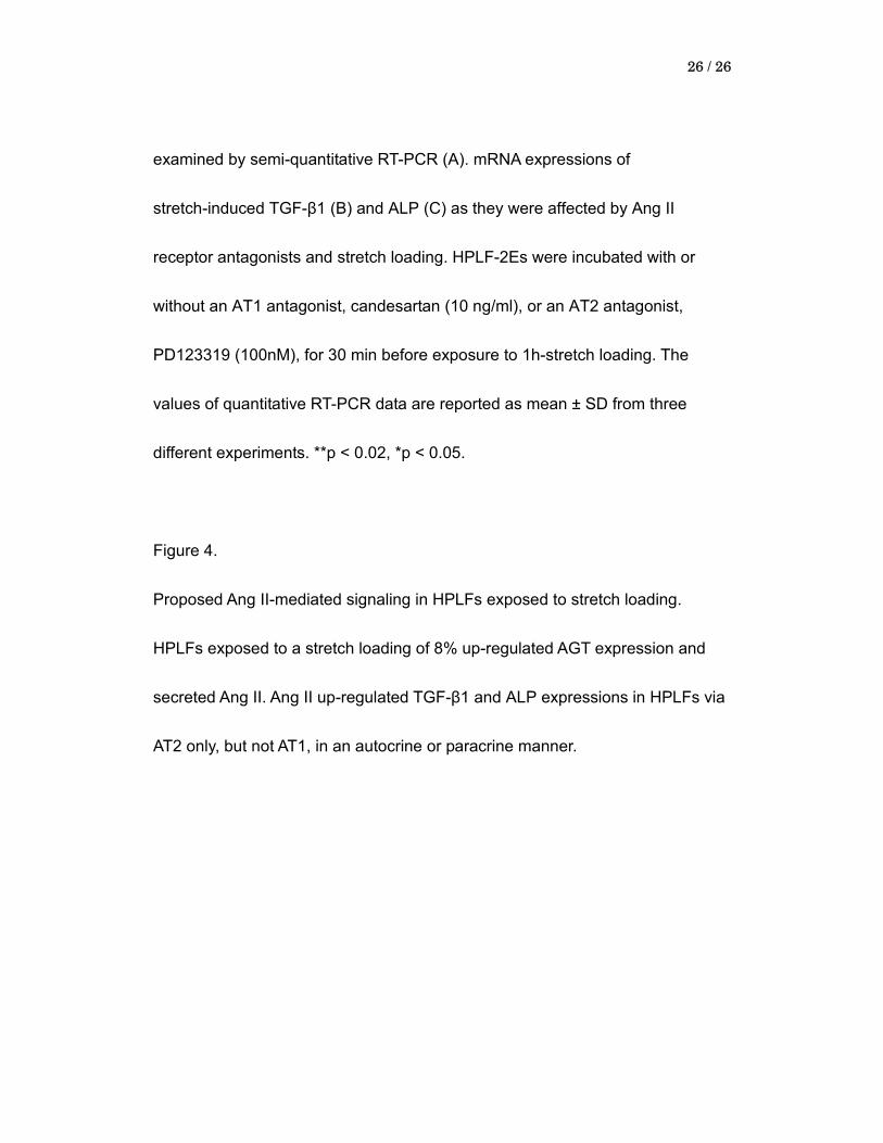
Figure 4.
Proposed Ang II-mediated signaling in HPLFs exposed to stretch loading.
HPLFs exposed to a stretch loading of 8% up-regulated AGT expression and
secreted Ang II. Ang II up-regulated TGF-β1 and ALP expressions in HPLFs via
AT2 only, but not AT1, in an autocrine or paracrine manner.

0
1
2
3
TGF-�1 ALP AGT
0%
0
1
2
3
TGF-�1 ALP AGT
0% 8%
** **
mR
NA
e
xp
ressio
n
(/�
-actin)
**
**
** m
RN
A
exp
ressio
n
(/�
-actin)
**
GAPDH 19c
RANKL 34c
0% 8% 12% 0% 8% 12%
GAPDH 19c
OPG 22c
Figure.1
0
1
2
0% 8% 12%
**
**
mR
NA
expre
ssio
n
(
RA
NK
L/G
AP
DH
) 0
1
2
3
0% 8% 12%
**
**
mR
NA
expre
ssio
n
(
OP
G/G
AP
DH
) (A) (B)
(C) (D)

0
1
2
3
TGF-�1 ALP
cont
.
D
PDL
B
(A) (B) (C)
Figure.2
(D) (E)
(F) (G)
mR
NA
e
xp
ressio
n
(/
�-a
ctin
)
**
**
* **
0
1
2
3 con
t
mR
NA
e
xp
ressio
n
(/�
-actin
)
**
**
**
(H)
0
1
2
3
TGF-�1 ALP
con
t.
**
**
mR
NA
e
xp
ressio
n
(/
�-a
ctin
)
0
1
2
3
4
0
1
2
3
4
mR
NA
expre
ssio
n
(
TG
F-�
1/�
-actin)
mR
NA
expre
ssio
n
(A
LP
/�-a
ctin)
stretch - + - + - + stretch - + - + - +
blocker - - AT1 AT1 AT2 AT2 blocker - - AT1 AT1 AT2 AT2
** *
���� ����
Figure.3
** **
���
��
��� ���
����
����

Stretch
PDL cell PDL cell
AGT Ang ll Ang ll
AT1
AT2 TGF-�1
ALP
Figure.4