Embed Size (px)
Citation preview

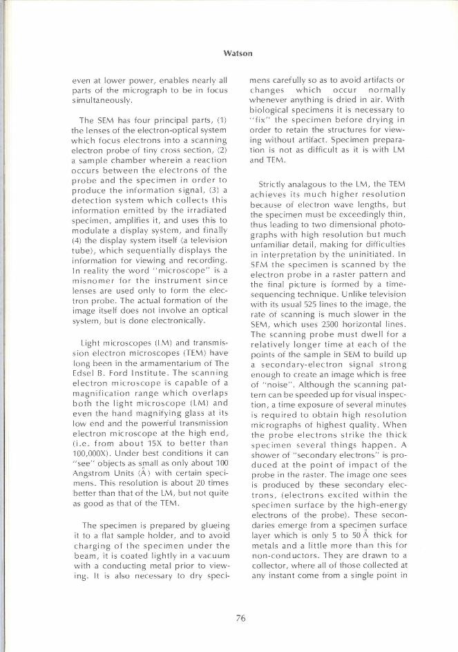
Henry Ford Hospital Medical Journal
Volume 21 | Number 2 Article 4
6-1973
The Scanning Electron Microscope: Applications atThe Edsel B. Ford Institute for Medical ResearchJohn H. L. Watson
Follow this and additional works at: https://scholarlycommons.henryford.com/hfhmedjournalPart of the Life Sciences Commons, Medical Specialties Commons, and the Public Health
Commons
This Article is brought to you for free and open access by Henry Ford Health System Scholarly Commons. It has been accepted for inclusion in HenryFord Hospital Medical Journal by an authorized editor of Henry Ford Health System Scholarly Commons.
Recommended CitationWatson, John H. L. (1973) "The Scanning Electron Microscope: Applications at The Edsel B. Ford Institute for Medical Research,"Henry Ford Hospital Medical Journal : Vol. 21 : No. 2 , 75-84.Available at: https://scholarlycommons.henryford.com/hfhmedjournal/vol21/iss2/4
H e n r y Ford H o s p . M e d . Journa l
V o l . 2 1 , N o . 2 , 1973
The Scanning Electron Microscope
Applications at The Edsel B. Ford Institute for Medical Research
J.H.L. W a t s o n , Ph D
The Henry Ford Hospital with its research partner. The Edsel B. Ford Institute for Medical Research, was the first private medical center in the State of Michigan to purchase a scanning electron microscope and to begin to apply it, a year ago, to the study of medical and biological problems in both the animal and the human. This article briefly describes how the Instrument works and shows some samples of micrographs produced.
'Department of Physics and Biophysics, Edsel B. Ford Institute for Medical Research
Address reprint request to Dr. Watson at EBFl, Henry Ford Hospital, 2799 W. Grand Blvd., Detroit, Ml 48202
D U R I N G the last 30 years the transmission electron microscope (TEM) ("transmission" because the electrons go t h r o u g h the spec imen) has rewr i t t en anatomy textbooks, and revolut ionized scientific thought wherever the ultra-structure of both biological and non-biological materials is important to the understanding of their propert ies. During the last five years a new type of microscope, the scanning electron microscope (SEM) has been reaching promi n e n c e . Pictures taken w i t h it have appeared more and more frequent ly in bo th the scient i f ic and popular press. The most s t r i k i ng f i rs t impress ion of these pictures (micrographs) is their three-dimensional characteristic usually completely absent in the two-d i m e n s i o n a l m i c rog raphs f r o m the t ransmiss ion type m ic roscope . O t h e r factors wh ich lead to a sense of reality about the scanning pictures are first, that they can be taken at very low magnificat ions, and second, they are taken of recognizable surfaces, both of which al low the obse rve r to " i d e n t i f y " w i t h t h e famil iar structures before using higher magn i f i ca t ions . In SEM it is re lat ively easy fo r an obse rve r to e x t e n d his in te rp re ta t ion f rom one d imens ion to the next al though the aspect of a picture and the informat ion it contains may be completely and dramatically changed as the magnification is increased. At the same t ime the greater depth of focus.
75
Watson
even at lower power, enables nearly all parts of the micrograph to be in focus simultaneously.
The SEM has four principal parts, (1) the lenses of the electron-optical system wh ich focus electrons in to a scanning electron probe of tiny cross sect ion, (2) a sample chamber whe re in a react ion occurs be tween the e lec t rons of t he p r o b e and the spec imen in o r d e r to p roduce the i n f o rma t i on s igna l , (3) a d e t e c t i o n system w h i c h co l lec ts th is i n fo rmat ion emi t ted by the i r radiated specimen, amplifies i t , and uses this to modulate a display system, and f inal ly (4) the display system itself (a television tube) , wh i ch sequent ia l ly displays the information for v iewing and recording. In reality the w o r d " m i c r o s c o p e " is a m i s n o m e r fo r t h e i n s t r u m e n t s ince lenses are used only to form the electron probe. The actual format ion of the image itself does not involve an optical system, but is done electronically.
Light microscopes (LM) and transmission e lect ron microscopes (TEM) have long been in the armamentarium of The Edsel B. Ford Ins t i t u te . The scann ing e l ec t r on m i c r o s c o p e is capab le of a m a g n i f i c a t i o n range w h i c h over laps b o t h the l i gh t m i c r o s c o p e (LM) and even the hand magn i f y ing glass at its low end and the powerful transmission e lec t ron mic roscope at the h igh e n d , ( i . e . f r o m a b o u t 15X to be t t e r t han 100,000X). Under best condit ions it can "see" objects as small as only about 100 Angstrom Units (A) wi th certain specimens. This resolut ion is about 20 times better than that of the LM, but not quite as good as that of the TEM.
The specimen is prepared by glueing it to a flat sample holder, and to avoid c h a r g i n g of t he spec imen u n d e r t h e beam, it is coated l ight ly in a vacuum wi th a conduct ing metal pr ior to viewing. It is also necessary to dry speci
mens carefully so as to avoid artifacts or changes w h i c h occur n o r m a l l y whenever anything is dried in air. Wi th biological specimens it is necessary to " f i x " the spec imen be fo re d r y i n g in order to retain the structures for viewing wi thout artifact. Specimen preparat ion is not as di f f icul t as it is w i th LM and TEM.
Strictly analagous to the LM, the TEM achieves its m u c h h ighe r r e s o l u t i o n because of electron wave lengths, but the specimen must be exceedingly th in , thus leading to two dimensional photographs w i th high resolut ion bu t much unfamiliar detai l , making for diff icult ies in in terpreta t ion by the un in i t ia ted. In SEM the spec imen is scanned by the e lect ron probe in a raster pat tern and the final picture is formed by a t ime-sequencing technique. Unl ike television wi th its usual 525 lines to the image, the rate of scanning is much slower in the SEM, which uses 2500 horizontal l ines. The scanning p robe must dwe l l fo r a re la t i ve ly l onger t i m e at each of the points of the sample in SEM to bui ld up a s e c o n d a r y - e l e c t r o n s ignal s t r ong enough to create an image which is free of "no i se " . A l though the scanning pattern can be speeded up for visual inspect i on , a t ime exposure of several minutes is requ i red to ob ta in h igh reso lu t i on micrographs of highest qual i ty . W h e n the p r o b e e l ec t r ons s t r i ke t h e t h i c k spec imen several th ings h a p p e n . A shower of "secondary electrons" is prod u c e d at t h e p o i n t of impac t of t he probe in the raster. The image one sees is produced by these secondary elect r o n s , (e lec t rons exc i t ed w i t h i n the spec imen surface by the h igh-energy electrons of the probe). These secondaries emerge from a specimen surface layer which is only 5 to 50 A thick for metals and a l i t t le more than th is fo r non -conduc to rs . They are d rawn to a col lector, where all of those collected at any instant come from a single point in
76
The Scanning Electron Microscope
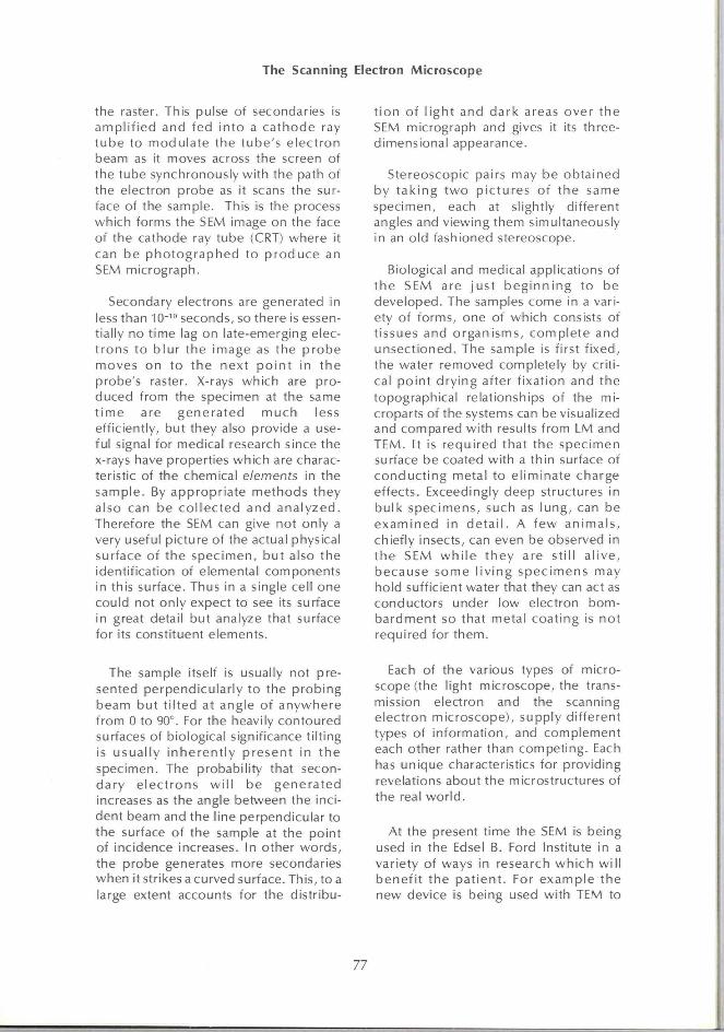
the raster. This pulse of secondaries is amp l i f i ed and fed in to a ca thode ray t ube to modu la te the tube 's e lec t ron beam as it moves across the screen of the tube synchronously w i th the path of the electron probe as it scans the surface of the sample. This is the process which forms the SEM image on the face of the cathode ray tube (CRT) where it can be p h o t o g r a p h e d to p r o d u c e an SEM micrograph.
Secondary electrons are generated in less than 10""' seconds, so there is essentially no t ime lag on late-emerging elect rons to b lu r the image as the p robe moves on to the next p o i n t in the probe's raster. X-rays which are produced f rom the specimen at the same t ime are gene ra ted m u c h less eff iciently, but they also provide a useful signal for medical research since the x-rays have properties which are characteristic of the chemical elements in the sample. By appropr ia te methods they also can be c o l l e c t e d and ana l yzed . Therefore the SEM can give not only a very useful picture of the actual physical surface of the spec imen, bu t also the identif ication of elemental components in this surface. Thus in a single cell one could not only expect to see its surface in great detail but analyze that surface for its constituent elements.
t i on of l i gh t and dark areas over the SEM micrograph and gives it its three-dimensional appearance.
Stereoscopic pairs may be obta ined by t a k i n g t w o p i c tu res of t he same specimen, each at slightly di f ferent angles and viewing them simultaneously in an old fashioned stereoscope.
Biological and medical applications of t he SEM are jus t b e g i n n i n g to be developed. The samples come in a variety of forms, one of which consists of t issues and o rgan isms, comp le te and unsec t ioned. The sample is f irst f i xed , the water removed completely by cri t ical po in t d ry ing after f ixat ion and the topographical relationships of the mi-croparts of the systems can be visualized and compared wi th results f rom LM and TEM. It is requ i red that the spec imen surface be coated wi th a th in surface of conduc t i ng metal to e l iminate charge effects. Exceedingly deep structures in bu lk spec imens, such as l ung , can be e x a m i n e d in d e t a i l . A f ew an ima ls , chiefly insects, can even be observed in the SEM w h i l e t hey are st i l l a l i ve , because some l i v i n g spec imens may hold sufficient water that they can act as conductors under low electron bombardment so that metal coat ing is no t required for them.
The sample itself is usually not presented perpendicu lar ly to the p rob ing beam but t i l t ed at angle of anywhere f rom 0 to 90°. For the heavily contoured surfaces of biological significance t i l t ing is usua l ly i n h e r e n t l y p resen t in t h e specimen. The probabil i ty that secondary e lec t rons w i l l be gene ra ted increases as the angle between the incident beam and the line perpendicular to the surface of the sample at the point of incidence increases. In other words, the probe generates more secondaries when it strikes a curved surface. This, to a large extent accounts for the distr ibu-
Each of the various types of microscope (the light microscope, the transmission electron and the scanning e lect ron microscope) , supply d i f ferent types of in format ion, and complement each other rather than compet ing. Each has unique characteristics for providing revelations about the microstructures of the real wo r ld .
At the present t ime the SEM is being used in the Edsel B. Ford Institute in a variety of ways in research w h i c h w i l l bene f i t the pa t i en t . For examp le the new device is being used wi th TEM to
77
Watson
study possible changes which occur on the inside surfaces of veins and arteries af ter they have been t r a n s p l a n t e d exper imenta l l y in labora tory an imals . The purpose of this work for the human is to improve the patient's chances for a successful arterial replacement in the surgical treatment of coronary th rombosis and for successful replacement of the femora l ar tery in a therosc le ro t i c occlusion. Similarly, possible changes in k idney s t ruc tu re are be ing s tud ied in laboratory animals when their k idneys are transplanted experimental ly.
The oppor tun i t i es for research w i t h SEM in the p r o b l e m s of c l i n i ca l m e d i c i n e are u n l i m i t e d . In h u m a n beings the instrument is being used to study surface changes and structure in diseased lung in order to relate them to a better understanding of the diseases
and their effects. The Institute has long been interested in problems connected wi th human breast cancer and the part wh ich viruses may or may not play in it. The SEM o f fe rs a new and p o w e r f u l method of approach to this problem by al lowing visualization of the surface of cancer cells taken f rom cancer patients and g r o w n in t issue c u l t u r e fo r SEM study. Another example of our current SEM Research (in concert w i th TEM), is the detailed study of the ultrastructure of t he sur face of human hair and its relationship to genetic defects in man. Some of the possible appl icat ions are suggested and illustrated in the microg raphs w h i c h accompany th is b r ie f desc r ip t i on of the ins t rument and its appl icat ion. The line in the micrographs indicates microns ip ) , which is equal to lO'^cms.
78
The Scanning Electron Microscope
Figures 1 A to 1 E
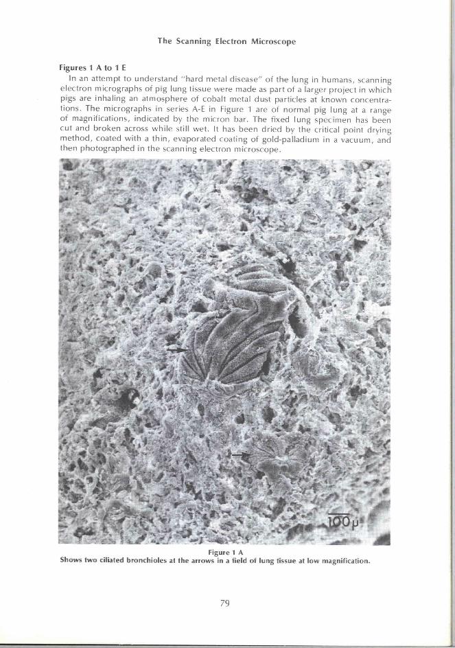
In an attempt to understand "hard metal disease" of the lung in humans, scanning electron micrographs of pig lung tissue were made as part of a larger project in which pigs are inhaling an atmosphere of cobalt metal dust particles at known concentrat ions. The micrographs in series A-E in Figure 1 are of normal pig lung at a range of magnif ications, indicated by the micron bar. The fixed lung specimen has been cut and broken across whi le still wet. It has been dr ied by the critical point drying method, coated wi th a th in , evaporated coating of gold-pal ladium in a vacuum, and then photographed in the scanning electron microscope.
r"
Figure 1 A Shows two ciliated bronchioles at the arrows in a field of lung tissue at low magnification.
79
Watson
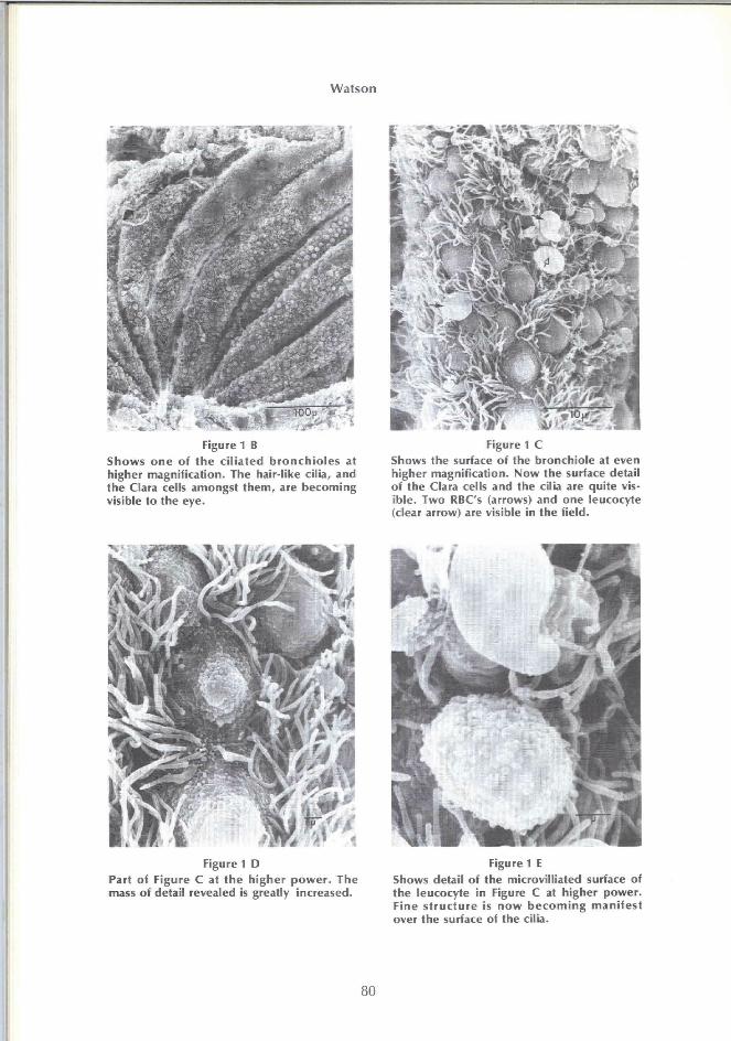
Figure 1 B Shows one of the ciliated bronchioles at higher magnification. The hair-like cilia, and the Clara cells amongst them, are becoming visible to the eye.
Figure 1 D Part of Figure C at the higher power. The mass of detail revealed is greatly increased.
Figure 1 C Shows the surface of the bronchiole at even higher magnification. Now the surface detail of the Clara cells and the cilia are quite visible. Two RBC's (arrows) and one leucocyte (clear arrow) are visible in the field.
Figure 1 E Shows detail of the microvilliated surface of the leucocyte in Figure C at higher power. Fine structure is now becoming manifest over the surface of the cilia.
80
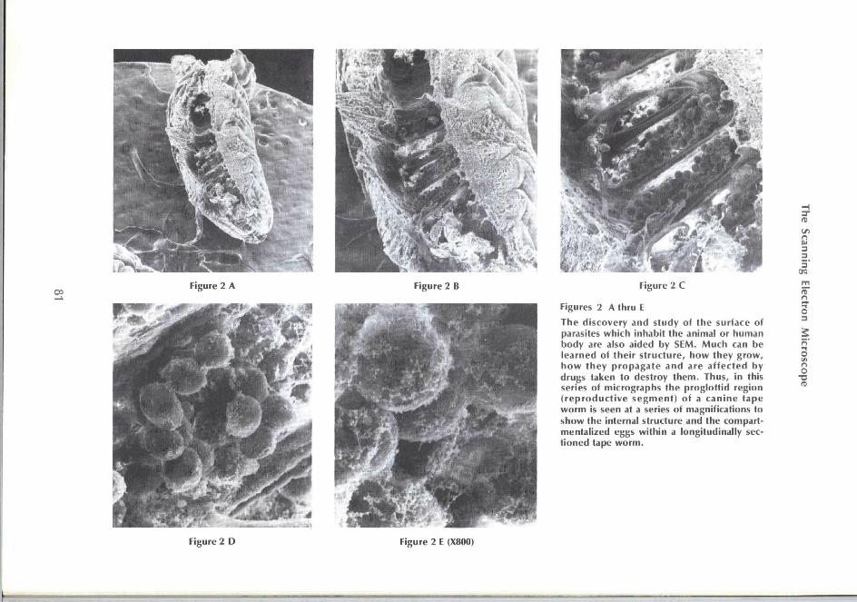
Figure 2 C
Figures 2 A thru E
The d iscovery and s tudy of the surface of parasites wh ich inhabit the animal or human body are also aided by SEM. Much can be learned of the i r s t ruc tu re , how they g row , h o w t hey p r o p a g a t e a n d are a f f e c t e d by drugs taken to destroy t hem. Thus, in this series of micrographs the p rog lo t t id region ( r e p r o d u c t i v e segmen t ) of a can ine tape w o r m is seen at a series of magnif ications to show the internal structure and the compartmental ized eggs w i th in a longi tudinal ly sect ioned tape w o r m .
IT) n 3
5' 95
3
n O
rt
Figure 2 D Figure 2 E (X800)
Watson
-4.
Figure 3 A Figure 3 B
Figure 3 C Figure 4
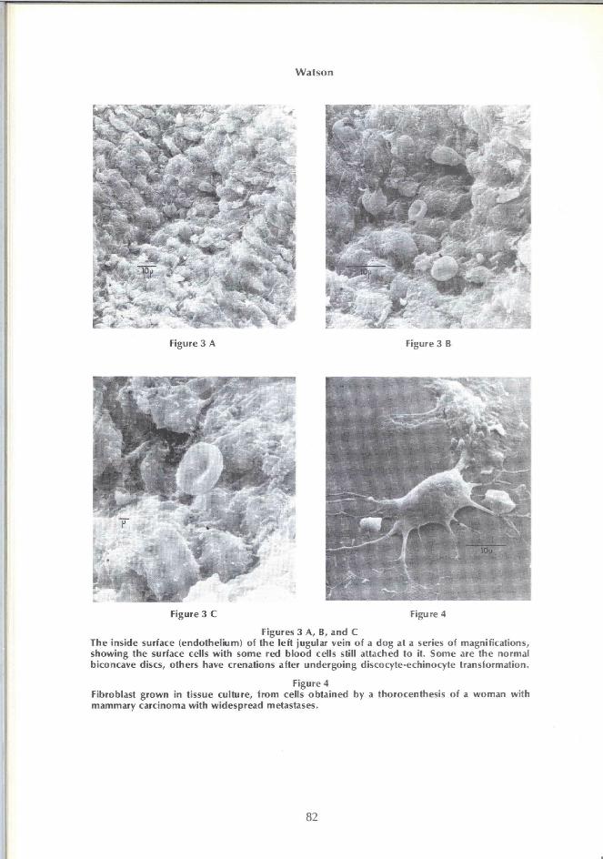
Figures 3 A, B, and C The inside surface (endothelium) of the left jugular vein of a dog at a series of magnifications, showing the surface cells with some red blood cells still attached to it. Some are the normal biconcave discs, others have crenations after undergoing discocyte-echinocyte transformation.
Figure 4 Fibroblast grown in tissue culture, from cells obtained by a thorocenthesis of a woman with mammary carcinoma with widespread metastases.
82
The Scanning Electron Microscope
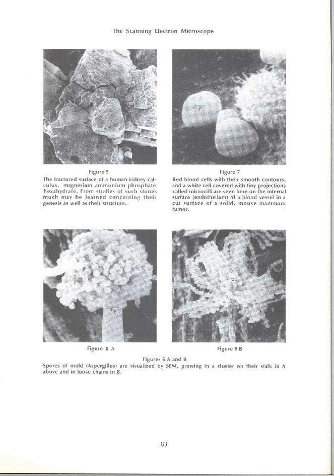
Figure 5
The fractured surface of a human kidney calcu l us , magnesium a m m o n i u m p h o s p h a t e hexahydra te . From studies of such stones m u c h may be l e a r n e d c o n c e r n i n g t h e i r genesis as wel l as their structure.
Figure 7
Red b lood cells w i th their smooth contours, and a wh i te cell covered w i th t iny project ions called microvi l l i are seen here on the internal surface (endothel ium) of a b lood vessel in a cu t su r f ace of a s o l i d , m o u s e m a m m a r y tumor .
Figures 6 A and B Spores of mo ld (Aspergillus) are visualized by SEM, g rowing in a cluster on their stalk in A above and in loose chains in B.
Watson
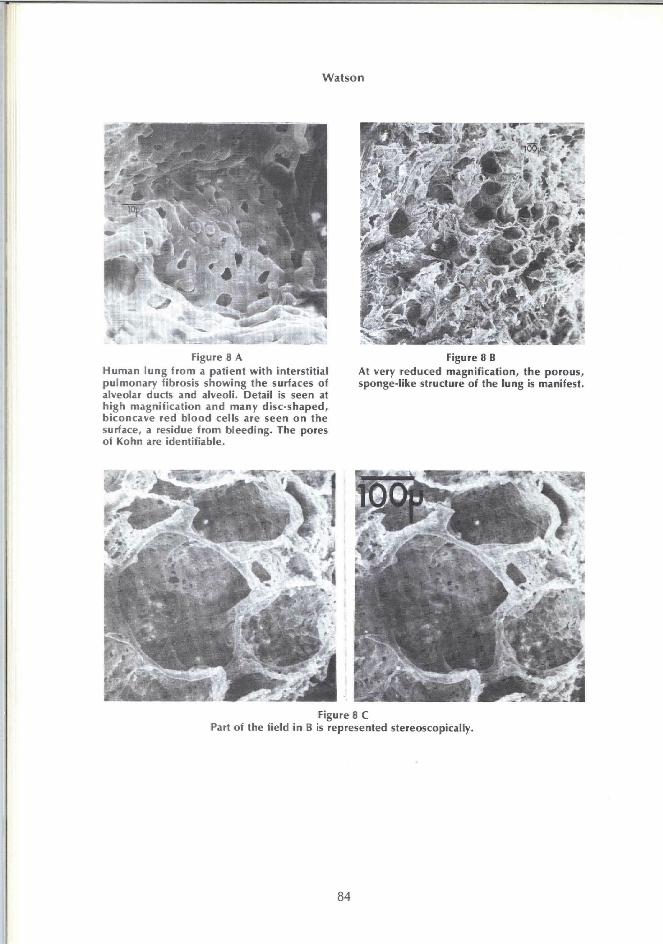
Figure 8 A Human lung from a patient with interstitial pulmonary fibrosis showing the surfaces of alveolar ducts and alveoli. Detail Is seen at high magnification and many disc-shaped, biconcave red blood cells are seen on the surface, a residue from bleeding. The pores of Kohn are identifiable.
Figure 8 B At very reduced magnification, the porous, sponge-like structure of the lung is manifest.
Figure 8 C Part of the field in B is represented stereoscopically.