Embed Size (px)
Citation preview
The Science Behind Zoom Whitening


In-Office WhiteningBenefit of Light vs No LightA Randomized, Parallel-Design Clinical Trial to Assess Tooth Bleaching Efficacy and Safety of Light versus non-Light Activated Chairside Whitening . . . . . . 7
Color Change of Vital Teeth Exposed to Bleaching Performed With and Without Supplementary Light . . . . . . . . . . . . . . . . . . . . . . . . 12
Effect of ZOOM! Advanced Power Lamp on Whitening . . . . . . . . . . . . . . . . . . . . . . . . . . . 14
Effect of Light Treatment on in vitro Tooth Bleaching Efficacy . . . . . . . . . . . . . . . . . . . . . . . 16
Clinical Study to Compare Two In-Office Whitening Systems . . . . . . . . . . . . . . . . . . . . . . . . . 18
Clinical Evaluation of a Novel Dental Whitening Lamp and Light-Catalyzed Peroxide Gel . . . . . . . . . . . . . . . . 20
Clinical Study of an In-Office Whitening System With and Without Light Procedures Utilizing a Split-Arch Design . . . . . . . . . . . . . . . . . . . . . . . . . . 22
Photo-Fenton and Conventional In-Office Dental Bleaching . . . . . . . . . . . . . . . . . . . . . . . . . . . 24
Clinical Trial: Photo-Fenton and Conventional In-Office Dental Bleaching . . . . . . . . . . . . . . . . 26
The Basic Chemistry Behind Hydrogen Peroxide Tooth Whitening . . . . . . . . . . . . . . . . . . . 28
EfficacyNovel Method for Efficacy Assessment of Whitening Agents . . . . . . . . . . . . . . . . . . . . . . . . . . . . . . . . 30
Light-Activated System for Bleaching Teeth . . . . . . . . . . . . . . 32
Clinical Evaluation Comparing Two H2O2 Concentration Used with a Light-Assisted Chairside Tooth Whitening System . . . . . . . . . . . . . . . . . . . . 35
Take-Home WhiteningBenefits of ACPInfluence of Bioactive Materials on Whitened Human Enamel Surface . . . . . . . . . . . . . . . . . . . . . 37
Influence of Five Home Whitening Gels and a Remineralizing Gel on the Enamel and Dentin Ultrastructure and Hardness . . . . . . . . . . . . . . . . . . . 39
Effect of Relief ACP on Dentin Microhardness and Surface Morphology . . . . . . . . . . . . . . . . . . . . . . . . . . . . 41
Fluoride and Potassium Nitrate-Fluoride Whitening Agents: in vitro Caries Study . . . . . . . . . . . . . . . . 43
Effect of Take-Home Whitening Agent on Enamel Microhardness . . . . . . . . . . . . . . . . . . . . . . . . . . . 45
Effect of Remineralizing Agents on Enamel Microhardness After Bleaching . . . . . . . . . . . . . . . . . 47
A 180-Day Clinical Investigation of the Tooth Whitening Efficacy of a Bleaching Gel with Added Amorphous Calcium Phosphate . . . . . . . . . . . . . 48
Whitening Agents with ACP Enamel Caries Formation and Progression . . . . . . . . . . . . . . . . . . . . 50
Tooth Surface Enhancement by a 16% Carbamide Peroxide Take-Home Bleaching Gel Containing ACP . . . . . . 52
The Clinical Performance of Professionally Dispensed Bleaching Gel With Added Amorphous Calcium Phosphate . . . . . . . . . . . . . . . . . . . . . . 54
Microhardness and Ultramorphological Changes after Whitening: Calcium and Phosphate Benefits . . . . . . . . 56
Whitening Treatment Combined With Bioactive Materials . . . . . . . . . . . . . . . . . . . . . . . . . . . . . . . . . 59
Table of Contents

4
5
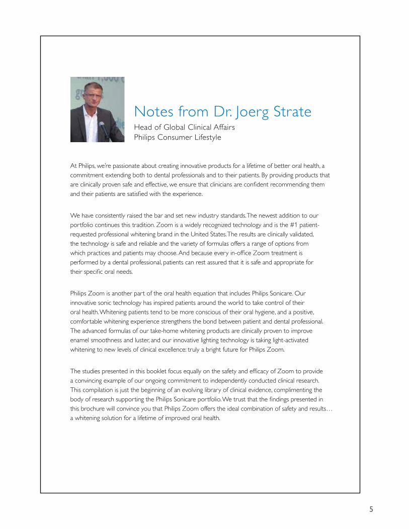
Notes from Dr. Joerg Strate Head of Global Clinical Affairs Philips Consumer Lifestyle
At Philips, we’re passionate about creating innovative products for a lifetime of better oral health, a commitment extending both to dental professionals and to their patients. By providing products that are clinically proven safe and effective, we ensure that clinicians are confident recommending them and their patients are satisfied with the experience.
We have consistently raised the bar and set new industry standards. The newest addition to our portfolio continues this tradition. Zoom is a widely recognized technology and is the #1 patient-requested professional whitening brand in the United States. The results are clinically validated, the technology is safe and reliable and the variety of formulas offers a range of options from which practices and patients may choose. And because every in-office Zoom treatment is performed by a dental professional, patients can rest assured that it is safe and appropriate for their specific oral needs.
Philips Zoom is another part of the oral health equation that includes Philips Sonicare. Our innovative sonic technology has inspired patients around the world to take control of their oral health. Whitening patients tend to be more conscious of their oral hygiene, and a positive, comfortable whitening experience strengthens the bond between patient and dental professional. The advanced formulas of our take-home whitening products are clinically proven to improve enamel smoothness and luster, and our innovative lighting technology is taking light-activated whitening to new levels of clinical excellence: truly a bright future for Philips Zoom.
The studies presented in this booklet focus equally on the safety and efficacy of Zoom to provide a convincing example of our ongoing commitment to independently conducted clinical research. This compilation is just the beginning of an evolving library of clinical evidence, complimenting the body of research supporting the Philips Sonicare portfolio. We trust that the findings presented in this brochure will convince you that Philips Zoom offers the ideal combination of safety and results… a whitening solution for a lifetime of improved oral health.

6

7
A Randomized, Parallel-Design Clinical Trial to Assess Tooth Bleaching Efficacy and Safety of Light versus non-Light Activated Chairside Whitening
In-OffIce WhItenIng
In-O
ffice
Whi
teni
ngEf
ficac
y
A Randomized, Parallel-Design Clinical Trial to Assess Tooth Bleaching Efficacy and Safety of Light versus non-Light Activated Chairside Whitening in vivo studyAuthors: Li Y, Lee S, Kwon S.R., Arambula M, Yang H, Li J, Delaurenti M, Jenkins W, Nelson M, Souza S, Ward M. Data on file, 2012
Objective
To characterize the extent to which the safety and efficacy profile of Philips Zoom WhiteSpeed (25% Hydrogen Peroxide) and Ultradent Opalescence Boost PF (40% Hydrogen Peroxide) cosmetic whitening regimens differ immediately following, and at seven and thirty days post bleaching application.
Methodology
One hundred thirty-five of 394 subjects screened completed an IRB-approved double-blind, randomized, parallel-design clinical trial in a population of healthy adults aged 18-75. Fifty-nine subjects were female, 76 were male; with a mean age of 50.0 years. Eligible subjects had a minimum of four maxillary anterior teeth with a tooth shade of A3 or darker assessed per VITA Classical (VC) shade guide. Sixty-eight subjects were randomized to Opalescence Boost and 69 to Philips Zoom. Efficacy was assessed by VITA EasyShade for ∆E characterization using a custom jig fabricated for a single anterior site in addition to VC and VITA Bleachedguide 3D-Master (BG) shade assessment. Safety was characterized by subject report of sensitivity, oral examination and subject use of sensitivity-reducing agents (Relief ACP or UltraEZ) applied and dispensed per manufacturer’s instructions. Study endpoints were assessed pre- and post-whitening, at Day 7 and Day 30.
Results
Median ∆E values per Kruskal-Wallis analysis for instrumental color change immediately post-whitening were 5.12 for Zoom and 2.55 for Boost (p<0.0001). At Day 7, ∆E outcomes were 6.34 for Zoom and 4.08 for Boost (p=0.0059). At Day 30, ∆E outcomes were 6.03 for Zoom and 3.44 for Boost (p=0.0019). The difference between treatments at each timepoint was statistically significant.
For VC visual shade assessment, LS Mean (SE) based on analysis of variance immediately post-whitening values were 5.86 (0.18) for Zoom and 4.47 (0.18) for Boost (p<0.0001). At Day 7, outcomes were 4.92 (0.20) for Zoom and 4.19 (0.20) for Boost (p=0.0106). At Day 30, outcomes were 4.45 for Zoom and 4.11 for Boost (p=0.2648).
For BG visual shade assessment, the median shade change per Kruskal-Wallis analysis immediately post-whitening was 3.17 for Zoom and 2.00 for Boost (p<0.0001). At Day 7, outcomes were 2.33 for Zoom and 1.67 for Boost (p=0.0198) and at Day 30, outcomes were 2.25 for Zoom and 1.83 for Boost (p=0.1195).
The percentage of subjects who reported ‘No Sensitivity’ immediately post-whitening was 98.5% for Zoom and 98.6% for

8
A Randomized, Parallel-Design Clinical Trial to Assess Tooth Bleaching Efficacy and Safety of Light versus non-Light Activated Chairside Whitening
In-OffIce WhItenIng
In-Office W
hiteningEfficacy
Boost. At Day 7, subject-reported values for ‘No Sensitivity’ were 82.1% for Zoom and 79.4% for Boost. Of those experiencing sensitivity, one subject rated sensitivity as ‘Moderate.’ All other reports were characterized as ‘Mild’ by subjects.
There were a total of 41 adverse events reported among 34 subjects. In general, these events were associated with sensitivity.
Subject use of post-whitening sensitivity gel (Relief ACP and UltraEZ) was low. Four subjects (two per treatment group) used the products at Day 1 post-bleaching and one subject used the product on Day 2. There are no other reports of use from Day 3 to Day 7.
Conclusions
Philips Zoom WhiteSpeed whitens teeth significantly better than Opalesence Boost immediately following bleaching, and at seven and thirty days post application as measured by ∆E VITA EasyShade.
Philips Zoom WhiteSpeed whitens teeth significantly better than Opalesence Boost immediately following and seven days post application per VITA Classical Shade Guide and VITA Bleachedguide 3D-Master assessment.
Philips Zoom WhiteSpeed and Ultradent Opalesence Boost are both well tolerated with low incidence of sensitivity and no significant differences in the safety profile between groups.
0
2
3
4
6
5
7
1
Post Whitening(p<0.0001)
Day 7(p=0.0059)
Day 30(p= 0.0019)
5.12
2.55
Median ∆E Post Whitening Day 7 and Day 30
Zoom WhiteSpeed Opalescence Boost
6.34
4.08
6.03
3.44
9
A Randomized, Parallel-Design Clinical Trial to Assess Tooth Bleaching Efficacy and Safety of Light versus non-Light Activated Chairside Whitening
In-OffIce WhItenIng
In-O
ffice
Whi
teni
ngEf
ficac
y
0
1
1.5
2
3
2.5
3.5
0.5
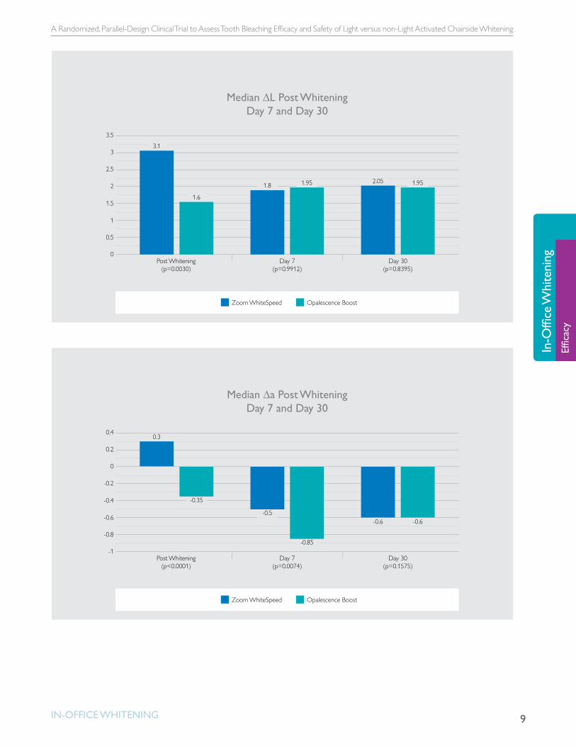
3.1
1.6
Median ∆L Post Whitening Day 7 and Day 30
Zoom WhiteSpeed Opalescence Boost
1.8 1.95 2.05 1.95
Post Whitening(p=0.0030)
Day 7(p=0.9912)
Day 30(p=0.8395)
-1
-0.6
-0.4
-0.2
0.2
0
0.4
-0.8
0.3
-0.35
Median ∆a Post Whitening Day 7 and Day 30
Zoom WhiteSpeed Opalescence Boost
-0.5
-0.85
-0.6 -0.6
Day 30(p=0.1575)
Post Whitening(p<0.0001)
Day 7(p=0.0074)

10
A Randomized, Parallel-Design Clinical Trial to Assess Tooth Bleaching Efficacy and Safety of Light versus non-Light Activated Chairside Whitening
In-OffIce WhItenIng
In-Office W
hiteningEfficacy
-5
-4
-3.5
-3
-2
-2.5
-1.5
-1
-0.5
0
-4.5
-2.8
-0.8
Median ∆b Post Whitening Day 7 and Day 30
Zoom WhiteSpeed Opalescence Boost
-4.6
-2.9
-4.15
-2.3
Post Whitening(p<0.0001)
Day 7(p=0.0019)
Day 30(p=0.0004)
0
2
3
4
6
5
7
1
Post Whitening(p<0.0001)
Day 7(p=0.0106)
Day 30(p=0.2648)
5.86 (0.18)
LSMeans VITA Classical Shade Reduction Post Whitening Day 7 and Day 30
Zoom WhiteSpeed Opalescence Boost
4.92 (0.20)
4.19 (0.20)4.47 (0.18) 4.45 (0.21)4.11 (0.21)
11
A Randomized, Parallel-Design Clinical Trial to Assess Tooth Bleaching Efficacy and Safety of Light versus non-Light Activated Chairside Whitening
In-OffIce WhItenIng
In-O
ffice
Whi
teni
ngEf
ficac
y0
40%
60%
80%
100%
90%
120%
20%
Zoom WhiteSpeed Opalescence Boost
1%
82%
16%
79%
21%
Maximum Sensitivity Experienced from Whitening Treatment
Mild Sensitivity No SensitivityModerate Sensitivity
0
1
1.5
2
3
2.5
3.5
0.5
Post Whitening(p<0.0001)
Day 7(p=0.0198)
Day 30(p=0.1195)
3.17
Median VITA Bleached Guide Shade Reduction Post Whitening Day 7 and Day 30
Zoom WhiteSpeed Opalescence Boost
2.33
1.672.0 1.83
2.25
12
Color Change of Vital Teeth Exposed to Bleaching Performed With and Without Supplementary Light
In-OffIce WhItenIng
In-Office W
hiteningBenefit of Light vs N
o Light
Color Change of Vital Teeth Exposed to Bleaching Performed With and Without Supplementary Lightin vivo studyOntiveros JC, Paravina RD. Color Change of Vital Teeth Exposed to Bleaching Performed With and Without Supplementary Light. Journal of Dentistry 2009;37:840-847.
Department of Restorative Dentistry and Biomaterials, University of Texas Dental Branch at Houston, TX, USA.
Objective:
To evaluate tooth color change after exposure to 25% hydrogen peroxide in-office tooth whitening system with and without supplementary light exposure.
Materials:
• 25% hydrogen peroxide whitening gel (Zoom! 2 Kit, Discus Dental, Inc.)
• Whitening lamp (Zoom! AP, Discus Dental, Inc.)
Methodology:
A total of 20 patients were enrolled for an in-office clinical tooth whitening study using an opposing-arch design. A total of 80 teeth were analyzed (one canine and central incisor for both arches). The order of arch (maxillary or mandibular) and treatment type (light or no light) was randomized at the patient’s initial bleaching and the opposite treatment was chosen for the second appointment. The 25% hydrogen peroxide bleaching gel (Zoom! 2 Kit, Discus Dental) was applied and repeated for three 15-minute cycles for a total exposure time of 45 minutes. Visual color matching was performed at baseline before bleaching and seven days after bleaching. Two different shade guides were used: Vitapan Classical (VC) and Vita Bleachedguide 3D-Master (BG). Evaluators were blind to the assigned intervention. Instrumental color monitoring was also performed before bleaching and seven days after bleaching using an intraoral spectrophotometer (Vita Easyshade, Vita Zahnfabrik, Bäd Sackingen, Germany). Data was analyzed using ANOVA, paired t-test, and Wilcoxon signed rank tests.
Results:
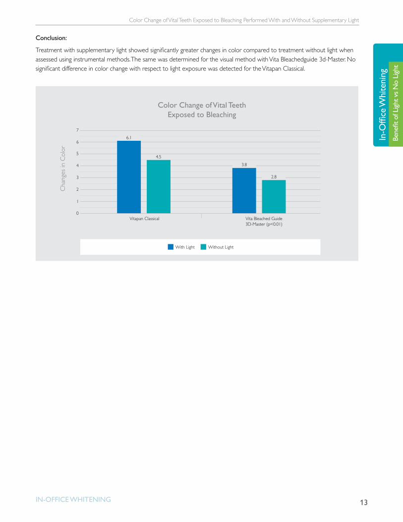
The instrumental measurement method revealed significant difference in color between treatment with light (∆E*ab = 6.0) and treatment without light (∆E*ab = 4.7) after seven days (p < 0.05). No differences were visually detected between treatment with light and without light using the VC (p = 0.56). However, a significant difference was recorded using the BG (p < 0.01). Instrumental measurements of color changes were in better accordance with visual findings using the BG guide (R2 = 0.60) rather than the VC guide (R2 = 0.20).
13
Color Change of Vital Teeth Exposed to Bleaching Performed With and Without Supplementary Light
In-OffIce WhItenIng
In-O
ffice
Whi
teni
ngBe
nefit
of L
ight v
s No
Light
Conclusion:
Treatment with supplementary light showed significantly greater changes in color compared to treatment without light when assessed using instrumental methods. The same was determined for the visual method with Vita Bleachedguide 3d-Master. No significant difference in color change with respect to light exposure was detected for the Vitapan Classical.
0
1
2
3
4
5
6
7
Vitapan Classical
Color Change of Vital Teeth Exposed to Bleaching
Cha
nges
in C
olor
Vita Bleached Guide3D-Master (p<0.01)
With Light Without Light
6.1
4.5
3.8
2.8
14
Effect of ZOOM! Advanced Power Lamp on Whitening
In-OffIce WhItenIng
In-Office W
hiteningBenefit of Light vs N
o Light
Effect of ZOOM! Advanced Power Lamp on Whiteningin vivo studyDiscus Dental, Inc., ZOOM! Advanced Power Chairside Whitening System Effect of ZOOM! Advanced Power Lamp on Whitening. The Dental Advisor. November 2007.
Objective:
To assess tooth color changes and patient satisfaction using ZOOM! Advanced Power Lamp compared to whitening treatment without the lamp.
Materials:
• 25% hydrogen peroxide gel (Zoom! Advanced Power Whitening System, Discus Dental, Inc.)
• Light activation lamp (Zoom! Advanced Power Lamp, Discus Dental, Inc.)
Methodology:
Seventeen patients received Zoom! Advanced Power Whitening System using Zoom! Advanced Power Lamp. Eleven patients received the whitening treatment without light activation. All patients had a start shade of Vita A3 or darker, and no tetracycline stains were treated. Tooth color was measured at baseline before and immediately following the in-office procedure using the Vita Classic value arranged shade guide. Shade change was measured again at 7-10 days and 30 days post-treatment. Patients were given a survey at the 30-day follow-up appointment to rate effectiveness of treatment, sensitivity, satisfaction, and overall opinion of the treatment.
Results:
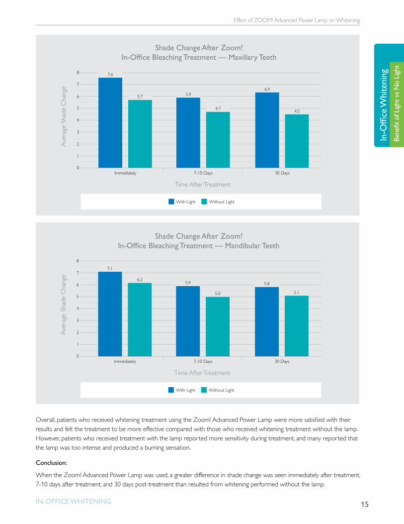
Upon evaluation immediately after whitening and 30 days post-treatment, whitening with the lamp resulted in an average of 1.9 shades lighter for maxillary teeth and 0.7 shades lighter for mandibular teeth compared to average whitening without the lamp.
15
Effect of ZOOM! Advanced Power Lamp on Whitening
In-OffIce WhItenIng
In-O
ffice
Whi
teni
ngBe
nefit
of L
ight v
s No
Light
0
2
1
4
5
Immediately
Time After Treatment
Shade Change After Zoom! In-Of�ce Bleaching Treatment — Maxillary Teeth
Ave
rage
Sha
de C
hang
e
7-10 Days 30 Days
6
7
8
3
7.6
5.7 5.9
4.7
6.4
4.5
With Light Without Light
0
2
1
4
5
Immediately
Time After Treatment
7-10 Days 30 Days
6
7
8
3
7.1
6.25.9
5.0
5.8
Ave
rage
Sha
de C
hang
e
5.1
With Light Without Light
Shade Change After Zoom! In-Of�ce Bleaching Treatment — Mandibular Teeth
Overall, patients who received whitening treatment using the Zoom! Advanced Power Lamp were more satisfied with their results and felt the treatment to be more effective compared with those who received whitening treatment without the lamp. However, patients who received treatment with the lamp reported more sensitivity during treatment, and many reported that the lamp was too intense and produced a burning sensation.
Conclusion:
When the Zoom! Advanced Power Lamp was used, a greater difference in shade change was seen immediately after treatment, 7-10 days after treatment, and 30 days post-treatment than resulted from whitening performed without the lamp.

16
Effect of Light Treatment on in vitro Tooth Bleaching Efficacy
In-OffIce WhItenIng
In-Office W
hiteningBenefit of Light vs N
o Light
Effect of Light Treatment on in vitro Tooth Bleaching Efficacyin vitro studyLi Y, Lee SS, Zheng M, Forde CA, Carino CM. Effect of Light Treatment on in vitro Tooth Bleaching Efficacy. J Dent Res 86 (Spec Iss A), 0275, 2007
Objective:
To evaluate the effects of light treatment on tooth bleaching efficacy of Zoom! 2 gel using extracted human incisors.
Materials:
• 20 extracted human incisors stored in 10% formalin solution
• Zoom! 2 Gel (Discus Dental)
• Zoom! 2 Light (Discus Dental)
Methodology:
Twenty extracted human incisors were randomized into two groups of ten each. The roots were embedded in denture resin to form a gingival contour. Each tooth was assessed for a baseline shade (Vitapan Classical Shade Guide, Vita Zahnfabrick GMbH, Sackingen, Germany) and L*a*b values (Shade Vision, X-Rite, Inc., Grandville, MI). Both groups of specimens were treated with Balancing Pre-Treatment Gel (Discus Dental) then covered with Zoom! 2 Gel (Discus Dental). Group A was exposed to Zoom! 2 Light (Discus Dental) for 15 minutes, while Group B did not receive light exposure. The gel application and light exposure (Group A) was repeated two additional times. Satin Finish Gel (Discus Dental) was applied to the enamel surfaces for five minutes after the final bleaching treatment. Post procedure Shade and L*a*b values were measured again and the data analyzed.
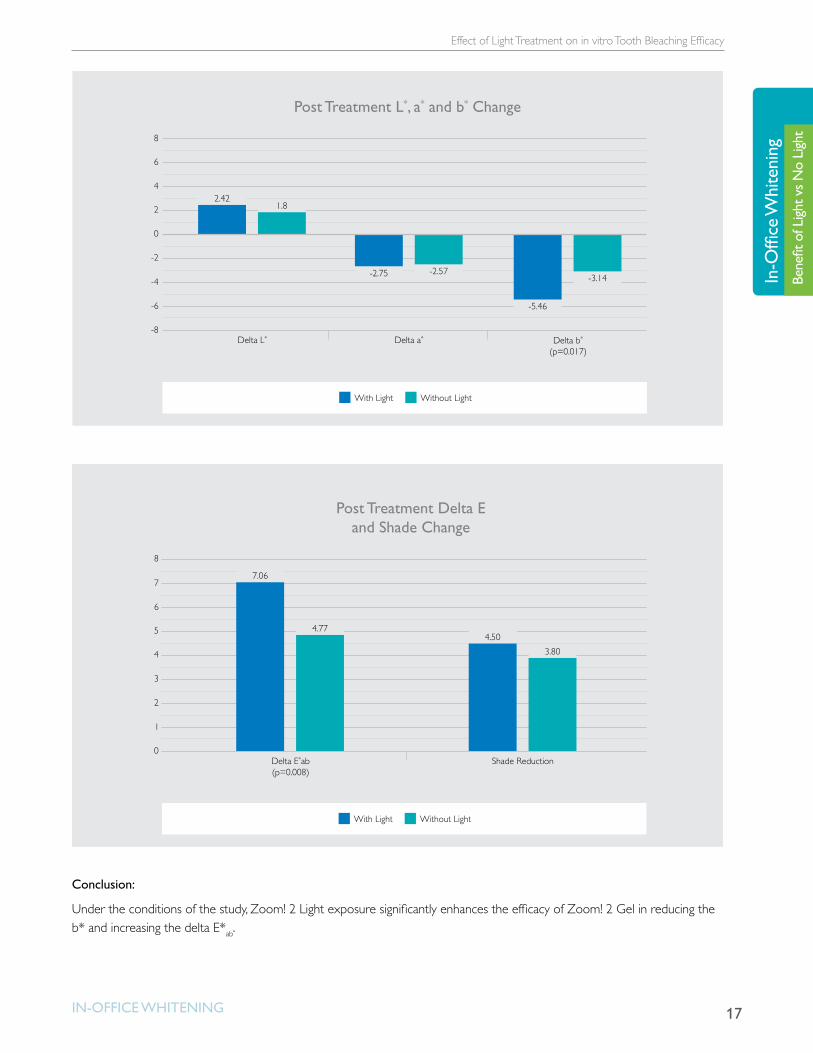
Results:
The shade reduction was 4.50 and 3.80 for Groups A and B respectively. The Shade Vision data showed significant differences between the two groups in delta b* and delta E*ab.
17
Effect of Light Treatment on in vitro Tooth Bleaching Efficacy
In-OffIce WhItenIng
In-O
ffice
Whi
teni
ngBe
nefit
of L
ight v
s No
Light
-8
-6
-4
-2
0
2
4
6
8
Delta L*
Post Treatment L*, a* and b* Change
Delta a* Delta b*
(p=0.017)
2.421.8
-2.75 -2.57
-5.46
-3.14
With Light Without Light
0
1
2
3
4
5
6
7
8
Delta E*ab(p=0.008)
Post Treatment Delta E and Shade Change
Shade Reduction
7.06
4.774.50
3.80
With Light Without Light
Conclusion:
Under the conditions of the study, Zoom! 2 Light exposure significantly enhances the efficacy of Zoom! 2 Gel in reducing the b* and increasing the delta E*ab.

18
Clinical Study to Compare Two In-Office Whitening Systems
In-OffIce WhItenIng
In-Office W
hiteningBenefit of Light vs N
o Light
Clinical Study to Compare Two In-Office Whitening Systemsin vivo studyGallagher A1, Maggio B1, Bowman J1, Felix H2, Clinical Study to Compare Two In-Office (Chairside) Whitening Systems. J Clin Dent 13:219-224, 2002.1Hill Top Research, Inc, West Palm Beach, FL, USA, 2Discus Dental, Inc, Culver City, USA
Objective:
To compare the efficacy of two in-office whitening systems: Discus Dental Zoom! Chairside System (25% hydrogen peroxide whitening gel) and Opalescence Xtra Boost Kit (38% hydrogen peroxide whitening gel).
Materials:
• 25% hydrogen peroxide whitening gel (Zoom! Discus Dental, Inc., Culver City, CA, USA)
• 38% hydrogen peroxide whitening gel (Opalescence Xtra Boost Kit, Ultradent, South Jordan, UT, USA)
Methodology:
Twenty-two healthy adults over the age of 18 were enrolled in a single-center, examiner-blind, randomized trial. At the outset of the study, all subjects had a tooth shade greater than or equal to A3 (Vita Shade guide, Vita Zahnfabrick GMbH, Sackingen, Germany) for a minimum of four of the six maxillary anterior teeth. Whitening procedures were completed using three applications per treatment according to the manufacturers’ instructions. Immediately following completion of the whitening treatment, Vita Shade assessments of the maxillary anterior teeth and chromameter shade assessments were recorded. Patients were questioned on their level of tooth sensitivity and the condition of the oral soft tissue was examined. The same procedures were performed on post-treatment Day 7.
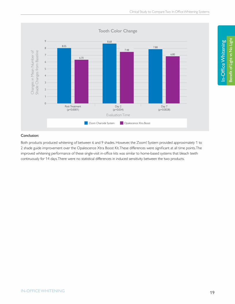
Results:
Both products achieved a statistically significant whitening from baseline (p < 0.0001) following treatment. After Day 7, mean changes of -7.8 and -6.8 shades were observed for the Zoom! System and the Opalescence Xtra Boost Kit respectively. The Zoom! Chairside System was an average of 1 to 2 shades better than the Opalescense Xtra Boost Kit at all time points. Results were directionally better at Day 2 (p < 0.08) and significantly better at Day 7 (p < 0.0025).
19
Clinical Study to Compare Two In-Office Whitening Systems
In-OffIce WhItenIng
In-O
ffice
Whi
teni
ngBe
nefit
of L
ight v
s No
Light
7
8
9
0
2
1
4
3
6
5
Post-Treatment(p<0.0001)
Evaluation Time
Tooth Color ChangeC
hang
es in
Mea
n N
umbe
r of
Shad
e C
hang
es fr
om B
asel
ine
Day 2(p=0.034)
Day 7(p=0.0028)
8.05
6.35
8.68
7.487.88
6.80
Zoom Chairside System Opalescence Xtra Boost
Conclusion:
Both products produced whitening of between 6 and 9 shades. However, the Zoom! System provided approximately 1 to 2 shade guide improvement over the Opalescence Xtra Boost Kit. These differences were significant at all time points. The improved whitening performance of these single-visit in-office kits was similar to home-based systems that bleach teeth continuously for 14 days. There were no statistical differences in induced sensitivity between the two products.

20
Clinical Evaluation of a Novel Dental Whitening Lamp and Light-Catalyzed Peroxide Gel
In-OffIce WhItenIng
In-Office W
hiteningBenefit of Light vs N
o Light
Clinical Evaluation of a Novel Dental Whitening Lamp and Light-Catalyzed Peroxide Gelin vivo studyZiemba SL1, Felix H1, MacDonald J1, and Ward M2, Clinical evaluation of a novel dental whitening lamp and light catalyzed peroxide gel. J Clin Dent 16:123-127, 2005.1Discus Dental, Inc, Culver City, USA, 2Houston, TX, USA
Objective:
To determine whether an ultraviolet light enhanced the whitening efficacy of a peroxide gel containing a photo-Fenton activator
Materials:
• 20% hydrogen peroxide gel with ultraviolet (UV) light (Zoom2, Discus Dental Inc.)
• 20% hydrogen peroxide gel without ultraviolet (UV) light
Methodology:
Fifty healthy male and female adults aged 18-70 years were enrolled into an IRB-approved randomized trial. The trial was conducted at two geographically separate study sites. At the outset of the study, all subjects had a tooth shade greater than or equal to A3 (Vita Shade guide, Vita Zahnfabrick GMbH, Sackingen, Germany) for all six non-restored maxillary anterior teeth. Participants had to agree not to use any other dental whitening product except toothpaste during the course of the study. Individuals also had to refrain from smoking and consuming coffee, cola drinks, grape juice and other food or drink that could stain teeth for seven days after treatment.
Subjects who met the inclusion criteria were randomly assigned to one of two groups: Light or No-light. The investigator applied the bleaching gel (Zoom 2 Discus Dental, Inc.) to the teeth of both groups for 15 minutes. At the end of the period, the gel was suctioned off and new gel was applied. This process was repeated twice more for a total of three applications for all study subjects. In the Light group, the six maxillary teeth were concurrently exposed to the whitening lamp during the gel application for a total light exposure of 45 minutes. The No-light group was not treated with the whitening lamp. Fluoride/potassium nitrate was applied to teeth according to manufacturer’s instructions for all subjects.
Subjects were examined before the whitening treatment, immediately after treatment (same day), one week after treatment and one month after treatment. Clinical data were collected on the gingival index, shade score and self-assessed dentinal hypersensitivity.
21
Clinical Evaluation of a Novel Dental Whitening Lamp and Light-Catalyzed Peroxide Gel
In-OffIce WhItenIng
In-O
ffice
Whi
teni
ngBe
nefit
of L
ight v
s No
Light
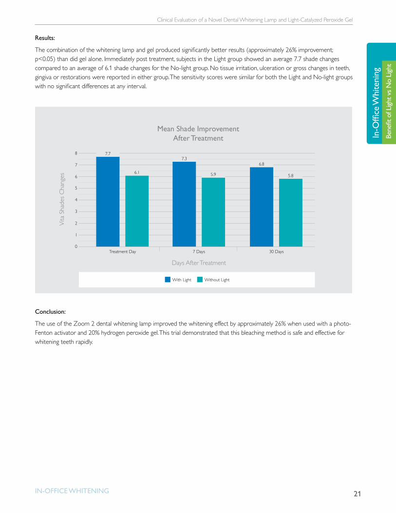
Results:
The combination of the whitening lamp and gel produced significantly better results (approximately 26% improvement; p<0.05) than did gel alone. Immediately post treatment, subjects in the Light group showed an average 7.7 shade changes compared to an average of 6.1 shade changes for the No-light group. No tissue irritation, ulceration or gross changes in teeth, gingiva or restorations were reported in either group. The sensitivity scores were similar for both the Light and No-light groups with no significant differences at any interval.
0
2
1
4
5
Treatment Day
Days After Treatment
Mean Shade Improvement After Treatment
Vita
Sha
des
Cha
nges
7 Days 30 Days
6
7
8
3
7.77.3
6.8
With Light Without Light
6.1 5.9 5.8
Conclusion:
The use of the Zoom 2 dental whitening lamp improved the whitening effect by approximately 26% when used with a photo-Fenton activator and 20% hydrogen peroxide gel. This trial demonstrated that this bleaching method is safe and effective for whitening teeth rapidly.
22
Clinical Study of an In-Office Whitening System With and Without Light Procedures Utilizing a Split-Arch Design
In-OffIce WhItenIng
In-Office W
hiteningBenefit of Light vs N
o Light
Clinical Study of an In-Office Whitening System With and Without Light Procedures Utilizing a Split-Arch Designin vitro studyMaggio B, Bowman JP, Felix H, Borden L, and Mason S. Clinical study of an In-Office Whitening System With and Without Light. J Dent Res 82 (Spec Iss A), 1031, 2003.
Objective:
To evaluate the bleaching efficacy of 25% hydrogen peroxide whitening gel (Discus Dental Zoom) with and without light activation by comparing tooth shade changes measured with Vita Shade Guide following treatment using a split-arch design.
Materials:
• 25% hydrogen peroxide whitening gel (Zoom, Discus Dental, Culver City, CA, USA)
• Light activator (Zoom Light Activator, Discus Dental, Culver City, CA, USA)
Methodology:
In this examiner-blind randomized pilot study, a split-arch design was used to compare an in-office teeth whitening product with and without light activation. Ten healthy adult subjects (five male and five female) were randomized into two groups to receive the light activator on the right or left side of the maxillary anterior teeth during treatment. One half of the maxillary anterior sextant was treated with light, and following isolation, the second half was treated without the light. Two separate rounds consisting of three whitening treatments apiece were conducted on each subject. Vita Shade scores were collected at baseline prior to treatment, immediately following treatment on the sample day, and one and seven days post-treatment.
Results:
The Zoom Chairside System with Zoom Light Activator was significantly better than the product application without the light at all time points: post-treatment (p = 0.0026); Day 1 (p > 0.0001); Day 7 (p = 0.0002).
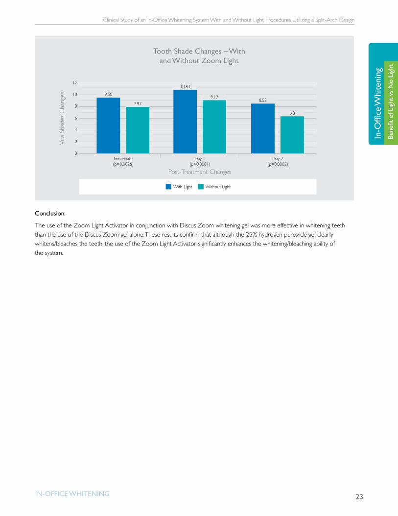
23
Clinical Study of an In-Office Whitening System With and Without Light Procedures Utilizing a Split-Arch Design
In-OffIce WhItenIng
In-O
ffice
Whi
teni
ngBe
nefit
of L
ight v
s No
Light
0
2
4
6
8
10
12
Immediate(p=0.0026)
Post-Treatment Changes
Tooth Shade Changes – With and Without Zoom Light
Vita
Sha
des
Cha
nges
Day 1(p>0.0001)
Day 7(p=0.0002)
9.50
7.97
10.83
9.178.53
6.3
With Light Without Light
Conclusion:
The use of the Zoom Light Activator in conjunction with Discus Zoom whitening gel was more effective in whitening teeth than the use of the Discus Zoom gel alone. These results confirm that although the 25% hydrogen peroxide gel clearly whitens/bleaches the teeth, the use of the Zoom Light Activator significantly enhances the whitening/bleaching ability of the system.
24
Photo-Fenton and Conventional In-Office Dental Bleaching
In-OffIce WhItenIng
In-Office W
hiteningBenefit of Light vs N
o Light
Photo-Fenton and Conventional In-Office Dental Bleachingin vivo studyCardoso PE, Muench A, and Pinheiro HB, Universidade de São Paulo, São Paulo, Brazil
FDI World Dental Congress, 2011 Poster #348
Objective:
To evaluate the results of two in-office whitening treatment methods, one based on the Photo-Fenton reaction and the other a conventional in-office system, verifying shade change (ΔE) and color stability.
Materials:
• 25% hydrogen peroxide with ferrous gluconate (Discus Dental) and ultraviolet (UV) light (Zoom AP Light, Discus Dental)
• 38% hydrogen peroxide (Opalescence Xtra Boost, Ultradent)
Methodology:
Forty healthy adult volunteers were randomly divided into 2 experimental groups of 20 subjects each (Group 1: ZAP; Group 2: OPX). The whitening treatment for both groups was performed with three consecutive 15-minute applications. The ZAP group received treatment with 25% hydrogen peroxide and ferrous gluconate and ultraviolet (UV) light (Discus Dental and Zoom AP Light); the OPX group received treatment with 38% hydrogen peroxide (Opalescence Xtra Boost, Ultradent).
Experimental Groups Description
ZAP (n=20)25% hydrogen peroxide with ferrous gluconate ultraviolet (UV) light (Discus Dental +Zoom AP Light) – 1 treatment session
OPX (n=20) 38% HP-Opalescence Xtra Boost (Ultradent) – 1 treatment session
The shade of superior incisors and canines was assessed for each subject using a digital Vita-Easyshade Spectrophotometer immediately before and after the whitening treatment at 7, 14 and 30 days.
25
Photo-Fenton and Conventional In-Office Dental Bleaching
In-OffIce WhItenIng
In-O
ffice
Whi
teni
ngBe
nefit
of L
ight v
s No
Light
The Vita-Easyshade device measures the color based on a tridimensional system that supplies numerical values, which are inserted into a formula to provide color or shade variation (also known as ∆E). A custom clear EVA tray was used to ensure that measurements were taken at the same spot on each tooth. Holes in the tray on the labial surface of incisors and canines were made with a specially designed six mm bur corresponding to the size of the tip of the optical spectrophotometer reader.
Results:
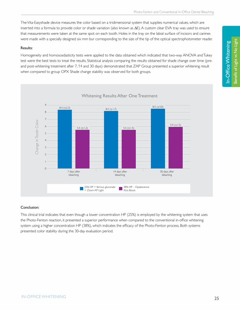
Homogeneity and homoscedasticity tests were applied to the data obtained which indicated that two-way ANOVA and Tukey test were the best tests to treat the results. Statistical analysis comparing the results obtained for shade change over time (pre- and post-whitening treatment after 7, 14 and 30 days) demonstrated that ZAP Group presented a superior whitening result when compared to group OPX Shade change stability was observed for both groups.
0
2
1
4
3
6
5
7
7 days after bleaching
5.5 (±1.2)
8.2 (±1.7)
Whitening Results After One Treatment
Cha
nge
in To
oth
Col
or
25% HP + ferrous gluconate + Zoom AP Light
38% HP – Opalescence Xtra Boost
14 days after bleaching
30 days after bleaching
8
98.4 (±2.3)
5.5 (±1.5)
8.5 (±3.0)
5.9 (±1.5)
Conclusion:
This clinical trial indicates that even though a lower concentration HP (25%) is employed by the whitening system that uses the Photo-Fenton reaction, it presented a superior performance when compared to the conventional in-office whitening system using a higher concentration HP (38%), which indicates the efficacy of the Photo-Fenton process. Both systems presented color stability during the 30-day evaluation period.
26
Clinical Trial: Photo-Fenton and Conventional In-Office Dental Bleaching
In-OffIce WhItenIng
In-Office W
hiteningBenefit of Light vs N
o Light
Clinical Trial: Photo-Fenton and Conventional In-Office Dental Bleachingin vivo studyCardoso PE, Muench A, and Pinheiro HB, Universidade de São Paulo, São Paulo, Brazil
Academy of Dental Materials Meeting, 2011
Objective:
To evaluate the efficacy of two in-office dental bleaching methods on shade change and color stability (∆E).
Materials:
• 25% hydrogen peroxide with light activation (Zoom2 + Zoom AP Light, Discus Dental, Inc.)
• 38% hydrogen peroxide (Opalescence Xtra Boost, Ultradent, Inc.)
Methodology:
Sixty healthy adult volunteers were randomly divided into three experimental groups of 20 subjects each. The groups were treated as follows: the ZAP1 group received one treatment with 25% hydrogen peroxide and light activation (Zoom2 + Zoom AP Light, Discus Dental); the OPX2 group received two treatments with 38% hydrogen peroxide (Opalescence Xtra Boost, Ultradent); the OPX3 group received one treatment with 38% hydrogen peroxide (Opalescence Xtra Boost, Ultradent). Each treatment consisted of three applications of bleaching gel for 15 minutes each.
Experimental Groups Description
ZAP1 (n=20)25% hydrogen peroxide with light (Zoom2 + Zoom AP Light, Discus Dental) – 1 treatment session
OPX2 (n=20) 38% HP-Opalescence Xtra Boost (Ultradent) – 2 treatment sessions
OPX1 (n=20) 38% HP-Opalescence Xtra Boost (Ultradent) – 1 treatment session
27
Clinical Trial: Photo-Fenton and Conventional In-Office Dental Bleaching
In-OffIce WhItenIng
In-O
ffice
Whi
teni
ngBe
nefit
of L
ight v
s No
Light
The shade of superior incisors and canines was assessed for each subject using a digital Vita-Easyshade Spectrophotometer immediately before and after the whitening treatment at 7, 14 and 30 days.
The Vita-Easyshade device measures the color based on a tridimensional system that supplies numerical values, which are inserted into a formula to provide color or shade variation (also known as ∆E). A custom clear EVA tray was used to ensure that measurements were taken at the same spot on each tooth. Holes in the tray on the labial surface of incisors and canines were made with a specially designed 6 mm bur corresponding to the size of the tip of the optical spectrophotometer reader.
Results:
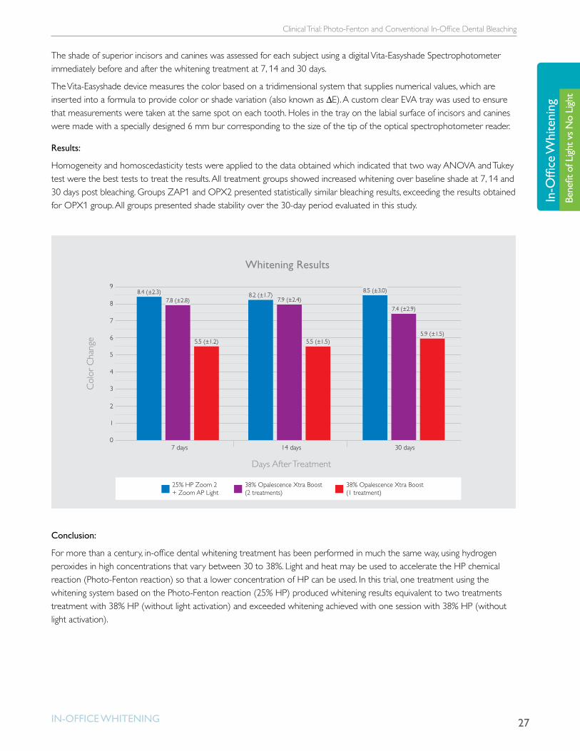
Homogeneity and homoscedasticity tests were applied to the data obtained which indicated that two way ANOVA and Tukey test were the best tests to treat the results. All treatment groups showed increased whitening over baseline shade at 7, 14 and 30 days post bleaching. Groups ZAP1 and OPX2 presented statistically similar bleaching results, exceeding the results obtained for OPX1 group. All groups presented shade stability over the 30-day period evaluated in this study.
0
2
1
4
5
7 days
Days After Treatment
Whitening Results
Col
or C
hang
e
14 days 30 days
6
7
8
9
3
25% HP Zoom 2 + Zoom AP Light
38% Opalescence Xtra Boost(2 treatments)
38% Opalescence Xtra Boost(1 treatment)
8.4 (±2.3)7.8 (±2.8)
5.5 (±1.2)
8.2 (±1.7)7.9 (±2.4)
5.5 (±1.5)
8.5 (±3.0)
7.4 (±2.9)
5.9 (±1.5)
Conclusion:
For more than a century, in-office dental whitening treatment has been performed in much the same way, using hydrogen peroxides in high concentrations that vary between 30 to 38%. Light and heat may be used to accelerate the HP chemical reaction (Photo-Fenton reaction) so that a lower concentration of HP can be used. In this trial, one treatment using the whitening system based on the Photo-Fenton reaction (25% HP) produced whitening results equivalent to two treatments treatment with 38% HP (without light activation) and exceeded whitening achieved with one session with 38% HP (without light activation).

28
The Basic Chemistry Behind Hydrogen Peroxide Tooth Whitening
In-OffIce WhItenIng
In-Office W
hiteningBenefit of Light vs N
o Light
The Basic Chemistry Behind Hydrogen Peroxide Tooth Whiteningin vitro studyYoung N, Fairley P, Mohan V, Jumeaux C. The chemistry behing hydrogen peroxide tooth whitening, J Dent Res 91(Spec Iss B):147, 20012 (www.dentalresearch.org).
Philips Research Laboratories, Cambridge, UK
Objective:
To examine the basic interactions between whitening agents and stain molecules in simple solutions and to give clarity on the basic chemistry and photochemistry that occurs during the process
Materials:
• Black tea stain solution
• Whitening agents of various compositions including hydrogen peroxide, ferrous gluconate, and potassium hydroxide (based on Zoom treatment, Discus Dental, Inc., Culver City, CA, USA)
• Blue light (465nm)
• Infrared light (850nm)
Methodology:
The absorbance of tea stain solution at 450nm was measured over a period of 40 minutes, with various compositions of whitening agent added (including hydrogen peroxide, ferrous gluconate and potassium hydroxide in the formulations) and at the same time the samples were subjected to blue light (465nm) or infra-red light (850nm) irradiation, or alternatively were heated.
Results:
The reaction rates between chromophores in the tea solution and hydrogen peroxide can be accelerated significantly using ferrous gluconate activator and blue light irradiation. Infra-red irradiation was not found to increase the reaction rate through photochemistry but increases the temperature. While raising the temperature can give a slight increase in reaction rate, it can easily lead to inefficiency through the acceleration of exothermic decomposition reactions of hydrogen peroxide.

29
The Basic Chemistry Behind Hydrogen Peroxide Tooth Whitening
In-OffIce WhItenIng
In-O
ffice
Whi
teni
ngBe
nefit
of L
ight v
s No
Light
0.0
0.2
10 20 30 40
0.4
0.6
Time (Minutes)
Abs
orba
nce
at 4
50nm
(Rel
ativ
e)
0.8
1.0
Effect of Ferrous Activators and Blue Light Irradiation on Whitening Process
465nm blue at 50mW/cm2 with FeG Dark without FeG
Conclusion:
By carrying out work in simple solution, it was possible to separate the basic chemistry of tooth whitening from the complex physical processes which occur in the tooth during whitening. Ferrous activators and blue light irradiation were shown unambiguously to significantly enhance the whitening process, whereas infrared irradiation or heating has a smaller effect.
30
Novel Method for Efficacy Assessment of Whitening Agents
In-OffIce WhItenIng
In-Office W
hiteningEfficacy Novel Method for Efficacy Assessment of Whitening Agents
in vitro studyCardoso PEC, Barros RMC, Marquez UML, Cardoso J. Novel Method for Efficacy Assessment of Whitening Agents. J Dent Res 89 (Spec Iss A), 0835, 2010.
Objective:
To evaluate the whitening efficacy of four in-office whitening systems using a direct measurement method, which evaluated the discoloration of a stain solution prepared with the most common food color used in beverages and industrial food
Materials:
• Caramel solution
• 25% hydrogen peroxide
• 25% hydrogen peroxide + ferrous complex
• UVA lamp
Methodology:
A caramel solution (CS) was prepared in water, using Caramel Class IV AP 100 (Sethness Products Company, USA), at a proportion of 0.1% in weight (w/v). Five experimental groups were designed: Group 1. Control: caramel solution (CS) not submitted to any discoloration process; Group 2. CS+HP 25% (Lase Peroxide, DMC, Brazil); Group 3. CS+HP 25% + visible light LED for 45 min (480nm, Whitening Lase Plus, DMC, Brazil); Group 4. CS+HP 25% and Fe2+ (Zoom 2 gel, Discus Dental, USA); 5. CS + HP 25% and Fe2+ + UVA light (360-400nm) during 45min (Zoom AP lamp, Discus Dental). Five repetitions were conducted for each experimental group and the results were measured using a Shimadzu spectrophotometer UV-1650PC (Shimadzu Scientific Instruments, Japan) and converted into a percent that indicates the color that remained in the solution.
Results:
One-Way ANOVA showed all groups were statistically different except for Groups 1 and 2, which were statistically similar.

31
Novel Method for Efficacy Assessment of Whitening Agents
In-OffIce WhItenIng
In-O
ffice
Whi
teni
ngEf
ficac
y
0
25
50
75
Ef�cacy Assessment ofWhitening Agents
Col
or R
emai
ning
in S
olut
ion
Control (Caramel solution)
25% HP
100
HP 25% + Visible Light
25% HP + Fe2+ 25% HP + Fe2+ + UVA
100% 99%
83%
37%
73%
Conclusion:
The 25% hydrogen peroxide gel associated with ferrous complex reached higher discoloration than the 25% hydrogen peroxide gel. The 25% hydrogen peroxide gel associated with ferric complex submitted to UVA light emission offered the highest whitening potential when compared to all the other groups.
32
Light-Activated System for Bleaching Teeth
In-OffIce WhItenIng
In-Office W
hiteningEfficacy Light-Activated System for Bleaching Teeth
in vivo studyWard M1, Ziemba SL2, Fleix H2. Light-Activated System for Bleaching Teeth. Academy of Dental Materials Meeting, October 23-25, 2006, São Paulo, Brazil.1Private Practice, Houston, TX, USA, 2Discus Dental, Inc., Culver City, CA, USA
Objective:
To examine the Zoom! Advanced Power light-assisted dental bleaching system, which combines a light source that emits strongly in the ultraviolet and blue-visible light wavelengths, with a 25% hydrogen peroxide gel.
Materials:
• 25% hydrogen peroxide gel (Zoom! Advance Power whitening gel, Discus Dental, Inc.)
• Light source (Zoom! Advanced Power whitening lamp, Discus Dental, Inc.)
Methodology:
Thirty-two healthy male and female patients age 18 to 70 years were enrolled in at a single site. Prior to treatment, all patients had a tooth shade greater than or equal to A3 for all six non-restored maxillary anterior teeth. After isolation of gingival and other oral soft tissue, 25% hydrogen peroxide gel was applied to the teeth, and the teeth were exposed to the Zoom! Advanced Power whitening lamp (Discus Dental, Inc., Culver City, CA, USA). After 15 minutes, the gel was removed and replaced twice with fresh gel and exposed to the light for three 15-minute sessions, for a total of 45 minutes. Vita Shades were assessed immediately before and after the treatment by the same examiner. A t-test for paired samples, using α = 0.05, was employed to compare shade changes (SPSS 11.0.1).
Results:
Overall, the 32 patients had an average Vita shade of D3 prior to treatment and A1 after treatment, for an average reduction of 7.8 Vita shades. One week after treatment, patients lost an average of 0.4 shades, making the average shade B2. Thus, the average shade change after one week was 7.4 shades. Importantly, 50% of all study patients achieved an ending shade of B1 or lighter immediately after treatment. Those patients who started with an average shade of A3, and had an average ending shade lighter than B1, had an average reduction of 8.3 shades Patients who did not achieve an ending shade of B1 or lighter had an average starting shade of B3, and in them we saw an ending shade of B2; an average reduction of 7.7 shades.
33
Light-Activated System for Bleaching Teeth
In-OffIce WhItenIng
In-O
ffice
Whi
teni
ngEf
ficac
y
0
2
1
4
5
Starting Shade
Power Shade Change
Vita
Sha
de C
hang
es
Ending Shade Shade Changes
6
7
8
3
9
10
10.4
2.6
7.8
0
2
4
6
8
10
12
Starting Shade
Power Shade Change (For Patients Whose Final Shade was B1 or Lighter)
Darker than B1 B1 or lighter
Ending Shade Shade Changes
11.2
9.6
2.6
1.3
7.28.3
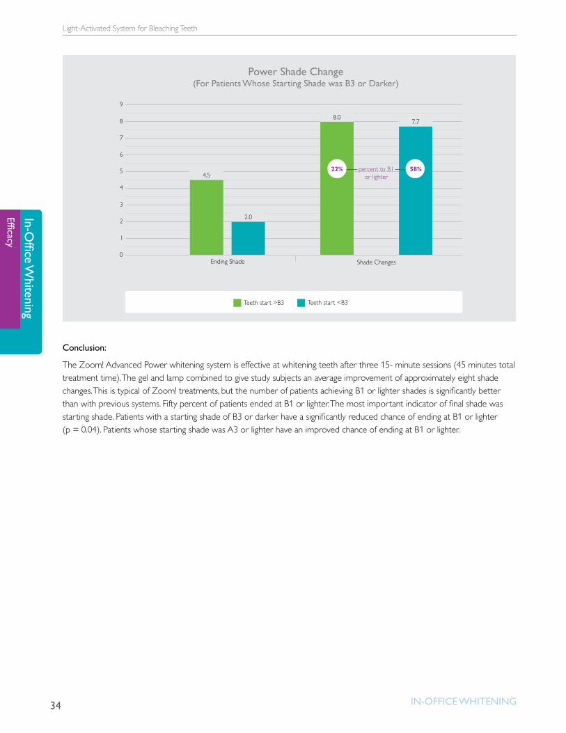
34
Light-Activated System for Bleaching Teeth
In-OffIce WhItenIng
In-Office W
hiteningEfficacy
0
1
2
3
4
5
6
7
Ending Shade
percent to B1or lighter
Shade Changes
8
9
22% 58%
Teeth start >B3 Teeth start <B3
Power Shade Change (For Patients Whose Starting Shade was B3 or Darker)
4.5
8.07.7
2.0
Conclusion:
The Zoom! Advanced Power whitening system is effective at whitening teeth after three 15- minute sessions (45 minutes total treatment time). The gel and lamp combined to give study subjects an average improvement of approximately eight shade changes. This is typical of Zoom! treatments, but the number of patients achieving B1 or lighter shades is significantly better than with previous systems. Fifty percent of patients ended at B1 or lighter. The most important indicator of final shade was starting shade. Patients with a starting shade of B3 or darker have a significantly reduced chance of ending at B1 or lighter (p = 0.04). Patients whose starting shade was A3 or lighter have an improved chance of ending at B1 or lighter.
35
Clinical Evaluation Comparing Two H202 Concentrations Used with a Light-Assisted Chairside Tooth Whitening System
In-OffIce WhItenIng
In-O
ffice
Whi
teni
ngEf
ficac
y
Clinical Evaluation Comparing Two H202 Concentrations Used with a Light-Assisted Chairside Tooth Whitening System in vivo studyWard M1, Felix H2, Clinical Evaluation Comparing two H202 Concentrations Used with a Light-Assisted Chairside Tooth Whitening Systems. Compendium 33(4): 286-291, 2012.1Private Practice, Houston, TX, USA, 2Discus Dental, Inc., Culver City, CA, USA
Objective:
To assess the efficacy of two concentrations of hydrogen peroxide (H202) gels in a split-arch protocol in a clinical setting when used in conjunction with the BriteSmile BS4000 light.
Materials:
• 15% hydrogen peroxide whitening gel
• 25% hydrogen peroxide whitening gel (Philips Discus Dental, Inc., Culver City, CA, USA)
• Chairside whitening lamp (BriteSmile BS4000, Philips Discus Dental, Inc., Culver City, CA, USA)
Methodology:
Fifteen healthy adults over age 18 with a tooth shade greater than or equal to A3 (Vita Shade guide, Vita Zahnfabrick GMbH, Sackingen, Germany) for all six nonrestored maxillary anterior teeth were enrolled in a single-center trial. Tooth shade was assessed before whitening treatment, immediately after whitening treatment (same day), and one week after treatment. All study subjects were treated with three 20-minute exposures of the BriteSmile BS4000 lamp with 15% H202 on three of the teeth and 25% H202 on the other three teeth. The gel was removed and reapplied between light exposures. Total exposure time was 60 minutes. Subjects were treated with ACP gel at the conclusion of treatment.
Results:
Both groups had significant lightening immediately after treatment. Changes in tooth shade were better for the teeth treated with 25% H2O2 (average of 8.0 shade changes) than the teeth treated with 15% H2O2 (average of 7.6 shade changes). The same held true one week post treatment (7.4 shade changes with 25% H2O2 versus 7.3 shade changes with 15% H2O2). However, the difference in shade change was not statistically significant immediately or at one week post treatment. There was no significant difference in tooth sensitivity scores between the two groups.

36
Light-Activated System for Bleaching Teeth
In-OffIce WhItenIng
In-Office W
hiteningEfficacy
0
2
4
6
8
10
Before Treatment
Mean Shade Guide Unit Measurement
25% Hydrogen Peroxide 15% Hydrogen Peroxide
Post Treatment Post Treatment1 week
10.2 10.2
2.2 2.6 2.8 2.9
Conclusion:
In-office treatment using a chairside whitening lamp and either 25% H2O2 or 15% H2O2 is a safe and effective method for whitening teeth within one hour.

Influence of Bioactive Materials on Whitened Human Enamel Surface
37Take-Home WHiTening
Take
-Hom
e W
hite
ning
Bene
fits o
f AC
P
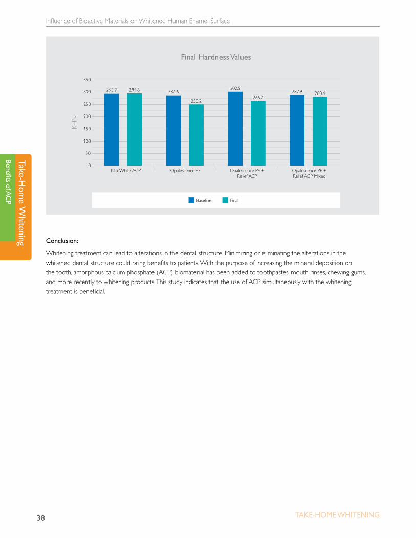
Influence of Bioactive Materials on Whitened Human Enamel Surfacein vitro studyPinheiro HB, Cardoso PEC, Universidade de São Paulo, São Paulo, Brazil
Academy of Dental Materials Meeting, 2011
Objective:
To investigate the influence of bioactive materials on whitened human enamel surface using Knoop hardness test
Materials:
• Five human teeth
• 16% carbarmide peroxide, potassium nitrate, fluoride (Opalescence PF, Ultradent)
• 16% carbamide peroxide 16%, potassium nitrate, fluoride, calcium, phosphate (NiteWhite ACP, Discus Dental)
• 15% carbamide peroxide 15%, potassium nitrate, fluoride + potassium nitrate, fluoride, calcium, phosphate (Opalescence PF, Ultradent + Relief ACP, Discus Dental)
• 15% carbamide peroxide, potassium nitrate, fluoride + potassium nitrate, fluoride, calcium, phosphate (Opalescence PF, Ultradent & Relief ACP, Discus Dental)
Methodology:
Five human teeth were sectioned into four slices per tooth. Whitening treatments were performed for 14 days according to manufacturers’ instructions. Six Knoop hardness measures were taken for each specimen, three before and three after treatments. The data were compared by Student’s t-test (α=0.01).
Results:
OPF and OPF + Relief ACP presented statistically significant hardness decrease; NiteWhite ACP and OPF & Relief ACP mixed at the time of application showed that enamel hardness was maintained.
Influence of Bioactive Materials on Whitened Human Enamel Surface
38 Take-Home WHiTening
Take-Hom
e Whitening
Benefits of ACP
0
100
150
200
300
250
350
50
NiteWhite ACP Opalescence PF Opalescence PF + Relief ACP
Opalescence PF + Relief ACP Mixed
293.7 294.6 287.6
250.2
302.5
266.7287.9 280.4
Final Hardness Values
KHN
Baseline Final
Conclusion:
Whitening treatment can lead to alterations in the dental structure. Minimizing or eliminating the alterations in the whitened dental structure could bring benefits to patients. With the purpose of increasing the mineral deposition on the tooth, amorphous calcium phosphate (ACP) biomaterial has been added to toothpastes, mouth rinses, chewing gums, and more recently to whitening products. This study indicates that the use of ACP simultaneously with the whitening treatment is beneficial.

39Take-Home WHiTening
Take
-Hom
e W
hite
ning
Bene
fits o
f AC
P
Influence of Five Home Whitening Gels and a Remineralizing Gel on the Enamel and Dentin Ultrastructure and Hardness
Influence of Five Home Whitening Gels and a Remineralizing Gel on the Enamel and Dentin Ultrastructure and Hardnessin vitro studyPinheiro HB, Cardoso PEC. Influence of Five Home Whitening Gels and a Remineralizing Gel on the Enamel and Dentin Ultrastructure and Hardness. Am J Dent 2011;24:131-137.
Universidade de São Paulo, São Paulo, Brazil
Objective:
To investigate the influence of calcium phosphate-enhanced home whitening agents on human enamel and dentin surface microhardness and ultramorphology.
Materials:
• Ten intact molar crowns
• 15% carbamide peroxide + potassium nitrate + fluoride (Opalescence PF, Ultradent)
• 16% carbamide peroxide + potassium nitrate + fluoride (Whiteness Perfect, FGM)
• Potassium nitrate + fluoride + calcium + phosphate (Relief ACP, Discus Dental)
• 16% carbamide peroxide + potassium nitrate + calcium + phosphate (NiteWhite ACP, Discus Dental)
• 7.5% hydrogen peroxide + potassium nitrate + calcium + phosphate (DayWhite ACP, Discus Dental)
• 7.5% hydrogen peroxide + potassium nitrate + fluoride + calcium (White Class Ca, FGM)
Methodology:
Five intact molar crowns were used for ultrastructural analysis and five for microhardness tests. Each resulting coronal structure was cut in slices. After measuring the baseline Knoop Hardness Number (KHN) of the enamel and dentin, the slices were divided into six experimental groups and one control group (n=5). The groups were as follows: G1 = 15% CP; G2 = 16% CP; G3 = Ca and PO4; G4 = 16% CP with Ca and POH4; G6 = 7.5% HP with Ca.
Results:
Conventional whitening agents (G1, G2) and the gel with calcium (G6) cause KHN decrease (p = <0.05). The remineralizing and whitening agents with calcium and phosphate (G3, G4, G5) did not change KHN. A change in morphology was observed on dentin surfaces in G1, G2, and G5.

Influence of Five Home Whitening Gels and a Remineralizing Gel on the Enamel and Dentin Ultrastructure and Hardness
40 Take-Home WHiTening
Take-Hom
e Whitening
Benefits of ACP
0
100
150
200
300
250
350
50
NiteWhite ACPDayWhite ACP Opalescence PF Relief ACP White Class CaWhiteness Perfect
320.7 316.2 315.7278.5
314.8291.2 289.7
Mean Enamel Hardness Before and After Treatment
KHN
Baseline Final
313.7 316.8 315.7310.1308.9
0
20
30
40
60
50
10
NiteWhite ACPDayWhite ACP Opalescence PF Relief ACP White Class CaWhiteness Perfect
53.7 52.648.2
44.1 46.5
34.8
44.8
Mean Dentin Hardness Before and After Treatment
KHN
Baseline Final
51.7 50.352.1
48.951.7
Conclusion:
The results indicated that the gels with calcium and phosphate added did not change the superficial enamel and dentin hardness nor did it change the tooth morphology. The conventional whitening gels that did not have remineralizing agents added to the formulation showed decreases on superficial hardness of enamel and dentin as well as morphological changes on tooth structure.
Effect of Relief ACP on Dentin Microhardness and Surface Morphology
41Take-Home WHiTening
Take
-Hom
e W
hite
ning
Bene
fits o
f AC
P
Effect of Relief ACP on Dentin Microhardness and Surface Morphologyin vitro studyLi Y, Lu H, Zhang W, Hou J, Devaraj A. Effect of Relief ACP on Dentin Microhardness and Surface Morphology. J Dent Res 86 (Spec Iss A), 1776, 2007.
Objective:
To investigate effects of Relief ACP on dentin microhardness and surface morphology of extracted human teeth compared to that of Satin Finish.
Materials:
• 20 dentin specimens
• Relief ACP (Discus Dental)
• Satin Finish (Discus Dental)
Methodology:
Twenty dentin specimens were prepared by grinding the enamel from the surface of human molars until the dentin was exposed. The sample surface was measured for Knoop Hardness Number (KHN) with a Leco Microhardness Tester (M-400-H1, St, Joseph, MI). The specimens were randomly assigned to three groups. Group A (N=4) served as the Control (100% humidity). Group B (N=8) was treated with Relief ACP (Discus Dental), while Group C (N=8) received treatments with Satin Finish (Discus Dental). The samples received 28 treatments of 30 minutes each. Prior to each treatment the samples were immersed in pooled human saliva for 20 minutes. The KHN was measured after the last treatment, and the specimens were then processes for the SEM evaluation. The KHN data were analyzed using the One-way ANOVA and Student-Newman-Keuls methods.
Effect of Relief ACP on Dentin Microhardness and Surface Morphology
42 Take-Home WHiTening
Take-Hom
e Whitening
Benefits of ACP
Results:
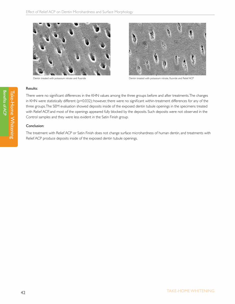
There were no significant differences in the KHN values among the three groups before and after treatments. The changes in KHN were statistically different (p=0.032); however, there were no significant within-treatment differences for any of the three groups. The SEM evaluation showed deposits inside of the exposed dentin tubule openings in the specimens treated with Relief ACP, and most of the openings appeared fully blocked by the deposits. Such deposits were not observed in the Control samples and they were less evident in the Satin Finish group.
Conclusion:
The treatment with Relief ACP or Satin Finish does not change surface microhardness of human dentin, and treatments with Relief ACP produce deposits inside of the exposed dentin tubule openings.
Dentin treated with potassium nitrate and fluoride Dentin treated with potassium nitrate, fluoride and Relief ACP

Fluoride and Potassium Nitrate-Fluoride Whitening Agents: in vitro Caries Study
43Take-Home WHiTening
Take
-Hom
e W
hite
ning
Bene
fits o
f AC
P
Fluoride and Potassium Nitrate-Fluoride Whitening Agents: in vitro Caries Study in vitro studyHicks J and Flaitz C. Effects of Whitening Agents with ACP-Fluoride and Potassium Nitrate-Fluoride on In Vitro Enamel Caries Initiation and Progression. J Dent Res 86 (Spec Iss A), 0497, 2007.
Objective:
To evaluate the effects of whitening agents containing amorphous calcium phosphate with fluoride (ACP-Fl) and potassium nitrate with fluoride (KN-Fl) on enamel caries initiation and progression
Materials:
• 15 human teeth
• 16% carbamide peroxide, ACP and fluoride (NiteWhite, ACP-FI, Discus Dental)
• 15% carbamide peroxide with potassium nitrate and fluoride (Opalescence, KN-Fl, Ultradent)
Methodology:
Fifteen human teeth with sound enamel surfaces were divided into 3 portions. Each tooth portion was assigned to a treatment group: Group 1) No Treatment Control; Group 2) NiteWhite 16% carbamide peroxide, ACP and fluoride (ACP-Fl, Discus Dental); Group 3) Opalescence 15% carbamide peroxide with potassium nitrate and fluoride (KN-Fl, Ultradent Products). The teeth were treated according to the manufacturer’s guidelines followed by synthetic saliva rinsing on a daily basis for 14 days. Control tooth portions were exposed only to synthetic saliva rinsing. A modified ten Cate solution was used for in vitro enamel caries initiation and progression. The teeth were treated prior to lesion initiation and before lesion progression. Longitudinal sections were taken after the lesion initiation period and the lesion progression period for polarized light study and statistical analysis (ANOVA, DMR).
Results:
For both the lesion initiation and progression periods, significant differences were found between ACP-Fl and KN-Fl.
Mean lesion depths:
• Lesion Initiation Period: Control 156±17um; KN-Fl 95±12um (P<.05); ACP-Fl 72±14um (P<.05)
• Progression Period: Control 306±29um; KN-Fl 172±18um (P<.05); ACP-Fl 108±14um (P<.05).

Fluoride and Potassium Nitrate-Fluoride Whitening Agents: in vitro Caries Study
44 Take-Home WHiTening
Take-Hom
e Whitening
Benefits of ACP
Mean Lesion Depths
Group 1: ControlGroup 2: Nite White — 16% carbamide peroxide, ACP and fluoride
Group 3: Opalescence — 15% carbamide peroxide with potassium nitrate and fluoride
Lesion initiation period 156±17um 72±14um (P<.05) 95±12um (P<.05)
Lesion progression period 306±29um 108±14um (P<.05) 172±18um (P<.05)
Conclusion:
Fluoride-containing whitening agents significantly reduce the susceptibility of enamel surfaces to in vitro caries initiation and progression compared with matched no treatment controls. When both ACP and fluoride (ACP-Fl) are present in the whitening agents, caries resistance was markedly improved over the whitening agent containing fluoride, but lacking ACP.
Effect of Take-Home Whitening Agent on Enamel Microhardness
45Take-Home WHiTening
Take
-Hom
e W
hite
ning
Bene
fits o
f AC
P
Effect of Take-Home Whitening Agent on Enamel Microhardnessin vitro studySung EC, Chung J, Chung EM, Caputo AA. Effect of Take Home Whitening Agent on Enamel Microhardness. J Dent Res 86 (Spec Iss A), 1769, 2007.
Objective:
To evaluate the effect of remineralizing agents such as fluoride or amorphous calcium phosphate (ACP) during bleaching on surface enamel microhardness.
Materials:
• 16% carbamide peroxide with ACP (NiteWhite ACP, Discus Dental)
• 15% carbamide peroxide with fluoride (Opalescence PF, Ultradent)
• 9% hydrogen peroxide with fluoride (Tres White, Ultradent)
Methodology:
Nine human central incisors were cut in half longitudinally for a total of 18 surfaces. The sections were embedded into acrylic with the facial surfaces exposed. The prepared specimens were randomly divided into three groups (six specimens each), and assigned treatment with one of the three whitening agents being tested. Group 1 was treated with 16% carbamide peroxide with ACP (NiteWhite ACP); Group 2 was treated with 15% carbamide peroxide with fluoride (Opalescence PF); Group 3 was treated with 9% hydrogen peroxide with fluoride (Tres White).
Before bleaching was initiated, baseline data were collected on the Vickers surface hardness (VHN) of the enamel using a MicroMet 2100 series tester (Buehler, Lake Bluff, IL). Six hardness tests were conducted with 500 gm load for 15 seconds on each specimen for a total of 36 measurements per group. The specimens then underwent six consecutive one-hour cycles of bleaching with the assigned whitening agent. At the completion of the bleaching, another six enamel hardness readings were taken for each specimen. The data gathered were analyzed using ANOVA with p < 0.05 for significant differences.
Effect of Take-Home Whitening Agent on Enamel Microhardness
46 Take-Home WHiTening
Take-Hom
e Whitening
Benefits of ACP
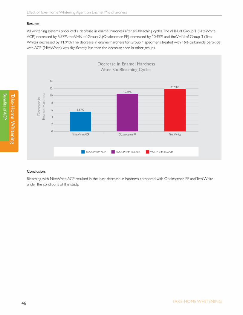
Results:
All whitening systems produced a decrease in enamel hardness after six bleaching cycles. The VHN of Group 1 (NiteWhite ACP) decreased by 5.57%, the VHN of Group 2 (Opalescence PF) decreased by 10.49% and the VHN of Group 3 (Tres White) decreased by 11.91%. The decrease in enamel hardness for Group 1 specimens treated with 16% carbamide peroxide with ACP (NiteWhite) was significantly less than the decrease seen in other groups.
0
4
2
8
10
NiteWhite ACP
Decrease in Enamel Hardness After Six Bleaching Cycles
Dec
reas
e in
En
amel
Har
dnes
s
Opalescence PF Tres White
12
14
6 5.57%
10.49%
11.91%
16% CP with ACP 16% CP with Fluoride 9% HP with Fluoride
Conclusion:
Bleaching with NiteWhite ACP resulted in the least decrease in hardness compared with Opalescence PF and Tres White under the conditions of this study.

Effect of Remineralizing Agents on Enamel Microhardness After Bleaching
47Take-Home WHiTening
Take
-Hom
e W
hite
ning
Bene
fits o
f AC
P
Effect of Remineralizing Agents on Enamel Microhardness After Bleaching in vitro studyOchiai K, Sung EC, Chung J, Caputo AA. Effect of Remineralizing Agents on Enamel Microhardness After Bleaching. J Dent Res 86 (Spec Iss A), 2007.
Objective:
To determine the potential of remineralizing agents to increase microhardness of enamel after bleaching
Materials:
• Five human incisors
• 15% carbamide peroxide (Opalescence, Ultradent)
• MI Paste (Ultradent)
• Relief ACP (Discus Dental)
Methodology:
Five human incisors were sectioned in half superior inferiorly. The halves were then mounted on cold cure acrylic for ease of manipulation. Six VH readings were then done per specimen on Micromet 2100 (Buehler, Lake Bluff, IL) with 500 gm load to establish baseline. Six cycles of bleaching with 15% carbamide peroxide (Opalescence, Ultradent) were then performed. Each cycle lasted one hour prior to placement of new solutions. The teeth again were tested for VH hardness with six readings per specimen. Upon completion of bleaching, the halves of the teeth were divided into either Group A (MI Paste, Ultradent) or Group B (Relief, Discus Dental). The systems were applied for 30 minutes then rinsed with tap water. The specimens again were tested for VH hardness with 6 readings per specimen. The procedure was repeated for a total of three remineralization cycles. All data gathered were analyzed using ANOVA with p<0.05 for significant differences.
Results:
After six cycles of bleaching, there was a significant decrease in hardness on all specimens from a VH of 313.1 to 280.8. After three applications of remineralization agents in both groups, all hardness measurements returned to baseline.
Conclusion:
There was a statistically significant decrease in enamel hardness for the bleaching system tested in this study. The application of remineralization agents reversed the decrease in enamel hardness to baseline after three applications.

A 180-Day Clinical Investigation of the Tooth Whitening Efficacy of a Bleaching Gel with Added Amorphous Calcium Phosphate
48 Take-Home WHiTening
Take-Hom
e Whitening
Benefits of ACP
A 180-Day Clinical Investigation of the Tooth Whitening Efficacy of a Bleaching Gel with Added Amorphous Calcium Phosphatein vivo studyGiniger M1, Spaid M1, MacDonald J2, Felix H2. A 180-Day Clinical Investigation of the Tooth Whitening Efficacy of a Bleaching Gel with Added Amorphous Calcium Phosphate. J Clin Dent 1611-16, 2005.1Martin Giniger & Company, New York, NY, USA, 2Discus Dental, Culver City, CA, USA
Objective:
To determine if there are any significant long-term clinical benefits or side effects caused by the addition of amorphous calcium phosphate (ACP) to a professional 16% carbamide peroxide bleaching gel.
Materials:
• 16% carbamide peroxide gel containing ACP (NiteWhite ACP, Discus Dental)
• 16% carbamide peroxide gel without ACP (NiteWhite Excel 3, Discus Dental)
Methodology:
This study examined the effect of bleaching gel with added ACP in a subset of subjects (n=27) from a previously published short-term (n=50) study, in which two groups were assigned to use either an experimental ACP-containing gel or a similar “control” gel. Both groups used the product for four hours (or overnight) daily for 14 days. In the present study, the long-term ACP effects on tooth color, gingival health and three measures of dentinal hypersensitivity at post-treatment days +90 and +180 were assessed.
Results:
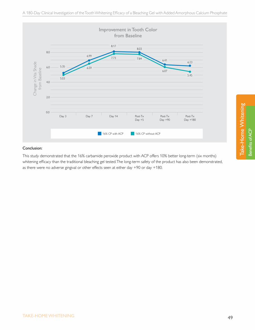
In the previously published study, the difference in tooth whitening efficacy at day +5 between the test group and the control group was only 0.19 shades relative to baseline, and was not statistically significant. In the present study, the differences between the groups had almost doubled at day +90, and were calculated to be 0.34 shades (statistically different t-test p=0.002). Furthermore, the differences had more than doubled again at day +180, with the ACP group subjects’ teeth being 0.78 shades lighter than the control group’s teeth (statistically different t-test p=0.002). Considered as a percentage, at day +180 the ACP group had retained nearly 10% more of their original whitening treatment result compared to control. There were no other significant differences found between the two groups. Tooth sensitivity, soft tissue health and gingival health remained similar to baseline levels.
A 180-Day Clinical Investigation of the Tooth Whitening Efficacy of a Bleaching Gel with Added Amorphous Calcium Phosphate
49Take-Home WHiTening
Take
-Hom
e W
hite
ning
Bene
fits o
f AC
P
0.0
2.0
Day 3 Day 7 Day 14 Post-TxDay +5
Post-TxDay +90
Post-TxDay +180
4.0
6.0
Cha
nge
in V
ita S
hade
fro
m B
asel
ine
8.0
Improvement in Tooth Colorfrom Baseline
16% CP with ACP 16% CP without ACP
8.03
5.35
5.03
6.99
6.59
8.17
7.73 7.84 6.41
6.07
6.23
5.45
Conclusion:
This study demonstrated that the 16% carbamide peroxide product with ACP offers 10% better long-term (six months) whitening efficacy than the traditional bleaching gel tested. The long-term safety of the product has also been demonstrated, as there were no adverse gingival or other effects seen at either day +90 or day +180.
Whitening Agents with ACP: Enamel Caries Formation and Progression
50 Take-Home WHiTening
Take-Hom
e Whitening
Benefits of ACP
Whitening Agents with ACP: Enamel Caries Formation and Progression in vitro studyHicks J, Flaitz C. Whitening Agents with ACP: Enamel Caries Formation and Progression. J Dent Res 85 (Spec Iss A), 0882, 2006.
Objective:
To evaluate the effect of whitening agents containing amorphous calcium phosphate (ACP) on human enamel caries formation and progression
Materials:
• 15 human teeth
• 9.5% hydrogen peroxide ACP (DayWhite Excel 3, Discus Dental)
• 6% hydrogen peroxide ACP (NiteWhite Turbo, Discus Dental)
• 16% carbamide peroxide ACP (NiteWhite, Discus Dental)
Methodology:
Fifteen teeth with sound enamel surfaces were divided into four portions. Each tooth portion was assigned to a treatment group: Group 1) No Treatment Control; Group 2) DayWhite Excel 3 9.5% hydrogen peroxide ACP; Group 3) NiteWhite Turbo 6% hydrogen peroxide ACP; Group 4) NiteWhite 16% carbamide peroxide ACP. The teeth were treated according to the manufacturer’s recommendations followed by synthetic saliva, on a daily basis for 14 days. Control tooth portions were exposed only to synthetic saliva. A modified ten Cate solution was used for in vitro enamel caries formation and progression. The teeth were treated prior to lesion formation, and before lesion progression 1 and lesion progression 2 periods. Longitudinal sections were taken after lesion formation, lesion progression 1 and lesion progression 2 periods for polarized light study and statistical analysis (ANOVA, DMR).
Whitening Agents with ACP: Enamel Caries Formation and Progression
51Take-Home WHiTening
Take
-Hom
e W
hite
ning
Bene
fits o
f AC
P
Results:
Mean lesion depths were:
• Lesion Formation Period: Control 108±15um; DayWhite 93±11um; NiteWhite Turbo 48±7um (P<.05); NiteWhite 16% 105±12um.
• Progression Period 1: Control 171±18um; DayWhite 126±13um (P<.05); NiteWhite Turbo 96±9um (P<.05); NiteWhite 16% 132±12um (P<.05).
• Progression Period 2: Control 228±20um; DayWhite 165±17um (P<.05); NiteWhite Turbo 129±11um (P<.05); NiteWhite 16% 152±16um (P<.05).
Mean Lesion Depths
Group 1: Control
Group 2: DayWhite Excel 3 - 9 .5% hydrogen peroxide ACP
Group 3: NiteWhite Turbo 6% hydrogen peroxide ACP
Group 4: NiteWhite 16% carbamide peroxide ACP
Lesion Formation Period
108±15um 93±11um(P<.05) 48±7um (P<.05) 105±12um(P<.05)
Progression Period 1
171±18um 126±13um(P<.05) 126±13um(P<.05) 132±12um (P<.05)
Progression Period 2
228±20um 165±17um (P<.05) 129±11um (P<.05) 152±16um (P<.05)
Conclusion:
Whitening agents containing calcium phosphate have a reduced susceptibility to in vitro enamel caries lesion initiation and progression.
Tooth Surface Enhancement by a 16% Carbamide Peroxide Take-Home Bleaching Gel Containing ACP
52 Take-Home WHiTening
Take-Hom
e Whitening
Benefits of ACP
Tooth Surface Enhancement by a 16% Carbamide Peroxide Take-Home Bleaching Gel Containing ACPin vivo studyGiniger M, et al. Tooth Surface Enhancement by a 16% Carbamide Peroxide Take Home Bleaching Gel. J Dent Res (Spec Iss A), 1793, 2005.
Objective:
To evaluate the effectiveness of a dual-barrel, 16% carbamide peroxide equivalent, take-home bleaching gel containing amorphous calcium phosphate (ACP) in enhancing tooth surface smoothness and gloss.
Materials:
• 16% carbamide peroxide equivalent gel containing ACP (NiteWhite Excel 3 ZCP, Discus Dental)
Methodology:
Ten healthy adults wore a custom tray containing the test ACP gel for a minimum of four hours daily (or overnight) for two weeks. Evaluations of tooth color, surface roughness and gloss were made at baseline, one week, two weeks and +five days post treatment. The surface gloss index (SGI) and surface roughness index (SRI) measurements were performed by a single experienced examiner who evaluated the anterior teeth. The SGI utilizes a six-point subjective scale, and the SRI uses a four-point scale. For the tooth color evaluation a 16-point Vita Shade Guide score index was used. Mean “before” and “after” tooth color scores, SGI scores and SRI scores were calculated. Mean differences between baseline values and after-treatment values were also reported. A t-test was used to determine significant differences with alpha set at 0.05.
Results:
Compared to the baseline control values, the ACP gel showed a significant (p < 0.01) longitudinal percent improvement in tooth surface gloss and roughness at one week (SGI = 10.1%; SRI + 6.15%) and two weeks (SGI + 22.4%; SRI = 15.4%). Tooth color was also improved significantly compared to baseline, reaching a maximum shade change of 8.13 (± 1.02) units at day 14 (t-test, p < 0.0001). At the +five days post-treatment evaluation, no significant changes in tooth color, SGI or SRI were found compared to day 14 results (p < 0.0001).

Tooth Surface Enhancement by a 16% Carbamide Peroxide Take-Home Bleaching Gel Containing ACP
53Take-Home WHiTening
Take
-Hom
e W
hite
ning
Bene
fits o
f AC
P
0
4
6
8
12
10
2
Baseline 7 Days 14 Days Day 5 Post
10.47
3.472.33 2.82
Mean Tooth Color Scores
NiteWhite Excel 3 ZCP
Vita
Sha
de G
uide
Sco
re
0
1.0
1.5
2.0
3.0
2.5
3.5
4.0
4.5
5.0
.5
Baseline 7 Days 14 Days Day 5 Post
3.77
2.75
4.15
2.92
4.61
3.17
4.50
3.12
Tooth Gloss and Roughness
Mean Gloss Score Mean Roughness Score
Conclusion:
The dual-chambered, 16% carbamide peroxide equivalent, ACP-containing bleaching gel has superior tooth whitening and surface properties that include excellent tooth-whitening ability (8+ Vita Shade Guide improvements from baseline) and improving teeth luster (significantly increased enamel gloss and decreased enamel roughness compared to baseline).
The Clinical Performance of Professionally Dispensed Bleaching Gel With Added Amorphous Calcium Phosphate
54 Take-Home WHiTening
Take-Hom
e Whitening
Benefits of ACP
The Clinical Performance of Professionally Dispensed Bleaching Gel With Added Amorphous Calcium Phosphatein vivo studyGiniger M1, MacDonald J2, Ziemba S2, Felix H2, The Clinical Performance of Professionally Dispensed Bleaching Gel With Added Amorphous Calcium Phosphate, JADA, 136, 2005.1Martin Giniger & Company, New York, NY, USA, 2Discus Dental, Culver City, CA, USA
Objective:
To measure how the addition of amorphous calcium phosphate (ACP) to a professionally dispensed 16% carbamide peroxide equivalent bleaching gel affects tooth color and dentinal hypersensitivity.
Materials:
• 16% carbamide peroxide equivalent gel with calcium and phosphate
• 16% carbamide peroxide equivalent gel (NiteWhite Excel 3 Regular, Discus Dental)
Methodology:
Fifty healthy subjects were enrolled in a parallel, double-blind, two-cell randomized clinical study. All subjects had maxillary anterior tooth discoloration equivalent to or darker than Vita shade 3. A baseline examination was performed which included medical history, oral soft tissue examination, Vita shade tooth color scoring, Gingival Index scoring and self-reported sensitivity scoring. Subjects were randomized into test and control groups that were balanced with respect to age, sex, tooth color and sensitivity scores. The subjects were instructed to use the gel they were given (according to the manufacturer’s instructions for the commercially-available control product) once a day for a minimum of three hours or overnight for 14 days. Clinical re-examinations were performed on days 3, 7, and 14 as well as +5 days post-treatment.
Results:
The test group demonstrated significantly lower (P < .05) mean thermal sensitivity scores with baseline (day 14: 0.21 versus 0.31; day +5: 0.06 versus 0.18). The test group also showed substantially lower (P < .05) tactile sensitivity scores (day 14: 0.26 versus 0.48; day +5: 0.06 versus 0.19). At the conclusion of the study twice as many subjects were free of thermal sensitivity in the test group (80%) compared with the control group (40%) (P < .001). There was a similar significant percentage difference for tactile sensitivity. Both groups demonstrated equivalent and significant tooth color enhancements as compared with base line (control: -7.73 shade change versus test: -8.12 shade change; P < .05).

The Clinical Performance of Professionally Dispensed Bleaching Gel With Added Amorphous Calcium Phosphate
55Take-Home WHiTening
Take
-Hom
e W
hite
ning
Bene
fits o
f AC
P
0.0
0.2
0.1
0.4
0.5
Baseline
Thermal Sensitivity
14 Days 19 Days
0.6
0.7
0.8
0.9
0.3 .27 .26
.58
.47.45
.32
Control Test
0.0
0.2
0.1
0.4
0.5
Baseline
Tactile Sensitivity
14 Days 19 Days
0.6
0.7
0.8
0.9
0.3
.39 .39
.87
.65
.57
.45
Control Test
Conclusion:
This study demonstrates that ACP could be added to a 16% carbamide peroxide equivalent bleaching gel and result in a significant reduction of clinical measures of dentinal hypersensitivity, both during and after treatment.
Microhardness and Ultramorphological Changes after Whitening: Calcium and Phosphate Benefits
56 Take-Home WHiTening
Take-Hom
e Whitening
Benefits of ACP
Microhardness and Ultramorphological Changes after Whitening: Calcium and Phosphate Benefits in vitro studyPinheiro HB, Cardoso PEC. Microhardness and Ultramorphological Changes after Whitening: Calcium and Phosphate Benefits. J Dent Res 89 (Spec Iss A), 0839, 2010.
Objective:
To investigate the influence of home bleaching agents with and without calcium and phosphate on human enamel and dentin surface microhardness and ultramorphology.
Materials:
• Five human molars
• 15% carbamide peroxide with potassium nitrate and fluoride (Opalescence PF, Ultradent)
• 16% carbamide peroxide with potassium nitrate and fluoride (Whiteness Perfect, FGM)
• 16% carbamide peroxide with potassium nitrate and ACP (NiteWhite ACP, Discus Dental)
• 7.5% hydrogen peroxide with potassium nitrate and ACP (DayWhite ACP, Discus Dental)
• 7.5% hydrogen peroxide with potassium nitrate, fluoride, and calcium (White Class Ca, FGM)
Methodology:
Five intact human third molars were sectioned into five wafers. All wafers had their baseline Knoop Hardness Number (KHN) of the enamel (E0) and dentin (D0) measured. Each wafer of the tooth was assigned to a group (n=5). G1=15% carbamide peroxide (CP) (Opalescence PF-Ultradent) applied for four hours per day; G2=16% CP (Whiteness Perfect, FGM), G3=16% CP with Ca and PO4 (NiteWhite ACP-Discus Dental), G2 and G3 followed same application time of group 1; G4=7.5% hydrogen peroxide (HP) with Ca and PO4 (Day White ACP-Discus Dental); G5=7.5% HP with Ca (White Class Ca-FGM), G4 and G5 were applied for one hour per day. After each session of bleaching treatment, specimens were stored in distillated water (37oC). The products were applied for two weeks, according to manufacturers’ instructions. Subsequent measurements of KHN were taken in the enamel (E1) and dentin (D1). Two specimens from each group were selected for ultramorphological investigation after final tests.

Microhardness and Ultramorphological Changes after Whitening: Calcium and Phosphate Benefits
57Take-Home WHiTening
Take
-Hom
e W
hite
ning
Bene
fits o
f AC
P
Results:
Conventional bleaching agents and the gel with calcium (Group 1 and Group 2) caused KHN decrease. Bleaching agents with calcium and PO4 (Group 3 and Group 4) did not change KHN. The obvious change of morphology was observed on enamel and dentin surfaces in Group 1, Group 2 and Group 5.
Control
NiteWhite ACP
Opalescence PF
DayWhite ACP
Whiteness Perfect
White Class Ca
0
102.9
154.3
205.7
308.6
257.1
360.0
51.4
KHN
KHN (Baseline) KHN (After treatment)
Opalescence PF
308.9
(15.5)278.5
(12.8)
Whiteness Perfect
316.8(8.0) 291.2
(12.9)
NiteWhite ACP
313.8(14.7)
315.7(16.0)
DayWhite ACP
320.7(10.4)
316.2(6.6)
White Class Ca
315.7(10.3) 289.7
(6.0)
Enamel
Enamel
Original magnification: 50,000X Insets: 100,000X
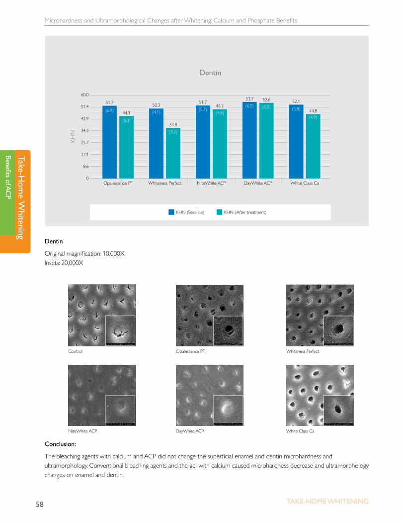
Microhardness and Ultramorphological Changes after Whitening: Calcium and Phosphate Benefits
58 Take-Home WHiTening
Take-Hom
e Whitening
Benefits of ACP
Conclusion:
The bleaching agents with calcium and ACP did not change the superficial enamel and dentin microhardness and ultramorphology. Conventional bleaching agents and the gel with calcium caused microhardness decrease and ultramorphology changes on enamel and dentin.
0
17.1
25.7
34.3
51.4
42.9
60.0
8.6
KHN
KHN (Baseline) KHN (After treatment)
Opalescence PF
51.7
(6.9) 44.1
(5.3)
Whiteness Perfect
50.3(4.1)
34.8(3.2)
NiteWhite ACP
51.7(5.7)
48.2(4.6)
DayWhite ACP
53.7(6.0)
52.6(5.0)
White Class Ca
52.1(5.8) 44.8
(4.9)
Dentin
Dentin
Original magnification: 10,000X Insets: 20,000X
Control
NiteWhite ACP
Opalescence PF
DayWhite ACP
Whiteness Perfect
White Class Ca
Whitening Treatment Combined With Bioactive Materials
59Take-Home WHiTening
Take
-Hom
e W
hite
ning
Bene
fits o
f AC
P
Whitening Treatment Combined With Bioactive Materialsin vitro studyPinheiro HB, Cardoso J and Cardoso PEC. Whitening Treatment Combined with Bioactive Materials - in situ study. J Dent Res (Spec Iss A), 0801, 2012.
Objective:
To investigate the influence of bioactive materials on whitened surfaces and dentin using Knoop hardness test
Materials:
• Eight human teeth
• 15% carbamide peroxide, potassium nitrate, fluoride (Opalescence PF, Ultradent)
• 16% carbamide peroxide, potassium nitrate, fluoride, calcium, phosphate (NiteWhite ACP, Discus Dental)
• 15% carbamide peroxide, potassium nitrate, fluoride & glass-ceramic crystalized glass P2O5 –Na2O–CaO–SiO2 (Opalescence PF, Ultradent & Biosilicate, Vitrovita)
• 15% carbamide peroxide, potassium nitrate, fluoride + potassium nitrate, fluoride, calcium, phosphate (Opalescence PF, Ultradent + Relief ACP, Discus Dental)
Methodology:
Eight human teeth were sectioned into five wafers per tooth and divided into five experimental groups (n=8). The specimens were treated and mounted in intra-oral palatal retainers. Whitening treatments were performed for 14 days according to manufacturer’s instructions. Six Knoop hardness measures were taken for each specimen, three before and three after treatments. The data were compared by Student’s t-test (α = 0.05).
Results:
Opalescence PF caused hardness decrease on enamel and dentin (p<0.05). NiteWhite ACP and the bioactive materials had a positive influence on the hardness of bleached enamel and dentin, except for the effect on enamel of the Biosilicate material when applied for five minutes one time per week, which showed a decrease in KHN.

Whitening Treatment Combined With Bioactive Materials
60 Take-Home WHiTening
Take-Hom
e Whitening
Benefits of ACP
250
260
270
280
290
Opalescence PFNiteWhite ACP Opalescence PF+ Bio Mixed
Opalescence PF+ Relief ACP
Opalescence PF+ Bio
279.7 279.6
267.8
289.6
265.8
278.5
Before After
281.4
277.0279.3
289.1KH
N
Pre- and Post-Treatment Enamel Hardness
KHN
38
41
44
47
50
Opalescence PFNiteWhite ACP Opalescence PF+ Bio Mixed
Opalescence PF+ Relief ACP
Opalescence PF+ Bio
45.2
42.5
38.9
44.3
40.541.4
Pre- and Post-Treatment Dentin Hardness
Before After
47.2
45.2
43.0
46.4
Conclusion:
Whitening treatment can lead to alterations in the dental structure. Minimizing or eliminating the alterations in the whitened dental structure could bring benefits to patients. Adding bioactive materials to whitening treatments can minimize or eliminate the alterations in the dental structure.

NOTES

NOTES


Philips and the Philips shield logo are a registered trademark of Koninklijke Philips Electronics, N.V. Zoom is a registered trademark of Discus Dental, LLC. All other trademarks are the property of their respective owners. For use by dental professionals only.©2013 Discus Dental, LLC. All rights reserved.US 7502540 CAN 7502541 * 021213


![Untitled-3 [] · Teeth whitening or bleaching. Several options are available, including the patented Bright Smile technique. This involves the use of a bleaching gel and a gas plasma](https://img.pdfslide.net/doc/110x75/5f0a950d7e708231d42c5759/untitled-3-teeth-whitening-or-bleaching-several-options-are-available-including.jpg)


![[2] Whitening Bleaching and Antioxidant](https://img.pdfslide.net/doc/110x75/563db985550346aa9a9e1e68/2-whitening-bleaching-and-antioxidant.jpg)
