Embed Size (px)
Citation preview

doi:10.1016/j.jmb.2011.10.021 J. Mol. Biol. (2011) 414, 499–510
Contents lists available at www.sciencedirect.com
Journal of Molecular Biologyj ourna l homepage: ht tp : / /ees .e lsev ie r.com. jmb
The Scope of Phage Display for Membrane Proteins
Rosemarie Vithayathil 1, Richard M. Hooy 1, Melanie J. Cocco 1
and Gregory A. Weiss 1, 2⁎1Department of Molecular Biology and Biochemistry, University of California, Irvine, CA 92697, USA2Department of Chemistry, University of California, Irvine, CA 92697, USA
Received 22 July 2011;received in revised form9 October 2011;accepted 12 October 2011Available online20 October 2011
Edited by J. Bowie
Keywords:phage display;membrane proteins;protein engineering;mutagenesis;β-barrel membrane proteins
*Corresponding author. DepartmenUniversity of California, Irvine, CAE-mail address: [email protected] used: TM, transme
membrane protein; ORF, open readinserum albumin; NFM, non-fat milk;and transcript release factor; PBS, psaline; RT, room temperature; HIV,immunodeficiency virus.
0022-2836/$ - see front matter © 2011 E
Numerous examples of phage display applied to soluble proteinsdemonstrate the power of the technique for protein engineering, affinityreagent discovery and structure–function studies. Recent reports haveexpanded phage display to include membrane proteins (MPs). The scopeand limitations of MP display remain undefined. Therefore, we report datafrom the phage display of representative types of membrane-associatedproteins including plasma, nuclear, peripheral, single and multipass. Theperipheral MP neuromodulin displays robustly with packaging byconventional M13-KO7 helper phage. The monotopic MP Nogo-66 canalso display on the phage surface, if packaged by the modified M13-KO7+
helper phage. The modified phage coat of KO7+ can better mimic thezwitterionic character of the plasma membrane. Four examples ofputatively α-helical, integral MPs failed to express as fusions to ananchoring phage coat protein and therefore did not display on the phagesurface. However, the β-barrel MPs ShuA (Shigella heme uptake A) andMOMP (major outer membrane protein), which pass through the membrane22 and 16 times, respectively, can display surprisingly well on the surfacesof both conventional and KO7+ phages. The results provide a guide forprotein engineering and large-scale mutagenesis enabled by the phagedisplay of MPs.
© 2011 Elsevier Ltd. All rights reserved.
Introduction
Reflecting their critical importance to cellularfunctions, membrane proteins (MPs) compriseapproximately 30% of the human proteome.1
Cellular processes relying on MPs range fromenergy generation to cell signaling. This preemi-nence also accounts for the large number of MPs
t of Chemistry,92697, USA.
mbrane; MP,g frame; BSA, bovinePTRF, polymerase Ihosphate-bufferedhuman
lsevier Ltd. All rights reserve
targeted by N50% of Food and Drug Administra-tion-approved therapeutics.2 Thus, understandingthe relationship between MP sequence and functioncould provide valuable insights in biology anddrug development.The properties of MPs challenge attempts to
produce sufficient quantities for biophysical stud-ies. For example, the Membrane Proteins of Known3D Structures† includes b300 unique MP structures,a low number limited by difficulties with expres-sion, isolation and purification of folded MPs.3Protein aggregation can result from the inherenthydrophobicity of MPs. Typical methods to solubi-lize MPs use strong detergents, which can destabi-lize the proteins.4 Furthermore, expression of MPsin prokaryotes can fail due to, among other reasons,
†http://blanco.biomol.uci.edu/mpstruc
d.

Fig. 1. A schematic diagram depicting the surfaces of KO7 and KO7+ helper phages. Positively and negatively chargedside chain functionalities are highlighted in blue and red, respectively. (a) The major coat protein, P8, of M13-KO7includes three carboxylate-bearing side chains at its N-terminus (Protein Data Bank accession code: 1ifd). (b) An insertionof a 4-mer peptide into each copy of P8 alters the surface charge of M13-KO7+. The resultant zwitterionic surface canbetter display monotopic MPs.
500 Scope of Phage Display for Membrane Proteins
the lack of posttranslational modifications requiredin eukaryotes.Phage display provides an alternative to the
expression of MPs for in vitro binding and drugdiscovery studies.5 Phage display allows identifica-tion of functional residues in the displayed proteinby high-throughput mutagenesis. Alanine shotgunscanning of the displayed protein, for example, canidentify residues contributing to protein bindinginterfaces.6,7 Furthermore, the technique can beused to reverse engineer soluble and functionalvariants of phage-displayed MPs8 and also toidentify MP ligands.9
Essential to the phage display approach, thephenotype of the phage-displayed protein is linkedto the encoded genotype, which is packaged withinthe phage particle.10 Proteins can be displayed onthe phage surface using a phage vector or a plasmid-based phagemid vector.11 Unlike the natural Ffphage genome of a phage vector, the phagemidencodes the open reading frame (ORF) with a singlecoat protein fused to the displayed protein. A helperphage provides the phage proteins required forvirus packaging and assembly.12 This report appliestwo helper phage, the conventional M13-KO7 andthe recently reported M13-KO7+ bacteriophage.13
KO7+ helper phage includes a tetrapeptide (AKAS)
Table 1. Phage display of membrane-associated proteins
Membrane-associated protein Molecular mass (kDa) Membrane
Nef (6) 23 PeriphNeuromodulin 25 PeriphNogo-66 7.5 MonoCaveolin-1 (15) 22 MonoEmerin 29 BitoMAN1 100 PolytNET25 57 PolytMatriptase-2 51.6 BitoShuA 72.5 PolytMOMP 40 Polyt
−, no display; ++, moderate display levels; +++, high display levels.a Putative secondary structure.
near the N-terminus of each copy of the major coatprotein (g8p or P8). Originally developed to reducebackground binding, this tetrapeptide insertion inthe P8 of KO7+ (Fig. 1) has also been shown to allowdisplay of two MPs, caveolin-1 and human immu-nodeficiency virus-1 (HIV-1) gp41.14
The successful display of a functional protein onthe phage surface requires protein translocation tothe periplasm, subsequent folding into its functionalconformation and incorporation of the phage-coat-fused protein during virus assembly. Although alarge number of soluble proteins have successfullydisplayed on the phage surface,12,15 some proteinsprove refractory to display.16 Failure in phagedisplay can result from protein aggregation17 orpotentially incomplete translocation into the peri-plasm due to “stop transfer” signals in the proteinsequence.18 Attempts to improve display levelsincludemutations to the anchoring P8 coat protein,19
co-expression of periplasmic chaperones20 andtranslocation of the displayed peptides to theperiplasm by signal recognition particle21 or Tattranslocation pathways.18
Despite successful display of soluble proteins,only a handful of membrane-associated proteinshave been displayed to date on phage.14 Here, wedefine the scope and current limitations in the
topology TM secondary structure Display Helper phage
eral +++ KO7eral ++ KO7topic +++ KO7+
topic ++ KO7+
pic α-Helicala − KO7/KO7+
opic α-Helicala − KO7/KO7+
opic α-Helicala − KO7/KO7+
pic α-Helicala − KO7/KO7+
opic β-Barrel ++ KO7/KO7+
opic β-Barrela +++ KO7

501Scope of Phage Display for Membrane Proteins
display of MPs on the phage surface. The proteinstested include plasma, nuclear, peripheral, singleand multipass membrane-associated proteins(Table 1). The accumulation of successes and failureswith phage-displayed,membrane-associated proteinsprovides a predictive model for the design of phage-based experiments to explore the structure andfunction of MPs.
Results and Discussion
Cloning MPs for display
The genes encoding the targeted MPs weresubcloned into a conventional phagemid vector forphage display initially as fusions to the N-terminusof P8. Fusion to the N-terminus of the minor coatprotein P3 (C-terminal domain) was also attemptedfor the display of the putatively α-helical transmem-brane (TM) proteins. Each displayed protein includ-ed a FLAG antibody epitope fused to the N-terminus for quantification of the relative displaylevels. DNA sequencing verified successful cloningof an ORF encoding the following sequences fromN-terminus to C-terminus: the periplasmic signalpeptide targeting the Sec translocation pathway, theFLAG epitope, the membrane-associated protein,the linker (amino acid sequence of GGGSGSSS) andthe anchoring phage coat protein. The phages werepackaged with wild-type KO7 or the variant KO7+
helper phage, as discussed below.A phage-based ELISA quantified the relative
display levels of the MPs. In the ELISA, microtiter-plate-adsorbed, anti-FLAG (α-Flag) antibody boundto the FLAG epitope of full-length proteins dis-played on phage surfaces. The negative and positivecontrols for all ELISA experiments yielded similarexpected results. Negative controls resulted in littleor no detectable binding; one negative controlexamined binding by the displayed protein to theblocking agent [bovine serum albumin (BSA), non-fat milk (NFM) or ovalbumin], and a second controltested binding to the anti-FLAG antibody by phagelacking a displayed protein (KO7 or KO7+ helperphage). Phage-displayed polymerase I and tran-script release factor (PTRF) provided the positivecontrol for successful display, as the proteindisplayed consistently well on the phage surfacesprovided by both KO7 and KO7+ helper phages.
Phage display of peripheral MPs
Peripheral MPs loosely and sometimes reversiblyassociate with the plasma membrane, throughinteractions with the membrane or other membrane-associated proteins.22 Posttranslational modifica-tions, including acylation,23 prenylation24 and
glycosylphophatidylinositol anchors,25 can covalentlyanchor the protein to the membrane, though proteinexpression in Escherichia coli precludes this possibility.Successful phage display of at least one peripheralMP has been reported previously. For example,the HIV-1 protein Nef, responsible for down-regulation of CD4, displays robustly on thephage surface.5 Display of Nef was reportedbefore the discovery of KO7+ for phage displayof MPs. Therefore, KO7 and KO7+ helper phageswere compared for the packaging of the peripheralMP, neuromodulin.Neuromodulin (GAP-43), a neurite-growth-stim-
ulating protein, associates with the membranethrough palmitoylation and electrostatic interactionsbetween the positively charged residues in themembrane binding domain at the N-terminus andthe headgroups of the lipid bilayer.26 For display onthe phage surface, the sites of palmitoylation,cysteine residues C3 and C4, were mutated toalanine. The neuromodulin-displayed phage waspackaged by either KO7 or KO7+ helper phage. AnELISA tomeasure display of the FLAG epitope at theN-terminus of neuromodulin demonstrates success-ful display with packaging by KO7, but not KO7+,helper phage (Fig. 2a and Fig. S1, respectively). Thedisplay of neuromodulin through KO7 packaging isconsistent with the display of the peripheral MPHIV-1 Nef also using KO7 helper phage.KO7 helper phage has three carboxylate-bearing
side chains near the surface-exposed N-terminus ofeach P8 to create a forest of negatively chargedresidues (Fig. 1a). Such side chains could facilitateelectrostatic interactions with the basic residues ofthe membrane binding domain of neuromodulin.However, the more positively charged surface ofKO7+ phage could negatively affect such interac-tions and thus interfere with the display of thisperipheral MP. The experimental results with twoperipheral MPs demonstrate that proteins with lipidbinding motifs can be readily packaged withconventional KO7 helper phage.Phage-displayed neuromodulin also retains its
functional conformation on the phage surface. Todemonstrate the functionality of displayed neuro-modulin, we performed a binding assay with aknown binding partner. F-actin binds to unpho-sphorylated neuromodulin with a dissociation con-stant (Kd) of ≈ 1.2 μM. 27 An ELISA withimmobilized f-actin and phage-displayed wild-typeneuromodulin demonstrated this interaction, thusconfirming the folding of the displayed protein (Fig.2b). Although neuromodulin binds to f-actin, phos-phorylation at Ser41 increases the binding affinity forf-actin about 7-fold (Kd=161 nM).27 To furtherdemonstrate functional folding of the phage-dis-played neuromodulin, we generated a constitutivelyactive neuromodulin variant through an S41Dsubstitution in the phage-displayed protein.
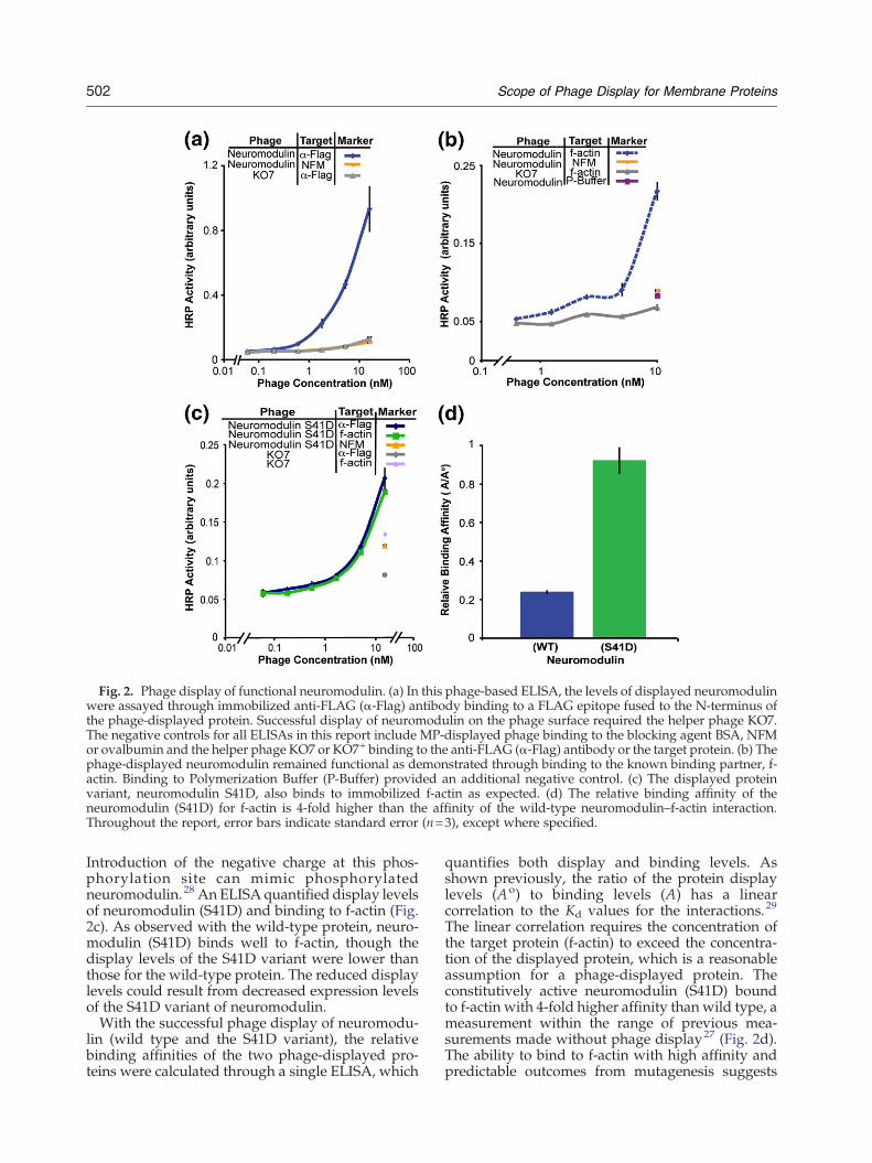
Fig. 2. Phage display of functional neuromodulin. (a) In this phage-based ELISA, the levels of displayed neuromodulinwere assayed through immobilized anti-FLAG (α-Flag) antibody binding to a FLAG epitope fused to the N-terminus ofthe phage-displayed protein. Successful display of neuromodulin on the phage surface required the helper phage KO7.The negative controls for all ELISAs in this report include MP-displayed phage binding to the blocking agent BSA, NFMor ovalbumin and the helper phage KO7 or KO7+ binding to the anti-FLAG (α-Flag) antibody or the target protein. (b) Thephage-displayed neuromodulin remained functional as demonstrated through binding to the known binding partner, f-actin. Binding to Polymerization Buffer (P-Buffer) provided an additional negative control. (c) The displayed proteinvariant, neuromodulin S41D, also binds to immobilized f-actin as expected. (d) The relative binding affinity of theneuromodulin (S41D) for f-actin is 4-fold higher than the affinity of the wild-type neuromodulin–f-actin interaction.Throughout the report, error bars indicate standard error (n=3), except where specified.
502 Scope of Phage Display for Membrane Proteins
Introduction of the negative charge at this phos-phorylation site can mimic phosphorylatedneuromodulin.28 An ELISA quantified display levelsof neuromodulin (S41D) and binding to f-actin (Fig.2c). As observed with the wild-type protein, neuro-modulin (S41D) binds well to f-actin, though thedisplay levels of the S41D variant were lower thanthose for the wild-type protein. The reduced displaylevels could result from decreased expression levelsof the S41D variant of neuromodulin.With the successful phage display of neuromodu-
lin (wild type and the S41D variant), the relativebinding affinities of the two phage-displayed pro-teins were calculated through a single ELISA, which
quantifies both display and binding levels. Asshown previously, the ratio of the protein displaylevels (Ao) to binding levels (A) has a linearcorrelation to the Kd values for the interactions.29
The linear correlation requires the concentration ofthe target protein (f-actin) to exceed the concentra-tion of the displayed protein, which is a reasonableassumption for a phage-displayed protein. Theconstitutively active neuromodulin (S41D) boundto f-actin with 4-fold higher affinity than wild type, ameasurement within the range of previous mea-surements made without phage display27 (Fig. 2d).The ability to bind to f-actin with high affinity andpredictable outcomes from mutagenesis suggests

Fig. 3. Functional Nogo-66 displayed on the phagesurface. (a) Display of Nogo-66 with KO7+ helper phage isassessed by ELISA with anti-FLAG (α-Flag) antibodyimmobilized on the microtiter plate. (b) The phage-displayed Nogo-66 remained functional, as demonstratedthrough binding to its known receptor, NgR, immobilizedon the microtiter plate.
503Scope of Phage Display for Membrane Proteins
that this peripheral protein is displayed in itsfunctional conformation. Furthermore, the phage-displayed neuromodulin could be amenable tofurther mutational analysis for structure–functionstudies.
Phage display of monotopic MPs
Monotopic MPs penetrate only one leaflet of theplasma membrane and do not traverse the bilayer.30
Such proteins associate tightly with the membraneeither through amphipathic helices31 or throughelectrostatic and hydrophobic interactions. 32Reported structures of monotopic MPs suggest thatboth basic residues and hydrophobic amino acids areinvolved in the anchoring of theMP.MonotopicMPsinclude both “shallow inserters” that interact withthe surface of the lipid bilayer and others that canpenetrate deeper into the bilayer, aided by ahydrophobic stretch and key basic residues.32
Phage display of two monotopic MPs demon-strates the capabilities of KO7+ helper phage topermit display of previously inaccessible MPs. Asreported previously, but not examined, successfulphage display of the monotopic MPs requirespackaging by KO7+ helper phage.14 At the physi-ological pH, the AKAS peptide inserted into eachcopy of the P8 phage coat protein provides apositively charged lysine side chain surrounded bythree negatively charged carboxylate-bearing sidechains. Extended to ≈2700 copies of P8 per phage,the protein displayed on the KO7+ surface will besurrounded by both positively and negativelycharged side chains (Fig. 1b). In this model, theAKAS insertion within P8 of KO7+ allows the phagesurface to better mimic the zwitterionic character ofthe phospholipid headgroups found at the plasmamembrane interface. The ɛ-amine of the lysine sidechain, for example, could substitute for the primaryand quaternary amines of the phospholipid head-groups.Our laboratory has previously reported successful
display of functional full-length caveolin-1 on thephage surface.14 Successful display of functionalcaveolin-1 on the phage surface allowed engineeringof a soluble, but functional, caveolin variant.8
Interestingly, the extensive mutagenesis of caveolinon the phage surface required solubilizing muta-tions targeting the aromatic residues in the intra-membrane domain. Such aromatic side chains couldform cation–π interactions with primary and qua-ternary amines of the phospholipid headgroups.The cation–π interactions provide a mechanistichypothesis for the efficacy of KO7+ in monotopicMP display.A second example of monotopic protein display,
Nogo-66, consists of the 66 residues of the extracel-lular domain of the neuronal protein Nogo. Asshown by circular dichroism and NMR spectra,
Nogo-66 requires dodecylphosphocholine micellesto fold.33 The structure of Nogo-66 determined byNMR shows a monotopic membrane topology. Thearomatic residues of Nogo-66 contribute to the lipidbinding through potential cation–π interactionswith the quaternary amines of phosphatidylcholine.Given the requirement for protein–lipid interactionsto assist folding, Nogo-66 represents a particularlychallenging test for display by M13 phages, whichlack an enveloping phospholipid.Using KO7+ as the helper phage, folded and
functional Nogo-66 successfully displayed on thephage surface (Fig. 3a). In this case, KO7+ contrib-utes two essential functions to allow successfulphage display. First, KO7+ mimics phospholipidheadgroups,which are required for the folding of theprotein. Binding to the Nogo receptor (NgR) byphage-displayed Nogo-66 (Fig. 3b) demonstrates

504 Scope of Phage Display for Membrane Proteins
functional folding of the displayed protein. Second,packaging with KO7+ helper phage reduced back-ground binding to NgR, which can result frominteractions between the conventional KO7 helperphage surface and high pI target proteins (the pI ofNgR is 8.4). Successful Nogo-66 display reinforcesthe hypothesis that the extra positive charge includ-ed with each copy of KO7+ P8 can mimic thenecessary zwitterionic character inherent to phos-pholipid headgroups.
Phage display of α-helical TM proteins
The α-helical TM proteins comprise N70% of theMPs in the Membrane Protein Data Bank.34 Thisclass of TM proteins can traverse the lipid bilayer ina single pass (bitopic) or cross two or more times inpolytopic TM proteins. Residing in cellular andnuclear membranes, α-helical TM proteins can formmonomers or oligomers. Through their extramem-brane domains, such proteins can perform a myriadof functions ranging from enzymatic activity tosignal transduction.35To examine different types of α-helical MPs, we
subcloned four MPs into a vector for phage display.The tested proteins include single and multipass TMproteins and also cytoplasmic and nuclear MPs(Table 1). Sub-cloned as fusions to the major coatprotein P8, the phages displaying the α-helical MPswere propagated using either KO7 or KO7+ helperphage. Attempts to display these TM proteins alsoinvolved fusion to the minor coat protein P3 andalso expression in different strains of E. coli bacteria(Table S2 in Supporting Information). As demon-strated by ELISA experiments, no combination ofhelper phage, coat protein and cell line allowedsuccessful display of the targeted α-helical integralMPs (Figs. S2 and S3).For an understanding of the basis for this inability
to display α-helical TM proteins, Western blotstargeting the FLAG epitope at the N-terminus ofeach protein examined protein expression levelsduring phage assembly. The phage coat proteins,including P3 and P8 fused to the displayed proteins,must insert into the bacterial membrane beforeassembly into the filamentous phage. Thus, theanalysis of fusion protein levels focused on the cellpellets where such MPs should localize. None of theα-helical TM proteins were detectable by Westernblot (Fig. S4). The positive control for the Westernblot, PTRF fused to P8, was readily detected in thecell pellets. The absence of the expected fusionproteins in the immuno-blot demonstrates that theα-helical TM proteins, emerin, MAN1, NET25 andmatriptase-2, fail to express sufficiently well forphage-based applications. Furthermore, this inabil-ity to express sufficient quantities of the fusionprotein results in the lack of display observed for α-helical TM proteins. In addition, the P8- and P3-
fused proteins could fail to translocate to theperiplasm, resulting in degradation of the fusionprotein in the cytosol. Despite these results, thedisplay of α-helical TM proteins might be successfulif the fusion protein can be expressed and targeted tothe periplasm.
Phage display of β-barrel TM proteins
β-Barrel MPs are observed only in the outermembranes of Gram-negative bacteria, mitochon-dria and chloroplasts. The proteins can form poresfor the transport of nutrients and can also possessenzymatic activity.36 Ranging from 8 to 22 β-strands, β-integral MPs are polytopic. Both mono-meric and oligomeric β-barrel TM proteins havebeen described. As described below, two β-barrelproteins, ShuA (Shigella heme uptake A), an outermembrane heme transporter from Shigella dysenter-iae and MOMP (major outer membrane protein), aporin from Chlamydia muridarum Nigg, displayedwell on the phage surface.ShuA belongs to the family of TonB-dependent
transporters.37 The C-terminal domain of ShuAforms a 22-stranded barrel structure. The N-terminaldomain provides a plug partitioning the extracellu-lar milieu from the periplasm.38 S. dysenteriae, aGram-negative pathogen, uses TonB-dependenttransporters to acquire heme from the host's heme-binding protein, hemoglobin.37
ShuA fused to P8 displayed robustly on the phagesurface with packaging by both KO7 and KO7+
helper phages (Fig. 4a and b). Improved display ofShuA resulted from phage propagation at 30 °C. Theslower growth at this reduced temperature couldallow for increased expression of the functionalfusion protein and could assist in the assembly andpackaging of phage particles. Furthermore, bindingto immobilized hemoglobin A demonstrated thatphage-displayed ShuA remains functionally folded(Fig. 4c and d). To our knowledge, ShuA is the firstexample of a functional full-length β-barrel integralMP successfully displayed on the phage surface.To demonstrate the generality of the approach, we
successfully displayed a second putative β-barrelMP. A cysteine-rich outer MP, MOMP of theChlamydia mouse pneumonitis (MoPn serovar) isexpected to form a 16-strand β-barrel secondarystructure. Functioning as a porin, MOMP likelyforms a homotrimer.39 To allow formation of atrimer on the phage surface, the phagemid ORFincluded the gene encoding MOMP, followed by anamber stop codon, before the linker and the phagecoat protein (P3). In an amber suppressor strain of E.coli, the amber stop is recognized as a glutamineresidue with 10–30% efficiency40 resulting in theproduction of both free and P3-fused MOMPmonomers. The two MOMP constructs can formtrimers in the periplasm prior to phage assembly.

Fig. 4. Functional display of full-length ShuA packagedwith (a) KO7 helper phage and (b) KO7+ helper phage. Displayof ShuA with KO7 and KO7+ helper phages is assessed by ELISA with anti-FLAG (α-Flag) antibody immobilized on theplate. (c and d) Binding between phage-displayed ShuA on KO7 and KO7+ and its binding partner hemoglobin (Hb) isassessed by ELISA with Hb immobilized on the plate.
505Scope of Phage Display for Membrane Proteins
An ELISA to determine display levels withimmobilized anti-FLAG antibody demonstratedthat MOMP displayed at high levels (Fig. 5a). Thenatural binding partners to MOMP remain uniden-tified, and we, therefore, demonstrate functionalityof the phage-displayed protein through binding totwo monoclonal antibodies with well-defined epi-topes. The first anti-MOMP antibody MoPn-40binds a linear epitope in one of the MOMP variabledomains, VD1, and the second MoPn-18b recog-nizes a structural epitope of the homotrimer.39 Thewild-type MOMP displayed well yet bound poorlyto the two antibodies (Fig. 5c and d).Modifications to the assay conditions and the gene
encoding MOMP allow MOMP-antibody bindingassays. Treatment of the phage-displayed wild-typeMOMP with 2 M urea and the reducing agent tris(2-carboxymethyl)phosphine (10 mM) producedphage-displayed MOMP that could be recognizedby MoPn-40, which requires a linear epitope ofMOMP (Fig. S5). The results confirm successfuldisplay of MOMP and implicate difficulties with
disulfide bonding as a complicating variable. Toimprove folding, we mutated the four free cysteinesof MOMP, C51, C136, C226 and C351, to alanine.The replacement of the free cysteines resulted in adisplayed and folded MOMP (Fig. 5b–d). TheMOMP variant protein MOMP (4CysAla) bound toboth MOMP antibodies, including the antibodyMoPn-18b, which recognizes a structural epitopeof the trimeric MOMP (Fig. 5d).Several factors unique to β-barrel MP transloca-
tion and folding could contribute to the successfuldisplay of folded β-barrel MPs. First, β-barrel TMdomains are less hydrophobic than α-helical TMproteins. Furthermore, the β-barrel TM proteins usealternate folding and membrane insertion path-ways, are targeted to the periplasm and remainunfolded with periplasmic chaperones, preventingtheir aggregation prior to insertion in the outermembrane.41,42 The moderate hydrophobicity andthe lack of aggregation allow the TM regions of suchproteins to insert spontaneously into the lipidbilayer. Such features complement the requirements
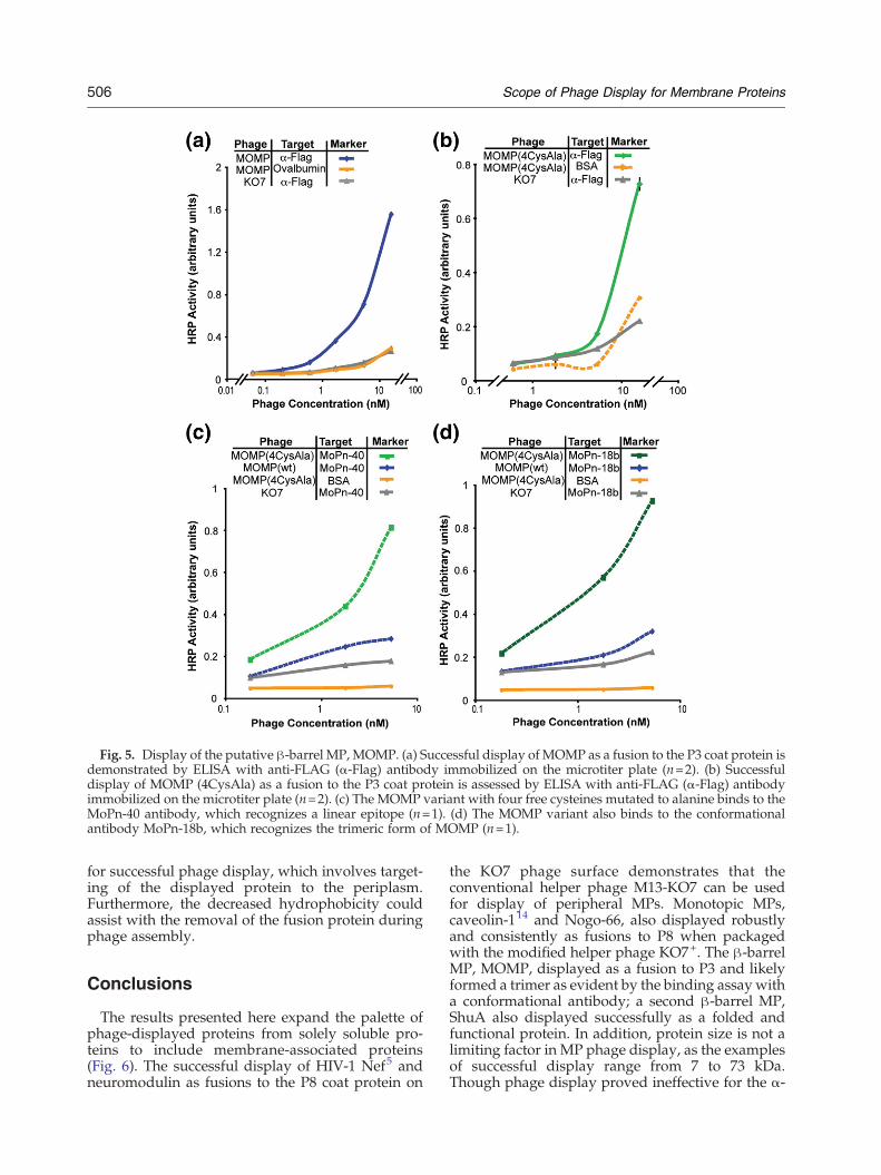
Fig. 5. Display of the putative β-barrel MP, MOMP. (a) Successful display of MOMP as a fusion to the P3 coat protein isdemonstrated by ELISA with anti-FLAG (α-Flag) antibody immobilized on the microtiter plate (n=2). (b) Successfuldisplay of MOMP (4CysAla) as a fusion to the P3 coat protein is assessed by ELISA with anti-FLAG (α-Flag) antibodyimmobilized on the microtiter plate (n=2). (c) The MOMP variant with four free cysteines mutated to alanine binds to theMoPn-40 antibody, which recognizes a linear epitope (n=1). (d) The MOMP variant also binds to the conformationalantibody MoPn-18b, which recognizes the trimeric form of MOMP (n=1).
506 Scope of Phage Display for Membrane Proteins
for successful phage display, which involves target-ing of the displayed protein to the periplasm.Furthermore, the decreased hydrophobicity couldassist with the removal of the fusion protein duringphage assembly.
Conclusions
The results presented here expand the palette ofphage-displayed proteins from solely soluble pro-teins to include membrane-associated proteins(Fig. 6). The successful display of HIV-1 Nef5 andneuromodulin as fusions to the P8 coat protein on
the KO7 phage surface demonstrates that theconventional helper phage M13-KO7 can be usedfor display of peripheral MPs. Monotopic MPs,caveolin-114 and Nogo-66, also displayed robustlyand consistently as fusions to P8 when packagedwith the modified helper phage KO7+. The β-barrelMP, MOMP, displayed as a fusion to P3 and likelyformed a trimer as evident by the binding assay witha conformational antibody; a second β-barrel MP,ShuA also displayed successfully as a folded andfunctional protein. In addition, protein size is not alimiting factor in MP phage display, as the examplesof successful display range from 7 to 73 kDa.Though phage display proved ineffective for the α-

Fig. 6. A guide to the phage display of MPs. As demonstrated here, peripheral MPs can require the negatively chargedsurface of wild-type KO7 helper phage. Monotopic MPs benefit from the zwitterionic surface of KO7+ helper phage.Unlike the requirements of peripheral and monotopic MPs, β-barrels MPs can be displayed through packaging witheither helper phage.
507Scope of Phage Display for Membrane Proteins
helical TM proteins, emerin, MAN1, matriptase-2and NET25, successful display of peripheral, mono-topic and β-barrel MPs illustrates the efficacy of theapproach. For the three classes of TM proteinsdisplayed on phage, we report at least two examplesof each class, which highlight functional foldingthrough binding to known ligands and receptors.Given the tremendous potential of phage-basedmutagenesis for protein engineering and structure–function studies, extending the technique to TMproteins should provide new possibilities for ex-ploring the world of MPs.
Materials and Methods
Phage display vector design
The genes encoding the MPs, emerin, MAN1, matrip-tase-2, MoPn MOMP, NET25, neuromodulin, mouseNogo-66 and ShuA were sub-cloned into the pM1165aphagemid between the ORF encoding a periplasmicsecretion signal peptide and P8. The pM1165a phagemidhas been previously reported43 and is based on pS1607phagemid.44 For display of the MPs as fusions to P3, themodified pS1602 phagemid45 was used. A FLAG epitopepeptide (amino acid sequence DYKDDDDK) fused to theN-terminus of the proteins provided an antibody epitopefor monitoring protein display levels. Throughout thisreport, successful cloning and mutagenesis were verifiedby DNA sequencing (Genewiz, Inc.).
Site-directedmutagenesis of neuromodulin andMOMP
Sequences of the mutagenic oligonucleotides appear inSupporting Information. Using the Stratagene QuikChangesite-directed mutagenesis protocol and oligonucleotidesNeuromodulinS41D-Fwd and NeuromodulinS41D-Rev,we generated the neuromodulin S41D gene. The C51A,C136A, C226A and C351A mutations were introduced intothe MOMP gene using oligonucleotide-directedmutagenesis.46 MOMP (4CysAla) was also subcloned intothe phagemid vector to encode a fusion to the P3 coat protein.
Purification of MP-displayed phage
As listed in Supporting Information, different strains ofE. coli were found to improve expression and display onthe phage surface (Table S2). K91, XL-1 or SS320 cells weretransformed with a phagemid encoding each of the MPs,and cells were grown at 30 or 37 °C in 2 ml 2YT mediumsupplemented with 50 μg/ml carbenicillin until theculture reached log-phase growth. KO7 or KO7+ helperphage (1010 phage/ml) was added, and the culture wasshaken at 37 °C for 0.5 h before transferring to 70 ml 2YTsupplemented with carbenicillin and kanamycin (25 μg/ml). The culture was incubated overnight for 21 or 16 h at30 or 37 °C respectively. Cells were removed bycentrifugation (10,000g, 10 min), and the phage precipi-tated from the supernatant by addition of one-fifthvolume of 20% polyethylene glycol 8000 in 2.5 M NaCl.The phages were recovered by centrifugation (10,000g for20 min and 5000g for 5 min), resuspended in phosphate-buffered saline (PBS) and 0.05% Tween-20 and isolated by

508 Scope of Phage Display for Membrane Proteins
centrifugation (10 min at 12,000g). The precipitation of thephage was repeated as described above with the additionof 20% polyethylene glycol 8000 in 2.5 M NaCl, followedby centrifugation and resuspension in PBS. Phage con-centrations were estimated spectrophotometrically(OD268=8.31 nM=5×10¹² phage/ml).
Phage-based ELISAs
A phage-based ELISA was used to assess relativedisplay and binding levels of the MPs. Specific wells of a96-well Nunc maxisorb plate were coated with anti-FLAGantibody (100 μl/well, 1:1000 dilution in 50 mM Na2CO3,pH 9.6; Stratagene) or the target protein (10 μg/ml) andincubated for 2 h at room temperature (RT) or overnight at4 °C. For ELISAs targeting f-actin, the actin protein(Sigma-Aldrich) was allowed to polymerize in Polymer-ization Buffer (2 mM MgCl2, 100 mM KCl, 0.2 mM DTT,0.2 mM ATP and 2 mM Tris, pH 7.6) for 1 h at RT beforecoating the plate.27 After removal of the coating solution,the wells were blocked for 30 min with BSA, ovalbumin orNFM (0.2%) in 50 mM PBS, pH 7.2. Control wells, whichwere not coated with the anti-FLAG antibody or targetprotein, were coated with the blocking buffer. The wellswere rinsed three times with wash buffer (0.05% Tween-20in PBS). Separately, serial dilutions of the MP displayingphage and KO7 or KO7+ phage (negative control) weremade in dilution buffer (0.1% Blocking agent with 0.05%Tween-20 in PBS). The phage solution was added to thecorresponding wells of target-coated ELISA plates andshaken on an orbital shaker for 1 h at RT. The wells werewashed five times with wash buffer and then incubatedwith horseradish-peroxidase-conjugated anti-M13 anti-body (100 μl/well, 1:5000 in the phage dilution buffer;GE Healthcare) for 30 min. The wells were washed fivetimes with wash buffer and twice with PBS. The boundreactants were detected by incubating with the substratesolution (100 μl/well; 2 mg/ml o-phenylenediaminedihydrochloride and 0.02% w/v H2O2, in citric acidbuffer, pH 5). Following an appropriate incubation time,the absorbance was measured spectrophotometrically at450 nm using a microtiter plate reader (μQuant; Bio-Tek).
Western blot of α-helical TM proteins
After the protocol described above for phage propaga-tion and isolation, samples of the E. coli cell pelletsexpressing the α-helical TM proteins, emerin, MAN1,matriptase-2, Net25 and the control, PTRF, were electro-phoretically separated by SDS-PAGE before transferringto a nitrocellulose membrane. The membrane was blockedwith 5% NFM in PBS for an hour. The primary antibodywas mouse anti-FLAG antibody (1:1000 dilution; Strata-gene), and the secondary antibody was horseradish-peroxidase-conjugated anti-mouse IgG antibody (1:1000dilution; Sigma). To visualize the immune blot, weincubated the membrane for 10 min with the addition ofa detection reagent (4-chloro-1-naphthol with 3,3′-diami-nobenzidine tetrahydrochloride).
Accession numbers
The National Center for Biotechnology Informationaccession numbers for emerin, MAN1 (LEMD3), matrip-
tase-2 (TMPRSS6), MOMP, Net25 (LEMD2), neuromodu-lin (GAP43), nogo-A (reticulon-4) and ShuA are BC000738,BC167864, BC039082, NP_296436, NP_851853, BC007936,NP_918943 and AAC27809, respectively.
Acknowledgements
We thank Drs. Larry Gerace for the NET25vector; Frank Torti for matriptase-2-encoding plas-mid; Luis de la Maza for the MOMP vector and theMoPn-40 and MoPn-18b antibodies; Angela Wilks,Thomas L. Poulos and Sarvind Tripathi for the ShuA vector; Sarvind Tripathi for the hemoglobinprotein; and Sachdev Sidhu for providing themodified pS1602 phagemid. We also thank Dr.Sudipta Majumdar for technical assistance andDavoud Mozhdehi for assistance with a figure.This work was supported by the National Institutesof Health, National Institute of General MedicalSciences (RO1-GM078528-01 to G.A.W.) and theRoman Reed Research Fund (RR05-155 to M.J.C.).
Supplementary Data
Supplementary data to this article can be foundonline at doi:10.1016/j.jmb.2011.10.021
References
1. Stevens, T. J. & Arkin, I. T. (2000). Do morecomplex organisms have a greater proportion ofmembrane proteins in their genomes? Proteins, 39,417–420.
2. Russell, R. B. & Eggleston, D. S. (2000). New roles forstructure in biology and drug discovery. Nat. Struct.Biol. 7, 928–930.
3. White, S. H. (2004). The progress of membrane proteinstructure determination. Protein Sci. 13, 1948–1949.
4. Popot, J. L. (2010). Amphipols, nanodiscs, andfluorinated surfactants: three nonconventional ap-proaches to studying membrane proteins in aqueoussolutions. Annu. Rev. Biochem. 79, 737–775.
5. Olszewski, A., Sato, K., Aron, Z. D., Cohen, F., Harris,A., McDougall, B. R. et al. (2004). Guanidine alkaloidanalogs as inhibitors of HIV-1 Nef interactions withp53, actin, and p56lck. Proc. Natl Acad. Sci. USA, 101,14079–14084.
6. Morrison, K. L. & Weiss, G. A. (2001). Combinatorialalanine-scanning. Curr. Opin. Chem. Biol. 5, 302–307.
7. Weiss, G. A., Watanabe, C. K., Zhong, A., Goddard, A.& Sidhu, S. S. (2000). Rapid mapping of proteinfunctional epitopes by combinatorial alanine scan-ning. Proc. Natl Acad. Sci. USA, 97, 8950–8954.
8. Hajduczki, A., Majumdar, S., Fricke, M., Brown, I. A.& Weiss, G. A. (2011). Solubilization of a membraneprotein by combinatorial supercharging. ACS Chem.Biol. 6, 301–307.

509Scope of Phage Display for Membrane Proteins
9. Majumdar, S., Hajduczki, A., Vithayathil, R., Olsen,T., Spitler, R., Mendez, A. et al. (2011). In vitroevolution of ligands to the membrane protein Caveo-lin. J. Am. Chem. Soc. 133, 9855–9862.
10. Smith, G. P. (1985). Filamentous fusion phage: novelexpression vectors that display cloned antigens on thevirion surface. Science, 228, 1315–1317.
11. Wells, J. A. & Lowman, H. B. (1992). Rapid evolutionof peptide and protein binding properties in vitro.Curr. Opin. Struct. Biol. 2, 597–604.
12. Kehoe, J. W. & Kay, B. K. (2005). Filamentous phagedisplay in the new millennium. Chem. Rev. 105,4056–4072.
13. Lamboy, J. A., Tam, P. Y., Lee, L. S., Jackson, P. J.,Avrantinis, S. K., Lee, H. J. et al. (2008). Chemical andgenetic wrappers for improved phage and RNAdisplay. ChemBioChem, 9, 2846–2852.
14. Majumdar, S., Hajduczki, A., Mendez, A. S. & Weiss,G. A. (2008). Phage display of functional, full-lengthhuman and viral membrane proteins. Bioorg. Med.Chem. Lett. 18, 5937–5940.
15. Clackson, T. & Wells, J. A. (1994). In vitro selectionfrom protein and peptide libraries. Trends Biotechnol.12, 173–184.
16. Wilson, D. R. & Finlay, B. B. (1998). Phage display:applications, innovations, and issues in phage andhost biology. Can. J. Microbiol. 44, 313–329.
17. Takahashi, T. T., Austin, R. J. & Roberts, R. W. (2003).mRNA display: ligand discovery, interaction analysisand beyond. Trends Biochem. Sci. 28, 159–165.
18. Paschke, M. & Hohne, W. (2005). A twin-argininetranslocation (Tat)-mediated phage display system.Gene, 350, 79–88.
19. Weiss, G. A., Wells, J. A. & Sidhu, S. S. (2000).Mutational analysis of the major coat protein of M13identifies residues that control protein display. ProteinSci. 9, 647–654.
20. Bothmann, H. & Pluckthun, A. (1998). Selection for aperiplasmic factor improving phage display andfunctional periplasmic expression. Nat. Biotechnol. 16,376–380.
21. Steiner, D., Forrer, P., Stumpp, M. T. & Pluckthun, A.(2006). Signal sequences directing cotranslationaltranslocation expand the range of proteins amenableto phage display. Nat. Biotechnol. 24, 823–831.
22. Singer, S. J. & Nicolson, G. L. (1972). The fluid mosaicmodel of the structure of cell membranes. Science, 175,720–731.
23. Resh, M. D. (1999). Fatty acylation of proteins: newinsights into membrane targeting of myristoylatedand palmitoylated proteins. Biochim. Biophys. Acta,1451, 1–16.
24. Jackson, J. H., Cochrane, C. G., Bourne, J. R., Solski,P. A., Buss, J. E. & Der, C. J. (1990). Farnesolmodification of Kirsten-ras exon 4B protein is essentialfor transformation. Proc. Natl Acad. Sci. USA, 87,3042–3046.
25. Ferguson, M. A. & Williams, A. F. (1988). Cell-surfaceanchoring of proteins via glycosyl-phosphatidylinosi-tol structures. Annu. Rev. Biochem. 57, 285–320.
26. Liang, X., Lu, Y., Neubert, T. A. & Resh, M. D. (2002).Mass spectrometric analysis of GAP-43/neuromodu-lin reveals the presence of a variety of fatty acylatedspecies. J. Biol. Chem. 277, 33032–33040.
27. He, Q., Dent, E. W. & Meiri, K. F. (1997). Modulationof actin filament behavior by GAP-43 (neuromodulin)is dependent on the phosphorylation status of serine41, the protein kinase C site. J. Neurosci. 17,3515–3524.
28. Chapman, E. R., Au, D., Alexander, K. A., Nicolson,T. A. & Storm, D. R. (1991). Characterization of thecalmodulin binding domain of neuromodulin. Func-tional significance of serine 41 and phenylalanine 42.J. Biol. Chem. 266, 207–213.
29. Rossenu, S., Leyman, S., Dewitte, D., Peelaers, D.,Jonckheere, V., Van Troys, M. et al. (2003). A phagedisplay-based method for determination of relativeaffinities of mutants. Application to the actin-bindingmotifs in thymosin beta 4 and the villin headpiece. J.Biol. Chem. 278, 16642–16650.
30. Blobel, G. (1980). Intracellular protein topogenesis.Proc. Natl Acad. Sci. USA, 77, 1496–1500.
31. Sapay, N., Montserret, R., Chipot, C., Brass, V.,Moradpour, D., Deleage, G. & Penin, F. (2006). NMRstructure and molecular dynamics of the in-planemembrane anchor of nonstructural protein 5A frombovine viral diarrhea virus. Biochemistry, 45,2221–2233.
32. Balali-Mood, K., Bond, P. J. & Sansom, M. S. (2009).Interaction of monotopic membrane enzymes with alipid bilayer: a coarse-grained MD simulation study.Biochemistry, 48, 2135–2145.
33. Vasudevan, S. V., Schulz, J., Zhou, C. & Cocco, M. J.(2010). Protein folding at the membrane interface, thestructure of Nogo-66 requires interactions with aphosphocholine surface. Proc. Natl Acad. Sci. USA,107, 6847–6851.
34. Raman, P., Cherezov, V. & Caffrey, M. (2006). TheMembrane Protein Data Bank. Cell. Mol. Life Sci. 63,36–51.
35. Eisenberg, D. (1984). Three-dimensional structure ofmembrane and surface proteins. Annu. Rev. Biochem.53, 595–623.
36. Schulz, G. E. (2000). β-Barrel membrane proteins.Curr. Opin. Struct. Biol. 10, 443–447.
37. Burkhard, K. A. & Wilks, A. (2007). Characterizationof the outer membrane receptor ShuA from the hemeuptake system of Shigella dysenteriae. Substrate spec-ificity and identification of the heme protein ligands. J.Biol. Chem. 282, 15126–15136.
38. Cobessi, D., Meksem, A. & Brillet, K. (2010). Structureof the heme/hemoglobin outer membrane receptorShuA from Shigella dysenteriae: heme binding by aninduced fit mechanism. Proteins, 78, 286–294.
39. Sun, G., Pal, S., Sarcon, A. K., Kim, S., Sugawara, E.,Nikaido, H. et al. (2007). Structural and functionalanalyses of the major outer membrane protein ofChlamydia trachomatis. J. Bacteriol. 189, 6222–6235.
40. Kelley, R. F., Totpal, K., Lindstrom, S. H., Mathieu, M.,Billeci, K., Deforge, L. et al. (2005). Receptor-selectivemutants of apoptosis-inducing ligand 2/tumor necro-sis factor-related apoptosis-inducing ligand reveal agreater contribution of death receptor (DR) 5 thanDR4 to apoptosis signaling. J. Biol. Chem. 280,2205–2212.
41. Bulieris, P. V., Behrens, S., Holst, O. & Kleinschmidt,J. H. (2003). Folding and insertion of the outermembrane protein OmpA is assisted by the chaperone

510 Scope of Phage Display for Membrane Proteins
Skp and by lipopolysaccharide. J. Biol. Chem. 278,9092–9099.
42. Lazar, S. W. & Kolter, R. (1996). SurA assists thefolding of Escherichia coli outer membrane proteins. J.Bacteriol. 178, 1770–1773.
43. Murase, K., Morrison, K. L., Tam, P. Y., Stafford, R. L.,Jurnak, F. & Weiss, G. A. (2003). EF-Tu bindingpeptides identified, dissected, and affinity optimizedby phage display. Chem. Biol. 10, 161–168.
44. Sidhu, S. S., Weiss, G. A. & Wells, J. A. (2000). Highcopy display of large proteins on phage for functionalselections. J. Mol. Biol. 296, 487–495.
45. Lee, C. V., Sidhu, S. S. & Fuh, G. (2004). Bivalentantibody phage display mimics natural immunoglob-ulin. J. Immunol. Methods, 284, 119–132.
46. Kunkel, T. A. (1985). Rapid and efficient site-specificmutagenesis without phenotypic selection. Proc. NatlAcad. Sci. USA, 82, 488–492.