Embed Size (px)
Citation preview

REVIEW ARTICLE
The sentinel node approach in gynaecological malignancies
Angela Collarino1,2• Sergi Vidal-Sicart3
• Germano Perotti1 •
Renato A. Valdes Olmos2,4,5
Received: 19 April 2016 / Accepted: 23 May 2016 / Published online: 13 June 2016
� The Author(s) 2016. This article is published with open access at Springerlink.com
Abstract This review discusses the state-of-the-art of
sentinel lymph node mapping in gynaecological malig-
nancies, including cervical cancer, endometrial cancer, and
vulvar cancer, with an emphasis on new technological
advances. For this objective, PubMed/MEDLINE was
searched for relevant studies about the sentinel lymph node
procedure in gynaecology. In particular, the use of preop-
erative lymphatic mapping with lymphoscintigraphy and
single photon emission tomography/computed tomography
(SPECT/CT) was identified in 18 studies. Other recent
advances as hybrid tracers (e.g. ICG-99mTc-nanocolloid)
and intraoperative tools (portable c-camera and 3D navi-
gation devices) appear to also represent a useful guide for
the surgeon during the operation. Concerning vulvar and
cervical cancers, the sentinel lymph node procedure has
been incorporated to the current guidelines in Europe and
North America, whereas for endometrial cancer it is con-
sidered investigative.
Keywords Cervical cancer � Endometrial cancer � Vulvarcancer � Sentinel lymph node � SPECT/CT
General introduction
In gynaecological tumours, the sentinel lymph node (SLN)
procedure is principally performed in vulvar cancer (VC),
cervical cancer (CC), and endometrial cancer (EC).
Although both preoperative lymphatic mapping and intra-
operative SLN detection are common parts of SLN pro-
cedure in gynaecological tumours, the type of injection and
lymphatic drainage is different for each one of these
malignancies (Fig. 1). In vulvar tumour, the lymphatic
drainage is predominantly superficial, and the first-draining
lymph nodes are usually located in the groin. Instead, the
lymphatic drainage of cervical and endometrial tumours is
deep, and SLNs are located along the iliac vessels as well
as in other areas with complex anatomy. Therefore, the use
of preoperative SPECT/CT appears to be mandatory in
cervical and endometrial tumours; whereas in vulvar
tumour, it is considered more optional. In addition, intra-
operative imaging, such as portable gamma-camera and
intraoperative 3D navigation SPECT/CT, represents com-
plementary tools useful to guide the surgeon in patients
with difficult SLN localization, such as those close to the
site of the injection or in complex anatomy areas. The new
hybrid tracer using indocyanine green with 99mTc-
nanocolloid (ICG99mTc-nanocolloid) improves the intra-
operative visualization of SLN, resulting useful during the
operation. All these particular aspects of SLN procedure in
gynaecological malignancies will be discussed in this
review. A research of the literature was performed on
PubMed/MEDLINE using the following keywords (MeSH
terms) to encounter the most relevant studies about the
& Renato A. Valdes Olmos
1 Institute of Nuclear Medicine, Universita Cattolica del Sacro
Cuore, Largo F. Vito, 1, 00168 Rome, Italy
2 Nuclear Medicine Section, Department of Radiology, Leiden
University Medical Center, Albinusdreef 2, 2333 ZA Leiden,
The Netherlands
3 Department of Nuclear Medicine, University Hospital Clınic
Barcelona, Villarroel, 170, 08036 Barcelona, Spain
4 Interventional Molecular Imaging Laboratory, Department of
Radiology, Leiden University Medical Center, Albinusdreef
2, 2333 ZA Leiden, The Netherlands
5 Department of Nuclear Medicine, The Netherlands Cancer
Institute-Antoni van Leeuwenhoek Hospital, Plesmanlaan
121, 1066 CX Amsterdam, The Netherlands
123
Clin Transl Imaging (2016) 4:411–420
DOI 10.1007/s40336-016-0187-6

SLN procedure in gynaecology: ‘‘SLN biopsy’’, ‘‘lym-
phatic mapping’’, ‘‘lymphoscintigraphy’’, ‘‘SPECT/CT’’,
‘‘intraoperative SLN detection’’, ‘‘hybrid tracer’’, ‘‘vulvar
cancer’’, ‘‘cervical cancer’’, and ‘‘endometrial cancer’’. The
search has been restricted to the English language. The
references of the retrieved articles were examined to
identify additional articles. This review also includes meta-
analyses published in the last five years.
Cervical cancer
Introduction
Cervical cancer (CC) is the third most common gynaeco-
logical cancer with an estimated of 12,990 new cases and
4120 deaths in the US, in 2016 [1]. The pattern of dissem-
ination of CC principally concerns the adjacent pelvic
organs, but can also spread to locoregional lymph nodes
(LN), while hematogenous spread to lung, liver, bone, and
brain is rare. The most important prognostic factor is the
presence of metastatic locoregional LN(s), including the
pelvic- and para-aortic lymph nodes [2, 3]. According to the
current guidelines, the preferred treatment for early-stage
disease (FIGO stages IA-2, IB-1, IIA-1) is radical hys-
terectomy and SLN mapping with or without bilateral pelvic
lymphadenectomy [4, 5]. The SLN(s) are the lymph
node(s) that receive direct drainage from the tumour [6];
thus, the tumour status of SLN(s) reflects the status of the
entire lymph node field. The SLN status plays an important
role, because when an SLN contains metastases at
histopathology, the best treatment approach would be based
on chemo-radiotherapy. In addition, when SLNs are nega-
tive for metastases, the pelvic lymph node dissection can be
safely avoided [4], reducing concomitant surgical morbidity.
The uterine cervix is a midline organ; thus, lymphatic
drainage is almost always bilateral and principally to the
pelvic region. The most frequent localization of pelvic
lymph node metastasis is the obturator followed by the
external iliac basins [7]. In addition, the lymphatic drainage
may spread to other areas, such as the common iliac and
para-aortic basins [8]. Nevertheless, it is rare to find ‘‘skip
metastasis’’ in the para-aortic basin without pelvic lymph
node metastases [9, 10]. Therefore, the SLN mapping is
useful for detection of lymphatic drainage patterns in par-
ticular to regions not routinely explored in conventional
surgery, such as para-aortic chains. The SLN mapping is
performed by peri-tumoural/peri-orificial injection of
radiocolloid (e.g. 99mTc-nanocolloid) in the four quadrants
of the cervix using a 20 or 22-gauge spinal needle. In the
case of previous conisation, the peri-cicatricial injection at
the four quadrants is recommended [5]. The most frequently
used tracer dose is approximately 110 MBq in a total vol-
ume of 2 mL [11]. The injection may be carried out the day
before surgery or on the same day of surgery. The Con-
ventional planar images are acquired for 3–5 min in anterior
and lateral views at 30 (early) and 60–120 (delayed) min
after injection [5]. The early images are used to visualize
lymphatic duct(s) and the first-draining lymph node(s). The
delayed images are used to differentiate the SLN(s) from
higher echelon nodes [12]. A higher echelon node is defined
as an LN draining from the SLN(s). The preoperative planar
lymphoscintigraphy does not give a precise anatomical
localization of the SLN(s) [13]. Therefore, SPECT in con-
junction with low-dose CT (SPECT/CT) is recommended
immediately after delayed imaging as a complementary
modality [5], providing not just better contrast and spatial
resolution in comparison to planar imaging, but also accu-
rate anatomical information (Fig. 2).
Fig. 1 Anatomical sentinel lymph-node (SLN) distribution in gynae-
cological malignancies. In vulvar cancer (Ref. [48]), SLNs are limited
to the groin and are predominantly found in the superior, central, and
medial inferior inguinal Daseler’s zones (a). By contrast, in cervical
cancer. (b) SLNs are mainly located along the iliac vessels (Ref.
[15]), whereas in endometrial cancer (c) also para-aortic drainage is
frequently observed (Ref. [17])
412 Clin Transl Imaging (2016) 4:411–420
123

Advantages of preoperative SPECT/CT imaging
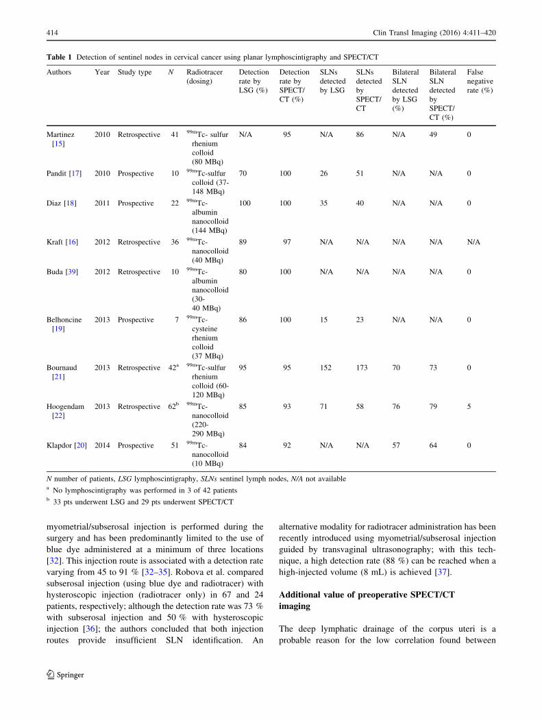
SPECT/CT has higher SLN detection rate compared to the
conventional planar images (98.6 vs. 85.3 %), as reported
in a recent meta-analysis, including eight studies [14]. In
general, SPECT/CT provides an accurate anatomical SLN
localization [15–18]. In particular, SPECT/CT images are
useful to detect SLN(s) close to injection sites, such as
parametrial SLNs, as well as SLN(s) in uncommon loca-
tions, such as the para-aortic and presacral basins
[15, 17, 19]. In addition, SPECT/CT leads to better
detection of bilateral SLN(s) compared to planar imaging
[20–22] (Table 1). Furthermore, Hoogendam et al. reported
that SPECT/CT provides a valuable surgical roadmap,
reducing the surgical time in cervical cancer in women
undergoing robotic-assisted surgery [22]. Recently, 99mTc
SPECT/MRI-fused images have been used for SLN map-
ping in preoperative assessment of SLN metastases in the
early-stage cervical cancer in women. The authors found a99mTc SPECT-MRI accuracy of 74.9 % (95 % CI
0.569–0.930) to non-invasively assess SLN metastases,
including 136 SLNs of which 13 (9.6 %) in 8/79 patients
(10.7 %) contained metastases [23].
Endometrial cancer
Introduction
Endometrial cancer (EC) is the most common malignancy
of gynaecological cancer with an estimated incidence of
60,050 new cases and 10,470 deaths in the US, in 2016 [1].
Lymph-node status is a key prognostic factor in
endometrial tumours. Indeed, the 5-year survival rate var-
ies from 44 to 52 % when pelvic- or para-aortic node
lymph nodes contained metastases [24]. Radical pelvic-
and para-aortic lymphadenectomies represent the standard
treatment in high-risk group (grade 3,[50 % myometrial
invasion) or high-risk tumour histology (papillary serous,
carcinosarcoma, and clear cell cancer) [25]. The SLN
technique may provide the surgical staging, avoiding the
morbidity of complete lymphadenectomy in patients with
negative SLN biopsy, but also ultra-staging assessment
(micro-metastases and isolated tumour cells) through
extensive immunochemistry. Although there are several
studies validating SLN mapping in EC, this technique is
not yet the standard of care in the early-stage EC (Stage I-II
high-risk) [5, 25]. One of the most controversial aspects for
SLN mapping is the modality of injection. Indeed, three
different modalities of injection have been described in the
currently literature: (i) cervical injection; (ii) endometrial
peri-tumoural injection assisted by hysteroscopy; and (iii)
myometrial/subserosal intraoperative injection. The radio-
tracer can be injected on the day prior to surgery, providing
lymphatic mapping with planar and SPECT/CT images.
The most common and easiest approach is the cervical
injection, which is performed peri-orificially into the four
quadrants as well as for CC. The detection rate related to
cervical injection is the highest of the three injection
modalities used in endometrial cancer, ranging from 62 to
100 % [26]. Endometrial radiotracer administration assis-
ted by hysteroscopy allows direct injection around the
tumour. This procedure is usually performed at the
beginning of the surgery, without the possibility to obtain
preoperative SLN mapping. The detection rate of this
injection modality varies from 40 to 95 % [27–31]. Finally,
Fig. 2 Cervical cancer. Planar
images show a bilateral
drainage in pelvic area (a–c).Volume-rendering image
displays the level of sentinel
nodes (d). SPECT/CT axial-
fused images showing two
separate nodes with high tracer
uptake in right obturator fossa
as well as three tiny nodes in
left side (e). Correspondingaxial CT slice (f)
Clin Transl Imaging (2016) 4:411–420 413
123
myometrial/subserosal injection is performed during the
surgery and has been predominantly limited to the use of
blue dye administered at a minimum of three locations
[32]. This injection route is associated with a detection rate
varying from 45 to 91 % [32–35]. Robova et al. compared
subserosal injection (using blue dye and radiotracer) with
hysteroscopic injection (radiotracer only) in 67 and 24
patients, respectively; although the detection rate was 73 %
with subserosal injection and 50 % with hysteroscopic
injection [36]; the authors concluded that both injection
routes provide insufficient SLN identification. An
alternative modality for radiotracer administration has been
recently introduced using myometrial/subserosal injection
guided by transvaginal ultrasonography; with this tech-
nique, a high detection rate (88 %) can be reached when a
high-injected volume (8 mL) is achieved [37].
Additional value of preoperative SPECT/CT
imaging
The deep lymphatic drainage of the corpus uteri is a
probable reason for the low correlation found between
Table 1 Detection of sentinel nodes in cervical cancer using planar lymphoscintigraphy and SPECT/CT
Authors Year Study type N Radiotracer
(dosing)
Detection
rate by
LSG (%)
Detection
rate by
SPECT/
CT (%)
SLNs
detected
by LSG
SLNs
detected
by
SPECT/
CT
Bilateral
SLN
detected
by LSG
(%)
Bilateral
SLN
detected
by
SPECT/
CT (%)
False
negative
rate (%)
Martinez
[15]
2010 Retrospective 41 99mTc- sulfur
rhenium
colloid
(80 MBq)
N/A 95 N/A 86 N/A 49 0
Pandit [17] 2010 Prospective 10 99mTc-sulfur
colloid (37-
148 MBq)
70 100 26 51 N/A N/A 0
Diaz [18] 2011 Prospective 22 99mTc-
albumin
nanocolloid
(144 MBq)
100 100 35 40 N/A N/A 0
Kraft [16] 2012 Retrospective 36 99mTc-
nanocolloid
(40 MBq)
89 97 N/A N/A N/A N/A N/A
Buda [39] 2012 Retrospective 10 99mTc-
albumin
nanocolloid
(30-
40 MBq)
80 100 N/A N/A N/A N/A 0
Belhoncine
[19]
2013 Prospective 7 99mTc-
cysteine
rhenium
colloid
(37 MBq)
86 100 15 23 N/A N/A 0
Bournaud
[21]
2013 Retrospective 42a 99mTc-sulfur
rhenium
colloid (60-
120 MBq)
95 95 152 173 70 73 0
Hoogendam
[22]
2013 Retrospective 62b 99mTc-
nanocolloid
(220-
290 MBq)
85 93 71 58 76 79 5
Klapdor [20] 2014 Prospective 51 99mTc-
nanocolloid
(10 MBq)
84 92 N/A N/A 57 64 0
N number of patients, LSG lymphoscintigraphy, SLNs sentinel lymph nodes, N/A not availablea No lymphoscintigraphy was performed in 3 of 42 patientsb 33 pts underwent LSG and 29 pts underwent SPECT/CT
414 Clin Transl Imaging (2016) 4:411–420
123
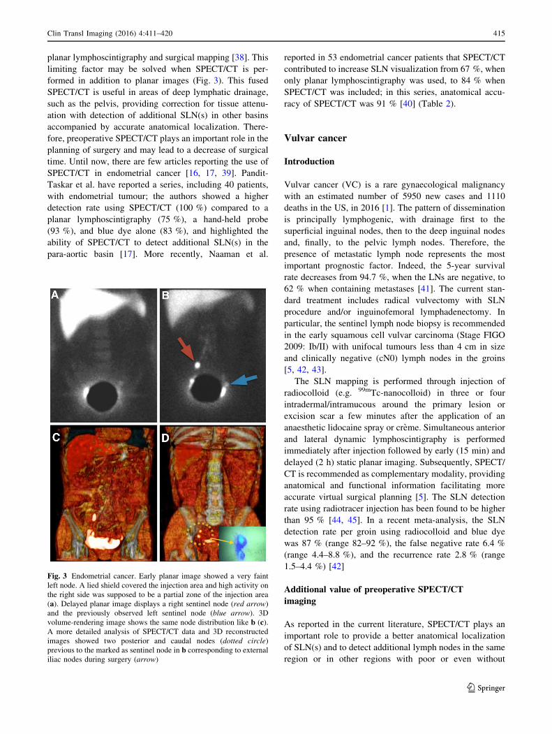
planar lymphoscintigraphy and surgical mapping [38]. This
limiting factor may be solved when SPECT/CT is per-
formed in addition to planar images (Fig. 3). This fused
SPECT/CT is useful in areas of deep lymphatic drainage,
such as the pelvis, providing correction for tissue attenu-
ation with detection of additional SLN(s) in other basins
accompanied by accurate anatomical localization. There-
fore, preoperative SPECT/CT plays an important role in the
planning of surgery and may lead to a decrease of surgical
time. Until now, there are few articles reporting the use of
SPECT/CT in endometrial cancer [16, 17, 39]. Pandit-
Taskar et al. have reported a series, including 40 patients,
with endometrial tumour; the authors showed a higher
detection rate using SPECT/CT (100 %) compared to a
planar lymphoscintigraphy (75 %), a hand-held probe
(93 %), and blue dye alone (83 %), and highlighted the
ability of SPECT/CT to detect additional SLN(s) in the
para-aortic basin [17]. More recently, Naaman et al.
reported in 53 endometrial cancer patients that SPECT/CT
contributed to increase SLN visualization from 67 %, when
only planar lymphoscintigraphy was used, to 84 % when
SPECT/CT was included; in this series, anatomical accu-
racy of SPECT/CT was 91 % [40] (Table 2).
Vulvar cancer
Introduction
Vulvar cancer (VC) is a rare gynaecological malignancy
with an estimated number of 5950 new cases and 1110
deaths in the US, in 2016 [1]. The pattern of dissemination
is principally lymphogenic, with drainage first to the
superficial inguinal nodes, then to the deep inguinal nodes
and, finally, to the pelvic lymph nodes. Therefore, the
presence of metastatic lymph node represents the most
important prognostic factor. Indeed, the 5-year survival
rate decreases from 94.7 %, when the LNs are negative, to
62 % when containing metastases [41]. The current stan-
dard treatment includes radical vulvectomy with SLN
procedure and/or inguinofemoral lymphadenectomy. In
particular, the sentinel lymph node biopsy is recommended
in the early squamous cell vulvar carcinoma (Stage FIGO
2009: Ib/II) with unifocal tumours less than 4 cm in size
and clinically negative (cN0) lymph nodes in the groins
[5, 42, 43].
The SLN mapping is performed through injection of
radiocolloid (e.g. 99mTc-nanocolloid) in three or four
intradermal/intramucous around the primary lesion or
excision scar a few minutes after the application of an
anaesthetic lidocaine spray or creme. Simultaneous anterior
and lateral dynamic lymphoscintigraphy is performed
immediately after injection followed by early (15 min) and
delayed (2 h) static planar imaging. Subsequently, SPECT/
CT is recommended as complementary modality, providing
anatomical and functional information facilitating more
accurate virtual surgical planning [5]. The SLN detection
rate using radiotracer injection has been found to be higher
than 95 % [44, 45]. In a recent meta-analysis, the SLN
detection rate per groin using radiocolloid and blue dye
was 87 % (range 82–92 %), the false negative rate 6.4 %
(range 4.4–8.8 %), and the recurrence rate 2.8 % (range
1.5–4.4 %) [42]
Additional value of preoperative SPECT/CT
imaging
As reported in the current literature, SPECT/CT plays an
important role to provide a better anatomical localization
of SLN(s) and to detect additional lymph nodes in the same
region or in other regions with poor or even without
Fig. 3 Endometrial cancer. Early planar image showed a very faint
left node. A lied shield covered the injection area and high activity on
the right side was supposed to be a partial zone of the injection area
(a). Delayed planar image displays a right sentinel node (red arrow)
and the previously observed left sentinel node (blue arrow). 3D
volume-rendering image shows the same node distribution like b (c).A more detailed analysis of SPECT/CT data and 3D reconstructed
images showed two posterior and caudal nodes (dotted circle)
previous to the marked as sentinel node in b corresponding to external
iliac nodes during surgery (arrow)
Clin Transl Imaging (2016) 4:411–420 415
123

visualization at planar lymphoscintigraphy (Fig. 4), as well
as to reduce the false positive rate possibly due to external
contamination or presence of radioactivity in enlarged
lymphatic vessels, [16, 19, 46, 47]. Recently, Collarino
et al. reported the use of SPECT/CT for anatomical map-
ping of lymphatic drainage in vulvar cancer. According to
the five Daseler zones using the inguinal saphenofemoral
junction as anatomical reference, the authors found that the
lymphatic drainage was principally to the medial inguinal
region (83 %), and the drainage to the lateral inferior groin
was only incidental (0.5 %) in 83 patients with cN0 vulvar
cancer (Fig. 1). Further drainage to higher echelon nodes
was visualized in the groin (15 %) and in the pelvis
(85 %). Therefore, SPECT/CT is able to personalize the
lymphatic mapping, and has a potential role in limiting the
extent of lymph node dissection to the lateral inferior zone
in patients with positive SLN(s) [48] (Table 3).
Advancements in intraoperative imagingand instrumentation
The indocyanine green (ICG) added to 99mTc-nanocolloid
in one signature represents a new hybrid tracer for the
detection of SLN. Recently, Matheron et al. reported the
use of a new hybrid tracer, in the SLN identification pro-
cedure in vulvar cancer. They showed that 98 % of the SNs
were radioactive at the time of excision, 96 % were fluo-
rescent, and only 65 % were blue in 15 patients. The
additional value of ICG is related to better intraoperative
visualization of SN to optimize the intraoperative SLN
visualization using the fluorescence component [49].
Nevertheless, there are no studies available on application
of this hybrid tracer in cervical cancer and endometrial
cancer. Indeed, a recently meta-analysis, including 67
studies in cervical cancer reported only the SLN detection
rate using the combination of radiotracer with blue dye
compared to the single use of radiotracer, blue dye, and
florescence imaging (92.3 vs. 90.9 vs. 80.9 vs 76.5 %)
[50]. In the future, the ICG-99mTc-nanocolloid may be used
in the SLN(s) identification procedure during the robot-
assisted laparoscopy [51, 52]. In addition, a portable c-camera might be a complementary tool during surgery in
gynaecological cancer. With a portable c-camera an
intraoperative real-time imaging is acquired before SLN
resection. Subsequently, an additional image of the surgi-
cal field is performed to confirm the absence of any
residual activity after excision. This device may improve
the intraoperative detection rate in patients with difficult
SLN localization in parametrial and precaval lymph-node
basins. Indeed, the hand-held c-probe has limitations to
detect parametrial SLN(s) due to their location in the
vicinity of the injection site, and in precavalTa
ble
2Detectionofsentinel
nodes
inendometrial
cancerusingplanar
lymphoscintigraphyandin
SPECT/CT
Authors
Year
Studytype
NInjection
site
Radiotracer
(dosing)
Detection
rate
byLSG
(%)
Detectionrate
bySPECT/CT
(%)
SLNs
detected
byLSG
SLNs
detectedby
SPECT/CT
Bilateral
SLN
detectionby
LSG
(%)
Bilateral
SLN
detectionby
SPECT/CT(%
)
False
negative
rate
(%)
Pandit
[17]
2010
Prospective
40
C99mTc-sulfur
colloid
(37-
148MBq)
75
100
67
207
N/A
N/A
0
Kraft
[16]
2012
Retrospective
21
C99mTc-nanocolloid
(40MBq)
86
86
N/A
N/A
N/A
N/A
N/A
Buda
[39]
2012
Prospective
25
C99mTc-albumin
nanocolloid
(30-
40MBq)
40
88
N/A
N/A
N/A
N/A
0
Naaman
[40]
2016
Retrospective
45a
C99mTc-albumin
nanocolloid
(7.4-
1.1
MBq)
67
84
N/A
N/A
29
49
0
Nnumber
ofpatients,C
cervix,LSG
lymphoscintigraphy,SLNssentinel
lymphnodes,N/A
notavailable
aOnly
37of45underwentSPECT/CT
416 Clin Transl Imaging (2016) 4:411–420
123

SLN(s) because of the liver activity. Vidal-Sicart et al.
reported the use of portable c-camera in gynaecological
cancer, showing a higher detection (92 %) when compared
to just hand-held c-probe (77 %) in the two cases of high-
risk endometrial cancer, three cases of cervical cancer and
one patients with vulvar cancer [53]. Furthermore, the
incorporation of co-registered SPECT/CT to 3D navigation
probe may be used during the operation offering a 3D
roadmap to the surgeon and facilitating the anatomical
localization of SLN(s). In the current literature on
radioguided surgery, this approach has been reported in
penile cancer by Brower et al. [54].
Conclusion
In conclusion, the SLN procedure has widely been vali-
dated in vulvar cancer and cervical cancer. Its application
in these malignancies is well standardized and has been
incorporated to the current guidelines in Europe and North
Fig. 4 In a patient with vulvar cancer, delayed planar imaging
(a) shows one SLN in the right groin (red arrow) corresponding with
one allocated SLN uptake (red arrow) on transversal-fused SPECT/
CT (b) and two not enlarged lymph nodes on transversal CT
(c) (double arrows). In another patient, delayed planar image
(d) shows unilateral lymphatic drainage with a single SLN in the
right groin (red arrow), while transversal-fused SPECT/CT (e) showsbilateral drainage with also a contralateral SLN (red arrow)
corresponding with a not enlarged lymph node in the left groin on
CT (f)
Table 3 Detection of sentinel nodes in vulvar cancer using planar lymphoscintigraphy and SPECT/CT
Authors Year Study type N Radiotracer (dosing) Detection
rate by LSG
(%)
Detection rate
by SPECT/CT
(%)
SLNs
detected by
LSG
SLNs detected
by SPECT/CT
False
negative
rate (%)
Beneder
[46]
2008 Prospective 10 99mTc-nanocolloid
(60 MBq)
N/A N/A 26 38 0
Kraft [16] 2012 Retrospective 7 99mTc-nanocolloid
(40 MBq)
100 100 N/A N/A N/A
Belhoncine
[19]
2013 Prospective 7 99mTc-cysteine
rhenium colloid
(37 MBq)
86 100 N/A N/A 0
Matheron
[49]
2013 Prospective 14 ICG-99mTc-
nanocolloid
(87 MBq)
N/A N/A 39 39 0
Collarino
[48]
2015 Retrospective 83 99mTc-nanocolloid
(81 MBq)
N/A N/A 192 217 0
N number of patients, LSG lymphoscintigraphy, SLNs sentinel lymph nodes, N/A not available, ICG indocyanine green
Clin Transl Imaging (2016) 4:411–420 417
123

America. By contrast, in endometrial cancer, there are
various controversial aspects (e.g. injection route) to be
clarified and the use of the SLN procedure needs to be
validated in larger clinical series. Beside these aspects, the
present review showed that recent technological advances,
such as preoperative and intraoperative use of SPECT/CT,
the contribution of the hybrid tracer ICG-99mTc-nanocol-
loid, and technological advances like SLN robotic-guided
procedure might play an increasing role to guide gynae-
cological cancer surgery in the future.
Compliance with ethical standards
Conflict of interest All four authors (Angela Collarino, Sergi Vidal-
Sicart, Germano Perotti, and Renato A. Valdes Olmos) declare that
they have no conflict of interest.
Ethical approval This article does not contain any studies with
human participants or animal performed by the any of the authors.
Open Access This article is distributed under the terms of the
Creative Commons Attribution 4.0 International License (http://crea
tivecommons.org/licenses/by/4.0/), which permits unrestricted use,
distribution, and reproduction in any medium, provided you give
appropriate credit to the original author(s) and the source, provide a
link to the Creative Commons license, and indicate if changes were
made.
References
1. Siegel RL, Miller KD, Jemal S (2016) Cancer statistics. CA
Cancer J Clin 66(1):7–30. doi:10.3322/caac.21332
2. Stehman FB, Bundy BN, DiSaia PJ, Keys HM, Larson JE, Fowler
WC (1991) Carcinoma of the cervix treated with radiation ther-
apy. I. A multi-variate analysis of prognostic variables in the
Gynecologic Oncology Group. Cancer 67(11):2776–2785
3. Macdonald OK, Chen J, Dodson M, Lee CM, Gaffney DK (2009)
Prognostic significance of histology and positive lymph node
involvement following radical hysterectomy in carcinoma of the
cervix. Am J Clin Oncol 32(4):411–416. doi:10.1097/COC.
0b013e31819142dc
4. Koh WJ, Greer BE, Abu-Rustum NR, Apte SM, Campos SM,
Cho KR et al (2015) Cervical cancer, version 2.2015. J Natl
Compr Canc Netw 13(4):395–404
5. Giammarile F, Bozkurt MF, Cibula D, Pahisa J, Oyen WJ, Paredes
P, Olmos RV, Sicart SV (2014) The EANM clinical and techni-
cal guidelines for lymphoscintigraphy and sentinel node local-
ization ingynaecological cancers. Eur J Nucl Med Mol Imaging
41(7):1463–1477. doi:10.1007/s00259-014-2732-8
6. Nieweg OE (2012) The sentinel lymph node concept in oncology
surgery. In: Mariani G, Manca G, Orsini P, Vidal-Sicar t S,
Valdes Olmos R (eds) Atlas of lymphoscintigraphy and sentinel
node mapping. Springer, Milan, pp 87–93
7. Benedetti-Panici P, Maneschi F, Scambia G, Greggi S, Cutillo G,
D’Andrea G, Rabitti C, Coronetta F, Capelli A, Mancuso S
(1996) Lymphatic spread of cervical cancer: an anatomical and
pathological study based on 225 radical hysterectomies with
systematic pelvic and aortic lymphadenectomy. Gynecol Oncol
62(1):19–24
8. Sakuragi N, Satoh C, Takeda N, Hareyama H, Takeda M,
Yamamoto R, Fujimoto T, Oikawa M, Fujino T, Fujimoto S
(1999) Incidence and distribution pattern of pelvic and paraaortic
lymph node metastasis in patients with stages IB, IIA, and IIB
cervical carcinoma treated with radical hysterectomy. Cancer
85(7):1547–1554
9. Lea JS, Sheets EE, Duska LR, Miller DS, Schorge JO (2002)
Early-stage cervical adenocarcinoma treated by surgical intent:
the role of paraaortic lymph node dissection. Gynecol Oncol
84(2):285–288
10. Bader AA, Winter R, Haas J, Tamussino KF (2007) Where to
look for the sentinel lymph node in cervical cancer. Am J Obstet
Gynecol 197(6):678.e1–7
11. El-Ghobashy AE, Saidi SA (2009) Sentinel lymph node sampling
in gynaecological cancers: techniques and clinical applications.
Eur J Surg Oncol 35(7):675–685. doi:10.1016/j.ejso.2008.09.004
12. Paredes P, Vidal-Sicart S (2012) Preoperative and intraoperative
lymphatic mapping for radioguided sentinel node biopsy in
cancers of the female reproductive system. In: Mariani G, Manca
G, Orsini P, Vidal-Sicar t S, Valdes Olmos R (eds). Atlas of
lymphoscintigraphy and sentinel node mapping. Springer, Milan,
pp 249–268
13. Vermeeren L, van der Ploeg IM, Olmos RA, Meinhardt W, Klop
WM, Kroon BB, Nieweg OE (2010) SPECT/CT for preoperative
sentinel node localization. J Surg Oncol 101(2):184–190. doi:10.
1002/jso.21439
14. Hoogendam JP, Veldhuis WB, Hobbelink MG, Verheijen RH,
van den Bosch MA, Zweemer RP (2015) 99 mTc SPECT/CT
versus planar lymphoscintigraphy for preoperative sentinel lymph
node detection in cervical cancer: a systematic review and
metaanalysis. J Nucl Med 56(5):675–680. doi:10.2967/jnumed.
114.152439
15. Martınez A, Zerdoud S, Mery E, Bouissou E, Ferron G, Querleu
D (2010) Hybrid imaging by SPECT/CT for sentinel lymph node
detection in patients with cancer of the uterine cervix. Gynecol
Oncol 119(3):431–435. doi:10.1016/j.ygyno.2010.08.001
16. Kraft O, Havel MD (2012) Detection of sentinel lymph nodes in
gynecologic tumours by planar scintigraphy and SPECT/CT. Mol
Imaging Radionucl Ther 21(2):47–55. doi:10.4274/Mirt.236
17. Pandit-Taskar N, Gemignani ML, Lyall A, Larson SM, Barakat
RR, Abu Rustum NR (2010) Single photon emission computed
tomography SPECT-CT improves sentinel node detection and
localization in cervical and uterine malignancy. Gynecol Oncol
117(1):59–64. doi:10.1016/j.ygyno.2009.12.021
18. Dıaz-Feijoo B, Perez-Benavente MA, Cabrera-Diaz S, Gil-Mor-
eno A, Roca I, Franco-Camps S, Fernandez MS, Garcıa-Jimenez
A, Xercavins J, Martınez-Palones JM (2011) Change in clinical
management of sentinel lymph node location in early stage cer-
vical cancer: the role of SPECT/CT. Gynecol Oncol
120(3):353–357. doi:10.1016/j.ygyno.2010.12.336
19. Belhocine TZ, Prefontaine M, Lanvin D, Bertrand M, Rachinsky
I, Ettler H, Zabel P, Stitt LW, Sugimoto A, Urbain JL (2013)
Added-value of SPECT/CT to lymphatic mapping and sentinel
lymphadenectomy in gynaecological cancers. Am J Nucl Med
Mol Imaging 3(2):182–193
20. Klapdor R, Mucke J, Schneider M, Langer F, Gratz KF, Hille-
manns P, Hertel H (2014) Value and advantages of preoperative
sentinel lymph node imaging with SPECT/CT in cervical cancer.
Int J Gynecol Cancer 24(2):295–302. doi:10.1097/IGC.
0000000000000032
21. Bournaud C, Le Bail-Carval K, Scheiber C, de Charry C,
Mathevet P, Moreau-Triby C (2013) Value of SPECT/CT in
lymphatic mapping in cervix and endometrial cancer. Med Nucl
37:387–396
22. Hoogendam JP, Hobbelink MG, Veldhuis WB, Verheijen RH,
van Diest PJ, Zweemer RP (2013) Preoperative sentinel node
mapping with (99 m)Tc-nanocolloid SPECT-CT significantly
reduces the intraoperative sentinel node retrieval time in robot
418 Clin Transl Imaging (2016) 4:411–420
123

assisted laparoscopic cervical cancer surgery. Gynecol Oncol
129(2):389–394. doi:10.1016/j.ygyno.2013.02.020
23. Hoogendam JP, Zweemer RP, HobbelinkMG, van den BoschMA,
Verheijen RH, Veldhuis WB (2016) 99mTc-nanocolloid SPECT-
MRI fusion for the selective assessment of non-enlarged sen-
tinel lymph nodes in patients with early stage cervical cancer.
J Nucl Med 57(4):551–556. doi:10.2967/jnumed.115.164780
24. Partridge EE, Shingleton HM, Menck HR (1996) The national
cancer data base report on endometrial cancer. J Surg Oncol
61(2):111–123
25. National Comprehensive Cancer Network, (2016) Uterine neo-
plasm, version 2.2016. http://www.nccn.org/professionals/physi
cian_gls/pdf/uterine.pdf. Accessed 28 March 2016
26. Cormier B, Rozenholc AT, Gotlieb W, Plante M, Giede C,
Communities of Practice (CoP) Group of Society of Society of
Gynecologic Oncology of Canada (GOC) (2015) Sen-
tinel lymph node procedure in endometrial cancer: a system-
atic review and proposal for standardization offuture research.
Gynecol Oncol 138(2):478–485. doi:10.1016/j.ygyno.2015.05.
039
27. Gien LT, Kwon JS, Carey MS (2005) Sentinel node mapping
with isosulfan blue dye in endometrial cancer. J Obstet Gynaecol
Can 27(12):1107–1112
28. Maccauro M, Lucignani G, Aliberti G, Villano C, Castellani MR,
Solima E, Bombardieri E (2005) Sentinel lymph node detection
following the hysteroscopic peritumoural injection of 99mTc-
labelled albumin nanocolloid in endometrial cancer. Eur J Nucl
Med Mol Imaging 32(5):569–574
29. Delaloye JF, Pampallona S, Chardonnens E, Fiche M, Lehr HA,
DeGrandi P, Delaloye AB (2007) Intraoperative lymphatic
mapping and sentinel node biopsy using hysteroscopy in patients
with endometrial cancer. Gynecol Oncol 106(1):89–93
30. Clement D, Bats AS, Ghazzar-Pierquet N, Le Frere Belda MA,
Larousserie F, Nos C, Lecuru F (2008) Sentinel lymph nodes in
endometrial cancer: is hysteroscopic injection valid? Eur J
Gynaecol Oncol 29(3):239–241
31. Solima E, Martinelli F, Ditto A, Maccauro M, Carcangiu M,
Mariani L, Kusamura S, Fontanelli R, Grijuela B, Raspagliesi F
(2012) Diagnostic accuracy of sentinel node in endometrial
cancer by using hysteroscopic injection of radiolabeled tracer.
Gynecol Oncol 126(3):419–423. doi:10.1016/j.ygyno.2012.05.
025
32. Frumovitz M, Bodurka DC, Broaddus RR, Coleman RL, Sood
AK, Gershenson DM, Burke TW, Levenback CF (2007) Lym-
phatic mapping and sentinel node biopsy in women with high-risk
endometrial cancer. Gynecol Oncol 104(1):100–103
33. Altgassen C, Pagenstecher J, HornungD Diedrich K, Hornemann
A (2007) A new approach to label sentinel nodes in endometrial
cancer. Gynecol Oncol 105(2):457–461
34. Li B, Li XG, Wu LY, Zhang WH, Li SM, Min C, Gao JZ (2007)
A pilot study of sentinel lymph nodes identification in patients
with endometrial cancer. Bull Cancer 94(1):E1–E4
35. Lopes LA, Nicolau SM, Baracat FF, Goncalves WJ, Santos HV,
Lopes RG, Lippi UG (2007) Sentinel lymph node in endometrial
cancer. Int J Gynecol Cancer 17(5):1113–1117
36. Robova H, Charvat M, Strnad P, Hrehorcak M, Taborska K,
Skapa P, Rob L (2009) Lymphatic mapping in endometrial can-
cer: comparison of hysteroscopic and subserosal injection and the
distribution of sentinel lymph nodes. Int J Gynecol Cancer
19(3):391–394. doi:10.1111/IGC.0b013e3181a1c0b1
37. Torne A, Pahisa J, Vidal-Sicart S, Martınez-Roman S, Paredes P,
Puerto B, Albela S, Fuste P, Perisinotti A, Ordi J (2013)
Transvaginal ultrasound-guided myometrial injection of radio-
tracer (TUMIR): a new method for sentinel lymph node detection
in endometrial cancer. Gynecol Oncol 128(1):88–94. doi:10.
1016/j.ygyno.2012.10.008
38. Ballester M, Rouzier R, Countant C, Kerrou K, Daraı E (2009)
Limits of lymphoscintigraphy for sentinel node biopsy in women
with endometrial cancer. Gynecol Oncol 112(2):348–352. doi:10.
1016/j.ygyno.2008.11.004
39. Buda A, Elisei F, Arosio M, Dolci C, Signorelli M, Perego P,
Giuliani D, Recalcati D, Cattoretti G, Milani R, Messa C (2012)
Integration of hybrid single-photon emission computed tomog-
raphy/computed tomography in the preoperative assessment of
sentinel node in patients with cervical and endometrial cancer:
our experience and literature review. Int J Gynecol Cancer
22(5):830–835. doi:10.1097/IGC.0b013e318253496f
40. Naaman Y, Pinkas L, Roitman S, Ikher S, Oustinov N, Vaisbuch
E, Yachnin A, Ben-Arie A (2016) The added value of SPECT/CT
in sentinel lymph nodes mapping for endometrial carcinoma. Ann
Surg Oncol 23(2):450–455. doi:10.1245/s10434-015-4877-5
41. Burger MP, Hollema H, Emanuels AG, Krans M, Pras E, Bouma
J (1995) The importance of the groin node status for the survival
of T1 and T2 vulval carcinoma patients. Gynecol Oncol
57(3):327–334
42. Covens A, Vella ET, Kennedy EB, Reade CJ, Jimenez W, Le T
(2015) Sentinel lymph node biopsy in vulvar cancer: systematic
review, meta-analysis and guideline recommendations. Gynecol
Oncol 137(2):351–361. doi:10.1016/j.ygyno.2015.02.014
43. National Comprehensive Cancer Network (2016) Vulvar cancer
(squamous cell carcinoma), version .2016 http://www.nccn.org/
professionals/physician_gls/pdf/vulvar.pdf. Accessed 28 March
2016
44. Hampl M, Hantschmann P, Michels W, Hillemanns P, German
Multicenter Study Group (2008) Validation of the accuracy of the
sentinel lymph node procedure in patients with vulvar cancer:
results of a multicenter study in Germany. Gynecol Oncol
111(2):282–288. doi:10.1016/j.ygyno.2008.08.007
45. Vidal-Sicart S, Puig-Tintore LM, Lejarcegui JA, Paredes P,
Ortega ML, Munoz A, Ordi J, Fuste P, Ortın J, Duch J, Martın F,
Pons F (2007) Validation and application of the sentinel lymph
node concept in malignant vulvar tumours. Eur J Nucl Med Mol
Imaging 34(3):384–391
46. Beneder C, Fuechsel FG, Krause T, Kuhn A, Mueller MD (2008)
The role of 3D fusion imaging in sentinel lymphadenectomy for
vulvar cancer. Gynecol Oncol 109(1):76–80. doi:10.1016/j.
ygyno.2007.11.045
47. Valdes Olmos RA, Rietbergen DD, Vidal-Sicart S, Manca G,
Giammarile F, Mariani G (2014) Contribution of SPECT/CT
imaging to radioguided sentinel lymph node biopsy in breast
cancer, melanoma, and other solid cancers: from ‘‘open and see’’
to ‘‘see and open’’. Q J Nucl Med Mol Imaging 58(2):127–139
48. Collarino A, Donswijk ML, van Driel WJ, Stokkel MP, Valdes
Olmos RA (2015) The use of SPECT/CT for anatomical mapping
of lymphatic drainage in vulvar cancer: possible implications for
the extent of inguinal lymph node dissection. Eur J Nucl Med
Mol Imaging 42(13):2064–2071. doi:10.1007/s00259-015-3127-1
49. Matheron HM, van den Berg NS, Brouwer OR, Kleinjan GH, van
DrielWJ TrumJW, Vegt E, Kenter G, van Leeuwen FW, Valdes
Olmos RA (2013) Multimodal surgical guidance towards the
sentinel node in vulvar cancer. Gynecol Oncol 131(3):720–725.
doi:10.1016/j.ygyno.2013.09.007
50. Kadkhodayan S, Hasanzadeh M, Treglia G, Azad A, Yousefi Z,
Zarifmahmoudi L, Sadeghi R (2015) Sentinel node biopsy for
lymph node staging of uterine cervix cancer: systematic review
and meta-analysis of the pertinent literature. Eur J Surg Oncol
41(1):1–20. doi:10.1016/j.ejso.2014.09.010
51. KleinJan GH, van den Berg NS, Brouwer OR, de Jong J, Acar C,
Wit EM, Vegt E, van der Noort V, Valdes Olmos RA, van
Leeuwen FW, van der Poel HG (2014) Optimisation of fluores-
cence guidance during robot-assisted laparoscopic sentinel node
Clin Transl Imaging (2016) 4:411–420 419
123

biopsy for prostate cancer. Eur Urol 66(6):991–998. doi:10.1016/
j.eururo.2014.07.014
52. KleinJan GH, van den Berg NS, de Jong J, Wit EM, Thygessen
H, Vegt E, van der Poel HG, van Leeuwen FW (2016) Multi-
modal hybrid imaging agents for sentinel node mapping as a
means to (re)connect nuclear medicine to advances made in
robot-assisted surgery. Eur J Nucl Med Mol Imaging
43(7):1278–87. doi:10.1007/s00259-015-3292-2
53. Vidal-Sicart S, Paredes P, Zanon G, Pahisa J, Martinez-Roman S,
Caparros X, Vilalta A, Rull R, Pons F (2010) Added value
of intraoperative real-time imaging in searches for difficult-to-
locate sentinel nodes. J Nucl Med 51(8):1219–1225. doi:10.
2967/jnumed.110.074880
54. Brouwer OR, van den Berg NS, Matheron HM, Wendler T, van
der Poel HG, Horenblas S, Valdes Olmos RA, van Leeuwen FW
(2014) Feasibility of intraoperative navigation to the sentinel
node in the groin using preoperatively acquired single photon
emission computerized tomography data: transferring functional
imaging to the operating room. J Urol 192(6):1810–1816. doi:10.
1016/j.juro.2014.03.127
420 Clin Transl Imaging (2016) 4:411–420
123