Embed Size (px)
Citation preview

Coagulation, Endoleak and Myocardial Injury after EVAR (CEMIE Study)
1
THE SHORT TERM EFFECTS OF ENDOVASCULAR ANEURYSM
REPAIR (EVAR) ON COAGULATION AND CARDIOVASCULAR
MORBIDITY AND MORTALITY IN PATIENTS WITH INFRA-RENAL
ABDOMINAL AORTIC ANEURYSMS
BY
ROBERT SCOTT MEREDITH DAVIES
A thesis presented for the degree of Doctor of Medicine,
Faculty of Medicine, University of Birmingham
From
University Department of Vascular Surgery, Heart of England NHS Foundation Trust,
Birmingham, UK, and the Department of Vascular Surgery, University Hospital Birmingham
NHS Foundation Trust, Birmingham, UK

University of Birmingham Research Archive
e-theses repository This unpublished thesis/dissertation is copyright of the author and/or third parties. The intellectual property rights of the author or third parties in respect of this work are as defined by The Copyright Designs and Patents Act 1988 or as modified by any successor legislation. Any use made of information contained in this thesis/dissertation must be in accordance with that legislation and must be properly acknowledged. Further distribution or reproduction in any format is prohibited without the permission of the copyright holder.

Coagulation, Endoleak and Myocardial Injury after EVAR (CEMIE Study)
3
!
‘If we knew what it was we were doing, it would not be called research,
would it? ‘
Albert Einstein
(Died from a ruptured abdominal aortic aneurysm 18th April, 1955)

Coagulation, Endoleak and Myocardial Injury after EVAR (CEMIE Study)
4
ACKNOWLEDGMENTS
The work for this thesis was carried out in the University Department of Vascular Surgery,
Heart of England NHS Foundation Trust, Birmingham and the Department of Vascular
Surgery, Selly Oak Hospital, Birmingham under the auspices of Mr. Donald Adam, Mr Rajiv
Vohra and Professor Andrew Bradbury to who I am indebted for their continuing inspiration
and support. I would also like to acknowledge the exceptional support given by the
Department of Haematology, Heart of England NHS Foundation Trust, and in particular to
Dr. Mark Hill for his support and encouragement in immunological techniques without which
this work would not have been possible. I am also grateful for the help given to me by the
nursing staff, both ward and theatre based, in the collection of samples and by the secretarial
staff for their help in arranging follow-up appointments for enrolled patients. Finally I am
indebted to Mr. Mohamed Abdelhamid for his help in the recruitment of patient cohort
number two.

Coagulation, Endoleak and Myocardial Injury after EVAR (CEMIE Study)
5
ABSTRACT
OBJECTIVE:
Patients undergoing open repair of asymptomatic abdominal aortic aneurysms (AAA)
demonstrate a prothrombotic state that initially deteriorates in the peri-operative period before
improving beyond the pre-operative state. We hypothesised that a similar haemostatic
improvement occurs following endovascular AAA repair (EVAR) and that the initial
prothrombotic derangement may increase the risk of myocardial injury.
METHODS:
60 patients [57 men; median (IQR) age, 77 (72-82) years] underwent EVAR. Patients were
assessed at baseline, 24-hours and 1-month post-procedure. Thrombin-antithrombin III-
complex (TAT), tissue-plasminogen activator (t-PA) and plasminogen activator inhibitor
(PAI-1), and soluble (s) P-selectin levels were assessed as biomarkers of coagulation,
fibrinolysis and platelet activity, respectively. Cardiac Troponin T (cTnT) levels were
assessed as a biomarker of myocardial injury.
RESULTS:
An increase in sP-selectin levels occurred between baseline [median (IQR), 80.5(68-128)
ng/ml], 24-hours [median (IQR), 89.5(73-112) ng/ml; p=0.003] and 1-month [median (IQR),
110(89-143) ng/ml; p=<0.0001] post-EVAR. There was a trend towards increased TAT levels
at 24-hours [median (IQR), 21.65(13-33.1) µg/l; p=0.069] compared to pre-operation [median
(IQR), 7.15(4.7-31.3) µg/l] followed by a significant decrease at 1-month [median (IQR), 8.1

Coagulation, Endoleak and Myocardial Injury after EVAR (CEMIE Study)
6
(5.4-14.85) µg/l; p=<0.0001]. cTnT levels were raised (>0.03ng/ml) in 16% of patients.
There was a positive correlation between cTnT and TAT levels at 24 hours post-EVAR
(r=0.38, p = 0.039, Kendall tau B = 0.26)
CONCLUSION:
These novel data suggest that the peri-operative pro-thrombotic state following EVAR may be
associated with an increased risk of myocardial injury.

Coagulation, Endoleak and Myocardial Injury after EVAR (CEMIE Study)
7
TABLE OF CONTENTS
CHAPTER TITLE PAGE NUMBER
INTRODUCTION
1.1 BACKGROUND 19
1.2 ENDOVASCULAR INFRA-RENAL 21
ABDOMINAL AORTIC ANEURYSM REPAIR
1.3 ENDOVASCULAR ANEURYSM REPAIR AND 23
HAEMOSTASIS
1.4 ENDOVASCULAR ANEURYSM REPAIR AND 25
MYOCARDIAL INJURY
1.5 HYPOTHESIS AND AIMS 26
1.6 SUMMARY 27
OVERVIEW OF HAEMOSTASIS
2.1 PLATELET ACTIVATION AND 29
THE COAGULATION CASCADE
2.2 ANTICOAGULANTS AND FIBRINOLYSIS 32
2.2.1 Anticoagulants 32
2.2.2 Fibrinolysis 33
2.3 SUMMARY 35

Coagulation, Endoleak and Myocardial Injury after EVAR (CEMIE Study)
8
CHAPTER TITLE PAGE NUMBER
THE IMPACT ON BLOOD COAGULATION, FIBRINOLYSIS AND PLATELET ACTIVATION OF
OPEN SURGICAL AND ENDOVASCULAR ANEURYSM REPAIR (EVAR) IN PATIENTS WITH
INFRA-RENAL ABDOMINAL AORTIC ANEURYSMS
REVIEW OF THE LITERATURE
3.1 INTRODUCTION 37
3.2 AIMS 38
3.3 METHODS 39
3.4 RESULTS 40
3.4.1 The effect of AAA on haemostasis 40
3.4.2 Association between AAA morphology and haemostasis 50
3.4.3 The effect of open surgical repair on biomarkers of 51
haemostasis
3.4.4 The effect of EVAR on biomarkers of haemostasis 52
3.5 DISCUSSION 53
3.6 SUMMARY 55
Coagulation, Endoleak and Myocardial Injury after EVAR (CEMIE Study)
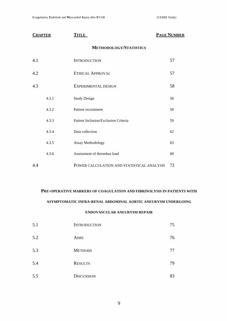
9
CHAPTER TITLE PAGE NUMBER
METHODOLOGY/STATISTICS
4.1 INTRODUCTION 57
4.2 ETHICAL APPROVAL 57
4.3 EXPERIMENTAL DESIGN 58
4.3.1 Study Design 58
4.3.2 Patient recruitment 58
4.3.3 Patient Inclusion/Exclusion Criteria 59
4.3.4 Data collection 62
4.3.5 Assay Methodology 63
4.3.6 Assessment of thrombus load 69
4.4 POWER CALCULATION AND STATISTICAL ANALYSIS 72
PRE-OPERATIVE MARKERS OF COAGULATION AND FIBRINOLYSIS IN PATIENTS WITH
ASYMPTOMATIC INFRA-RENAL ABDOMINAL AORTIC ANEURYSM UNDERGOING
ENDOVASCULAR ANEURYSM REPAIR
5.1 INTRODUCTION 75
5.2 AIMS 76
5.3 METHODS 77
5.4 RESULTS 79
5.5 DISCUSSION 83

Coagulation, Endoleak and Myocardial Injury after EVAR (CEMIE Study)
10
CHAPTER TITLE PAGE NUMBER
THE SHORT TERM EFFECTS OF ENDOVASCULAR ANEURYSM REPAIR ON MARKERS OF
COAGULATION, FIBRINOLYSIS AND PLATELET ACTIVITY IN PATIENTS WITH ASYMPTOMATIC
INFRA-RENAL ABDOMINAL AORTIC ANEURYSMS
6.1 INTRODUCTION 88
6.2 AIMS 90
6.3 METHODS 91
6.4 RESULTS 94
6.5 DISCUSSION 103
PERI-OPERATIVE MYOCARDIAL INJURY AND HAEMOSTASIS IN PATIENTS UNDERGOING
ENDOVASCULAR ANERUYSM REPAIR FOR ASYMPTOMATIC INFRA-RENAL ABDOMINAL
AORTIC ANEURYSM
7.1 INTRODUCTION 110
7.2 AIMS 111
7.3 METHODS 112
7.4 RESULTS 115
7.5 DISCUSSION 120

Coagulation, Endoleak and Myocardial Injury after EVAR (CEMIE Study)
11
CHAPTER TITLE PAGE NUMBER
8 AREAS OF FUTURE RESEARCH 123
8.1 THE RELATIONSHIP BETWEEN MURAL 124
THROMBUS AND HAEMOSTASIS
8.2 BIOMARKER OF ENDOLEAK 126
8.3 MYOCARDIAL INJURY AND EVAR 127
9 APPENDICES 131
A1 Classification of endoleaks 132
A2 Lost samples 133
A3 Patient information leaflet 134
A4 Patient consent form 138
A5 GP letter 139
10 BIBLIOGRAPHY 140

Coagulation, Endoleak and Myocardial Injury after EVAR (CEMIE Study)
12
PUBLICATIONS
Davies RSM, Abdelhamid M, Wall ML, Vohra RK, Bradbury AW, Adam, DJ
Peri-operative myocardial injury and haemostasis in patients undergoing endovascular
aneurysm repair (EVAR) for asymptomatic infra-renal abdominal aortic aneurysm
Vasc Endovascular Surg. 2011 Nov;45(8):712-716
Davies RSM, Abdelhamid M, Wall ML, Vohra RK, Bradbury AW, Adam, DJ
Coagulation, Fibrinolysis and Platelet Activation in patients undergoing Open and
Endovascular Repair of Abdominal Aortic Aneurysm
J Vasc Surg. 2011 Sep;54(3):865-78
PRESENTATIONS TO LEARNED SOCIETIES
Davies RSM, Abdelhamid MJ, Wall ML, Vohra RK, Bradbury AW, Adam DJ
Short-term platelet activity and fibrinolysis in patients undergoing endovascular abdominal
aortic aneurysm repair.
European Society of Surgical Research, Geneva, Switzerland, June 2010,
Davies RSM, Wall ML, Abdelhamid M, Vohra RK, Bradbury AW, Adam DJ
Short Term Platelet Activity and Fibrinolysis in Patients Undergoing Endovascular
Abdominal Aortic Aneurysm Repair
Society of Academic and Research Surgery, Bristol, UK, January 2009

Coagulation, Endoleak and Myocardial Injury after EVAR (CEMIE Study)
13
LIST OF TABLES Table 1.1: AAA rupture rates based on maximum transverse diameter
Table 3.4.1: Summary of studies investigating the association between AAA and levels of
fibrinogen and biomarkers of fibrinolysis. (* symptomatic unruptured AAA, **
manufacturer’s range, *** t-PA activity)
Table 3.4.2: Summary of studies investigating the association between AAA and biomarkers
of thrombin generation. (* symptomatic unruptured AAA, ** manufacturer’s range)
Table 3.4.3: Summary of studies investigating the association between AAA and vWF,
Platelet count and sP-Selectin. (* symptomatic unruptured AAA, ** manufacturer’s range)
Table 3.4.4: Summary of studies investigating the association between AAA morphology and
biomarkers of haemostasis.
Table 3.4.5: Summary of studies investigating the effects of open surgery on biomarkers of
haemostasis. [* Manufacturer’s range, ** mean follow up of 26 month (19-37)]
Table 5.4.1: Patient pre-operative co-morbidities and anti-platelet/statin status.
Table 5.4.2: Pre-operative values of markers of haemostasis. The arrows indicate
comparisons to normal reference ranges.
Table 5.5.1: Latest studies investigating the association between AAA and levels of
fibrinogen and biomarkers of fibrinolysis. and thrombin generation (* symptomatic
unruptured AAA, ** manufacturer’s range, *** t-PA activity)
Table 6.4.1: Effect of EVAR on coagulation, fibrinolysis and platelet activity parameters.
Table 6.5.1: Summary of studies investigating the effects of EVAR on biomarkers of
haemostasis (* manufacturer’s range)

Coagulation, Endoleak and Myocardial Injury after EVAR (CEMIE Study)
14
LIST OF FIGURES
Figure 2.1.1: Platelet activation and aggregation at the site of endothelial cell injury. (TF =
tissue factor, ADP = adenosine diphosphate, vWF = von Willebrand factor, Fib. = fibrinogen)
Figure 2.1.2: Coagulation Cascade. Biomarkers described chapter 3 are highlighted in red.
(Plt= platelets, sP-selectin= soluble P-selectin, vWF= von Willebrand factor, TF= tissue
factor, APC-PCI=activated protein C-protein C inhibitor complex, PS= protein s, APC=
activated protein C, TM-IIa= thrombomodulin-thrombin complex, TAT= Thrombin-
antithrombin complex, ATIII= antithrombin III, F1+2= prothrombin fragments 1 & 2, FM-F=
fibrin monomer=fibrinogen complex)
Figure 2.2.1: Diagrammatic representation of the fibrinolytic pathway. Biomarkers described
in chapter 3 are highlighted in red. (t-PA= tissue plasminogen activator, PAI-1= plasminogen
activator inhibitor type-1, FDP= fibrinogen degradation product, PIC= !2-plasmin inhibitor
complex, IIa= Thrombin)
Figure 4.3.1: Triturus" (Grifols) fully automated enzyme immunoassay analyser
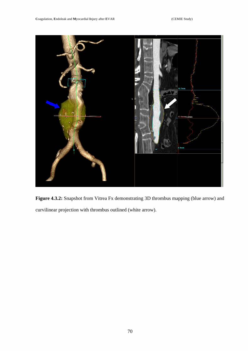
Figure 4.3.2: Snapshot from Vitrea Fx demonstrating 3D thrombus mapping (blue arrow) and
curvilinear projection with thrombus outlined (white arrow).
Figure 4.3.3: Manual remapping of lumen (white line) and thrombus (green line) contours
using Vitrea Fx workstation.
Figure 6.4.1: Comparison of plasma levels of PAI-1at pre-operation, 24 hours post-operation
and 1-month post-operation.
Figure 6.4.2: Median (IQR) differences of PAI-1 levels between baseline and 1-month post-
EVAR in patients with or without endoleak.

Coagulation, Endoleak and Myocardial Injury after EVAR (CEMIE Study)
15
Figure 6.4.3: Comparison of plasma levels of PAI-1at pre-operation, 24 hours post-operation
and 1-month post-operation.
Figure 6.4.4: Comparison of plasma levels of t-PA activity at pre-operation, 24 hours post-
operation and 1-month post-operation.
Figure 6.4.5: Comparison of plasma levels of sP-selectin at pre-operation, 24 hours post-
operation and 1-month post-operation.
Figure 6.4.6: Comparison of plasma levels of TAT at pre-operation, 24 hours post-operation
and 1-month post-operation.
Figure 7.4.1: Individual patient data points for cTnT. The red points represent the two
patients with clinical/ECG signs of cardiac injury.
Figure 7.4.2: Box and Whisker plot of haemostatic variables pre- and 24 hours post-EVAR.
Spearman’s rank correlation coefficient (r) and p-value for each haemostatic variable vs.
cTnT is shown.

Coagulation, Endoleak and Myocardial Injury after EVAR (CEMIE Study)
16
LIST OF ABBREVIATIONS
AAA: Abdominal aortic aneurysm
AB: Antibody
ADP: Adenosine diphosphate
APC: Activated protein C
CENTRAL: Cochrane Central Register of Controlled Trials
CTa: Computed tomographic aortography
cTn: Cardiac troponin
cTnT: Cardiac troponin-T
DREAM: Dutch Randomised Endovascular Aneurysm Management
ECG: Electrocardiography
EVAR: Endovascular aneurysm repair
EVAR-1/ EVAR-2: EndoVascular Aneurysm Repair Trial 1 and 2
F1+2: Prothrombin fragment 1 +2
FM-F: Fibrin-monomer-fibrinogen complex
GP-1b: Glycoprotein-1b
HBV: Hepatitis B virus
HIV: Human immunodeficiency virus
IL: Interleukin
IQR: Inter-quartile range

Coagulation, Endoleak and Myocardial Injury after EVAR (CEMIE Study)
17
ITP: Idiopathic thrombocytopenic purpura
LMWH: Low molecular weigh heparin
MMP-3: Metalloproteinase-3
MMP-9: Metalloproteinase-9
Ng/ml: Nanogram per millilitre
OS: Open surgical
PAI-1: Plasminogen Activator Inhibitor-1
PCI: Protein-C inhibitor
sP-Selectin: Soluble P-selectin
t-PA: Tissue Plasminogen Activator
TAFI: Thrombin Activatable Fibrinolysis Inhibitor
TAT: Thrombin-antithrombin III-complex
TNF: Tumour necrosis factor
TxA2: Thromboxane-A2
USS: Ultrasound scan
vWF: von Willebrand Factor

Coagulation, Endoleak and Myocardial Injury after EVAR (CEMIE Study)
18
CHAPTER 1
THE SHORT TERM EFFECTS OF ENDOVASCULAR ANEURYSM REPAIR (EVAR) ON
COAGULATION AND CARDIOVASCULAR MORBIDITY AND MORTALITY IN PATIENTS
WITH INFRA-RENAL ABDOMINAL AORTIC ANEURYSMS
INTRODUCTION

Coagulation, Endoleak and Myocardial Injury after EVAR (CEMIE Study)
19
1.1 BACKGROUND
The incidence of abdominal aortic aneurysm (AAA), defined as an enlargement of at least
3cm, increases with age and affects approximately 4% of the elderly general population (>65
years) within the United Kingdom. The age-specific prevalence is six times greater in men
than women with an estimated prevalence of 5-8% for men aged 65-80 years old. 1 2 3 The
majority of abdominal aortic aneurysms affect the infra-renal aorta and 75% are
asymptomatic at the time of presentation having been identified incidentally during routine
health checks or investigations for other pathologies.
The natural history of AAA is that of gradual accelerating, asymptomatic expansion until
rupture occurs. The best-known predictor of rupture rate is the maximum AAA diameter; the
annual risk of rupture for an aneurysm with a diameter greater than 6cm may be as high as
25%. (See table 1.1)
DIAMETER 5-YEAR RISK OF RUPTURE
<5 cm 5%
5-6 cm 25%
6-7 cm 35%
>7 cm 75%
Table 1.1: AAA rupture rates based on maximum transverse diameter 4

Coagulation, Endoleak and Myocardial Injury after EVAR (CEMIE Study)
20
Approximately 2500 men aged >60 years die as a result of a ruptured AAA in England and
Wales per year; 2.5% of all deaths in males aged >60 years. Of these deaths approximately
half occur prior to hospital treatment. 1 5 For patients who survive to reach hospital alive,
despite advances in peri-operative management, in-hospital mortality rates following
emergency open surgery for ruptured AAA remain devastatingly high. In our experience,
open repair of rAAA is associated with an in-hospital mortality rate of 40%. 6 Therefore in the
United Kingdom elective repair of an infrarenal abdominal aortic aneurysm, in a suitably fit
patient, is recommended when the aneurysm measures >5.5cm, or greater than 4.5cm with a
growth rate >0.5cm/six months. 4

Coagulation, Endoleak and Myocardial Injury after EVAR (CEMIE Study)
21
1.2 ENDOVASCULAR INFRA-RENAL ABDOMINAL AORTIC ANEURYSM REPAIR (EVAR)
Following the first reported endovascular infra-renal abdominal aortic aneurysm repair
(EVAR) by Juan Parodi in 1990, EVAR has been widely promoted as a less invasive and
safer alternative treatment option to conventional open AAA repair, especially in the high-risk
patient. It is now well established following the publications of the UK EndoVascular
Aneurysm Repair Trials 1 and 2 (EVAR-1 & EVAR-2) and the Dutch Randomised
Endovascular Aneurysm Management (DREAM) trial that elective endovascular repair of an
AAA reduces peri-operative mortality by approximately 3% compared with open surgical
repair in the fit and anatomically suitable patient. 7 8 9 10 However exponents have argued that
these initial operative mortality benefits are offset by the long-term economic cost due to late
graft-related complications.13
Endoleak is a complication unique to EVAR and results in perfusion of the aneurysm sac and
subsequent risk of rupture. (See appendix 1) The EVAR-1 trial reported 22% (118 cases) of
EVARs were complicated by endoleak and 35% require a secondary procedure to maintain
complete aneurysm exclusion within 3 years of the procedure. 7 A systematic review by Drury
et al reported 17.5% and 21.3% of all EVARs demonstrate endoleak at 30 days and 12 months
respectively. 11 However, the endoleak rate is influenced by a variety of factors including pre-
procedural planning, type of stent graft deployed, aortic morphology and technical expertise.
12 For these reasons, and the risk of other stent-related complications including stent
migration, regular outpatient follow-up with computed tomographic aortography (CTA) is
necessary to identify aneurysm exclusion failure. Thus, the short-term peri-operative
mortality/morbidity benefits of EVAR may be outweighed by its long-term morbidity in
terms of re-intervention as well as the associated economic burden. 11 13

Coagulation, Endoleak and Myocardial Injury after EVAR (CEMIE Study)
22
Currently a number of different approaches have been instigated to reduce the economic cost
of EVAR in order to maximise its short-term benefit. A number of institutions have adopted
serial USS and plain radiographs instead of computed tomography as a means of graft
surveillance. This requires dedicated ultrasonographers thereby potentially restricting follow-
up to a specific centres, and continues to pose a significant long-term burden on an
institution’s resources. Thus alternative, less onerous, methods of identifying stent-graft
failure, and specifically endoleak, would be desirable.
Coagulation, Endoleak and Myocardial Injury after EVAR (CEMIE Study)
23
1.3 ENDOVASCULAR ANEURYSM REPAIR AND HAEMOSTASIS
It is reported that patients with asymptomatic AAA demonstrate a hypercoagulable and
hypofibrinloytic state. 28 It is speculated that the pre-operative haemostatic derangement and
subsequent improvement following open repair represents activation of the coagulation
system by intraluminal aneurysmal sac thrombus. This would correlate with reports of a
positive relationship between the size of the aneurysm sac thrombus and the extent of the
haemostatic derangement. 14 Previous work by our research group has demonstrated an
attenuation of the prothrombotic diathesis evident in patients affected by AAA follows open
repair; Adam et al reported that elective open repair of AAA is associated with intense
thrombin generation and inhibition of systemic fibrinolysis that is present pre-operatively and
persists until day-2 peri-operatively where upon it largely resolves. 29 There are currently no
robust studies investigating the effect of EVAR on the hypercoagulable and hypofibrinolytic
state observed in these patients.
Recently there has been interest in utilising changes in circulating levels of biomarkers as an
alternative means of identifying stent-graft failure following EVAR. Two separate studies
report circulating levels of metalloproteinase-3 and -9 (MMP-3 & MMP-9) as being
significantly elevated in patients affected by AAA and successful EVAR has been shown to
be associated with a decrease in MMP levels. 15 16 The presence of an endoleak and aneurysm
sac pressurisation may cause an increase in MMP levels. To date only one study has
investigated a biomarker of thrombin generation and degradation as a marker of stent-graft
failure. Serino et al reported elevated levels of D-dimer in patients with type-1 endoleaks and
stable/increasing sac diameters compared with patients who had endoleaks and decreasing sac
diameters. 17 However, this study has limited value due to methodology deficiencies and lack
of study power. No studies have investigated the use of modern biomarkers of coagulation,

Coagulation, Endoleak and Myocardial Injury after EVAR (CEMIE Study)
24
fibrinolysis and platelet function as an alternative/complementary method of identifying
patients with endoleak.

Coagulation, Endoleak and Myocardial Injury after EVAR (CEMIE Study)
25
1.4 ENDOVASCULAR ANEURYSM REPAIR AND MYOCARDIAL INJURY
Elective open infrarenal AAA repair is associated with a peri-operative mortality rate of 3-
10%. 18 19 20 21 22 Previous work from this institution has demonstrated that over one-quarter
of patients undergoing elective open AAA repair suffer peri-operative myocardial injury as
determined by raised cardiac troponin (cTn) levels. 23 Furthermore, similar haemostatic
derangements to those reported by Adam et al are known to be independently associated with
myocardial injury, stroke and multiple organ failure. 24 25 26 27 Thus, the majority of peri-
operative mortality following elective open AAA repair may be secondary to micro- and
macro- vascular thrombosis causing myocardial injury, thromboembolism and multiple organ
failure. 28 29 While a positive association between the peri-operative hypofibrinolytic state
and myocardial injury has been demonstrated in patients undergoing emergency open repair
of ruptured AAA, to date, the relationship between coagulation and fibrinolysis and
myocardial injury after EVAR has not been studied. 30

Coagulation, Endoleak and Myocardial Injury after EVAR (CEMIE Study)
26
1.5 HYPOTHESIS AND AIMS
We hypothesise that following successful aneurysm sac exclusion by EVAR the underlying
hypercoagulable state witnessed in these patients will be attenuated as a result of exclusion of
the intraluminal thrombus from the systemic circulation. Furthermore, we speculate the re-
establishment of aneurysm sac perfusion secondary to an endoleak results in the re-
establishment of the pre-operative prothrombotic state secondary to exposure of the sac
thrombus to the systemic circulation. We speculate that the novel use of biomarkers of
haemostasis can provide endoleaks in patients with EVAR.
The aims of this thesis are to:
1. Establish if intra-luminal thrombus volume is related to the pro-thrombotic diathesis
evident in patients affected by AAA.
2. Investigate changes to the resting pro-coagulant and hypofibrinloytic state following
elective endovascular aneurysm repair for an asymptomatic infra-renal AAA.
3. Investigate and correlate any peri-operative haemostatic changes with peri-operative
myocardial injury.
4. Accumulate pilot data on which to base future studies pertaining to the use of
haemostatic biomarkers as a method of identifying endoleak.

Coagulation, Endoleak and Myocardial Injury after EVAR (CEMIE Study)
27
1.3 SUMMARY
The proposed study is, to the best of our knowledge, the first to investigate changes to
coagulation, fibrinolysis and platelet activity following EVAR for infra-renal AAA. The study
will not only attempt to corroborate previously published findings regarding open AAA
repair, namely that successful aneurysm sac exclusion attenuates the resting hypercoagulable
state in patients with AAA, but will extend our investigation to patients that suffer endoleaks.
The proposed study will, therefore, attempt to determine if EVAR attenuates the
hypercoagulable state in patients with AAA thereby providing a scientific foundation to base
future investigation into whether or not an endoleak results in the re-establishment of this pro-
thrombotic diathesis.

Coagulation, Endoleak and Myocardial Injury after EVAR (CEMIE Study)
28
CHAPTER 2
AN OVERVIEW OF THE NORMAL COAGULATION AND FIBRINOLYTIC SYSTEMS

Coagulation, Endoleak and Myocardial Injury after EVAR (CEMIE Study)
29
2.1 PLATELET ACTIVATION AND THE COAGULATION CASCADE
Normal haemostasis represents a complex interaction between the damaged vessel wall,
circulating cells (platelets and leukocytes) and circulating proteins/zymogens (coagulation
factors). Injury to the vessel wall is the major stimulus for coagulation and results in the
activation of endothelial cells and exposure of sub-endothelial collagen to circulating
platelets. The activated endothelial cells express P-selectin on their surfaces and release von
Willebrand factor (vWF) which aids in the localisation of thrombus formation at the site of
injury. 31 Tissue factor (TF) is also released from the endothelium and plays a vital role in the
activation of the coagulation cascade through the extrinsic pathway. Platelets bind to the
exposed sub-endothelial collagen resulting in their activation and creation of a platelet plug.
This is mediated by vWF which binds to both platelet membrane glycoprotein (Gp) Ib!
receptor and sub-endothelial collagen. 32 Fibrinogen and vWF binds to Gp-IIb and Gp-IIIa
receptors on adjacent platelets creating a lattice (aggregation) of platelets. 33 Activated
platelets shed the contents of their stored !-granules and dense bodies (platelet granules),
which include vWF, TF, adenosine diphosphate (ADP) and coagulation zymogens receptors.
Activated platelets also undergo a morphological change from spherical to stellate that assists
in the assembly of coagulation factors on their surface. (See figure 2.1.1)

Coagulation, Endoleak and Myocardial Injury after EVAR (CEMIE Study)
30
Figure 2.1.1: Platelet activation and aggregation at the site of endothelial cell injury. (TF =
tissue factor, ADP = adenosine diphosphate, vWF = von Willebrand factor, Fib. = fibrinogen)
The formation of thrombin (Factor IIa) is vital to ensure stability of the initial platelet plug
through its ability to convert fibrinogen to fibrin. 34 The formation of fibrin is achieved
through activation of the coagulation cascade predominantly through the extrinsic pathway by
the release of TF from endothelial cells and activated platelets. TF complexes and activates
the zymogen factor VII (TF-FVIIa). This greatly enhances the catalytic ability of FVII which
activates factors IX and X. Through a cascade effect that occurs on the surface of the
activated platelets, the production of thrombin (factor IIa) occurs (See figure 2.1.2). Initially
only trace amounts of thrombin are generated, however through a process of feedback
amplification exponentially increasing amounts of thrombin are generated with each feedback
loop. 35 Thrombin acts upon fibrinogen to create fibrin monomers, which in the presence of
FXIIIa undergo polymerisation to form fibrin that strengthens and stabilises the plug. 36

Coagulation, Endoleak and Myocardial Injury after EVAR (CEMIE Study)
31
Figure 2.1.2: Coagulation Cascade. Biomarkers described in chapter 3 are highlighted in
red. (Plt= platelets, sP-selectin= soluble P-selectin, vWF= von Willebrand factor, TF= tissue
factor, APC-PCI=activated protein C-protein C inhibitor complex, PS= protein s, APC=
activated protein C, TM-IIa= thrombomodulin-thrombin complex, TAT= Thrombin-
antithrombin complex, ATIII= antithrombin III, F1+2= prothrombin fragments 1 & 2, FM-F=
fibrin monomer=fibrinogen complex)

Coagulation, Endoleak and Myocardial Injury after EVAR (CEMIE Study)
32
2.2 ANTICOAGULANTS AND FIBRINOLYSIS
Diffuse intravascular thrombosis is prevented by the actions of localised and systemic
anticoagulants and fibrinolytic mechanisms:
2.2.1 Anticoagulants
The two main natural anticoagulants are Antithrombin (AT) III and activated protein C
(APC). AT III is the central physiological anticoagulant and prevents the coagulation cascade
amplificatory effects of thrombin; these include the cleavage of fibrinogen to form fibrin, the
activation of factors V and VIII and the mediation of platelet activation and aggregation.
Thrombin binds to a receptor (thrombomodulin) presented on the surface of intact
endothelium to form a thrombin-thrombomodulin complex that activates protein C as well as
inhibiting the effects of thrombin. APC in the presence of its co-factor protein S acts as an
anticoagulant through its ability to inactivate Factors Va and VIIIa. 37 38 In addition APC
heightens fibrinolysis through two major mechanisms: APC forms a tight complex with
plasminogen activator inhibitor type-1 (PAI-1) thereby preventing its inhibitory effect on
tissue plasminogen activator (t-PA), secondly through its ability to limit thrombin generation
it reduces the activation of thrombin activatable fibrinolysis inhibitor (TAFI). 39 40 Tissue
factor pathway inhibitor (TFPI)/lipoprotein-associated coagulation inhibitor also acts as a
reversible anticoagulant through its ability to complex to and inactivate factor Xa. TFPI-Xa
complex also has the ability to inhibit factor TF-VIIa complex thereby arresting the extrinsic
pathway. 41!

Coagulation, Endoleak and Myocardial Injury after EVAR (CEMIE Study)
33
2.2.2 Fibrinolysis
Plasmin is the main fibrinolytic enzyme formed by the activation of the zymogen
plasminogen through a variety of mechanisms. The main activator of plasminogen is t-PA
which is produced by the vascular endothelium in response to thrombin. When incorporated
into a forming clot t-PA is a potent catalyst for conversion of plasminogen to plasmin. 42 (See
figure 2.2.1)
Figure 2.2.1: Diagrammatic representation of the fibrinolytic pathway. Biomarkers described
in chapter 3 are highlighted in red. (t-PA= tissue plasminogen activator, PAI-1= plasminogen
activator inhibitor type-1, FDP= fibrinogen degradation product, PIC= !2-plasmin inhibitor
complex, IIa= Thrombin)

Coagulation, Endoleak and Myocardial Injury after EVAR (CEMIE Study)
34
Urokinase-type plasminogen activator, APC and factors XIa, XIIa and Kallikrein can also
independently activate plasminogen. Plasmin degrades polymerised fibrin to form fibrin
degradation products (fragment E and D-Dimer). The activation of plasminogen and fibrin
degradation by plasmin is finely balanced by PAI-1 and TAFI. PAI-1 is released by the
vascular endothelium in response to thrombin and inhibits t-PA. TAFI eliminates
plasminogen receptor sites on partially degraded fibrin and thus slows the binding and
activation of plasminogen and thus fibrinolysis. 42 !-Antiplasmin is also an inhibitor of
plasmin.

Coagulation, Endoleak and Myocardial Injury after EVAR (CEMIE Study)
35
2.3 SUMMARY
Haemostasis requires the interaction of platelets, coagulation factors and fibrinolytic factors.
Platelet and endothelial cell activation at the site of injury initiates clot formation. The
coagulation cascade is activated predominantly through the extrinsic pathway to form fibrin,
which is vital in creating a stable platelet plug. The fibrinolytic and anticoagulant systems
ensure thrombus formation is limited to the site of vascular injury.

Coagulation, Endoleak and Myocardial Injury after EVAR (CEMIE Study)
36
CHAPTER 3
THE IMPACT ON BLOOD COAGULATION, FIBRINOLYSIS AND PLATELET
ACTIVATION OF OPEN SURGICAL AND ENDOVASCULAR ANEURYSM REPAIR
(EVAR) IN PATIENTS WITH INFRA-RENAL ABDOMINAL AORTIC ANEURYSMS
A REVIEW OF THE LITERATURE
Coagulation, Endoleak and Myocardial Injury after EVAR (CEMIE Study)
37
3.1 INTRODUCTION
Major surgery causes a pro-thrombotic derangement in the peri-operative period with elevated
levels of Factor VIII, Fibrinogen, Thrombin-antithrombin III-complex (TAT), von Willebrand
Factor (vWF), deranged fibrinolysis and platelet hyperactivity. 43 44 45 46 47 48 49
Patients undergoing elective open surgical infra-renal AAA repair have an associated
operative mortality rate of 3-10%. 19 18 20 21 22 The majority of deaths are due to tissue damage
and ischaemic-reperfusion injury, which result in an extensive and uncontrolled inflammatory
response with micro- and macro- vascular thrombosis that may cause myocardial injury,
thromboembolism and multiple organ failure. 29 50
Endovascular abdominal aortic aneurysm repair (EVAR) provides a less invasive and safer
alternative treatment to open surgical repair in the fit and anatomically suitable patient. The
EVAR-1, EVAR-2 and DREAM trials reported EVAR was associated with a 60% reduction
in peri-operative mortality when compared to open surgical repair. 7 8 9 10 These
improvements may relate to an attenuation of the inflammatory response and pro-thrombotic
diathesis associated with open repair through a reduction in tissue damage and ischaemic-
reperfusion injury. However Swartbol et al have reported EVAR as promoting a systemic
inflammatory response equal to if not in excess of that witnessed after open repair. 51 This
has been hypothesised to be secondary to cytokine release from the aneurysm sac thrombus as
a result of introducer and catheter manipulation. 51 52 The use of contrast media has also been
reported to induce arterial endothelial damage. 53 54

Coagulation, Endoleak and Myocardial Injury after EVAR (CEMIE Study)
38
3.2 AIMS
The aim of this chapter is to review and compare the effects of abdominal aortic aneurysm,
open surgical repair and EVAR on coagulation, fibrinolysis and platelet activation as reported
in the English-language scientific literature at the time of starting this thesis.

Coagulation, Endoleak and Myocardial Injury after EVAR (CEMIE Study)
39
3.3 METHODS
We performed a MEDLINE, EMBASE and Cochrane Central Register of Controlled Trials
(CENTRAL) databases search looking for English-language articles between January 1970
and August 2006 relating to abdominal aortic aneurysm, open surgical repair and EVAR, and
their effects on haemostatic mechanisms. The terms coagulation, clotting, fibrinolysis,
thrombosis and platelets were included amongst others. These were linked with terms such as
abdominal aortic aneurysm, open repair, endovascular aneurysm repair and EVAR. Further
articles were identified by following MEDLINE links, by cross-referencing from the
reference lists of major articles and by following citations for these studies. Studies were
specifically rejected if a) patient cohort was less than seven and b) values of assessed
biomarkers of haemostasis were not included in the presented results. The studies were then
graded and prioritised according to the level of the evidence presented.

Coagulation, Endoleak and Myocardial Injury after EVAR (CEMIE Study)
40
3.4 RESULTS
Studies examining the effect of AAA and open surgical or EVAR on blood coagulation,
fibrinolysis and platelet activity are summarised in Tables 3.4.1-5. The coagulation cascade
and fibrinolysis pathways are summarised in figures 3.4.1 and 3.4.2
3.4.1 Effect of abdominal aortic aneurysm on haemostasis
AAA represents a chronic inflammatory pathology and is usually characterised by the
presence of mural thrombus directly proportional to the maximum AAA diameter. 55 In
contrast to atherosclerotic occlusive disease, blood flow is maintained through the mural
thrombus thereby providing an interface for exchange between the systemic circulation and
thrombus. The structure of the thrombus is highly complex with a network of inter-
connecting canaliculi capable of delivering macromolecules between the intra-luminal and
thrombus-arterial wall surfaces. 56 The canaliculi often contain cellular infiltrates including
neutrophils, macrophages and platelets, often in state of degranulation. This may lead to
consumption of platelets and coagulation factors such that in this group of patients a sub-
clinical state of disseminated intravascular coagulation (DIC) may exist. 57 Thus the mural
thrombus represents a biologically active entity with the ability to trap polymorphonuclear
leukocytes, absorb circulating plasma components and aggregate platelets as well as being a
source of proteolysis and fibrinolytic activity thought to be implicit in AAA progression. 56 58
59 Several prospective comparative studies have examined the effects of AAA on direct and
indirect biomarkers of thrombin generation, fibrinolysis and platelet activity. (See tables
3.4.1-3).

Coagulation, Endoleak and Myocardial Injury after EVAR (CEMIE Study)
41
Table 3.4.1: Summary of studies investigating the association between AAA and levels of
fibrinogen and biomarkers of fibrinolysis. (* symptomatic unruptured AAA, **
manufacturer’s range, *** t-PA activity)

Coagulation, Endoleak and Myocardial Injury after EVAR (CEMIE Study)
42
Table 3.4.2: Summary of studies investigating the association between AAA and biomarkers of thrombin generation. (* symptomatic
unruptured AAA, ** manufacturer’s range)

Coagulation, Endoleak and Myocardial Injury after EVAR (CEMIE Study)
43
Table 3.4.3: Summary of studies investigating the association between AAA and vWF, Platelet count and sP-Selectin. (* symptomatic
unruptured AAA, ** manufacturer’s range)

Coagulation, Endoleak and Myocardial Injury after EVAR (CEMIE Study)
44
Biomarker Study Maximum
diameter of AAA
Worst angle along length of
AAA
Maximum thickness of intraluminal
AAA thrombus
Total Intra-luminal AAA
thrombus volume
APC-PCI Kolbel et al85 r=0.22, p=0.001 r=0.123, p=0.142
Shindo et al69 r=0.208, p=NS r=0.208, p=NS D-Dimer
Yamazumi et al14 r=0.644, p=0.0001 r=-0.411, p=0.009 r=0.650, p=0.0001
F-TFPI Yamazumi et al14 r=0.408, p=0.016 r=-0.583, p=0.0006
Shindo et al69 r=0.208, p=NS r=0.171, p=NS FDP
Yamazumi et al14 r=0.561, p=0.0009 r=0.513, p=0.0024
FM-FC Hosaka et al70 r=0.128, p=0.381 r=0.233, p=0.125
PIC Yamazumi et al14 r=0.413, p=0.0146 r=0.484, p=0.042
TAT Yamazumi et al14 r=0.566, p=0.001 r=-0.366, p=0.0305 r=0.677, p=<0.0001
Table 3.4.4: Summary of studies investigating the association between AAA morphology and biomarkers of haemostasis.

Coagulation, Endoleak and Myocardial Injury after EVAR (CEMIE Study)
45
Table 3.4.5: Summary of studies investigating the effects of open surgery on biomarkers of haemostasis. [* Manufacturer’s range, **
mean follow up of 26 month (19-37)]

Coagulation, Endoleak and Myocardial Injury after EVAR (CEMIE Study)
46
An elevated level of plasma fibrinogen is an independent risk factor for stroke and myocardial
infarction as well as cardiovascular mortality. 60 61 62 An association between elevated
fibrinogen and atherosclerotic peripheral vascular disease has been widely reported and
elevated levels of plasma fibrinogen are found in patients who subsequently develop
peripheral artery disease. 24 61 63 64 The association between non-ruptured AAA and plasma
fibrinogen levels have been extensively investigated. 14 18 65 66 67 68 69 70 71 72 73 74 (See table
3.4.1) Six from twelve studies have reported significantly elevated levels of fibrinogen in
patients with asymptomatic AAA. Singh et al reported significantly increased levels of
plasma fibrinogen in patients with AAA. 68 On multivariate analysis Lee et al reported an
independent association between AAA and plasma fibrinogen (p=0.05, OR=1.51). 72 While
Singh et al reported similar findings but only in the studies male cohort; male (p = <0.001,
OR = 1.42) vs. females (p = 0.18, OR = 1.23). 68
Hosaka et al reported elevated levels of both fibrinogen and fibrin-monomer-fibrinogen (FM-
F) complex in patients with AAA indicating increased thrombin activity. During the cleavage
of fibrinogen by thrombin fibrinopeptide A and B are released to create fibrin monomer (FM)
that undergoes polymerisation –catalysed by factor XIIIa- to form insoluble fibrin clots. 75
When a pro-thrombotic state exists and thrombin generation is high FM forms soluble
complexes with fibrinogen (FM-F complex) due to excess plasma fibrinogen. 76 Thus elevated
levels of FM-F represent heightened thrombin activity. Furthermore, a sufficiently elevated
concentration of FM-F may itself add to the pro-thrombotic diathesis as it precipitates from
plasma. 76
Adam et al reported significantly elevated levels of plasma fibrinogen in patients with
symptomatic, but non-ruptured AAAs. 71 In a separate study, the same group reported no
difference in pre-operative plasma fibrinogen levels between those patients with ruptured

Coagulation, Endoleak and Myocardial Injury after EVAR (CEMIE Study)
47
AAA and asymptomatic, non-ruptured AAA (median (range), 2.27 (0.86-3.75) vs. 2.8 (1.59-
6.02) g/l, p = NS). 29 However, these studies involved small numbers of patients.
Ten studies were identified investigating the association between AAA and fibrinolysis. 14 65 69
70 71 72 73 77 78 79 (See table 3.4.1) Nine studies assessed the impact of AAA on plasma D-dimer
levels. The degradation of fibrin by plasmin ultimately results in the formation of two
fragment-D molecules that covalently link to form a dimmer: D-dimer. Thus the presence of
D-dimer in the circulation represents ongoing clot formation and fibrinolysis. All studies
report elevated levels of plasma D-dimer in patients with AAA indicating ongoing clot
formation and fibrinolysis. On multivariate analysis Lee et al (p = <0.001, OR = 3.75)
reported an independent association between circulating D-dimer levels and AAA. 72
Tissue Plasminogen Activator (t-PA) is released from vascular endothelium in response to
thrombin generation and becomes incorporated into the forming fibrin clot. When bound to
fibrin t-PA is a potent activator of plasminogen and is therefore a marker of fibrinolysis. PAI-
1 is also released from the vascular endothelium in the presence of thrombin and acts to
maintain a balance between clot formation and lysis by inhibiting t-PA. Four studies have
reported the effect of AAA on both D-dimer levels and t-PA and/or Plasminogen Activator
Inhibitor-1 (PAI-1). 65 71 72 77 All three studies reporting t-PA antigen levels found no difference
in circulating levels in patients with or without an AAA. However, t-PA antigen levels reflect
both the unbound, active t-PA and the bound, inactive t-PA/PAI-1 complexes. Therefore it is
important to measure t-PA activity as opposed to t-PA antigen to avoid the paradox in which
elevated levels of t-PA antigen are associated with a pro-coagulant state due to increase PAI-1
activity. 29 Only one study has analysed both t-PA antigen and PAI-1 activity levels and
reports no significant difference compared to their control populations although elevated D-
dimer levels in the AAA cohort was found. 77 Adam et al are the only group to report on t-PA

Coagulation, Endoleak and Myocardial Injury after EVAR (CEMIE Study)
48
activity levels that were found to be comparable to the manufactures normal reference range
as was PAI-1 activity in symptomatic, non-ruptured AAA. 71
Thrombin plays an intricate part in coagulation initiation and platelet activation. More
recently thrombin has been implicated in smooth muscle cell mitogenesis, with intimal
vascular smooth muscle cells showing increased thrombin receptor expression. 80 The
conversion of prothrombin to thrombin by prothrombinase complex results in the production
of a degradation product with a half life of <90 mins; prothrombin fragment 1+2 (F1+2). 81
Thrombin released through this enzymatic process is inactivated by antithrombin-III to form a
Thrombin-antithrombin III-complex (TAT). 82 Thus, F1+2 and TAT are biomarkers of
coagulation activation and thrombin generation. This review identified six studies that
reported on the effect of AAA on the levels of one or both of these biomarkers. 14 70 71 73 77 78
(See table 3.4.2) All studies reporting TAT found elevated circulating levels in patients
affected by AAA. Holmberg et al report elevated levels of TAT, but normal levels of F1+2. 77
This may be explained by the short half-life of F1+2; elevated TAT levels are more indicative
of ongoing thrombosis whereas F1+2 may be more reflective of a single acute thrombotic
event.
Activated protein-C (APC) is a natural anticoagulant and is produced on the surface of intact
endothelium following the binding of thrombin to the endothelial cell cofactor:
thrombomodulin. 83 APC in the presence of protein-S cofactor reduces Xase and
prothrombinase activity through its ability to cleave factors Va and VIIIa, thereby limiting
thrombin production to the site of endothelial injury. 37 38 APC is inactivated through binding
to protein-C inhibitor (PCI) to form APC-PCI complex. This complex is detectable in the
blood for <20 minutes after formation and is an indicator of thrombin generation. 84 Kolbel et
al report a three-fold increase in median APC-PCI complex levels in patients with AAA when

Coagulation, Endoleak and Myocardial Injury after EVAR (CEMIE Study)
49
compared to healthy controls (p = <0.0001), indicating that patients with AAA would appear
to undergo periods of acute thrombus formation on background of chronically increased
thrombin generation. 85
Adhesion of platelets to exposed sub-endothelial collagen leads to platelet activation and
aggregation. Von Willebrand Factor (vWF) through its platelet receptor Glycoprotein-1b (GP-
1B) mediates this interaction. 32 These activated platelets release prothrombotic mediators
form dense bodies and alpha-granules that include vWF, adenosine diphosphate (ADP) and
Thromboxane-A2 (TxA2). They are also rich in FVa and FVIIa receptors and undergo
aggregation by the binding of fibrinogen to platelet GP-IIb/-IIIa receptors on adjacent
platelets. 33 Thus, the central role of platelet-vessel wall interaction is the initiation and
progression of thrombosis. Six studies report on the effects of AAA on platelets. 14 67 69 71 73 86
Three of six studies reports significantly lower levels of platelets in patients with AAA. Milne
et al, in addition to finding a lower a platelet count, reported elevated levels of Glycocalicin, a
biomarker for GP-1B levels and suggested that this represented increased activation and
destruction of platelets as a result of adherence and incorporation into the thrombus mass. 86
This would appear to be supported by Mukaiyama et al who demonstrated uptake of
radiolabelled platelets by the AAA lumen. 87 Platelets activated through exposure to sub-
endothelial collagen without concomitant aggregation at the site of injury are removed from
the circulation by the reticulo-endothelial system irrespective of platelet age. 88 Thus the
aneurysm thrombus mass may also induce platelet activation without platelet adhesion
leading to their subsequent destruction and GP-1B release. Blann et al demonstrated increased
levels of soluble P-selectin, a marker of platelet activation, and vWF in AAA patients, which
would lend support to the theory of increased platelet activation. 67

Coagulation, Endoleak and Myocardial Injury after EVAR (CEMIE Study)
50
3.4.2 Association between AAA morphology and haemostasis
Three studies have reported on the correlation between AAA maximum diameter and
thrombus volume, and markers of haemostasis. 14 69 70 (See table 3.4.4). AAA size and total
volume/maximum thrombus thickness appear to correlate with the aforementioned changes in
thrombin generation and fibrinolysis. Yamazumi et al reported a correlation between AAA
tortuosity and markers of thrombosis. 89 This association may represent the association
between blood flow velocity changes, particularly turbulent flow, and red blood cell (RBC)
activation. Shindo et al reported a correlation between RBC counts and the AAA lumen
volume. 69 Activated RBCs release ADP resulting in platelet aggregation and activation. 90
Thus the consumptive coagulopathy, resulting from the thrombus mass and the abnormal flow
field in a tortuous lumen may both contribute to the haemostatic derangement reported in
patients with AAA.

Coagulation, Endoleak and Myocardial Injury after EVAR (CEMIE Study)
51
3.4.3 Effect of open surgical repair on biomarkers of haemostasis
Major surgery is known to produce a pro-thrombotic derangement in the peri-operative period
with raised levels of FVIII, Fibrinogen, TAT, vWF, platelet hyper-activity, and evidence of
deranged fibrinolysis. 43 44 46 47 48 49 91 Patients undergoing major lower limb arterial
reconstructive surgery exhibit defective endogenous fibrinolytic activity: increased levels of
PAI-1 and t-PA antigen that return to pre-operative levels by one week post-procedure
suggestive of a consumptive hypofibrinolytic state. 92 93 94
Studies reporting the effect of open surgical AAA repair on haemostasis are summarised in
table 3.4.5. Yamazumi et al reported a significant decrease in circulating levels of TAT and
D-dimer at 3-months post repair when compared to pre-operative levels (p = <0.01). 14
However, the levels remained elevated when compared to those of the control group (p =
<0.01) suggestive of ongoing up-regulation of thrombin generation and lysis. Holmberg et al
reported similar findings with significantly elevated levels of TAT and F1+2 reported peri-
operatively which returned to pre-operative levels by one-week post surgery, but once again
remained elevated compared to age-matched controls. 77 In a separate study the same group
reported open surgical repair attenuating the pre-operative thrombotic derangement in the
long term with a reduction in TAT and D-dimer plasma levels at a median follow up of 26
months. 66 However, the values remained slightly higher than the normal healthy reference
ranges suggesting that there was ongoing haemostatic derangement despite thrombus volume
reduction.

Coagulation, Endoleak and Myocardial Injury after EVAR (CEMIE Study)
52
3.4.4 Effect of EVAR on biomarkers of haemostasis
To date only two studies has investigated the effect of EVAR on haemostasis. 17 95 Serino et al
report the short-term effects of EVAR on circulating D-dimer levels in nine patients assessed
pre-operatively and on day four post-operatively. D-dimer levels were elevated in seven
patients and decreased in two patients. The median level for the entire patient cohort did not
demonstrate a statistically significant difference between baseline and day four post-operation
[563+/-521 ng/ml vs. 799+/-443 ng/ml (p = 0.19)]. This lack of statistical significance may
represent a type-II error due to the study’s lack of power. Odegard et al reported increased
platelet activation following the administration of contrast media intra-operatively and this
remained increased during variable lengths of hospital stays. 95

Coagulation, Endoleak and Myocardial Injury after EVAR (CEMIE Study)
53
3.5 DISCUSSION
AAA is associated with increased thrombin generation, activity and fibrin turnover as
demonstrated by increased plasma levels of TAT, APC-PCI, FM-F, F1+2, fibrinogen and D-
dimer. Increased fibrin turnover occurs on the surface of intra-luminal AAA thrombus.
Several authors have reported a positive correlation between the extent of haemostatic
derangement and AAA size and thrombus load. 14 85 Thus the pro-thrombotic diathesis
witnessed in this patient population may be secondary to continuous intra-luminal thrombus
remodelling.
Open surgical repair causes significant haemostatic derangement in the peri-operative period
with increased thrombin generation and activity, and increased fibrin turnover. By 3-months
post-repair, the pro-thrombotic diathesis is similar to if not improved compared to the pre-
operative state. However, open surgical repair does not tend to complete resolution of the pre-
operative haemostatic derangement. Plasma levels of TAT and D-dimer remain elevated after
surgery compared to healthy controls suggestive that the surgical prosthesis may be capable
of inducing a prothrombotic derangement. Other factors that may contribute to these ongoing
haemostatic derangements include atherosclerotic disease affecting other vascular beds with a
number of studies reporting a positive correlation between the extent of atherosclerotic vessel
disease and a pro-thrombotic state. 96 97 98 Red blood cell concentrates have been shown to
have high levels of cytokines including IL-1, IL-6, and TNF which are potent coagulation
stimulators and thus the volume of blood transfusion may contribute to the peri-operative
coagulation derangement witnessed after aneurysm repair. 66 99 Increasing age has also been
reported to influence biomarkers of coagulation and fibrinolysis. 100 101
Only one study has studied the effect EVAR on coagulation/fibrinolysis in the peri-operative
period. The small patient numbers and lack statistical power did not allow any firm

Coagulation, Endoleak and Myocardial Injury after EVAR (CEMIE Study)
54
conclusions to be drawn from this study. However, the majority of patients demonstrated
increased fibrin turnover with elevated levels of circulating D-dimer in the peri-operative
phase. This may occur due to aneurysm sac exclusion with sac thrombosis resulting in
increased coagulation and fibrinolysis activation, and platelet consumption; contrast media
induced endothelial cell injury and platelet activation; or endothelial damage due to the
passage of guide-wires and catheters as has been demonstrated after coronary and peripheral
angioplasty. 53 54 102 103 104

Coagulation, Endoleak and Myocardial Injury after EVAR (CEMIE Study)
55
3.6 SUMMARY
AAA is associated with a pro-thrombotic diathesis proportional to the volume of intra-luminal
thrombus. The pro-coagulant state is exaggerated in the immediate peri-operative period
following open repair but is attenuated at medium-term follow-up although not normalised.
These changes may account for the high level of thrombotic complications observed in this
cohort of patients. There are currently no robust studies investigating the effect of EVAR on
the underlying pro-thrombotic diathesis evident in AAA patients.

Coagulation, Endoleak and Myocardial Injury after EVAR (CEMIE Study)
56
CHAPTER 4
THE SHORT TERM EFFECTS OF ENDOVASCULAR ANEURYSM REPAIR (EVAR) ON
COAGULATION AND CARDIOVASCULAR MORBIDITY AND MORTALITY IN PATIENTS
WITH INFRA-RENAL ABDOMINAL AORTIC ANEURYSMS
METHODOLOGY

Coagulation, Endoleak and Myocardial Injury after EVAR (CEMIE Study)
57
4.1 INTRODUCTION
This chapter describes the methodology involved in the investigation of the hypotheses
described in chapter 1. The premise for this study is that the AAA intra-luminal thrombus
load causes a pro-thrombotic diathesis that is attenuated upon exclusion of the aneurysm
through endovascular stent-graft repair (EVAR).
4.2 ETHICAL APPROVAL
Ethical approval was granted by Birmingham East, North and Solihull Research Ethics
Committee and South Birmingham Research Ethics Committee for all aspects of work
contained within this thesis (reference: 06/Q2703/44). The study was named Coagulation,
Endoleak and Myocardial Injury after EVAR (CEMIE) study.

Coagulation, Endoleak and Myocardial Injury after EVAR (CEMIE Study)
58
4.3 EXPERIMENTAL DESIGN
4.3.1 Study Design
This is a prospective, non-randomized, multi-center study designed to investigate the short-
term haemostatic derangement in patients with abdominal aortic aneurysms undergoing
elective EVAR. AAA–for this protocol- is defined as an asymptomatic infra-renal aortic
aneurysm with a maximum anterior-posterior diameter measuring >5.5cm on ultrasonography
or computed tomographic angiography (CTA).
4.3.2 Patient recruitment
Initially all patients presenting to the Vascular Surgery Units of the Heart of England NHS
Foundation Trust and University Hospital Birmingham NHS Trust between April 2006 and
October 2007 and undergoing EVAR were considered for inclusion in the study. However,
due to a freezer malfunction the majority of samples/results collected from this patient cohort
were lost or invalidated. (See appendix 2) As a result a second cohort of patients using the
same methods and protocols for enrolment was recruited between January 2008 and
December 2009. Thus this thesis presents two equal cohorts of patients as separate datasets:
cohort one (April 2006- October 2007) and cohort two (January 2008- December 2009).
Patients were primarily identified through a) liaising with lead clinicians in their respective
vascular surgery departments and radiology departments, b) attendance at weekly vascular
multidisciplinary team meetings, and c) attendance at outpatient clinics in the aforementioned
hospitals.

Coagulation, Endoleak and Myocardial Injury after EVAR (CEMIE Study)
59
Following identification of a suitable study patient (see inclusion and exclusion criteria
below) the patient was interviewed by the lead investigator (RSM Davies) and provided with
a comprehensive CEMIE study information leaflet. (See appendix 3) The patient was then
allowed at least 24 hours to decide whether or not they wished to participate in the study.
Those patients willing to participate underwent a subsequent written fully informed consent
process with a concomitant study participation notice forwarded to the patients’ general
practitioner. (See appendices 4 & 5)
4.3.3 Patient Inclusion/Exclusion Criteria
a) Inclusion
All patients met the following criteria:
1. Males and females 50-90 years of age inclusive
2. Asymptomatic AAA
3. Radiological (USS/CTA) diagnosis of an infra-renal AAA > 5.5cm
4. Suitable for EVAR
5. Smoking status stable for ! 3 months
6. Patient was committed and able to follow the protocol requirements as evidenced by
written informed consent

Coagulation, Endoleak and Myocardial Injury after EVAR (CEMIE Study)
60
b) Exclusion
Patients were excluded if they met any of the following criteria:
1. A non-cardiac endovascular intervention procedure other than a diagnostic
percutaneous trans-femoral/-brachial digital subtraction angiography within the
previous 3 months.
2. Patients with concomitant thoracic or suprarenal aortic aneurysm or dissection
3. Patients with significant lower limb peripheral vascular disease (Fontaine
Classification IIb-IV)
3. Presence of the following conditions that could confound coagulation studies:
• History of malignancy and/or chemotherapy within the last 5 years
• Coagulation disorders:
! Hereditary coagulopathy e.g. Haemophilia, von Willebrand disease
! Acquired e.g. liver disease, malabsorption syndromes
• Thrombocytopenia e.g. bone marrow failure, idiopathic thrombocytopenic purpura
(ITP), hypersplenism
• Kidney Disease Outcomes Quality Initiative (KDOQI) Stage 3-5 chronic renal
failure
• History of deep venous thrombosis and/or venous ulceration within last 3 months
• Evidence of sepsis or systemic inflammatory response syndrome
• Patients with immuno-compromised conditions, organ transplant recipients, or
those being treated with immunosuppressive pharmacotherapy

Coagulation, Endoleak and Myocardial Injury after EVAR (CEMIE Study)
61
4. Patients who have undergone previous surgery within the last 6 months
5. Patients who have undergone coronary artery angiography/stent insertion within the
last 3 months
6. Patients who have been on treatment dose anti-coagulation pharmacotherapy within
the last 6 months e.g. Heparin, low molecular weight heparin (LMWH), warfarin
7. Patients with known blood borne infections e.g. Hepatitis B virus (HBV), Human
immunodeficiency virus (HIV)
8. Patients who have undergone a blood transfusion within the last month

Coagulation, Endoleak and Myocardial Injury after EVAR (CEMIE Study)
62
4.3.4 Data collection
The following patient clinico-pathological data were collected prospectively and stored in an
encrypted database: patient age and sex; co-morbidities and medications; pre-operative
imaging investigations; procedural details; post-operative data including imaging
investigations, re-interventions, complications and mortality. Specifically cardiovascular
morbidity and mortality at each study time point was recorded and all patients underwent
ECG pre-operatively and at 24-hours post-operatively.
Blood Sample Collection
All patients had a resting venous blood sample drawn from an antecubital fossa vein without
tourniquet at the following time points for haemostatic assays: pre-procedure, 24-hours post-
procedure, and 1-month post-procedure. Blood for the assay of myocardial injury was drawn
pre-procedure, and 24 hours post-procedure. Post-procedure is defined as the time following
the successful completion of EVAR.
Blood was drawn into a standard syringe utilising a 21g hypodermic needle and then
immediately transferred to specific tubes; a 2.7 ml sample was collected into EDTA
anticoagulant (1.6mg/ml), a 3ml sample into sodium citrate anticoagulant (0.106 mol/l), a 9ml
sample into a tube containing Z-serum clot activator. Samples were placed immediately on ice
and transferred to the laboratory within 30 minutes of collection. Plasma was separated by
centrifugation at 3,000 revolutions per minute for 30 minutes at a temperature of 4o C
(equivalent to 1400g). Plasma and serum were separated utilising a standard 1 ml graduated
pasteur pipette and stored in cryogenic vials at - 80 C for subsequent batch analysis.

Coagulation, Endoleak and Myocardial Injury after EVAR (CEMIE Study)
63
4.3.5 Assay Methodology
a) Markers of Haemostasis
Commercially available assays were used to measure circulating levels of Thrombin-
antithrombin III-complex (TAT), Plasminogen activator inhibitor 1 (PAI-1) antigen, tissue
plasminogen activator (t-PA) activity and soluble P-selectin (sP-Selectin).
Figure 4.3.1: Triturus! (Grifols) fully automated enzyme immunoassay analyser
All assays for biomarkers of coagulation, fibrinolysis and platelet activity were batch
analysed utilising an open system Triturus! (Grifols) fully automated enzyme immunoassay
analyser. (See figure 4.3.1) The following plasma circulating biomarkers were analysed at the
aforementioned time points:

Coagulation, Endoleak and Myocardial Injury after EVAR (CEMIE Study)
64
1) Marker of Coagulation
Name: Thrombin-antithrombin III (TAT) Complex
Biomarker of: Thrombin generation
Information: Thrombin plays an intricate part in coagulation initiation and platelet activation.
Prothrombinase complex (Va-Xa) in the presence of Ca2+ acts as a catalyst converting
prothrombin (II) into Thrombin (IIa). Thrombin released through this enzymatic process is
inactivated by antithrombin III to form a Thrombin-antithrombin III-complex (TAT). 82 Thus
circulating plasma levels of TAT reflect thrombin generation.
Test Kit: AssayMax Human Thrombin-antithrombin (TAT) Complexes ELISA Kit
Company: Assaypro!
Principal of assay: This assay employs a quantitative sandwich enzyme immunoassay
technique utilising an immobilised monoclonal antibody (AB) specific for antithrombin and a
polyclonal AB -with a covalently attached biotin tag (biotinylated) - specific for thrombin.
TAT complexes are sandwiched by the immobilized AB and biotinylated polyclonal AB that
is recognized by an enzyme-bound streptavidin (streptavidin-peroxidase). Unbound material
is then washed away and a peroxidase enzyme substrate catalyst is added to generate a
coloured product whose intensity is measured.

Coagulation, Endoleak and Myocardial Injury after EVAR (CEMIE Study)
65
2) Markers of Fibrinolysis
Name: Plasminogen Activator Inhibitor Type-1 (PAI-1) antigen
Biomarker of: Fibrinolysis
Information: PAI-1 is released from the vascular endothelium in the presence of thrombin
and is the principal inhibitor of tissue plasminogen activator (t-PA). Through its ability to
inhibit the activators of plasminogen, PAI-1 acts to maintain balance between clot formation
and lysis and thus is a primary regulator of fibrinolysis.
Test Kit: Spectrolyse !PAI-1
Company: American Diagnostica Inc. !
Healthy Range: 7-43 ng/ml
Principal of assay: This is a two-stage, indirect chromogenic assay based upon the work by
Chmielewska and Eriksson. 105 106 The first stage consists of adding a known amount of t-PA
(40 IU/ml) to the plasma allowing reaction with PAI-1. The second stage involves calculating
the residual t-PA activity. This is achieved through the addition of human glu-plasminogen,
poly-D-lysine and a chromogenic substrate for plasmin. The residual t-PA activity catalyses
the conversion of plasminogen to plasmin stimulated by poly-D-lysine. The conversion of
plasminogen to plasmin hydrolyses the chromogenic substrate causing a colour reaction that
is proportional to PAI-1 content.

Coagulation, Endoleak and Myocardial Injury after EVAR (CEMIE Study)
66
Name: Tissue plasminogen activator (t-PA) activity
Biomarker of: Fibrinolysis
Information: t-PA is a 70,000 Dalton glycoprotein released from the vascular endothelium in
response to thrombin generation. It is highly fibrin specific and when bound efficiently
cleaves Glu-plasminogen to form plasmin and, therefore, serves as the major activator of
fibrinolysis. The majority of circulating t-PA is complexed to PAI-1 and thus inactive.
Therefore the active, non-complexed t-PA reflects true t-PA activity in the calculation.
Test Kit: t-PA actibind ELISA kit
Company: Technoclone. !
Healthy Range: 0 U/ml
Principal of assay: An AB that does not interfere with t-PA functional activity is coated onto
a microtitre plate and used to bind t-PA contained in the test plasma sample to the plate
surface. Following an incubation period, non-bound plasma components are washed away.
Active non-complexed t-PA is photometrically quantified through the addition and incubation
of an activity substrate solution containing Glu-plasminogen, cyanogen bromide fragments of
fibrinogen and a chromogenic plasmin. The AB bound t-PA activates Glu-plasminogen to
yield plasmin that interacts with the chromogenic plasmin substrate to release a coloured
product. The concentration of the coloured product is proportional to the amount of non-
complexed, active t-PA in the test sample.

Coagulation, Endoleak and Myocardial Injury after EVAR (CEMIE Study)
67
3) Marker of Platelet Function
Name: Soluble P-Selectin (sP-selectin)
Biomarker of: Platelet activation
Information: Plasma sP-selectin is an adhesion molecule predominantly found in "-granules
of unstimulated platelets. Upon platelet activation, sP-selectin is expressed on the platelet
surface and subsequently shed by cleavage over a short period of time. 107 108 Circulating
levels of sP-selectin are not influenced by different anticoagulants, although levels are
influenced by anti-platelet agents. 109 This, in combination with its ability to resist ex-vivo
activation enables it to act as a more reliable marker of in-vivo platelet activation than
alternative methods in patients affected by thrombotic conditions, including light
transmittance aggregometry.
Test Kit: Human sP-selectin ELISA
Company: Immuno-Biological Laboratories, Inc !
Healthy Range: 92-212 ng/ml
Principal of assay: This is a chromogenic assay utilising an AB sandwich immunoassay
technique. Anti-human sP-selectin AB is immobilised onto microwells. The plasma sample is
then added with the sP-selectin binding to the immobilised antibodies. Horseradish peroxidase
(HRP) conjugated anti-human sP-selectin AB is then added sandwiching the bound sP-
selectin. Unbound HRP-conjugated anti-human sP-selectin is then removed and tetramethyl-
benzidine is added that reacts with HRP to form a coloured product proportional to the sP-
selectin levels present in the sample.

Coagulation, Endoleak and Myocardial Injury after EVAR (CEMIE Study)
68
b) Marker of Myocardial injury
Name: Cardiac troponin T (cTnT)
Biomarker of: Micro-myocardial infarction
Information: Troponins are normal regulatory muscle proteins involved in the calcium
regulated actin-myosin interactions. Three sub-types exist (T, I and C) of which Troponin T
binds to tropomyosin attaching the troponin complex to the thin filament. 110 Troponin T
exists as a distinct cardiac structural protein subtype (cTnT) that is released following cardiac
muscle cell necrosis; numerous studies have demonstrated cTnT to be a highly sensitive and
specific biomarker for micro-myocardial injury. 110 111 112 Our research group has previously
reported that over a third of patients undergoing surgery for critical lower limb ischaemia
sustain peri-operative micro-myocardial injury as evidenced by elevated plasma cardiac
troponin levels. 113 Furthermore, none of these patients demonstrated any clinical evidence of
cardiac injury or had evidence of myocardial ischaemia on ECG. cTnT is detected 3-6 hours
after myocardial damage and reaches peak level at 12-48 hours.
Test Kit: Cardiac troponin T enzyme immunoassay kit using the ES300 fully automated
multichannel immuno-assay analyzer
Company: Boehringer Mannheim
Lower limit of detection: < 0.01 ng/ml
Principal of assay: This test employs a standard sandwich immunoassay technique with
streavidin-coated tubes and two monoclonal antibodies; one AB acts as the capture AB and
binds to the streavidin-coated test tube, the second AB act as the signal AB and is labelled
with horseradish peroxidase.

Coagulation, Endoleak and Myocardial Injury after EVAR (CEMIE Study)
69
4.3.6 Assessment of thrombus load
All computed tomographic aortograms (CTA) were performed according to a standard
acquisition protocol using an Aquilion system (Toshiba). Images were uploaded to a Vitrea
Fx (Vital Images) workstation for post-processing thrombus load calculation. The Vitrea Fx
workstation has a semi-automated thrombus load estimation function that estimates total
aneurysm, lumen and thrombus volumes through a process of interpolation. The start and
endpoints were standardised to the aorta immediately distal to the lowest renal artery and the
aortic bifurcation, respectively. (See figures 4.3.2) If the software incorrectly identified
thrombus as lumen and vice versa, thrombus/lumen contours were manually corrected prior to
thrombus calculation. (4.3.3) Thrombus volume is measured in cubic centimetres (CC).
Coagulation, Endoleak and Myocardial Injury after EVAR (CEMIE Study)
70
Figure 4.3.2: Snapshot from Vitrea Fx demonstrating 3D thrombus mapping (blue arrow) and
curvilinear projection with thrombus outlined (white arrow).

Coagulation, Endoleak and Myocardial Injury after EVAR (CEMIE Study)
71
Figure 4.3.3: Manual remapping of lumen (white line) and thrombus (green line) contours
using Vitrea Fx workstation.

Coagulation, Endoleak and Myocardial Injury after EVAR (CEMIE Study)
72
4.4 POWER CALCULATION AND STATISTICAL ANALYSIS
Statistical advice was provided by Mr. Peter Nightingale (Statistician, University Hospital
Birmingham). A priori power analysis was performed based upon previously published data
by our research group utilising techniques described by Gottfried E Noether. A minimum
sample size of 17 patients was calculated as being required to demonstrate a similar degree of
haemostatic improvement following EVAR as open surgical repair with a power of 80% ("-
error =0.8) and a significance of <0.05 ("-error=0.05). 114 Based on this calculation, in
combination with an expected drop out rate of 30% (based on our units experience in running
research studies in similar patient populations), we estimated a minimum cohort recruitment
of 25 patients was required.
All analyses were carried out using StatsDirect version 2.7.2 (StatsDirect Ltd, Cheshire, UK).
Although data sets were expected to show non-Gaussian/normal distribution, all data were
assessed for Gaussian/normal distribution utilising the Shapiro-Wilk W test; p-value <0.05 for
W rejects the assumption of normality. 115
Differences between patient characteristics were assessed using Fisher’s exact test for
categorical variables and Mann-Whitney test for continuous variables. The effect of surgical
treatment was analysed with Wilcoxon signed-ranked test. Correlation analyses were
performed using the Spearman rank test. Where levels of biomarkers were below the limit of
detection of the assay, the minimum detection concentration was assigned to that sample and
statistical analysis performed using this definition. Data are presented as median (inter-
quartile range) unless stated otherwise. A probability value of less than 0.05 was considered
statistically significant.

Coagulation, Endoleak and Myocardial Injury after EVAR (CEMIE Study)
73
CHAPTER 5
PRE-OPERATIVE MARKERS OF COAGULATION AND FIBRINOLYSIS IN PATIENTS WITH
ASYMPTOMATIC INFRA-RENAL ABDOMINAL AORTIC ANEURYSM UNDERGOING
ENDOVASCULAR ANEURYSM REPAIR

Coagulation, Endoleak and Myocardial Injury after EVAR (CEMIE Study)
74
“Blood alone moves the wheels of history.”
Martin Luther King

Coagulation, Endoleak and Myocardial Injury after EVAR (CEMIE Study)
75
5.1 INTRODUCTION
Abdominal aortic aneurysms (AAA) represent a chronic inflammatory pathology and is
usually characterised by the presence of biologically active mural thrombus directly
proportional to the maximum AAA diameter. 55 This mural thrombus is thought to cause
systemic haemostatic derangement characterised by increased thrombin production and
reduced fibrinolysis. 29 The extent of this prothrombotic diathesis is reported to correlate with
the aneurysm sac thrombus volume. 14 70 85 At the time of starting this thesis only one study
had investigated the effect of infra-renal AAA on plasma levels of sP-selectin, a biomarker of
platelet activity and no study has attempted to correlate plasma sP-selectin levels with
aneurysm sac thrombus volume. 67

Coagulation, Endoleak and Myocardial Injury after EVAR (CEMIE Study)
76
5.2 AIMS
The aims of this study are to 1) report the effect of the presence of an infra-renal AAA on the
circulating plasma levels of novel biomarkers of coagulation, fibrinolysis and platelet activity,
and 2) analyse whether any haemostatic derangement including platelet activation is related to
AAA diameter or sac thrombus volume.

Coagulation, Endoleak and Myocardial Injury after EVAR (CEMIE Study)
77
5.3 METHODS
Patients presenting to the Vascular Surgery Units of the Heart of England NHS Foundation
Trust and University Hospital Birmingham NHS Trust between April 2006 and October 2007
(cohort 1) and January 2008 and December 2009 (cohort 2) with an infra-renal AAA
measuring >5.5 cm in maximum diameter and due to undergo endovascular AAA repair were
assessed for study inclusion suitability. (See chapter 4.3.3)
Blood sample collection
A resting venous blood sample was drawn from an antecubital fossa vein 24 hours prior to
EVAR. The samples were placed immediately on ice and centrifuged within 30 minutes of
collection at 3,000 revolutions per minute for 30 minutes at a temperature of 4o C (equivalent
to 1400g). Plasma was separated and stored at - 80 C for later batch analysis.
Markers of Thrombin generation, Fibrinolysis and Platelet activity
Plasma thrombin antithrombin III-complex (TAT) (healthy range, <4.2 µg/L) was assayed as
a marker of thrombin generation. Plasma plasminogen activator inhibitor 1 (PAI-1) (healthy
range, 7-43 ng/ml) and tissue plasminogen activator (t-PA) activity (healthy range, 0 U/ml)
were assayed as markers of fibrinolysis. Soluble P-selectin (sP-selectin) (healthy range, 92-
212 ng/ml) was assayed as a marker of platelet activity. The assay’s manufacturer determined
the healthy range for each haemostatic marker. (For further details of these commercially
available assays please see chapter 4.3.5)

Coagulation, Endoleak and Myocardial Injury after EVAR (CEMIE Study)
78
Assessment of thrombus load
AAA thrombus volume, measured in cubic centimetres (CC), was calculated utilising the
Vitrea Fx post-processing workstation. (See chapter 4.3.6)
Statistics
Differences between patient characteristics were assessed using Fisher’s exact test for
categorical variables and Mann-Whitney test for continuous variables. Correlation analyses
were performed using the Spearman rank test. A probability value of less than .05 was
regarded as statistically significant. Data are presented as median (inter-quartile range). All
analyses were carried out using StatsDirect version 2.7.2 (StatsDirect Ltd, Cheshire, UK).

Coagulation, Endoleak and Myocardial Injury after EVAR (CEMIE Study)
79
5.4 RESULTS
Patient Cohort 1
30 patients (28 men, 2 women of median (IQR) age 77.5 (72-82) years) who underwent
elective EVAR for infra-renal AAA were studied. Patient co-morbidities are shown in table
5.4.1. The median (IQR) pre-operative plasma levels of PAI-1 were significantly higher than
the normal range: 51 (12.7-51) ng/ml vs. 7-43 ng/ml; p=0.05. There was no correlation
between pre-operative anti-platelet (p = 0.71) or statin (p = 0.59) pharmacotherapy and PAI-1
levels.
Median (IQR) maximum aneurysm diameter was 62.5 (59-69) mms. Median (IQR) total
aneurysm volume and aneurysm lumen volume were 210 (164-235) cc and 101 (77-125) cc.
Median (IQR) aneurysm sac thrombus volume was 112 (95-148) cc. There was no correlation
between aneurysm thrombus volume and maximum aneurysm diameter: r = -0.03, p = 0.89.
There was no correlation between aneurysm thrombus volume and PAI-1 plasma levels: r =
0.211, p = 0.41.

Coagulation, Endoleak and Myocardial Injury after EVAR (CEMIE Study)
80
Table 5.4.1: Patient pre-operative co-morbidities and anti-platelet/statin status.
Patient cohort
1 (n = 30) 2 (n = 30) p-value
Co-morbidities
Renal failure (CKD Stage = <3) 6 4 0.51
Diabetes 7 4 0.34
Hypercholesterolaemia 7 2 0.09
Ischaemic Heart Disease 10 10 >0.99
Smoker 5 1 0.11
Peripheral vascular disease (fontaine = <IIb) 11 1 0.0013
Medication
Antiplatelet 16 16 >0.99
Aspirin 16 14 0.62
Clopidogrel 0 1 0.5
Dual therapy 0 1 0.5
Statin 19 16 0.45

Coagulation, Endoleak and Myocardial Injury after EVAR (CEMIE Study)
81
Patient Cohort 2
30 patients (29 men, 1 women of median (IQR) age 75 (71-82) years) who underwent elective
EVAR for infra-renal AAA were prospectively studied. The pre-operative plasma levels of
PAI-1 (median (IQR) = 20.9 (8.4-50.7) ng/ml) and t-PA activity (median (IQR) = 0 (0-0)
U/ml) were within the normal values.
The pre-operative plasma levels of sP-selectin were significantly lower than the normal range:
median (IQR) = 80.5 (68-128) ng/ml), p = <0.0001. The pre-operative use of a statin (median
(IQR); 72 (66-81) vs. 84 (80-117) ng/ml, p = 0.0165) resulted in lower plasma sP-selectin
levels. The pre-operative use of an anti-platelet (median (IQR); 73 (66-81) ng/ml vs. 84 (76-
99) ng/ml, p = 0.076) tended to result in lower sP-selectin levels.
Table 5.4.2: Pre-operative values of markers of haemostasis. The arrows indicate
comparisons to normal reference ranges.
Pre-operative Values (median (IQR) Assay Normal Range
Cohort 1 Cohort 2
PAI-1 7-43 ng/ml 51 (12.7-51) # 20.9 (8.4-50.7)
t-PA 0 U/ml NA 0 (0-0)
sP-Selectin 92-212 ng/ml NA 80.5 (68-128) $
TAT <4.2 µg/L NA 7.15 (4.7-31.3) #

Coagulation, Endoleak and Myocardial Injury after EVAR (CEMIE Study)
82
The pre-operative plasma levels of TAT were significantly higher than the normal reference
range: median (IQR) = 7.15 (4.7-31.3) µg/L, P = <0.001. There was no correlation between
pre-operative anti-platelet (p = 0.13) or statin (p = 0.21) pharmacotherapy and TAT levels.
(See table 5.4.2)
Median (IQR) maximum aneurysm diameter was 66.5 (57.3-70.3) mms. Median (IQR) total
aneurysm volume and aneurysm lumen volume were 188 (147-247) cc and 80 (54.3-107) cc.
Median (IQR) aneurysm sac thrombus volume was 97.6 (63-127) cc. There was no
correlation between aneurysm thrombus volume and maximum aneurysm diameter: r = 0.45,
p = 0.15. There was no correlation between aneurysm thrombus volume and PAI-1 (r = -0.25,
p = 0.47), sP-Selectin (r = 0.26, p = 0.43) or TAT plasma levels (r = -0.21, p = 0.54).

Coagulation, Endoleak and Myocardial Injury after EVAR (CEMIE Study)
83
5.5 DISCUSSION
Several prospective studies report the effects of AAA on a variety of different biomarkers of
coagulation and fibrinolysis.
To date only thirteen studies in the English language literature report the association between
AAA and fibrinolysis. 14 65 69 71 70 72 73 77 78 79 116 117 118 (See tables 3.4.1 & 5.5.1) Nine
studies report often-contradictory effects of AAA on levels of PAI-1 and/or t-PA antigen. 65 29
71 72 77 79 118 116 117 The majority of studies report no difference in circulating t-PA antigen
levels in patients with AAA compared to control populations. The one exception is
Wanhainen et al who report elevated levels of t-PA antigen in patients with screen detected
AAA suggestive of increased fibrinolysis in this cohort of patients. 117 Sofi et al report the
largest series analysing the effects of AAA on PAI-1 levels and describes a significant
elevation in those affected with AAA compared to a control population. The majority of our
study’s patients have either normal or slightly increased levels of PAI-1 when compared to
the normal reference range. Only three studies have analysed both t-PA antigen and PAI-1
levels and all three studies report no significant difference compared to their control
populations. 77 116 118 We analysed t-PA activity levels as a true reflection of systemic
fibrinolysis and found comparable results in that levels were analogous to the normal range.
Thus despite PAI-1 levels being elevated in some patients overall systemic fibrinolysis was
normal.

Coagulation, Endoleak and Myocardial Injury after EVAR (CEMIE Study)
84
Table 5.5.1: Latest studies investigating the association between AAA and levels of fibrinogen and biomarkers of fibrinolysis. and
thrombin generation (* symptomatic unruptured AAA, ** manufacturer’s range, *** t-PA activity)
Coagulation, Endoleak and Myocardial Injury after EVAR (CEMIE Study)
85
In our study patients with AAA demonstrated increased thrombin generation as evidenced by
elevated levels of TAT. Similar findings have been reported in five of seven English language
studies. 14 29 70 71 77 78 116 118 (See table 3.4.2 & 5.5.1) The largest study reported elevated
levels of TAT and F1+2 on both univariate and multivariate analysis in men with small AAA:
median (range) maximum diameter 41mm (30-55 mm). 118 In our patient cohort TAT levels
were elevated independent of anti-platelet or statin pharmacotherapy.
To date only one study has reported on the effects of AAA on platelet activation through the
measurement of plasma sP-selectin. 67 P-selectin is an adhesion molecule and is expressed on
the cell surface upon activation; plasma sP-selectin derives predominantly from platelets and
is an accurate marker of in vivo platelet activation. 119 Blann et al reported elevated levels of
sP-selectin in 21 patients with uncomplicated AAA implying increased platelet activation.
The current study appears to contradict this finding in that patients with AAA had reduced
plasma sP-selectin levels and thus depressed platelet activity. This may reflect differences in
the proportion of patients with hypercholesterolaemia and/or treated with a statin; the majority
of patient cohort reported by Blann et al demonstrated cholesterol levels in excess of 5.8
mmol/l. Patients with hypercholesteraemia have increased platelet cell-membrane cholesterol
concentrations which results in a heightened sensitivity to platelet-aggregation inducing
agents with increased plasma levels of P-selectin and other platelet derived micro-particles
reported. 120 Statins inhibit platelet activation and aggregation through both cholesterol
dependent and cholesterol independent pathways. 121 By reducing plasma cholesterol
concentration statins disrupt platelet cell membrane phospholipid and attenuates ADP-
induced platelet aggregation. Statins have also been reported to inhibit platelet activation
through interactions with the RhoA-Rho kinase pathway. 122 We found a direct correlation
between statin pharmacotherapy and platelet activation.

Coagulation, Endoleak and Myocardial Injury after EVAR (CEMIE Study)
86
To date six studies have investigated the effects of AAA thrombus volume/maximum
thickness on haemostasis, often with contradictory results. 14 69 70 123 124 125 (See table 5.5.2)
Both Yamazumi et al and Aho et al reported a correlation between AAA total thrombus
volume/maximum thrombus thickness and changes in biomarkers of systemic fibrinolysis.
However, only Yamazumi et al reported a positive correlation between maximum thickness of
intraluminal AAA thrombus and heightened thrombin generation. 14
In direct contradiction, Kolbel et al, using APC-PCI as a marker of thrombin generation,
reported no correlation between thrombus volume or thrombus intra-luminal surface area and
the underlying pro-coagulant state. 124 The present study failed to show a correlation between
systemic thrombin generation and total aneurysm thrombus volume. These discrepancies may
reflect differing methods of thrombus volume measurement. We utilised a semi-automated
interpolation method using a Vitrea Fx post-processing workstation. This compares to Kolbel
et al who estimated thrombus volume using the formula for measuring the volume of a
cylindrical tube, and Yamazumi who utilised 2D computed tomography. Thus we believe our
methodology to better reflect the true thrombus volume as it allows for varying vessel
morphologies in the calculation.
In conclusion, the present study confirms that patients with AAA demonstrate haemostatic
derangement, but the extent of the haemostatic derangement does not correlate with the
volume of intra-luminal aneurysm thrombus.

Coagulation, Endoleak and Myocardial Injury after EVAR (CEMIE Study)
87
CHAPTER 6
THE SHORT TERM EFFECTS OF ENDOVASCULAR ANEURYSM REPAIR ON MARKERS
OF COAGULATION, FIBRINOLYSIS AND PLATELET ACTIVITY IN PATIENTS WITH
ASYMPTOMATIC INFRA-RENAL ABDOMINAL AORTIC ANEURYSMS

Coagulation, Endoleak and Myocardial Injury after EVAR (CEMIE Study)
88
6.1 INTRODUCTION
Elective EVAR for asymptomatic AAA is associated with a 60% reduction in peri-operative
mortality when compared to open surgical repair in the fit and anatomically suitable patient. 7
126 However, exponents of EVAR argue that these initial short-term benefits are offset by the
long-term propensity for stent-related complications, in particular endoleak. 13 Endoleak is a
complication unique to EVAR and results in aneurysm sac reperfusion. The EVAR-1 trial
reported one quarter of EVARs were complicated by endoleak, with one third requiring a
secondary procedure to maintain complete aneurysm exclusion within 3 years of the index
procedure. 7 Thus it is necessary for all patents to be entered into a stent-graft surveillance
program to identify stent-graft failure prior to aneurysm sac rupture. Despite imaging
protocols undergoing considerable evolution this need for long-term follow-up poses a
significant socio-economic burden on the treating institution. 11
Major surgery is known to produce a pro-thrombotic derangement with elevated plasma
biomarkers of thrombosis reported in the post-operative period. 43 44 46 47 48 49 127 Patients
undergoing major lower limb arterial reconstructive surgery exhibit defective endogenous
fibrinolytic activity; increased levels of PAI-1 and t-PA antigen that return to pre-operative
levels by one week post-procedure. 92 93 94
Open surgical repair of an infra-renal AAA causes significant haemostatic derangement in the
peri-operative period with increased thrombin generation and activity, and increased fibrin
turnover reported during the first week. This pro-coagulant state would appear to attenuate
thereafter such that by 3-months post-repair the pro-thrombotic diathesis is similar to, if not
improved, compared to the pre-operative haemostatic state. However, open surgical repair
does not lead to resolution of the pre-operative derangement.

Coagulation, Endoleak and Myocardial Injury after EVAR (CEMIE Study)
89
Careful review of the literature has revealed no studies investigating the effect of EVAR on
biomarkers of coagulation, fibrinolysis and platelet activity. We hypothesised that 1) similar
changes to systemic coagulation, fibrinolysis, and platelet activity would occur following
EVAR and 2) failure to exclude the aneurysm sac from the systemic circulation, in the form
of an endoleak, would result in the re-establishment of the heightened pre-operative
thrombotic state.

Coagulation, Endoleak and Myocardial Injury after EVAR (CEMIE Study)
90
6.2 AIMS
The aim of this chapter is to 1) investigate peri-operative changes in plasma levels of
biomarkers of coagulation, fibrinolysis and platelet activity following EVAR for
asymptomatic AAA, and 2) examine the relationship between peri- and post-operative
changes in coagulation, fibrinolysis and platelet function following the development of
endoleak after EVAR.

Coagulation, Endoleak and Myocardial Injury after EVAR (CEMIE Study)
91
6.3 METHODS
Patients presenting to the Vascular Surgery Units of the Heart of England NHS Foundation
Trust and University Hospital Birmingham NHS Trust between April 2006 and October 2007
(cohort 1), and January 2008 and December 2009 (cohort 2) with an infra-renal AAA
measuring > 5.5 cm in maximum diameter and due to undergo endovascular AAA repair were
assessed for study inclusion suitability. (For inclusion and exclusion criteria see chapter 4.3.3)
Blood sample collection
A resting venous blood sample was drawn from an antecubital fossa vein 24 hours prior to,
24-hours after and 1-month following EVAR. The samples were placed immediately on ice
and centrifuged within 30 minutes of collection at 3,000 revolutions per minute for 30
minutes at a temperature of 4o C (equivalent to 1400g). Plasma was separated and stored at -
80 C for later batch analysis.
Markers of Thrombin generation, fibrinolysis and Platelet activity
Plasma thrombin antithrombin III-complex (TAT) (healthy range, <4.2 µg/L) was assayed as
a marker of thrombin generation. Plasma plasminogen activator inhibitor 1 (PAI-1) (healthy
range, 7-43 ng/ml) and tissue plasminogen activator (t-PA) activity (healthy range, 0 U/ml)
were assayed as a marker of fibrinolysis. Soluble P-selectin (sP-selectin) (healthy range, 92-
212 ng/ml) was assayed as a marker of platelet activity. The assay’s manufacturer determined
the healthy range for each haemostatic marker. (For further details of these commercially
available assays please see chapter 4.3.5)

Coagulation, Endoleak and Myocardial Injury after EVAR (CEMIE Study)
92
Assessment for Endoleak
All patients underwent computed tomography angiography assessment at 24 hours and 1-
month post-EVAR to assess for endoleak.

Coagulation, Endoleak and Myocardial Injury after EVAR (CEMIE Study)
93
Statistics
Differences between patient characteristics were assessed using Fisher’s exact test for
categorical variables and Mann-Whitney test for continuous variables. Wilcoxon signed-
ranked test was used to assess the effects of EVAR at different time points. Correlation
analyses were performed using the Spearman rank test. Where levels of assays were below
the lower limit of assay detection, this level was assigned for statistical analysis. A probability
value of less than .05 was regarded as statistically significant. Data are presented as median
(inter-quartile range). All analyses were carried out using StatsDirect version 2.7.2
(StatsDirect Ltd, Cheshire, UK).

Coagulation, Endoleak and Myocardial Injury after EVAR (CEMIE Study)
94
6.4 RESULTS
Patient Cohort 1
30 patients (28 men 2 women of median (IQR) age 77.5 (72-82) years) underwent EVAR. 22
patients underwent insertion of a Zenith Endovascular stent-graft (Cook, Bloomington,
Indiana) under loco-regional anaesthesia and 8 patients underwent insertion of a Gore
Excluder stent-graft (W. L. Gore & Associates, Flagstaff, Arizona) under general anaesthesia.
EVAR was successfully completed in all patients and there were no in-hospital deaths.
Median (IQR) levels of PAI-1 pre-operative, 24-hours post-operation and 1-month post-
operation were 51 (12.7-51) ng/ml, 43.8 (22.2-51) ng/ml and 36 (31.7-45.1) ng/ml. There was
no significant difference in plasma levels of PAI-1 between pre-operation and 24-hours post-
operation (p = 0.23), pre-operation and 1-month post-operation (p = 0.67) or 24-hours post-
operation and 1-month post-operation (p = 0.37). (See figure 6.4.1).
Median (IQR) levels of PAI-1 pre-operative, 24-hours post-operation and 1-month post-
operation for patients undergoing EVAR under general anesthesia/Gore Excluder were 20.3
(9.2-50.2) ng/ml, 45 (22.6-51) ng/ml and 33.8 (31.2-44.9) ng/ml, respectively. Median (IQR)
levels of PAI-1 pre-operative, 24-hours post-operation and 1-month post-operation for
patients undergoing EVAR under loco-regional anesthesia (Zenith stent) were 51 (40.6-51)
ng/ml, 42 (13.8-51) ng/ml and 36.9 (31.7-47.7) ng/ml, respectively. There was no significant
difference in changes to plasma PAI-1 levels at 24-hours (p = 0.47) or 1-month (p = 0.21)
post-operation when stratified for those EVARs using Zenith or Gore Excluder stent-grafts.
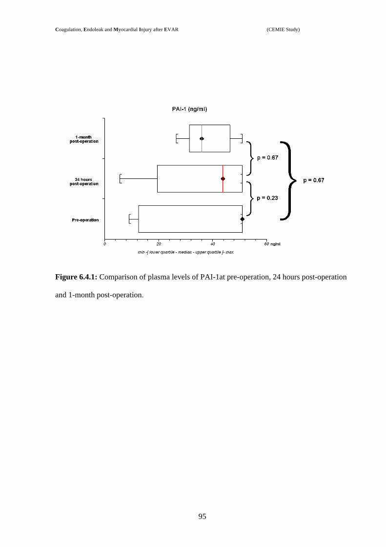
Coagulation, Endoleak and Myocardial Injury after EVAR (CEMIE Study)
95
Figure 6.4.1: Comparison of plasma levels of PAI-1at pre-operation, 24 hours post-operation
and 1-month post-operation.

Coagulation, Endoleak and Myocardial Injury after EVAR (CEMIE Study)
96
Effect of Endoleak
Seven patients demonstrated evidence of an endoleak at 1-month follow-up post-EVAR: type-
2 = 6, type-1 = 1. Median (IQR) difference in plasma levels of PAI-1 between baseline and 1-
month post-EVAR for patients with and without endoleak were -13.1 (-25-14.1) ng/ml and
12.3 (0-17.3) and ng/ml, respectively (p = 0.043). (See figure 6.4.2) Two patients with
endoleaks had positive differences (+14.1 ng/ml and +1.1 ng/ml) between PAI-1 levels at 1-
month and baseline
Figure 6.4.2: Median (IQR) differences of PA-1 levels between baseline and 1-month post-
EVAR in patients with or without endoleak.

Coagulation, Endoleak and Myocardial Injury after EVAR (CEMIE Study)
97
Patient Cohort 2
30 patients (29 men and 1 women of median (IQR) age 75 (71-82) years) underwent EVAR.
All EVARs were performed using a Zenith Endovascular stent-graft (Cook) and were
performed under loco-regional anaesthesia. There were no in-hospital deaths and EVAR was
successfully completed in all patients. Changes in coagulation, fibrinolysis and platelet
activity parameters are illustrated in table 6.4.1
Table 6.4.1: Effect of EVAR on coagulation, fibrinolysis and platelet activity parameters.
POST-OPERATIVE VALUES
BIOMARKERS NORMAL RANGE
PRE-OPERATIVE VALUES
24 HOURS 1-MONTH
PAI-1(ng/ml) 7-43 ng/ml 20.9 (8.4-50.7) 30.75 (19.8-46.8) 30.25 (15.8-56.9)
t-PA (U/ml) 0 U/ml 0 (0-0) 0 (0-0) 0 (0-0.06)
sP-Selectin (ng/ml) 92-212 ng/ml 80.5 (68-128) 89.5 (73-112) 110 (89-143)
TAT (µg /ml) <4.2 µg/L 7.15 (4.7-31.3) 21.65 (13-33.1) 8.1 (5.4-14.85)

Coagulation, Endoleak and Myocardial Injury after EVAR (CEMIE Study)
98
We noted no significant differences between pre-operative, 24-hours post-operative and 1-
month post-operative levels of PAI-1. A significant increase in sP-selectin levels occurred
between pre-operation and 24-hours post-operation (p = 0.003), pre-operation and 1-month
post-operation (p = <0.0001) and 24-hours and 1-month post-operation (p = 0.0003). There
was increased t-PA activity at 1-month post-operation compared to pre-operation (p = 0.0012)
and 24-hours post-operation (p = 0.0012). There was a trend towards increased thrombin
generation at 24 hours compared to pre-operation (p = 0.069) followed by a significant
decrease at 1-month (p = <0.0001) such that thrombin generation returned to pre-operative
levels by 1-month post-EVAR. (See figures 6.4.3 – 6.4.6).
Effect of Endoleak
No endoleaks occurred in this patient cohort at 1-month post-procedure. Combining the
results from patient cohorts 1 and 2 median (IQR) difference in plasma levels of PAI-1
between baseline and 1-month post-EVAR for patients with and without endoleak were -13.1
(-25-14.1) ng/ml and 2.8 (-9.4-16) ng/ml, respectively (p = 0.079).

Coagulation, Endoleak and Myocardial Injury after EVAR (CEMIE Study)
99
Figure 6.4.3: Comparison of plasma levels of PAI-1at pre-operation, 24 hours post-operation
and 1-month post-operation.

Coagulation, Endoleak and Myocardial Injury after EVAR (CEMIE Study)
100
Figure 6.4.4: Comparison of plasma levels of t-PA activity at pre-operation, 24 hours post-
operation and 1-month post-operation.

Coagulation, Endoleak and Myocardial Injury after EVAR (CEMIE Study)
101
Figure 6.4.5: Comparison of plasma levels of sP-selectin at pre-operation, 24 hours post-
operation and 1-month post-operation.

Coagulation, Endoleak and Myocardial Injury after EVAR (CEMIE Study)
102
Figure 6.4.6: Comparison of plasma levels of TAT at pre-operation, 24 hours post-operation
and 1-month post-operation.

Coagulation, Endoleak and Myocardial Injury after EVAR (CEMIE Study)
103
6.5 DISCUSSION
Only five studies to date have examined the effects of EVAR on coagulation and fibrinolysis
and platelet activity. 17 93 125 128 129
(Studies reporting the effects of EVAR on coagulation, fibrinolysis and platelet levels are
summarised in table 6.5.1)
Aho et al investigated the impact of EVAR on haemostasis in seventeen patients with AAA.
125 They reported a three-fold increase in TAT levels at 24 hours followed by a gradual
decline over the ensuing week. A similar transient increase was demonstrated for prothrombin
fragments 1 & 2 (F 1+2), a marker of factor Xa activity and thus thrombin generation. Levels
of both TAT and F 1+2 returned to pre-operative values by 1-week post-EVAR, however
levels remained elevated when compared to the normal ranges. The current study reports
similar findings with intense thrombin generation occurring within 24-hours of EVAR
followed by a return to up-regulated pre-operative levels by 1-month. Engleberger et al
demonstrated similar effects with elevated levels of plasma markers of thrombin generation
(TAT) and activity (fibrin monomer (FM) and fibrinopeptide A (FPA)) occurring in the peri-
operative period. 129 Maximum levels of TAT and FPA were found at the end of surgery
whereas FM levels were at their highest at 24 hours post-EVAR. This may reflect the
differing plasma kinetics of these markers; TAT and FPA reflect acute thrombin generation
and activity due to their short half-life (TAT t1/2 = 15 minutes, FPA t1/2= 3-5 minutes) whereas
FM reflects the overall coagulation process due to its much longer half –life (t1/2 > 3 hours).

Coagulation, Endoleak and Myocardial Injury after EVAR (CEMIE Study)
104
Serino et al reported the short-term effects of EVAR on circulating D-dimer levels in nine
patients assessed pre-operatively and day four post-operatively. 17 D-dimer levels were
elevated in seven patients and decreased in two patients; the median level for the entire
patient cohort did not demonstrate a statistically significant difference between baseline and
day four post-operation; this may reflect the study’s lack of statistical power.
Monaco et al reported a significant decrease in levels of platelets, fibrinogen, plasminogen
and prothrombin activity in patients undergoing EVAR during the first 10 days post-
procedure. 128 This was coupled with increased D-dimer and FDP levels suggestive of a
coagulation factor/platelet consumption coupled with systemic hyper-fibrinolysis during the
peri-operative period in patients undergoing EVAR. By one-month post-EVAR all
biomarkers returned to pre-operative levels with the exception of fibrinogen, which peaked at
one-month and remained significantly elevated at six-month post-EVAR.
To date only Aho et al have reported the effect of EVAR on systemic fibrinolysis through the
investigation of t-PA antigen and PAI-1 antigen levels. This group reported elevated levels of
both t-PA antigen and PAI-1 antigen during the first 24-hours post-operatively compared to
pre-operatively, thereafter returning to baseline levels by 1-week. However, t-PA antigen and
PAI-1 antigen levels remained within the normal reference ranges throughout. The current
study is the first to investigate t-PA activity as a marker of fibrinolysis following EVAR. t-PA
activity better reflects the true state of systemic fibrinolysis compared to t-PA antigen which
reflects the level of inactive circulating t-PA/PAI complexes. Thus despite normal levels of
PAI-1 antigen we report a proportion of patients demonstrate heightened systemic fibrinolysis
at one month post-operation as evidenced by elevated levels of t-PA activity. This finding is
consistent with elevated levels of D-dimer reported at 3-months by Aho et al despite PAI-I
antigen and t-PA antigen levels returning to baseline values at 1-week post-EVAR. 125
Coagulation, Endoleak and Myocardial Injury after EVAR (CEMIE Study)
105
Only one previous study has reported the effects of EVAR on platelet activity utilising novel
markers of platelet activity. Aho et al reported P-selectin levels as reducing during the first
three days post-operative and then returning to baseline levels by one-week. P-selectin levels
measured at 3-months were elevated in the EVAR group. 125 We report elevated platelet
activity as evidenced by increased levels of sP-selectin at both 24-hours and 1-month post-
operation. However, at both time points levels remained within normal reference ranges.
The Perioperative Ischemia Randomized Anesthesia Trial demonstrated that patients
undergoing lower limb revascularisation under epidural anaesthetic did not show any change
between pre- and peri-operative PAI-1 levels and suffered fewer post-operative graft
thromboses. 130 This was in comparison to patients operated under general anaesthesia where
PAI-1 levels were significantly higher in the peri-operative period as was the early graft
thrombosis rate. Other studies have shown fewer post-operative thrombotic complications in
patients undergoing major surgery under loco-regional anaesthesia. 131 The ability of loco-
regional anaesthesia to attenuate the catecholamine and cortisol biochemical response to
trauma may account for the reported haemostatic benefits when compared to general
anaesthesia. However, this apparent benefit has not been uniformly reported; Parrson et al
report elevated levels of PAI-1 in patients undergoing infra-inguinal vascular reconstruction
under epidural anaesthesia in the first 48 hours post-operative. 132 The current study is the first
to report the effects of differing methods of anaesthesia on haemostasis in patients undergoing
EVAR. We found no significant differences in peri-operative plasma PAI-1 levels between
patients undergoing EVAR under loco-regional or general anaesthesia. A number of
confounding factors may account for this lack of difference including differing peri-operative
analgesic regimes and type of stent-graft deployed. Furthermore the study was not powered to
detect such differences.

Coagulation, Endoleak and Myocardial Injury after EVAR (CEMIE Study)
106
To date, radiological imaging is required to identify endoleak and other stent related
complications. There has been interest in utilising changes in circulating levels of biomarkers
as an alternative means of identifying endoleak following EVAR. Sangiorgi et al and Lorelli
et al have both reported elevated levels of circulating metalloproteinase-3 and -9 (MMP-3 &
MMP-9) in patients affected by AAA. 15 16 Furthermore, successful endovascular repair
causes a decrease in MMP levels, whereas the presence of an endoleak post-EVAR appears to
attenuate this decrease. Only one study has investigated the effects of endoleak on levels of a
haemostatic biomarker in patients undergoing EVAR. 17 Serino et al reported patients with a
type-1 endoleak and stable/increasing aneurysm sac diameter demonstrated heightened
circulating D-dimer levels compared to patients with an endoleak with a decreasing aneurysm
sac diameter. The current study suggests that patients undergoing EVAR complicated by
endoleak demonstrate a relative increase in systemic fibrinolysis as evidenced by a reduction
in PAI-1 plasma levels at 1-month compared to pre-operative levels. Further research is
required in larger groups of patients.

Coagulation, Endoleak and Myocardial Injury after EVAR (CEMIE Study)
107
Table 6.5.1: Summary of studies investigating the effects of EVAR on biomarkers of haemostasis (* manufacturer’s range)

Coagulation, Endoleak and Myocardial Injury after EVAR (CEMIE Study)
108
In conclusion, the principal finding of this study is that EVAR causes significant haemostatic
derangement in the peri-operative period with increased platelet activity and thrombin
generation (as evidenced by increased sP-selectin and TAT levels) demonstrated at 24-hours
post-operation. At 1 month the amplified thrombin generation has largely resolved with TAT
levels returning to pre-operative levels, although remaining elevated above the normal healthy
range. Conversely, platelet activity continues to increase during the first month post-
operation. Furthermore, a proportion of patients demonstrate increased systemic fibrinolysis
at 1-month post-EVAR as evidenced by elevated levels of t-PA activity.

Coagulation, Endoleak and Myocardial Injury after EVAR (CEMIE Study)
109
CHAPTER 7
PERI-OPERATIVE MYOCARDIAL INJURY AND HAEMOSTASIS IN PATIENTS
UNDERGOING ENDOVASCULAR ANERUYSM REPAIR FOR ASYMPTOMATIC INFRA-
RENAL ABDOMINAL AORTIC ANEURYSM

Coagulation, Endoleak and Myocardial Injury after EVAR (CEMIE Study)
110
7.1 INTRODUCTION
Cardiac related complications, including myocardial ischaemic injury, are reported to occur in
5% of patients in the peri-operative period following EVAR. 11 This may represent a
significant under-estimation of the true rate of myocardial injury due to sub-clinical events
remaining undiagnosed. 113 133 134 135
To date no study has reported on the routine use of cardiac troponins as a method for
identifying patients with sub-clinical myocardial injury following EVAR, or attempted to
correlate cTn levels with the haemostatic derangement that can occurs as a result of EVAR.

Coagulation, Endoleak and Myocardial Injury after EVAR (CEMIE Study)
111
7.2 AIMS
The aim of this study is to 1) investigate the incidence of myocardial injury in patients
undergoing EVAR through the routine measurement of peri-operative cTn, and 2) investigate
and correlate any increase in cTn with concomitant haemostatic derangement.

Coagulation, Endoleak and Myocardial Injury after EVAR (CEMIE Study)
112
7.3 METHODS
All patients presenting to the Vascular Surgery Units of the Heart of England NHS
Foundation Trust and University Hospital Birmingham NHS Trust between January 2007 and
January 2009 with an infra-renal AAA measuring >5.5 cm in maximum diameter and due to
undergo EVAR were assessed for study inclusion suitability. (For study inclusion and
exclusion criteria see chapter 4.3.3)
Assessment for Myocardial Injury
Patients were assessed pre-operatively for cardiovascular disease including a full history and
examination. Pre-operative electrocardiograms (ECG’s) were performed. Post-operative
complications including myocardial infarction, cardiac failure, unstable angina and cardiac
dysrhythmias were assessed for clinically. All patients underwent routine ECG at 24 hours
and severe ischaemia was defined as ST-elevation/depression >2mm in two or more leads.

Coagulation, Endoleak and Myocardial Injury after EVAR (CEMIE Study)
113
Blood sample collection A resting venous blood sample was drawn from an antecubital fossa vein 24 hours prior to
EVAR and 24 hours post EVAR. The samples were placed immediately on ice and
centrifuged within 30 minutes of collection at 3,000 revolutions per minute for 30 minutes at
a temperature of 4o C (equivalent to 1400g). Plasma was separated and stored at - 80 C for
later batch analysis.
Cardiac troponin-T levels
All patients underwent routine cardiac troponin T (cTnT) measurement 24 hours post-
procedure using an ES300 fully automated second-generation multichannel immuno-assay
analyzer (Boehringer Mannheim). This second-generation test does not detect cross-reacting
isoforms from skeletal muscle. The lower limit for cTnT detection using this analyzer is
<0.01 ng/ml; cTnT values equal to or greater than 0.03 ng/ml were considered positive for
myocardial injury. 136
Markers of Thrombin generation, fibrinolysis and Platelet activity
Plasma thrombin antithrombin III-complex (TAT) (healthy range, <4.2 µg/L) was assayed as
a marker of thrombin generation. Plasma plasminogen activator inhibitor 1 (PAI-1) (healthy
range, 7-43ng/ml) and tissue plasminogen activator (t-PA) activity (healthy range, 0 U/ml)
were assayed as markers of fibrinolysis. Soluble P-selectin (sP-selectin) (healthy range, 92-
212 ng/ml) was assayed as a marker of platelet activity. The assay manufacturer determined
the healthy range for each haemostatic marker. (For further details of these commercially
available assays please see chapter 4.3.5)

Coagulation, Endoleak and Myocardial Injury after EVAR (CEMIE Study)
114
Statistics Differences between patient characteristics were assessed using Fisher’s exact test for
categorical variables and Mann-Whitney test for continuous variables. Correlation analyses
were performed using the Spearman rank test to test the null hypothesis and Kendall rank
correlation to test the strength of dependence between two variables if the null hypothesis was
rejected. Where levels of cTnT were below the limit of detection of the assay, a value of 0.01
ng/ml was assigned. A probability value (p-value) of less or equal to 0.05 was regarded as
statistically significant. Data are presented as median (inter-quartile range). All analyses were
carried out using StatsDirect version 2.7.2 (StatsDirect Ltd, Cheshire, UK).

Coagulation, Endoleak and Myocardial Injury after EVAR (CEMIE Study)
115
7.4 RESULTS
General
30 patients (29 men and 1 women of median (IQR) age 75 (71-82) years) who underwent
elective EVAR for infra-renal AAA were prospectively studied. Ten patients had ischaemic
heart disease co-morbidity, four patients suffered with diabetes, four patients suffered with
chronic renal failure (Stage <3), one patient was an active smoker and one patient suffered
with peripheral vascular disease (Fontaine Classification < IIb). Sixteen patients were being
treated with an anti-platelet agent (aspirin = 14, clopidogrel = 1, aspirin & clopidogrel = 1);
sixteen patients were being treated with a statin.

Coagulation, Endoleak and Myocardial Injury after EVAR (CEMIE Study)
116
Myocardial Injury
Two patients (7%) suffered with significant cardiac complication within 24 hours of EVAR.
One patient suffered with central chest pain and hypotension accompanied by ischaemic ECG
changes (ST-depression) and an elevated cTnT level (0.042 ng/ml). He was successfully
treated for acute coronary syndrome with a glyceryl trinitrate (GTN) infusion for 24 hours.
One patient developed fast atrial fibrillation requiring rate control pharmacotherapy; cTnT at
24 hours post-procedure was significantly elevated; cTnT = 0.178 ng/ml. Three patients
without clinical or ECG evidence of myocardial injury had cTnT levels >0.03 ng/ml; patient 2
cTnT = 0.04 ng/ml, patient 8 cTnT = 0.134 ng/ml, patient 25 cTnT = 0.058 ng/ml. Six
patients without clinical or ECG evidence of myocardial injury had cTnT levels >0.01 ng/ml;
patient 7 cTnT = 0.023 ng/ml, patient 11 cTnT = 0.012 ng/ml, patient 18 cTnT = 0.019 ng/ml,
patient 19 cTnT = 0.021 ng/ml, patient 27 cTnT = 0.021 ng/ml, patient 29 cTnT = 0.021
ng/ml. (See figure 7.4.1) There was no association between pre-operative anitplatelet or statin
usage and myocardial injury (p = NS)

Coagulation, Endoleak and Myocardial Injury after EVAR (CEMIE Study)
117
Figure 7.4.1: Individual patient data points for cTnT. The red points represent the two
patients with clinical/ECG signs of cardiac injury.

Coagulation, Endoleak and Myocardial Injury after EVAR (CEMIE Study)
118
Relationship between haemostatic markers and cTnT
Median (IQR) plasma levels of sP-selectin were 80.5 (68-86) ng/ml pre-operatively and 89.5
(73-112) ng/ml 24 hours post-EVAR (p = 0.0028). There was no correlation between raised
cTnT levels and pre-operative (r = -0.06, p = 0.76) or 24 hours post-EVAR (r = 0.23, p =
0.22) levels of sP-selectin.
Median (IQR) plasma levels of PAI-I were 20.9 (8.4-50.7) ng/ml pre-operatively and 30.75
(19.8-46.8) ng/ml 24 hours post-EVAR (p = 0.75). There was no correlation between raised
cTnT levels and pre-operative (r = 0.03, p = 0.87) or 24 hours post-EVAR (r = 0.12, p = 0.54)
levels of PAI-I. Median (IQR) plasma levels of t-PA were 0 (0-0) U/ml pre-operatively and 0
(0-0) U/ml 24 hours post-EVAR (p = 0.6875).
Median (IQR) plasma levels of TAT were 7.15 (4.7-31.3) µg/l pre-operatively and 21.65 (13-
33.1) µg/l 24 hours post-EVAR (p = 0.069). There was no correlation between raised cTnT
levels and pre-operative (r = 0.11, p = 0.56) levels of TAT. There was a positive correlation
between cTnT and TAT levels at 24 hours post-EVAR (r=0.38, p = 0.039, Kendall tau B =
0.26) (see figure 7.4.2)

Coagulation, Endoleak and Myocardial Injury after EVAR (CEMIE Study)
119
Figure 7.4.2: Box and Whisker plot of haemostatic variables pre- and 24 hours post-EVAR.
Spearman’s rank correlation coefficient (r) and p-value for each haemostatic variable vs.
cTnT is shown.

Coagulation, Endoleak and Myocardial Injury after EVAR (CEMIE Study)
120
7.5 DISCUSSION
This is the first study to investigate the routine use of cTnT as a marker of myocardial injury
and its association with altered peri-operative haemostasis following EVAR. The principle
findings of the present study are a) EVAR is associated with a significant risk of myocardial
injury [5 of 30 patients (16%)], as demonstrated by elevated levels of cTnT, and b) the pro-
thrombotic state that occurs in the peri-operative period after EVAR is associated with
myocardial injury.
Cardiac troponins (cTn) are highly sensitive and specific markers of myocardial necrosis. An
elevated peri-operative cTnT is an independent predictor of peri-operative and long-term
mortality. 30 136 137 134 Kertai el al reported peri-operative cTnT levels >0.1 ng/ml were
independently associated with a two-fold increased risk of long-term mortality following
successful major vascular surgery. 30 Landesberg et demonstrated that even minor elevations
in post-operative cTnT levels was a predictor of increased mortality; cTnT levels >0.1 ng/ml
and >0.03ng/ml independently predicted a 2.06 and 1.89 fold increase in mortality following
major vascular surgery, respectively. 136 In our study, utilising 0.03ng/ml as the cut-off level
for myocardial injury, 5 patients suffered peri-operative myocardial injury as opposed to 2
patients if the conventional cut-off of 0.1 ng/ml was utilised. This is considerably higher than
the 5% peri-operative cardiovascular morbidity reported by Drury et al in their Cochrane
review of endovascular aneurysm repair.
A number of studies have shown that routine cTnT surveillance in the peri-operative period
identifies a significant number of patients with sub-clinical myocardial injury following major
vascular surgery. 113 133 134 135 Hobbs et al reported 38% of patients undergoing surgery for
critical limb ischaemia had evidence of peri-operative myocardial injury as evidenced by
raised cardiac troponin levels; only one third of these cases were clinically apparent. 113

Coagulation, Endoleak and Myocardial Injury after EVAR (CEMIE Study)
121
Haggart et al reported that more than one quarter of patients undergoing elective open surgical
AAA repair suffer peri-operative myocardial injury. 23 However, 50% of these events
remained sub-clinical, identified only through routine monitoring of cardiac troponins. 23 In
our study only 2 of 5 patients with peri-operative levels of cTnT >0.03 ng/ml demonstrated
clinical evidence of cardiac dysfunction. Thus 10% of patients with peri-operative myocardial
injury would have gone undetected if routine surveillance with cTnT had not been
undertaken.
The present study demonstrates a correlation between myocardial injury and increased peri-
operative thrombin generation. Gibbs et al reported an association between the peri-operative
hypercoagulable state following open abdominal aortic surgery and peri-operative myocardial
infarction. 138 However, this study was limited to Q-wave infarcts whereas the majority of
peri-operative MI are non-Q-wave. 133 In the present study there were no Q-wave infarcts, but
the association between myocardial injury as defined by cTnT >0.03 ng/ml and a
hypercoagulable state was maintained. This may suggest that the elevated cTnT levels, with
the exception of the patient who developed atrial fibrillation, are secondary to a true
thrombotic myocardial injury. However, platelet activation and aggregation is the initial step
in the formation of clot and the present study did not demonstrate any correlation between
elevated cTnT and peri-operative platelet activation. The cTnT elevation witnessed in the
peri-operative phase of the present study may represent myocardial stress from causes other
than thrombosis.

Coagulation, Endoleak and Myocardial Injury after EVAR (CEMIE Study)
122
In conclusion, EVAR is associated with a significant risk of peri-operative myocardial injury
that is under-detected clinically. The resultant pro-coagulopathic state following EVAR may
be associated with an increased risk of myocardial injury. The routine use peri-operative cTnT
as marker of myocardial injury testing may allow for the identification of patients at increased
risk of mortality and the judicious use of peri-operative anti-coagulantion may help prevent
myocardial injury.

Coagulation, Endoleak and Myocardial Injury after EVAR (CEMIE Study)
123
CHAPTER 8
AREAS OF FUTURE RESEARCH

Coagulation, Endoleak and Myocardial Injury after EVAR (CEMIE Study)
124
8.1 THE RELATIONSHIP BETWEEN MURAL THROMBUS AND HAEMOSTASIS
The current study has not demonstrated the expected correlation between aneurysm thrombus
volume and biomarkers of haemostasis. However, these results do not necessarily exclude the
thrombus from being of importance in the prothrombotic state we report in patients with
AAA. Vascular surgeons operating on AAA will note that the thrombus composition and
density is often considerably different between patients. On occasion the thrombus can be
removed en masse with little fracturing, whereas in other patients the thrombus is very fragile
and semi-viscous in consistency. Microscopic examination of the mural thrombus reveals a
highly complex network of inter-connecting canaliculi. These exponentially increase the
surface area in direct contact with the luminal contents in a similar fashion to the villi and
microvilli of the gastrointestinal tract. Thus, the mural thrombus is a biologically active entity
that is not uniform in architecture, composition or density at a macro- or microscopic level.
These differences may directly impact the level of haemostatic derangement an AAA exerts
on the systemic circulation and may account for the conflicting studies reported in the English
literature. (see table 3.4.4)
To date only Kolbel et al have investigated the relationship between thrombin activation and
thrombus luminal surface area (TLS). 85 This group reported no correlation between the APC-
PCI levels and surface area in 130 patients. The study was limited by its methodology in that
it did not quantify the additional surface area contributed by macroscopic thrombus surface
irregularities and microscopic canaliculi thereby grossly underestimating the TLS. In order to
investigate this relationship comprehensively an accurate method for measuring true TLS is
required. This is extremely difficult and is akin to measuring the surface area of grass on a
football field. Currently, to the best of our knowledge, no non-invasive/radiographical method
is available to measure the surface area of the thrombus with the canaliculi taken into account.

Coagulation, Endoleak and Myocardial Injury after EVAR (CEMIE Study)
125
One potential solution is the utilisation of microscopic optical planimetry as described by
Spring et al. 139 The technique utilises optical cross-sectional imaging in multiple planes at
1!m intervals through a tissue sample allowing an accurate computer 3D- reconstruction.
From this 3D- reconstruction surface area and volume can be extrapolated. In the case of
mural thrombus the luminal surface area of a known volume of thrombus biopsied during
open repair can be utilised to estimate the luminal surface area for the total volume of
thrombus. Furthermore, the method of calculating thrombus volume described in Chapter 3
could be validated by the direct measurement of thrombus volume removed at open operation.
The current study did not investigate the differing thrombus biological qualities/variables and
in particular the TLS. We speculate that the TLS may demonstrate correlation with markers of
haemostasis if assessed appropriately and that further investigation is required to
establish/refute any such relationship.

Coagulation, Endoleak and Myocardial Injury after EVAR (CEMIE Study)
126
8.2 BIOMARKER OF ENDOLEAK
The current study investigated the novel use of biomarkers of haemostasis as a potential
marker of endoleak as a secondary outcome. However, the results from this secondary
outcome are of limited clinical value due to a) the study was not powered to investigate this
relationship, and b) the lack of endoleak occurring in the second cohort of patients.
We report endoleak occurring in 12% of patients within one month post-procedure. This
compares to the reported incidence of 22% in the EVAR-1 trial. 7 However, it is the clinically
significant endoleaks, those associated with sac expansion, that we wish to identify and only
one (2%) patient from the current study required re-intervention secondary to a type-1
endoleak. In order for a study to be sufficiently powered to investigate a relationship between
clinically significant endoleaks and novel markers of haemostasis we estimate a patient cohort
of >1000 would be required. Thus, a multi-centre, multi-regional study is required to
investigate this relationship further.

Coagulation, Endoleak and Myocardial Injury after EVAR (CEMIE Study)
127
8.3 MYOCARDIAL INJURY AND EVAR
Perhaps the most clinically relevant finding of the current study is patients undergoing EVAR
are at significant risk of peri-operative myocardial injury of which significant proportions are
clinically occult. It is established that patients with peri-operative cardiac troponin elevation
with or without clinical evidence of myocardial injury demonstrate adverse cardiovascular
short- and medium term-outcomes. Therefore the ability to pre-operatively identify
individuals at increased risk of peri-operative myocardial injury may allow targeted
optimisation to occur.
B-type natriuretic peptide (BNP) and its pro-hormone N-terminal fragment pro-BNP (NT-pro-
BNP) have recently been investigated as a potential pre-operative predictor of peri-operative
myocardial injury. 140 141 142 Rajagopalan et al demonstrated a pre-operative cut-off value for
NT-pro-BNP of 308 pg/ml correctly indentified patients at heightened risk of peri-operative
myocardial injury as identified by a cardiac troponin rise. 143 Feringa et al reported a pre-
operative NT-pro-BNP value of >533 pg/ml was independently associated and more accurate
than pre-operative dobutamine stress echocardiography in predicting early post-operative
adverse cardiac events. 142 To date no study has utilised NT-pro-BNP as a pre-operative risk
stratification method for peri-operative cardiac injury in patients undergoing EVAR.
The best means of pre-operative cardiac optimisation in patients at risk for peri-operative
adverse cardiac events remains unknown. A number of studies have investigated the use of
peri-operative !-blockers as a cardio-protective pharmacotherapy in patients undergoing non-
cardiac surgery. The PeriOperative ISschemic Evaluation (POISE) study reported a reduction
in peri-operative cardiovascular death following the administration of metoprolol in patients
with or at risk of atherosclerotic disease and undergoing non-cardiac surgery. 144 However,
the study protocol did not include routine peri-operative cardiac troponin measurement and

Coagulation, Endoleak and Myocardial Injury after EVAR (CEMIE Study)
128
was underpowered to assess for sub-group analysis i.e. patients undergoing major vascular
surgery. The Dutch Echocardiographic Cardiac Risk Evaluation Applying Stress
Echocardiography Study group (DECREASE) trial 1 reported a significant reduction in major
cardiovascular events following the administration of peri-operative bisoprolol. 145 The study
is limited through the utilisation of CK-MB as the marker of myocardial injury as opposed to
cardiac troponin resulting in a likely underestimation of the incidence of myocardial injury.
The only trial to employ routine peri-operative cardiac troponin assessment following major
infra-renal vascular surgery is the Perioperative !-Blockade (POBBLE) trial. 146 This trial
reported one third of patients suffered peri-operative myocardial ischaemia and that peri-
operative !-blockade did not reduce the 30-day major cardiovascular event rate. The study
protocol required a combination of cTnT >0.1ng/ml and persistent ECG ischaemic changes
before the diagnosis of myocardial injury could be inferred. Landesberg et al demonstrated
that even isolated minor elevations in post-operative cTnT (>0.03ng/ml) levels independently
predicted a two-fold increase in mortality following major vascular surgery. 136 Thus, the
POBBLE trial may be underestimating the actual incidence of clinically significant
myocardial injury in the study population. Only Rajagopalan et al have reported the
relationship between peri-operative !-blockade and isolated peri-operative cTnI levels in
patients undergoing major vascular surgery. 143 This longitudinal observational study
reported the incidence of peri-operative cardiac troponin rise was not affected by peri-
operative !-Blockade.
The aetiology of peri-operative myocardial injury is complex. Traditionally it has been
thought that prolonged myocardial stress in the setting of coronary artery atherosclerotic
disease combined with plaque rupture account for the majority of peri-operative myocardial
ischaemic events. However, as demonstrated in the current study, the resultant peri-operative

Coagulation, Endoleak and Myocardial Injury after EVAR (CEMIE Study)
129
hypercoagulable state following major vascular surgery may also contribute to the
development of myocardial injury in the peri-operative period. This may partially explain
why pre-operative stress testing is relatively inaccurate for predicting post-operative
myocardial ischaemic injury. 147
During open infra-renal AAA repair cross-clamping of the aorta places considerable strain on
the myocardium by dramatically increasing systemic vascular resistance and arterial pressure.
148 It is thought !-blockade attenuates this physiological response by lowering the heart rate
and improving coronary artery diastolic perfusion thereby reducing myocardial stress and
improving myocardial oxygenation. During EVAR the infra-renal aorta is rarely totally
occluded, thus any benefit inferred by the peri-operative use of !-blockade during open AAA
repair may be negated in patients undergoing EVAR. To date no study has investigated the
effects of peri-operative B-blockade on myocardial injury as defined by elevated post-
operative cardiac troponin levels in patients undergoing EVAR.

Coagulation, Endoleak and Myocardial Injury after EVAR (CEMIE Study)
130
APPENDICES

Coagulation, Endoleak and Myocardial Injury after EVAR (CEMIE Study)
131
APPENDIX 1
Classification of Endoleak
Type-I: Failure of stent-graft to seal against the aortic wall proximally (type-Ia) or
against the iliac arteries distally (type-Ib)
Type-II: Retrograde blood flow through one (type-IIa) or more (type-IIb) normal
branches of the excluded aortic segment e.g. inferior mesenteric artery or lumbar arteries.
Type-III: Structural failure of the stent-graft secondary to fabric disruption (type-IIIa),
separation of modular components (type-IIIb), suture holes in fabric (type-IIIc)
Type-IV: Caused by graft porosity typically identified at the time of stent-graft
deployment.

Coagulation, Endoleak and Myocardial Injury after EVAR (CEMIE Study)
132
APPENDIX 2: LOST SAMPLES
A B
Unfortunately due to the departments research freezer being turned-off by a member of the
domestic services department the majority of the initial patient cohort plasma samples (figures
A & B) were invalidated prior to batch analysis.

Coagulation, Endoleak and Myocardial Injury after EVAR (CEMIE Study)
133
APPENDIX 3: PATIENT INFORMATION LEAFLET

Coagulation, Endoleak and Myocardial Injury after EVAR (CEMIE Study)
134

Coagulation, Endoleak and Myocardial Injury after EVAR (CEMIE Study)
135

Coagulation, Endoleak and Myocardial Injury after EVAR (CEMIE Study)
136

Coagulation, Endoleak and Myocardial Injury after EVAR (CEMIE Study)
137
APPENDIX 4: PATIENT CONSENT FORM
Coagulation, Endoleak and Myocardial Injury after EVAR (CEMIE Study)
Consent Form Authorisation and Signatures I,________________________________, have been invited to participate in a study (The CEMIE study) investigating the short term changes to coagulation (clotting) following my endovascular aneurysm repair under the direction of Mr DJ Adam, in which I voluntarily consent to participate. a. The implications of my voluntary participation in this study, its nature, the duration, the purpose, the methods and means by which it is to be conducted, and the hazards which may be expected have been thoroughly explained to me by Mr Robert Davies b. I have read and understand all the written materials that have been provided to me describing this study and its potential risks and benefits. c. I have been given an opportunity to ask any questions I wish concerning the procedures, and all such questions have been answered to my complete satisfaction. I understand that my participation in this evaluation can be terminated at any time upon my request. d. I agree to allow authorised personnel to examine my medical Records and understand that this will be done in the strictest confidence. Date ________/_________/________ Day Month Year _______________________ _______________________ Patient signature Patient name printed _______________________ _______________________ Doctor Signature Doctor name printed

Coagulation, Endoleak and Myocardial Injury after EVAR (CEMIE Study)
138
APPENDIX 5: GP LETTER
Dear Sir/Madam I am writing to inform you that your patient_______________________ has agreed to
participate in the following study:
Coagulation, Endoleak and Myocardial Injury after EVAR (CEMIE Study)
The study involves obtaining peri-procedural blood samples (10-20ml) to assess altered
haemostasis and myocardial injury. The study does not involve any new therapeutic
pharmacotherapy or modality and will not influence the clinical treatment of your patient.
Your patient will be followed up during their routine clinic appointments where possible.
I have enclosed an information sheet providing additional information. Should you have any
queries please feel free to contact me.
Kind regards
Mr. Robert Davies
Vascular Research Fellow
Patient Sticker Here

Coagulation, Endoleak and Myocardial Injury after EVAR (CEMIE Study)
139
BIBLIOGRAPHY

Coagulation, Endoleak and Myocardial Injury after EVAR (CEMIE Study)
140
1 Scott RA, Ashton HA, Kay DN. Abdominal aortic aneurysm in 4237 screened patients:
prevalence, development and management over 6 years. Br J Surg. 1991 Sep;78(9):1122-5.
2 Collin J, Araujo L, Walton J, Lindsell D. Oxford screening programme for abdominal aortic
aneurysm in men aged 65 to 74 years. Lancet. 1988 Sep 10;2(8611):613-5.
3 O'Kelly TJ, Heather BP. General practice-based population screening for abdominal aortic
aneurysms: a pilot study. Br J Surg. 1989 May;76(5):479-80.
4 The UK Small Aneurysm Trial Participants. Mortality results for randomised controlled trial
of early elective surgery or ultrasonographic surveillance for small abdominal aortic
aneurysms. Lancet. 1998 Nov 21;352(9141):1649-55.
5 TBM Wilmink, CRG Quick, CS Hubbard and NE Day, The influence of screening on the
incidence of ruptured abdominal aortic aneurysms, J Vasc Surg 30 (1999), pp. 203–208.
6 Davies RS, Dawlatly S, Clarkson JR, Bradbury AW, Adam DJ. Outcome in patients
requiring renal replacement therapy after open surgical repair for ruptured abdominal aortic
aneurysm. Vasc Endovascular Surg. 2010 Apr;44(3):170-3.
7 EVAR trial participants. Endovascular aneurysm repair versus open repair in patients with
abdominal aortic aneurysm (EVAR trial 1): randomised controlled trial. Lancet. 2005 Jun 25-
Jul 1;365(9478):2179-86.
8 Greenhalgh RM, Brown LC, Kwong GP, Powell JT, Thompson SG; EVAR trial
participants. Comparison of endovascular aneurysm repair with open repair in patients with
abdominal aortic aneurysm (EVAR trial 1), 30-day operative mortality results: randomised
controlled trial. Lancet. 2004 Sep 4-10;364(9437):843-8.
9 EVAR trial participants. Endovascular aneurysm repair and outcome in patients unfit for
open repair of abdominal aortic aneurysm (EVAR trial 2): randomised controlled trial.
Lancet. 2005 Jun 25-Jul 1;365(9478):2187-92.
Coagulation, Endoleak and Myocardial Injury after EVAR (CEMIE Study)
141
10 Prinssen M, Verhoeven EL, Buth J, et al: A randomized trial comparing conventional and
endovascular repair of abdominal aortic aneurysms. Dutch Randomized Endovascular
Aneurysm Managemtn (DREAM) Trial Group. N Engl J Med 351:1607, 2004.
11 Drury D, Michaels JA, Jones L, Ayiku L. Systematic review of recent evidence for the
safety and efficacy of elective endovascular repair in the management of infrarenal abdominal
aortic aneurysm. Br J Surg. 2005 Aug;92(8):937-46.
12 Ghouri M, Krajcer Z. Endoluminal abdominal aortic aneurysm repair: the latest advances in
prevention of distal endograft migration and type 1 endoleak. Tex Heart Inst J. 2010;37(1):19-
24.
13 Michaels JA, Drury D, Thomas SM. Cost-effectiveness of endovascular abdominal aortic
aneurysm repair. Br J Surg. 2005 Aug;92(8):960-7.
14 Yamazumi K, Ojiro M, Okumura H, Aikou T. An activated state of blood coagulation and
fibrinolysis in patients with abdominal aortic aneurysm. Am J Surg. 1998 Apr;175(4):297-
301.
15 Sangiorgi G, D’Averio R, Mauriello A, Bondio M, Pontillo M, Castelvecchio S, Trimarchi
S, Tolva V, Nano G, Rampoldi V, Spagnoli LG, Inglese L. Plasma levels of
metalloproteinases-3 and -9 as markers of successful abdominal aortic aneurysm exclusion
after endovascular graft treatment. Circulation. 2001;104:I-288–I-295.
16 Lorelli DR, Jean-Claude JM, Fox CJ, Clyne J, Cambria RA, Seabrook GR, Towne JB.
Response of plasma matrix metalloproteinase-9 to conventional abdominal aortic aneurysm
repair or endovascular exclusion: implications for endoleak. J Vasc Surg. 2002;35:916–922.
17 Serino F, Abeni D, Galvagni E, Sardella SG, Scuro A, Ferrari M, Ciarafoni I, Silvestri L,
Fusco A. Noninvasive diagnosis of incomplete endovascular aneurysm repair: D-dimer assay
to detect type I endoleaks and nonshrinking aneurysms. J Endovasc Ther. 2002 Feb;9(1):90-7.
18 Bradbury AW, Adam DJ, Makhdoomi KR, Stuart WP, Murie JA, Jenkins AM, Ruckley
CV. A 21-year experience of abdominal aortic aneurysm operations in Edinburgh. Br J Surg.
1998 May;85(5):645-7.

Coagulation, Endoleak and Myocardial Injury after EVAR (CEMIE Study)
142
19 Dardik A, Lin JW, Gordon TA, Williams GM, Perler BA. Results of elective abdominal
aortic aneurysm repair in the 1990s: A population-based analysis of 2335 cases. J Vasc Surg.
1999 Dec;30(6):985-95.
20 Kazmers A, Jacobs L, Perkins A, Lindenauer SM, Bates E. Abdominal aortic aneurysm
repair in Veterans Affairs medical centers. J Vasc Surg. 1996 Feb;23(2):191-200.
21 Sayers RD, Thompson MM, Nasim A, Healey P, Taub N, Bell PR. Surgical management of
671 abdominal aortic aneurysms: a 13 year review from a single centre. Eur J Vasc Endovasc
Surg. 1997 Mar;13(3):322-7.
22 Dueck AD, Kucey DS, Johnston KW, Alter D, Laupacis A. Long-term survival and
temporal trends in patient and surgeon factors after elective and ruptured abdominal aortic
aneurysm surgery. J Vasc Surg. 2004 Jun;39(6):1261-7.
23 Haggart PC, Adam DJ, Ludman PF, Bradbury AW. Comparison of cardiac troponin I and
creatine kinase ratios in the detection of myocardial injury after aortic surgery. Br J Surg
2001; 88: 1196-1200.
24 Meade TW, Ruddock V, Stirling Y, Chakrabarti R, Miller GJ. Fibrinolytic activity, clotting
factors, and long-term incidence of ischaemic heart disease in the Northwick Park Heart
Study. Lancet 1993;342:1076-9.
25 Vervloet MG, Thijs LG, Hack CE. Derangements of coagu- lation and fibrinolysis in
critically ill patients with sepsis and septic shock. Semin Thromb Hemost 1998;24:33-44.
26 Penner JA. Disseminated intravascular coagulation in patients with multiple organ failure
of non-septic origin. Semin Thromb Hemost 1998;24:45-52.
27 Lindgren A, Lindoff C, Norrving B, Astedt B, Johansson BB. Tissue plasminogen activator
and plasminogen activator inhibitor-1 in stroke patients. Stroke 1996;27:1066-71.29.
28 Davies MG, Murphy WG, Murie JA, Elton RA, Bell K, Gillon JG, et al. Preoperative
coagulopathy in ruptured abdominal aortic aneurysm predicts poor outcome. Br J Surg
1993;80:974-6.

Coagulation, Endoleak and Myocardial Injury after EVAR (CEMIE Study)
143
29 Adam DJ, Ludlam CA, Ruckley CV, Bradbury AW. Coagulation and fibrinolysis in
patients undergoing operation for ruptured and nonruptured infrarenal abdominal aortic
aneurysms. J Vasc Surg. 1999 Oct;30(4):641-50.
30 Kertai MD, Boersma E, Klein J, van Urk H, Bax JJ, Poldermans D. Long-term Prognostic
Value of Asymptomatic Cardiac Troponin T Elevations in Patients After Major Vascular
Surgery. Eur J Vasc Endovasc Surg 2004; 28: 59-66.
31 Polgar J, Matuskova J, Wagner DD. The P-selectin, tissue factor, coagulation triad. J
Thromb Haemost. 2005 Aug;3(8):1590-6.
32 Hickey MJ, Williams SA, Roth GJ. Human platelet glycoprotein IX: an adhesive prototype
of leucine-rich glycoproteins with flank-center-flank structures. Proc Natl Acad Sci U S A.
1989 Sep;86(17):6773-7.
33 Savage B, Ruggeri ZM. Selective recognition of adhesive sites in surface-bound fibrinogen
by glycoprotein IIb-IIIa on nonactivated platelets. J Biol Chem. 1991 Jun 15;266(17):11227-
33.
34 Furie B, Furie BC. In vivo thrombus formation. J Thromb Haemost. 2007 Jul;5 Suppl 1:12-
7
35 Furie B, Furie BC. Mechanisms of thrombus formation. N Engl J Med. 2008 Aug
28;359(9):938-49.
36 Green D. Coagulation cascade. Hemodial Int. 2006 Oct;10 Suppl 2:S2-4.
37 Marlar RA, Kleiss AJ, Griffin JH. Mechanism of action of human activated protein C, a
thrombin-dependent anticoagulant enzyme. Blood. 1982 May;59(5):1067-72.
38 Dahlbäck B, Villoutreix BO. The anticoagulant protein C pathway. FEBS Lett. 2005 Jun
13;579(15):3310-6. Epub 2005 Mar 13.
39 Bajzar L, Morser J, Nesheim M: TAFI or plasma procarboxypeptidase B, couples the
coagulation and fibrinolytic cascades through the thrombin–thrombomodulin complex. J Biol
Chem 1996, 271:16603–16608.

Coagulation, Endoleak and Myocardial Injury after EVAR (CEMIE Study)
144
40 Sakata Y, Curriden S, Lawrence D, Griffin JH, Loskutoff DJ: Activated protein C
stimulates the fibrinolytic activity of cultured endothelial cells and decreases antiactivator
activity. Proc Natl Acad Sci USA 1985, 82:1121–1125.
41 Broze GJ Jr, Warren LA, Novotny WF, Higuchi DA, Girard JJ, Miletich JP. The
lipoprotein-associated coagulation inhibitor that inhibits the factor VII-tissue factor complex
also inhibits factor Xa: insight into its possible mechanism of action. Blood. 1988
Feb;71(2):335-43.
42 Rijken DC, Sakharov DV. Basic principles in thrombolysis: regulatory role of plasminogen.
Thromb Res. 2001 Sep 30;103 Suppl 1:S41-9.
43 Seyfer AE, Seaber AV, Dombrose FA, Urbaniak JR. Coagulation changes in elective
surgery and trauma. Ann Surg. 1981 Feb;193(2):210-3.
44 Knight MT, Dawson R, Melrose DG. Fibrinolytic response to surgery. Labile and stable
patterns and their relevance to post-operative deep venous thrombosis. Lancet. 1977 Aug
20;2(8034):370-3.
45 McDaniel MD, Pearce WH, Yao JS, Rossi EC, Fahey VA, Green D, Flinn WR, Bergan
JJ.Sequential changes in coagulation and platelet function following femorotibial bypass. J
Vasc Surg. 1984 Mar;1(2):261-8.
46 Collins GJ Jr, Barber JA, Zajtchuk R, Vanek D, Malogne LA. The effects of operative
stress on the coagulation profile. Am J Surg. 1977 May;133(5):612-6.
47 Woodburn KR, Rumley A, Lowe GD, Pollock JG. Fibrinogen and markers of fibrinolysis
and endothelial damage following resolution of critical limb ischaemia. Eur J Vasc Endovasc
Surg. 1995 Oct;10(3):272-8.
48 Rosc D, Zekanowska E, Kotschy M, Hryncewicz W, Jurkowska M. Effect of vascular
surgery on values of thrombin-antithrombin III complexes in patients with atherosclerosis of
lower limb arteries. Pol Tyg Lek. 1994 Apr 18-25;49(16-17):360-2.

Coagulation, Endoleak and Myocardial Injury after EVAR (CEMIE Study)
145
49 Naesh O, Friis JT, Hindberg I, Winther K. Platelet function in surgical stress. Thromb
Haemost. 1985 Dec 17;54(4):849-52.
50 Galle C, De Maertelaer V, Motte S, Zhou L, Stordeur P, Delville JP, Li R, Ferreira J,
Goldman M, Capel P, Wautrecht JC, Pradier O, Dereume JP. Early inflammatory response
after elective abdominal aortic aneurysm repair: a comparison between endovascular
procedure and conventional surgery. J Vasc Surg. 2000 Aug;32(2):234-46.
51 Swartbol P, Norgren L, Albrechtsson U, Cwikiel W, Jahr J, Jonung T, Pärsson H, Ribbe E,
Thörne J, Truedsson L, Zdanowski Z. Biological responses differ considerably between
endovascular and conventional aortic aneurysm surgery. Eur J Vasc Endovasc Surg. 1996
Jul;12(1):18-25.
52 Swartbol P, Truedsson L, Norgren L. Adverse reactions during endovascular treatment of
aortic aneurysms may be triggered by interleukin 6 release from the thrombotic content. J
Vasc Surg. 1998 Oct;28(4):664-8.
53 Zhang H, Holt CM, Malik N, Shepherd L, Morcos SK. Effects of radiographic contrast
media on proliferation and apoptosis of human vascular endothelial cells. Br J Radiol.
2000;73(874):1034-41.
54 Polanowska R, Wilczynska M, Slawinski W, Goch JH, Augustyniak W, Cierniewski CS.
Changes in platelet activity and tissue plasminogen activator during arteriography in patients
with chronic limb ischaemia. Thromb Res. 1992;65(4-5):663-5.
55 Hans SS, Jareunpoon O, Balasubramaniam M, Zelenock GB. Size and location of thrombus
in intact and ruptured abdominal aortic aneurysms. J Vasc Surg. 2005 Apr;41(4):584-8.
56 Adolph R, Vorp DA, Steed DL, Webster MW, Kameneva MV, Watkins SC Cellular
content and permeability of intraluminal thrombus in abdominal aortic aneurysm. J Vasc
Surg. 1997 May;25(5):916-26.
57 Gibney EJ, Bouchier-Hayes D. Coagulopathy and abdominal aortic aneurysm. Eur J Vasc
Surg. 1990 Dec;4(6):557-62.

Coagulation, Endoleak and Myocardial Injury after EVAR (CEMIE Study)
146
58 Fontaine V, Jacob MP, Houard X, Rossignol P, Plissonnier D, Angles-Cano E, Michel JB.
Involvement of the mural thrombus as a site of protease release and activation in human aortic
aneurysms. Am J Pathol. 2002 Nov;161(5):1701-10.
59 Carrell TW, Burnand KG, Booth NA, Humphries J, Smith A. Intraluminal thrombus
enhances proteolysis in abdominal aortic aneurysms.Vascular. 2006 Jan-Feb;14(1):9-16.
60 Danesh J, Lewington S, Thompson SG et al Plasma fibrinogen level and the risk of major
cardiovascular diseases and nonvascular mortality: an individual participant meta-analysis.
JAMA. 2005 Oct 12;294(14):1799-809.
61 Ernst E. Plasma fibrinogen--an independent cardiovascular risk factor. J Intern Med. 1990
Jun;227(6):365-72.
62 Smith FB, Lee AB, Fowkes FG et al. Hemostatic factors as predictors of ischemic heart
disease and stroke in the Edinburgh Artery Study. Arterioscler Thromb Vasc Biol. 1997
Nov;17(11):3321-5.
63 Kannel WB, Wolf PA, Castlli WP, D’Agostino RB. Fibrinogen and risk of cardiovascular
disease. The Framingham Study. nJAMA. 1987 Sep 4;258(9):1183-6.
64 Dormandy JA, Hoare E, Colley J, Arrowsmith DE, Dormandy TL. Clinical,
haemodynamic, rheological, and biochemical findings in 126 patients with intermittent
claudication. Br Med J. 1973 Dec 8;4(5892):576-81.
65 Fowkes FG, Anandan CL, Lee AJ, Smith FB, Tzoulaki I, Rumley A, Powell JT, Lowe GD.
Reduced lung function in patients with abdominal aortic aneurysm is associated with
activation of inflammation and hemostasis, not smoking or cardiovascular disease. J Vasc
Surg. 2006 Mar;43(3):474-80.
66 Holmberg A, Bergqvist D, Westman B, Siegbahn A. Cytokine and fibrinogen response in
patients undergoing open abdominal aortic aneurysm surgery. Eur J Vasc Endovasc Surg.
1999 Apr;17(4):294-300.

Coagulation, Endoleak and Myocardial Injury after EVAR (CEMIE Study)
147
67 Blann AD, Devine C, Amiral J, McCollum CN. Soluble adhesion molecules, endothelial
markers and atherosclerosis risk factors in abdominal aortic aneurysm: a comparison with
claudicants and healthy controls. Blood Coagul Fibrinolysis. 1998 Sep;9(6):479-84.
68 Singh K, Bønaa KH, Jacobsen BK, Bjørk L, Solberg S. Prevalence of and risk factors for
abdominal aortic aneurysms in a population-based study : The Tromsø Study. Am J
Epidemiol. 2001 Aug 1;154(3):236-44.
69 Shindo S, Matsumoto H, Kubota K, Kojima A, Matsumoto M, Satoh K, Ozaki Y. Is the
size of an abdominal aortic aneurysm associated with coagulopathy? World J Surg. 2005
Jul;29(7):925-9
70 Hosaka A, Miyata T, Aramoto H, Shigematsu H, Nakazawa T, Okamoto H, Shigematsu K,
Nagawa H. Clinical implication of plasma level of soluble fibrin monomer-fibrinogen
complex in patients with abdominal aortic aneurysm. J Vasc Surg. 2005 Aug;42(2):200-5.
71 Adam DJ, Haggart PC, Ludlam CA, Bradbury AW. Hemostatic markers before operation in
patients with acutely symptomatic nonruptured and ruptured infrarenal abdominal aortic
aneurysm. J Vasc Surg. 2002 Apr;35(4):661-5.
72 Lee AJ, Fowkes FG, Lowe GD, Rumley A. Haemostatic factors, atherosclerosis and risk of
abdominal aortic aneurysm. Blood Coagul Fibrinolysis. 1996 Oct;7(7):695-701.
73 Jelenska MM, Szmidt J, Bojakowski K, Grzela T, Palester-Chlebowczyk M. Compensated
activation of coagulation in patients with abdominal aortic aneurysm: effects of heparin
treatment prior to elective surgery. Thromb Haemost. 2004 Nov;92(5):997-1002.
74 Fisher DF Jr, Yawn DH, Crawford ES. Preoperative disseminated intravascular coagulation
associated with aortic aneurysms. A prospective study of 76 cases. Arch Surg. 1983
Nov;118(11):1252-5
75 Brass EP, Forman WB, Edwards RV, Lindan O. Fibrin formation: the role of the
fibrinogen-fibrin complexes. Thromb Haemost 1976;36:37-48

Coagulation, Endoleak and Myocardial Injury after EVAR (CEMIE Study)
148
76 Nakahara K, Kazahaya Y, Shintani Y, Yamazumi K, Eguchi Y, Koga S, Wada H, Matsuda
M. Measurement of soluble fibrin monomer-fibrinogen complex in plasmas derived from
patients with various underlying clinical situations.Thromb Haemost. 2003 May; 89(5):832-
6.
77 Holmberg A, Siegbahn A, Westman B, Bergqvist D. Ischaemia and reperfusion during
open abdominal aortic aneurysm surgery induce extensive thrombin generation and activity.
Eur J Vasc Endovasc Surg. 1999 Jul;18(1):11-6.
78 Ihara A, Kawamoto T, Matsumoto K, Kawamoto J, Katayama A, Yoshitatsu M, Izutani H,
Ihara K. Relationship between hemostatic markers and circulating biochemical markers of
collagen metabolism in patients with aortic aneurysm. Pathophysiol Haemost Thromb. 2003
Jul-2004 Aug;33(4):221-4.
79 Sofi F, Marcucci R, Giusti B, Pratesi G, Lari B, Sestini I, Lo Sapio P, Pulli R, Pratesi C,
Abbate R, Gensini GF. High levels of homocysteine, lipoprotein (a) and plasminogen
activator inhibitor-1 are present in patients with abdominal aortic aneurysm. Thromb
Haemost. 2005 Nov;94(5):1094-8.
80 Nelken NA, Soifer SJ, O'Keefe J, Vu TK, Charo IF, Coughlin SR. Thrombin receptor
expression in normal and atherosclerotic human arteries.J Clin Invest. 1992;90(4):1614-21.
81 Haeberli A. Prothrombin fragment F1+2. In:Jespersen J, Bertina RM, Haverkater F
(eds):Laboratory techniques in thrombosis - A manual. Dordrecht-Boston-London, Kluwer,
1999, pp 217-222.
82 Lau HK, Rosenberg RD. The isolation and characterization of a specific antibody
population directed against the thrombin antithrombin complex. J Biol Chem 1980;255:5885-
5893
83 Esmon CT, Owen WG. The discovery of thrombomodulin. J Thromb Haemost. 2004
Feb;2(2):209-13.

Coagulation, Endoleak and Myocardial Injury after EVAR (CEMIE Study)
149
84 Strandberg K, Kjellberg M, Knebel R, Lilja H, Stenflo J. A sensitive immunochemical
assay for measuring the concentration of the activated protein C-protein C inhibitor complex
in plasma: use of a catcher antibody specific for the complexed/cleaved form of the inhibitor.
Thromb Haemost. 2001 Aug;86(2):604-10.
85 Kölbel T, Strandberg K, Mattiasson I, Stenflo J, Lindblad B. Activated protein C-protein C
inhibitor complex: a new biological marker for aortic aneurysms. J Vasc Surg. 2006
May;43(5):935-9
86 Milne AA, Adam DJ, Murphy WG, Ruckley CV. Effects of asymptomatic abdominal aortic
aneurysm on the soluble coagulation system, platelet count and platelet activation. Eur J Vasc
Endovasc Surg. 1999 May;17(5):434-7
87 Mukaiyama H, Shionoya S, Ikezawa T, Kamiya T, Hamaguchi M, Saito H. Abdominal
aortic aneurysm complicated with chronic disseminated intravascular coagulopathy: a case of
surgical treatment. J Vasc Surg. 1987 Dec;6(6):600-4
88 Fox JE. Shedding of adhesion receptors from the surface of activated platelets. Blood
Coagul Fibrinolysis. 1994 Apr;5(2):291-304
89 Wang XF, Liu L, Cheng TO, Li ZA, Deng YB, Wang JE. The relationship between
intracardiovascular smoke-like echo and erythrocyte rouleaux formation. Am Heart J. 1992
Oct;124(4):961-5
90 Hwang JJ, Ko FN, Li YH, Ma HM, Wu GJ, Chang H, Wang SM, Schie JT, Tseng YZ,
Kuan P, et al. Clinical implications and factors related to left atrial spontaneous echo contrast
in chronic nonvalvular atrial fibrillation. Cardiology. 1994;85(2):69-75
91 McDaniel MD, Pearce WH, Yao JS, Rossi EC, Fahey VA, Green D, Flinn WR, Bergan
JJ.Sequential changes in coagulation and platelet function following femorotibial bypass. J
Vasc Surg. 1984 Mar;1(2):261-8
92 Killewich LA, Macko RF, Gardner AW, Cox K, Lilly MP, Flinn WR. Defective
fibrinolysis occurs after infrainguinal reconstruction. J Vasc Surg. 1997 May;25(5):858-64

Coagulation, Endoleak and Myocardial Injury after EVAR (CEMIE Study)
150
93 Parsson H, Holmberg A, Siegbahn A, Bergqvist D. Activation of coagulation and
fibrinolytic systems in patients with CLI is not normalized after surgical revascularisation.
Eur J Vasc Endovasc Surg. 2004 Feb;27(2):186-92
94 Perler BA, Christopherson R, Rosenfeld BA, Norris EJ, Frank S, Beattie C, Williams GM.
The influence of anesthetic method on infrainguinal bypass graft patency: a closer look. Am
Surg. 1995 Sep;61(9):784-9
95 Odegård A, Lundbom J, Myhre HO, Hatlinghus S, Bergh K, Waage A, Bjerve KS, Mollnes
TE, Aadahl P, Lie TA, Videm V.The inflammatory response following treatment of
abdominal aortic aneurysms: a comparison between open surgery and endovascular repair.
Eur J Vasc Endovasc Surg. 2000 May;19(5):536-44
96 Speiser W, Speiser P, Minar E, Korniger C et al. Activation of coagulation and fibrinolysis
in patients with arteriosclerosis: relation to localization of vessel disease and risk factors.
Thromb Res. 1990 Jul 1;59(1):77-88
97 Strano A, Hoppensteadt D, Walenga JM et al. Plasma levels of the molecular markers of
coagulation and fibrinolysis in patients with peripheral arterial disease. Semin Thromb
Hemost. 1996;22 Suppl 1:35-40
98 Koksch M, Zeiger F, Wittig K, Siegemund A et al. Coagulation, fibrinolysis and platelet P-
selectin expression in peripheral vascular disease. Eur J Vasc Endovasc Surg. 2001
Feb;21(2):147-54
99 Kristiansson M, Soop M, Saraste L, Sundqvist KG. Cytokines in stored red blood cell
concentrates: promoters of systemic inflammation and simulators of acute transfusion
reactions? Acta Anaesthesiol Scand. 1996 Apr;40(4):496-501
100 Hashimoto Y, Kobayashi A, Yamazaki N, Sugawara Y, Takada Y, Takada A. Relationship
between age and plasma t-PA, PA-inhibitor, and PA activity. Thromb Res. 1987 Jun
1;46(5):625-33
101 Van der Bom JG, Bots ML, van Vliet HHDM, Pols HAP et al. Antithrombin and
atherosclerosis in the Rotterdam Study.Arterioscler Thromb Vasc Biol. 1996 Jul;16(7):864-7

Coagulation, Endoleak and Myocardial Injury after EVAR (CEMIE Study)
151
102 Barani J, Gottsater A, Mattiasson I, Lindblad B. Platelet and leukocyte activation during
aortoiliac angiography and angioplasty. Eur J Vasc Endovasc Surg. 2002;23(3):220-5
103 Namiki A, Toma H, Nakamura M, Matsuda K, Hara H, Hara H, Asahara T, Soumitsu Y,
Kobayashi N, Yamaguchi T. Hemostatic and fibrinolytic activation is less following cutting
balloon angioplasty of the coronary arteries. Jpn Heart J. 2004;45(3):409-17
104 Tschopl M, Tsakiris DA, Marbet GA, Labs KH, Jager K. Role of hemostatic risk factors
for restenosis in peripheral arterial occlusive disease after transluminal angioplasty.
Arterioscler Thromb Vasc Biol. 1997 ;17(11):3208-14
105 Eriksson, E., et al. Determination of plasminogen activator inhibitor5 in plasma using t-PA
and chromogenic single point poly-D-lysine stimulated assay. Thromb Res 1989, 50: 91-101.
106 Chmielewska, K., Rånby, M., and Wiman, B. Evidence for a rapid inhibitor to tissue
plasminogen activator in plasma. Thromb Res 1983, 31: 427-436.
107 L.C. Dunlop, M.P. Skinner and L.J. Bendall et al., Characterization of GMP-140 (P-
selectin) as a circulating plasma protein, J Exp Med 175 (1992), pp. 1147–1150.
108 G. Berger, D.W. Hartwell and D.D. Wagner, P-selectin and platelet clearance, Blood 92
(1998), pp. 4446–4452.
109 P. Ferroni, F.M. Pulcinelli, L. Lenti and P.P. Gazzaniga, Is soluble P-selectin
determination a more reliable marker of in vivo platelet activation than CD62P flow
cytometric analysis?, Thromb Haemost 81 (1999), pp. 472–473.
110 Haggart PC, Ludman PF, Bradbury AW. Cardiac troponin: a new biochemical marker for
peri-operative myocardial injury.Eur J Vasc Endovasc Surg. 2001 Oct;22(4):301-5.
111 Sharma S, Jackson PG, Makan J. Cardiac troponins. J Clin Pathol. 2004 Oct;57(10):1025-
6.
112 Ammann P, Pfisterer M, Fehr T, Rickli H. Raised cardiac troponins. BMJ. 2004 May
1;328(7447):1028-9.

Coagulation, Endoleak and Myocardial Injury after EVAR (CEMIE Study)
152
113 Hobbs SD, Yapanis M, Burns PJ, Wilmink AB, Bradbury AW, Adam DJ. Peri-operative
myocardial injury in patients undergoing surgery for critical limb ischaemia. Eur J Vasc
Endovasc Surg. 2005 Mar;29(3):301-4.
114 Noether GE, Sample Size Determination for Some Common Nonparametric Tests. Journal
of the American Statistical Association, 1987 Jun; 82(398):645-647.
115 Shapiro SS, Wilk MB. An analysis of variance test for normality. Biometrika
1965;52(3):591-9.
116 Wallinder J, Bergqvist D, Henriksson AE. Haemostatic markers in patients with abdominal
aortic aneurysm and the impact of aneurysm size. Thromb Res. 2009 Sep;124(4):423-6.
117 Wanhainen A, Nilsson TK, Bergqvist D, Boman K, Björck M. Elevated tissue
plasminogen activator in patients with screening-detected abdominal aortic aneurysm. J Vasc
Surg. 2007 Jun;45(6):1109-13.
118 Parry DJ, Al-Barjas HS, Chappell L, Rashid T, Ariëns RA, Scott DJ. Haemostatic and
fibrinolytic factors in men with a small abdominal aortic aneurysm. Br J Surg. 2009
Aug;96(8):870-7.
119 Ferroni P, Martini F, Riondino S, La Farina F, Magnapera A, Ciatti F, Guadagni F. Soluble
P-selectin as a marker of in vivo platelet activation. Clin Chim Acta. 2009 Jan;399(1-2):88-91
120 Tomizuka T, Yamamoto K, Hirai A, Tamura Y, Yoshida S. Hypersensitivity to
thromboxane A2 in cholesterol-rich human platelets. Thromb Haemost. 1990 Dec
28;64(4):594-9.
121 Miida T, Takahashi A, Ikeuchi T. Prevention of stroke and dementia by statin therapy:
experimental and clinical evidence of their pleiotropic effects. Pharmacol Ther. 2007
Feb;113(2):378-93.
122 Casani L, Sanchez-Gomez S, Vilahur G, Badimon L. Pravastatin reduces thrombogenicity
by mechanisms beyond plasma cholesterol lowering. Thromb Haemost. 2005
Nov;94(5):1035-41.

Coagulation, Endoleak and Myocardial Injury after EVAR (CEMIE Study)
153
123 Al-Barjas HS, Ariëns R, Grant P, Scott JA. Raised plasma fibrinogen concentration in
patients with abdominal aortic aneurysm. Angiology. 2006 Oct-Nov;57(5):607-14.
124 Kölbel T, Donath T, Strandberg K, Flondell-Sité D, Kühme T, Gottsäter A, Lindblad B. Is
increased thrombin activation in patients with abdominal aortic aneurysms dependent on area
or volume of aneurysm thrombus mass? Angiology. 2010 Feb-Mar;61(1):113-8.
125 Aho PS, Niemi T, Piilonen A, Lassila R, Renkonen R, Lepäntalo M. Interplay between
coagulation and inflammation in open and endovascular abdominal aortic aneurysm repair--
impact of intra-aneurysmal thrombus. Scand J Surg. 2007;96(3):229-35
126 Prinssen M, Verhoeven EL, Buth J, et al: A randomized trial comparing conventional and
endovascular repair of abdominal aortic aneurysms. Dutch Randomized Endovascular
Aneurysm Managemtn (DREAM) Trial Group. N Engl J Med 351:1607, 2004.
127 McDaniel MD, Pearce WH, Yao JS, Rossi EC, Fahey VA, Green D, Flinn WR, Bergan
JJ.Sequential changes in coagulation and platelet function following femorotibial bypass. J
Vasc Surg. 1984 Mar;1(2):261-8.
128 Monaco M, Di Tommaso L, Stassano P, Smimmo R, De Amicis V, Pantaleo A, Pinna GB,
Iannelli G. Impact of blood coagulation and fibrinolytic system changes on early and mid
term clinical outcome in patients undergoing stent endografting surgery. Interact Cardiovasc
Thorac Surg. 2006 Dec;5(6):724-8.
129 Englberger L, Savolainen H, Jandus P, Widmer M, Do do D, Haeberli A, Baumgartner I,
Carrel TP, Schmidli J. Activated coagulation during open and endovascular abdominal aortic
aneurysm repair. J Vasc Surg. 2006 Jun;43(6):1124-9.
130 Rosenfeld BA, Beattie C, Christopherson R, Norris EJ, Frank SM, Breslow MJ, Rock P,
Parker SD, Gottlieb SO, Perler BA, et al. The effects of different anesthetic regimens on
fibrinolysis and the development of postoperative arterial thrombosis. Perioperative Ischemia
Randomized Anesthesia Trial Study Group. Anesthesiology. 1993 Sep;79(3):435-43.

Coagulation, Endoleak and Myocardial Injury after EVAR (CEMIE Study)
154
131 Tuman KJ, McCarthy RJ, March RJ, DeLaria GA, Patel RV, Ivankovich AD. Effects of
epidural anesthesia and analgesia on coagulation and outcome after major vascular surgery.
Anesth Analg. 1991 Dec;73(6):696-704.
132 Parsson H, Holmberg A, Siegbahn A, Bergqvist D. Activation of coagulation and
fibrinolytic systems in patients with CLI is not normalized after surgical revascularisation.
Eur J Vasc Endovasc Surg. 2004 Feb;27(2):186-92.
133 Landesberg G, Mosseri M, Zahger D, Wolf Y, Perouansky M, Anner H, Drenger B, Hasin
Y, Berlatzky Y, Weissman C. Myocardial infarction after vascular surgery: the role of
prolonged stress-induced, ST depression-type ischemia. J Am Coll Cardiol. 2001 Jun
1;37(7):1839-45.
134 Kim LJ, Martinez EA, Faraday N, Dorman T, Fleisher LA, Perler BA, Williams GM, Chan
D, Pronovost PJ. Cardiac troponin I predicts short-term mortality in vascular surgery patients.
Circulation. 2002 Oct 29;106(18):2366-71.
135 Adams JE 3rd, Sicard GA, Allen BT, Bridwell KH, Lenke LG, Dávila-Román VG, Bodor
GS, Ladenson JH, Jaffe AS. Diagnosis of perioperative myocardial infarction with
measurement of cardiac troponin I. N Engl J Med. 1994 Mar 10;330(10):670-4.
136 Landesberg G, Shatz V, Akopnik I, Wolf YG, Mayer M, Berlatzky Y, Weissman C,
Mosseri M. Association of cardiac troponin, CK-MB, and postoperative myocardial ischemia
with long-term survival after major vascular surgery. J Am Coll Cardiol. 2003 Nov
5;42(9):1547-54.
137 Barbagallo M, Casati A, Spadini E, Bertolizio G, Kepgang L, Tecchio T, Salcuni P, Rolli
A, Orlandelli E, Rossini E, Fanelli G. Early increases in cardiac troponin levels after major
vascular surgery is associated with an increased frequency of delayed cardiac complications. J
Clin Anesth. 2006 Jun;18(4):280-5.
138 Gibbs NM, Crawford GP, Michalopoulos N. Postoperative changes in coagulant and
anticoagulant factors following abdominal aortic surgery. J Cardiothorac Vasc Anesth. 1992
Dec;6(6):680-5.

Coagulation, Endoleak and Myocardial Injury after EVAR (CEMIE Study)
155
139 Spring KR, HoPe A. Fluid transport and dimensions of cells and interspaces of living
Necatrus gallbladder. J.Gen.Physiol. 73:287-305
140 Gibson SC, Payne CJ, Byrne DS, Berry C, Dargie HJ, Kingsmore DB. B-type natriuretic
peptide predicts cardiac morbidity and mortality after major surgery. Br J Surg 2007;94:903-9.
141 Yeh HM, Lau HP, Lin JM, Sun WZ, Wang MJ, Lai LP. Preoperative plasma N-terminal pro-
brain natriuretic peptide as a marker of cardiac risk in patients undergoing elective non-cardiac
surgery. Br J Surg 2005;92:1041-5.
142 Feringa HH, Bax JJ, Elhendy A, de Jonge R, Lindemans J, Schouten O, et al. Association of
plasma N-terminal pro-B-type natriuretic peptide with postoperative cardiac events in patients
undergoing surgery for abdominal aortic aneurysm or leg bypass. Am J Cardiol 2006; 98:111-5.
143 Rajagopalan S, Croal BL, Bachoo P, Hillis GS, Cuthbertson BH, Brittenden J. N-terminal pro
B-type natriuretic peptide is an independent predictor of postoperative myocardial injury in
patients undergoing major vascular surgery. J Vasc Surg. 2008 Oct; 48(4):912-7
144 Devereaux PJ, Yang H, Yusuf S, et al. Effects of extended-release metoprolol succinate in
patients undergoing non-cardiac surgery (POISE trial): a randomised controlled trial. Lancet
2008; 371: 1839–47
145 Poldermans D, Boersma E, Bax JJ, et al. The effect of bisoprolol on perioperative
mortality and myocardial infarction in high-risk patients undergoing vascular surgery. Dutch
Echocardiographic Cardiac Risk Evaluation Applying Stress Echocardiography Study Group.
N Engl J Med 1999; 341: 1789–94
146 Brady AR, Gibbs JS, Greenhalgh RM, Powell JT, Sydes MR. Perioperative beta-blockade

Coagulation, Endoleak and Myocardial Injury after EVAR (CEMIE Study)
156
(POBBLE) for patients undergoing infrarenal vascular surgery: results of a randomized
double-blind controlled trial. J Vasc Surg 2005; 41: 602–9
147 Chassot PG, Delabays A, Spahn DR. Preoperative evaluation of patients with, or at risk of,
coronary artery disease undergoing non-cardiac surgery. Br J Anaesth 2002; 89:747–59
148 Gelman S. The pathophysiology of aortic cross-clamping and unclamping. Anesthesiology.
1995; 82(4):1026-60.


![Best power point evar! [autosaved]](https://img.pdfslide.net/doc/110x75/548bdd07b479592b788b460b/best-power-point-evar-autosaved.jpg)