Embed Size (px)
Citation preview
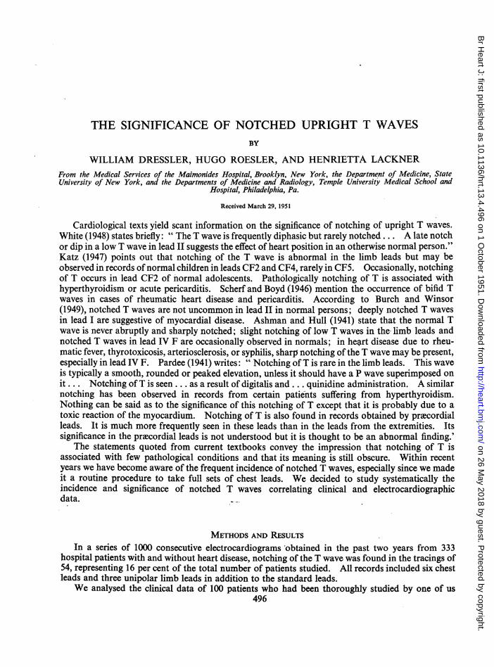
THE SIGNIFICANCE OF NOTCHED UPRIGHT T WAVESBY
WILLIAM DRESSLER, HUGO ROESLER, AND HENRIETTA LACKNERFrom the Medical Services of the Maimonides Hospital, Brooklyn, New York, the Department of Medicine, StateUniversity of New York, and the Departments of Medicine and Radiology, Temple University Medical School and
Hospital, Philadelphia, Pa.
Received March 29, 1951
Cardiological texts yield scant information on the significance of notching of upright T waves.White (1948) states briefly: " The T wave is frequently diphasic but rarely notched ... A late notchor dip in a low T wave in lead II suggests the effect of heart position in an otherwise normal person."Katz (1947) points out that notching of the T wave is abnormal in the limb leads but may beobserved in records ofnormal children in leads CF2 and CF4, rarely in CF5. Occasionally, notchingof T occurs in lead CF2 of normal adolescents. Pathologically notching of T is associated withhyperthyroidism or acute pericarditis. Scherf and Boyd (1946) mention the occurrence of bifid Twaves in cases of rheumatic heart disease and pericarditis. According to Burch and Winsor(1949), notched T waves are not uncommon in lead II in normal persons; deeply notched T wavesin lead I are suggestive of myocardial disease. Ashman and Hull (1941) state that the normal Twave is never abruptly and sharply notched; slight notching of low T waves in the limb leads andnotched T waves in lead IV F are occasionally observed in normals; in heart disease due to rheu-matic fever, thyrotoxicosis, arteriosclerosis, or syphilis, sharp notching of the T wave may be present,especially in lead IV F. Pardee (1941) writes: " Notching ofT is rare in the limb leads. This waveis typically a smooth, rounded or peaked elevation, unless it should have a P wave superimposed onit ... Notching ofT is seen ... as a result of digitalis and. . . quinidine administration. A similarnotching has been observed in records from certain patients suffering from hyperthyroidism.Nothing can be said as to the significance of this notching of T except that it is probably due to atoxic reaction of the myocardium. Notching of T is also found in records obtained by prxcordialleads. It is much more frequently seen in these leads than in the leads from the extremities. Itssignificance in the precordial leads is not understood but it is thought to be an abnormal finding.'
The statements quoted from current textbooks convey the impression that notching of T isassociated with few pathological conditions and that its meaning is still obscure. Within recentyears we have become aware of the frequent incidence of notched T waves, especially since we madeit a routine procedure to take full sets of chest leads. We decided to study systematically theincidence and significance of notched T waves correlating clinical and electrocardiographicdata.
METHODS AND RESULTSIn a series of 1000 consecutive electrocardiograms 'obtained in the past two years from 333
hospital patients with and without heart disease, notching of the T wave was found in the tracings of54, representing 16 per cent of the total number of patients studied. All records included six chestleads and three unipolar limb leads in addition to the standard leads.
We analysed the clinical data of 100 patients who had been thoroughly studied by one of us496
on 26 May 2018 by guest. P
rotected by copyright.http://heart.bm
j.com/
Br H
eart J: first published as 10.1136/hrt.13.4.496 on 1 October 1951. D
ownloaded from
THE SIGNIFICANCE OF NOTCHED UPRIGHT T WAVES
during the past few years and whose electrocardiograms presented notched T waves in limb or chestleads. A full set of leads was available in 74 cases of this series; CF, CR, andV leads were represen-ted in fairly equal numbers. In 46 patients the unipolar limb leads were obtained.
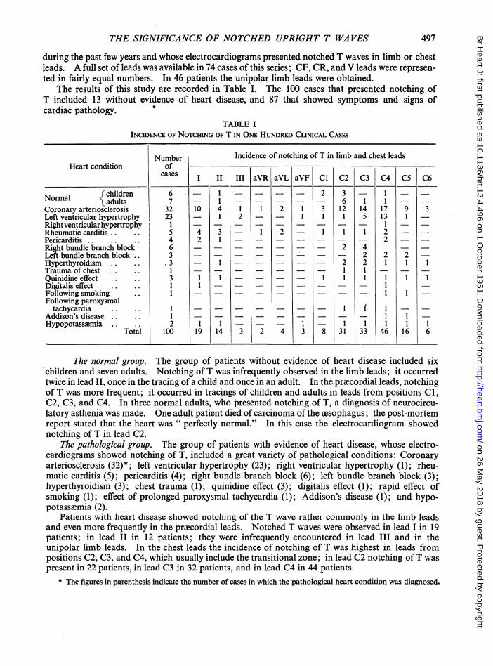
The results of this study are recorded in Table I. The 100 cases that presented notching ofT included 13 without evidence of heart disease, and 87 that showed symptoms and signs ofcardiac pathology. 0
TABLE IINCIDENCE OF NOTCHING OF T IN ONE HUNDRED CLINICAL CASES
Number Incidence of notching of T in limb and chest leadsHeart condition of
icases |I II III aVR aVL aVF Cl C2 C3 C4 C5 C6
fchildren 6-1 2 - 1--INormal Iadults 7 -16 1 1 -
Coronary arteriosclerosis 32 10 4 1 1 2 1 3 12 14 17 9 3Left ventricular hypertrophy 23 1 2 1 1 1 5 13 1 -Rightventricularhypertrophy I1. . . . . . . ...1 -Rheumatic carditis.. .. 5 4 3 - 1 2 1 1 1 2 -
Pericarditis.. .. .. 4 2 1 . . . . . . . 2 -Right bundle branch block 6 ..- 2 4 -Left bundle branch block.. 3 _ _. . 2 2 2Hyperthyroidism .. .. 3 - 2 2 1 1 1Trauma of chest .. .. 11 1 - - -Quinidine effect .. .. 3 1 1 - - 1 1 1 1 1 1Digitalis effect .. .. 1 11 -_ _Following smoking .. 1 . 1 1Following paroxysmal
tachycardia I.... 1 _ 1 - -Addison's disease.. .. 1 . 1 1Hypopotassemia .. .. 2 1 1 - - 1 1 1 1 1 1
Total 100 19 14 3 2 4 3 8 31 33 46 16 6._ _I~~~~~~~~~~~~~~~~~~________________________________________I__________________________________________
The normal group. The group of patients without evidence of heart disease included sixchildren and seven adults. Notching of T was infrequently observed in the limb leads; it occurredtwice in lead II, once in the tracing of a child and once in an adult. In the prxcordial leads, notchingof T was more frequent; it occurred in tracings of children and adults in leads from positions Cl,C2, C3, and C4. In three normal adults, who presented notching of T, a diagnosis of neurocircu-latory asthenia was made. One adult patient died of carcinoma of the cesophagus; the post-mortemreport stated that the heart was " perfectly normal." In this case the electrocardiogram showednotching of T in lead C2.
The pathological group. The group of patients with evidence of heart disease, whose electro-cardiograms showed notching of T, included a great variety of pathological conditions: Coronaryarteriosclerosis (32)*; left ventricular hypertrophy (23); right ventricular hypertrophy (1); rheu-matic carditis (5); pericarditis (4); right bundle branch block (6); left bundle branch block (3);hyperthyroidism (3); chest trauma (1); quinidine effect (3); digitalis effect (1); rapid effect ofsmoking (1); effect of prolonged paroxysmal tachycardia (1); Addison's disease (1); and hypo-potassvmia (2).
Patients with heart disease showed notching of the T wave rather commonly in the limb leadsand even more frequently in the prxcordial leads. Notched T waves were observed in lead I in 19patients; in lead II in 12 patients; they were infrequently encountered in lead III and in theunipolar limb leads. In the chest leads the incidence of notching of T was highest in leads frompositions C2, C3, and C4, which usually include the transitional zone; in lead C2 notching ofT waspresent in 22 patients, in lead C3 in 32 patients, and in lead C4 in 44 patients.
* The figures in parenthesis indicate the number of cases in which the pathological heart condition was diagnosed.
497
on 26 May 2018 by guest. P
rotected by copyright.http://heart.bm
j.com/
Br H
eart J: first published as 10.1136/hrt.13.4.496 on 1 October 1951. D
ownloaded from
DRESSLER, ROESLER, AND LACKNER
THE MEANING OF NOTCHING OF TA striking parallelism can be observed in the incidence of notching and inversion of the T waves.
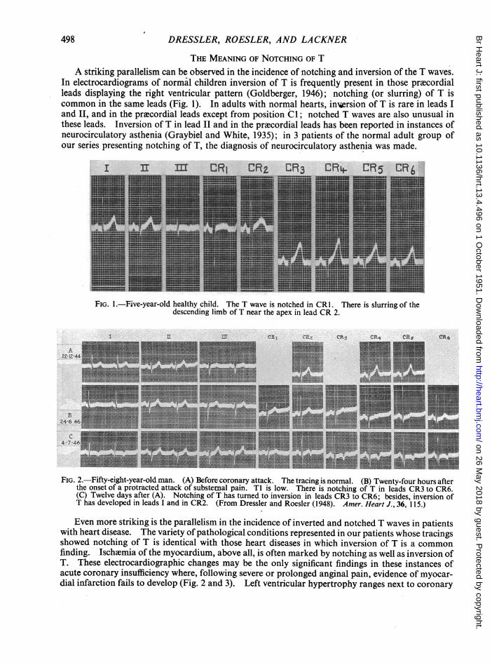
In electrocardiograms of normal children inversion of T is frequently present in those precordialleads displaying the right ventricular pattern (Goldberger, 1946); notching (or slurring) of T iscommon in the same leads (Fig. 1). In adults with normal hearts, in%ersion of T is rare in leads Iand II, and in the prlcordial leads except from position Cl; notched T waves are also unusual in-these leads. Inversion of T in lead II and in the precordial leads has been reported in instances ofneurocirculatory asthenia (Graybiel and White, 1935); in 3 patients of the normal adult group ofour series presenting notching of T, the diagnosis of neurocirculatory asthenia was made.
I Ir m CRi CRFIiIIIIIIiUIi
I
IiiIII1.II
I
MRt4 CR5 CRA
FIG. 1.-Five-year-old healthy child. The T wave is notched in CR1. There is slurring of thedescending limb of T near the apex in lead CR 2.
1 'a rl e9.'1 ¢T26 & 97 rit. trp
A2Z-12-44
24-6 46
C4-7-4
FIG. 2.-Fifty-eight-year-old man. (A) Before coronary attack. The tracing is normal. (B) Twenty-four hours afterthe onset of a protracted attack of substernal pain. TI is low. There is notching of T in leads CR3 to CR6.(C) Twelve days after (A). Notching of T has turned to inversion in leads CR3 to CR6; besides, inversion ofT has developed in leads I and in CR2. (From Dressler and Roesler (1948). Amer. Heart J., 36, 115.)
Even more striking is the parallelism in the incidence of inverted and notched T waves in patientswith heart disease. The variety of pathological conditions represented in our patients whose tracingsshowed notching of T is identical with those heart diseases in which inversion of T is a commonfinding. Ischimia of the myocardium, above all, is often marked by notching as well as inversion ofT. These electrocardiographic changes may be the only significant findings in these instances ofacute coronary insufficiency where, following severe or prolonged anginal pain, evidence of myocar-dial infarction fails to develop (Fig. 2 and 3). Left ventricular hypertrophy ranges next to coronary
498
A...
4.
m M.---
iiiiiiii"
WN 1-mili-a
v-r. L-sl........2 Ln3 u n, L;j %IL-6rRKA
on 26 May 2018 by guest. P
rotected by copyright.http://heart.bm
j.com/
Br H
eart J: first published as 10.1136/hrt.13.4.496 on 1 October 1951. D
ownloaded from
THE SIGNIFICANCE OF NOTCHED UPRIGHT T WA VES
I 1 m CrVR aVL d1VF VI V_ V3 V4 V5 V6*HEU inin~~~~ ~~EEUEEEinI *.12==H Pi
mi'*-inUEM!_ 9- HEEl I** rn. **u. 3 !=|--| 11mmmmmalilg |lA IEEEBEX-5--:a 2 US====F., mEE--- =ml
26-1-50 S..Mls| lE i.gii= l SWW !l- _ -- - 3 "= ^= -u*-Eusr
IEEE iE lE ilE...=IEEE :!-3l=U2==U *i lE_
IKE IEEE E*.u ..-5_e_ -==.. =5==3n
UinU ~ufl!-l l5E X i. in.in=: s_,_-_ =. I__ InEB=*E - -- -nuEE .._.. IE WEa I m
-_*-=5_= _-*___U 5=5=5=3 __W
30150"-==--.--==_*e__ i*_E ~~_===--r.=:
w,u=w - "-. s~ 3"~
*33 81S@i30Elig |_His M1. *5= I1F5=M~ =; 15=535=EN1ibiflE~~~~~~~u.~~~~u' i~~~~~~i mm ~~~~~ ~~.: :z: L~~~~~~~~E ..~~~~E~~QE*'F-1
FIG.3EFEEKma. ye ElmrtiEnalp= * -%11-INIMA-1MM w ffm-~ 1~*
am5.-. M.-5=33.-'Kro I=5-....*-Z=.IUin1-E
M= =E3 Mo3353==5M==Wa=--.-- E =: -MMiIE U-:~~~j-~~~~~=CmminUEE
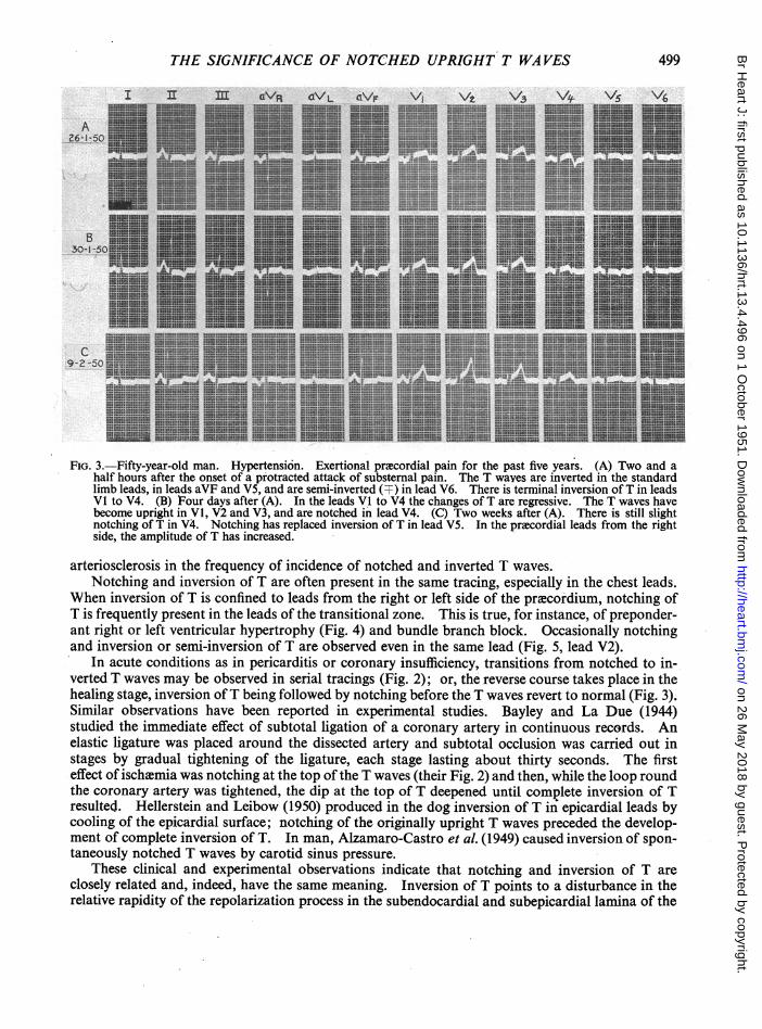
half hours after the onset of a protracted attack of substemnal pain. The T waves are inverted in the standardlimb leads, in leads aVF and V5, and are semi-inverted (F) in lead V6. There is terminal inversion of T in leadsVl to V4. (B) Four days after (A). In the leads Vl to V4 the changes of T are regressive. The T waves havebecome upright in Vl, V2 and V3, and are notched in lead V4. (C) Two weeks after (A). There is still slightnotching of T in V4. Notching has replaced inversion of T in lead V5. In the prxcordial leads from the rightside, the amplitude of T has increased.
arteriosclerosis in the frequency of incidence of notched and inverted T waves.Notching and inversion of T are often present in the same tracing, especially in the chest leads.
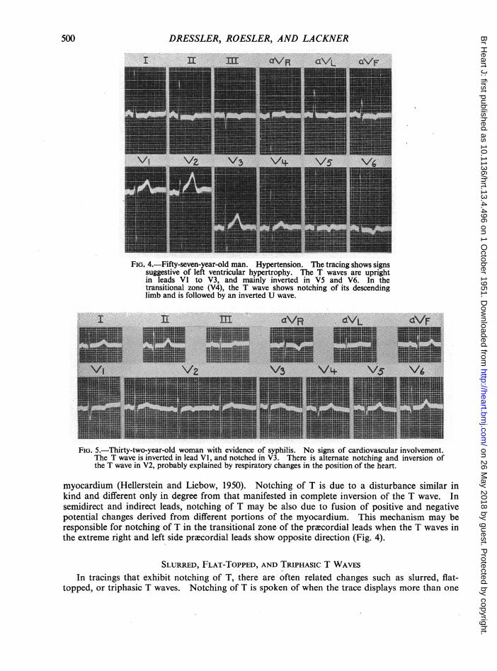
When inversion of T is confined to leads from the right or left side of the pr~ecordium, notching ofT is frequently present in the leads of the transitional zone. This is true, for instance, of preponder-ant right or left ventricular hypertrophy (Fig. 4) and bundle branch block. Occasionally notchingand inversion or semi-inversion of T are observed even in the same lead (Fig. 5, lead V2).
In acute conditions as in pericarditis or coronary insufficiency, transitions. from notched to in-verted T waves may be observed in serial tracings (Fig. 2); or, the reverse course takes place in thehealing stage, inversion of T being followed by notching before the T waves revert to normal (Fig. 3).Similar observations have been reported in experimental studies. Bayley and La Due (1944)studied the immediate effect of subtotal ligation of a coronary artery in continuous records. Anelastic ligature was placed around the dissected artery and subtotal occlusion was carried out instages by gradual tightening of the ligature, each stage lasting about thirty seconds. The firsteffect of ischicmia was notching at the top of the T waves (their Fig. 2) and then, while the loop roundthe coronary artery was tightened, the dip at the top of T deepened until complete inversion of Tresulted. Hellerstein and Leibow (1950) produced in the dog inversion of T in epicardial leads bycooling of the epicardial surface; notching of the originally upright T waves preceded the develop-ment of complete inversion of T. In man, Alzamaro-Castro et al. (1949) caused inversion of spon-taneously notched T waves by carotid sinus pressure.
These clinical and experimental observations indicate that notching and inversion of T areclosely related and, indeed, have the same meaning. Inversion of T points to a disturbance in therelative rapidity of the repolarization process in the subendocardial and subepicardial lamina of the
499
on 26 May 2018 by guest. P
rotected by copyright.http://heart.bm
j.com/
Br H
eart J: first published as 10.1136/hrt.13.4.496 on 1 October 1951. D
ownloaded from
DRESSLER, ROESLER, AND LACKNER
Tr
FIG. 4.-Fifty-seven-year-old man. Hypertension. The tracing shows signssuggestive of left ventricular hypertrophy. The T waves are uprightin leads VI to V3, and mainly inverted in V5 and V6. In thetransitional zone (V4), the T wave shows notching of its descendinglimb and is followed by an inverted U wave.
I I[ m
_ _ . ...._VIt VPwl--
lII
FIG. 5.-Thirty-two-year-old woman with evidence of syphilis. No signs of cardiovascular involvement.The T wave is inverted in lead VI, and notched in V3. There is alternate notching and inversion ofthe T wave in V2, probably explained by respiratory changes in the position of the heart.
myocardium (Hellerstein and Liebow, 1950). Notching of T is due to a disturbance similar inkind and different only in degree from that manifested in complete inversion of the T wave. Insemidirect and indirect leads, notching of T may be also due to fusion of positive and negativepotential changes derived from different portions of the myocardium. This mechanism may beresponsible for notching of T in the transitional zone of the precordial leads when the T waves inthe extreme right and left side precordial leads show opposite direction (Fig. 4).
SLURRED, FLAT-TOPPED, AND TRIPHASIC T WAVESIn tracings that exhibit notching of T, there are often related changes such as slurred, flat-
topped, or triphasic T waves. Notching of T is spoken of when the trace displays more than one
500
4dVL
V.L-. V-IT
on 26 May 2018 by guest. P
rotected by copyright.http://heart.bm
j.com/
Br H
eart J: first published as 10.1136/hrt.13.4.496 on 1 October 1951. D
ownloaded from
THE SIGNIFICANCE OF NOTCHED UPRIGHT T WAVES
I 31II m :... RaV/L C\/F '
l--l
CRI CR,2 CR ORL4 CR CR-6
-~~~~~ Fi 3 5
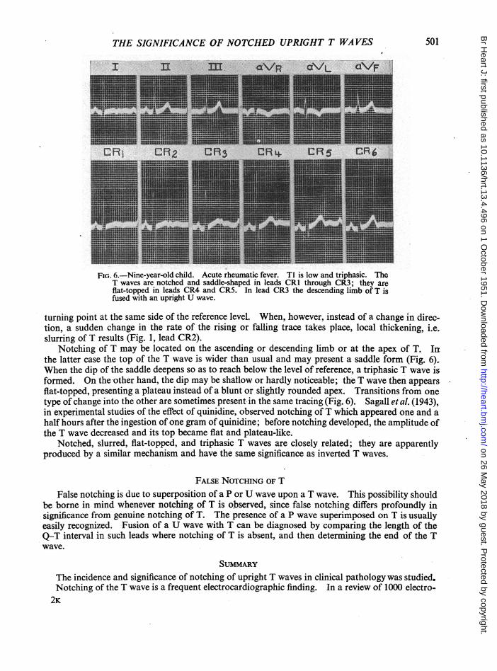
FIG. 6.-Nine-year-old child. Acute rheumatic fever. Ti is low and triphasic. TheT waves are notched and saddle-shaped in leads CR1 through CR3; they areflat-topped in leads CR4 and CR5. In lead CR3 the descending limb of T isfused with an upright U wave.
turning point at the same side of the reference level. When, however, instead of a change in direc-tion, a sudden change in the rate of the rising or falling trace takes place, local thickening, i.e.slurring of T results (Fig. 1, lead CR2).
Notching of T may be located on the ascending or descending limb or at the apex of T. Irthe latter case the top of the T wave is wider than usual and may present a saddle form (Fig. 6).,When the dip of the saddle deepens so as to reach below the level of reference, a triphasic T wave isformed. On the -other hand, the dip may be shallow or hardly noticeable; th'e T wave then appearsflat-topped, presenting a plateau instead of a blunt or slightly rounded apex. Transitions from onetype of change into the other are sometimes present-in the same tracing (Fig. 6). Sagall et al. (1943),in experimental studies of the effect of quinidine, observed notching of T which appeared one and ahalf hours after the ingestion of one gram of quinidine; before notching develo'ped, the amplitude ofthe T wave decreased and its top became flat 'and plateau-like.
Notched, slurred, flat-topped, and triphasic T waves are closely related; they are apparentlyproduced by a similar mechanism and have the same significance as inverted T waves.
FALSE NOTCHING oF T
False notching is due to superposition of a P or U wave upon a T wave. This possibility shouldbe borne in mind whenever notching of T is observed, since false notching diffe.rs profoundly insignificance from genuine notching of T. The presence of a P wave superimposed on T is usuallyeasily recognized. Fusion of a U wave with T can be diagnosed by comparing the length of theQ-T interval in such leads where notching of T is absent, and then determining the end of the Twave.
The incidence and significance of notching of upright T waves in clinical pathology was studied.Notching of the T wave is a frequent electrocardiographic finding. In a review of 1000 electro-
2K
501
on 26 May 2018 by guest. P
rotected by copyright.http://heart.bm
j.com/
Br H
eart J: first published as 10.1136/hrt.13.4.496 on 1 October 1951. D
ownloaded from
DRESSLER, ROESLER, AND LACKNER
cardiograms, that were obtained from 333 hospital patients with and without heart disease, notchingof T was present in 16 per cent of the patients.
In the absence of heart disease, notching of T is rare in the limb leads, but is not infrequent inprecordial leads especially in'children. When heart disease is present, notching of T is oftenobserved in the limb leads and is even more freqw,nt in the chest leads. It occurs in a great varietyof pathological conditions, most often with coronary insufficiency and/or left ventricular hyper-trophy.
Notching of T is observed under the same conditions as inversion of T. Both features of Tmay occur in the same lead either simultaneously or in serial tracings during the evolution of apathological condition. Experimentally, certain effects such as progressive diminution of thecoronary blood supply may produce first notching and then inversion of T. Thus, clinical andexperimental evidence indicate that notching of T is due to a similar mechanism and has the samesignificance as inversion of T. Slurred, flat-topped, and triphasic T waves are closely related tonotched T waves and have the same meaning.
Genuine notching of T should be distinguished from false notching that is caused by superposi-tion of P or U waves upon the T wave.
REFERENCESAlzamora-Castro, V., Rubio, C., Battilana, G., and Subiria, R., (1949). Amer. Heart J., 37, 927.Ashman, R., and Hull, E. (1941). Essentials of Electrocardiography, 2nd ed., p. 158. McMillan Co., New York.Bayley, R. H., and La Due, J. S. (1944). Amer. Heart J., 28, 54Burch, G. E., and Winsor, T. (1949). A Primer ofElectrocardiography, 2nd ed., pp. 93 and 95. Lea and Febiger,
Philadelphia.Goldberger, Emanuel (1946). Amer. J. Dis. Child., 71, 618.Graybiel, A., and White, P. D. (1935). Amer. Heart J., 10, 349.Hellerstein, H. K., and Liebow, I. M. (1950). Amer. Heart J., 39, 34.Katz, L. N. (1947). Electrocardiography, 2nd ed., pp. 136, 169, 404. Lea and Febiger, Philadelphia.Pardee, H. E. B. (I1941). tlinical Aspects of the Electrocardiogram, 4th ed., p. 154. P. B. Hoeber, New York.Sagall, E. L., Horn, C. D., and Riseman, J. E. F. (1943). Arch. intern. Med., 71. 460.Scherf, D., and Boyd, L. J. (1946). Clinical Electrocardiography, 2nd ed., pp. 57, 68, 124. J. B. Lippincott, Phila-
delphia.Wendkos, M. H. (1944). Amer. Heart J., 28, 549.White, P. D. (1948). Heart Disease, 3rd ed., p. 207. McMillan Co., New York.
502
on 26 May 2018 by guest. P
rotected by copyright.http://heart.bm
j.com/
Br H
eart J: first published as 10.1136/hrt.13.4.496 on 1 October 1951. D
ownloaded from