Embed Size (px)
Citation preview

The Skeletal System

Imagine for a moment that people did not have skeletons .
What comes to mind? Probably that each of us would be a little heap on the floor, much like a jellyfish out of
water .the most obvious function of the skeleton: to support the body. it is a framework for the body, the skeleton is not at all like the wooden beams that support a house.
Bones are living organs that actively contribute to the maintenance of the internal environment of the
body .

The skeletal system consists of bones and other structures that make up the joints of
the skeleton .
The types of tissue present are bone tissue, cartilage, and fibrous connective tissue, which forms the ligaments that connect bone to bone.

FUNCTIONS OF THE SKELETON1 .Provides a framework that supports the
body; the muscles that are attached to bones move the skeleton.
2 .Protects some internal organs from mechanical injury; the rib cage protects the heart and lungs, for example.
3 .Contains and protects the red bone marrow, the primary hemopoietic (blood-forming) tissue.
4 .Provides a storage site for excess calcium. Calcium may be removed from bone to maintain a normal blood calcium level, which is essential for blood clotting and proper functioning of muscles and nerves.

TYPES OF BONE TISSUE
Bone cells are called osteocytes, and the matrixof bone is made of calcium salts and collagen .
The calcium salts are -Calcium carbonate (CaCO3) and
-Calcium phosphate (Ca3(PO4)2) ,Which give bone the strength required to perform its supportive and protective functions.
Bone matrix is non-living, but it changes constantly, with calcium that is taken from bone into the blood replaced by calcium from the diet.
In normal circumstances, the amount of calcium thatis removed is replaced by an equal amount of calcium
deposited .
This is the function of osteocytes, to regulate the amount of calcium that is deposited in, or removed from, the bone matrix.
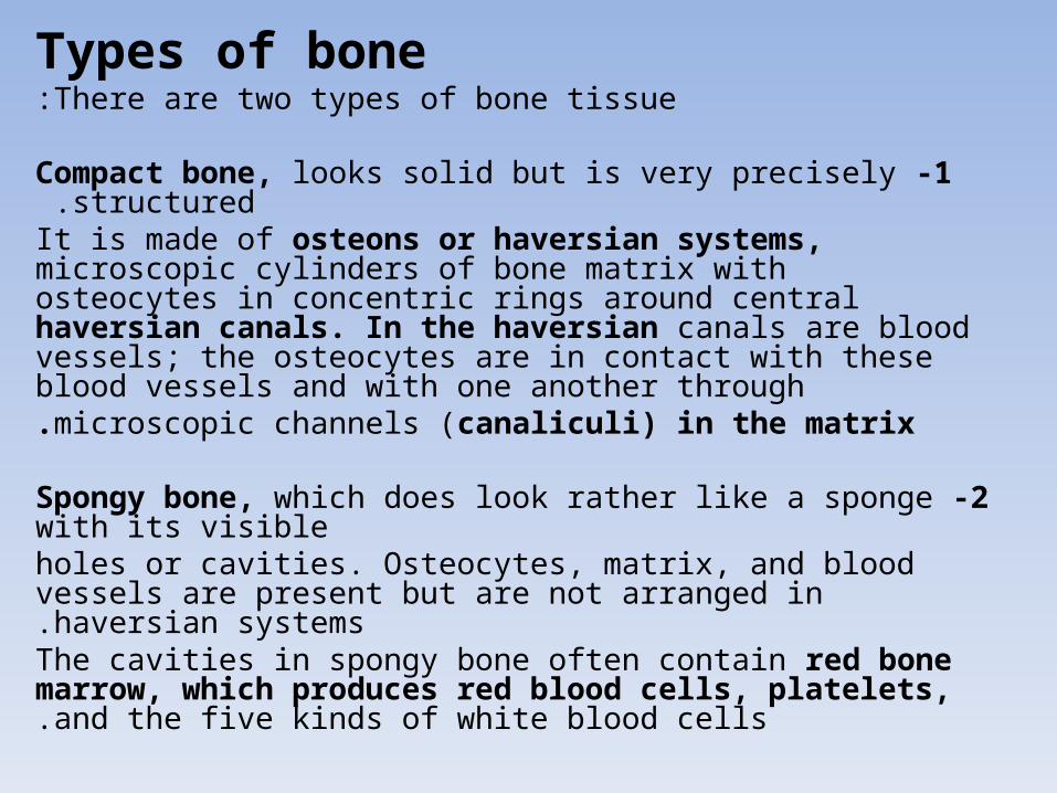
Types of boneThere are two types of bone tissue:
1 -Compact bone, looks solid but is very precisely structured .
It is made of osteons or haversian systems, microscopic cylinders of bone matrix with osteocytes in concentric rings around central haversian canals. In the haversian canals are blood vessels; the osteocytes are in contact with these blood vessels and with one another throughmicroscopic channels (canaliculi) in the matrix.
2 -Spongy bone, which does look rather like a sponge with its visibleholes or cavities. Osteocytes, matrix, and blood vessels are present but are not arranged in haversian systems.
The cavities in spongy bone often contain red bone marrow, which produces red blood cells, platelets, and the five kinds of white blood cells.

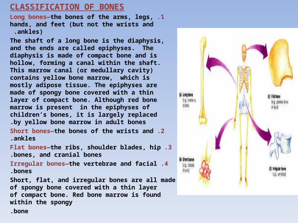
CLASSIFICATION OF BONES1 .Long bones—the bones of the arms, legs, hands,
and feet (but not the wrists and ankles) .
The shaft of a long bone is the diaphysis, and the ends are called epiphyses. The diaphysis is made of compact bone and is hollow, forming a canal within the shaft. This marrow canal (or medullary cavity) contains yellow bone marrow, which is mostly adipose tissue. The epiphyses are made of spongy bone covered with a thin layer of compact bone. Although red bone marrow is present in the epiphyses of children’s bones, it is largely replaced by yellow bone marrow in adult bones.
2 .Short bones—the bones of the wrists and ankles.
3 .Flat bones—the ribs, shoulder blades, hip bones, and cranial bones.
4 .Irregular bones—the vertebrae and facial bones.
Short, flat, and irregular bones are all made of spongy bone covered with a thin layer of compact bone. Red bone marrow is found within the spongybone.

FACTORS THAT AFFECT BONE GROWTH AND MAINTENANCE
1 .Heredity—each person has a genetic potential for height, that is, a maximum height, with genes
inherited from both parents .
2 .Nutrition—nutrients are the raw materials of which bones are made .
-Calcium, phosphorus, and protein become part of the bone matrix itself.
-Vitamin D is needed for the efficient absorption of calcium and phosphorus by the small intestine .
-Vitamins A and C do not become part of bone but are necessary for the process of bone matrix formation
(ossification) .Without these and other nutrients, bones cannot grow
properly .
3 .Hormones—endocrine glands produce hormones that stimulate specific effects in certain cells.

THE SKELETONThere are 206 bones in total, and the human skeleton has
two divisions :1 -The axial skeleton, which forms the axis of the body,
consists of the skull, vertebral column, and rib cage .
2 -The appendicular skeleton, which supports the limbs. The bones of the arms and legs and the shoulder and
pelvic girdles make up the appendicular skeleton .
-Many bones are connected to other bones across joints by ligaments, which are strong cords or sheets of fibrous connective tissue.
-The importance of ligaments becomes readily apparent when a joint is sprained.


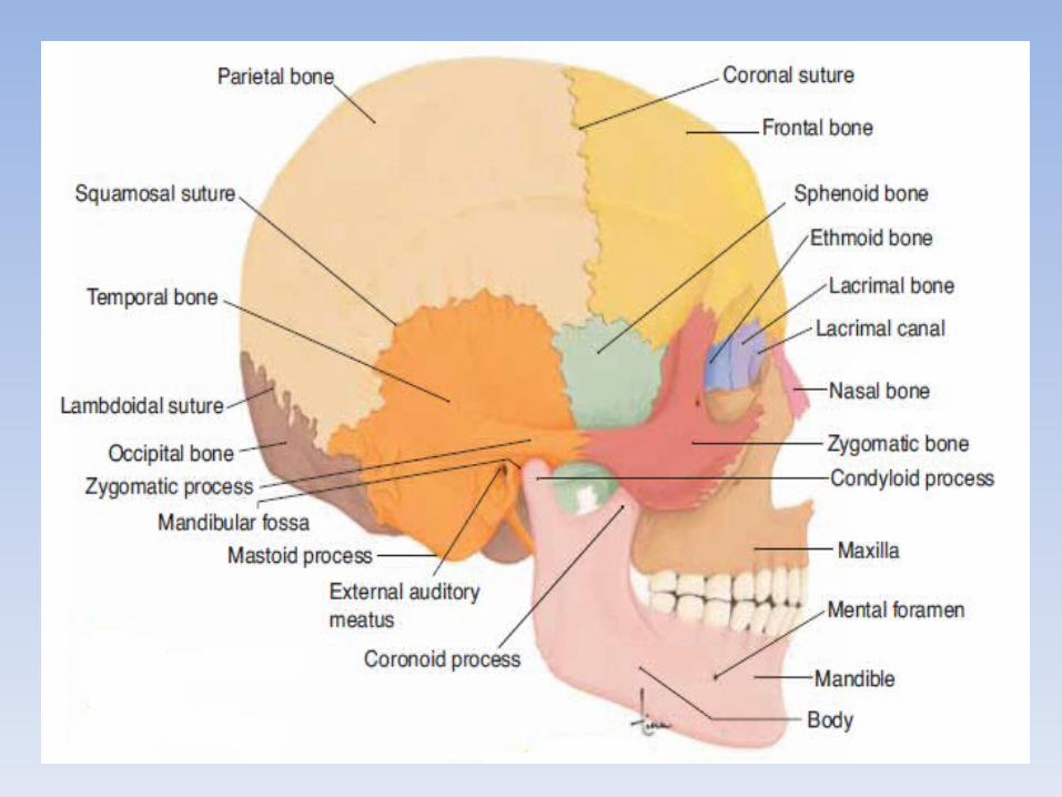
SKULLThe skull consists of 8 cranial bones and 14 facial bones. Also in the head are three small bones in each middle ear cavity and the
hyoid bone that supports the base of the tongue .
A- The cranial bones, form the braincase (lined with the meninges) that encloses and protects the brain, eyes, and ears. These are:
1 -The frontal bone, forms the forehead and the anterior part of the top of the skull .
2 -The parietal bones (two), Parietal means “wall,” form the posterior top and much of the side walls of the skull .
3 -The temporal bones (two), on the side of the skull contains an external auditory meatus (ear canal), a middle ear cavity, and an inner ear labyrinth.

4 -The occipital bone, forms the lower, posterior part of the braincase .
Function: Its foramen magnum is a large opening for the spinal cord, and the two condyles (rounded projections) on either side articulate with the atlas, the first cervical vertebra, enabling movement of the head
relative to the spine .

Skull. Inferior view with mandible removed.

5 -The sphenoid bone: is said to be shaped like a bat, and the greater wing is visible on the side of the skull between the frontal and temporal bones. The body of the bat has a depression called the sella turcica, which encloses the pituitary gland.
Function: Articulates with the frontal, parietal and temporal bones .
6 -The ethmoid bone has a vertical projection called the crista galli (“rooster’s comb”) that anchors the cranial meninges. The rest of the ethmoid bone forms the roof and upper walls of the nasal cavities, and the upper part of the nasal septum.
Function: Forms part of the nasal cavity and the orbits.Main support structure of the nasal cavity

All of the joints between cranial bones are immovable joints called sutures.
It may seem strange to refer to a joint without movement, but the term joint (or articulation) is used for any junction of two bones.
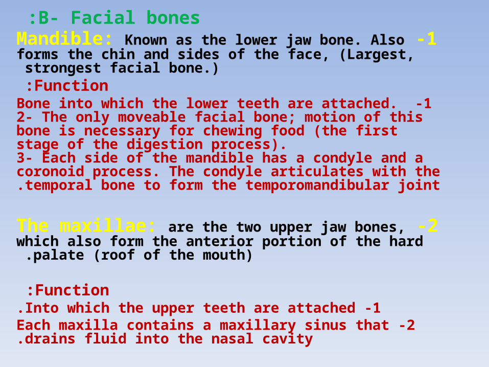
B- Facial bones :1 -Mandible: Known as the lower jaw bone. Also forms the chin and
sides of the face, (Largest, strongest facial bone.) Function:
1 -Bone into which the lower teeth are attached. 2- The only moveable facial bone; motion of this bone is necessary for chewing food (the first stage of the digestion process).3- Each side of the mandible has a condyle and a coronoid process. The condyle articulates with the temporal bone to form the temporomandibular joint.
2 -The maxillae: are the two upper jaw bones, which also form the
anterior portion of the hard palate (roof of the mouth) .
Function :1 -Into which the upper teeth are attached.
2 -Each maxilla contains a maxillary sinus that drains fluid into the nasal cavity.
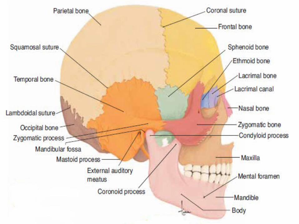

3 -The two nasal bones form the bridge of the nose
Function: They articulate with the frontal bone (the rest of the nose is supported by cartilage).
4 -The two lacrimal bone: each of the bone present at the medial side of each orbit ;
Function: the lacrimal canal contains the lacrimal sac, a passageway for tears
5 -The two zygomatic bones: each of them forms the point of a cheek, and articulates with the maxilla, frontal bone, and
temporal bone .
Function: Articulates with the frontal, maxilla, sphenoid and temporal bones.
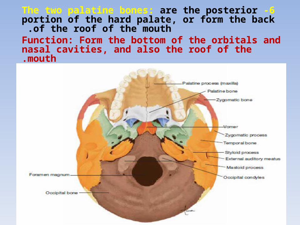
6 -The two palatine bones: are the posterior portion of the hard palate, or form the back of the roof of the
mouth .Function: Form the bottom of the orbitals and nasal cavities, and also the roof of the mouth.
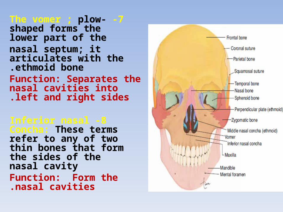
7 -The vomer ; plow-shaped forms the lower part of thenasal septum; it articulates with the ethmoid bone.
Function: Separates the nasal cavities into left and right sides.
8 -Inferior nasal Concha: These terms refer to any of two thin bones that form the sides of the nasal cavityFunction: Form the nasal cavities.

SINUSESIs a sac or cavity in any organ or tissue.
The sinuses functions are :
1 -They make the skull lighter in weight, because air is lighter than bone.
2 -They provide resonance for the voice, meaningmore air to vibrate and thus deepen the pitch of the voice.
1 -Paranasal sinuses: air cavities in the cranial bone located in the maxillae and frontal, sphenoid, and
ethmoid bones .
As the name paranasal suggests, they open into the nasal cavities and are lined with ciliated epithelium
continuous with the mucosa of the nasal cavities .
We are aware of our sinuses only when they become “stuffed up,” which means that the mucus they produce cannot drain into the nasal cavities. Thismay happen during upper respiratory infections such as colds, or with allergies such as hay fever (is an
allergic inflammation of the nasal airways) .


2 -The mastoid sinuses are air cavities in the mastoid process of each temporal bone; they open into the middle ear. Before the availability of antibiotics, middle ear infections often caused mastoditis, infection of these sinuses.
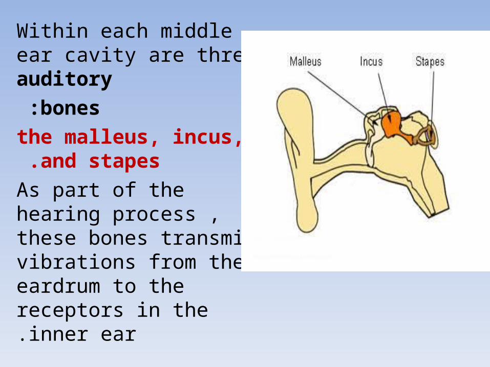
Within each middle ear cavity are three auditory
bones :
the malleus, incus, and stapes.
As part of the hearing process , these bones transmit vibrations from the eardrum to the receptors in the inner ear.

29