Embed Size (px)
Citation preview

Understanding the Pathophysiology of Diabetic Skin May lead to Early Topical Intervention Resulting in Reduced Wounding and Loss of Limb
Darlene E. McCord, Ph.D., FAPWCA, Steven R. Kravitz, DPM, FAPWCA
Abstract
Purpose: Elucidate early and late stage diabetic skin pathophysiologies that lead
to skin breakdown, wounding and potential loss of limb. Glycation, oxidation,
inflammation, loss of microcirculation commence the cascade that leads to
ischemia, neuropathy, collagen cross-linking and pruritus. The etiologies of this
disease on the skin’s immune system may best be directed to the field of
biochemistry where micronutrients that have demonstrated efficacy against these
disorders are now being incorporated into topical corneotherapeutic products.
Materials and Methods: A review of scientific and medical databases was used
and the data was cross referenced. Micronutrients with a strong scientific basis
for use against each skin disorder were cataloged. For example, antioxidants
were evaluated to determine their effectiveness against oxidative stress known to
participate in premature skin cell death, aging, chronic disease and diabetes.
Histamine blocking topical micronutrients were evaluated as part of the
inflammatory and pruritic process. Results: Corneotherapeutic products provide
for the administration of micronutrients and medicine by absorption through the
skin. Micronutrients with a molecular weight of less that 500 Daltons can enter
or exit the skin (500 Dalton Rule). These micronutrients include amino acids,
vitamins, antioxidants and polyunsaturated fatty acids (PUFAs) comprised as a
balance of n-3/n-6. Research demonstrated that these micronutrients, when
applied topically, act at a biochemical level within the skin and reduce the impact
1Accepted for publication in Advances in Skin & Wond Care

of oxidative damage, inflammation, and glycation. Corneotherapeutic products
used at the earliest stages of diabetes may dramatically reduce the risks
associated with skin breakdown, wounding and the eventual loss of limb.
Abbreviations PUFAs - polyunsaturated fatty acids AGE - advanced glycoslation end-products FL - fructoselysine CML - carboxymethyl lysine ROS - reactive oxygen species CGRP - calcitonin gene-related peptide ORAC - oxygen radical absorbance capacity MDA - malondialdehyde PMNs - polymorphonuclear leukocytes MMP - matrix metalloproteinase RA - retinoic acid IGF-I – Insulin-like growth factor RCS - reactive carbonyl species
Background
Diabetes is primarily a metabolic disease and its complication is sequelae of
broad-based derangements in fuel metabolism. There is discussion on the
sequence of cellular and metabolic events related to diabetes and this speaks to
the complexity of the disease. One hypothesis is that carbonyl stress precedes
oxidative stress.1 Both are involved in the pathogenesis of diabetic skin
complication. The body of evidence may favor oxidative stress as the metabolic
pathway to diabetic skin complications. 1
The skin and its healing processes are altered in patients with diabetes. The
physiological and biochemical implication of the effects of glucose have not been
fully elucidated. The skin’s altered state leads to chronic complications that
2

include loss of limbs and mortality. Hyperglycemia, insulin and IGF-I are
abnormal in diabetic skin keratinocytes. Exposure to high glucose is associated
with changes in cellular morphology, as well as with decreased proliferation and
enhancement of Ca2+. Hyperglycemia and impaired insulin signaling might be
directly involved in the development of chronic complications of diabetes, such
as impaired wound healing. By impeding glucose utilization of skin,
keratinocytes as well as skin proliferation and differentiation are diminished. 2
Oxidative stress and oxidative damage that lead to premature skin cell death are
presupposed in aging, chronic disease and diabetes. Oxidative stress has a
primary role in the pathogenesis of diabetic complication and end-stage tissue
damage in the diabetic. This oxidative process may account for increased
glycoxidation and lipoxidation products in tissue proteins. These findings have
lead Baynes, et al to hypothesize that increased chemical modification of
proteins by carbohydrates and lipids in diabetes is the result of overload on
metabolic pathways involved in detoxification of reactive carbonyl species
(RCS). The overload is apparent in a general build up in steady-state levels of
reactive carbonyl species. The increase in oxidation in glucose and lipids of
tissue proteins in diabetes may therefore be viewed as the result of increased
carbonyl stress. The use of lipoic acids for treatment of neuropathy associated
with carbonyl stress may provide one pathway to improved tissue physiology.1
Glycation (cross-linking of proteins and sugars to form non-functioning
structures in the body) modifies tissue proteins, resulting in chemical reactions
3

between glucose and primary amino groups. The Maillard reaction (also known
as advanced glycoslation end-products [AGE products]) is used to measure
modification of insoluble collagen associated with oxidative stress and glycation.
End product chemicals present according to the stage of glycation. Early stage
glycation is identified with fructoselysine (FL). The glycoxidation products are
N-epsilon (carboxymethyl) lysine (CML) and pentosidine which form later in the
process.3 Studies support the description of diabetes as a disease characterized by
accelerated chemical aging of long-lived tissue proteins.4
Dyer DG, et al studied Maillard reaction products in skin collagen from 39 type
1 diabetic patients and 52 non-diabetic control subjects. The Maillard reaction is
created by damaged proteins and is associated with aging of the extracellular
matrix and diabetes. Proteins undergo increased stiffening and loss of elasticity
during the process. In non-diabetic patients glycation of collagen FL content
increased 33% between age group 20-85 years of age. In contrast CML,
pentosidine and fluorescence increased five fold, correlating directly with age. In
diabetic patients, collagen FL was increased threefold compared to non-diabetic
subjects, correlating strongly with glycated hemoglobin but not with age.
Collagen CML, pentosidine and fluorescence were increased up to two fold in
diabetic subjects compared with control subjects. This can be explained by the
increase in glycation alone. Increased glycation can be invoked without
increased oxidative stress. There is a strong correlation between age-dependent
CML, pentosidine and fluorescence modification of collagen via the Maillard
4

reaction and acceleration in the process chemical tissue aging in diabetes.
Diabetic patients who are also under high oxidative stress, and are therefore rapid
accumulators of glycoxidation products (AGE), may be particularly vulnerable to
the development of complications associated with skin collagen and poor wound
healing.
Oxidative reactions are a normal attribute of aerobic life that causes structural
damage to DNA, proteins, carbohydrates and lipids. When this oxidative
damage is inflicted by reactive oxygen species (ROS) it is known as “oxidative
stress.” Oxidative stress occurs when the balance of prooxidant/ antioxidant
forces favor the prooxidant process.5 Oxidative stress is apparent in the
inflammatory process, aging and diabetes and is implicated in increased
glycoxidation and lipoxidation leading to cell death.
Diabetic vascular complications are also resultant of oxidative stress. Oxidative
stress may play an important role in the pathogenesis of vascular diseases
characterized by an increased formation of free radicals and a corresponding
depletion of antioxidant reserves.6 Events that take place in macrovascular
disease cascade leading to microvascular disease events. Complications related
to microvascular blood flow create their own sequelae of endothelial dysfunction,
neuropathy, foot ulceration and amputations.
5

Diabetic microangiopathy progresses slowly due to a combination of sequential
circulatory changes including; (1) altered basement membrane, (2) altered
cellular function, (3) cell metabolic changes, (4) altered blood flow properties,
(5) disturbed hemostasis, (6) altered oxygen transport, and (7) altered hormone
production.7
Skin blood flow is altered in diabetes. Some of the functional disturbances are
improved through metabolic control. These microvascular alterations reduce
blood flow to the skin and result in a reduction of micronutrient support.8 It
appears that disturbances in the microvascular system may play a role in the
pathogenesis of diabetic neuropathy.6
Dermal neurovascular dysfunction stems in part from decreased microvascular
blood flow and increased vascular resistance. These alterations involve impaired
dilator response to substance P (a neuropeptide-neurotransmitter), calcitonin
gene-related peptide (CGRP) and reactivity to nociceptive stimulation.
Unmyelinated C-fibers, which constitute the central reflex pathway, are assumed
to be damaged in diabetic neuropathy.9 Insulin/C-peptide deficiency in diabetic
neuropathy is linked to the metabolic abnormality of oxidative stress.10
Neuropathies affect more than half of all diabetics. As the disease progresses,
the deterioration results in peripheral and autonomic nerve dysfunction. Diabetic
neuropathy is the most common cause of non-traumatic amputations affecting
15% of all diabetic patients with neuropathic disease.11
6

The pathophysiologies of diabetes commence with alteration in the metabolic
processes. These unseen changes lead progressively to a disease state that
involves the loss of limb and life. Early intervention with corneotherapeutic
products may spare diabetics this fate.
Discussion
Considerable evidence exists demonstrating the role hyperglycemia plays in the
production of ROS. ROS leads to increased oxidative stress in diabetic tissue.
When the endogenous antioxidant network becomes overwhelmed (redox
imbalance) intercellular signaling pathways are activated. Redox reactions
involve the transfer of electrons. With redox, the compound that loses an
electron is said to be “oxidized” while the compound that gains an electron is
said to be “reduced.” A major consequence is the production of gene products
that cause cellular damage that lead to late-stage diabetic complications. Studies
suggest that treatment with exogenous antioxidants may be an effective treatment
against redox imbalance providing a balance between prooxidant/antioxidant
forces.12
ROS (including free radical damage) tissue damage first presents in the sub-
epidermal tissues with damage of proteins, lipids, and DNA. The endogenous
antioxidant system has become overwhelmed causing cell apoptosis slowly
progressing to present as skin lesions and more serious wounds. The
7

microvascular system responsible for nourishing the epidermis is in a state of
oxidative stress due to redox imbalance.
The issue becomes how can intervention reverse the process and create a more
homeostatic environment for the dermis, epidermis and stratum corneum? The
epidermis is metabolically regulated by the integrity of the stratum corneum.
When the barrier permeability is altered (lesions) the epidermis becomes
compromised.
Corneotherapy
The term corneotherapy was coined by Albert M. Kligman, MD, in the mid-
1990’s.13 Today, it is a field of research that is leading to an improved
understanding of the skin’s role as gatekeeper. In the past decade, researchers
have discovered which topically applied substances will pass over its threshold.
According to Kligman, “The ultimate development in the quest for understanding
the stratum corneum was corneotherapy, focusing therapy toward correcting the
defective horny layers associated with chronic diseases.” 14 Among the
complement of corneotherapeutic substances that can be delivered via the
stratum corneum as “active agents” are amino acids, lipids, vitamins and
extracts.15
500 Dalton Rule
8

Corneotherapy is made possible by what is known as the 500 Dalton Rule. The
500 Dalton Rule is used in the development of topical drugs and transdermal
delivery systems. The skin’s barrier is effective in blocking molecules with an
atomic weight greater than 500 Daltons, but molecules with less weight pass
through the skin’s barrier. Topical drugs like cyclosporine, tacrolimus and
ascomycins can be effectively delivered through the skin because the molecules
of the drugs are all under 500 Daltons.
While the stratum corneum’s physicochemical barrier resists the penetrations of
large molecules, smaller molecules with a molecular weight of less than 500
Daltons surpass transcutaneously.16 Molecular size is an important factor
governing passage of substances through the skin, giving substances with higher
molecular weights self-limiting properties.17 Passive delivery of substances, due
to their low molecular weight, provides novel delivery opportunities.18 Included
in these low molecular weight substances are vitamins, amino acids, essential
fatty acids n3 and n6, and antioxidants like hydroxytyrosol.
Essential Fatty Acids
The inflammatory response is among the physiological events at the site of
injury. Fatty acids are involved in the modulation of wound (injury) closure
either in the form of phospholipids anchored in the cell membrane or as soluble
lipoic mediators. This favorable modulation demonstrates a role for fatty acids
as therapeutic agents at the wound site.19
9

There are many plant oils that provide n3 and n6 fatty acids to the skin for
therapeutic applications. Among them are olive oil and canola oil which were
the focus of our research. Olive oil is unique in that it delivers an additional
benefit as a free radical scavenger. 3,4-dihydroxyphenyl ethanol or
hydroxytyrosol, is a potent anti-inflammatory and antioxidant found in olive oil
as well as the pulp of green olives.20
In a study to evaluate olive oil, with its hydroxytyrosol component, it was found
to block activation of nuclear factor-kappaB, signal transducer an activator of
transcription-1 alpha and interferon regulatory factor-1. In addition, it down-
regulated inducible nitric oxide and COX-2 gene expression by preventing
nuclear factor-kappaB. These findings suggest that olive oil/hydroxytyrosol may
represent a non-toxic agent for the control of pro-inflammatory genes.21
Olive phenolics possess the highest antioxidant activities of all known
antioxidants. They scavenge superoxide and other reactive oxygen species,
inhibit neutrophil respiratory burst, and increase plasma antioxidant capacity.22
Hydroxytyrosol has an oxygen radical absorbance capacity (ORAC) value nearly
six times that of alpha lipoic acid (antioxidant).
Table I
10
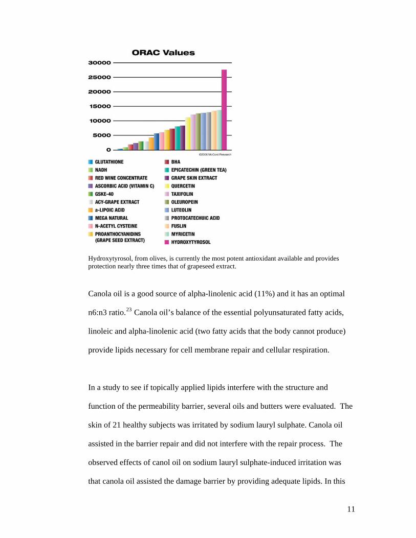
Hydroxytyrosol, from olives, is currently the most potent antioxidant available and provides protection nearly three times that of grapeseed extract.
Canola oil is a good source of alpha-linolenic acid (11%) and it has an optimal
n6:n3 ratio.23 Canola oil’s balance of the essential polyunsaturated fatty acids,
linoleic and alpha-linolenic acid (two fatty acids that the body cannot produce)
provide lipids necessary for cell membrane repair and cellular respiration.
In a study to see if topically applied lipids interfere with the structure and
function of the permeability barrier, several oils and butters were evaluated. The
skin of 21 healthy subjects was irritated by sodium lauryl sulphate. Canola oil
assisted in the barrier repair and did not interfere with the repair process. The
observed effects of canol oil on sodium lauryl sulphate-induced irritation was
that canola oil assisted the damage barrier by providing adequate lipids. In this
11

study canola oil out performed borage oil, sunflower seed oil, shea butter and
petrolatum.24
Amino Acids
Less than two dozen amino acids join together to orchestrate the body’s tissue
and skeletal structures. These small molecules join forces to create
macromolecules like collagen, elastin and fibronectin. Amino acids are also
involved in thousands of chemical reactions each second that have nothing to do
with tissue or skeletal formation. Two of the body’s most vital amino acids are
L-taurine and L-cysteine.
Taurine (2-aminoethane sulfonic acid) is of importance in the management of
diabetic skin. Taurine is a sulfur-containing amino acid found in almost all
tissues. 25 It is responsible for a myriad of important physiological roles in each
organ. Taurine plays a pivotal role as; a) an antioxidant and detoxifying agent
against ROS, b) enhancer of cell proliferation, c) reducer of inflammation, d) key
as an osmosregulator and, e) a stimulatory of glycolysis and glycogenesis. The
role of taurine is determined by the cell type.26 At the wound site, taurine
increases tensile strength by reducing lipid peroxide formation-malondialdehyde
(MDA). Further, taurine is vital to neurotransmission and serves as a
neuroprotectant.
12
Taurine prevents high-glucose-induced vascular endothelial cell apoptosis or
programmed cell death. In vitro and in vivo studies have demonstrated that high
glucose selectively triggers apoptosis. Taurine has cytoprotective properties
through its actions as an antioxidant, osmoregulator and intracellular Ca2+ flux
regulator. Taurine has been shown to reduce the cell damage associated with the
ischemia-reperfusion phenomena (ROS and no-flow of fluid after injury).
Subjects with insulin-dependent diabetes have been found to be deplete in
taurine, giving this amino acid pharmacological value in the treatment of diabetic
patients.27
AGEs accumulate earlier and faster in long-term diabetes than in aging. The
accumulation of AGE and oxidative stress enhance the synthesis of extracellular
matrix and the release of toxic cytokines. Cysteine, in combination with taurine,
was effective in preventing AGE in the treatment of advanced diabetes.28
Peripheral nerve conduction velocity deficits are dependent on decreased nerve
perfusion which is related to free radical activity that is not impaired via
endogenous protection in the redox cycle. In a controlled study using diabetic
rats that had been either treated with cysteine or left untreated, followed by the
application of a liquid nitrogen-cooled probe to form a lesion on the myelinated
nerve fiber, it was found that the cysteine treatment prevented damage.29 This
study has implications in the treatment of diabetic neuropathy.
13

Vitamins
Vitamins are organic compounds that are required by the body. Our bodies are
incapable of synthesizing vitamins and vitamin deficiency is associated with
many diseases. The body is better able to transform the vitamins provided
through the food we eat when we are young. With age, vitamin synthesis
decreases and vitamins like A, B, C and D are not available for many of the
metabolic processes associated with cell viability.
According to Vernon R. Young, Ph.D. of the Massachusetts Institute of
Technology (MIT), one area of exciting research deals with vitamin D.
According to Young, vitamin D requirements of our cells are met in part by the
conversion of a precursor of cholesterol through the imposition of ultraviolet
light. This precursor of cholesterol is converted to a product which subsequently
undergoes a metabolic transformation in the liver. Then it leaves the liver and
goes to the kidney, and finally the active form of vitamin D, 1,25-
dihydroxyvitamin D (1,25[OH]2D) is formed. Vitamin D is carried to the
tissues where it is required by a protein in the blood stream. Studies suggest that
with age the precursor of vitamin D declines reducing the conversion of 7-
dehydrocholesterol to the active form of vitamin D. Diabetes is a disease that,
due to the creation of ROS, accelerates the aging process on a cellular level.
1,25 (OH) 2D down regulates inflammatory markers and is important for the
paracrine regulation of cell differentiation and function. For this reason vitamin
14

D deficiency can play a role in the pathogenesis of auto-immune diseases like
diabetes.30 Vitamin D deficiency predisposes individuals to type 1 and 2
diabetes and has been shown to impair insulin synthesis and secretion in humans
suggesting that it has a role in type 2diabetes. Vitamin D deficiency may,
therefore, be involved in the pathogenesis of both forms of diabetes.31
Studies to ascertain the capacity of the epidermis to produce Vitamin D3 in
young and aged skin was studied. The skin of both groups was exposed to
ultraviolet light and then evaluated for Vitamin D3 levels. When comparing
Vitamin D3 levels in the skin of subjects between the ages of 8-18 years of age
with those of subjects between 77-88 years of age, it was shown that Vitamin D3
epidermal levels were reduced two-fold in the aged subjects.32 Such research
may reinforce the need for corneotherapeutic Vitamin D3 in patients with
diabetes and/or aged skin.
Retinyl palmitate (Vitamin A) is essential for normal skin development and is
known to have physiological and biochemical effects. It serves as an important
regulator of keratinocytes terminal differentiation. It has the potential to alter the
expression of protein molecules in both the epidermis and dermis. It is directly
involved in collagen synthesis and the type of collagen synthesized. A study
using topical administration of retinyl palmitate (0.1%-5% w/w) for 14 days on
hairless mice demonstrated an up to 128% increase of collagen per unit of skin
surface area in response to retinyl palmitate administration when compared to
15

control. There was a significant thickening of the epidermis and in increase in
DNA content.33
It is well known that retinoids are effective in the treatment of dermatological
disorders. It has been reported that retinoids have inhibitory effects on the
generation of superoxide O2 (ROS generated during inflammation) by stimulated
polymorphonuclear leukocytes (PMNs). In a study to evaluate the effect of
retinoids on PMNs and ROS, it was found that retinoids served as effective
antioxidants and reduced PMN inflammation associated with inflammatory skin
disorders.34
Vitamin A can be absorbed through the skin in physiologically significant
quantities.35 Diabetics have an increased susceptibility to chronic skin
ulcerations. The etiology of chronic wound formation in diabetic individuals is
multifactoral but may be accelerated by changes in the structure and function of
the skin secondary to impaired fibroblast proliferation, decreased collagen
synthesis and increased matrix metalloproteinase (MMP) expression. In a study
to determine the effects of retinoids on diabetic skin, 2mm skin biopsies from the
hip or ankle were obtained and then incubated for 9 days in the presents of
retinoic acid (RA). The study’s data suggest that RA has the capacity to improve
the structure and function of diabetic skin, and that a major effect is on reduction
of collagen-degrading MMPs.36
16

A feature of aged skin is the flattening of the epidermal-dermal junction,
evidenced in histological sections as a loss of rete ridges and the disappearance
of papillary projections. Diabetics suffer from premature skin aging are subject
to this loss of dermal-epidermal junction integrity. In a study to determine if
topically applied Vitamin C could increase the density of dermal papillae in aged
human skin, it was determined that Vitamin C had the potential to enhance the
density of dermal papillae, perhaps through the mechanism of angiogenesis.
Further, it was determined that topically applied Vitamin C may have
therapeutical effects for correction of the regressive structural changes associated
with the aging process.37
Vitamin C is known for its antioxidant potential and activity in collagen
biosynthetic pathways. The photo-protective properties of topically applied
Vitamin C have also been demonstrated, placing this small molecule as a
potential candidate for use in the prevention and treatment of skin aging.38 In
addition, Vitamin C selectively restores the impaired endothelium-dependent
vasodilatation in the vessels of patients with insulin-dependent diabetes mellitus
through its antioxidant effects against NO degradation.39
Niacinamide (Vitamin B3) prevents or delays insulin-deficient diabetes in several
animal models of type 1 diabetes and improves cell function.40 Vitamin B3 acts
as an antioxidant and can reduce inflammation and may also improve glucose
control in type 2 diabetics. In multiple chronic clinical studies, topical Vitamin
17

BB
3 has been observed to be well tolerated by skin and to provide a broad array of
improvements for aging skin. 41
Vitamin B6 participates as a cofactor in the synthesis of prostaglandin hormones
that, in part, dertermine the smoothness and texture of skin. Virtually all B-
Vitamins are required at sufficient doses to ensure healthy development of skin
cells. Deficiencies in B-Vitamins directly result in various types of skin
conditions, skin diseases and alterations in the normal appearance of skin.42
Conclusion
Corneotherapy is an effective adjunctive treatment in patients with diabetes for
the prevention of skin breakdown and treatment of the peri-wound area. The
literature supports the positive effects of topically administered small molecules
such as amino acids, fatty acids, vitamins and specific antioxidants such as
hydroxytyrosol. Topical products should be reviewed to determine their ability
to deliver corneotherapeutic ingredients to diabetic patients for the prevention of
skin breakdown and wounding.
References:
1 Baynes JW, Thorpe SR: Perspectives in Diabetes Role of Oxidative Stress in Diabetic Complication A New Perspective on an Old Paradigm. Diabetes 48:1-9. 2 Spravchikow N, Sizyakow G, Gartsbein M, Accili D, Tennenbaum T, Wertheimer E: Glucose Effects on Skin Keratinocytes. Diabetes 50:1627-1635
18

3 Dyer DG, Dunn JA, Thorpe SR, Bailie KE, Lyons TJ, McCance DR, Baynes JW: Accumulation of Maillard Reaction Products in Skin Collagen in Diabetes and Aging. J. Clin Invest. 91:2463-2469 4 Dunn JA, Patrick JS, Thorpe SR, Baynes JW Oxidation of Glycated Proteins; Age-Dependent accumulation of N-epsilon (carboxylmethyl) lysine in Lens Proteins. Biochemistry 28: 9464-9468 5 Sies, H,: Review Biochemistry of Oxidative Stress. Agewandte Chemie International Edition in English 48:1058-1071. 6 Ceolotto G, Bevilacqua M, Papparella I, Baritono E, Franco L, Corvaja C, Mazzoni M, Semplicini A, Avogaro A: Insulin Generates Free Radicals by an NAD(P)H, Phosphatidylinositol 3’ – Kinase – Dependent Mechanism in Human Skin Fibroblasts Ex Vivo. Diabetes 53:1344-1351. 7 McMillan DE: Deterioration of the microcirculation in diabetes. Diabetes: 24 8 Forst T, Kunt T, Pohlmann T, Goltom K, Engelbach M, Beyer J, Pfuitzner A. Biological Actitity of C-Peptide on the Skin Microcirculation in Patients with Insulin-dependent Diabetes Mellitus J. Clin. Invest. 101:2036-2041 9 Vinik AI, Erbas T, Park TS, Stansberry KB, Scanelli JA, Pittenger GL: Dermal Neurovascular Dysfunction in Type 2 Diabetes. Diabetes Care 24:1468-1475 10 Sima AF: Diabetic neuropathy: pathogenetic background, current and future therapies. Dok:1:225-238 11 Feldman EL: Oxidative stress and diabetic neuropathy: a new understanding of an old problem. J. Clin. Invest. 111:431-433 12 Evans JL, Goldfine ID, Maddux BA, Grodsky GM: Oxidative Stress and Stress-Activated Signaling Pathways: A Unifying Hypothesis of Type 2 Diabetes. Endocrine Reviews. 23:599-622 13 Lautenschlager H: Spezielle Wirkstoffe und Grundlagen in der Korneotherapie. Kosmetische Medizin. 02:82-84 14 Kapes B: Treatment from the outside-in. Dermatology Times. July 1, 2004 15 Lautenschlager H: Corneotherapy - …more than just a surface application. Kosmetik Konzept. 2004 (8) 16 Bos JD, Meinardi MM. The 500 Dalton rule for the skin penetration of chemical compounds and drugs. Exp Dermatol. 2000 Jun;9(3): 165-9 17 Bos JD: Non-steroidal topical immunomodulators provide skin-selective, self-limiting treatment in atopic dermatitis. Eur J Dermatol 2003; 13: 455-61 18 Brown MB, Martin GP, Jones SA, Akomeah FK: Dermal and Transdermal Drug Delivery Systems: Current and Future Prospects. Drug Delivery. 13(3): 175-87. 19 Cardoso CR, Souza MA, Ferro MV, Favoreto S, Pena JD: Influence of topical administration of n3 and n6 essential and n9 nonessential fatty acids on the healing of cutaneous wounds. Wound Repair and Regeneration . March-April 2004. 20 Bitler CM, Viale TM, Damaj B, Crea R: Hydrolyzed olive vegetation water in mice has anti-inflammatory activity. J Nutr. 2005 Jun;135(6):1475-9. 21 Maiuri MC, DeStefano D, Meglio P, Irace C, Savarese M, Sacchi R, Cinelli MP, Carnuccio R: Hydroxytyrosol, a phenolic compound from virgin olive oil, prevents macrophage activation. Naunyn Schmiedebergs Arch Pharmacol. 2005 Jun;371 (6):457-65. Epub 2005 Jul 16. 22 Bitler CM, Viaale TM, Damaj B, Crea R: Hydrolyzed Olive Vegetation Water in Mice Has Anti-Inflammatory Activity. American Society for Nutritional Sciences. 2005: 1475-79 23 Simopoulos AP: Essential fatty acids in health and chronic diseases. Am J Clin Nutr. 70: 5605-95 24 Loden M, Andersson AC: Effect of topically applied lipids on surfactant-irritated skin. British Journal of Dermatology 134(2)1996:215-20(6). 25 Degim Z, Celebi N, Sayan H, Babul A, Erdogan D, Take G: An investigation on skin wound healing in mice with a taurine-chitosan gel formulation. Amino Acids (2002) 22: 187-98. 26 Stapleton PP, O’Flaherty L, Redomon HP, Bouchier-Hayes DJ: Host defense—a role for the amino acid taurine? JEPN 22 (1); 42-48. 27 Wu QD, Wang JH, Fennessy F, Redmond HP, Bouchier-Hayes D: Taurine prevents high-glucose-induced human vascular endothelial cell apoptosis. Ajpcell.physiology.org June 8, 06
19

28 Odetti P, Pesce C, Traverso N, Menini S, Maineri EP, Cosso L, Valentini S, Patriarca S, Cottalasso D, Marinari UM, Pronzato MA: Comparative Trial of N-Acetyl-Cysteine, Taruine and Oxerutin on Skin and Kidney Damage in Long-Term Experimental Diabetes. Diabetes 52;499-505 29 Love A, Cotter MA, Cameron NE: Effects of the sulphydryl donor N-acetyl-L-cysteine on nerve conduction, perfusion, maturation and regeneration following freeze damage in diabetic rats. Eur J clin Invest. 1996 Aug;26(8):698-706. 30 Lips P: Vitamin D physiology. Prog. Biophys Mol Biol. 2006 Feb 28. 31 Mathieu C, Gysemans C., Giulietti A., Bouillon R.: Vitamin D and diabetes. Diabetologia. 2005 (48) 7; 1247-57. 32 MacLaughlin J, Holick MF: Aging Decreases the Capacity of Human Skin to Produce Vitamin D3. J. Clin. Invest. 1985 (76); 1536-38. 33 Counts DF, Skreko F, McBee J, Wich AG: The effect of retinyl palmitate on skin compostion and morphometry. J. Soc. Sosmet. Chem. 1988 (39); 235-40. 34 Yoshioka A, Miyachi Y, Imamura S, Niwa Y: Anti-oxidant effects of retinoids on inflammatory skin diseases. Archives of Dermatological Research. 1986 (278) 3; 177-83. 35 Mandelbaum J, Schlessinger L. Absorption of Vitamin A Through Human Skin. Arch Derm Syph. 2002; 431-42. 36 Lateef H, Stevens MJ, Varani J: All-trans Retinoic Acid Supresses Matrix Metalloproteinase Activity and Increases Collagen Synthesis in Diabetic Human Skin Organ Culture. Am Journal of Pathology. 2004 (165) 1; 167-74. 37 Sauermann K, Jaspers S, Koop U, Wenck H. Topicall applied vitamin C increases the density of dermal papillae in aged human skin. BMC Dermatology. 2004, 4:13. 38 Humbert PG, Haftek M, Creidi P, Lapiere C, Nusgens B, Richard A, Schmitt D, Rougier A, Zahouani H: Topical ascorbic acid on photogaed skin. Clinical, topographical and ultrastructural evaluation: double- blind study vs. placebo. Experimental Dermatology. 2003 (12) 3; 237-44. 39 Timimi FK, Ting HH, Haley EA, Roddy MA, Ganz P, Creager MA: Vitamin C improves endothelium-dependent vasodilation in patients with insulin-dependent diabetes mellitus. J Am Coll Cardiol. 1998 Mar 1;31 (30; 552-7. 40 Kolb H, Burkart V: Nicotinamide in Type 1 Diabetes. J. Diabetes. 2006
41 Bissett DL, Oblong JE, Berge CA: Niacinamide: A B Vitamin that Improves Aging Facial Skin Appearance. Dermatologic Surgery. 2005 (31); 860. 42 Meschino J: Vitamin Supplmentation in the Prevention and Management of Skin Conditions. Nutra Therapeutics. 2001;Vol 23.
20

Table I
21