Embed Size (px)
Citation preview


102 ✦ CHAPTER SIX
The skin is the one system that can be inspected in itsentirety without requiring surgery or special equip-
ment. The skin not only gives clues to its own health butalso reflects the health of other body systems. Althoughthe skin may be viewed simply as a membrane envelopingthe body, it is far more complex than the other epithelialmembranes described in Chapter 4.
The skin is associated with accessory structures, alsoknown as appendages, which include glands, hair, andnails. Together with blood vessels, nerves, and sensory or-gans, the skin and its associated structures form the in-tegumentary (in-teg-u-MEN-tar-e) system. This name isfrom the word integument (in-TEG-u-ment), which means“covering.” The term cutaneous (ku-TA-ne-us) also refersto the skin. The functions of this system are discussed laterin the chapter after a description of its structure.
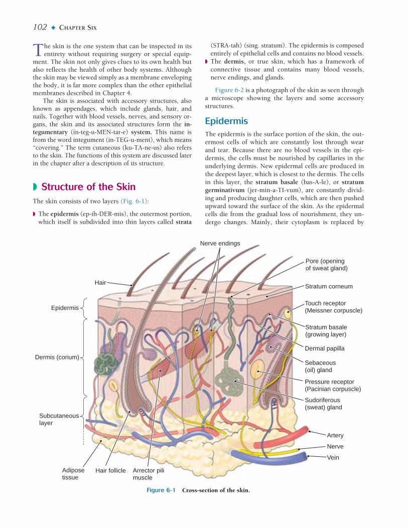
◗ Structure of the SkinThe skin consists of two layers (Fig. 6-1):
◗ The epidermis (ep-ih-DER-mis), the outermost portion,which itself is subdivided into thin layers called strata
(STRA-tah) (sing. stratum). The epidermis is composedentirely of epithelial cells and contains no blood vessels.
◗ The dermis, or true skin, which has a framework ofconnective tissue and contains many blood vessels,nerve endings, and glands.
Figure 6-2 is a photograph of the skin as seen througha microscope showing the layers and some accessorystructures.
EpidermisThe epidermis is the surface portion of the skin, the out-ermost cells of which are constantly lost through wearand tear. Because there are no blood vessels in the epi-dermis, the cells must be nourished by capillaries in theunderlying dermis. New epidermal cells are produced inthe deepest layer, which is closest to the dermis. The cellsin this layer, the stratum basale (bas-A-le), or stratumgerminativum (jer-min-a-TI-vum), are constantly divid-ing and producing daughter cells, which are then pushedupward toward the surface of the skin. As the epidermalcells die from the gradual loss of nourishment, they un-dergo changes. Mainly, their cytoplasm is replaced by
Figure 6-1 Cross-section of the skin.
Subcutaneouslayer
Dermis (corium)
Epidermis
Sebaceous(oil) gland
Pore (openingof sweat gland)
Nerve endings
Dermal papilla
Touch receptor(Meissner corpuscle)
Stratum basale(growing layer)
Stratum corneum
Sudoriferous(sweat) gland
Arrector pilimuscle
Adiposetissue
Hair follicle
Pressure receptor(Pacinian corpuscle)
Artery
Nerve
Vein
Hair

THE SKIN IN HEALTH AND DISEASE ✦ 103
large amounts of a protein called keratin (KER-ah-tin),which serves to thicken and protect the skin (Fig. 6-3).
By the time epidermal cells approach the surface, theyhave become flat, filled with keratin, and horny, formingthe uppermost layer of the epidermis, the stratumcorneum (KOR-ne-um). The stratum corneum is a pro-tective layer and is deeper in thick skin than in thin skin.Cells at the surface are constantly being lost and replacedfrom below, especially in areas of the skin that are subjectto wear and tear, as on the scalp, face, soles of the feet,and palms of the hands. Although this process of exfolia-tion (eks-fo-le-A-shun) occurs naturally at all times,many cosmetics companies sell products to promote ex-foliation, presumably to “enliven” and “refresh” the skin.
Between the stratum basale and the stratum corneumthere are additional layers of stratified epithelium thatvary in number and quantity depending on the thicknessof the skin.
Cells in the deepest layer of the epidermis producemelanin (MEL-ah-nin), a dark pigment that colors theskin and protects it from the harmful rays of sunlight.The cells that produce this pigment are the melanocytes(MEL-ah-no-sites). Irregular patches of melanin arecalled freckles.
DermisThe dermis, the so-called “true skin,” has a framework ofelastic connective tissue and is well supplied with bloodvessels and nerves. Because of its elasticity, the skin canstretch, even dramatically as in pregnancy, with littledamage. Most of the accessory structures of the skin, in-cluding the sweat glands, the oil glands, and the hair, arelocated in the dermis and may extend into the subcuta-neous layer under the skin.
The thickness of the dermis also varies in differentareas. Some places, such as the soles of the feet and thepalms of the hands, are covered with very thick layers ofskin, whereas others, such as the eyelids, are covered withvery thin and delicate layers. (See Box 6-1, Thick andThin Skin: Getting a Grip on Their Differences.)
Portions of the dermis extend upward into the epi-dermis, allowing blood vessels to get closer to the surfacecells (see Figs. 6-1 and 6-2). These extensions, or dermalpapillae, can be seen on the surface of thick skin, such asat the tips of the fingers and toes. Here they form a dis-tinct pattern of ridges that help to prevent slipping, suchas when grasping an object. The unchanging patterns ofthe ridges are determined by heredity. Because they areunique to each person, fingerprints and footprints can beused for identification.
6
Figure 6-2 Microscopic view of thin skin. Tissue layers andsome accessory structures are labeled. (Reprinted with permis-sion from Cormack DH. Essential Histology. 2nd ed. Philadel-phia: Lippincott Williams & Wilkins, 2001.)
Hair follicle Sebaceous gland
Sweat gland
Subcutaneousadipose tissue
Epidermis
Dermis
Figure 6-3 Upper portion of the skin. Layers of keratin inthe stratum corneum are visible at the surface. Below are layersof stratified squamous epithelium making up the remainder ofthe epidermis. (Reprinted with permission from Cormack DH.Essential Histology. 2nd ed. Philadelphia: Lippincott Williams &Wilkins, 2001.)
Keratin instratum corneum
Stratum basale
Epidermis
Dermis
Checkpoint 6-1 The skin and all its associated structures comprisea body system. What is the name of this system?
Subcutaneous LayerThe dermis rests on the subcutaneous (sub-ku-TA-ne-us)layer, sometimes referred to as the hypodermis or the su-perficial fascia (see Fig. 6-1). This layer connects the skinto the surface muscles. It consists of loose connective tis-sue and large amounts of adipose (fat) tissue. The fatserves as insulation and as a reserve supply for energy.Continuous bundles of elastic fibers connect the subcuta-neous tissue with the dermis, so there is no clear bound-ary between the two.
Checkpoint 6-2 The skin itself is composed of two layers. Movingfrom the superficial to the deeper layer, what are the names of thesetwo layers?

104 ✦ CHAPTER SIX
The blood vessels that supply the skin with nutrientsand oxygen and help to regulate body temperature runthrough the subcutaneous layer. This tissue is also rich innerves and nerve endings, including those that supplynerve impulses to and from the dermis and epidermis.The thickness of the subcutaneous layer varies in differ-ent parts of the body; it is thinnest on the eyelids andthickest on the abdomen.
sac is referred to as a sebaceous cyst. Usually, it is not dif-ficult to remove such tumorlike cysts by surgery.
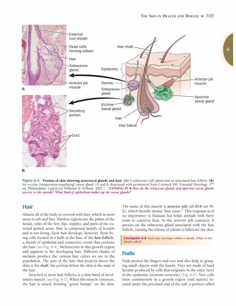
Sudoriferous (Sweat) GlandsThe sudoriferous (su-do-RIF-er-us) glands, or sweatglands, are coiled, tubelike structures located in the der-mis and the subcutaneous tissue (see Fig. 6-4 B). Most ofthe sudoriferous glands function to cool the body. Theyrelease sweat, or perspiration, that draws heat from theskin as the moisture evaporates at the surface. These ec-crine (EK-rin) type sweat glands are distributed through-out the skin. Each gland has a secretory portion and an ex-cretory tube that extends directly to the surface and opensat a pore (see also Fig. 6-1). Because sweat contains smallamounts of dissolved salts and other wastes in addition towater, these glands also serve a minor excretory function.
Present in smaller number, the apocrine (AP-o-krin)sweat glands are located mainly in the armpits (axillae)and groin area. These glands become active at pubertyand release their secretions through the hair follicles inresponse to emotional stress and sexual stimulation. Theapocrine glands release some cellular material in their se-cretions. Body odor develops from the action of bacteriain breaking down these organic cellular materials.
Several types of glands associated with the skin aremodified sweat glands. These are the ceruminous (seh-RU-min-us) glands in the ear canal that produce ear wax,or cerumen; the ciliary (SIL-e-er-e) glands at the edges ofthe eyelids; and the mammary glands.
The skin is the largest organ in the body, weighing about 4kg. Though it appears uniform in structure and function,
its thickness in fact varies, from less than 1 mm covering theeyelids to more than 5 mm on the upper back. Many of thefunctional differences between skin regions reflect the thick-ness of the epidermis and not the skin’s overall thickness.Based on epidermal thickness, skin can be categorized as thick(about 1 mm deep) or thin (about 0.1 mm deep).
Areas of the body exposed to significant wear and tear (thepalms, fingertips, and bottoms of the feet and toes) are cov-ered with thick skin. It is composed of a thick stratumcorneum and an extra layer not found in thin skin, the stra-tum lucidum, both of which make thick skin resistant to abra-sion. Thick skin is also characterized by epidermal ridges (e.g.,fingerprints) and numerous sweat glands, but lacks hair and
sebaceous (oil) glands. These adaptations make the thick skincovering the hands and feet effective for grasping or gripping.Thick skin’s dermis also contains many sensory receptors, giv-ing the hands and feet a superior sense of touch.
Thin skin covers areas of the body not exposed to muchwear and tear. It has a very thin stratum corneum and lacks adistinct stratum lucidum. Though thin skin lacks epidermalridges and has fewer sensory receptors than thick skin, it hasseveral specializations that thick skin does not. Thin skin iscovered with hair, which may help prevent heat loss from thebody. In fact, hair is most densely distributed in skin that cov-ers regions of great heat loss—the head, axillae (armpits), andgroin. Thin skin also contains numerous sebaceous glands,making it supple and free of cracks that may let infectious or-ganisms enter.
Box 6-1 A Closer Look
Thick and Thin Skin: Getting a Grip on Their DifferencesThick and Thin Skin: Getting a Grip on Their Differences
Checkpoint 6-3 What is the composition of the subcutaneouslayer?
◗ Accessory Structures of the SkinThe integumentary system includes some structures asso-ciated with the skin—glands, hair, and nails—that notonly protect the skin itself but have some more general-ized functions as well.
Sebaceous (Oil) GlandsThe sebaceous (se-BA-shus) glands are saclike in struc-ture, and their oily secretion, sebum (SE-bum), lubricatesthe skin and hair and prevents drying. The ducts of thesebaceous glands open into the hair follicles (Fig. 6-4 A).
Babies are born with a covering produced by theseglands that resembles cream cheese; this secretion is calledthe vernix caseosa (VER-niks ka-se-O-sah), which literallymeans “cheesy varnish.” Modified sebaceous glands, mei-bomian (mi-BO-me-an) glands, are associated with theeyelashes and produce a secretion that lubricates the eyes.
Blackheads consist of a mixture of dried sebum andkeratin that may collect at the openings of the sebaceousglands. If these glands become infected, pimples result. Ifa sebaceous gland becomes blocked, a sac of accumulatedsebum may form and gradually increase in size. Such a
Checkpoint 6-4 Some skin glands produce an oily secretion calledsebum. What is the name of these glands?
Checkpoint 6-5 What is the scientific name for the sweat glands?
THE SKIN IN HEALTH AND DISEASE ✦ 105
HairAlmost all of the body is covered with hair, which in mostareas is soft and fine. Hairless regions are the palms of thehands, soles of the feet, lips, nipples, and parts of the ex-ternal genital areas. Hair is composed mainly of keratinand is not living. Each hair develops, however, from liv-ing cells located in a bulb at the base of the hair follicle,a sheath of epithelial and connective tissue that enclosesthe hair (see Fig. 6-4). Melanocytes in this growth regionadd pigment to the developing hair. Different shades ofmelanin produce the various hair colors we see in thepopulation. The part of the hair that projects above theskin is the shaft; the portion below the skin is the root ofthe hair.
Attached to most hair follicles is a thin band of invol-untary muscle (see Fig. 6-1). When this muscle contracts,the hair is raised, forming “goose bumps” on the skin.
The name of this muscle is arrector pili (ah-REK-tor PI-li), which literally means “hair raiser.” This response is ofno importance to humans but helps animals with furrycoats to conserve heat. As the arrector pili contracts, itpresses on the sebaceous gland associated with the hairfollicle, causing the release of sebum to lubricate the skin.
6
Figure 6-4 Portion of skin showing associated glands and hair. (A) A sebaceous (oil) gland and its associated hair follicle. (B)An eccrine (temperature-regulating) sweat gland. (A and B, Reprinted with permission from Cormack DH. Essential Histology. 2nd
ed. Philadelphia: Lippincott Williams & Wilkins, 2001.) ZOOMING IN ✦ How do the sebaceous glands and apocrine sweat glandssecrete to the outside? What kind of epithelium makes up the sweat glands?
Hair shaft
Epidermis
Dermis
Eccrinesweat gland
Sebaceousgland
Apocrinesweat gland
Hair follicle
Hair
Arrector pilimuscle
B
A
Hair
Externalroot sheath
Sebaceousgland
Arrector pilimuscle
Dead cellsforming sebum
Secretoryportion
Duct
Checkpoint 6-6 Each hair develops within a sheath. What is thissheath called?
NailsNails protect the fingers and toes and also help in grasp-ing small objects with the hands. They are made of hardkeratin produced by cells that originate in the outer layerof the epidermis (stratum corneum) (Fig. 6-5). New cellsform continuously in a growth region (nail matrix) lo-cated under the proximal end of the nail, a portion called

106 ✦ CHAPTER SIX
the nail root. The remainder of the nail plate rests on anail bed of epithelial tissue. The color of the dermisbelow the nail bed can be seen through the clear nail. Thepale lunula (LU-nu-lah), literally “little moon,” at theproximal end of the nail appears lighter because it liesover the thicker growing region of the nail. The cuticle,an extension of the stratum corneum, seals the space be-tween the nail plate and the skin above the root.
Nails of both the toes and the fingers are affected bygeneral health. Changes in nails, including abnormalcolor, thickness, shape, or texture (e.g., grooves or split-ting), occur in chronic diseases such as heart disease, pe-ripheral vascular disease, malnutrition, and anemia.
◗ Functions of the SkinAlthough the skin has many functions, the following areits four major functions:
◗ Protection against infection◗ Protection against dehydration (drying)
◗ Regulation of body temperature◗ Collection of sensory information
Protection Against InfectionIntact skin forms a primary barrier against invasion ofpathogens. The cells of the stratum corneum form atight interlocking pattern that is resistant to penetration.The surface cells are constantly being shed, causing themechanical removal of pathogens. Rupture of this bar-rier, as in cases of wounds or burns, invites infection ofdeep tissues. The skin also protects against bacterial tox-ins (poisons) and some harmful chemicals in the envi-ronment.
Protection Against DehydrationBoth keratin in the epidermis and the oily sebum releasedto the surface of the skin from the sebaceous glands helpto waterproof the skin and prevent water loss by evapora-tion from the surface.
Regulation of Body TemperatureBoth the loss of excess heat and protection from cold are important functions of the skin. Indeed, most of the blood supply to the skin is concerned with tempera-ture regulation. In cold conditions, vessels in the skinconstrict (become narrower) to reduce the flow of bloodto the surface and diminish heat loss. The skin may be-come visibly pale under these conditions. Special vesselsthat directly connect arteries and veins in the skin of theears, nose, and other exposed locations provide the vol-ume of blood flow needed to prevent freezing.
To cool the body, the skin forms a large surface for ra-diating body heat to the surrounding air. When the bloodvessels dilate (widen), more blood is brought to the sur-face so that heat can be dissipated.
The other mechanism for cooling the body involvesthe sweat glands, as noted above. The evaporation of per-spiration draws heat from the skin. A person feels un-comfortable on a hot and humid day because water doesnot evaporate as readily from the skin into the surround-ing air. A dehumidifier makes one more comfortable evenwhen the temperature remains high.
As is the case with so many body functions, tempera-ture regulation is complex and involves several parts ofthe body, including certain centers in the brain.
Collection of Sensory InformationBecause of its many nerve endings and other special re-ceptors, the skin may be regarded as one of the chief sen-sory organs of the body. Free nerve endings detect painand moderate changes in temperature. Other types of sen-sory receptors in the skin respond to light touch and deeppressure. Figure 6-1 shows some free nerve endings, atouch receptor (Meissner corpuscle), and a deep pressurereceptor (Pacinian corpuscle) in a section of skin.
Figure 6-5 Nail structure. (A) Photograph of a nail, superiorview. (B) Midsagittal section of a fingertip. (A, Reprinted withpermission from Bickley LS. Bates’ Guide to Physical Examina-tion and History Taking. 8th ed. Philadelphia: LippincottWilliams & Wilkins, 2003.)
Lunula
CuticleNail plate
Nail bed
Nail root
Growth region(nail matrix)
Distal boneof finger
B
A
LunulaNail plate CuticleFree edge
A

THE SKIN IN HEALTH AND DISEASE ✦ 107
Many of the reflexes that make it possible for humansto adjust themselves to the environment begin as sensoryimpulses from the skin. As elsewhere in the body, theskin works with the brain and the spinal cord to accom-plish these important functions.
Other Activities of the SkinSubstances can be absorbed through the skin in limitedamounts. Some drugs, for example, estrogens, othersteroids, anesthetics, and medications to control motionsickness, can be absorbed from patches placed on theskin. (See Box 6-2, Medication Patches: No Bitter Pill toSwallow.) Most medicated ointments used on the skin,however, are for the treatment of local conditions only.Even medication injected into the subcutaneous tissues isabsorbed very slowly.
There is also a minimal amount of excretion throughthe skin. Water and electrolytes (salts) are excreted insweat (perspiration). Some nitrogen-containing wastesare eliminated through the skin, but even in disease, theamount of waste products excreted by the skin is small.
Vitamin D needed for the development and mainte-nance of bone tissue is manufactured in the skin underthe effects of ultraviolet radiation in sunlight.
Note that the human skin does not “breathe.” Thepores of the epidermis serve only as outlets for perspi-ration from the sweat glands and sebum (oil) from thesebaceous glands. They are not used for exchange ofgases.
◗ Observation of the SkinWhat can the skin tell you? What do its color, texture,and other attributes indicate? Is there any damage? Muchcan be learned by an astute observer. In fact, the first in-dication of a serious systemic disease (such as syphilis)may be a skin disorder.
ColorThe color of the skin depends on a number of factors, in-cluding the following:
◗ Amount of pigment in the epidermis◗ Quantity of blood circulating in the surface blood vessels◗ Composition of the circulating blood, including:
◗ Quantity of oxygen◗ Concentration of hemoglobin◗ Presence of bile, silver compounds, or other chemi-
cals
Pigment The main pigment of the skin, as we havenoted, is called melanin. This pigment is also found in thehair, the middle coat of the eyeball, the iris of the eye, andcertain tumors. Melanin is common to all races, butdarker people have a much larger quantity in their tis-sues. The melanin in the skin helps to protect againstdamaging ultraviolet radiation from the sun. Thus, skinthat is exposed to the sun shows a normal increase in thispigment, a response we call tanning.
Sometimes, there are abnormal increases in the quan-tity of melanin, which may occur either in localized areasor over the entire body surface. For example, diffusespots of pigmentation may be characteristic of some en-
6
For most people, pills are a convenient way to take medica-tion, but for others, they have drawbacks. Pills must be
taken at regular intervals to ensure consistent dosing, andthey must be digested and absorbed into the bloodstream be-fore they can begin to work. For those who have difficultyswallowing or digesting pills, transdermal (TD) patches offeran effective alternative to oral medications.
TD patches deliver a consistent dose of medication that dif-fuses at a constant rate through the skin into the bloodstream.There is no daily schedule to follow, nothing to swallow, andno stomach upset. TD patches can also deliver medication tounconscious patients, who would otherwise require intra-venous drug delivery. TD patches are used in hormone re-placement therapy, to treat heart disease, to manage pain, andto suppress motion sickness. Nicotine patches are also used aspart of programs to quit smoking.
TD patches must be used carefully. Drug diffusion
through the skin takes time, so it is important to know howlong the patch must be in place before it is effective. It isalso important to know how long the medication’s effectstake to disappear after the patch is removed. Because thebody continues to absorb what has already diffused into theskin, removing the patch does not entirely remove the med-icine.
A recent advance in TD drug delivery is iontophoresis.Based on the principle that like charges repel each other, thismethod uses a mild electrical current to move ionic drugsthrough the skin. A small electrical device attached to thepatch uses positive current to “push” positively charged drugmolecules through the skin, and a negative current to pushnegatively charged ones. Even though very low levels of elec-tricity are used, people with pacemakers should not use ion-tophoretic patches. Another disadvantage is that they canmove only ionic drugs through the skin.
Medication Patches: No Bitter Pill to Swallow
Box 6-2 Clinical Perspectives
Medication Patches: No Bitter Pill to Swallow
Checkpoint 6-7 What two mechanisms are used to regulate tem-perature through the skin?

108 ✦ CHAPTER SIX
docrine disorders. In albinism (AL-bih-nizm), a heredi-tary disorder that affects melanin production, there islack of pigment in the skin, hair, and eyes.
Another pigment that imparts color to the skin iscarotene, a pigment obtained from carrots and other or-ange and yellow vegetables. Carotene is stored in fatty tis-sue and skin. Also visible is hemoglobin, the pigment thatgives blood its color, which can be seen through the ves-sels in the dermis.
Discoloration Pallor (PAL-or) is paleness of the skin,often caused by reduced blood flow or by reduction inhemoglobin, as occurs in cases of anemia. Pallor is mosteasily noted in the lips, nail beds, and mucous mem-branes. Flushing is redness of the skin, often related tofever. Signs of flushing are most noticeable in the faceand neck.
When there is not enough oxygen in circulatingblood, the skin may take on a bluish discoloration termedcyanosis (si-ah-NO-sis) (Fig. 6-6 A). This is a symptomof heart failure and of breathing problems, such as asthmaor respiratory obstruction.
A yellowish discoloration of the skin may be due to thepresence of excessive amounts of bile pigments, mainlybilirubin (BIL-ih-ru-bin), in the blood (Fig. 6-6 B). (Bile isa substance produced by the liver that aids in the digestionof fats; see Chapter 19.) This condition, called jaundice(JAWN-dis) (from the French word for “yellow”), may bea symptom of a number of disorders, such as the following:
◗ A tumor pressing on the common bile duct or a stonewithin the duct, either of which would obstruct theflow of bile into the small intestine
◗ Inflammation of the liver (hepatitis), commonly causedby a virus
◗ Certain diseases of the blood in which red blood cellsare rapidly destroyed (hemolyzed)
◗ Immaturity of the liver. Neonatal (newborn) jaundice oc-curs when the liver is not yet capable of processing biliru-bin (bile pigment). Most such cases correct themselveswithout treatment in about a week, but this form of jaun-dice may be treated by exposure to special fluorescentlight that helps the body to get rid of the bilirubin.
Another possible cause of a yellowish discoloration ofthe skin is the excessive intake of carrots and other deeplycolored vegetables. This condition is known as carotene-mia (kar-o-te-NE-me-ah).
Certain types of chronic poisoning may cause gray orbrown discoloration of the skin. A peculiar bronze cast ispresent in Addison disease (malfunction of the adrenalgland). Many other disorders cause discoloration of the skin,but their discussion is beyond the scope of this chapter.
Figure 6-6 Discoloration of the skin. (A) Cyanosis is a bluish discoloration due to lack of oxygen. (B) Jaundice is a yellowishdiscoloration due to bile pigments in the blood. (Reprinted with permission from Bickley LS. Bates’ Guide to Physical Examinationand History Taking. 8th ed. Philadelphia: Lippincott Williams & Wilkins, 2003.) ZOOMING IN ✦ What color is associated withcyanosis? What color is associated with jaundice?
A B
Checkpoint 6-8 What are some pigments that impart color to theskin?
LesionsA lesion (LE-zhun) is any wound or local damage to tis-sue. In examining the skin for lesions, it is important tomake note of their type, arrangement, and location. Le-sions may be flat or raised or may extend below the sur-face of the skin.
Surface Lesions A surface lesion is often called a rashor, if raised, an eruption (e-RUP-shun). Skin rashes maybe localized, as in diaper rash, or generalized, as inmeasles and other systemic infections. Often, these le-sions are accompanied by erythema (er-eh-THE-mah), orredness of the skin. The following are some terms used todescribe surface skin lesions:
◗ Macule (MAK-ule). A macule is a spot that is neitherraised nor depressed. Macules are typical of measlesand descriptive of freckles (Fig. 6-7 A).
◗ Papule (PAP-ule). A papule is a firm, raised area, as insome stages of chickenpox and in the second stage ofsyphilis (see Fig. 6-7 B). A pimple is a papule. A largefirm papule is called a nodule (NOD-ule).
◗ Vesicle (VES-ih-kl). A vesicle is a blister or small sacthat is full of fluid, such as may be found in some of theeruptions of chickenpox or shingles (see Fig. 6-7 C).Another term for a vesicle is a bulla (BUL-ah).
◗ Pustule (PUS-tule). A pustule is a vesicle filled withpus. Pustules may develop if vesicles become infected(see Fig. 6-7 D).

THE SKIN IN HEALTH AND DISEASE ✦ 109
Deeper Lesions A deeper lesion of the skin may de-velop from a surface lesion or may be caused by trauma(TRAW-mah), that is, a wound or injury. Because suchbreaks may be followed by infection, wounds should becared for to prevent the entrance of pathogens and toxinsinto deeper tissues and body fluids. Deeper injuries to theskin include the following:
◗ Excoriation (eks-ko-re-A-shun), which is a scratch intothe skin
◗ Laceration (las-er-A-shun), which is a rough, jaggedwound made by tearing of the skin
◗ Ulcer (UL-ser), which is a sore associated with disinte-gration and death of tissue (Fig. 6-8 A)
◗ Fissure (FISH-ure), which is a crack in the skin. Ath-lete’s foot, for example, can produce fissures. Tonguefissures may be normal variations in the tongue’s sur-face (see Fig. 6-8 B), but may also appear on the lips ortongue as a result of injury or disease.
6
Figure 6-7 Surface lesions. (A) Macules on the dorsal sur-face of the hand, wrist, and forearm. (B) Papules on the knee.(C) Vesicles on the chin. (D) Pustules on the palm. (Pho-tographs reprinted with permission from Bickley LS. Bates’Guide to Physical Examination and History Taking. 8th ed.Philadelphia: Lippincott Williams & Wilkins, 2003. Line draw-ings reprinted with permission from Cohen BJ. Medical Termi-nology. 4th ed. Philadelphia: Lippincott Williams & Wilkins,2004.)
A Macule
B Papule
D Pustule
C Vesicle
Figure 6-8 Deeper lesions. (A) Tongue ulcer. (B) Tonguefissures. (Photographs reprinted with permission from LanglaisRP, Miller CS. Color Atlas of Common Oral Diseases. 3rd ed.Philadelphia: Lippincott Williams & Wilkins, 2002. Line draw-ings reprinted with permission from Cohen BJ. Medical Termi-nology. 4th ed. Philadelphia: Lippincott Williams & Wilkins,2004.)
A Ulcer
B Fissures
Checkpoint 6-9 What is a lesion?
BurnsMost burns are caused by contact with hot objects, explo-sions, or scalding with hot liquids. They may also be caused

110 ✦ CHAPTER SIX
by electrical injuries, contact with harmful chemicals, orabrasion. Burns are assessed in terms of the depth of dam-age and the percentage of body surface area (BSA) involved.Depth of tissue destruction is categorized as follows:
◗ Superficial partial-thickness, which involves the epi-dermis and perhaps a portion of the dermis. The tissueis reddened and may blister, as in cases of sunburn.
◗ Deep partial-thickness, which involves the epidermisand portions of the dermis. The tissue is blistered andbroken, with a weeping surface. Causes include scald-ing and exposure to flame.
◗ Full-thickness, which involves the full skin and some-times subcutaneous tissue and underlying tissues aswell. The tissue is broken, dry and pale, or charred.These injuries may require skin grafting and may resultin loss of digits or limbs.
The above classification replaces an older system ofranking burns as first-, second-, and third-degree accord-ing to the depth of tissue damage.
The amount of body surface area involved in a burnmay be estimated by using the rule of nines, in whichareas of body surface are assigned percentages in multi-ples of nine (Fig. 6-9). The more accurate Lund andBrowder method divides the body into small areas and es-timates the proportion of BSA that each contributes.
Infection is a common complication of burns, becausethe skin, a major defense against invasion of microorgan-isms, is damaged. Respiratory complications may becaused by inhalation of smoke and toxic chemicals, andcirculatory problems may result from loss of fluids andelectrolytes. Treatment of burns includes respiratory care,administration of fluids, wound care, and pain control.Patients must be monitored for circulatory complications,infections, and signs of posttraumatic stress.
Sunburn Sunlight can cause chemical and biologicchanges in the skin. On exposure, the skin first becomesreddened (erythematous) and then may become swollenand blistered. (See Box 6-3, The Dark Side of the Sun.)Sunlight can cause severe burns that result in serious ill-ness. Continued excessive exposure to the sun is a risk fac-tor in skin cancer. Tanning requires the skin to protect it-
Figure 6-9 The rule of nines. This method is used to estimatepercentages of body surface area (BSA) in treatment of burns.
Anterior
18%
1%
4.5%
4.5%
9% 9%
4.5%
18%
1%
4.5%
4.5%
9% 9% 9% 9%
4.5% 4.5%
4.5%
18%
9% 9%
4.5% 4.5% 4.5%
4.5%
18%
Posterior
The three most common forms of skin cancer—basal cellcarcinoma, squamous cell carcinoma, and malignant
melanoma—share a common risk factor: excessive exposureto the ultraviolet radiation (UV) found in sunlight. UV raysalso cause premature aging of the skin, including wrinkling,discoloration (“age spots” or “liver spots”), and a change intexture most often referred to as “leathery skin.” Excessivesun exposure is also a risk factor for cataracts and other eyeproblems.
The damaging radiation found in sunlight occurs in two dif-ferent forms, ultraviolet-A (UVA) and ultraviolet-B (UVB).UVA damages the skin’s deeper layers, resulting in a loss ofelasticity and a general decrease in blood flow to the skin.UVB damages the skin’s outermost layers, causing the ery-thema (redness), inflammation, and peeling common to theaverage “sunburn.” Excessive UV exposure causes geneticmutations in skin cells that make them unable to repair them-
selves and possibly cancerous. Tanning booths also produceUVA and UVB rays and are no safer than sun tanning.
You can reduce the damage caused by UVA and UVB by thefollowing:
◗ Limit exposure during midday when the level of UV radia-tion is highest.
◗ Cover up with a hat, long pants, and a long-sleeved shirtwhen outdoors.
◗ Wear sunglasses that block UV rays.◗ Apply a sunscreen with an SPF (sun protection factor) of 15
or higher 30 minutes before going outdoors. Reapply duringexposure, especially after swimming.
◗ Stay in the shade, where exposure to UVA and UVB is sig-nificantly decreased.
◗ Avoid tanning booths.
The Dark Side of the Sun
Box 6-3 • Health Maintenance
The Dark Side of the Sun

THE SKIN IN HEALTH AND DISEASE ✦ 111
self by producing considerably more than usual amounts ofmelanin. This increase in pigmentation may reduce thebody’s ability to profit from smaller amounts of sun avail-able during some parts of the year.
◗ Tissue RepairTrue tissue regeneration after injury can occur only inareas that have actively dividing cells or cells that can betriggered to divide by injury. Specifically, these tissues arethe epithelial and connective tissues. Even among theconnective tissues, repair occurs more slowly in tissuesthat are not very active metabolically, in cartilage for ex-ample. Muscle tissue and nervous tissue, which stop di-viding early in life, generally do not restore themselves,although some types can carry out minimal regeneration.When muscle and nervous tissues are injured, they aregenerally replaced by connective tissue.
Repair of a skin wound or lesion begins after bloodhas clotted and a scab has formed at the surface to protectunderlying tissue. From damaged capillaries, new vesselsbranch and grow into the injured tissue. Fibroblasts (cellsthat produce fibers) manufacture collagen to close thegap made by the wound. A large wound requires exten-sive growth of new connective tissue, which developsfrom within the wound. This new tissue forms a scar, alsocalled a cicatrix (SIK-ah-triks).
After the upper layer of epithelium has regenerated,the scab is released. The underlying scar tissue may thencontinue to show at the surface as a white line. Scar tis-sue is strong but is not as flexible as normal tissue anddoes not function like the tissue it replaces. Suturing(sewing) the edges of a clean wound together, as is donein the case of operative wounds, decreases the amount ofconnective tissue needed for repair and thus reduces thesize of the resulting scar.
Excess production of collagen in the formation of ascar may result in the development of keloids (KE-loyds),tumorlike masses or sharply raised areas on the surface ofthe skin. These are not dangerous but may be removedfor the sake of appearance.
Wound healing is affected by:
◗ Nutrition—A complete and balanced diet will providethe nutrients needed for cell regeneration. All requiredvitamins and minerals are important, but especially vi-tamins A and C, which are needed for collagen.
◗ Blood supply—The blood brings oxygen and nutrientsto the tissues and also carries away waste materials andtoxins (poisons) that might form during the healingprocess. White blood cells attack invading bacteria atthe site of the injury. Poor circulation, as occurs in casesof diabetes, for example, will delay wound healing.
◗ Infection—Contamination prolongs inflammation andinterferes with the formation of materials needed forwound repair.
◗ Effects of Aging on theIntegumentary SystemAs people age, wrinkles, or crow’s feet, develop around theeyes and mouth owing to the loss of fat and collagen in theunderlying tissues. The dermis becomes thinner, and theskin may become transparent and lose its elasticity, the ef-fect of which is sometimes called “parchment skin.” The for-mation of pigment decreases with age. However, there maybe localized areas of extra pigmentation in the skin with theformation of brown spots (“liver spots”), especially on areasexposed to the sun (e.g., the back of the hands). Circulationto the dermis decreases, so white skin looks paler.
The hair does not replace itself as rapidly as beforeand thus becomes thinner on the scalp and elsewhere onthe body. Decreased melanin production leads to gray orwhite hair. The texture of the hair changes as the hairshaft becomes less dense, and hair, like the skin, be-comes drier with a decrease in sebum production.
The sweat glands decrease in number, so there is lessoutput of perspiration and lowered ability to withstandheat. The elderly are also more sensitive to cold becauseof less fat in the skin and poor circulation. The fingernailsmay flake, become brittle, or develop ridges, and toenailsmay become discolored or abnormally thickened.
◗ Care of the SkinThe most important factors in caring for the skin are thosethat ensure good general health. Proper nutrition and ad-equate circulation are vital to the maintenance of the skin.Regular cleansing removes dirt and dead skin debris andsustains the slightly acid environment that inhibits bacte-rial growth on the skin. Careful hand washing with soapand water, with attention to the under-nail areas, is a sim-ple measure that reduces the spread of disease.
The skin needs protection from continued exposure tosunlight to prevent premature aging and cancerouschanges. Appropriate applications of sunscreens beforeand during time spent in the sun can prevent skin damage.
◗ Skin DisordersSkin disorders range from simple superficial nuisances,such as acne and rashes, to more deep-seated problemsthat may lead to systemic disease.
6Checkpoint 6-10 What two categories of tissues repair themselvesmost easily?
◗ Age—Healing is generally slower among the elderly dueto a slower rate of cell replacement. The elderly alsomay have a lowered immune response to infection.

112 ✦ CHAPTER SIX
DermatitisDermatosis (der-mah-TO-sis) is a general term referringto any skin disease. Inflammation of the skin is called der-matitis (der-mah-TI-tis). It may be due to many kinds ofirritants, such as the oil of poison oak or poison ivyplants, detergents, and strong acids, alkalis, or otherchemicals. Prompt removal of the irritant is the most ef-fective method of prevention and treatment. A thoroughcleansing as soon as possible after contact with plant oilsmay prevent the development of itching eruptions.
Atopic Dermatitis Atopic dermatitis (ah-TOP-ik der-mah-TI-tis) or eczema (EK-ze-mah) is characterized byintense itching and skin inflammation (Fig. 6-10). The af-fected areas show redness (erythema), blisters (vesicles),pimplelike lesions (papules), and scaling and crusting ofthe skin surface. Scratching (excoriation) of the skin canlead to a secondary bacterial infection. Atopic dermatitiscommonly first occurs in early childhood, with the recur-rence of acute episodes throughout life. The skin may beexcessively sensitive to many soaps, detergents, roughfabrics, or perspiration. The person with atopic dermati-tis may also be subject to allergic disorders, such as hayfever, asthma, and food allergies.
PsoriasisPsoriasis (so-RI-ah-sis) is a chronic overgrowth of the epi-dermis leading to large, sharply outlined, red (erythema-
tous), flat areas (plaques) covered with silvery scales (Fig. 6-11). The cause of this chronic, recurrent skin disease is un-known, but there is sometimes a hereditary pattern, and animmune disorder may be involved. Psoriasis is treated withtopical corticosteroids and exposure to ultraviolet (UV) light.
Figure 6-10 Atopic dermatitis (eczema). Scratches (excori-ation) are visible in the photo. (Reprinted with permission fromBickley LS. Bates’ Guide to Physical Examination and HistoryTaking. 8th ed. Philadelphia: Lippincott Williams & Wilkins,2003.)
Figure 6-11 Psoriasis. Silvery surface scales are visible.(Reprinted with permission from Bickley LS. Bates’ Guide toPhysical Examination and History Taking. 8th ed. Philadelphia:Lippincott Williams & Wilkins, 2003.)
Checkpoint 6-11 What is the difference between dermatosis anddermatitis?
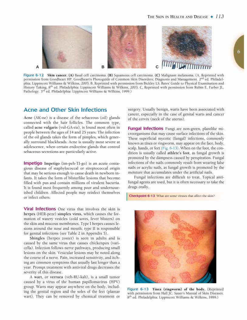
CancerSkin cancer is the most common form of cancer in theUnited States. Exposure to sunlight predisposes to devel-opment of skin cancer, which, in the United States, ismost common among people who have fair skin and wholive in the Southwest, where exposure to the sun is con-sistent and may be intense.
Basal cell and squamous cell carcinomas arise in theepidermis and generally appear on the face, neck, andhands (Fig. 6-12 A, B). Early detection and treatment inthese cases usually results in cure, although squamouscell carcinoma is the more likely to metastasize.
Melanoma (mel-ah-NO-mah) is a malignant tumor ofmelanocytes (melanin-forming cells). This type of canceroriginates in a nevus (NE-vus), a mole or birthmark, any-where in the body (see Fig. 6-12 C). Unlike a normal mole,which has an evenly round shape and well-defined border,a melanoma may show irregularity in shape. Other signs ofmelanoma are a change in color or uneven color and in-crease in size of a mole. A predisposing factor for melanomais severe, blistering sunburn, although these cancers can ap-pear in areas not sun-exposed, such as the soles of the feet,between fingers and toes, and in mucous membranes.
Checkpoint 6-12 What is the name for a cancer of the skin’s pig-ment-producing cells?
THE SKIN IN HEALTH AND DISEASE ✦ 113
Acne and Other Skin InfectionsAcne (AK-ne) is a disease of the sebaceous (oil) glandsconnected with the hair follicles. The common type,called acne vulgaris (vul-GA-ris), is found most often inpeople between the ages of 14 and 25 years. The infectionof the oil glands takes the form of pimples, which gener-ally surround blackheads. Acne is usually most severe atadolescence, when certain endocrine glands that controlsebaceous secretions are particularly active.
Impetigo Impetigo (im-peh-TI-go) is an acute conta-gious disease of staphylococcal or streptococcal originthat may be serious enough to cause death in newborn in-fants. It takes the form of blisterlike lesions that becomefilled with pus and contain millions of virulent bacteria.It is found most frequently among poor and undernour-ished children. Affected people may reinfect themselvesor infect others.
Viral Infections One virus that involves the skin isherpes (HER-peze) simplex virus, which causes the for-mation of watery vesicles (cold sores, fever blisters) onthe skin and mucous membranes. Type I herpes causes le-sions around the nose and mouth; type II is responsiblefor genital infections (see Table 2 in Appendix 5).
Shingles (herpes zoster) is seen in adults and iscaused by the same virus that causes chickenpox (vari-cella). Infection follows nerve pathways, producing smalllesions on the skin. Vesicular lesions may be noted alongthe course of a nerve. Pain, increased sensitivity, and itch-ing are common symptoms that usually last longer than ayear. Prompt treatment with antiviral drugs decreases theseverity of this disease.
A wart, or verruca (veh-RU-kah), is a small tumorcaused by a virus of the human papillomavirus (HPV)group. Warts may appear anywhere on the body, includ-ing the genital region and the soles of the feet (plantarwart). They can be removed by chemical treatment or
surgery. Usually benign, warts have been associated withcancer, especially in the case of genital warts and cancerof the cervix (neck of the uterus).
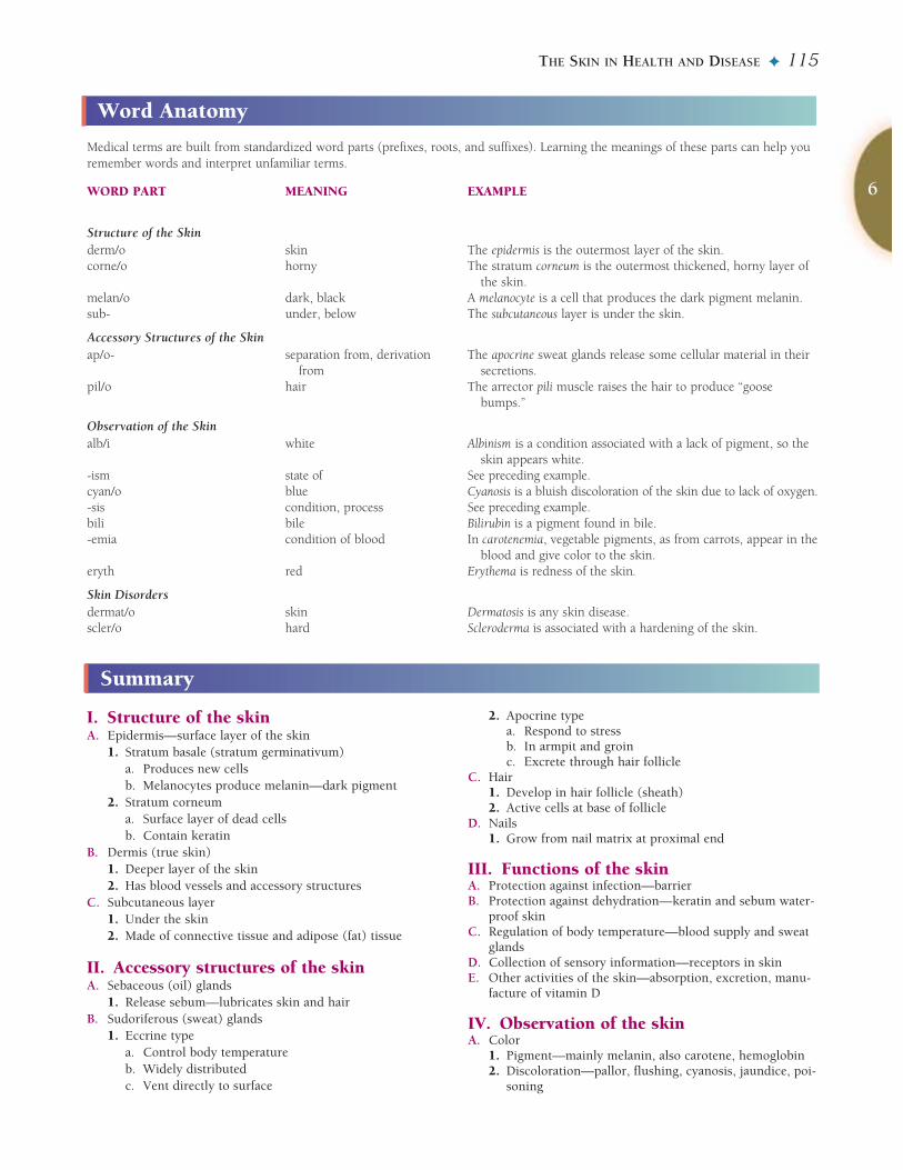
Fungal Infections Fungi are non-green, plantlike mi-croorganisms that may cause surface infections of the skin.These superficial mycotic (fungal) infections, commonlyknown as tinea or ringworm, may appear on the face, body,scalp, hands, or feet (Fig. 6-13). When on the foot, the con-dition is usually called athlete’s foot, as fungal growth ispromoted by the dampness caused by perspiration. Fungalinfections of the nails commonly result from wearing falsenails or acrylic nails, as fungal growth is promoted by themoisture that accumulates under the artificial nails.
Fungal infections are difficult to treat. Topical anti-fungal agents are used, but it is often necessary to take thedrugs orally.
6
Figure 6-12 Skin cancer. (A) Basal cell carcinoma. (B) Squamous cell carcinoma. (C) Malignant melanoma. (A, Reprinted withpermission from Goodheart HP. Goodheart’s Photoguide of Common Skin Disorders; Diagnosis and Management. 2nd ed. Philadel-phia: Lippincott Williams & Wilkins, 2003. B, Reprinted with permission from Bickley LS. Bates’ Guide to Physical Examination andHistory Taking. 8th ed. Philadelphia: Lippincott Williams & Wilkins, 2003. C, Reprinted with permission from Rubin E, Farber JL.Pathology. 3rd ed. Philadelphia: Lippincott Williams & Wilkins, 1999.)
A B C
Checkpoint 6-13 What are some viruses that affect the skin?
Figure 6-13 Tinea (ringworm) of the body. (Reprintedwith permission from Hall JC. Sauer’s Manual of Skin Diseases.8th ed. Philadelphia: Lippincott Williams & Wilkins, 1999.)

114 ✦ CHAPTER SIX
ease of connective tissue. The more widespread form ofthe disease, systemic lupus erythematosus (SLE), in-volves the skin and other organs. The discoid form(DLE) involves only the skin. It is seen as rough, raised,violet-tinted papules, usually limited to the face andscalp. There may also be a butterfly-shaped rash acrossthe nose and cheeks, described as a malar (cheekbone)rash. The skin lesions of lupus are worsened by exposureto the ultraviolet radiation in sunlight. SLE is moreprevalent in women than in men and has a higher inci-dence among Asians and blacks than in other popula-tions.
Scleroderma is a disease of unknown cause that in-volves overproduction of collagen with thickening andtightening of the skin. Sweat glands and hair follicles arealso involved. A very early sign of scleroderma is numb-ness, pain, and tingling on exposure to cold caused byconstriction of blood vessels in the fingers and toes. Skinsymptoms first appear on the forearms and around themouth. Internal organs become involved in a diffuse formof scleroderma called progressive systemic sclerosis(PSS).
Pressure UlcersPressure ulcers are skin lesions that appear where thebody rests on skin that covers bony projections, such asthe spine, heel, elbow, or hip. The pressure interrupts cir-culation leading to ulceration and death of tissue. Poorgeneral health, malnutrition, age, obesity, and infectioncontribute to the development of pressure ulcers.
Lesions first appear as redness of the skin. If ignored,they may penetrate the skin and underlying muscle, ex-tending even to bone and requiring months to heal.
Pads or mattresses to relieve pressure, regular cleans-ing and drying of the skin, frequent change in position,and good nutrition help to prevent pressure ulcers. Pre-vention of pressure ulcer by these methods is far easierthan treatment of an established ulcer.
Other terms for pressure ulcers are decubitus ulcerand bedsore. Both of these terms refer to lying down, al-though pressure ulcers may appear in anyone with lim-ited movement, not only those who are confined tobed.
Checkpoint 6-14 What causes tinea or ringworm infections?
Alopecia (Baldness)Alopecia (al-o-PE-she-ah), or baldness, may be due to anumber of factors. The most common type, known asmale pattern baldness, is an expression of heredity andaging; it is influenced by male sex hormones. Topical ap-plications of the drug minoxidil (used as an oral medica-tion to control blood pressure) have produced growth ofhair in this type of baldness. Alopecia may be the result ofa systemic disease, such as uncontrolled diabetes, thyroiddisease, or malnutrition. In such cases, control of the dis-ease results in regrowth of hair. A growing list of drugshas been linked with baldness, including the chemother-apeutic drugs used in treating neoplasms.
Allergy and Other ImmuneDisordersAllergy, also known as hypersensitivity, is an unfavorableimmune response to a substance that is normally harm-less to most people (see Chapter 17). Foods, drugs, cos-metics, and a variety of industrial substances can provokeallergic responses in some people. Often the skin is in-volved in such responses, showing inflammation, rashes,vesicles, or other forms of eruptions, usually accompa-nied by severe pruritus (pru-RI-tus), or itching.
Urticaria (ur-tih-KA-re-ah), or hives, is an allergic re-action characterized by the temporary appearance of ele-vated red patches known as wheals.
Autoimmune Disorders An autoimmune disease re-sults from an immune reaction to one’s own tissues. Thefollowing diseases that involve the skin are believed to becaused, at least in part, by autoimmune reactions.
Pemphigus (PEM-fi-gus) is characterized by the for-mation of blisters, or bullae (BUL-e) in the skin and mu-cous membranes caused by a separation of epidermalcells from underlying layers. Rupture of these lesionsleaves deeper areas of the skin unprotected from infectionand fluid loss, much as in cases of burns. Pemphigus isfatal unless treated by methods to suppress the immunesystem.
Lupus erythematosus (LU-pus er-ih-the-mah-TO-sus) (LE) is a chronic, inflammatory, autoimmune dis-
Checkpoint 6-15 What are several autoimmune disorders that in-volve the skin?
THE SKIN IN HEALTH AND DISEASE ✦ 115
I. Structure of the skinA. Epidermis—surface layer of the skin
1. Stratum basale (stratum germinativum)a. Produces new cellsb. Melanocytes produce melanin—dark pigment
2. Stratum corneuma. Surface layer of dead cellsb. Contain keratin
B. Dermis (true skin)1. Deeper layer of the skin2. Has blood vessels and accessory structures
C. Subcutaneous layer1. Under the skin2. Made of connective tissue and adipose (fat) tissue
II. Accessory structures of the skinA. Sebaceous (oil) glands
1. Release sebum—lubricates skin and hairB. Sudoriferous (sweat) glands
1. Eccrine typea. Control body temperatureb. Widely distributedc. Vent directly to surface
2. Apocrine typea. Respond to stressb. In armpit and groinc. Excrete through hair follicle
C. Hair1. Develop in hair follicle (sheath)2. Active cells at base of follicle
D. Nails1. Grow from nail matrix at proximal end
III. Functions of the skinA. Protection against infection—barrierB. Protection against dehydration—keratin and sebum water-
proof skinC. Regulation of body temperature—blood supply and sweat
glandsD. Collection of sensory information—receptors in skinE. Other activities of the skin—absorption, excretion, manu-
facture of vitamin D
IV. Observation of the skinA. Color
1. Pigment—mainly melanin, also carotene, hemoglobin2. Discoloration—pallor, flushing, cyanosis, jaundice, poi-
soning
6
Word Anatomy
Summary
Medical terms are built from standardized word parts (prefixes, roots, and suffixes). Learning the meanings of these parts can help youremember words and interpret unfamiliar terms.
WORD PART MEANING EXAMPLE
Structure of the Skinderm/o skin The epidermis is the outermost layer of the skin.corne/o horny The stratum corneum is the outermost thickened, horny layer of
the skin.melan/o dark, black A melanocyte is a cell that produces the dark pigment melanin.sub- under, below The subcutaneous layer is under the skin.
Accessory Structures of the Skinap/o- separation from, derivation The apocrine sweat glands release some cellular material in their
from secretions.pil/o hair The arrector pili muscle raises the hair to produce “goose
bumps.”
Observation of the Skinalb/i white Albinism is a condition associated with a lack of pigment, so the
skin appears white.-ism state of See preceding example.cyan/o blue Cyanosis is a bluish discoloration of the skin due to lack of oxygen.-sis condition, process See preceding example.bili bile Bilirubin is a pigment found in bile.-emia condition of blood In carotenemia, vegetable pigments, as from carrots, appear in the
blood and give color to the skin.eryth red Erythema is redness of the skin.
Skin Disordersdermat/o skin Dermatosis is any skin disease.scler/o hard Scleroderma is associated with a hardening of the skin.

Building Understanding
Fill in the blanks1. Cells of the stratum corneum contain large amounts ofa protein called ______.2. Sweat glands located in the axillae and groin are called______ sweat glands.
3. The name of the muscle that raises the hair is ______.4. A dark-colored pigment that protects the skin from ul-traviolet light is called ______.5. A medical term that means “scar” is ______.
116 ✦ CHAPTER SIX
B. Lesions—wound or local damage1. Surface lesions (rash, eruption)
a. Macule (spot), papule (firm, raised), vesicle (blister),pustule (pus-filled)
2. Deeper lesionsa. excoriation (scratch), laceration (tear), ulcer (sore),
fissure (crack)C. Burns
1. Evaluated by depth of damage and amount body surfacearea (BSA) involved
2. Sunburn—risk factor in skin cancer
V. Tissue repair1. Requires actively dividing cells2. Easiest in epithelial and connective tissue3. Fibrous material forms scar (cicatrix)4. Influenced by nutrition, blood supply, infection, age
VI. Effects of aging on the integumentarysystem
VII. Care of the skin—good nutrition,cleansing, sun protection
VIII. Skin disordersA. Dermatitis—inflammation
1. Atopic dermatitis (eczema)B. PsoriasisC. Cancer
1. Basal cell carcinoma2. Squamous cell carcinoma3. Melanoma—cancer of melanocytes
D. Acne and other skin infections1. Acne—disease of sebaceous glands related to increased
endocrine secretions2. Impetigo—infectious disease of infants and children3. Viral infections—herpes viruses, shingles, human papil-
loma virus4. Fungal (mycotic) infections—tinea (ringworm)
E. Alopecia—baldnessF. Allergy and other immune disorders
1. Allergy—hypersensitivitya. Urticaria (hives)
2. Autoimmune disorders—pemphigus, lupus erythemato-sus, scleroderma
G. Pressure ulcers (decubitus ulcer, bedsore)1. Caused by pressure on skin over bone
Questions for Study and Review
MatchingMatch each numbered item with the most closely related lettered item.___ 6. Skin sensitivity characterized by intense itching and inflammation___ 7. A viral infection that follows nerve pathways, producing small lesions
on the overlying skin___ 8. Severe itching of the skin___ 9. Allergic reaction characterized by the appearance of wheals___ 10. Chronic skin disease characterized by red flat areas covered with
silvery scales
a. urticariab. pruritisc. shinglesd. psoriasise. eczema
Multiple choice___ 11. The epidermis is ______ to the dermis.
a. superficialb. deepc. laterald. medial
___ 12. Acne is an infection of aa. sudoriferous glandb. sebaceous glandc. ceruminous glandd. meibomian gland
___ 13. The medical term for baldness isa. alopeciab. pemphigus
c. verrucad. dermatitis
___ 14. Accumulation of bile pigments in the bloodcausesa. pallorb. cyanosisc. jaundiced. carotenemia
___ 15. Basal cell and squamous cell carcinomas arecancers ofa. epidermal cellsb. dermal cellsc. melanocytesd. subcutaneous fat

THE SKIN IN HEALTH AND DISEASE ✦ 117
6
Understanding Concepts16. Compare and contrast the epidermis, dermis, andhypodermis. How are the outermost cells of the epidermisreplaced?17. What are the four most important functions of theskin?18. Describe the location and function of the two types ofskin glands.19. Describe the events associated with skin wound heal-ing.20. What changes may occur in the skin with age?21. What is the difference between the terms dermatosisand dermatitis? List examples of irritants that can causedermatitis.
22. Discuss some ways to prevent and control athlete’s foot.23. What is a decubitus ulcer? List the two best measuresfor preventing decubitus ulcer.
Conceptual Thinking24. Skin is the largest organ in your body. Explain why itis an organ.25. Remember Mr. Baker from last chapter? He sustainedfull-thickness burns to his legs while lighting a fire withgasoline. After Mr. Baker is informed that he will requireskin grafting, he asks you why his own skin won’t heal byitself. How would you answer his question? Using therule of nines, estimate Mr. Baker’s percentage body sur-face area burned.