Embed Size (px)
Citation preview
The SNMMI and EANM Practice Guideline for Small-Boweland Colon Transit 1.0*
Alan H. Maurer1 (Chair), Michael Camilleri2, Kevin Donohoe3, Linda C. Knight1, Jan L. Madsen4, Giuliano Mariani5,Henry P. Parkman1, and Janice Van Dolsen1
1Temple University School of Medicine, Philadelphia, Pennsylvania; 2Mayo Clinic, Rochester, Minnesota; 3Beth Israel DeaconessMedical Center, Boston, Massachusetts; 4Hvidovre Hospital, Hvidovre, Denmark; and 5University of Pisa Medical School, Pisa, Italy
PREAMBLE
The Society of Nuclear Medicine and Molecular Imaging (SNMMI)is an international scientific and professional organization foundedin 1954 to promote the science, technology, and practical appli-cation of nuclear medicine. Its 18,000 members are physicians,technologists, and scientists specializing in the research and prac-tice of nuclear medicine. In addition to publishing journals, news-letters, and books, the SNMMI also sponsors international meetingsand workshops designed to increase the competencies of nuclearmedicine practitioners and to promote new advances in the scienceof nuclear medicine. The European Association of Nuclear Medicine(EANM) is a professional nonprofit medical association that facil-itates communication worldwide between individuals pursuingclinical and research excellence in nuclear medicine. The EANMwas founded in 1985.The SNMMI/EANM will periodically define new guidelines for
nuclear medicine practice to help advance the science of nuclearmedicine and to improve the quality of service to patients. Exist-ing practice guidelines will be reviewed for revision or renewal asappropriate, on their fifth anniversary or sooner, if indicated.Each practice guideline, representing a policy statement by the
SNMMI/EANM, has undergone a thorough consensus process inwhich it has been subjected to extensive review. The SNMMI/EANMrecognizes that the safe and effective use of diagnostic nuclearmedicine imaging requires specific training, skills, and techniques,as described in each document.The EANM and SNMMI have written and approved these guide-
lines to promote the use of nuclear medicine procedures with highquality. These guidelines are intended to assist practitioners in providingappropriate nuclear medicine care for patients. They are not in-flexible rules or requirements of practice and are not intended, norshould they be used, to establish a legal standard of care. For thesereasons and those set forth below, the SNMMI/EANM cautions
against the use of these guidelines in litigation in which the clini-cal decisions of a practitioner are called into question.The ultimate judgment regarding the propriety of any specific
procedure or course of action must be made by medical profes-
sionals taking into account the unique circumstances of each case.
Thus, there is no implication that an approach differing from the
guidelines, standing alone, is below the standard of care. To the
contrary, a conscientious practitioner may responsibly adopt a
course of action different from that set forth in the guidelines
when, in the reasonable judgment of the practitioner, such course
of action is indicated by the condition of the patient, limitations of
available resources, or advances in knowledge or technology sub-
sequent to publication of the guidelines.The practice of medicine involves not only the science but also
the art of dealing with the prevention, diagnosis, alleviation, and
treatment of disease. The variety and complexity of human con-
ditions make it impossible to always reach the most appropriate
diagnosis or to predict with certainty a particular response to treat-
ment. Therefore, it should be recognized that adherence to these
guidelines will not ensure an accurate diagnosis or a successful
outcome. All that should be expected is that the practitioner will
follow a reasonable course of action based on current knowledge,
available resources, and the needs of the patient to deliver effec-
tive and safe medical care. The sole purpose of these guidelines is
to assist practitioners in achieving this objective.
I. INTRODUCTION
During the clinical evaluation of gastrointestinal symptoms
suspected to be caused by a motility disorder, it may be difficult
for clinicians to determine whether the symptoms are caused by
upper or lower gastrointestinal tract dysfunction. In clinical prac-
tice, it is therefore helpful to evaluate motility throughout the
entire gastrointestinal tract. At present, whole-gut transit scintig-
raphy (combined gastric emptying, small-bowel transit, and colon
transit) is a relatively easy study to perform and in some centers is
a frequently used and validated method to assess motility through-
out the gut. Treatment selection may be guided by the finding of
upper, lower, or combined gastrointestinal transit abnormalities. In
addition, in patients with chronic constipation who are being con-
sidered for surgical colectomy, an assessment of upper gastrointes-
tinal motility is important since upper gastrointestinal dysmotility
may reduce the clinical response to surgical treatment.Small- and large-bowel motility is the result of complex gastro-
intestinal contractions that promote the aboral movement of intestinalchyme and indigestible solids. The most physiologic measurement ofgastrointestinal tract transit has been shown to be through imaging
Received Jul. 29, 2013; accepted Jul. 29, 2013.For correspondence or reprints contact: Alan Maurer, Temple University
Hospital/School of Medicine, 3401 N. Broad St., Broad and Ontario Streets,Philadelphia, PA 19140.E-mail: [email protected] online Oct. 3, 2013.*NOTE: YOU CAN ACCESS THIS GUIDELINE THROUGH THE SNMMI
WEB SITE (http://www.snmmi.org/guidelines).COPYRIGHT ª 2013 by the Society of Nuclear Medicine and Molecular
Imaging, Inc.DOI: 10.2967/jnumed.113.129973
2004 THE JOURNAL OF NUCLEAR MEDICINE • Vol. 54 • No. 11 • November 2013
normal foods labeled with a small amount of radiotracer. With-minor modifications, small-bowel and colon transit scintigraphystudies are easily performed as a continuation of the standard gastric-emptying study. Other methods for measuring bowel transit includesubstrate-hydrogen breath tests for small-bowel transit, radioopaquemarkers for colon transit, and a wireless motility capsule for as-sessment of gastric, small-bowel, and large-bowel transit. How-ever, indigestible solid particles such as the radioopaque markers(;4 mm in diameter) and wireless motility capsule (;10 mm inits longest axis) may not move through the gastrointestinal tract inthe same manner as a physiologic meal (1,2).A recent task force committee on gastrointestinal transit studies
has stated that “the scintigraphic method is the only one that re-liably allows the determination of both total and regional transittimes” for gastrointestinal and colon transit (3). A position paperfrom the American Neurogastroenterology and GastrointestinalMotility Society and the European Society of Neurogastroenter-ology and Motility states that scintigraphy is recommended for“detection of altered small-intestine transit in subjects with sus-pected diffuse gastrointestinal motility disorder” and that colontransit scintigraphy “offers reproducible and accurate performance,”as it measures whole-gut and regional colon transit in patients withsuspected colonic motility disorders or more diffuse disorders in-volving the stomach or small intestine (4). The clinical valueof performing small-bowel and colon transit studies usinga dual-isotope solid–liquid gastric-emptying meal has been con-firmed by demonstrating that these studies can alter the clinicaldiagnosis and management of patients with constipation in ap-proximately 50% of cases (5,6).Although scintigraphic methods for measuring small-bowel
and colon transit have been in use for at least 20 y, they have notgained widespread use because of the lack of standardizedmethods for the studies and, in the United States, the lack ofspecified Current Procedural Terminology codes required forreimbursement. In the United States, there is an approvednuclear medicine Current Procedural Terminology code onlyfor gastric emptying, and these studies thus are currently donewith additional imaging and analysis after a standard solid orliquid gastric-emptying study without additional reimburse-ment (7–11).
II. GOALS
The purpose of this guideline is to assist nuclear medicine im-aging practitioners in recommending, performing, interpreting, andreporting the results of transit studies of the small bowel and colon.
III. DEFINITIONS
Dual-isotope imaging: Acquisition of simultaneous images, typi-cally using the separate imaging peaks of 99mTc and 111In.Whole-gut transit study: Measurement of gut motility from the
stomach through the large bowel. This study is usually performedusing a dual-isotope solid-phase (99mTc) and liquid-phase (111In)gastric-emptying study followed by small-bowel and colon follow-through using the 111In energy peak.Small-bowel transit study: The performance either of a single-
isotope liquid gastric-emptying study alone followed by additio-nal imaging to record small-bowel transit or a dual-isotope study(combined solid–liquid gastric-emptying studies) followed by ad-ditional imaging to record small-bowel transit of the liquid meal.In either case, a liquid-phase meal is used, because liquid gastric
emptying is only rarely so delayed as to affect measurement ofsmall-bowel transit.Colon transit study: Use of orally ingested liquid (usually 111In-
diethylenetriamine pentaacetic acid [111In-DTPA]) to monitor colontransit (5). Some institutions use specially prepared 111In capsulesthat dissolve at more alkaline pH and release 111In tracer activityin the terminal ileum (12).Terminal ileum reservoir filling: Measurement of radiotracer
activity that accumulates in the terminal ileum reservoir, as anindex of small-bowel transit. The terminal ileum reservoir is thatarea in the distal small bowel where intestinal chyme accumulatesbefore a bolus transfers into the colon. This method shortens thetime needed for imaging small-bowel transit compared with meth-ods that require imaging of the accumulation of colon activity.Small-bowel transit time: An alternative approach to measuring
small-bowel transit that quantifies the accumulation of radiotracerin the colon to calculate either small-bowel transit time 10% (de-fined as the time elapsed between 10% gastric emptying and 10%colon filling [a surrogate for small-bowel transit time 10% is theproportion arriving in the colon at 6 h]) or small-bowel transit time50% (defined as the time elapsed between 50% gastric emptyingand 50% colon filling).Colon geometric center: Aweighted average of the radioactivity
counted in specific segments of the bowel, that is, the ascending,transverse, descending, and rectosigmoid colon and in the stool(regions 1–5) (12,13). Some authors further divide the large bowelto distinguish activity in hepatic and splenic flexures (regions 1–7)(5,14). The geometric center is then calculated as the sum of theweighted fraction of counts in each region, where the fractions ineach region are based on the decay-corrected total counts availableto fill the colon. It is similar to a center of mass of the distributedactivity in the colon, providing a quantitative measure of the pro-gression of radiolabeled stool as it moves through the colon.
IV. COMMON CLINICAL INDICATIONS
Indications for small-bowel and colon transit scintigraphy include,but are not limited to, evaluation of gastrointestinal and colon transitabnormalities as a cause of symptoms in patients with known orsuspected gastroparesis, dyspepsia, irritable bowel syndrome, chronicconstipation, chronic diarrhea, chronic idiopathic intestinalpseudoobstruction, scleroderma, celiac disease, and malabsorptionsyndromes. In the evaluation of patients with constipation, transitmeasurements may demonstrate a motility disorder or slow colontransit or may provide evidence to support a diagnosis of defeca-tion disorder or functional rectosigmoid obstruction (3).
V. QUALIFICATIONS AND RESPONSIBILITY OF PERSONNEL
See the SNM Guideline for General Imaging.
VI. PROCEDURE/SPECIFICATIONS OF THE EXAMINATION
A. Study requisition
The requisition for the bowel motility study shouldinclude sufficient information to justify performance of theprocedure and to direct the attention of the physicianreading the study to the site of any suspected abnormality.Common indications are described above. This informationmay include symptoms or the presence of other systemicillness such as diabetes, celiac sprue, or scleroderma.Pertinent additional test results should also be reviewed when
GUIDELINE FOR BOWEL TRANSIT • Maurer et al. 2005
available, such as other radiographic studies and results fromendoscopy. It is particularly important to ensure that there isno obstruction or partial obstruction in a patient with delayedgastrointestinal transit. Relevant history, such as priorsurgical procedures and current medications, should alsobe available for review, as well as a detailed gastrointestinalhistory including frequency of bowel movements and anyroutine use of laxatives.
B. Patient preparation and precautions
The preparation for small-bowel and colon transitstudies is similar to that for a standard gastric-emptyingstudy, which is usually acquired simultaneously (15). Beforethe study begins, patients should be questioned about foodallergies, especially to eggs or gluten, and about other di-etary restrictions. Patients are required to fast overnightor minimally for 8 h before the beginning of the procedure.They should discontinue medications that affect motility atleast 48–72 h before the start of the procedure unless motilitywhile on those medications is being investigated. Medica-tions that should be stopped include opiate analgesics andanticholinergic medications, which slow gastrointestinaltransit, and prokinetic agents (metoclopramide, domperi-done, erythromycin), which accelerate gastrointestinal tran-sit.
Diabetic patients should be instructed to bring theirinsulin and glucometer, if used, with them. They shouldadminister their scheduled dose of insulin just beforeingesting the test meal unless the supervising physicianfeels some modification in insulin dose is needed. Mea-surement of blood glucose in diabetics before and duringthe test may be helpful. Bowel motility studies should notbe performed if the blood glucose level is high, for example,more than 250 mg/dL, at the time of the study sincehyperglycemia can delay gastric emptying and affect small-bowel transit measurements (16).
For colon transit studies, medications that can affectgastrointestinal transit are typically withheld before andduring the entire test. Patients are instructed not to takelaxatives and are told to consume their typical diet for 2d before the test and for the 4 d of sequential colon imaging.No other diagnostic studies should be scheduled during the4 d after the start of imaging when small-bowel and colontransit imaging is being performed. This rule appliesparticularly to studies that would require alteration in nor-mal eating patterns or ingestion of other agents, such asbowel radiologic contrast material.
C. Radiopharmaceuticals
Three radionuclide methods have been proposed formeasuring small-bowel and colon transit:
1. Use of a standard mixed solid–liquid gastric-emptyingmeal, which has shown reproducible results in measurementof both solid and liquid small-bowel transit and also allowsevaluation of colon transit (7,8). This method involvesradiolabeling the liquid phase of a gastric-emptying meal,typically with 111In-DTPA. As the liquid mixes with theintestinal chyme, transit of the radiolabel is used to assesssmall-bowel and colon transit.
2. Use of a specially prepared, delayed-release, methac-rylate resin-coated capsule containing 111In-labeled acti-vated charcoal particles for measuring colon transit. Thecoated capsule dissolves on reaching the alkaline terminalileum, releasing the radioisotope into the lumen (9).
3. Use of 67Ga-complexes (10,11). This method is men-tioned for completeness but is not included in this guidelineas it has had only limited use and is not as well standardizedas methods 1 and 2.
The 2 radioisotopes most commonly used for bowelmotility studies are 99mTc and 111In. When 99mTc is attachedto sulfur colloid and bound to egg or given as 99mTc-DTPA,it will be nonabsorbable in the bowel. 111In-DTPA is alsononabsorbable in the bowel.
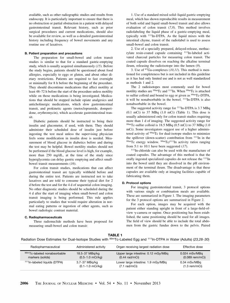
The suggested activity range for 111In-DTPA is 3.7 MBq(0.1 mCi) to 37 MBq (1.0 mCi) (Table 1). This dose isusually administered only for colon transit studies requiringmore than 1 d of imaging. The suggested activity range for99mTc-sulfur colloid is 18.5 MBq (0.5 mCi) to 37 MBq (1.0mCi). Some investigators suggest use of a higher adminis-tered activity of 99mTc for dual-isotope studies to minimizethe spillover (down-scatter) contribution from 111In in the99mTc energy window. 99mTc/111In activity ratios rangingfrom 5:1 to 10:1 have been suggested (17).
111In-chloride can also be used with the manufacture ofcoated capsules. The advantage of this method is that theorally ingested specialized capsules do not release the 111Ininto the bowel until they are dissolved in the pH environ-ment of the terminal ileum. The disadvantage is that thesecapsules are available only at imaging facilities capable offabricating them.
D. Protocol options
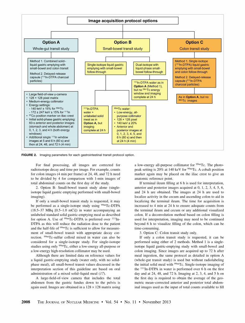
For imaging gastrointestinal transit, 3 protocol optionswith various single or combination meals are available.These are summarized in Figure 1. The imaging parametersfor the 3 protocol options are summarized in Figure 2.
For each option, images may be acquired with thepatient either standing upright in front of a large-field-of-view g-camera or supine. Once positioning has been estab-lished, the same positioning should be used for all images.The field of view should be able to include the total abdo-men from the gastric fundus down to the pelvis. Paired
TABLE 1Radiation Dose Estimates for Dual-Isotope Studies with 99mTc-Labeled Egg and 111In-DTPA in Water (Adults) (23,28–30)
Radiopharmaceutical Administered activity Organ receiving largest radiation dose Effective dose
99mTc-labeled nonabsorbable
markers (solids)
18.5–37 MBq/kg
(0.5–1.0 mCi/kg)
Upper large intestine: 0.12 mGy/MBq
(0.44 rad/mCi)
0.024 mSv/MBq
(0.089 rem/mCi)
111In-labeled liquids (DTPA) 3.7–37 MBq/kg
(0.1–1.0 mCi/kg)
Lower large intestine: 1.9 mGy/MBq
(7.1 rad/mCi)
0.34 mSv/MBq
(1.3 rem/mCi)
2006 THE JOURNAL OF NUCLEAR MEDICINE • Vol. 54 • No. 11 • November 2013

anterior and posterior images are acquired for all imagingtimes to calculate the geometric mean of region counts tocompensate for photon attenuation, which may vary as ac-tivity transits throughout the abdomen.
E. Technical notes on the protocol options
1. Option A: Combined gastric-emptying, small-bowel,and colon transit study, or whole-gut transit (mixed solid–liquid dual-isotope radiolabeled meal).
When small-bowel and colon transit studies are per-formed as part of a combined solid–liquid gastric-emptyingstudy, the solid-phase meal recommended is liquid eggwhite labeled with 99mTc as per the SNMMI PracticeGuideline for Adult Solid-Meal Gastric-Emptying Study.
The complete dual-isotope, labeled test meal consistsof a sandwich with 99mTc-sulfur colloid–labeled egg white(120 g [4 oz]; Egg Beaters [ConAgra Foods] or generic) and300 mL of water containing 3.7–7.4 MBq (0.1–0.2 mCi) of111In-DTPA. The solid meal includes 18.5–37 MBq (0.5–1.0 mCi) of 99mTc-sulfur colloid–labeled egg white, 2 slicesof white or wheat bread, 30 g of strawberry jam, and 120mL of water. The isotope is added to the liquid egg mixture,which then is cooked in a microwave or on a skillet withintermittent stirring during cooking to promote mixing. Theegg is served with the 2 slices of bread, the jam, and thewater. The meal consists of 255 kcal, 24% protein, 2% fat,72% carbohydrate, and 2% fiber.
Images are obtained in a 128 · 128 pixel matrix usinga medium-energy collimator. The photopeak setting for99mTc is 15% at 140 keV. For dual-isotope imaging, boththe 172-keV and the 247-keV peaks for 111In with 15%
windows can be used to maximize counts. If there is signif-icant spillover from the 99mTc activity into the lower-energy172-keV 111In photopeak, a single 247-keV peak for 11In canbe used.
Each imaging site will need to individually evaluatepotential spillover between the 2 energy windows. A cobaltposition marker placed on the iliac crest helps to give ananatomic reference point.
As per the SNMMI Practice Guideline for Adult Solid-Meal Gastric-Emptying Study, initial solid-phase gastric-emptying images of 60 s each are acquired in anterior andposterior projections for 4 h to calculate geometric meanactivity in the stomach and whole abdomen (15). Simulta-neous anterior and posterior whole-abdomen images of theliquid phase using the 111In-DTPA peaks are also acquiredup to 4 h. After completion of the solid gastric-emptyingimages (4 h), additional anterior and posterior single-iso-tope 111In-DTPA images at 5 and 6 h are acquired again for60 s.
After the imaging for gastric-emptying and small-boweltransit, the patients resume their normal diet and activity. Ifmedications are needed, the patients may take them withsmall sips of water during the 6 h required to complete thegastric-emptying and small-bowel studies. The patientsreturn 24, 48, and 72 h after ingesting the meal for a singleset of paired anterior and posterior whole-abdomen imagesof the 111In-DTPA activity to assess colon transit. Imagesare obtained in a 128 · 128 pixel matrix, again using a me-dium-energy collimator for 4 min. Photopeak settings areunchanged. A cobalt position marker placed on the iliac crestagain helps to give an anatomic reference point.
FIGURE 1. Gastrointestinal transit imaging options with various single or combination meals.
GUIDELINE FOR BOWEL TRANSIT • Maurer et al. 2007
For final processing, all images are corrected forradioisotope decay and time per image. For example, countsfor colon images (4 min per frame) at 24, 48, and 72 h needto be divided by 4 for comparison with 1-min images oftotal abdominal counts on the first day of the study.
2. Option B: Small-bowel transit study alone (single-isotope liquid gastric emptying performed with small-bowelimaging).
If only a small-bowel transit study is requested, it maybe performed as a single-isotope study using 99mTc-DTPA(18.5–37 MBq [0.5–1.0 mCi]) in water accompanying anunlabeled standard solid gastric-emptying meal as describedfor option A. Use of 99mTc-DTPA is preferred over 111In-DTPA as this will reduce the radiation dose to the patientand the half-life of 99mTc is sufficient to allow for measure-ment of small-bowel transit with appropriate decay cor-rection. 99mTc-sulfur colloid mixed in water can also beconsidered for a single-isotope study. For single-isotopestudies using only 99mTc, either a low-energy all-purpose ora low-energy high-resolution collimator may be used.
Although there are limited data on reference values fora liquid gastric-emptying study (water only, with no solid-phase meal), all small-bowel transit values discussed in theinterpretation section of this guideline are based on oraladministration of a mixed solid–liquid meal (17).
A large-field-of-view camera that includes the totalabdomen from the gastric fundus down to the pelvis isagain used. Images are obtained in a 128 · 128 matrix using
a low-energy all-purpose collimator for 99mTc. The photo-peak setting is 20% at 140 keV for 99mTc. A cobalt positionmarker again may be placed on the iliac crest to give ananatomic reference point.
If terminal ileum filling at 6 h is used for interpretation,anterior and posterior images acquired at 0, 1, 2, 3, 4, 5, 6,and 24 h are obtained. The images at 24 h are used tolocalize activity in the cecum and ascending colon to aid inlocalizing the terminal ileum. The time for acquisition isincreased to 4 min at 24 h to ensure adequate counts fromthe terminal ileum and cecum or any additional visualizedcolon. If a deconvolution method based on colon filling isused for interpretation, imaging may need to be continuedbeyond 6 h to visualize filling of the colon, which can betime-consuming.
3. Option C: Colon transit study only.If only a colon transit study is requested, it can be
performed using either of 2 methods. Method 1 is a single-isotope liquid gastric-emptying study with small-bowel andcolon imaging. Since images are acquired up to 72 h aftermeal ingestion, the same protocol as detailed in option A(whole-gut transit study) is used but without radiolabelingthe initial solid meal with 99mTc. Single-isotope imaging ofthe 111In-DTPA in water is performed over 6 h on the firstday and at 24, 48, and 72 h. Imaging at 2, 3, 4, and 5 h onthe first day is required to obtain the average of the geo-metric mean–corrected anterior and posterior total abdom-inal images used as the input of total counts available to fill
FIGURE 2. Imaging parameters for each gastrointestinal transit protocol option.
2008 THE JOURNAL OF NUCLEAR MEDICINE • Vol. 54 • No. 11 • November 2013

the colon needed to calculate the colon geometric center(Fig. 3). Analysis and interpretation are the same as forthe combined study, but gastric-emptying and small-bowelanalysis need not be performed.
Method 2 is a colon transit study using the coated-capsulemethod. This method requires preparation of a capsulecoated with a pH-sensitive polymer, methacrylate, which isdesigned to dissolve and release its contents in the terminalileum. This method is therefore limited to sites that canprepare such a capsule. Preparation requires mixing a slurryof 5 mg of activated charcoal with 111InCl3. The mixture isdried at 90�C, and the dried charcoal is placed into a size 1gelatin capsule and coated with pH-sensitive methacrylate(9,18). This measurement of colon transit may be used to-gether with the standard 99Tc-sulfur colloid–labeled eggmeal for simultaneous measurement of gastric emptyingand small-bowel transit of a solid meal as described foroption A.
For a whole-gut transit study using a dual-isotope 99mTc-sulfur colloid–labeled solid meal with the pH-sensitivemethacrylate-coated capsule containing 3.7 MBq (0.1mCi) of 111In-labeled activated charcoal particles, the pro-cedure is conducted as detailed for option A (whole-guttransit study). Images are obtained in a 128 · 128 pixelmatrix using a medium-energy collimator for 4 min (2 minanterior and 2 min posterior). Photopeak settings are un-changed. A position marker placed on the iliac crest pro-vides an anatomic reference point.
In contrast to the method using a dual-isotope mixedradiolabeled meal, where no additional meal is given,a lunch meal (550 kcal, chicken sandwich with potato andpudding) is consumed at 4 h after the start of the study (19).
F. Interpretation
Interpretation of small-bowel and colon transit studiesrequires both analysis of quantitative parameters and visualinspection of the images. The regions of interest (ROIs)should be checked for correct positioning, and visual analysisof tracer progression through the bowel should be confirmedby quantitative analysis.
G. Quantitative Analysis
1. Total abdominal counts analysis.The 111In-DTPA liquid-phase meal
is generally used for small-bowel anal-ysis because emptying of liquids fromthe stomach is more rapid than empty-ing of solids and only rarely is sodelayed as to affect small-bowel transitanalysis. A manually drawn large ROI toinclude the entire abdomen is used toobtain average total abdominal countsbetween 2 and 5 h, when all liquidhas left the stomach and is distributedin the abdomen (Fig. 3). This averageof the total abdominal counts is used todetermine the counts available to fill theterminal ileum at 6 h (for small-boweltransit) and to enter the colon (for geo-metric center analysis). This average isobtained from the decay-corrected geo-metric mean total abdominal counts at
2, 3, 4, and 5 h after meal ingestion (Fig. 4).For dual-isotope studies each imaging lab should de-
termine the percentage of 111In counts contributing to the99mTc counts in the 140-keV window or 99mTc counts con-tributing to the 111In window and perform any needed cor-rections for simultaneously acquired energy windows.
As shown in Figure 3, a large, rectangular ROI is used todetermine total abdominal 111In counts. At 5 h (300 min)after meal ingestion, there is clear accumulation of 111Inactivity in the terminal ileum reservoir. An ROI to deter-mine the percentage filling of the terminal ileum at 6 h ismanually drawn (dotted circle and arrow in the figure). Anyactivity that has passed through the terminal ileum and into
FIGURE 3. Total abdominal and terminal ileum ROIs.
FIGURE 4. Total abdominal geometric mean counts.
GUIDELINE FOR BOWEL TRANSIT • Maurer et al. 2009
the cecum or colon at 6 h should be included in the ROI asthis activity has passed through the small bowel.
There is typically some variation in total abdominalcounts over the course of the study, but usually the countvariation is less than 10%. The average of the decay-corrected geometric mean 111In counts between 2 and 6 h isused to quantify the total abdominal counts available toenter the terminal ileum and colon.
2. Small-bowel transit analysis.It is well recognized that small-bowel motility is complex,
with contraction patterns that differ between the proximalsmall bowel and distal small bowel. Although these patternsare complex, methods based on terminal ileum or colonfilling have been proposed that provide simplified quantita-tive indices of overall small-bowel transit (12). To minimizethe effect of variable output from the stomach, which canaffect terminal ileum- and colon-filling methods, more com-plex methods that incorporate deconvoluting gastric empty-ing and colon filling have been described (1,20). These meth-ods are less practical than simple measurement of terminalileum or colon filling. Because of the need to measure colonfilling, the latter requires significantly increased imaging timeto ensure adequate colon filling.
3. Terminal ileum filling method.Because of the stasis that occurs in the terminal ileum
reservoir, a simple measurement of the total amount of 111Inactivity accumulated in the terminal ileum or transited intothe cecum and ascending colon at 6 h is used as an index ofsmall-bowel transit. Studies have shown that the most rapidtransit is in the proximal small bowel, with slowing occur-ring in the distal ileum. The terminal ileum serves as a res-ervoir and fills with activity before it crosses the ileocecalvalve and enters the colon (21,22). Visually, the 111In-la-beled liquid meal activity is identified in the reservoir areaand terminal ileum, and a manual ROI is drawn to encom-pass all activity in the terminal ileum (Fig. 3). If activity hasprogressed into the cecum or colon, that activity is consid-ered to have transited through the small bowel and a largerROI to include the terminal ileum and the cecum/colon isdrawn to measure all the activity that has passed through thesmall bowel.
Imaging is continued up to 360 min on the first day toquantify the 111In activity that transits into the terminalileum reservoir or passes into the cecum or ascending colon.The patient returns 24 h after the beginning of the study(usually the next morning) for one anteroposterior image setof the abdomen to help with visual identification of thelocations of the colon and terminal ileum. Anterior andposterior manual ROIs are drawn to include the 111In activ-ity in the terminal ileum or any activity that has passed intothe colon at 6 h. The geometric mean decay-corrected per-centage of activity that has passed into the terminal ileumreservoir (or into the colon) is calculated as follows:
% activity transited through small bowel 5
total counts in terminal ileum1 colon�
average total abdominal counts� ð225 h after meal ingestionÞ
*Geometric mean and decay-corrected counts.
If more than 40% of the total abdominal 111In countsadministered has moved through the small bowel into theterminal ileum or colon, small-bowel transit is normal (5).
Visual interpretation should also accompany the quan-titative analysis. Delayed small-bowel transit is typicallyseen as residual activity in multiple loops of small bowelwithout clear activity having progressed into the terminalileum or colon. The 24-h image (second day) is used tovisualize the colon in questionable cases for which theterminal ileum is not clearly identified in earlier images. Inmost cases of delayed small-bowel transit, activity is seen topersist in loops of small bowel, with no clear arrival in theterminal ileum or colon at 6 h. Rapid small-bowel transitcan visually be detected by identifying early cecal filling($10% of administered activity in the cecum) in less than70 min, with a reference range of between 72 and 392 minfor cecal arrival in the dual-isotope meal method (23).
4. Colon-filling method.The amount of colon filling at 6 h has also been used as
an index of small-bowel transit. The range for normal fillingof the colon at 6 h using nondigestible particles is 11%–70%. The range for digestible solids is 43%–95%, withrapid small-bowel transit defined as cecal arrival at less than90 min (12).
5. Deconvolution method incorporating gastric emptyingand colon filling.
Deconvolution was first proposed by Malagelada in 1984(24) and was modified by Brinch et al. in 1999 (20). Thesemethods result in an expected small-intestine time–activitycurve if gastric emptying were instantaneous. The curve canbe used to calculate a mean transit time for the bulk ofradiolabeled material. This protocol, however, requires fre-quent (every 30 min) imaging and lengthy acquisitions untilall tracer has passed into the colon. A simplification to thismethod was proposed by Read et al. in 1986 and is based onsubtraction of 50% gastric-emptying time from the time to50% colon filling (22).
H. Small-bowel transit time
Normal small-bowel transit time can vary depending onthe methods used. Using resin pellets mixed with a meal,small-bowel transit time in healthy individuals reportedlyranged from 151 to 290 min (18). Using the liquid phase ofa mixed solid–liquid meal, small-bowel transit time rangedfrom 72 to 392 min in healthy individuals (23). In somecases, there is prolonged stasis of activity in the terminalileal reservoir, particularly if no second meal is adminis-tered. This may result in the need for imaging beyond 6hours—which is also impractical—to see progression intothe cecum or ascending colon.
I. Interpretation of colon transit scintigraphy
Various approaches to recording colon transit have beenreported. Some advocate imaging at 24, 48, 72, and 96 h (6).Others suggest imaging only up to 48 h. Healthy individualstypically show complete colon evacuation by 72 h. Imagingup to 72 h is needed particularly to be able to diagnosefunctional rectosigmoid outlet obstruction and to localizeany sites of functional obstruction in the colon (5).
Quantification of colon transit is based on serialmeasurement of the geometric center of a liquid meal as
2010 THE JOURNAL OF NUCLEAR MEDICINE • Vol. 54 • No. 11 • November 2013
it moves through the colon. The geometric center isa weighted average of the radioactivity counted over specificsegments of the colon, that is, the ascending, transverse,descending, and rectosigmoid colon. The geometric centeris calculated as the sum of a weighted fraction representedby the counts in each region multiplied by the regionnumber divided by the total counts.
Two methods have been most widely reported forquantifying geometric-center colon transit scintigraphy(5,25).
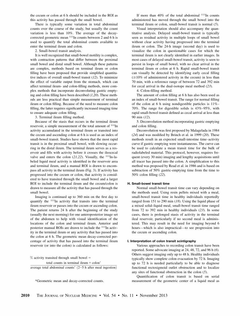
1. Charcoal in delayed-release capsule.This first method designates numbers from 1 to 5 for
counts in the colonic regions, as well as the activity indefecated stool, as weighting factors. This method is basedon use of the special delayed-release capsule that deliversradiolabeled charcoal into the ileocolonic region (Fig. 5).With this method, reference values are a geometric center ofless than 1.4 at 4 h, with a range of 1.6–3.8 at 24 h and 3.0–4.8 at 48 h (12,13). Slow colon transit is defined as a geo-metric center less than these reference values at 24 and 48 h.Table 2 provides the reference values published using thismethod (25).
2. Liquid-phase meal.The second method is based on oral administration of
an 111In-DTPA–radiolabeled liquid meal and is usually
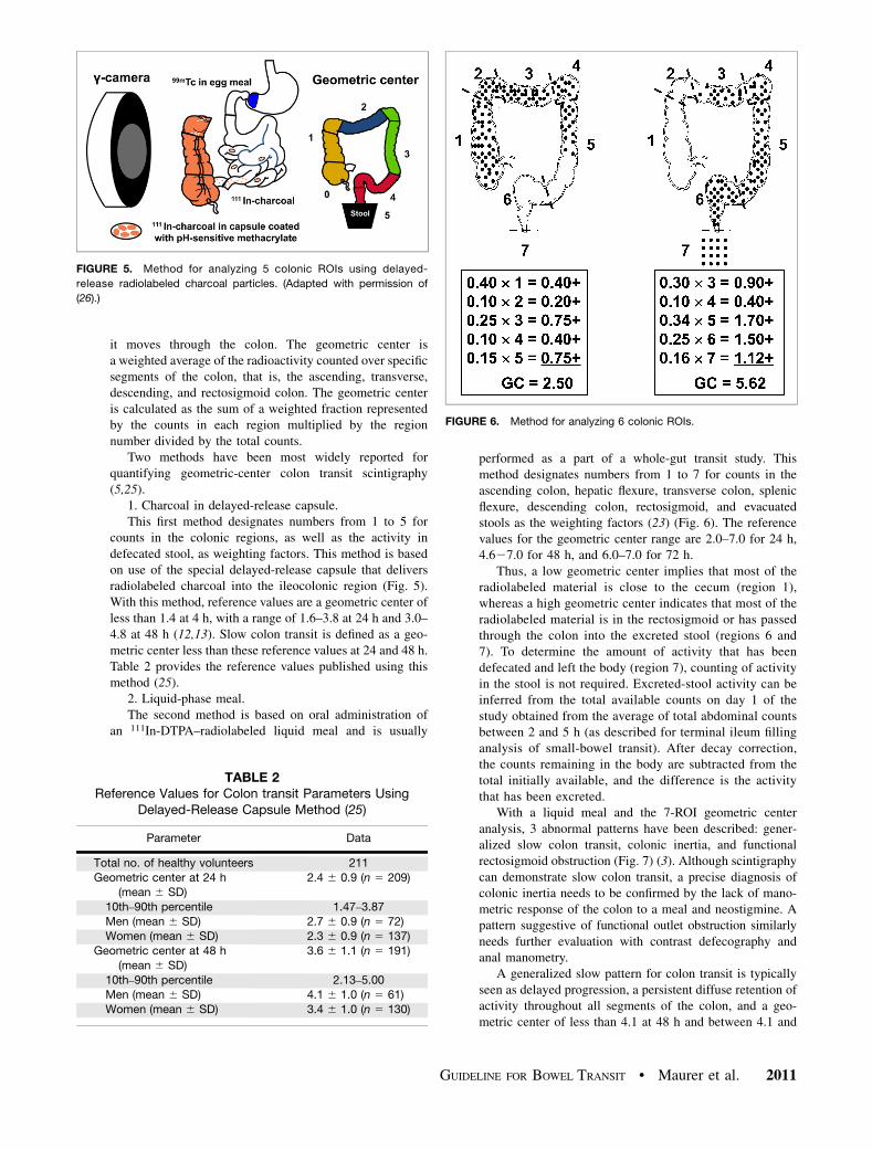
performed as a part of a whole-gut transit study. Thismethod designates numbers from 1 to 7 for counts in theascending colon, hepatic flexure, transverse colon, splenicflexure, descending colon, rectosigmoid, and evacuatedstools as the weighting factors (23) (Fig. 6). The referencevalues for the geometric center range are 2.0–7.0 for 24 h,4.627.0 for 48 h, and 6.0–7.0 for 72 h.
Thus, a low geometric center implies that most of theradiolabeled material is close to the cecum (region 1),whereas a high geometric center indicates that most of theradiolabeled material is in the rectosigmoid or has passedthrough the colon into the excreted stool (regions 6 and7). To determine the amount of activity that has beendefecated and left the body (region 7), counting of activityin the stool is not required. Excreted-stool activity can beinferred from the total available counts on day 1 of thestudy obtained from the average of total abdominal countsbetween 2 and 5 h (as described for terminal ileum fillinganalysis of small-bowel transit). After decay correction,the counts remaining in the body are subtracted from thetotal initially available, and the difference is the activitythat has been excreted.
With a liquid meal and the 7-ROI geometric centeranalysis, 3 abnormal patterns have been described: gener-alized slow colon transit, colonic inertia, and functionalrectosigmoid obstruction (Fig. 7) (3). Although scintigraphycan demonstrate slow colon transit, a precise diagnosis ofcolonic inertia needs to be confirmed by the lack of mano-metric response of the colon to a meal and neostigmine. Apattern suggestive of functional outlet obstruction similarlyneeds further evaluation with contrast defecography andanal manometry.
A generalized slow pattern for colon transit is typicallyseen as delayed progression, a persistent diffuse retention ofactivity throughout all segments of the colon, and a geo-metric center of less than 4.1 at 48 h and between 4.1 and
TABLE 2Reference Values for Colon transit Parameters Using
Delayed-Release Capsule Method (25)
Parameter Data
Total no. of healthy volunteers 211
Geometric center at 24 h(mean 6 SD)
2.4 6 0.9 (n 5 209)
10th–90th percentile 1.47–3.87
Men (mean 6 SD) 2.7 6 0.9 (n 5 72)Women (mean 6 SD) 2.3 6 0.9 (n 5 137)
Geometric center at 48 h
(mean 6 SD)
3.6 6 1.1 (n 5 191)
10th–90th percentile 2.13–5.00Men (mean 6 SD) 4.1 6 1.0 (n 5 61)
Women (mean 6 SD) 3.4 6 1.0 (n 5 130)
FIGURE 6. Method for analyzing 6 colonic ROIs.
FIGURE 5. Method for analyzing 5 colonic ROIs using delayed-
release radiolabeled charcoal particles. (Adapted with permission of
(26).)
GUIDELINE FOR BOWEL TRANSIT • Maurer et al. 2011
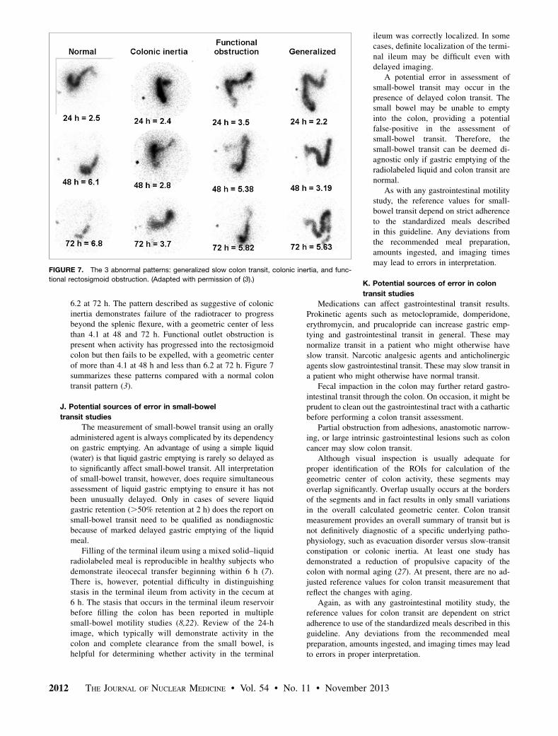
6.2 at 72 h. The pattern described as suggestive of colonicinertia demonstrates failure of the radiotracer to progressbeyond the splenic flexure, with a geometric center of lessthan 4.1 at 48 and 72 h. Functional outlet obstruction ispresent when activity has progressed into the rectosigmoidcolon but then fails to be expelled, with a geometric centerof more than 4.1 at 48 h and less than 6.2 at 72 h. Figure 7summarizes these patterns compared with a normal colontransit pattern (3).
J. Potential sources of error in small-bowel
transit studies
The measurement of small-bowel transit using an orallyadministered agent is always complicated by its dependencyon gastric emptying. An advantage of using a simple liquid(water) is that liquid gastric emptying is rarely so delayed asto significantly affect small-bowel transit. All interpretationof small-bowel transit, however, does require simultaneousassessment of liquid gastric emptying to ensure it has notbeen unusually delayed. Only in cases of severe liquidgastric retention (.50% retention at 2 h) does the report onsmall-bowel transit need to be qualified as nondiagnosticbecause of marked delayed gastric emptying of the liquidmeal.
Filling of the terminal ileum using a mixed solid–liquidradiolabeled meal is reproducible in healthy subjects whodemonstrate ileocecal transfer beginning within 6 h (7).There is, however, potential difficulty in distinguishingstasis in the terminal ileum from activity in the cecum at6 h. The stasis that occurs in the terminal ileum reservoirbefore filling the colon has been reported in multiplesmall-bowel motility studies (8,22). Review of the 24-himage, which typically will demonstrate activity in thecolon and complete clearance from the small bowel, ishelpful for determining whether activity in the terminal
ileum was correctly localized. In somecases, definite localization of the termi-nal ileum may be difficult even withdelayed imaging.
A potential error in assessment ofsmall-bowel transit may occur in thepresence of delayed colon transit. Thesmall bowel may be unable to emptyinto the colon, providing a potentialfalse-positive in the assessment ofsmall-bowel transit. Therefore, thesmall-bowel transit can be deemed di-agnostic only if gastric emptying of theradiolabeled liquid and colon transit arenormal.
As with any gastrointestinal motilitystudy, the reference values for small-bowel transit depend on strict adherenceto the standardized meals describedin this guideline. Any deviations fromthe recommended meal preparation,amounts ingested, and imaging timesmay lead to errors in interpretation.
K. Potential sources of error in colon
transit studies
Medications can affect gastrointestinal transit results.Prokinetic agents such as metoclopramide, domperidone,erythromycin, and prucalopride can increase gastric emp-tying and gastrointestinal transit in general. These maynormalize transit in a patient who might otherwise haveslow transit. Narcotic analgesic agents and anticholinergicagents slow gastrointestinal transit. These may slow transit ina patient who might otherwise have normal transit.
Fecal impaction in the colon may further retard gastro-intestinal transit through the colon. On occasion, it might beprudent to clean out the gastrointestinal tract with a catharticbefore performing a colon transit assessment.
Partial obstruction from adhesions, anastomotic narrow-ing, or large intrinsic gastrointestinal lesions such as coloncancer may slow colon transit.
Although visual inspection is usually adequate forproper identification of the ROIs for calculation of thegeometric center of colon activity, these segments mayoverlap significantly. Overlap usually occurs at the bordersof the segments and in fact results in only small variationsin the overall calculated geometric center. Colon transitmeasurement provides an overall summary of transit but isnot definitively diagnostic of a specific underlying patho-physiology, such as evacuation disorder versus slow-transitconstipation or colonic inertia. At least one study hasdemonstrated a reduction of propulsive capacity of thecolon with normal aging (27). At present, there are no ad-justed reference values for colon transit measurement thatreflect the changes with aging.
Again, as with any gastrointestinal motility study, thereference values for colon transit are dependent on strictadherence to use of the standardized meals described in thisguideline. Any deviations from the recommended mealpreparation, amounts ingested, and imaging times may leadto errors in proper interpretation.
FIGURE 7. The 3 abnormal patterns: generalized slow colon transit, colonic inertia, and func-
tional rectosigmoid obstruction. (Adapted with permission of (3).)
2012 THE JOURNAL OF NUCLEAR MEDICINE • Vol. 54 • No. 11 • November 2013
VII. DOCUMENTATION/REPORTING
For the goals of a nuclear medicine report and information ondirect communication, see the SNMMI Guideline for General Imaging.
VIII. EQUIPMENT SPECIFICATIONS
See the SNMMI Guideline for General Imaging.
IX. QUALITY CONTROL AND IMPROVEMENT
See the SNMMI Guideline for General Imaging.
X. SAFETY, INFECTION CONTROL, AND PATIENT
EDUCATION CONCERNS
See the SNMMI Guideline for General Imaging.
XI. RADIATION SAFETY IN IMAGING
See the SNMMI Guideline for General Imaging.
XII. ACKNOWLEDGMENTS
Dr. Italia Paglianiti (Regional Center of Nuclear Medicine,University Hospital of Pisa, Pisa, Italy) helped with defining theflowcharts to illustrate the technical and imaging options A, B, andC included in this guideline.
XIII. REFERENCES
1. Madsen JL, Larsen N, Hilsted J, Worning H. Scintigraphic determination of
gastrointestinal transit times: a comparison with breath hydrogen and radiologic
methods. Scand J Gastroenterol. 1991;26:1263–1271.
2. Southwell BR, Clarke M, Sutcliffe J, Hutson J. Colonic transit studies: normal
values for adults and children with comparison of radiological and scintigraphic
methods. Pediatr Surg Int. 2009;25:559–572.
3. Lin HC, Prather C, Fisher R, et al. Measurement of gastrointestinal transit: AMS
task force committee on gastrointestinal transit. Dig Dis Sci. 2005;50:989–1004.
4. Rao SS, Camilleri M, Hasler W, et al. Evaluation of gastrointestinal transit in
clinical practice: position paper of the American and European neurogastroen-
terology and motility societies. Neurogastroenterol Motil. 2011;23:8–23.
5. Bonapace ES, Maurer AH, Davidoff S, Krevsky B, Fisher RS, Parkman HP.
Whole gut transit scintigraphy in the clinical evaluation of patients with upper
and lower gastrointestinal symptoms. Am J Gastroenterol. 2000;95:2838–2847.
6. Mclean RG, King D, Talley N, Tait A, Freiman J. The utilization of colon transit
scintigraphy in the diagnostic algorithm for patients with chronic constipation.
Dig Dis Sci. 1999;44:41–47.
7. Bennink R, Peeters M, Van den Maegdenbergh V, et al. Evaluation of small-
bowel transit for solid and liquid test meal in healthy men and women. Eur J
Nucl Med. 1999;26:1560–1566.
8. Krevsky B, Maurer A, Niewiarowski T, Cohen S. Effect of verapamil on human
intestinal transit. Dig Dis Sci. 1992;37:919–924.
9. Burton DD, Camilleri M, Mullan B, Forstrom L, Hung J. Colonic transit scin-
tigraphy labeled activated charcoal compared with ion exchange pellets. J Nucl
Med. 1997;38:1807–1810.
10. Bartholomeusz D, Chatterton B, Bellen J, Gaffney R, Hunter A. Segmental co-
lonic transit after oral 67Ga-citrate in healthy subjects and those with chronic
idiopathic constipation. J Nucl Med. 1999;40:277–282.
11. Bellen JC, Chatterton B, Penglis S, Tsopelas C. Gallium-67 complexes as radio-
active markers to assess gastric and colonic transit. J Nucl Med. 1995;36:
513–517.
12. Szarka LA, Camilleri M. Methods for the assessment of small-bowel and colonic
transit. Semin Nucl Med. 2012;42:113–123.
13. Proano M, Camilleri M, Phillips S, Brown M, Thomforde G. Transit of solids
through the human colon: regional quantification in the unprepared bowel. Am J
Physiol. 1990;258:G856–G862.
14. Mariani G, Pauwels EK, AlSharif A, et al. Radionuclide evaluation of the lower
gastrointestinal tract. J Nucl Med. 2008;49:776–787.
15. Donohoe KJ, Maurer A, Ziessman H, Urbain J, Royal H, Martin-Comin J. Pro-
cedure guideline for adult solid-meal gastric-emptying study 3.0. J Nucl Med
Technol. 2009;37:196–200.
16. Schvarcz E, Palmer M, Aman J, Horowitz M, Stridsberg M, Berne C. Physiolo-
gical hyperglycemia slows gastric emptying in normal subjects and patients with
insulin-dependent diabetes mellitus. Gastroenterology. 1997;113:60–66.
17. Ziessman HA, Chander A, Clarke J, Ramos A, Wahl R. The added diagnostic
value of liquid gastric emptying compared with solid emptying alone. J Nucl
Med. 2009;50:726–731.
18. Camilleri M, Zinsmeister A, Greydanus M, et al. Towards a less costly but ac-
curate test of gastric emptying and small bowel transit. Dig Dis Sci. 1991;36:
609–615.
19. Prather CM, Camilleri M, Zinsmeister A, McKinzie S, Thomforde G. Tegaserod
accelerates orocecal transit in patients with constipation-predominant irritable
bowel syndrome. Gastroenterology. 2000;118:463–468.
20. Brinch K, Larsson H, Madsen J. A deconvolution technique for processing small
intestinal transit data. Eur J Nucl Med. 1999;26:272–276.
21. Connel A. Propulsion in the small intestine. Rendic R Gastroenterol. 1970;2:
38–46.
22. Read NW, Al-Janabi M, Holgate A, Barber D, Edwards C. Simultaneous mea-
surement of gastric-emptying, small bowel residence and colonic filling of a solid
meal by the use of the gamma camera. Gut. 1986;27:300–308.
23. Maurer AH, Krevsky B. Whole-gut transit scintigraphy in the evaluation of
small-bowel and colon transit disorders. Semin Nucl Med. 1995;25:326–338.
24. Malagelada J, Robertson J, Brown M, et al. Intestinal transit of solid and liquid
components of a meal in health. Gastroenterology. 1984;87:1255–1264.
25. Nullens S, Nelson T, Camilleri M, et al. Regional colon transit in patients with
dyssynergic defecation or slow transit in patients with constipation. Gut. 2012;
61:1132–1139.
26. Vazquez-Roque MI, Camilleri M, Carlson P, et al. HLA-DQ genotype is
associated with accelerated small bowel transit in patients with diarrhea-
predominant irritable bowel syndrome. Eur J Gastroenterol Hepatol. 2011;
23:481–487.
27. Madsen JL, Graff J. Effects of ageing on gastrointestinal motor function. Age
Ageing. 2004;33:154–159.
28. International Commission on Radiological Protection. ICRP Publication 80:
Radiation Dose to Patients from Radiopharmaceuticals. St. Louis, MO: Elsevier;
1999.
29. Siegel JA, Wu RK, Knight LC, et al: Radiation dose estimates for oral agents
used in upper gastrointestinal disease. J Nucl Med. 1983;24:835–837.
30. Stabin MG, Sparks RB, Crowe E. OLINDA/EXM: the second-generation per-
sonal computer software for internal dose assessment in nuclear medicine. J Nucl
Med. 2005;46:1023–1027.
XIV. APPROVAL
This practice guideline was approved by the Board of Direc-
tors of the SNMMI on September 29, 2013, and by the EANM
Executive Committee on August 28, 2013.
GUIDELINE FOR BOWEL TRANSIT • Maurer et al. 2013