Embed Size (px)
Citation preview
These articles have been accepted for publication in the British Journal of Dermatology and are currently being edited and typeset. Readers should note that articles published below have been fully refereed, but have not been through the copy-editing and proof correction process. Wiley-Blackwell and the British Association of Dermatologists cannot be held responsible for errors or consequences arising from the use of information contained in these articles; nor do the views and opinions expressed necessarily reflect those of Wiley-Blackwell or the British Association of Dermatologists Accepted Date : 26-Jul-2010 Article type : Original Article
THE SPECTRUM OF HISTOPATHOLOGICAL FEATURES IN ACUTE
GENERALISED EXANTHEMATOUS PUSTULOSIS: A STUDY OF 102
CASES
S. Halevy1 MD, S. H. Kardaun2 MD, B. Davidovici1 MD,
J. Wechsler3 MD, for the EuroSCAR and RegiSCAR study group 1 Department of Dermatology and Venereology, Soroka Medical Center, Faculty of Health Sciences, Ben-Gurion University of the Negev, Beer-Sheva, Israel. 2 Department of Dermatology, University Medical Center Groningen, University of Groningen, Groningen, the Netherlands. 3 Department of Pathology, Henri-Mondor Hospital, Faculty of Medicine, Paris XII University, Creteil, France Running head: Histopathological features in AGEP Keywords: Acute Generalised Exanthematous Pustulosis; AGEP; Cutaneous Adverse Drug Reaction; Histopathology E-mail addresses: [email protected]; [email protected]; [email protected]; [email protected] Corresponding author: Sylvia H. Kardaun, MD University Medical Center Groningen, dept. Dermatology Hanzeplein 1, 9713 GZ Groningen, The Netherlands. Tel +31 50 3612520 Fax +31 50 3619247 ([email protected]) Previously presented as: 1. Halevy S, Kardaun S, Davidovici B, Wechsler J. Histopathology of the skin in
Acute generalized Exanthematous Pustulosis (AGEP) – A study of 102 cases. 6th Annual European Society for Dermatological Research Meeting, September 7-9, 2006, Paris, France. The Journal of Investigative Dermatology 2006;126, (Suppl. 3):114s (Abstract # 671)
2. Halevy S, Kardaun S, Davidovici B, Wechsler J. Histopathology of the skin in Acute generalized Exanthematous Pustulosis (AGEP) – A study of 102 Cases. 32th Annual Meeting of the Israel Society of Dermatology and Venereology, June 18-19, 2008, Tel-Aviv, Israel. (Abstract Book)
Funding/Support: The EuroSCAR study was funded by the following institutions/companies: ADIR & Cie, Bayer Pharma / AG / Vital, Boehringer Ingelheim, Cassenne, Ciba Geigy / Novartis, Cilag GmbH, Dr. Willmar Schwabe, Goedecke Parke Davis, Glaxo Wellcome / GlaxoSmithKline, Hoechst AG / Hoechst Marion Roussel / Aventis, Hoffmann-La-Roche, IRIS Servier, Jouveinal Lab., LEO, LILLY, MSD Sharp & Dohme, Pfizer, Rhone Poulenc Rorer, Sanofi Winthrop / Sanofi Synthelabo GmbH , Schering AG. Funding from Pharmaceutical Companies in France was managed through contract with INSERM (Institut National de la Santé et de la Recherche Médicale), French Ministry of Health (PHRC AOM 98027). The German Registry (the basis for EuroSCAR in Germany) was funded in part by the Federal Institute for Drugs and Medical Devices (BfArM). The RegiSCAR study was funded by grants from the European Commission (QLRT-2002-01738), GIS-Institut des Maladies Rares and INSERM (4CH09G) in France, DFG (FOR 534) in Germany and by a consortium of Pharmaceutical companies (Bayer Vital, Boehringer-Ingelheim, GlaxoSmithKline, MSD Sharp and Dohme, Merck Novartis, Pfizer, Roche, Sanofi-Aventis, Servier). Role of the Sponsors: The sponsors had no role in the design or conduct of the study, in the collection, analysis, and interpretation of data, or in the preparation, review, or approval of the manuscript. Financial Disclosure: “None reported.” Acknowledgment: We are indebted to all SCAR-patients and all the collaborating hospitals and colleagues for their important contribution to this work. The authors also thank the following EuroSCAR and RegiSCAR principal study investigators for their important contributory comments and assistance: Prof. Jean-Claude Roujeau, Department of Dermatology, Henri Mondor Hospital, Paris XII University, Créteil, France; Mrs. Peggy Sekula, Institute of Medical Biometry and Informatics (IMBI), University Medical Center Freiburg, Freiburg, Germany; Mrs. Ariane Dunant, Biostatistics and Epidemiology Unit, Institut Gustave-Roussy, Villejuif and Reference Center for Toxic and Autoimmune Blistering Diseases, Department of Dermatology, Henri-Mondor Hospital, Paris What's already known about this topic?
• Knowledge of the histopathology of AGEP is based primarily on case reports and a few clinical studies.
What does this study add?
• The present study includes a large collection of validated AGEP cases that were collected in a systematized manner within two international studies. The histopathological evaluation of AGEP was based on a standardised grading system. The paper shows the prevalence of the various histopathological criteria and provides unique diagnostic clues that support the diagnosis of AGEP.
SUMMARY
Background: Acute Generalised Exanthematous Pustulosis (AGEP) is a rare severe pustular
reaction pattern with a typical clinical picture.
Objectives: To characterise the histopathological features of AGEP in a large series of cases
with a validated diagnosis.
Patients and methods: A multi-national retrospective histopathological study was
conducted. It included 102 hospitalised patients (recruited within the EuroSCAR and
RegiSCAR studies) with a validated diagnosis of probable or definite AGEP. A systematic
description of the histopathologic features in AGEP was done based on a standardised
grading system.
Results: Sub/intracorneal pustules (41%), intraepidermal pustules (20%) or combinations of
them (38%) were observed in 102 cases. The pustules were usually large (>15 keratinocytes)
(82% and 89%, respectively) and regularly contained eosinophils (36% and 32%,
respectively). Spongiform features were less prominent in the sub/intracorneal pustules
compared to the intraepidermal pustules (44% and 95%, respectively). The main epidermal
features were necrotic keratinocytes (67%), including incidental segmental necrosis (7%),
and spongiosis (80%) with neutrophil exocytosis (77%). The main dermal features were
papillary oedema (88%) and mixed superficial (100%), interstitial (93%), and mid/deep-
dermal infiltrates (95%) containing neutrophils (100%) and eosinophils (81%). Follicular
pustules were also seen (23%), but vasculitis generally was absent. Classical features of
plaque-type psoriasis were infrequent and usually mild. No significant differences were
observed between a sub-group of 16 cases with and 86 cases without psoriasis.
Conclusions: The present histopathologic study concerns a large series of cases with a
validated diagnosis of AGEP. It provides diagnostic clues in favour of AGEP in patients with
a pustular eruption.
INTRODUCTION
Acute generalised exanthematous pustulosis (AGEP) is a rare, severe, acute-onset pustular
reaction pattern characterised by a typical clinical picture and course. AGEP is attributed
mostly to drugs, although other etiologies such as viral infections due to human parvovirus
B19, cytomegalovirus, and Coxsackie B4, hypersensitivity to mercury and spider bite have
been implicated.1-12
Clinically, AGEP is characterised by the sudden appearance of dozens of sterile, non-
follicular pinhead sized pustules arising on oedematous erythema with a predilection to the
big folds, or widespread distribution. Mild, non-erosive mucous membrane involvement
(mostly oral) may occur in about 20% of the cases. Other skin symptoms, such as marked
oedema of the face, purpura, “atypical target-like lesions” and blisters have been described
but are not typical for AGEP. The course of AGEP is characterised in most of the cases by
fever (≥ 38°C) and elevated blood neutrophil count (≥ 7000/µ/L). Mild eosinophilia may be
present in about one third of the patients.13 Pustules resolve spontaneously within a few days,
followed typically by post-pustular, pin-point desquamation. The reaction resolves fully in ≤
15 days. Internal organs are generally not involved and the disease has a favourable
prognosis, although secondary infection might pose a danger to patients in poor general
medical condition. The reported mortality is 5%.14
Eruptions similar to AGEP have been described in the literature as toxic pustuloderma or
pustular drug eruption,15-21 or have been interpreted as special variants of other pustular
diseases, such as exanthematic pustular psoriasis (PP), suspected to be triggered by drugs or
infections.22,23
Knowledge of the histopathology of AGEP is based primarily on case reports and a few
clinical studies.1,3,4,13,24-29
The aim of the present study was to characterise the histopathological features in a large
series of cases with a validated diagnosis of AGEP.
PATIENTS AND METHODS
Source of patients
The AGEP patients came from two multi-national studies devoted to Severe Cutaneous
Adverse Reactions (SCAR): The EuroSCAR study, conducted in France, Germany, Italy, the
Netherlands, Austria, Spain and Israel during the years 1997-20012,30 and the RegiSCAR
study, conducted in France, Germany, Italy, the Netherlands, Austria and Israel since
2003.31,32 In both studies AGEP cases were actively detected in a network of hospitals in
Europe and Israel. Potential AGEP cases were patients admitted to hospital due to acute
pustular skin reactions (i.e. community cases) or who developed such reactions during a
hospital stay (i.e. hospital cases). They had dozens of pustules that could not be attributed to
another definitive diagnosis. All patients signed informed consent to participate in those
studies. The study was approved by the Helsinki Committee of each participating center that
recruited patients.
Case validation
An international committee of experts validated the diagnosis of AGEP based on a special
standardised scoring system that was developed in the EuroSCAR study, the AGEP
validation score.2,33 Based on the score, patients were either excluded from the study or
classified as definite, probable or possible cases.
Inclusion of cases
The present study population was comprised of patients with a definite or probable
diagnosis of AGEP and a skin biopsy with slides available for histopathological investigation.
Histopathologic evaluation
The histopathological study was performed on haematoxylin-eosin (HE) stained sections.
All four readers viewed the same slide with a multi-headed microscope and discussed it
together at that time. When several sections were available for a particular patient only the
most informative specimen was chosen, based on the proper representation of the epidermis
and dermis and the presence of an acute inflammatory process, preferentially including a
pustule. When several pustules were present in a section, the largest was evaluated.
Evaluation was based on a standardised list of histopathological parameters used for the
diagnosis of AGEP. A severity scale of the various histopathological parameters, ranging
from 0 to 3 was developed (Table 1), and the degree of severity was determined by
consensus.
Analysis and statistics
Data were analysed using SPSS (version 12, SPSS, Chicago, IL). The frequencies of
different variables in two subgroups (with and without a background of psoriasis) were
compared using the t-test for continuous variables, or the chi-square or Fisher's exact test for
differences in proportions, as appropriate.
RESULTS
The present study included 102 cases with a definite or probable diagnosis of AGEP: 86 of
134 cases from the EuroSCAR study and 16 of 70 cases from phase I of the RegiSCAR study
(cases enrolled in the study until the end of 2004). Seventy cases (68.6%) originated from
France, 22 (21.6%) from Israel (see reference no. 5 for the clinical profile of nine cases), and
10 (9.8%) from the Netherlands. A personal history of psoriasis was recorded in 16 cases
(15.6%) (11 from the EuroSCAR study and five from the RegiSCAR study).
The skin biopsies were taken from a known clinical lesion in only 45 of the 102 cases
(44%): 40 biopsies (39%) were obtained from pustules (sometimes associated with erythema,
oedema, or purpura) and five (5%) from non-pustular clinical lesions, described as erythema
or oedema.
The prevalence rates of the broad range of histopathological parameters seen in the 102
cases are presented in Table 2. Pustules were found in 94 cases (92%) and the location of the
pustules was sub/intracorneal in 41%, intraepidermal in 20%, and combined in 38%. In
addition to subcorneal pustules, subcorneal pustules contiguous with intracorneal pustules,
and intracorneal pustules were also seen. The intraepidermal pustules were located in the
upper part of the epidermis, most often contiguous with subcorneal or sub/intracorneal
pustules. The sub/intracorneal and intraepidermal pustules were usually large (>15
keratinocytes) in 82% and 89%, respectively, and regularly contained eosinophils (36% and
32%, respectively). Spongiform features were less prominent in the sub/intracorneal pustules
compared to the intraepidermal pustules (44% and 95%, respectively). Follicular pustules
were seen in 22 cases (23%). They were accessory, predominant, or alone. The main
epidermal features (Figures 1, 2, 3) were necrotic keratinocytes (67%) including segmental
necrosis (7%), and spongiosis (80%) with neutrophil exocytosis (77%). The main dermal
features were papillary oedema (88%), mixed superficial (100%), interstitial (93%), and
mid/deep-dermal infiltrates (95%) containing neutrophils (100%) and eosinophils (81%).
Erythrocyte extravasation (54%) was also observed, but vasculitis occurred only once (1%).
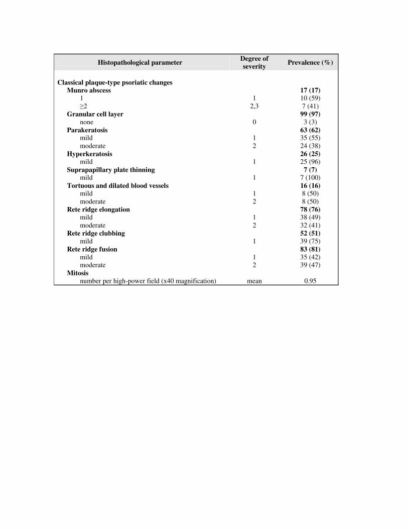
Classical features of plaque-type psoriasis were infrequent and usually mild. These included
the presence of Munro abscesses (17%), parakeratosis (62%), suprapapillary plate thinning
(7%), tortuous and dilated blood vessels (16%), and absence of the granular layer (3%). The
calculated mean mitosis was 0.95/high-power field at magnification x40.
Three levels of prevalence were observed. All AGEP cases showed superficial infiltrates
(mostly moderate) and dermal neutrophils (mostly scattered). Additional features observed in
80-99% of AGEP cases were large sub/intracorneal or intraepidermal pustules, spongiform
intraepidermal pustules, spongiosis (mostly mild), papillary oedema, mid/deep-dermal
infiltrates (mostly discrete), interstitial infiltrates (mostly discrete to moderate), dermal
eosinophils (usually just a few), absence of vasculitis, presence of granular cell layer, rete
ridges fusion (mild to moderate), and absence of classical features of plaque-type psoriasis
(Munro abscesses, suprapapillary plate thinning, tortuous and dilated blood vessels, and a
high mitotic rate). Additional features observed in 50-79% of AGEP cases were necrotic
keratinocytes, parakeratosis, extravasation of erythrocytes, exocytosis of neutrophils (usually
just a few), rete ridge elongation (mild to moderate), and clubbing (mostly mild).
There were no statistically significant differences in the prevalence of histopathological
parameters between a sub-group of 16 AGEP cases (15.6%) with a personal history of
psoriasis and the 86 AGEP cases with no personal history of psoriasis (data not shown).
DISCUSSION
The present study, which focused on the histopathological evaluation of AGEP, included
unique features in design and methodology: a) the study population consisted of patients
recruited in two multi-national studies with a validated diagnosis of probable or definite
AGEP; b) validation of the diagnosis was based on a special standardised scoring system, the
AGEP validation score;2,33 c) it included the largest series of AGEP cases; d) the
histopathological evaluation of AGEP was based on direct investigation of the slides by four
investigators using a multi-headed microscope; and e) evaluation was based on a standardised
grading system related to pustules, epidermis, dermis, and psoriasis-like changes developed
by the authors.
The main histopathological findings, in a previous clinical study of 63 cases of AGEP13
with 64 biopsies from 48 patients reviewed by two investigators, were superficial spongiform
pustules (66%), papillary oedema (61%), polymorphous perivascular infiltrate with
eosinophils (34%), and leukocytoclastic vasculitis with fibrinoid deposits (20%). Focal
necrosis of keratinocytes was observed in 25% and the epidermis was normal or spongiotic
without psoriasiform hyperplasia in 61%.
In comparison, the present study of 102 AGEP cases disclosed several unique
histopathological features: 1) sub/intracorneal pustules and intraepidermal pustules, often
contiguous with sub/intracorneal pustules; 2) the pustules showed a higher prevalence of
spongiform features (95% of intraepidermal pustules); 3) a higher prevalence of necrotic
keratinocytes (67%), papillary oedema (88%), and dermal eosinophils (81%); (4) a marked
prevalence of interstitial and mid/deep-dermal infiltrates (93% and 95%, respectively) and of
dermal neutrophils (100%), not emphasised previously; 5) psoriasiform hyperplasia (rete
ridge elongation, clubbing, and fusion at rates of 76%, 51%, 81%, respectively) was usually
mild, although more common than previously reported. Munro abscesses, which are generally
associated with psoriasis, were observed in 17% of the cases; 6) on the other hand, vasculitis,
which was strictly defined by the presence of vascular fibrinoid alteration and
leucocytoclasia, was observed in the present study only once, indicating that vasculitis is not
a diagnostic feature of AGEP. Since erythrocyte extravasation occurred in 54% of the cases,
the previously reported high rate of vasculitis in AGEP might be attributed to
misinterpretation of leukocytoclasia and/or erythrocyte extravasation as vasculitis, or to a
diagnostic confusion of AGEP with pustular vasculitis.34; 7) histologically, follicular pustules
were found in 23% of the cases. Although the distribution of the pustular eruption in AGEP is
mostly non-follicular,13 the occurrence of follicular pustules in association with non-follicular
pustules has been reported.3,5 Thus, the presence of follicular pustules would appear not to
exclude the diagnosis of AGEP.
Differences between the histopathological features of AGEP reported in various case
reports and clinical studies might be attributed to case definition or different stages in the
evolution of the skin lesions analysed. It was shown in a study of 21 AGEP cases26 that the
histopathological features vary in relation to the age of the skin lesion. Thus, biopsies of early
lesions showed marked to moderate papillary dermal oedema and a mixed dermal
inflammatory infiltrate, often with erythrocyte extravasation, and some leukocytoclasia.
Biopsies of well-developed lesions showed spongiform pustules within the epidermis and
occasional dyskeratotic cells with residual perivascular dermal oedema. Although no
definitive vasculitis was seen, leukocytoclasia was observed within the dermal infiltrate in the
majority of biopsy specimens obtained more than 48 hours after the onset of the eruption.
A wide spectrum of pustular reactions can easily be differentiated from AGEP both
clinically and histologically (e.g. bacterial folliculitis, acne, dermatophyte infections,
impetigo, infantile chronic acropustulosis, Sweet’s syndrome, IgA pemphigus, necrolytic
migratory erythema, bowel bypass syndrome, Behçet’s disease, and staphylococcal scalded
skin syndrome). However, the differential diagnosis between AGEP and generalised PP,
especially the acute von Zumbusch type, may be difficult clinically and histologically..
Various histological features in PP bear similarity to AGEP, including superficial spongiform
pustules, neutrophils beneath the stratum corneum, acanthosis, and papillary oedema. On the
other hand, characteristic for PP is the spongiform macropustule, arising from neutrophils
that migrate from the dermal papillary capillaries into the epidermis, while dermal infiltrates
are superficial and lymphocytic, usually lacking eosinophils. In addition, classical epidermal
changes of psoriasis vulgaris vary and may be rather prominent in PP.35,36
Several histopathological features that were observed in the present study, may point to
the diagnosis of AGEP. These include superficial spongiform pustules, spongiosis, exocytosis
of neutrophils, necrotic keratinocytes, papillary oedema, mixed dermal infiltrates, including
mid/deep-dermal and interstitial infiltrates, containing neutrophils and eosinophils, and the
paucity of classical plaque-type psoriatic changes (i.e., Munro abscesses, absence of granular
layer, suprapapillary plate thinning, tortuous and dilated blood vessels). The diagnosis of
AGEP may be based on these key histopathological features combined with clinical signs in
favour of AGEP including an abrupt onset, a short duration (≤ 15 days), association with
recently introduced drugs, spontaneous resolution after withdrawal of the culprit drugs, and a
non-recurrent tendency.2,6,13
It has been reported that AGEP may occur in patients with psoriasis.2,13 Accordingly,
AGEP has been alleged to be a variant of PP, that could be triggered by a variety of
exogenous triggers such as drugs or infections.13,22-25,37 In the present study a personal history
of psoriasis was recorded in 16 (15.6%) of the 102 AGEP cases. No significant differences
were observed between the sub-group of 16 AGEP cases with a personal history of psoriasis
and the other 86 AGEP cases. Nevertheless, our study does not support the assumption that
any acute pustular eruption, occurring in patients with a psoriatic background, is necessarily
PP.
Several of the prevalent key features in favour of AGEP may imply its aetiopathogenesis:
a) The prominent presence of eosinophils in the skin of AGEP patients, both within the
pustules and in the dermis, is in agreement with the presence of blood eosinophilia
observed in about a third of AGEP patients.13 The presence of tissue and blood
eosinophilia, which is a hallmark of many drug-induced allergic reactions, suggests
that AGEP is a hypersensitivity reaction, probably drug-induced.38,39 Eosinophilia
observed in AGEP may be attributed to the rare presence of IL8/CXCL8 producing
T-cell clones, which display a Th2-type cytokine profile with high IL-4 and IL-5
secretion.40-42
b) The presence of necrotic keratinocytes in AGEP, has been reported in other drug
eruptions including exanthematic drug eruptions and drug eruptions characterised
primarily by interface dermatitis such as lichenoid drug eruptions, Stevens Johnson
syndrome (SJS), toxic epidermal necrolysis (TEN), and fixed drug eruptions.43
Although SJS or TEN are drug-induced reactions manifested by full thickness
epidermal necrosis and only a very sparse inflammatory infiltrate, some similarity
may exist between AGEP and SJS or TEN.27,44 The necrotic keratinocytes observed
in AGEP can be induced by cytotoxic drug-specific T-cells (CD8+ or CD4+).45
c) The neutrophilic inflammation observed in AGEP is unusual in allergic drug
reactions. The prominent presence of dermal neutrophils in AGEP may reflect their
recruitment by the potent neutrophil-attracting chemokine IL-8/CXCL8, secreted by
drug-specific T-cells (CD4+ and CD8+) and keratinocytes. Factors produced by the
IL8/CXCL8-producing T-cells reduce neutrophil apoptosis, thus enhancing
neutrophil survival and leading to the sterile pustular eruption found in AGEP.39-42
d) The mid/deep-dermal perivascular infiltrates and extravasation of erythrocytes,
which were observed in AGEP have been reported in other drug-induced eruptions,
even in the absence of vasculitis, and may point to a drug etiology.38,46
In conclusion, the present study, conducted in a large series of AGEP patients with a
validated diagnosis, disclosed a spectrum of histopathological features that provides
additional support for the concept that AGEP is a separate entity that can occur as an acute
episode, even in patients with psoriasis.
REFERENCES
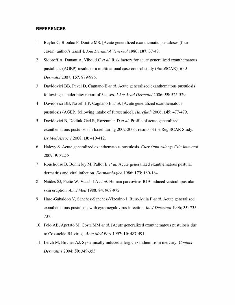
1 Beylot C, Bioulac P, Doutre MS. [Acute generalized exanthematic pustuloses (four
cases) (author's transl)]. Ann Dermatol Venereol 1980; 107: 37-48.
2 Sidoroff A, Dunant A, Viboud C et al. Risk factors for acute generalized exanthematous
pustulosis (AGEP)-results of a multinational case-control study (EuroSCAR). Br J
Dermatol 2007; 157: 989-996.
3 Davidovici BB, Pavel D, Cagnano E et al. Acute generalized exanthematous pustulosis
following a spider bite: report of 3 cases. J Am Acad Dermatol 2006; 55: 525-529.
4 Davidovici BB, Naveh HP, Cagnano E et al. [Acute generalized exanthematous
pustulosis (AGEP) following intake of furosemide]. Harefuah 2006; 145: 477-479.
5 Davidovici B, Dodiuk-Gad R, Rozenman D et al. Profile of acute generalized
exanthematous pustulosis in Israel during 2002-2005: results of the RegiSCAR Study.
Isr Med Assoc J 2008; 10: 410-412.
6 Halevy S. Acute generalized exanthematous pustulosis. Curr Opin Allergy Clin Immunol
2009; 9: 322-8.
7 Rouchouse B, Bonnefoy M, Pallot B et al. Acute generalized exanthematous pustular
dermatitis and viral infection. Dermatologica 1986; 173: 180-184.
8 Naides SJ, Piette W, Veach LA et al. Human parvovirus B19-induced vesiculopustular
skin eruption. Am J Med 1988; 84: 968-972.
9 Haro-Gabaldon V, Sanchez-Sanchez-Vizcaino J, Ruiz-Avila P et al. Acute generalized
exanthematous pustulosis with cytomegalovirus infection. Int J Dermatol 1996; 35: 735-
737.
10 Feio AB, Apetato M, Costa MM et al. [Acute generalized exanthematous pustulosis due
to Coxsackie B4 virus]. Acta Med Port 1997; 10: 487-491.
11 Lerch M, Bircher AJ. Systemically induced allergic exanthem from mercury. Contact
Dermatitis 2004; 50: 349-353.
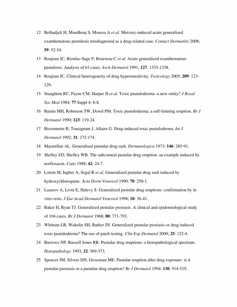
12 Belhadjali H, Mandhouj S, Moussa A et al. Mercury-induced acute generalized
exanthematous pustulosis misdiagnosed as a drug-related case. Contact Dermatitis 2008;
59: 52-54.
13 Roujeau JC, Bioulac-Sage P, Bourseau C et al. Acute generalized exanthematous
pustulosis. Analysis of 63 cases. Arch Dermatol 1991; 127: 1333-1338.
14 Roujeau JC. Clinical heterogeneity of drug hypersensitivity. Toxicology 2005; 209: 123-
129.
15 Staughton RC, Payne CM, Harper JI et al. Toxic pustuloderma--a new entity? J Royal
Soc Med 1984; 77 Suppl 4: 6-8.
16 Rustin MH, Robinson TW, Dowd PM. Toxic pustuloderma: a self-limiting eruption. Br J
Dermatol 1990; 123: 119-24.
17 Bissonnette R, Tousignant J, Allaire G. Drug-induced toxic pustuloderma. Int J
Dermatol 1992; 31: 172-174.
18 Macmillan AL. Generalised pustular drug rash. Dermatologica 1973; 146: 285-91.
19 Shelley ED, Shelley WB. The subcorneal pustular drug eruption: an example induced by
norfloxacin. Cutis 1988; 42: 24-7.
20 Lotem M, Ingber A, Segal R et al. Generalized pustular drug rash induced by
hydroxychloroquine. Acta Derm Venereol 1990; 70: 250-1.
21 Lazarov A, Livni E, Halevy S. Generalized pustular drug eruptions: confirmation by in
vitro tests. J Eur Acad Dermatol Venereol 1998; 10: 36-41.
22 Baker H, Ryan TJ. Generalized pustular psoriasis. A clinical and epidemiological study
of 104 cases. Br J Dermatol 1968; 80: 771-793.
23 Whittam LR, Wakelin SH, Barker JN. Generalized pustular psoriasis or drug-induced
toxic pustuloderma? The use of patch testing. Clin Exp Dermatol 2000; 25: 122-4.
24 Burrows NP, Russell Jones RR. Pustular drug eruptions: a histopathological spectrum.
Histopathology 1993; 22: 569-573.
25 Spencer JM, Silvers DN, Grossman ME. Pustular eruption after drug exposure: is it
pustular psoriasis or a pustular drug eruption? Br J Dermatol 1994; 130: 514-519.
26 Smith K, Norwood C, Skelton H. Do the physical and histologic features and time course
in acute generalized exanthematous pustulosis reflect a pattern of cytokine
dysregulation? J Cutan Med Surg 2003; 7: 7-12.
27 Cohen AD, Cagnano E, Halevy S. Acute generalized exanthematous pustulosis
mimicking toxic epidermal necrolysis. Int J Dermatol 2001; 40: 458-461.
28 Kardaun SH, de Monchy JG. Acute generalized exanthematous pustulosis caused by
morphine, confirmed by positive patch test and lymphocyte transformation test. J Am
Acad Dermatol 2006; 55: S21-3.
29 Paradisi A, Bugatti L, Sisto T et al. Acute generalized exanthematous pustulosis induced
by hydroxychloroquine: three cases and a review of the literature. Clin Ther 2008; 30:
930-940.
30 Mockenhaupt M, Viboud C, Dunant A et al. Stevens-Johnson syndrome and toxic
epidermal necrolysis: Assessment of medication risks with emphasis on recently
marketed drugs. The EuroSCAR-study. J Invest Dermatol 2007: (in press).
31 Kardaun SH, Sidoroff A, Valeyrie-Allanore L et al. Variability in the clinical pattern of
cutaneous side-effects of drugs with systemic symptoms: does a DRESS syndrome really
exist? Br J Dermatol 2007; 156: 609-11.
32 Valeyrie-Allanore L, Poulalhon N, Fagot JP et al. Stevens-Johnson syndrome and toxic
epidermal necrolysis induced by amifostine during head and neck radiotherapy.
Radiother Oncol 2008; 87: 300-3.
33 Sidoroff A, Halevy S, Bavinck JN et al. Acute generalized exanthematous pustulosis
(AGEP)--a clinical reaction pattern. J Cutan Pathol 2001; 28: 113-119.
34 Rockl H. [Leukocytoclastic vasculitis due to drug allergy presenting as generalized
pustular exanthema]. Hautarzt 1981; 32: 467-70.
35 Mobini N, Toussaint S, Kamino H. Noninfectious erythemathous, papular, and squamous
diseases. In: Lever's Histology of the Skin (Elder DE, ed), 9th edn. Philadelphia, PA:
Lippincott Williams & Wilkins. 2005.
36. Ackerman AB, Böer A, Bennin B, Gottlieb GJ. Histologic diagnosis of inflammatory skin
diseases. 3rd ed. New York:Ardor Scribendi;2005.
37 Beylot C, Doutre MS, Beylot-Barry M. Acute generalized exanthematous pustulosis.
Semin Cutan Med Surg 1996; 15: 244-249.
38 Ramdial PK, Naidoo DK. Drug-induced cutaneous pathology. J Clin Pathol 2009; 62:
493-504.
39 Pichler WJ, Yawalkar N, Britschgi M et al. Cellular and molecular pathophysiology of
cutaneous drug reactions. Am J Clin Dermatol 2002; 3: 229-238.
40 Britschgi M, Pichler WJ. Acute generalized exanthematous pustulosis, a clue to
neutrophil-mediated inflammatory processes orchestrated by T cells. Curr Opin Allergy
Clin Immunol 2002; 2: 325-331.
41 Britschgi M, Steiner UC, Schmid S et al. T-cell involvement in drug-induced acute
generalized exanthematous pustulosis. J Clin Invest 2001; 107: 1433-1441.
42 Schaerli P, Britschgi M, Keller M et al. Characterization of human T cells that regulate
neutrophilic skin inflammation. J Immunol 2004; 173: 2151-2158.
43 Horn TD, Hiatt KM. Cutaneous toxicities of drugs. In: Lever's Histology of the Skin
(Elder DE, ed), 9th edn. Philadelphia, PA: Lippincott Williams & Wilkins. 2005.
44 Meiss F, Helmbold P, Meykadeh N et al. Overlap of acute generalized exanthematous
pustulosis and toxic epidermal necrolysis: response to antitumour necrosis factor-alpha
antibody infliximab: report of three cases. J Eur Acad Dermatol Venereol 2007; 21: 717-
719.
45 Schmid S, Kuechler PC, Britschgi M et al. Acute generalized exanthematous pustulosis:
role of cytotoxic T cells in pustule formation. Am J Pathol 2002; 161: 2079-86.
46 Pang BK, Su D, Ratnam KV. Drug-induced purpura simplex: clinical and histological
characteristics. Ann Acad Med Singapore 1993; 22: 870-2.
LEGENDS TO FIGURES
Figure 1. Large non-spongiform subcorneal pustule, papillary oedema, and erythrocyte
extravasation (haematoxylin-eosin stain, original magnification x 200).
Figure 2. Small subcorneal pustule, presence of neutrophils and eosinophils in the epidermis
and in the superficial dermis (haematoxylin-eosin stain, original magnification x
400).
Figure 3. Large spongiform intraepidermal pustule with necrotic keratinocytes and spongiosis
in the lower part of the epidermis. In the dermis there is discrete leucocytoclasia, but
no vasculitis (haematoxylin-eosin stain, original magnification x 200).
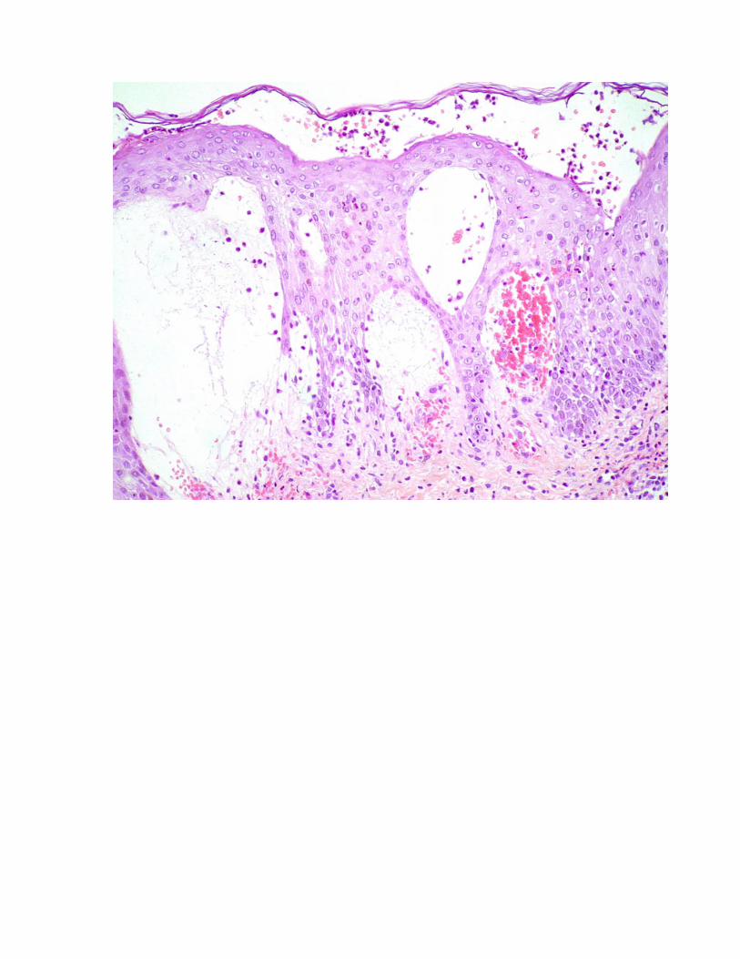
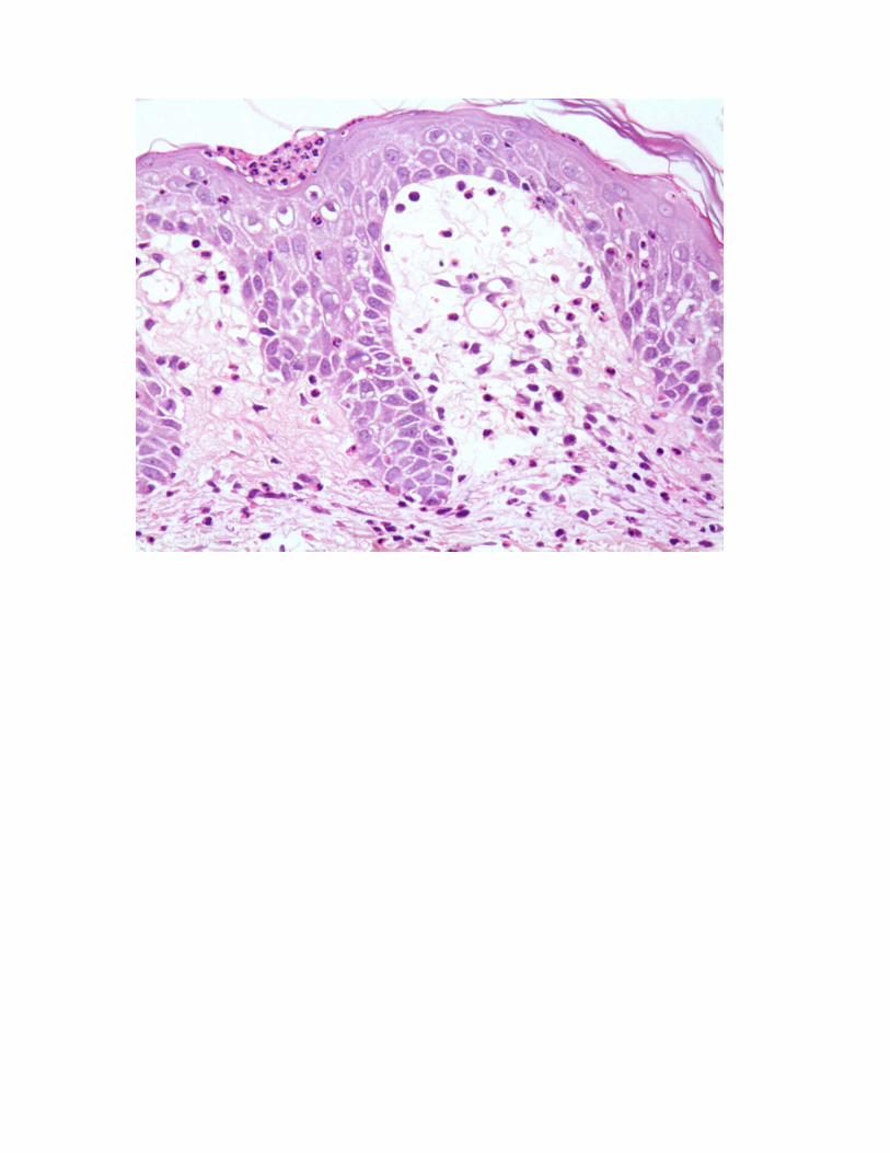
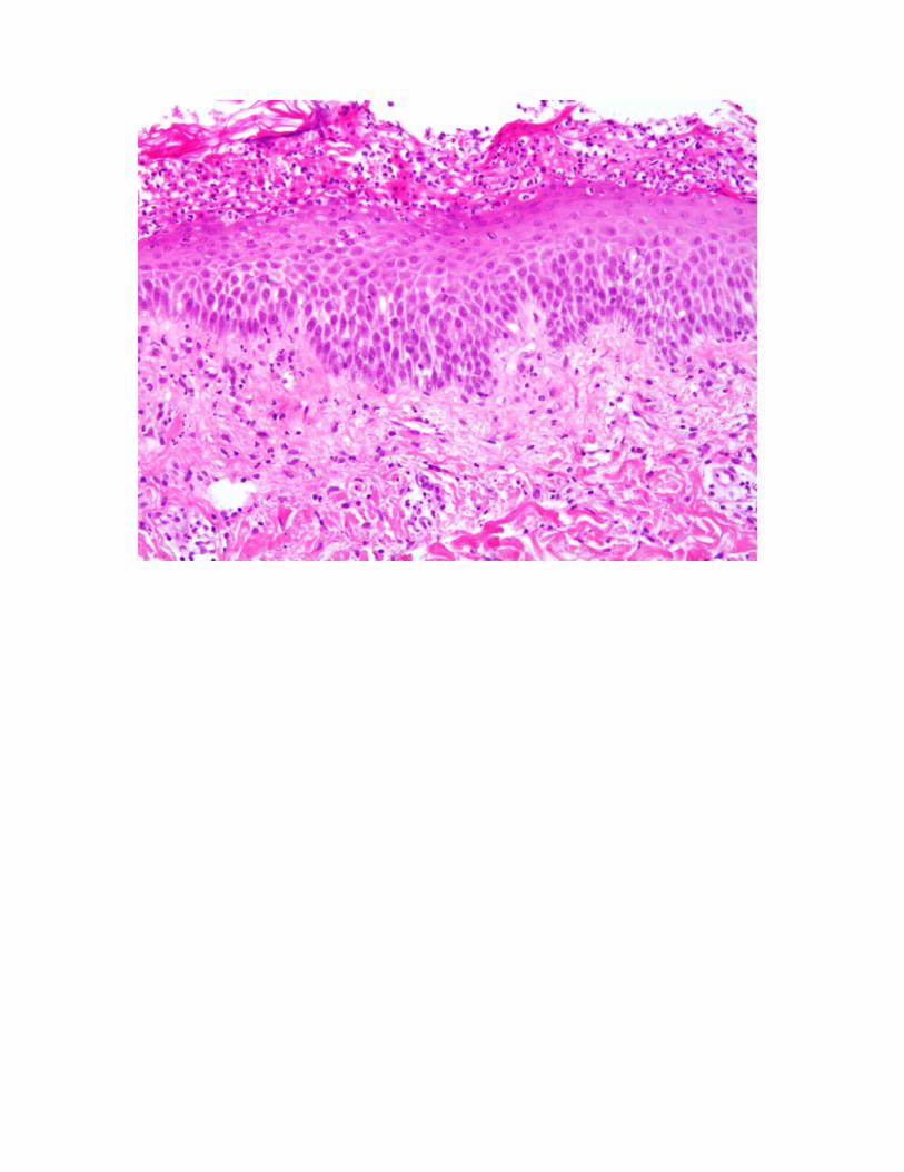
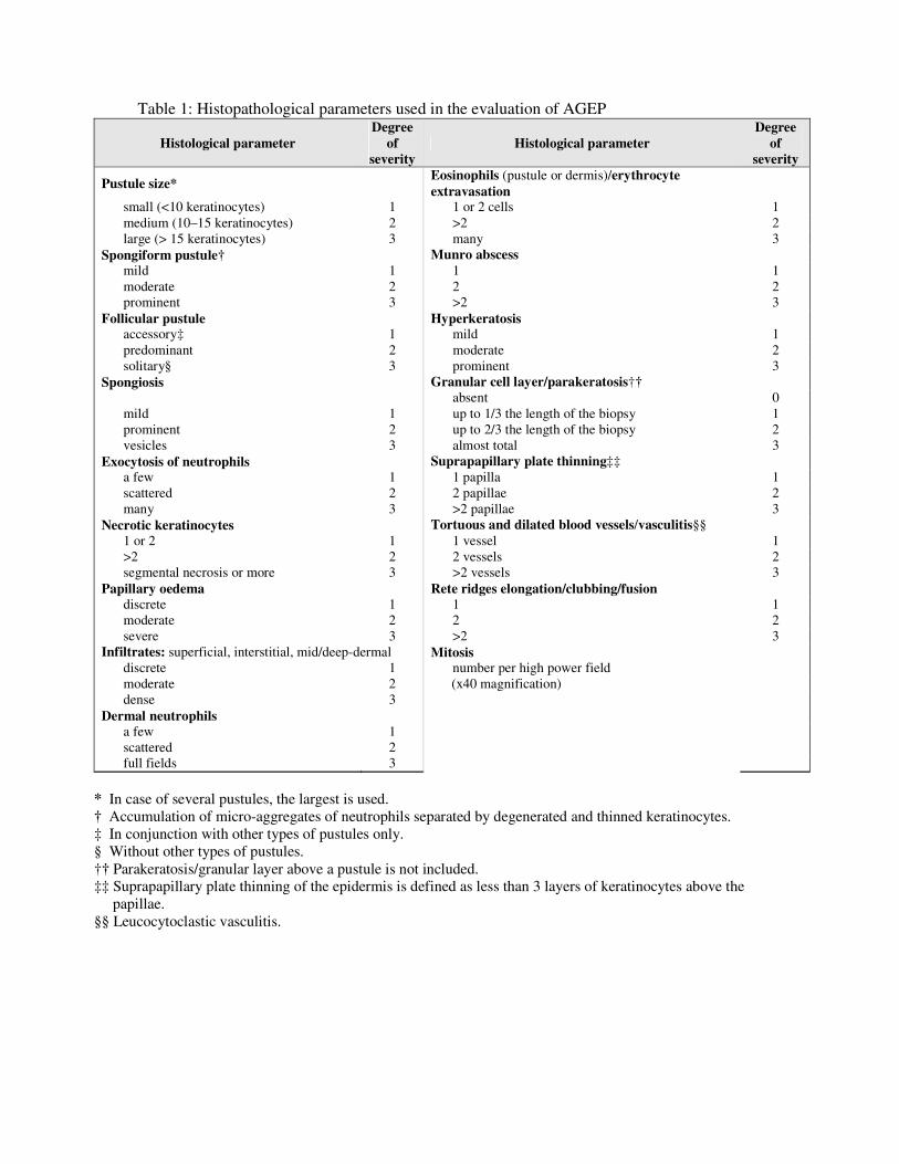
Table 1: Histopathological parameters used in the evaluation of AGEP
Histological parameter Degree
of severity
Histological parameter Degree
of severity
Pustule size* Eosinophils (pustule or dermis)/erythrocyte extravasation
small (<10 keratinocytes) 1 1 or 2 cells 1 medium (10–15 keratinocytes) 2 >2 2 large (> 15 keratinocytes) 3 many 3
Spongiform pustule† Munro abscess mild 1 1 1 moderate 2 2 2 prominent 3 >2 3
Follicular pustule Hyperkeratosis accessory‡ 1 mild 1 predominant 2 moderate 2 solitary§ 3 prominent 3
Spongiosis Granular cell layer/parakeratosis†† absent 0 mild 1 up to 1/3 the length of the biopsy 1 prominent 2 up to 2/3 the length of the biopsy 2 vesicles 3 almost total 3
Exocytosis of neutrophils Suprapapillary plate thinning‡‡ a few 1 1 papilla 1 scattered 2 2 papillae 2 many 3 >2 papillae 3
Necrotic keratinocytes Tortuous and dilated blood vessels/vasculitis§§ 1 or 2 1 1 vessel 1 >2 2 2 vessels 2 segmental necrosis or more 3 >2 vessels 3
Papillary oedema Rete ridges elongation/clubbing/fusion discrete 1 1 1 moderate 2 2 2 severe 3 >2 3
Infiltrates: superficial, interstitial, mid/deep-dermal Mitosis discrete 1 number per high power field moderate 2 (x40 magnification) dense 3
Dermal neutrophils a few 1 scattered 2 full fields 3
* In case of several pustules, the largest is used. † Accumulation of micro-aggregates of neutrophils separated by degenerated and thinned keratinocytes. ‡ In conjunction with other types of pustules only. § Without other types of pustules. †† Parakeratosis/granular layer above a pustule is not included. ‡‡ Suprapapillary plate thinning of the epidermis is defined as less than 3 layers of keratinocytes above the
papillae. §§ Leucocytoclastic vasculitis.
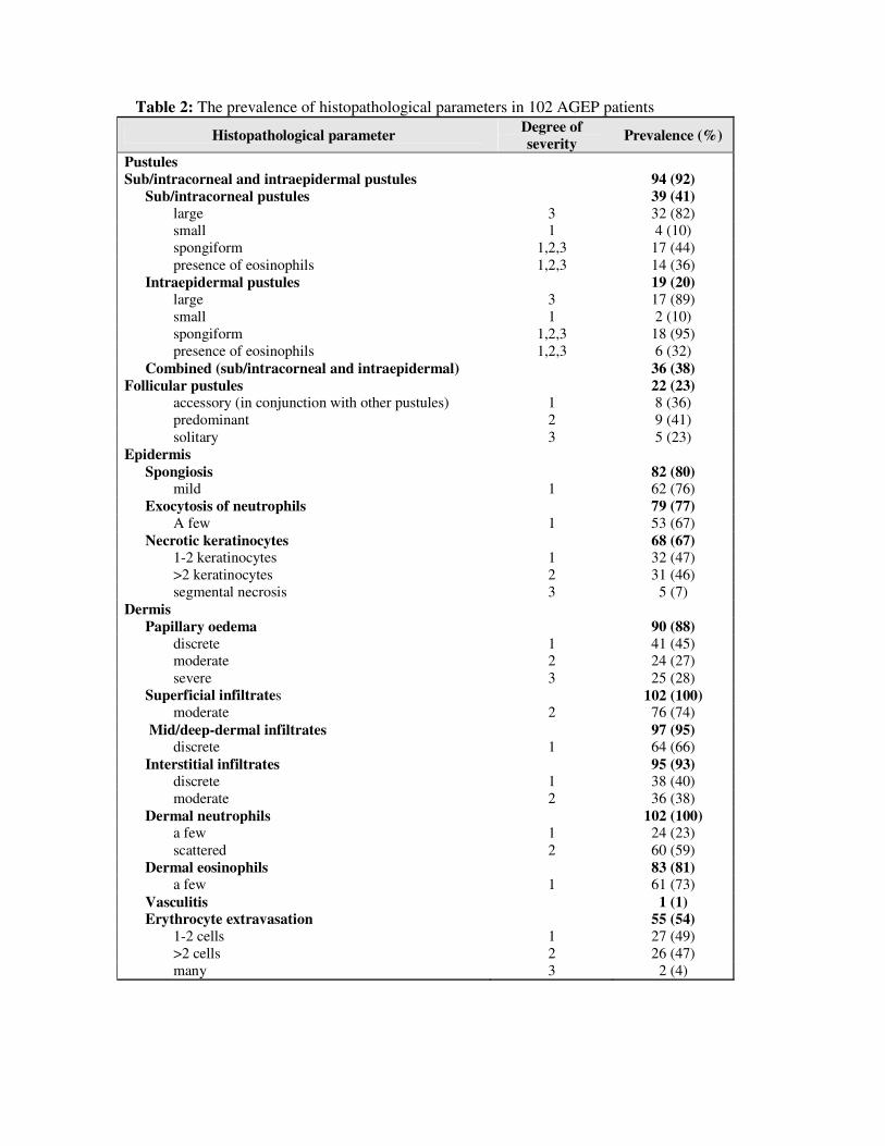
Table 2: The prevalence of histopathological parameters in 102 AGEP patients
Histopathological parameter Degree of severity Prevalence (%)
Pustules Sub/intracorneal and intraepidermal pustules 94 (92)
Sub/intracorneal pustules 39 (41) large 3 32 (82) small 1 4 (10) spongiform 1,2,3 17 (44) presence of eosinophils 1,2,3 14 (36)
Intraepidermal pustules 19 (20) large 3 17 (89) small 1 2 (10) spongiform 1,2,3 18 (95) presence of eosinophils 1,2,3 6 (32)
Combined (sub/intracorneal and intraepidermal) 36 (38) Follicular pustules 22 (23)
accessory (in conjunction with other pustules) 1 8 (36) predominant 2 9 (41) solitary 3 5 (23)
Epidermis Spongiosis 82 (80)
mild 1 62 (76) Exocytosis of neutrophils 79 (77)
A few 1 53 (67) Necrotic keratinocytes 68 (67)
1-2 keratinocytes 1 32 (47) >2 keratinocytes 2 31 (46) segmental necrosis 3 5 (7)
Dermis Papillary oedema 90 (88)
discrete 1 41 (45) moderate 2 24 (27) severe 3 25 (28)
Superficial infiltrates 102 (100) moderate 2 76 (74)
Mid/deep-dermal infiltrates 97 (95) discrete 1 64 (66)
Interstitial infiltrates 95 (93) discrete 1 38 (40) moderate 2 36 (38)
Dermal neutrophils 102 (100) a few 1 24 (23) scattered 2 60 (59)
Dermal eosinophils 83 (81) a few 1 61 (73)
Vasculitis 1 (1) Erythrocyte extravasation 55 (54)
1-2 cells 1 27 (49) >2 cells 2 26 (47) many 3 2 (4)
Histopathological parameter Degree of severity Prevalence (%)
Classical plaque-type psoriatic changes
Munro abscess 17 (17) 1 1 10 (59) ≥2 2,3 7 (41)
Granular cell layer 99 (97) none 0 3 (3)
Parakeratosis 63 (62) mild 1 35 (55) moderate 2 24 (38)
Hyperkeratosis 26 (25) mild 1 25 (96)
Suprapapillary plate thinning 7 (7) mild 1 7 (100)
Tortuous and dilated blood vessels 16 (16) mild 1 8 (50) moderate 2 8 (50)
Rete ridge elongation 78 (76) mild 1 38 (49) moderate 2 32 (41)
Rete ridge clubbing 52 (51) mild 1 39 (75)
Rete ridge fusion 83 (81) mild 1 35 (42) moderate 2 39 (47)
Mitosis number per high-power field (x40 magnification) mean 0.95


![Doru Davidovici - Lumi Galactice [Ibuc.info]](https://img.pdfslide.net/doc/110x75/563db787550346aa9a8be70c/doru-davidovici-lumi-galactice-ibucinfo.jpg)


![Davidovici Doru - Intrarea Actorilor [Reader 6]](https://img.pdfslide.net/doc/110x75/563dd11755034635058b47a3/davidovici-doru-intrarea-actorilor-reader-6.jpg)
![Doru Davidovici - Zeita de oricalc [1977].pdf](https://img.pdfslide.net/doc/110x75/55cf9146550346f57b8c3b59/doru-davidovici-zeita-de-oricalc-1977pdf.jpg)