Embed Size (px)
Citation preview

The Spinal Cord (CNS) and Peripheral Nervous System
Spinal Cord (CNS)
• Meninges and Injections/Punctures
• Tracts and Information Pathways
• Spinal nerves
• Medical conditions: Spinal cord injury, shingles
Peripheral Nervous System
• Structure of a Nerve and Its Wrappings
• The Twelve Cranial Nerves
• Reflexes: Monosynaptic and Polysynaptic
• Branches of the Peripheral System
o Comparison between autonomic and somatic
Sympathetic division
Parasymphathetic division
• Developmental Aspects of the Nervous System

Spinal Cord Extends from the medulla
oblongata to the region of T12
Below L2 is the cauda equina (a collection of spinal nerves)
Enlargements occur in the cervical and lumbar regions
31 pairs of spinal nerves branch off it
Meninges follow cord all the way to the spinal hiatus of the sacrum
Figure 7.18
at L2
C7

Lumbar Puncture vs. Epidural Space Injection
Lumbar puncture for CSF below L2 in larger subarachnoid space
3
2
1
Epidural injection (steroids, anaesthetic) into epidural space

Spinal Cord Anatomy Exterior white mater – conduction tracts (axons) – is myelinated
Internal gray matter - mostly cell bodies, is unmyelinated
• Dorsal (posterior) horns (mostly association/interneurons)
• Anterior (ventral) horns (motor nerves of somatic system)
Central canal filled with cerebrospinal fluid

The Spinal Cord (CNS) and Peripheral Nervous System
Spinal Cord (CNS)
• Meninges and Injections/Punctures
• Tracts and Information Pathways
• Spinal nerves
• Medical conditions: Spinal cord injury, shingles
Peripheral Nervous System
• Structure of a Nerve and Its Wrappings
• The Twelve Cranial Nerves
• Reflexes: Monosynaptic and Polysynaptic
• Branches of the Peripheral System
o Comparison between autonomic and somatic
Sympathetic division
Parasymphathetic division
• Developmental Aspects of the Nervous System
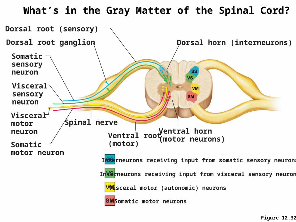
Figure 12.32
Somaticsensoryneuron
Dorsal root (sensory)
Dorsal root ganglion
Visceralsensory neuron
Somaticmotor neuron
Spinal nerve
Ventral root(motor)
Ventral horn(motor neurons)
Dorsal horn (interneurons)
Visceralmotorneuron
Interneurons receiving input from somatic sensory neurons
Interneurons receiving input from visceral sensory neurons
Visceral motor (autonomic) neurons
Somatic motor neurons
What’s in the Gray Matter of the Spinal Cord?

White Matter: Spinal Cord Information Pathways
Dorsal columns carry only ascending/afferent sensory tracts
Lateral and anterior columns carry both
• Ascending/afferent sensory tracts
• Descending/efferent motor tracts
Pathways are composed of 2-3 neurons in a chain or relay
Pathways cross from one side of the CNS to the other (decussate)
Ascending fibers add on laterally in cord as they join - they “map” in the tract based on where they enter or exit (somatotopy)
Ascending/Afferent Sensory Tracts only in
posterior columns Ascending/ Afferent Sensory) AND
Descending/Efferent Motor) Tracts in
lateral and anterior columns
Dorsal/posterior
Ventral/anterior

Three Ascending Pathways to Somatosensory Cortex
Dorsal column-medial lemniscal pathway
Precise, “straight-through” transmission of inputs from single, localized body surface receptors. In dorsal columns.
Conveys information about muscle or tendon stretch. Do not contribute to conscious sensation. In lateral columns.
Transmission of pain, temperature, and coarse touch. In anterior-ventral and lateral columns.
Anterolateral (spinothalmic) pathways
Spinocerebellar tracts

Descending Pathways and Tracts
Direct pyramidal pathways stimulate skeletal muscles
Indirect (Extrapyramidal system), e.g. the rubrospinal tract

The Spinal Cord (CNS) and Peripheral Nervous System
Spinal Cord (CNS)
• Meninges and Injections/Punctures
• Tracts and Information Pathways
• Spinal nerves
• Medical conditions: Spinal cord injury, shingles
Peripheral Nervous System
• Structure of a Nerve and Its Wrappings
• The Twelve Cranial Nerves
• Reflexes: Monosynaptic and Polysynaptic
• Branches of the Peripheral System
o Comparison between autonomic and somatic
Sympathetic division
Parasymphathetic division
• Developmental Aspects of the Nervous System

Spinal Nerves
There is a pair of spinal nerves at the level of each vertebrae (8 cervical, 12 thoracic, 5 lumbar, 5 sacral, 1 coccygeal) = a total of 31 pairs
Spinal nerves are formed by the combination of the ventral and dorsal roots of the spinal cord
Spinal nerves are named for the region from which they arise
Nerve C8 emergesBelow vertebra C7

Anatomy of Spinal Nerves Spinal nerves divide soon after
leaving the spinal cord
• Dorsal ramus – serves the skin and muscles of the posterior trunk
• Ventral ramus – forms a complex of networks (plexus) for the anterior part of the body spinal
nerve
posterior
anterior

The Spinal Cord (CNS) and Peripheral Nervous System
Spinal Cord (CNS)
• Meninges and Injections/Punctures
• Tracts and Information Pathways
• Spinal nerves
• Medical conditions: spinal cord injury, shingles
Peripheral Nervous System
• Structure of a Nerve and Its Wrappings
• The Twelve Cranial Nerves
• Reflexes: Monosynaptic and Polysynaptic
• Branches of the Peripheral System
o Comparison between autonomic and somatic
Sympathetic division
Parasymphathetic division
• Developmental Aspects of the Nervous System

Areas of the Skin Served By Single Spinal Nerves
Dermatomes
• Used to diagnose spinal cord injury
• Pain in a particular skin area reflects trouble in a specific spinal nerve and spinal location
• Can help to locate the site of damage in the spinal cord
• Most dermatomes overlap, so destruction of a single spinal nerve will not cause complete numbness

Shingles (Herpes zoster) is a viral infection of sensory neurons to the skin
Scaly, painful blisters confined to a narrow strip of skin on one side of the body trunk
Infects sensory skin neurons
Caused by latent infection (resurgence) of chicken pox virus when immune system is weakened as an adult
Mostly in people over 50.

The Spinal Cord (CNS) and Peripheral Nervous System
Spinal Cord (CNS)
• Meninges and Injections/Punctures
• Tracts and Information Pathways
• Spinal nerves
• Medical conditions: Spinal cord injury, shingles
Peripheral Nervous System
• Structure of a Nerve and Its Wrappings
• The Twelve Cranial Nerves
• Reflexes: Monosynaptic and Polysynaptic
• Branches of the Peripheral System
o Comparison between autonomic and somatic
Sympathetic division
Parasymphathetic division
• Developmental Aspects of the Nervous System

Peripheral Nervous System (The PNS)
Nerves and ganglia outside the central nervous system
Nerve = bundle of neuron fibers
Neuron fibers are bundled by connective tissue; one fiber = 1 axon or cell
Endoneurium surrounds each fiber, just outside of Schwann cells
Groups of fibers are bound into fascicles by perineurium
Fascicles are bound together by epineurium
one neuron (nerve fiber)

Classification of Nerves Mixed nerves – both sensory and motor fibers
present in the nerve
Afferent (sensory) nerves – carry impulses toward the CNS
Efferent (motor) nerves – carry impulses away from the CNS

The Twelve Cranial Nerves (I-V) I Olfactory nerve – purely sensory for smell; ask
patient to identify oil of cloves and vanilla
II Optic nerve – purely sensory for vision; observe eye, test patient with eye chart
III Oculomotor nerve – mostly motor fibers to eye muscles, some proprioreceptive afferents; examine pupil size and reflex, ability to follow objects with the eye
IV Trochlear – mostly motor fibers to extrinsic eye muscles; test patient’s ability to follow objects with eye
V Trigeminal nerve – 3 divisions:
• Opthalmic (tested by corneal reflex) carrying sensory for skin of anterior scalp, eyelid, nose
• Maxillary (tested with pain, touch temperature using safety pin) carrying sensory from nasal cavity, palate, upper lip, cheek
• Mandibular (test by teeth clenching, move jaw) carrying sensory from lower teeth, masseter, temporalis On Old Olympus' Towering Top a Frisky Virile Gymnast Vaults And Hops
Oh, Oh, Oh, To Touch And Feel Very Good Velvet: AH!Oh, Oh, Oh, To Touch And Feel And Grip Vegas' Slot Handles! On Occasion, Our Trusty Truck Acts Funny - Very Good Vehicle Anyhow.
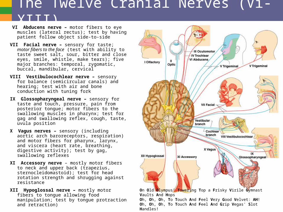
The Twelve Cranial Nerves (Vi-XIII) VI Abducens nerve – motor fibers to eye muscles
(lateral rectus); test by having patient follow object side-to-side
VII Facial nerve – sensory for taste; motor fibers to the face (test with ability to taste sweet salt, sour, bitter and close eyes, smile, whistle, make tears); five major branches: temporal, zygomatic, buccal, mandibular, cervical
VIII Vestibulocochlear nerve – sensory for balance (semicircular canals) and hearing; test with air and bone conduction with tuning fork
IX Glossopharyngeal nerve – sensory for taste and touch, pressure, pain from posterior tongue; motor fibers to the swallowing muscles in pharynx; test for gag and swallowing reflex, cough, taste, uvula position
X Vagus nerves – sensory (including aortic arch baroreceptors, respiration) and motor fibers for pharynx, larynx, and viscera (heart rate, breathing, digestive activity); test by gag, swallowing reflexes
XI Accessory nerve – mostly motor fibers to neck and upper back (trapezius, sternocleidomastoid); test for head rotation strength and shrugging against resistance
XII Hypoglossal nerve – mostly motor fibers to tongue allowing food manipulation; test by tongue protraction and retraction)
On Old Olympus' Towering Top a Frisky Virile Gymnast Vaults And HopsOh, Oh, Oh, To Touch And Feel Very Good Velvet: AH!Oh, Oh, Oh, To Touch And Feel And Grip Vegas' Slot Handles!On Occasion, Our Trusty Truck Acts Funny - Very Good Vehicle Anyhow.

The Spinal Cord (CNS) and Peripheral Nervous System
Spinal Cord (CNS)
• Meninges and Injections/Punctures
• Tracts and Information Pathways
• Spinal nerves
• Medical conditions: Spinal cord injury, shingles
Peripheral Nervous System
• Structure of a Nerve and Its Wrappings
• The Twelve Cranial Nerves
• Reflexes: Monosynaptic and Polysynaptic
• Branches of the Peripheral System
o Comparison between autonomic and somatic
Sympathetic division
Parasymphathetic division
• Developmental Aspects of the Nervous System

Reflexes Classification by Acquisition
• Inborn (intrinsic) reflex: a rapid, involuntary, predictable motor response to a stimulus
o E.g. withdrawing your arm when fingers burned
• Learned (acquired) reflexes result from practice or repetition
o Example: driving skills, knitting
Classification by Function
• Somatic (drives skeletal muscle)
• Autonomic/visceral if activates visceral effectors in glands or smooth and cardiac muscle
Classification by Number of Synapses
• Monosynaptic - e.g. any stretch reflex like knee-jerk reflex
• Polysynaptic - e.g.
o Golgi tendon reflex causing muscle relaxation and lengthening
o Flexor/withdrawal reflexes causing flexion of muscles to pull limb away
o Crossed-extensor reflexes casuing withdrawal and opposing extensor activity like sudden pain in the foot and shifting of weight

Figure 13.14
Receptor
Sensory neuron
Integration center
Motor neuron
Effector
Spinal cord(in cross section)
Interneuron
Stimulus
Skin
1
2
3
4
5
Basic Reflex Arc

Figure 13.17 (1 of 2), step1
Stretched muscle spindles initiate a stretch reflex,causing contraction of the stretched muscle and inhibition of its antagonist.
The events by which muscle stretch is damped
Initial stimulus(muscle stretch)
Cell body ofsensory neuron
Sensoryneuron
Muscle spindleAntagonist muscle
Spinal cord
Stretch Reflexes are Monosynaptic Pathways Triggered By Muscle Stretching

Figure 13.17 (1 of 2), step1
Stretched muscle spindles initiate a stretch reflex,causing contraction of the stretched muscle and inhibition of its antagonist.
When muscle spindles are activatedby stretch, the associated sensoryneurons (blue) transmit afferent impulsesat higher frequency to the spinal cord.
The events by which muscle stretch is damped
Initial stimulus(muscle stretch)
Cell body ofsensory neuron
Sensoryneuron
Muscle spindleAntagonist muscle
Spinal cord
1
Stretch Reflexes are Monosynaptic Pathways Triggered By Muscle Stretching

Figure 13.17 (1 of 2), step 2
Stretched muscle spindles initiate a stretch reflex,causing contraction of the stretched muscle andinhibition of its antagonist.
When muscle spindles are activatedby stretch, the associated sensoryneurons (blue) transmit afferent impulsesat higher frequency to the spinal cord.
The sensory neurons synapse directly with alphamotor neurons (red), which excite extrafusal fibersof the stretched muscle. Afferent fibers alsosynapse with interneurons (green) that inhibit motorneurons (purple) controlling antagonistic muscles.
The events by which muscle stretch is damped
Initial stimulus(muscle stretch)
Cell body ofsensory neuron
Sensoryneuron
Muscle spindleAntagonist muscle
Spinal cord
12

Figure 13.17 (1 of 2), step 3a
Stretched muscle spindles initiate a stretch reflex,causing contraction of the stretched muscle andinhibition of its antagonist.
When muscle spindles are activatedby stretch, the associated sensoryneurons (blue) transmit afferent impulsesat higher frequency to the spinal cord.
The sensory neurons synapse directly with alphamotor neurons (red), which excite extrafusal fibersof the stretched muscle. Afferent fibers alsosynapse with interneurons (green) that inhibit motorneurons (purple) controlling antagonistic muscles.
The events by which muscle stretch is damped
Efferent impulses of alpha motor neuronscause the stretched muscle to contract,which resists or reverses the stretch.
Initial stimulus(muscle stretch)
Cell body ofsensory neuron
Sensoryneuron
Muscle spindleAntagonist muscle
Spinal cord
12
3a

Figure 13.17 (1 of 2), step 3b
Stretched muscle spindles initiate a stretch reflex,causing contraction of the stretched muscle andinhibition of its antagonist.
When muscle spindles are activatedby stretch, the associated sensoryneurons (blue) transmit afferent impulsesat higher frequency to the spinal cord.
The sensory neurons synapse directly with alphamotor neurons (red), which excite extrafusal fibersof the stretched muscle. Afferent fibers alsosynapse with interneurons (green) that inhibit motorneurons (purple) controlling antagonistic muscles.
The events by which muscle stretch is damped
Efferent impulses of alpha motor neuronscause the stretched muscle to contract,which resists or reverses the stretch.
Efferent impulses of alpha motorneurons to antagonist muscles arereduced (reciprocal inhibition).
Initial stimulus(muscle stretch)
Cell body ofsensory neuron
Sensoryneuron
Muscle spindleAntagonist muscle
Spinal cord
12
3a 3b
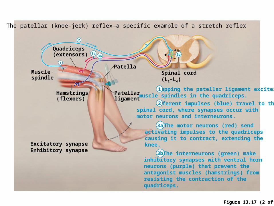
Figure 13.17 (2 of 2)
The patellar (knee-jerk) reflex—a specific example of a stretch reflex
Musclespindle
Quadriceps(extensors)
Hamstrings(flexors)
Patella
Patellarligament
Spinal cord(L2–L4)
Tapping the patellar ligament excitesmuscle spindles in the quadriceps.
The motor neurons (red) sendactivating impulses to the quadricepscausing it to contract, extending theknee.
Afferent impulses (blue) travel to thespinal cord, where synapses occur withmotor neurons and interneurons.
The interneurons (green) makeinhibitory synapses with ventral horn neurons (purple) that prevent theantagonist muscles (hamstrings) fromresisting the contraction of thequadriceps.
Excitatory synapseInhibitory synapse
+
–
1
2
3a
3b
1
2
3a3b 3b

Figure 13.18
+ Excitatory synapse– Inhibitory synapse
Quadriceps strongly contracts. Golgi tendon organs are activated.
Afferent fibers synapse with interneurons in the spinal cord.
Efferent impulses to muscle with stretched tendon are damped. Muscle relaxes, reducing tension.
Efferent impulses to antagonist muscle cause it to contract.
Interneurons
Spinal cord
Quadriceps(extensors)
Golgitendon
organHamstrings
(flexors)
1 2
3a 3b
Golgi Tendon Reflex: A Polysynaptic Reflex

Figure 13.19
Afferentfiber
Efferentfibers
Extensorinhibited
Flexorstimulated
Site of stimulus: a noxiousstimulus causes a flexorreflex on the same side,withdrawing that limb.
Site of reciprocalactivation: At thesame time, theextensor muscleson the oppositeside are activated.
Armmovements
Interneurons
Efferentfibers
FlexorinhibitedExtensorstimulated
+ Excitatory synapse– Inhibitory synapse
Crossed Extensor Reflex: A Polysynaptic Reflex

The Spinal Cord (CNS) and Peripheral Nervous System
Spinal Cord (CNS)
• Meninges and Injections/Punctures
• Tracts and Information Pathways
• Spinal nerves
• Medical conditions: Spinal cord injury, shingles
Peripheral Nervous System
• Structure of a Nerve and Its Wrappings
• The Twelve Cranial Nerves
• Reflexes: Monosynaptic and Polysynaptic
• Branches of the Peripheral System
o Comparison between autonomic and somatic
Sympathetic division
Parasymphathetic division
• Developmental Aspects of the Nervous System

Autonomic Nervous System The involuntary branch of
the nervous system
Consists of only motor nerves
Innervate smooth and cardiac muscle and glands
Make adjustments to ensure optimal support for body activities
Operate via subconscious control
Divided into two divisions
• Sympathetic division
• Parasympathetic division
Skin, muscle, and
joint sensors
Visceral organ
sensors
somatic sensory fibers
visceral sensory fibers
(visceral afferents)
and reflexes
inhibitory stimulatory
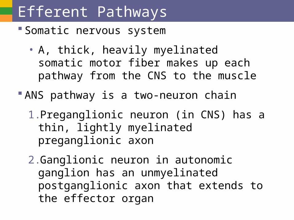
Efferent Pathways Somatic nervous system
• A, thick, heavily myelinated somatic motor fiber makes up each pathway from the CNS to the muscle
ANS pathway is a two-neuron chain
1. Preganglionic neuron (in CNS) has a thin, lightly myelinated preganglionic axon
2. Ganglionic neuron in autonomic ganglion has an unmyelinated postganglionic axon that extends to the effector organ
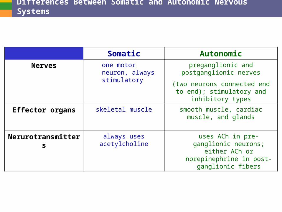
Differences Between Somatic and Autonomic Nervous Systems
Somatic Autonomic
Nerves one motor neuron, always stimulatory
preganglionic and postganglionic nerves
(two neurons connected end to end); stimulatory and inhibitory types
Effector organs skeletal muscle smooth muscle, cardiac muscle, and glands
Nerurotransmitters always uses acetylcholine uses ACh in pre-ganglionic neurons; either ACh or
norepinephrine in post-ganglionic fibers

Comparison of Somatic and Autonomic Nervous Systems
Closer to spinal cord than target
Closer to target than spinal cord

Anatomy of the Sympathetic Division Originates from T1 through L2
Short pre-ganglionic neuron and long postganglionic neuron transmit impulses from CNS to the effector
Ganglia are closer to the spinal cord than the target
Norepinephrine and epinephrine are neurotransmitters to the effector organs

Anatomy of the Parasympathetic Division Originates from the brain
stem (cranial nerves III,VII, IX, X) and S1 through S4
Long pre-ganglionic neuron and short postganglionic neuron transmit impulses from CNS to the effector
Terminal ganglia are at the effector organs (closer to target than spinal cord)
Always uses acetylcholine as a neurotransmitter

Autonomic Functioning Sympathetic – “fight-or-flight”
• Response to unusual stimulus
• Takes over to increase activities
• Remember as the “E” division = exercise, excitement, emergency, and embarrassment
Parasympathetic – housekeeping activites
• Conserves energy
• Maintains daily necessary body functions
• Remember as the “D” division - digestion, defecation, and diuresis

Sympathetic and Parasympathetic Compared
Sympathetic Division 14.2 Parasympathetic Division 14.6
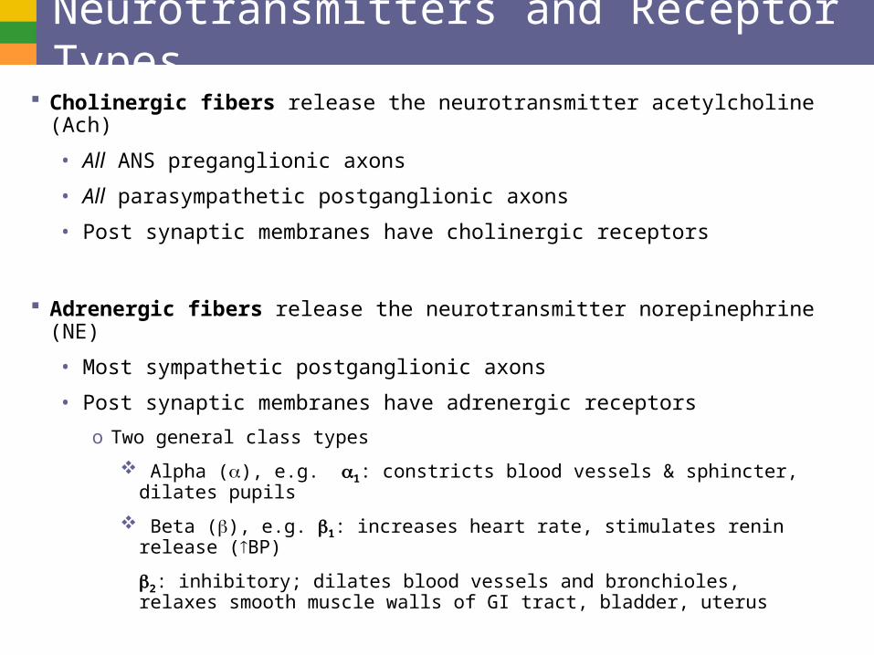
Neurotransmitters and Receptor Types Cholinergic fibers release the neurotransmitter acetylcholine (Ach)
• All ANS preganglionic axons
• All parasympathetic postganglionic axons
• Post synaptic membranes have cholinergic receptors
Adrenergic fibers release the neurotransmitter norepinephrine (NE)
• Most sympathetic postganglionic axons
• Post synaptic membranes have adrenergic receptors
o Two general class types
Alpha (), e.g. 1: constricts blood vessels & sphincter, dilates pupils
Beta (), e.g. 1: increases heart rate, stimulates renin release (BP)
2: inhibitory; dilates blood vessels and bronchioles, relaxes smooth muscle walls of GI tract, bladder, uterus

The Spinal Cord (CNS) and Peripheral Nervous System
Spinal Cord (CNS)
• Meninges and Injections/Punctures
• Tracts and Information Pathways
• Spinal nerves
• Medical conditions: Spinal cord injury, shingles
Peripheral Nervous System
• Structure of a Nerve and Its Wrappings
• The Twelve Cranial Nerves
• Reflexes: Monosynaptic and Polysynaptic
• Branches of the Peripheral System
o Comparison between autonomic and somatic
Sympathetic division
Parasymphathetic division
• Developmental Aspects of the Nervous System

Interactions of the Autonomic Divisions Most visceral organs have dual innervation
Dynamic antagonism allows for precise control of visceral activity
• Sympathetic division increases heart and respiratory rates, and inhibits digestion and elimination
• Parasympathetic division decreases heart and respiratory rates, and allows for digestion and the discarding of wastes

Four Steps of Regeneration of Peripheral Nerves
Axon becomes fragmented at injury site
Macrophages clean off the dead axon distal to the injury
Axon sprouts or filaments grow through a regeneration tube formed by Schwann cells
Axon regenerated and a new myelin sheath forms

Developmental Aspects of Fetal Nervous System
Measles (rubella)
Lack of oxygen
Radiation and drugs
Lack of folic acid
Premature birth problems
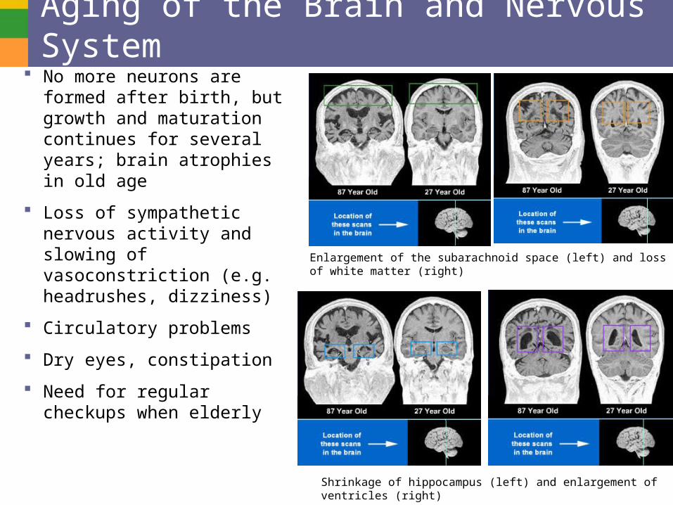
Aging of the Brain and Nervous System No more neurons are formed
after birth, but growth and maturation continues for several years; brain atrophies in old age
Loss of sympathetic nervous activity and slowing of vasoconstriction (e.g. headrushes, dizziness)
Circulatory problems
Dry eyes, constipation
Need for regular checkups when elderly
Enlargement of the subarachnoid space (left) and loss of white matter (right)
Shrinkage of hippocampus (left) and enlargement of ventricles (right)
The Spinal Cord (CNS) and Peripheral Nervous System
Spinal Cord (CNS)
• Meninges and Injections/Punctures
• Tracts and Information Pathways
• Spinal nerves
• Medical conditions: Spinal cord injury, shingles
Peripheral Nervous System
• Structure of a Nerve and Its Wrappings
• The Twelve Cranial Nerves
• Reflexes: Monosynaptic and Polysynaptic
• Branches of the Peripheral System
o Comparison between autonomic and somatic
Sympathetic division
Parasymphathetic division
• Developmental Aspects of the Nervous System


![SciForschen ISSN 2379-7150 · CNS tumors [11]. Neurosurgical approach Surgical intervention for spinal cord ependymomas remains the mainstay treatment option. Due to spinal cord compression](https://img.pdfslide.net/doc/110x75/5f0d49097e708231d43996e0/sciforschen-issn-2379-7150-cns-tumors-11-neurosurgical-approach-surgical-intervention.jpg)