Embed Size (px)
Citation preview
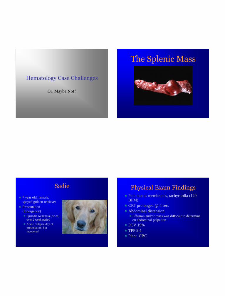
Hematology Case Challenges
Or, Maybe Not?
The Splenic Mass
Sadie
7 year old, female,
spayed golden retriever
Presentation
(Emergency)
Episodic weakness (twice)
over 2 week period
Acute collapse day of
presentation, but
recovered
Pale mucus membranes, tachycardia (120 BPM)
CRT prolonged @ 4 sec.
Abdominal distension
Effusion and/or mass was difficult to determine on abdominal palpation
PCV 19%
TPP 5.4
Plan: CBC
Physical Exam Findings
CBC Results
WBC 19.1 (6.0 – 17.0) x 103 RBC 2.51 (5.4 – 7.8) x 106
Neuts 15.2 (3.0 – 11.5) x 103 HGB 6.8 (13.0 – 19.0) g/dL
Bands 0.900 (0.0 - 0.3) x 103 HCT 18.2 (37.0 – 54.0) %
Lym. 0.700 (1.0 – 4.8) x 103 MCV 76.2 (66 – 75) fL
Mon. 2.3 (0.15 – 1.35) x 103 MCHC36.3 (34.0 – 36.0) g/dL
Eos. 0.0 (0.1 – 1.25) x 103 Plts 25.0 (150 – 430) x 103
Reticulocyte count = 150,600 / µl (>80,000 = regenerative)
Blood film evaluation
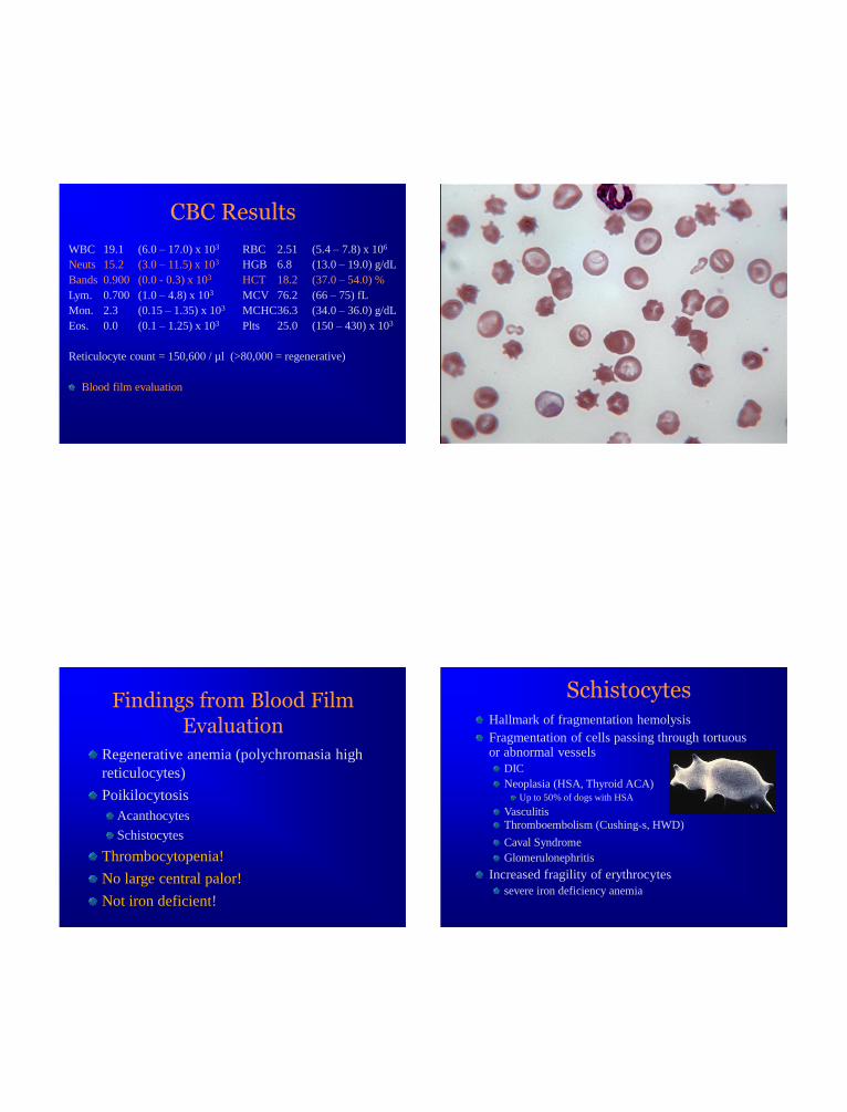
Findings from Blood Film Evaluation
Regenerative anemia (polychromasia high
reticulocytes)
Poikilocytosis
Acanthocytes
Schistocytes
Thrombocytopenia!
No large central palor!
Not iron deficient!
Schistocytes Hallmark of fragmentation hemolysis
Fragmentation of cells passing through tortuous or abnormal vessels
DIC
Neoplasia (HSA, Thyroid ACA)Up to 50% of dogs with HSA
Vasculitis
Thromboembolism (Cushing=s, HWD)
Caval Syndrome
Glomerulonephritis
Increased fragility of erythrocytes
severe iron deficiency anemia
Abdominal Ultrasound
Free abdominal fluid
Large mass in cranial abdomen (14 cm)
Cavitated with mixed echogenicity
Appeared to be associated with the spleen
What is your working diagnosis?
1. Hemangioma
2. Hemangiosarcoma
3. Splenic torsion or
vascular thrombosis
4. Hematoma
5. Lymphoma
No firm diagnosis?
Hemangioma, hemangiosarcoma,
hematoma, lymphoma?
Imaging not always helpful
Hematological abnormalities indicate HSA
Anemia seen in 80% of dogs with splenic HSA
Dogs with splenic masses and evidence of
anemia, fragmentation hemolysis and
thrombocytopenia
Significantly greater risk of having HSA (90%)
Fine-needle Aspiration of Splenic Mass
Potential for definitive, presurgical
diagnosis
Potential for complications
Seeding the abdomen with tumor cells
Hemorrhage
Ultrasound guided aspirate with 25 gauge
needle
Stay away from cavitated areas of mass
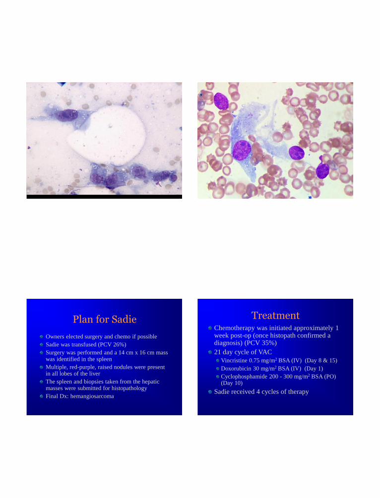
Plan for Sadie
Owners elected surgery and chemo if possible
Sadie was transfused (PCV 26%)
Surgery was performed and a 14 cm x 16 cm mass was identified in the spleen
Multiple, red-purple, raised nodules were present in all lobes of the liver
The spleen and biopsies taken from the hepatic masses were submitted for histopathology
Final Dx: hemangiosarcoma
TreatmentChemotherapy was initiated approximately 1 week post-op (once histopath confirmed a diagnosis) (PCV 35%)
21 day cycle of VAC
Vincristine 0.75 mg/m2 BSA (IV) (Day 8 & 15)
Doxorubicin 30 mg/m2 BSA (IV) (Day 1)
Cyclophosphamide 200 - 300 mg/m2 BSA (PO) (Day 10)
Sadie received 4 cycles of therapy
Prognosis
Long-term prognosis extremely poor
Death from exsanguination from rupture of
metastatic site
Surgery alone rarely curative with MST of 1
to 3 months
Multi-drug chemotherapy MST 6 to 9
months
Sadie
Sadie was found dead in her bed 9 months
after splenic surgery
Likely the result of ruptured metastatic
lesion
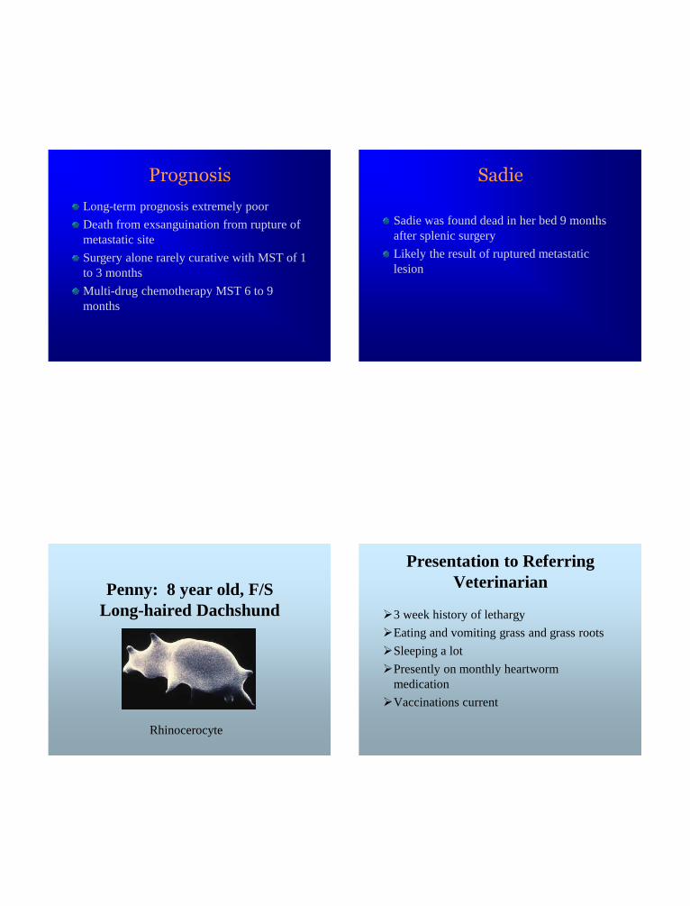
Penny: 8 year old, F/S
Long-haired Dachshund
Rhinocerocyte
Presentation to Referring
Veterinarian
3 week history of lethargy
Eating and vomiting grass and grass roots
Sleeping a lot
Presently on monthly heartworm
medication
Vaccinations current
Penny’s Exam
Temp. 101.50F
Respiration 35 PM
HR 90 BPM
Pale mucus membranes
No history of trauma or evidence of hemorrhage
Fecal parasites check negative
Heartworm test negative
CBC & Biochemical profile
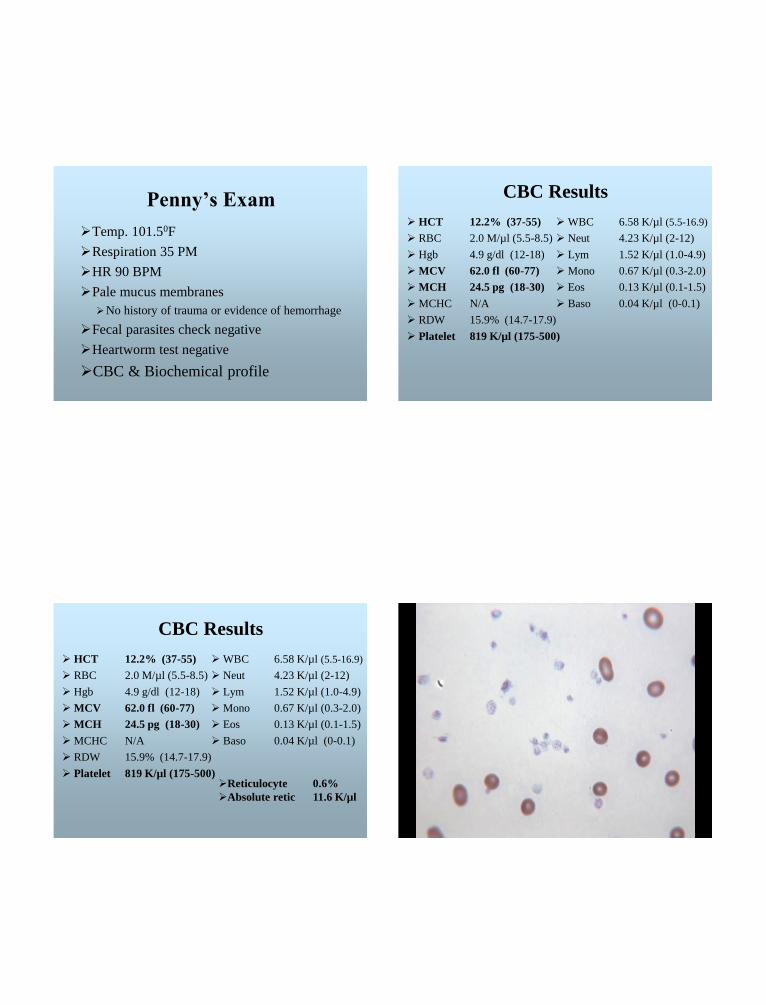
CBC Results
HCT 12.2% (37-55)
RBC 2.0 M/µl (5.5-8.5)
Hgb 4.9 g/dl (12-18)
MCV 62.0 fl (60-77)
MCH 24.5 pg (18-30)
MCHC N/A
RDW 15.9% (14.7-17.9)
Platelet 819 K/µl (175-500)
WBC 6.58 K/µl (5.5-16.9)
Neut 4.23 K/µl (2-12)
Lym 1.52 K/µl (1.0-4.9)
Mono 0.67 K/µl (0.3-2.0)
Eos 0.13 K/µl (0.1-1.5)
Baso 0.04 K/µl (0-0.1)
Reticulocyte 0.6%
Absolute retic 11.6 K/µl
Biochemical Profile
WNL
Problem List
Severe nonregenerative anemia
Thrombocytosis
Pica
Vomiting
Plan
Problem List
Severe nonregenerative anemia
Thrombocytosis
Pica
Vomiting
Plan
Refer to UF-VMC for further evaluation
Penny’s Presentation
1 month history of lethargy and decreased appetite
Eating grass and grass roots and vomiting them
Physical exam
BW 4.6 kg
Quite but alert
Temp 102.1
HR 80, Resp. 32
MM white with no CRT available
Normal fecal color
Fecal occult blood test (Hemocult) negative
Diagnostic Plan
MDB
CBC, UA, Biochemical profile
Reticulocyte count
Coombs’ test
Cross match
CBC Results
HCT 15.1% (37-55)
RBC 2.31 M/µl (5.4-7.8)
Hgb 5.4 g/dl (13-19)
MCV 66.4 fl (66-75)
MCH 23.4 pg (18-30)
MCHC 35.8 g/dl (34-36)
RDW 12.1% (11-13)
Platelet 735 K/µl (150-430)
Plasma Protein 6.9 g/dl
WBC 6.92 K/µl (6.0-17)
Neut 4.5 K/µl (2-12)
Bands .21 K/µl (0-.3)
Lym 1.80 K/µl (1.0-4.9)
Mono 0.45 K/µl (0.3-2.0)
Eos 0.1 K/µl (0.1-1.5)
Baso 0.0 K/µl (0-0.1)
Reticulocyte 0.1%
Absolute retic 2.3 K/µl
Iron Levels
Serum iron 374 ug/dl (70-264)
TIBC 559 ug/dl (246-504)
% saturation 67 (19-79)
Biochemical Profile
If Penny was bleeding (e.g. chronic
hemorrhage) what abnormality would you
see in the profile?
Low total protein (albumin, pos. globulin)
High BUN, normal creatinine
Penny was normal, no abnormalities seen
Diagnostic Plan
Cross-matched transfusion
Adapted to chronic anemia
Concern for anesthesia
General anesthesia
Imaging
Thoracic and abdominal radiographs
(neoplasia?)
Ultrasound of abdomen
Bone marrow collection and evaluation
Results
Pre transfusion PCV 15.1%
Post transfusion PCV 20.0%
Imaging: no abnormalities seen
Bone marrow aspirate
Dry tap (Why?)
Core biopsy of marrow
Role core on slide for cytological evaluation
Submit core for histopathology
Sent patient home awaiting biopsy results
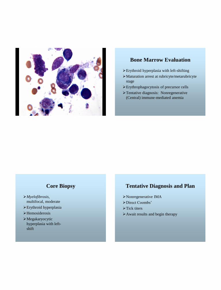
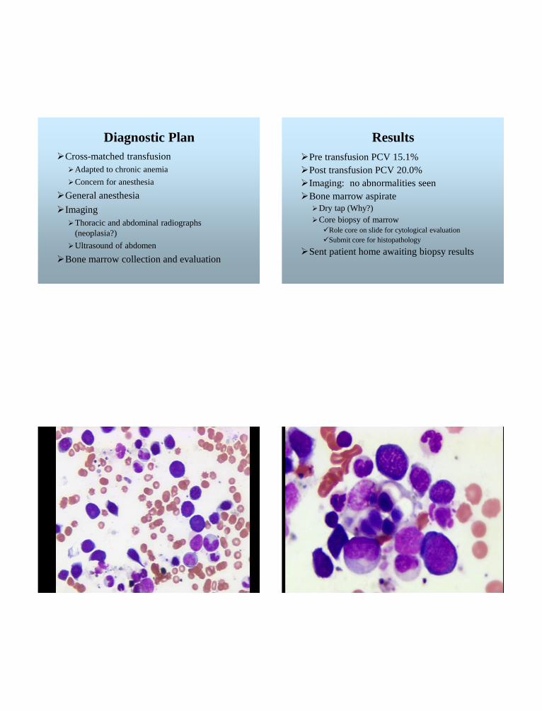
Bone Marrow Evaluation
Erythroid hyperplasia with left-shifting
Maturation arrest at rubricyte/metarubricyte
stage
Erythrophagocytosis of precursor cells
Tentative diagnosis: Nonregenerative
(Central) immune-mediated anemia
Core Biopsy
Myelofibrosis,
multifocal, moderate
Erythroid hyperplasia
Hemosiderosis
Megakaryocytic
hyperplasia with left-
shift
Tentative Diagnosis and Plan
Nonregenerative IMA
Direct Coombs’
Tick titers
Await results and begin therapy
Recheck
Return visit 1 week after diagnostic evaluation
Penny more active after transfusion, but getting lethargic again
PCV 15% with low number of spherocytes on blood smear
Direct Coombs’ negative at 1:2
Serology for E. canis and A. phagocytophilum -negative
Treatment Plan
Azathioprine 2mg/kg for two weeks
Prednisone 1 mg/kg BID
Famotidine 5 mg per day (pepcid AC)
2 week Recheck
Penny more alert than before
PCV 22%
No spherocytes noted on smear
Continue on current dose until PCV normal
Nonregenerative IMHA
Estimated 33% to 58% of IMHA are nonregenerative
Anemia typically severe, (median PCV 11%) with majority being <20%
Age 10 mo. to 12 yr. (median 6.5 yr.)
Female over-represented in most studies
Dachshunds over-represented at UF (> 10 cases)
Two forms PRCA: more severe less common form
Evidence of erythropoiesis in bone marrow (>90%)
Stokol et al., JAVMA 216:14291436, 2000. (43 cases)
Chronicity of Anemia
Important in establishing diagnosis
Most dogs have clinical signs of 7 or more
days
Lethargy, anorexia, pallor, weakness, pica,
vomiting
Animals tolerant of very low red cell
mass
Laboratory Analysis
Normocytic, normochromic anemia
Spherocytes seen in small percent (16%), numbers vary
0 to 7 NRBC’s / 100 WBC (no / rare polychromasia)
Normal leukocyte count
50% had mild left-shift
Platelets most often increased
Decreased in 22% of cases
Direct Coombs’ test positive in 30 to 50% (low titer
1:4)
ANA positive in 23%
Bone Marrow Evaluation (Stokol et al.)
Bone marrows difficult to aspirate in 21 of 43 dogs
No spicules
Dry tap
16 dogs had core biopsies, all had myelofibrosis (reversible)
Erythroid precursors may be absent (5% PRCA), normal numbers, or increased (most common)
Erythroid hyperplasia was common among dogs with myelofibrosis
Maturation arrest at rubiricytes and metarubricytes with few to no polychromatophilic cells
Most dogs had large amounts of iron
Treatment for NRIMHA
(Stokol, et al., JAVMA 216:1429-1436;2000)
Combination chemotherapy
Pred. & Cytoxan (73% remission)
Pred. & Azathioprine (52% remission)
Pred. alone (25% remission)
Response rate
Complete remission (55%)
Partial remission (18%)
Poor response (27%)
Response Time and MortalityResponse to treatment seen in 1 to 10
weeks
Median 2 weeks
18 cases with extensive follow-ups
5 off all medication within 2 years
9 on alternate day pred / azathioprine for 3 years
6 relapsed when drug dosage / frequency was reduced
If drugs are reduced, maintain at reduced level for prolonged period of time (60 days?)
Mortality rate 28%
Summary
NR-IMHA should be considered in dogs with
severe, chronic, nonregenerative anemia
Normal WBC and normal or increased platelets
Bone marrow evaluation may aid in confirming
diagnosis
Fibrosis, maturation arrest, erythrophagia
Myelofibrosis may complicate marrow aspiration
Treatment should include combination
chemotherapy
Response to therapy may take weeks to months
The End?